Embed Size (px)
Citation preview
1
UNIVERSIDADE FEDERAL DE SANTA MARIA CENTRO DE CIÊNCIAS NATURAIS E EXATAS
PROGRAMA DE PÓS-GRADUAÇÃO EM CIÊNCIAS BIOLÓGICAS
EFEITO HEPATOPROTETOR CAUSADO PELO 3-ALQUINIL SELENOFENO CONTRA O DANO OXIDATIVO INDUZIDO POR
AGENTES QUÍMICOS EM RATOS
DISSERTAÇÃO DE MESTRADO
Ethel Antunes Wilhelm
Santa Maria, RS, Brasil 2009
PDF created with pdfFactory Pro trial version www.pdffactory.com
2
EFEITO HEPATOPROTETOR CAUSADO PELO 3-ALQUINIL
SELENOFENO CONTRA O DANO OXIDATIVO INDUZIDO POR AGENTES QUÍMICOS EM RATOS
por
Ethel Antunes Wilhelm
Dissertação apresentada ao Programa de Pós-Graduação em Ciências Biológicas, Área de Concentração em Bioquímica
Toxicológica, da Universidade Federal de Santa Maria (UFSM, RS), como requisito parcial para a obtenção do grau de
Mestre em Bioquímica Toxicológica.
Orientadora: Profa. Dra. Lucielli Savegnago Co-orientadora: Profa. Dra. Cristina Wayne Nogueira
Santa Maria, RS, Brasil
PDF created with pdfFactory Pro trial version www.pdffactory.com
3
Universidade Federal de Santa Maria Centro de Ciências Naturais e Exatas
Programa de Pós-Graduação em Ciências Biológicas A Comissão Examinadora, abaixo assinada, aprova a Dissertação de
mestrado:
EFEITO HEPATOPROTETOR CAUSADO PELO 3-ALQUINIL SELENOFENO CONTRA O DANO OXIDATIVO INDUZIDO POR
AGENTES QUÍMICOS EM RATOS
Elaborada por Ethel Antunes Wilhelm como requisito parcial para a obtenção do grau de Mestre em Bioquímica Toxicológica
COMISSÃO EXAMINADORA:
_________________________________________ Profª. Drª. Cristina Wayne Nogueira (Co-orientadora)
_________________________________________ Profª. Drª. Carla Bonan
________________________________________ Prof. Dr. Alexandre Mazzanti
Santa Maria, fevereiro de 2009.
PDF created with pdfFactory Pro trial version www.pdffactory.com
4
AGRADECIMENTOS
Agradeço primeiramente à minha família, meu alicerce. Agradeço em especial e
com muita saudade ao meu pai (in memoriam) por ter sido meu exemplo de garra, força
de vontade, honestidade, humildade e dedicação. Mãe e La obrigada por estarem
sempre presentes, me apoiando e principalmente sempre acreditando em mim! Eu amo
vocês!!!
Juliano, meu amor, a você tenho uma enorme gratidão por todo companheirismo,
amizade e amor dedicado à mim. Você sempre esteve ali, juntinho, nos momentos mais
difíceis, enxugando muitas e muitas vezes minhas lágrimas. Obrigada por me dar a
oportunidade de conviver e aprender contigo! Você é muito especial!!! Te amo muito!!!
À Lu, minha orientadora, meus sinceros agradecimentos pelo tempo dedicado,
pelo incentivo, amizade, colaboração em vários trabalhos, sugestões, críticas,
compreensão. Enfim, obrigada pela orientação!!!
Cris, obrigada por acreditar em mim. Por ter sido mais que co-orientadora, por ter
sido minha amiga. Obrigada pela compreensão que você teve neste ano tão difícil pra
mim e minha família. Obrigada pelo tempo dedicado, pelas sugestões, pelos conselhos,
pelo exemplo de profissional, de competência e dedicação que você é.
Cristiano, à você não basta um “muito obrigada” para expressar minha gratidão,
neste um ano, você foi um exemplo de amigo, colega, parceiro e gerador de muitas
idéias. Obrigada Cris, pelo companheirismo, paciência e principalmente pela alegria e
bem estar que você espalha aos que estão perto de você. Mesmo nos dias mais tristes
você conseguiu me fazer sorrir. Obrigada do fundo do coração por tudo!
À Crisinha, minha IC querida, pela amizade, pela ajuda, dedicação, por ser essa
pessoa maravilhosa que você é !!! Te adoro muito!!!
Ao GZ, pelo incentivo, amizade e exemplo de dedicação. Ao pessoal do seu
laboratório, pela amizade e companheirismo, e principalmente pelo tempo que
dispuseram para a síntese dos “selenofenos”.
À Marina, minha mãe científica, por todos os ensinamentos, parceria no primeiro
trabalho e pela sincera amizade.
PDF created with pdfFactory Pro trial version www.pdffactory.com
5
Aos colegas Ricardo, Cristiane, Silvane, Érico, Simone, Jú, Aninha, Bibi, Carmine,
Marlon, Xorão, maninho Cézar, pela amizade, pela colaboração, pelo companheirismo.
A todos os professores do Programa de Pós Graduação em Bioquímica
Toxicológica, pela atenção.
À Angélica, pela dedicação com que cuida da parte burocrática, sempre com bom
humor. Ao Rinaldo, por cuidar dos nossos animais e pela sua amizade.
À CAPES, pelo auxílio financeiro durante a realização deste trabalho.
A todos, que de alguma forma colaboraram para a realização deste trabalho, muito
obrigada!!!
PDF created with pdfFactory Pro trial version www.pdffactory.com
6
RESUMO
Dissertação de Mestrado Programa de Pós-Graduação em Ciências Biológicas
Universidade Federal de Santa Maria, RS, Brasil
EFEITO HEPATOPROTETOR CAUSADO PELO 3-ALQUINIL SELENOFENO CONTRA O DANO OXIDATIVO INDUZIDO POR AGENTES QUÍMICOS EM RATOS
AUTOR: Ethel Antunes Wilhelm
ORIENTADORA: Lucielli Savegnago DATA E LOCAL DA DEFESA: Santa Maria, fevereiro de 2009.
O fígado apresenta extraordinária pluralidade funcional, destacando-se no controle de produção de energia, defesa imunológica e reserva volêmica. No meio ambiente e ocupacionalmente, o ser humano está exposto a uma variedade de compostos hepatotóxicos, como por exemplo, no uso de tintas e seus derivados (2-nitropropano, 2-NP), reagentes químicos (tetracloreto de carbono, CCl4) e na exposição ao cigarro (2-NP). Portanto, é interessante o estudo de terapias que previnam ou até mesmo revertam a intoxicação causada por estes compostos. Considerando que as espécies reativas de oxigênio (EROs) apresentam importante papel sobre diversas patologias, em especial nas doenças hepáticas, o uso de terapias antioxidantes deve ser considerada. Neste contexto, destacam-se os compostos heterocíclicos contendo selênio em sua estrutura. Deste modo, neste estudo investigou-se a atividade antioxidante de 3-alquinil selenofenos em modelos de dano oxidativo in vitro e ex vivo em ratos (Wistar, machos, pesando entre 200 – 300 g). Para esse fim, testou-se uma classe de compostos 3-alquinil selenofeno, com diferentes substituições na estrutura química, com o objetivo de avaliar o perfil antioxidante e seu possível efeito tóxico in vitro em ratos. Como resultado, 3-alquinil selenofenos tiveram atividade antioxidante, porém esta atividade foi dependente da presença de um alquino terminal na molécula ou da fácil conversão da molécula a um alquino terminal. Além disso, o possível efeito tóxico dos 3-alquinil selenofenos foi avaliado através da atividade da enzima δ-aminolevulinato desidratase (δ-ALA-D) in vitro. Os resultados obtidos demonstraram que nenhum dos 3-alquinil selenofenos testados inibiu a atividade desta enzima, sugerindo que esta classe de compostos não apresentou toxicidade sobre a atividade da δ-ALA-D. A partir destes resultados, selecionou-se o selenofeno h (que obteve melhor atividade antioxidante in vitro) para a avaliação do seu efeito protetor contra o dano oxidativo induzido por 2-NP e CCl4 em ratos (ex vivo). O selenofeno h (25 mg/kg) protegeu contra o aumento dos marcadores de dano hepático (aspartato aminotranferase (AST) e alanina aminotransferase (ALT)) e de estresse oxidativo induzidos pela administração do 2-NP. O 2-NP induziu alterações microscópicas avaliadas por inspeções histopatológicas as quais foram protegidas pelo composto. O selenofeno h demonstrou efeito protetor contra o aumento da peroxidação lipídica e inibição da atividade da δ-ALA-D nos animais tratados com 2-NP. Além disso, o selenofeno h protegeu contra o dano oxidativo induzido pelo CCl4 em ratos. Uma única dose de CCl4 causou significante hepatotoxicidade, evidenciada por elevação da atividade plasmática das enzimas AST e ALT, aumento da incidência de lesões histopatológicas, aumento dos níveis de peroxidação lipídica e da atividade da enzima glutationa-S-transferase (GST), bem como diminuição dos níveis de ácido ascórbico e da atividade das enzimas catalase e δ-ALA-D. A partir dos resultados demonstrados, verificou-se que o selenofeno h protegeu contra todas estas alterações, confirmando o seu efeito hepatoprotetor. Considerando os resultados obtidos, pode-se sugerir que o
PDF created with pdfFactory Pro trial version www.pdffactory.com
7
selenofeno h, uma molécula com atividade antioxidante, pode ser uma útil terapia contra o dano oxidativo induzido pelos hepatotoxicantes: 2-NP e CCl4.
Palavras-chave: dano hepático, selênio, 3-alquinil selenofeno, tetracloreto de carbono, 2-nitropropano.
PDF created with pdfFactory Pro trial version www.pdffactory.com
8
ABSTRACT Dissertation of Master’s Degree
Federal University of Santa Maria, RS, Brazil
HEPATOPROTECTIVE EFFECT OF 3-ALKYNYL SELENOPHENE AGAINST OXIDATIVE DAMAGE INDUCED BY CHEMICAL INDUCTORS IN RATS
AUTHOR: Ethel Antunes Wilhelm
ADVISOR: Lucielli Savegnago DATE AND PLACE OF THE DEFENSE: Santa Maria, 2009.
The liver presents extraordinary functional diversity, particularly in the control of energy production, immune defense and volemic reserve. The human being is exposed occupationally and in the environment to a variety of hepatotoxic compounds, such as the use of paints and their derivatives (2-nitropropane, 2-NP), chemical reagents (carbon tetrachloride, CCl4) and exposure to cigarette (2-NP). Therefore, it is interesting the study of therapies to prevent or even reverse the poisoning caused by these compounds. Considering that reactive oxygen species (ROS) have an important role in various diseases, especially in liver diseases, the use of antioxidant therapies should be considered. In this context, the heterocyclic compounds containing selenium in their structures have attracted the attention of researchers. Thus, this study investigated the antioxidant activity of 3-alkynyl selenophenes in models of oxidative damage in vitro and ex vivo in rats (Wistar, male, weighing 200-300g). A class of 3-alkynyl selenophene compounds with different substitutions was tested, with the objective to assess their antioxidant profile and their possible toxic effect in vitro. As a result, 3-alkynyl selenophenes had antioxidant activity, but this activity was dependent on the presence of terminal alkynes in the molecule or easy conversion to it. The possible toxic effect of 3-alkynyl selenophenes was evaluated through the activity of the enzyme δ-aminolevulinate dehydratase (δ-ALA-D) in vitro. The results showed that none of 3-alkynyl selenophenes inhibited the activity of this enzyme, suggesting that this class of compound did not present toxicity on this enzyme. From these results, selenophene h (compound that had the best antioxidant activity in vitro) was selected for the evaluation of its protective effect against oxidative damage induced by 2-NP and CCl4 (ex vivo). Selenophene h (25 mg/kg) protected against the increase of markers of liver damage (aspartate aminotransferase (AST) and alanine aminotransferase (ALT) activities) and oxidative stress induced by administration of 2-NP in rats. 2-NP induced microscopic changes, evaluated by histopathological inspections, that were protected by this compound. Selenophene h showed a protective effect against the increase of lipid peroxidation and inhibition of activity of δ-ALA-D in animals treated with 2-NP. Selenophene h protected against oxidative damage induced by CCl4 in rats. A single dose of CCl4 caused significant hepatotoxicity, evidenced by elevated plasma enzyme activity of AST and ALT, increased incidence of histopathological lesions, increased lipid peroxidation levels and the activity of Glutathione-S-transferase (GST), decreased levels of ascorbic acid and the activity of catalase and δ-ALA-D. In conclusion, 3-alkynyl selenophene protected from all these changes, confirming its hepatoprotective effect. Considering the results, we suggest that 3-alkynyl selenophene, an antioxidant, may be a useful therapy for the oxidative damage induced by 2-NP or CCl4 . Keywords: liver damage, selenium, 3-alkynyl selenophene, carbon tetrachloride, 2-nitropropane.
PDF created with pdfFactory Pro trial version www.pdffactory.com
9
LISTA DE FIGURAS
REVISÃO BIBLIOGRÁFICA
Figura 1. Estrutura química do AZT 16
Figura 2: Estrutura química do ebselen. 17
Figura 3: Estrutura química do D-501036 17
Figura 4: Estrutura química do selenofeno h 18
Figura 5: Localização do fígado no organismo humano. 19
Figura 6: Representação esquemática dos mecanismos da evolução
do dano hepático.
22
Figura 7: Representação esquemática dos mecanismos de dano
oxidativo induzido por 2-NP na presença de metais.
Figura 8: Representação esquemática dos mecanismos de dano
oxidativo induzido por tetracloreto de carbono.
Figura 9: Fármacos contendo unidade heterocíclica.
Figura 10: Exemplos de calcogenofenos
24
25
31
31
MANUSCRITO 1
Figura 1: Chemical structures of selenophenes. 66
PDF created with pdfFactory Pro trial version www.pdffactory.com
10
Figura 2: Photomicrography of segment of the hepatic lobe (A) of an
animal control; note the hepatic tissue with normal aspect, (B) of animal
treated with selenophene h 100 mg/kg; observe the hepatocyte strings,
centrillobular vein and sinusoid capillaries with normal aspect, (C) of
animal treated with 2-NP; note intense infiltration of inflammatory cells
(arrow-head) in the hepatic tissue and the loss of cellular architecture
(*), (D) of animal treated with selenophene h 25 mg/kg+2NP; note the
reduction of infiltration of inflammatory cells (arrow-head) in the hepatic
tissue with normal appearance. Hepatocyte strings (arrow);
Centrillobular vein (CV); Sinusoid capillaries (sc). Both H.E. 100X.
67
MANUSCRITO 2
Figure 1: Photomicrography of segment of the hepatic lobe (A) of a
control animal. Note the hepatocyte strings (arrow), the centrilobular
vein (CV) and sinusoid capillaries (*) with normal aspect, (B) of an
animal treated with CCl4, note intense ballooning degeneration (arrow)
and (*) infiltration of inflammatory cells, (C) of an animal treated with
selenophene h (50 mg/kg), note the hepatocyte strings (arrow), the
centrilobular vein and some inflammatory cells (*), (D) of an animal
treated with CCl4 + 25 mg/kg; observe around the centrilobular vein
some hepatocytes with vacuolation (head-arrows), ballooning
degeneration (arrow) and the presence of inflammatory cells (*) in the
sinusoid capillaries, (E) of an animal treated with CCl4 + 50 mg/kg.
Observe the hepatocyte strings (arrow), the centrilobular vein and
sinusoid capillaries (*) with normal aspect. H.E. 100X.
92
PDF created with pdfFactory Pro trial version www.pdffactory.com
11
LISTA DE TABELAS
MANUSCRITO 1
Tabela 1: Effect of selenophenes on iron/EDTA-induced lipid
peroxidation in vitro.
61
Tabela 2: Effect of selenophene h on liver of rats. 62
Tabela 3: Effect of selenophene h in liver and plasma of 2-NP
exposed rats.
Tabela 4: Effect of selenophene h at different doses on
biochemical parameters in rats
63
64
MANUSCRITO 2
Tabela 1: Effect of selenophene h on ALT and AST activities
and ascorbic acid and TBARS levels, CAT, GST and δ-ALA-D
activities.
89
Tabela 2: Effect of selenophene h on histopathological scores
in hepatic injury induced by CCl4 in rats.
90
PDF created with pdfFactory Pro trial version www.pdffactory.com

12
LISTA DE ESQUEMAS
DISCUSSÃO
Esquema 1: Visão geral dos efeitos do selenofeno h frente
aos compostos hepatotóxicos (2-NP e CCl4 ) estudados neste
trabalho.
97
PDF created with pdfFactory Pro trial version www.pdffactory.com
13
LISTA DE ABREVIATURAS
δ-ALA-D - delta-aminolevulinato desidratase ou porfobilinogênio sintase
(PhSe)2 – disseleneto de difenila
2-NP - 2-nitropropano
ALT – alanina aminotransferase
ANOVA – análise de variância
AST – aspartato aminotransferase
ATP – Adenosina trifosfato
AZT - azidovudina
CAT – catalase
CYP-450 - sistema P-450
DL50 - dose letal para 50 % dos animais
ERNs - espécies reativas de nitrogênio
EROs - espécies reativas de oxigênio
GST – glutationa S-transferase
i.p. – intraperitoneal
MDA - malondialdeído
R-SeH – selenóis
R-SH – tióis
S.D – standard deviation
S.E.M – standard error of the mean
S1 – sobrenadante
TBARS – espécies reativas ao ácido tiobarbitúrico
v.o. – via oral
PDF created with pdfFactory Pro trial version www.pdffactory.com
14
SUMÁRIO
1. INTRODUÇÃO 15
2. OBJETIVOS 18
3. REVISÃO BIBLIOGRÁFICA 19
3.1. Fígado 3.1.1. Hepatotoxicidade
19
21
3.1.2. Indutores químicos de hepatotoxicidade 3.1.2.1. 2-Nitropropano (2-NP)
3.1.2.2. Tetracloreto de carbono (CCl4) 3.2. Estresse Oxidativo 3.2.1. Hepatotoxicidade x Estresse Oxidativo 3.3. Selênio
3.3.1. Selênio x hepatotoxicidade 3.4. Compostos heterocíclicos 3.4.1. Compostos heterocíclicos x Selênio 4. MANUSCRITOS
4.1. Selenofeno protege contra dano oxidativo induzido por 2-nitropropano em fígado de ratos.
4.1.1. Manuscrito 1: Selenophene protects against oxidative damage induced by 2-nitropropane in liver of rats.
4.2. Efeito hepatoprotetor do 3-alquinil selenofeno contra o dano no
fígado induzido pelo tetracloreto de carbono em ratos.
44.2.1. Manuscrito 2: Hepatoprotective effect of 3-alkynyl selenophene against carbon tetrachloride-induced liver damage in rats.
5. DISCUSSÃO
6. CONCLUSÕES 7. REFERÊNCIAS BIBLIOGRÁFICAS
22
22
24
26
27
27
30
30
32
34
35
36
68
69
93
98
99
PDF created with pdfFactory Pro trial version www.pdffactory.com
15
1. INTRODUÇÃO
O estresse oxidativo corresponde a uma excessiva formação endógena de
espécies reativas de oxigênio (EROs) associada a uma diminuição nas defesas
antioxidantes. As EROs podem induzir um grande número de alterações nos constituintes
celulares, incluindo inativação de enzimas, danos às bases nitrogenadas dos ácidos
nucléicos e às proteínas, além de peroxidação dos lipídios de membrana (Ha et al., 2006).
O conceito formulado por Sies (1997) define estresse oxidativo como sendo um
desequilíbrio entre a produção de agentes oxidantes e antioxidantes, em favor dos
oxidantes, com potencial para ocasionar dano celular. Nos últimos anos, evidências têm
demonstrado o papel central exercido pelas EROs em um variado número de reações
biológicas fundamentais, sugerindo que muitas doenças e processos degenerativos
podem estar associados com a superprodução das EROs (Young e Woodside, 2001).
Dentre essas doenças, cabe salientar as hepáticas, as quais são um problema de
saúde pública mundial. No meio ambiente e ocupacionalmente, o ser humano está
exposto a uma variedade de compostos hepatotóxicos, como por exemplo, no uso de
tintas e seus derivados (2-nitropropano, 2-NP), reagentes químicos (tetracloreto de
carbono, CCl4) e na exposição ao cigarro (2-NP). Portanto, é interessante o estudo de
terapias que previnam ou até mesmo revertam a toxicidade causada por estes compostos
(Henry, 1999; Kalil et al., 2001).
Evidências crescentes relacionam as EROs com a cascata de eventos que regulam
o início e a progressão das doenças hepáticas, independentemente do agente que as
originou (Loguercio e Frederico, 2003; Vitaglione et al., 2004). Assim, o uso de terapias
antioxidantes (Lima et al., 2007), de drogas que interferem no sistema de metabolização
(Sistema P-450) do agente hepatotóxico e aumentam a atividade de sistemas enzimáticos
de defesa são as opções terapêuticas usadas no estudo da hepatotoxicidade (Weber et
al., 2003).
Considerando que as EROs apresentam importante papel sobre diversas
patologias, em especial nas doenças hepáticas, é conveniente ressaltar a importância
exercida por compostos que apresentam atividade antioxidante. Em vista disso, tem sido
alvo de interesse de muitos pesquisadores a busca por novos compostos que possuam
PDF created with pdfFactory Pro trial version www.pdffactory.com
16
atividade biológica com o mínimo de toxicidade e efeitos adversos (Mugesh et al., 2001;
Xu et al., 2006).
Nesse contexto, cabe salientar que 85% dos fármacos disponíveis na medicina
moderna são de origem sintética, destes, 62% são compostos heterocíclicos (Barreiro et
al., 2001). Os compostos heterocíclicos possuem grande importância, uma vez que
muitos processos que sustentam a vida no planeta possuem a participação indispensável
destes compostos, os quais estão distribuídos em grande número na natureza. Além
disso, de maneira geral, esses compostos apresentam enormes aplicações farmacêuticas,
agroquímicas, entre outras (Barreiro et al., 2001).
Um fato que vem reforçar a importância crescente dos compostos heterocíclicos é a
notoriedade da aplicação de alguns representantes desta categoria no combate a doenças
que invariavelmente levam a morte de milhares de pessoas (Cao et al., 2008; Clercq,
2008).
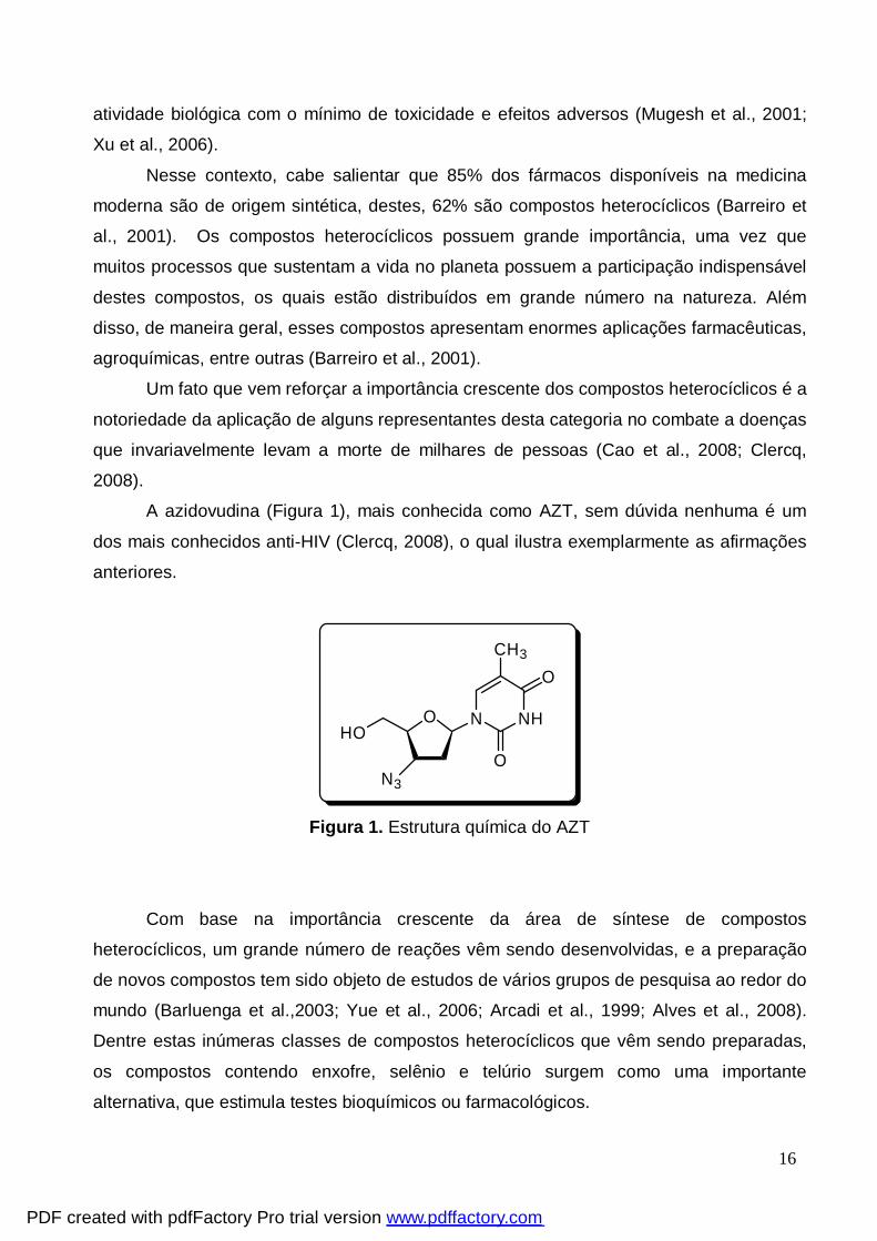
A azidovudina (Figura 1), mais conhecida como AZT, sem dúvida nenhuma é um
dos mais conhecidos anti-HIV (Clercq, 2008), o qual ilustra exemplarmente as afirmações
anteriores.
OHO
N NH
O
O
N3
CH3
Figura 1. Estrutura química do AZT
Com base na importância crescente da área de síntese de compostos
heterocíclicos, um grande número de reações vêm sendo desenvolvidas, e a preparação
de novos compostos tem sido objeto de estudos de vários grupos de pesquisa ao redor do
mundo (Barluenga et al.,2003; Yue et al., 2006; Arcadi et al., 1999; Alves et al., 2008).
Dentre estas inúmeras classes de compostos heterocíclicos que vêm sendo preparadas,
os compostos contendo enxofre, selênio e telúrio surgem como uma importante
alternativa, que estimula testes bioquímicos ou farmacológicos.
PDF created with pdfFactory Pro trial version www.pdffactory.com
17
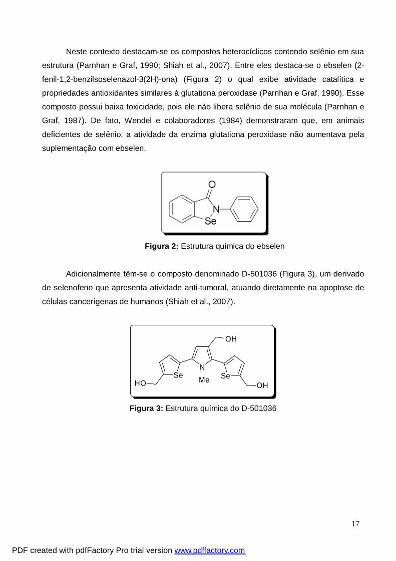
Neste contexto destacam-se os compostos heterocíclicos contendo selênio em sua
estrutura (Parnhan e Graf, 1990; Shiah et al., 2007). Entre eles destaca-se o ebselen (2-
fenil-1,2-benzilsoselenazol-3(2H)-ona) (Figura 2) o qual exibe atividade catalítica e
propriedades antioxidantes similares à glutationa peroxidase (Parnhan e Graf, 1990). Esse
composto possui baixa toxicidade, pois ele não libera selênio de sua molécula (Parnhan e
Graf, 1987). De fato, Wendel e colaboradores (1984) demonstraram que, em animais
deficientes de selênio, a atividade da enzima glutationa peroxidase não aumentava pela
suplementação com ebselen.
Figura 2: Estrutura química do ebselen
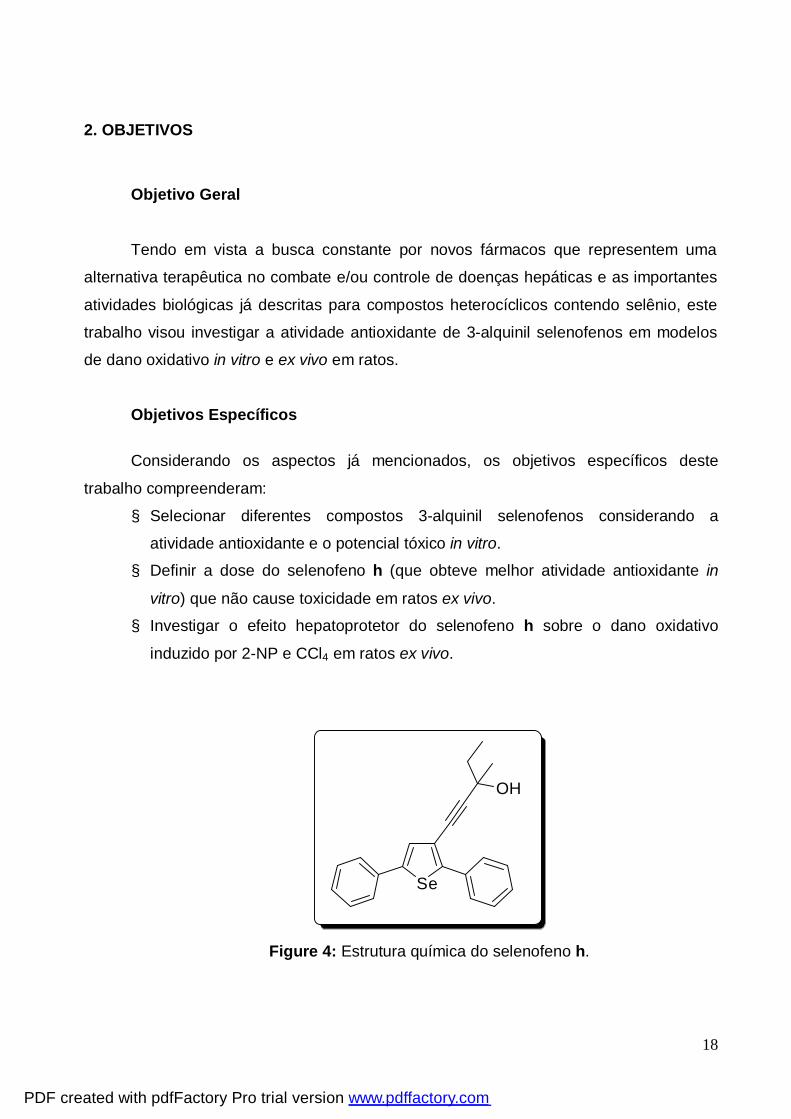
Adicionalmente têm-se o composto denominado D-501036 (Figura 3), um derivado
de selenofeno que apresenta atividade anti-tumoral, atuando diretamente na apoptose de
células cancerígenas de humanos (Shiah et al., 2007).
SeHO
NSe
OH
OHMe
Figura 3: Estrutura química do D-501036
PDF created with pdfFactory Pro trial version www.pdffactory.com
18
2. OBJETIVOS
Objetivo Geral
Tendo em vista a busca constante por novos fármacos que representem uma
alternativa terapêutica no combate e/ou controle de doenças hepáticas e as importantes
atividades biológicas já descritas para compostos heterocíclicos contendo selênio, este
trabalho visou investigar a atividade antioxidante de 3-alquinil selenofenos em modelos
de dano oxidativo in vitro e ex vivo em ratos.
Objetivos Específicos Considerando os aspectos já mencionados, os objetivos específicos deste
trabalho compreenderam:
§ Selecionar diferentes compostos 3-alquinil selenofenos considerando a
atividade antioxidante e o potencial tóxico in vitro.
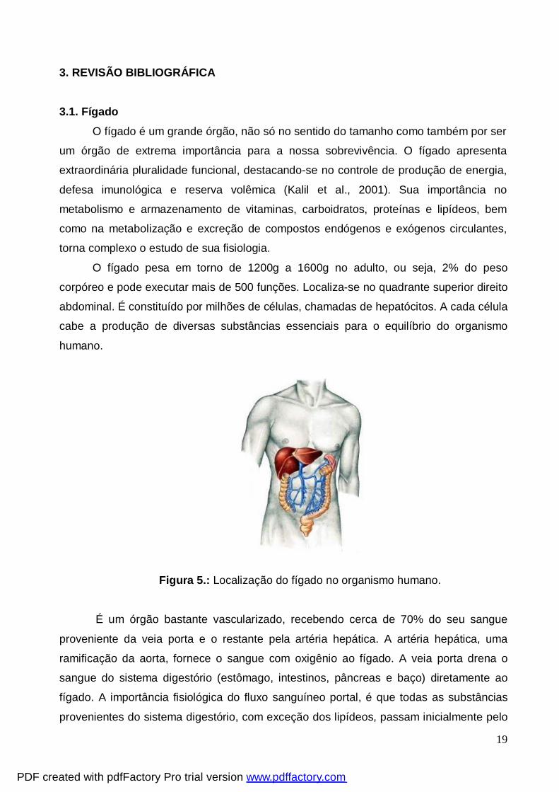
§ Definir a dose do selenofeno h (que obteve melhor atividade antioxidante in
vitro) que não cause toxicidade em ratos ex vivo.
§ Investigar o efeito hepatoprotetor do selenofeno h sobre o dano oxidativo
induzido por 2-NP e CCl4 em ratos ex vivo.
Se
OH
Figure 4: Estrutura química do selenofeno h.
PDF created with pdfFactory Pro trial version www.pdffactory.com
19
3. REVISÃO BIBLIOGRÁFICA
3.1. Fígado
O fígado é um grande órgão, não só no sentido do tamanho como também por ser
um órgão de extrema importância para a nossa sobrevivência. O fígado apresenta
extraordinária pluralidade funcional, destacando-se no controle de produção de energia,
defesa imunológica e reserva volêmica (Kalil et al., 2001). Sua importância no
metabolismo e armazenamento de vitaminas, carboidratos, proteínas e lipídeos, bem
como na metabolização e excreção de compostos endógenos e exógenos circulantes,
torna complexo o estudo de sua fisiologia.
O fígado pesa em torno de 1200g a 1600g no adulto, ou seja, 2% do peso
corpóreo e pode executar mais de 500 funções. Localiza-se no quadrante superior direito
abdominal. É constituído por milhões de células, chamadas de hepatócitos. A cada célula
cabe a produção de diversas substâncias essenciais para o equilíbrio do organismo
humano.
Figura 5.: Localização do fígado no organismo humano.
É um órgão bastante vascularizado, recebendo cerca de 70% do seu sangue
proveniente da veia porta e o restante pela artéria hepática. A artéria hepática, uma
ramificação da aorta, fornece o sangue com oxigênio ao fígado. A veia porta drena o
sangue do sistema digestório (estômago, intestinos, pâncreas e baço) diretamente ao
fígado. A importância fisiológica do fluxo sanguíneo portal, é que todas as substâncias
provenientes do sistema digestório, com exceção dos lipídeos, passam inicialmente pelo
PDF created with pdfFactory Pro trial version www.pdffactory.com
20
fígado antes de atingir o sistema circulatório. O fígado possui uma estrutura anatômica
única. As células hepáticas estão em contato com a circulação sanguínea de um lado e o
canalículo biliar de outro. Deste modo, o hepatócito tem uma grande área de contato
tanto com um sistema nutriente proveniente dos sinusóides da veia porta e um sistema
de escoamento quanto com o canalículo biliar que transporta as secreções e excreções
dos hepatócitos (Motta et al., 2000; Kalil et al., 2001).
Os nutrientes absorvidos pelo intestino chegam ao fígado pela via linfática. No
fígado são metabolizados e acumulados. As substâncias tóxicas absorvidas são
neutralizadas e eliminadas através da bile. O fígado possui atividade endócrina e
exócrina. A fisiologia hepática é altamente especializada no cumprimento de diversas
funções conhecidas, tais como: metabólicas, excretoras, secretoras, armazenamento,
protetoras, circulatórias e de coagulação sanguínea. Dentre as funções citadas, uma das
mais relevantes consiste na função de desintoxicação, através do sistema microssomal
de biotransformação de xenobióticos (sistema P-450 ou CYP-450) (Motta et al., 2000;
Kalil et al., 2001).
Devido à grande amplitude funcional hepática, este órgão é constantemente
exposto a substâncias do meio externo, atuando como órgão alvo de diversos
xenobióticos (Motta et al., 2000). Os sinais e sintomas que refletem algum transtorno
hepático são: astenia, dor abdominal, náusea, vômito e icterícia. Além destes sintomas,
exames laboratoriais são utilizados para confirmar o diagnóstico de doença hepática e
sua severidade, sendo que a insuficiência hepática pode levar a falência de outros
órgãos como o encéfalo, rins e coração (Kim et al., 1998).
A biotransformação de xenobióticos consiste na conversão de substâncias
lipofílicas em substâncias polares, passíveis de excreção. Esse processo de
biotransformação é crucial para eliminação de compostos tóxicos. O metabolismo é
realizado por enzimas, muitas das quais são específicas e estão localizadas
principalmente no retículo endoplasmático. Algumas estão localizadas no citosol e
poucas são encontradas em outras organelas como, por exemplo, as mitocôndrias
(Timbrell, 1991).
As reações envolvidas na biotransformação dos xenobióticos podem ser
agrupadas em duas fases distintas: Fase Pré-Sintética ou Fase I, onde ocorrem reações
de oxidação, redução e hidrólise e Fase Sintética, de Conjugação ou Fase II, onde
ocorrem reações de conjugação. Na primeira fase, as oxidações são, geralmente,
catalisadas por uma classe de enzimas monooxigenases encontradas no retículo
PDF created with pdfFactory Pro trial version www.pdffactory.com
21
endoplasmático e conhecidas como enzimas microssomais, que são enzimas complexas,
inespecíficas (oxidam diferentes tipos de compostos) e que necessitam de NADPH e O2
para agirem. Durante a oxidação elas exigem uma molécula de oxigênio para cada
molécula de fármaco a ser oxidado (um átomo de oxigênio é incorporado ao fármaco
ocorrendo a oxidação e outro é, geralmente, combinado com H2, formando água). A
enzima ou sistema enzimático principal na oxidação de xenobióticos é o sistema P-450.
Ele recebe os elétrons provenientes de outras fases da reação, se reduz e se liga ao O2
e ao fármaco, promovendo de fato a oxidação do composto. As reações de fase II são
reações de conjugação que envolvem a adição de grupos endógenos aos xenobióticos,
os quais geralmente são polares. Os grupos doados nas reações de conjugação incluem
derivados de carboidratos, aminoácidos, glutationa e sulfato (Timbrell, 1991; Motta et al.,
2000). Esse processo de biotransformação é conhecido como processo de
desintoxicação, entretanto em alguns casos podem ser formados metabólitos reativos
que são mais tóxicos que os originais. As reações de fase I são as mais comumente
envolvidas nesse processo (Timbrell, 1991).
3.1.1. Hepatotoxicidade
As doenças hepáticas são um problema de saúde pública mundial, sendo que a
evolução das mesmas inicia-se com a hepatite, esteatose, fibrose, cirrose até o
carcinoma hepatocelular (Loguercio e Frederico, 2003; Vitaglione et al., 2004). A
confirmação do dano hepático é realizada por meio de exames diagnósticos sorológicos
específicos, como a dosagem da atividade enzimática das transaminases (AST e ALT),
fosfatase alcalina, γ-glutamil-transferase (Henry et al., 1999). Estas são enzimas
presentes em vários tecidos, de função intracelular, e sua presença no sangue é
conseqüência de liberação anormal para a circulação. A elevação das transaminases no
soro pode ocorrer mesmo após agressão celular mínima. A utilização destes marcadores
pode delinear o tipo de dano hepático, sua extensão e o prognóstico da doença hepática.
Entretanto, somente as técnicas histopatológicas confirmam e complementam a
avaliação do dano, auxiliando também na verificação precoce da efetividade de terapias
que protejam e/ou revertam a injúria hepática causada por agentes indutores de dano
hepático (Henry et al., 1999).
PDF created with pdfFactory Pro trial version www.pdffactory.com
22
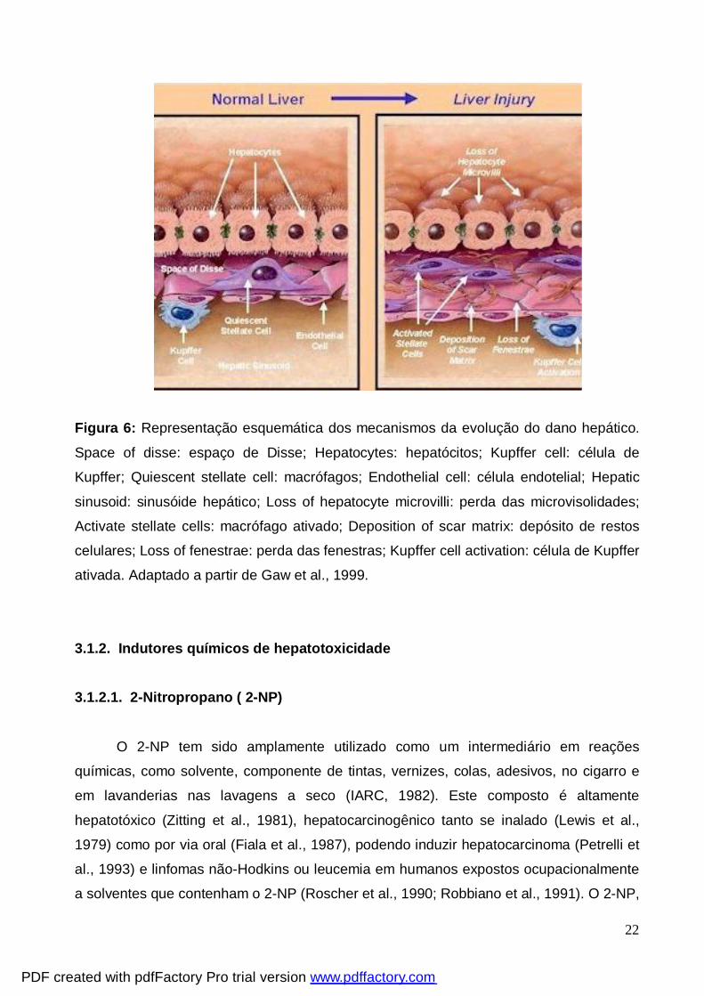
Figura 6: Representação esquemática dos mecanismos da evolução do dano hepático.
Space of disse: espaço de Disse; Hepatocytes: hepatócitos; Kupffer cell: célula de
Kupffer; Quiescent stellate cell: macrófagos; Endothelial cell: célula endotelial; Hepatic
sinusoid: sinusóide hepático; Loss of hepatocyte microvilli: perda das microvisolidades;
Activate stellate cells: macrófago ativado; Deposition of scar matrix: depósito de restos
celulares; Loss of fenestrae: perda das fenestras; Kupffer cell activation: célula de Kupffer
ativada. Adaptado a partir de Gaw et al., 1999.
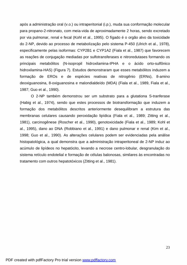
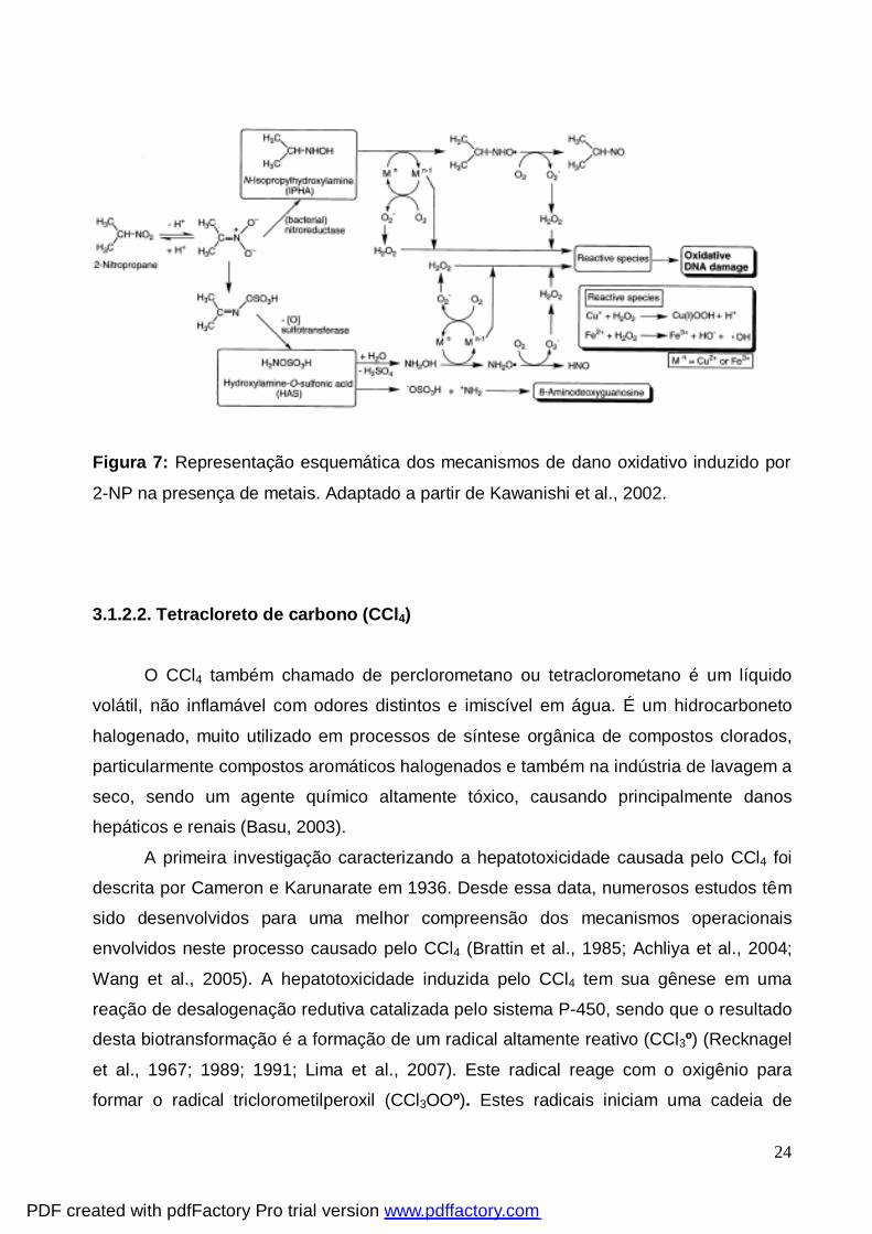
3.1.2. Indutores químicos de hepatotoxicidade 3.1.2.1. 2-Nitropropano ( 2-NP)
O 2-NP tem sido amplamente utilizado como um intermediário em reações
químicas, como solvente, componente de tintas, vernizes, colas, adesivos, no cigarro e
em lavanderias nas lavagens a seco (IARC, 1982). Este composto é altamente
hepatotóxico (Zitting et al., 1981), hepatocarcinogênico tanto se inalado (Lewis et al.,
1979) como por via oral (Fiala et al., 1987), podendo induzir hepatocarcinoma (Petrelli et
al., 1993) e linfomas não-Hodkins ou leucemia em humanos expostos ocupacionalmente
a solventes que contenham o 2-NP (Roscher et al., 1990; Robbiano et al., 1991). O 2-NP,
PDF created with pdfFactory Pro trial version www.pdffactory.com
23
após a administração oral (v.o.) ou intraperitonial (i.p.), muda sua conformação molecular
para propano-2-nitronato, com meia-vida de aproximadamente 2 horas, sendo excretado
por via pulmonar, renal e fecal (Kohl et al., 1995). O fígado é o orgão alvo da toxicidade
do 2-NP, devido ao processo de metabolização pelo sistema P-450 (Ulrich et al., 1978),
especificamente pelas isoformas: CYP2B1 e CYP1A2 (Fiala et al., 1987) que favorecem
as reações de conjugação mediadas por sulfotransferases e nitroredutases formando os
principais metabólitos (N-isopropil hidroxilamina-IPHA e o ácido orto-sulfônico
hidroxilamina-HAS) (Figura 7). Estudos demonstraram que esses metabólitos induzem a
formação de EROs e de espécies reativas de nitrogênio (ERNs), 8-amino
deoxiguanosina, 8-oxiguanosina e malondialdeído (MDA) (Fiala et al., 1989, Fiala et al.,
1987; Guo et al., 1990).
O 2-NP também demonstrou ser um substrato para a glutationa S-tranferase
(Habig et al., 1974), sendo que estes processos de biotransformação que induzem a
formação dos metabólitos descritos anteriormente desequilibram a estrutura das
membranas celulares causando peroxidação lipídica (Fiala et al., 1989; Zitting et al.,
1981), carcinogênese (Roscher et al., 1990), genotoxicidade (Fiala et al., 1989; Kohl et
al., 1995), dano ao DNA (Robbiano et al., 1991) e dano pulmonar e renal (Kim et al.,
1998; Guo et al., 1990). As alterações celulares podem ser evidenciadas pela análise
histopatológica, a qual demonstra que a administração intraperitoneal de 2-NP induz ao
acúmulo de lipídeos no hepatócito, levando a necrose centro-lobular, desgranulação do
sistema retículo endotelial e formação de células balonosas, similares às encontradas no
tratamento com outros hepatotóxicos (Zitting et al., 1981).
PDF created with pdfFactory Pro trial version www.pdffactory.com
24
Figura 7: Representação esquemática dos mecanismos de dano oxidativo induzido por
2-NP na presença de metais. Adaptado a partir de Kawanishi et al., 2002.
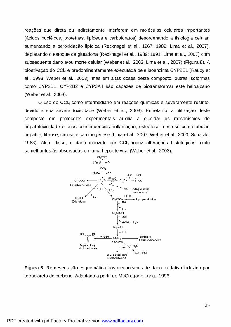
3.1.2.2. Tetracloreto de carbono (CCl4)
O CCl4 também chamado de perclorometano ou tetraclorometano é um líquido
volátil, não inflamável com odores distintos e imiscível em água. É um hidrocarboneto
halogenado, muito utilizado em processos de síntese orgânica de compostos clorados,
particularmente compostos aromáticos halogenados e também na indústria de lavagem a
seco, sendo um agente químico altamente tóxico, causando principalmente danos
hepáticos e renais (Basu, 2003).
A primeira investigação caracterizando a hepatotoxicidade causada pelo CCl4 foi
descrita por Cameron e Karunarate em 1936. Desde essa data, numerosos estudos têm
sido desenvolvidos para uma melhor compreensão dos mecanismos operacionais
envolvidos neste processo causado pelo CCl4 (Brattin et al., 1985; Achliya et al., 2004;
Wang et al., 2005). A hepatotoxicidade induzida pelo CCl4 tem sua gênese em uma
reação de desalogenação redutiva catalizada pelo sistema P-450, sendo que o resultado
desta biotransformação é a formação de um radical altamente reativo (CCl3º) (Recknagel
et al., 1967; 1989; 1991; Lima et al., 2007). Este radical reage com o oxigênio para
formar o radical triclorometilperoxil (CCl3OOº). Estes radicais iniciam uma cadeia de
PDF created with pdfFactory Pro trial version www.pdffactory.com
25
reações que direta ou indiretamente interferem em moléculas celulares importantes
(ácidos nucléicos, proteínas, lipídeos e carboidratos) desordenando a fisiologia celular,
aumentando a peroxidação lipídica (Recknagel et al., 1967; 1989; Lima et al., 2007),
depletando o estoque de glutationa (Recknagel et al., 1989; 1991; Lima et al., 2007) com
subsequente dano e/ou morte celular (Weber et al., 2003; Lima et al., 2007) (Figura 8). A
bioativação do CCl4 é predominantemente executada pela isoenzima CYP2E1 (Raucy et
al., 1993; Weber et al., 2003), mas em altas doses deste composto, outras isoformas
como CYP2B1, CYP2B2 e CYP3A4 são capazes de biotransformar este haloalcano
(Weber et al., 2003).
O uso do CCl4 como intermediário em reações químicas é severamente restrito,
devido a sua severa toxicidade (Weber et al., 2003). Entretanto, a utilização deste
composto em protocolos experimentais auxilia a elucidar os mecanismos de
hepatotoxicidade e suas consequências: inflamação, esteatose, necrose centrolobular,
hepatite, fibrose, cirrose e carcinogênese (Lima et al., 2007; Weber et al., 2003; Schatzki,
1963). Além disso, o dano induzido por CCl4 induz alterações histológicas muito
semelhantes às observadas em uma hepatite viral (Weber et al., 2003).
Figura 8: Representação esquemática dos mecanismos de dano oxidativo induzido por
tetracloreto de carbono. Adaptado a partir de McGregor e Lang., 1996.
PDF created with pdfFactory Pro trial version www.pdffactory.com
26
3.2. Estresse Oxidativo
O estresse oxidativo corresponde a uma excessiva formação endógena de EROs
associada a uma diminuição nas defesas antioxidantes (Dawson e Dawson, 1996;
Halliwel, 1992). As EROs são capazes de gerar estresse oxidativo em conseqüência de
suas propriedades oxidantes e reação com os constituintes celulares (Josephy, 1997;
Timbrell, 2000). Estas são geradas por uma variedade de processos, podendo atacar
uma diversidade de biomoléculas alvo, tais como, DNA, lipídeos e proteínas (Josephy,
1997; Timbrell, 2000).
As membranas biológicas apresentam uma estrutura geral comum. Estas são
constituídas de uma bicamada lipídica as quais estão associadas a proteínas. As
proteínas presentes na membrana celular são responsáveis pelo transporte de moléculas
específicas através da bicamada lipídica. Além disso, essas proteínas podem agir como
catalisadoras de reações associadas às membranas, como a síntese de ATP (Alberts et
al., 1994). As membranas biológicas são constituídas principalmente por fosfolipídeos, os
quais possuem uma cabeça polar e duas caudas hidrofóbicas. Geralmente, as caudas
hidrofóbicas são compostas por ácidos graxos, que podem diferir no comprimento e na
configuração em que se apresentam, podendo uma das caudas apresentar uma ou mais
ligações duplas (insaturações) (Alberts et al., 1994; Halliwell e Gutteridge, 1989). Quando
as EROs reagem com esses ácidos graxos insaturados, modificam os lipídeos e a
membrana perde suas características arquitetônicas, tornando-se menos firme e menos
flexível, criando-se verdadeiras fendas iônicas que alteram sua semipermeabilidade, o
que favorece a entrada e saída indiscriminada de metabólitos e detritos da célula,
provocando sua ruptura e lise com necrose (Josephy, 1997; Timbrell, 2000).
As principais EROs vinculadas ao estresse oxidativo são: o radical ânion
superóxido (O-2), radical hidroxil (.OH), peróxido de hidrogênio (H2O2), óxido nítrico (NO)
e peroxinitrito (ONOO-). Estes por sua vez são neutralizados por um elaborado sistema
de defesa antioxidante constituído de enzimas tais como a catalase, a superóxido
dismutase, a glutationa peroxidase, além de inúmeros sistemas de defesas não-
enzimáticas incluindo as vitaminas A, E e C, flavonóides, ubiquinonas e o conteúdo de
glutationa reduzida (Alexi et al., 1998; Gianni et al., 2004).
PDF created with pdfFactory Pro trial version www.pdffactory.com
27
3.2.1. Hepatotoxicidade x Estresse Oxidativo
Recentemente, o estresse oxidativo tem sido sugerido como uma das principais
causas de lesão tecidual (Loguercio e Frederico, 2003). Segundo Lee e colaboradores
(2001), o estresse oxidativo tem um papel fundamental no início e desenvolvimento das
patologias hepáticas. O reconhecimento do envolvimento das EROs em diversas
enfermidades tem levado à implementação da terapia antioxidante (Young e Woodside,
2001).
Antioxidantes como α-tocoferol podem bloquear a fibrogênese hepática (Lee et al.,
2001). Os antioxidantes são compostos que funcionam como bloqueadores dos
processos óxido-redutivos desencadeados pelas EROs. Portanto, funcionam em vários
tipos de processos degenerativos. Nutrientes dietéticos com propriedades antioxidantes
estão assumindo grande significado no contexto de certas doenças, como a
aterosclerose (Bem et al., 2008). Antioxidantes sintéticos têm potencial uso na química,
indústria alimentícia e medicina (Packer e Cadenas, 1997). Alguns desses compostos
contêm um grupo funcional quimicamente análogo ao de antioxidantes “naturais”, e
introduzem novos grupos químicos que aumentam sua amplitude de ação celular ou
melhoram sua biodisponibilidade. Por outro lado, outros antioxidantes sintéticos não
apresentam analogia estrutural aos naturais, mas exercem alta reatividade para com as
EROs e/ou protegem seletivamente alguns tecidos (Packer e Cadenas, 1997).
3.3. Selênio
O selênio (Se) foi descoberto em 1817, pelo químico sueco J. J. Berzelius. O Se é
um elemento do grupo 16 da tabela periódica, podendo apresentar-se sob quatro
estados de oxidação: selenato (Se+6), selenito (Se+4), selênio elementar (Se0) e seleneto
(Se-2).
O Se compartilha propriedades químicas e físicas com o enxofre (S). Esta
similaridade permite que o Se substitua o S, promovendo interações Se-S nos sistemas
biológicos. Por outro lado, as diferenças nas propriedades fisico-químicas entre Se e S
constituem a base de seus papéis biológicos específicos (Stadtman,1980). Os selenóis
(R-SeH) são as formas correspondentes aos tióis (R-SH), onde ocorre a substituição do
átomo de S pelo átomo de Se (Klayman e Günther,1973).
PDF created with pdfFactory Pro trial version www.pdffactory.com
28
O selênio é um elemento traço essencial, cuja essencialidade nutricional foi
demonstrada em 1957, em ratos (Schwartz e Foltz, 1957). Nos últimos anos, têm sido
descrito que baixos níveis de selênio podem levar à predisposição para o
desenvolvimento de algumas doenças, tais como câncer, esclerose, doença
cardiovascular, cirrose e diabetes (Navarro-Alarcón e López-Martinez, 2000). Neste
contexto, a suplementação de dietas com selênio, tanto para animais quanto para
humanos, tem sido aceita pela comunidade científica. Para humanos, a Junta de
Alimentação e Nutrição da Academia de Ciências dos Estados Unidos propõe uma
ingestão diária de 50-200 μg, a qual é considerada segura e saudável para adultos. Por
outro lado, sabe-se que a concentração alimentar requerida de selênio é muito próxima
da dose que pode ser tóxica (Oldfield, 1987). De fato, estudos demonstraram que altas
doses de selênio podem ser citotóxicas, uma vez que possuem a habilidade de oxidar
grupos –SH e gerar radicais livres (Barbosa et al. 1998; Nogueira et al. 2004). Este
elemento pode ser encontrado nos seguintes alimentos: castanha-do-pará, alho, cebola,
brócolis, cogumelos, cereais, pescados, ovos e carnes (Dumont et al., 2006).
Este calcogênio apresenta um grande número de funções biológicas, sendo a
mais importante a de antioxidante. Sabe-se que as moléculas contendo selênio, como
por exemplo o disseleneto de difenila (PhSe)2, podem ser melhores antioxidantes do que
os antioxidantes clássicos (Arteel e Sies, 2001). Já é conhecido que o selênio está
presente como resíduo de selenocisteína no sítio ativo das enzimas glutationa
peroxidase (Wingler e Brigelius-Flohé, 1999), tioredoxina redutase (Holmgren, 1985), 5’-
deiodinase (Behne e Kyriakopoulos, 1990) e selenoproteína P (Ursini et al., 1990). A
atividade redox do selênio tem importância fundamental porque ele faz parte do sítio
ativo dessas enzimas.
Nos mamíferos, o selênio parece ser rapidamente absorvido no duodeno, seguido
pelo jejuno e íleo. Além do trato gastrointestinal, o selênio pode ser absorvido por tecidos
cutâneos e inalação. Estas duas últimas vias de absorção estão relacionadas com a
exposição e intoxicação ocupacional por compostos de selênio (Whanger et al., 1976).
Após a absorção, os maiores níveis de selênio estão localizados nos eritrócitos,
baço, coração, unha e esmalte de dentes (Martin e Gerlack, 1972). Na intoxicação
crônica em animais, o selênio é depositado principalmente nos rins e fígado, seguido
pelo pâncreas, baço e pulmões (Wilber, 1980). A primeira evidência de metabolização
dos compostos de selênio em animais foi determinada após um longo período de
PDF created with pdfFactory Pro trial version www.pdffactory.com
29
tratamento com o selenito de sódio. Os animais apresentavam odor gárlico
característico, que posteriormente demonstrou-se ter sido causado pelo seleneto de
dimetila (Klayman e Gunther, 1973). Este metabólito pode ser resultado do processo de
detoxificação do selênio, o qual envolve uma série de metilações dependentes da S-
adenosilmetionina (Hoffman e McConnell, 1986).
O selênio pode ser excretado por três vias: urina, fezes e ar expirado. A excreção
urinária deste composto pode auxiliar em casos de intoxicações ou de exposições a altos
níveis deste elemento (Valentine et al., 1978). Recentemente, foi demonstrado que
dentro dos níveis normais de selênio, ou seja, não tóxicos, a principal forma encontrada
na urina é como seleno-açúcar. Entretanto, nos casos de doses tóxicas de selênio, o
marcador biológico encontrado na urina é o trimetilselenônio (Suzuki et al., 2006). Em
indivíduos expostos acidentalmente a altos níveis de selênio, pode ser realizada a
detecção do composto volátil seleneto de dimetila (Mozier et al., 1988).
Devido à tentativa crescente de desenvolvimento de compostos que possuam
atividades biológicas e aplicações farmacológicas (Parnham e Graf, 1990), têm chamado
bastante atenção os compostos orgânicos de selênio (organocalcogênios) com
propriedades antioxidantes, que em geral, são inibidores da peroxidação lipídica (Sies,
1993; Kanda et al., 1999).
Durante as últimas décadas, o interesse por esta classe de compostos tem sido
intensificado, principalmente devido ao fato de que uma variedade destes compostos
possui propriedades farmacológicas (Nogueira et al., 2004). De fato, estudos em animais
de laboratório têm demonstrado que estes compostos apresentam propriedades
antiúlcera (Savegnago et al., 2006), antiinflamatória e antinociceptiva (Savegnago et al.,
2007a), antidepressiva e ansiolítica (Savegnago et al., 2007b), neuroprotetora (Ghisleni
et al., 2003), hepatoprotetora (Borges et al., 2005; 2006, Wilhelm et al., 2008), anti-
hiperglicêmica (Barbosa et al., 2006) e pode retardar o desenvolvimento de câncer
(Barbosa et al., 2008). Além disso, apresenta efeitos antioxidantes em diversos modelos
de toxicidade induzida por estresse oxidativo (Meotti et al. 2004; Luchese et al., 2007),
incluindo exposições ao cádmio (Santos et al., 2004; 2005; Borges et al., 2008).
PDF created with pdfFactory Pro trial version www.pdffactory.com
30
3.3.1. Selênio x hepatotoxicidade
A associação da importância do selênio com a hepatoproteção foi demonstrada
em meados de 1957, graças a estudos pioneiros desenvolvidos por Schwartz e Foltz,
onde foi demonstrado que ratos alimentados com dieta pobre em selênio poderiam
desenvolver necrose hepática. Este interessante estudo levou ao reconhecimento que
doenças oriundas da privação de nutrientes, poderiam ser causadas por deficiência de
selênio na dieta (Oldfield, 1987). Outra pesquisa relevante demonstrou que a
administração oral de ebselen, pode inibir as lipoxigenases em um modelo experimental
de indução de hepatite pela administração da endotoxina galactosamina (Wendel et al.,
1984). De fato o ebselen demonstrou suas propriedades hepatoprotetoras em diversos
modelos de dano hepático, tais como os induzidos por paracetamol (Li et al., 1994;
Rocha et al., 2005), CCl4 (Wasser et al., 2001), lipopolissacarídeo e Propionibacterium
acnes (Koyanagi et al., 2001), etanol (Kono et al., 2001) e isquemia e reperfusão (Ozaki
et al., 1997).
Além disso, nosso grupo de pesquisa demonstrou que o disseleneto de difenila
apresenta efeito hepatoprotetor contra dano hepático induzido por 2-NP (Borges et al.,
2005, 2006; Wilhelm et al., 2008), cádmio (Borges et al., 2008) e mostra-se efetivo contra
dano oxidativo induzido por acetaminofen (paracetamol) em ratos (Wilhelm et al., 2009).
3.4. Compostos heterocíclicos
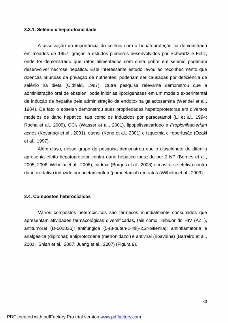
Vários compostos heterocíclicos são fármacos mundialmente consumidos que
apresentam atividades farmacológicas diversificadas, tais como, inibidor do HIV (AZT),
antitumoral (D-501036); antifúngica (5-(3-buten-1-inil)-2,2’-bitienila); antinflamatória e
analgésica (dipirona); antiprotozoária (metronidazol) e antiviral (ribavirina) (Barreiro et al.,
2001; Shiah et al., 2007; Juang et al., 2007) (Figura 9).
PDF created with pdfFactory Pro trial version www.pdffactory.com
31
OHO
N NH
O
O
N3
CH3
SeHO
NSe
OH
OHMe
S S
OHO
N
OHHO
NN
OH2N
NN
N Me
SO3-Na+
Me
Me
O
N
NOH
MeO2N
RibavirinaDipirona Metronidazol
AZT D-501036 5-(3-buten-1-inil)-2,2'-bitienila
Figura 9. Fármacos contendo unidade heterocíclica
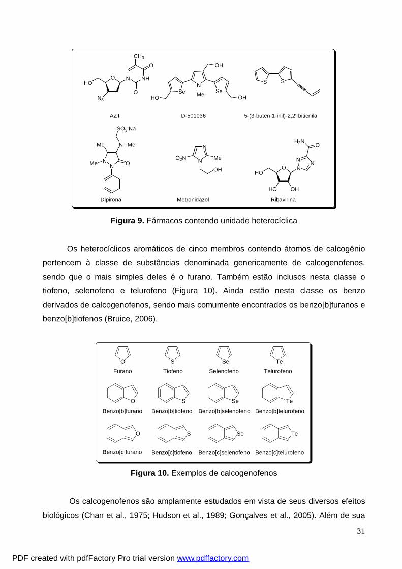
Os heterocíclicos aromáticos de cinco membros contendo átomos de calcogênio
pertencem à classe de substâncias denominada genericamente de calcogenofenos,
sendo que o mais simples deles é o furano. Também estão inclusos nesta classe o
tiofeno, selenofeno e telurofeno (Figura 10). Ainda estão nesta classe os benzo
derivados de calcogenofenos, sendo mais comumente encontrados os benzo[b]furanos e
benzo[b]tiofenos (Bruice, 2006).
O S TeSe
O
O S Se Te
S Se Te
Furano Tiofeno Selenofeno Telurofeno
Benzo[b]furano
Benzo[c]furano Benzo[c]tiofeno Benzo[c]selenofeno Benzo[c]telurofeno
Benzo[b]tiofeno Benzo[b]selenofeno Benzo[b]telurofeno
Figura 10. Exemplos de calcogenofenos
Os calcogenofenos são amplamente estudados em vista de seus diversos efeitos
biológicos (Chan et al., 1975; Hudson et al., 1989; Gonçalves et al., 2005). Além de sua
PDF created with pdfFactory Pro trial version www.pdffactory.com
32
atividade atioxidante (Meotti et al., 2004), os calcogenofenos apresentam propriedades
antinociceptiva e antiinflamatória (Zeni et al., 2001; Meotti et al., 2003; Gonçales et al.,
2005)
Entre os calcogenofenos, furanos, tiofenos e seus derivados têm despertado o
interesse de pesquisadores na química orgânica sintética, pois suas ocorrências em
produtos naturais que apresentam alguma atividade biológica são relativamente
freqüentes, incentivando a procura de metodologias para a síntese destes compostos
(Sperry e Wright, 2005; Tachibana et al., 2008; Tran et al., 2008). Os selenofenos,
telurofenos e seus derivados vêm recebendo menos atenção da comunidade científica
quando comparados com seus análogos tiofenos e furanos. De fato, os telurofenos e
selenofenos são escassamente relatados na literatura tanto na área biológica, quanto na
área de síntese e reatividade destes compostos. Este fato incentiva estudos que
busquem demonstrar possibilidades de síntese bem como a busca de compostos com
possíveis atividades biológicas.
3.4.1. Compostos heterocíclicos x Selênio
Uma vez que moléculas contendo selênio podem ser melhores antioxidantes do
que os antioxidantes clássicos (Arteel e Sies, 2001), a incorporação do átomo de selênio
em moléculas orgânicas permite a preparação de inúmeros compostos, com
propriedades farmacológicas bastante amplas.
Neste contexto destacam-se os compostos heterocíclicos contendo selênio em
sua estrutura. Como mencionado anteriormente, o ebselen (Figura 2) apresenta
importantes atividades biológicas: exibe atividade catalítica e propriedades antioxidantes
similares à glutationa peroxidase (Parnhan e Graf, 1990), possui baixa toxicidade
(Parnhan e Graf, 1987), reage com grupos tióis, como a glutationa (Ulrich et al., 1996),
inibe a peroxidação lipídica (Parnhan e Graf, 1987; Rossato et al., 2002; Nowak et al.,
2006), inibe a lipoxigenase (Parnhan e Graf, 1987), bloqueia a produção de ânion
superóxido e desempenha um papel protetor contra o peroxinitrito (Masumoto e Sies,
1996). Além disso, o ebselen tem sido usado como antioxidante, como neuroprotetor em
culturas de neurônios (Osaki et al., 1997; Takasago et al.,1997; Kondoh et al., 1999; Imai
et al., 2001; Porciúncula et al., 2003), no tratamento clínico de pacientes com isquemia
aguda (Yamaguchi et al., 1998; Kondoh et al., 1999), em modelos de Parkinson
PDF created with pdfFactory Pro trial version www.pdffactory.com
33
(Moussaoui et al., 2000) e como antiinflamatório (Parnhan e Graf, 1987; Walther et al.,
1999; Haddad et al., 2002; Mugesh et al., 2001).
Outro composto heterocíclico que destaca-se é o D-501036 (Figura 3), um
derivado de selenofeno que apresenta atividade anti-tumoral, atuando diretamente na
apoptose de células cancerígenas de humanos (Shiah et al., 2007).
PDF created with pdfFactory Pro trial version www.pdffactory.com
34
4. MANUSCRITOS
Os resultados que fazem parte desta dissertação estão apresentados sob a forma
de manuscritos, os quais se encontram assim organizados. Os itens Materiais e
Métodos, Resultados, Discussão dos Resultados e Referências Bibliográficas
encontram-se nos próprios manuscritos. O manuscrito 1 e 2 estão dispostos da mesma
forma que foram submetidos para avaliação.
PDF created with pdfFactory Pro trial version www.pdffactory.com
35
4.1. Selenofeno protege contra dano oxidativo induzido por 2-nitropropano em fígado de
ratos.
4.1.1. Manuscrito 1
SELENOPHENE PROTECTS AGAINST OXIDATIVE DAMAGE INDUCED
BY 2-NITROPROPANE IN LIVER OF RATS
(Submetido à Journal of Pharmacological Science)
PDF created with pdfFactory Pro trial version www.pdffactory.com
36
Selenophene Protects Against Oxidative Damage Induced
by 2-Nitropropane in Liver of Rats
Ethel A. Wilhelm, Marina Prigol, Diego Alves, Lucielli Savegnago, Cristina W. Nogueira∗
Departamento de Química, Centro de Ciências Naturais e Exatas, Universidade Federal
de Santa Maria, Santa Maria, CEP 97105-900, RS, Brazil
Running title: Antioxidant effect of selenophenes
∗Correspondence should be sent to:
Cristina Nogueira
Departamento de Química,
Centro de Ciências Naturais e Exatas,
Universidade Federal de Santa Maria, 97105-900, Santa Maria, RS, Brazil.
Phone: 55-55-3220-8140
FAX: 55-55-3220-8978
E-mail: [email protected]
PDF created with pdfFactory Pro trial version www.pdffactory.com
37
Abstract The aim of this study was the in vitro study of the antioxidant function of recently
synthesized selenophenes. It was evaluated using iron/EDTA-induced thiobarbituric acid
reactive species (TBARS) and δ-aminolevulinate dehydratase (δ-ALA-D) assays.
Selenophenes b, c, d, e, f, and i presented poor antioxidant profiles in the TBARS assay
(IC50 > 400 µM) when compared to a, g and h (IC50 = 313, 233 and 237 µM, respectively).
Selenophenes a, g and h presented maximal inhibition (Imax) of lipid peroxidation of
75%. The antioxidant activity of selenophenes was dependent of a terminal alkyne
bonded directly at 3-position of selenophene. A second objective was to investigate the
antioxidant action of selenophene h, against oxidative damage induced by 2-nitropropane
(2-NP) in liver of rats. Selenophene h 25 mg/kg protected against the increase in TBARS
levels and in aspartate aminotransferase (AST) and alanine aminotransferase (ALT)
activities induced by 2-NP (100 mg/kg of body weight). Compound h 25mg/kg significantly
attenuated 2-NP-induced hepatic histopathological alterations. The inhibition of δ-ALA-D
activity caused by 2-NP was protected by selenophene h. This study proved the
antioxidant effect of selenophene h at a concentration of 25 mg/kg in a model of oxidative
damage induced by 2-NP in rats.
Keywords: selenophene, selenium, antioxidant, oxidative stress, liver.
PDF created with pdfFactory Pro trial version www.pdffactory.com
38
Introduction
Aerobic life is characterized by a steady formation of pro-oxidants balanced by a
similar rate of their consumption by antioxidants. To maintain homeostasis, there is a
requirement for the continuous regeneration of antioxidant capacity, and if this is not met,
oxidative damage occurs (1). Oxidative stress is characterized by a significantly increased
concentration of intracellular oxidizing species, such as reactive oxygen species (ROS)
and is often accompanied by the simultaneous loss of antioxidant defense capacity (2).
To counteract ROS levels more effectively, exogenous compounds should combine a
range of antioxidant activities in one chemically simple molecule (3).
In the last two decades the interest in organoselenium chemistry and biochemistry
has increased, mainly due to the fact that these compounds have been described to
possess very interesting biological activities (4, 5, 6, 7). Several reports have been
published on glutathione peroxidase (GPX)-mimetic compounds, which, like the native
enzyme, rely on the redox cycling of selenium (4, 8, 9, 10). In fact, scientists have paid
more attention to glutathione peroxidase artificial imitation in view of its instability, poor
availability and high molecular weight, which have limited its therapeutic use (11, 12).
Ebselen is the best-known mimic of GPX (13). In this context, diphenyl diselenide has
also been reported as a good GPX mimic and an antioxidant in different experimental
models of oxidative damage (14, 15, 16, 17, 18).
Although the peroxidase-like activity of organoselenium compounds may account
for their antioxidant properties, the SH–selenide exchange catalyzed by
organochalcogens may contribute to their toxicological properties by oxidizing relevant
SH-containing metabolites and proteins without consuming toxic substances such as
PDF created with pdfFactory Pro trial version www.pdffactory.com
39
peroxides (19). Selenides can react with -SH groups, forming selenosulfide or –SeH and
disulfides (19).
Chalcogenophenes, a class of organochalcogen heterocycles containing a five-
membered ring in the structure, have drawn the attention of researchers in view of their
interesting biological activities (20, 21, 22, 23) including antioxidant properties (24, 25,
26). Among chalcogenophenes, selenophenes play an important role in organic synthesis
(27) because of their excellent electrical properties and environmental stability.
Based on the important chemistry and pharmacological properties of
organoselenium compounds, the aim of this study was to evaluate the in vitro antioxidant
activity of recently synthesized selenophenes (27, 28). Considering the results obtained in
vitro, a second objective of this study was to investigate the antioxidant action of
selenophene h against the oxidative liver damage induced by 2-nitropropane (2-NP) in
rats. 2-NP, a nitroalkane, is known to be an acute hepatotoxicant (29) and a potent
hepatocarcinogen in rodents (30, 31). The mechanism by which 2-NP causes toxicity is
not completely elucidated, but accumulating evidence suggests that generation of
reactive oxygen species via the metabolism of 2-NP-nitronate to acetone and nitrite plays
an important role for the carcinogenic effect of 2-NP (32, 33).
Materials and Methods
Chemicals
Selenophenes (a-i) (Fig. 1) were prepared according to literature methods (27,
28). For in vitro experiments, selenophenes were dissolved in dimethylsulphoxide
(DMSO). For ex vivo experiments, selenophene h and 2-nitropropane (2-NP) were
dissolved in canola oil.
PDF created with pdfFactory Pro trial version www.pdffactory.com
40
δ-Aminolevulinic acid (δ-ALA), p-dimethylaminobenzaldehyde and 2-nitropropane
were purchased from Sigma (St. Louis, MO, USA). All other chemicals were obtained
from standard commercial suppliers.
Animals
Male adult Wistar rats, weighing 200-300g, were obtained from a local breeding
colony. The animals were kept in separate animal rooms, on a 12 h light/dark cycle, in an
air conditioned room (22 ± 2°C). Commercial diet (GUABI, RS, Brazil) and tap water were
supplied ad libitum. This study was approved by the Ethics and Animal Welfare
Committee of Universidade Federal de Santa Maria.
In vitro model
In vitro experiments were carried out to screen selenophenes (a-i) by using
iron/EDTA-induced thiobarbituric acid reactive species (TBARS) levels and δ-
aminolevulinate dehydratase (δ-ALA-D) activity. For this end, rats were euthanized and
liver tissues were rapidly homogenized in 50 mM Tris-HCl, pH 7.4 (1/10, w/v) and
centrifuged at 2,400×g for 15 min. The supernatants (S1) were separated and used to
determine the effect of different concentrations of selenophenes (a-i) in iron/EDTA-
induced TBARS and δ-ALA-D activity assays.
Ex vivo model
Considering the in vitro results and the reaction conditions, selenophene h was
chosen to evaluate its antioxidant activity in ex vivo experiments. It is important to point
out that selenophenes a, g and h had similar IC50 and Imax values, but selenophene h
was the most cheapest selenophene obtained.
PDF created with pdfFactory Pro trial version www.pdffactory.com
41
In the first set of experiments, selenophene h was administered at different doses
to rats to find a dose which does not induce toxicity. For these experiments, rats were
randomly divided into four groups consisting of five to eight animals each. In group I,
canola oil (5 ml/kg of body weight) was administered to rats. In groups II, III and IV rats
received selenophene h at 25, 50 and 100 mg/kg of body weight, respectively.
Selenophene h was administered by intragastric gavage as a single oral dose.
In the second set of experiments, selenophene h (25 mg/kg) was used to test its
antioxidant property against oxidative damage induced by 2-NP in the liver of rats. For
these experiments, rats were randomly divided into four groups consisting of five to eight
animals each. In group V, rats received two doses of canola oil (5 ml/kg). In group VI, rats
received canola oil and 24 h later 2-NP was administered. Animals belonging to group VII
were exposed to selenophene h and 24 h later received canola oil. In group VIII, rats
received selenophene h and 24 h later 2-NP was administered. Selenophene h (25
mg/kg) and 2-NP (100 mg/kg) were administered to rats as a single oral dose by
intragastric gavage. The dosage of 2-NP was based on Borges et al. (16).
Seventy two hours after selenophene h administration (one sets of experiments) or
twenty-four hours after 2-NP administration (two sets of experiments) all rats were
anesthetized for blood collection by heart puncture (hemolyzed plasma was discharged).
After this procedure, rats were euthanized and the liver of animals was removed,
dissected and kept on ice until the time of assay. The samples of liver were homogenized
in 50 mM Tris-HCl, pH 7.4 (1/10, w/v), centrifuged at 2,400×g for 15 min. The
supernatants (S1) were separated and used for biochemical assays. To histological
evaluation, at sacrifice, all rats were slightly anesthetized and subjected to a through
necropsy evaluation.
PDF created with pdfFactory Pro trial version www.pdffactory.com
42
Lipid peroxidation
To determine the antioxidant effect of selenophenes (a-i), FeSO4 and EDTA were
used as classical inductors of lipid peroxidation. An aliquot of 200 µl of homogenate (S1)
was added to the reaction mixture containing: 50 µM FeCl2, 100 µM EDTA and
selenophenes (a-i) at different concentrations (50 - 400 µM). After that, the mixture was
pre-incubated for 1 h at 37°C. The reaction product was determined using 500 μl
thiobarbituric acid (0.8%), 200 μl SDS (sodium dodecyl sulfate, 8.1%) and 500 μl acetic
acid (pH 3.4), after the incubation for 2 h at 95°C. MDA reacts with thiobarbituric acid to
generate a colored product that can be measured optically at 532 nm. TBARS were
determined as described by Ohkawa et al. (34) and expressed as nmol equivalents of
MDA (malondialdehyde)/mg protein .
In ex vivo experiments, an aliquot of S1 (200 µl) from rats belonging to the
experimental groups was reacted as described above except for the pre-incubation step.
δ-ALA-D activity
Persuasive evidence has indicated that δ-ALA-D is extremely sensitive to the
presence of pro-oxidant agents (35, 36, 37), which oxidize –SH groups essential for the
enzyme activity (38, 39). Since this enzyme is very sensitive to organoselenium
compounds, δ-ALA-D activity was used as a marker of toxicity.
δ-ALA-D activity was assayed according to the method described by Sassa (40) by
measuring the rate of product porphobilinogen (PBG) formation. In in vitro experiments,
an aliquot of 200 µl of S1 was pre-incubated for 10 min at 37°C in the presence or
absence of selenophenes (a-i) at different concentrations (50 - 500 µM). The enzymatic
reaction was initiated by adding the substrate (δ-ALA) to a final concentration of 2.4 mM
in a medium containing 100 mM phosphate buffer, pH 6.8 and incubated for 1 h at 37°C.
PDF created with pdfFactory Pro trial version www.pdffactory.com
43
The reaction product was determined using modified Erlich’s reagent at 555 nm. The
enzymatic activity was expressed as nmol PBG/mg protein/hour.
In ex vivo experiments, an aliquot of S1 (200 µl) from animals belonging to the
experimental groups was reacted as described above except for the pre-incubation step.
Ascorbic acid determination
Ascorbic acid determination was performed as described by Jacques-Silva et al.
(41). Proteins were precipitated in 10 volumes of a cold 4 % trichloroacetic acid solution.
An aliquot of homogenate at a final volume of 1 ml of the solution was incubated for 3 h at
38°C then 1 ml H2SO4 65 % (v/v) was added to the medium. The reaction product was
determined using a color reagent containing 4.5 mg/ml dinitrophenyl hydrazine and
CuSO4 (0.075 mg/ml) at 520 nm. The content of ascorbic acid is related to tissue amount.
Ascorbic acid content was expressed as µmol ascorbic acid/g tissue.
Catalase activity
Catalase activity was assayed spectrophotometrically by the method of Aebi (42),
which involves monitoring the disappearance of H2O2 in the homogenate at 240 nm.
Enzymatic reaction was initiated by adding an aliquot of 20 µl of S1 and the substrate
(H2O2) to a concentration of 0.3 mM in a medium containing 50 mM phosphate buffer, pH
7.0. The enzymatic activity was expressed in as UI/mg protein.
Aspartate aminotransferase (AST) and alanine aminotransferase (ALT) activity
Plasma enzymes AST and ALT were used as the biochemical markers for the early
acute hepatic damage (43), using a commercial kit (LABTEST, Diagnostica S.A., Minas
Gerais, Brazil).
PDF created with pdfFactory Pro trial version www.pdffactory.com
44
Histopathological analysis
Small pieces of liver tissues from individual rats were fixed in 10% formalin. For
light microscopy examination, tissues were embedded in paraffin, sectioned at 4µm and
stained with hematoxylin and eosin. To histological evaluation, four animals per group
were used.
Protein quantification
Protein concentration was measured by the method of Bradford (44), using bovine
serum albumin as the standard.
Statistical analysis
Statistical analysis of in vitro data was performed using a one-way analysis of
variance (ANOVA), followed by the Duncan’s multiple range test when appropriate. IC50
(concentration inhibiting 50% of lipid peroxidation) was determined by linear regression
from individual experiments using “GraphPad Software”. The IC50 values were reported as
geometric means accompanied by their 95% confidence limits. Maximal inhibition (Imax)
values were calculated at the most effective dose used using “GraphPad Software”
(GraphPad software, San Diego, CA, USA).
Ex vivo data were analysed by using a one-way analysis of variance (ANOVA) for
assays of selenophene h toxicity and two-way analysis of variance (ANOVA)
(selenophene h x 2-NP) for 2-NP induced damage followed by Duncan’s Multiple Range
Test when appropriate. Main effects are presented only when the second order
interaction was non-significant.
PDF created with pdfFactory Pro trial version www.pdffactory.com

45
All data of in vitro and ex vivo experiments were expressed as means ± S.D.
Values of p<0.05 were considered statistically significant.
Results Lipid peroxidation
As shown in Table 1, selenophenes a, f, g and h reduced iron/EDTA-induced lipid
peroxidation at concentrations of 100 µM and greater. Selenophene b decreased
iron/EDTA-induced lipid peroxidation only at concentrations of 300 and 400 µM.
Selenophenes c, d, e, and i did not reduce iron/EDTA-induced lipid peroxidation at all
concentrations tested.
Selenophenes b, c, d, e, f, and i presented poor antioxidant profiles (IC50 >
400µM) when compared to a, g and h that had IC50 = 313, 233 and 237 µM, respectively.
Selenophenes a, g and h presented maximal inhibition (Imax) of 75%.
Selenophene h at 25 mg/kg decreased basal lipid peroxidation levels in rat liver
when compared to the control group. At doses of 50 and 100 mg/kg selenophene h did
not alter basal lipid peroxidation levels (Table 2).
Two-way ANOVA of basal lipid peroxidation levels yielded a significant main effect
of 2-NP (F1,20 = 33.081; p<0.001) and of selenophene h (F1,20 = 39.424; p<0.01) (Table 3).
Post hoc comparisons showed that 2-NP increased basal TBARS levels (p<0.05) and
selenophene h significantly decreased the basal levels in liver of rats when compared to
the 2-NP group (Table 3).
δ- ALA-D activity
Selenophenes a-i at different concentrations (50 – 500 µM) did not significantly
inhibit hepatic δ-ALA-D activity (data not shown).
PDF created with pdfFactory Pro trial version www.pdffactory.com
46
δ-ALA-D activity was not altered in the liver of rats which received selenophene h
25 and 50 mg/kg. Selenophene h 100 mg/kg inhibited the activity of this enzyme when
compared to the control group (Table 2).
Two-way ANOVA of δ-ALA-D activity revealed a significant selenophene h and 2-
NP interaction (F1,22 = 15.414; p<0.001). Post hoc comparisons demonstrated that 2-NP
inhibited δ-ALA-D activity (p<0.05) and selenophene h completely protected δ-ALA-D
inhibition induced by 2-NP (p<0.05) (Table 3).
Ascorbic acid
The levels of ascorbic acid were not changed by selenophene h (Table 2).
Catalase activity
Administration of selenophene h did not change catalase activity in liver (Table 2).
A non-significant dose-dependent increase was observed.
2-NP alone did not affect catalase activity (Table 3). Post hoc comparisons showed
that selenophene h and 2-NP significantly increased hepatic catalase activity (30.66
%)(p<0.05) when compared to the 2-NP group (Table 3) but the enzyme activity was
similar to the control group (Table 3).
ALT and AST activities
Selenophene h significantly increased plasma ALT and AST activities at 50 and
100 mg/kg when compared to the control group (Table 4). ALT and AST activities were
not altered by 25 mg/kg of selenophene h when compared to the control group (Table 4).
Two-way ANOVA of plasma ALT activity showed a significant main effect of 2-NP
(F1,17 = 5.130; p<0.001). Post hoc comparisons revealed that 2-NP increased ALT activity
PDF created with pdfFactory Pro trial version www.pdffactory.com
47
(234.86 %) (p<0.05) compared to the control group. Selenophene h significantly reduced
2-NP modulated ALT activity (p<0.05) (Table 3).
Two-way ANOVA of plasma AST activity yielded a significant main effect of 2-NP
(F1,16 = 21.940; p<0.001). Post hoc comparisons demonstrated that 2-NP increased AST
activity (120.03 %) (p<0.05) compared to the control group. In fact, selenophene h was
effective in completely preventing AST activity increased by 2-NP (p<0.05) (Table 3).
Histological examination
The severity of the liver morphological changes induced by 2-NP treatment is
shown in Fig. 2C. The liver tissues from rats treated with 2-NP revealed extensive
injuries, characterized by intense infiltration of inflammatory cells and loss of cellular
architecture (tumefation) (Fig. 2C). Selenophene h 25 mg/kg significantly attenuated 2-NP
-induced hepatic histopathological alterations. It was observed that selenophene h
administration at the highest dose (100 mg/kg, p.o.) did not cause appreciable changes in
the morphology of liver (Fig. 2B).
Discussion
In this study we reported the antioxidant activity of selenophenes, an important
class of organochalcogen compounds. The interest in natural and synthetic antioxidant
compounds that could potentially retard the development of diseases has grown
considerably in the scientific community in the last decades. Accordingly, data from our
research group have demonstrated that organochalcogens presented important
pharmacological activities (21, 22, 25, 26, 45).
PDF created with pdfFactory Pro trial version www.pdffactory.com
48
A closer inspection of the in vitro results revealed that the antioxidant activity was
sensitive to substitution on the selenophene ring. Furthermore, literature data have
indicated that the chemical structure of organochalcogens has an important role in
establishing their antioxidant activity (46). Therefore, selenophene derivatives containing
R= C6H5 - selenophene c; R= C5H11 - selenophene d; R= CCH2OCH2CH3 - selenophene
e as substitution and with no substitutent on the selenophene ring - selenophene i
exhibited poor antioxidant activity in the TBARS assay (IC50 > 400 µM). In fact,
selenophene i did not exert antioxidant effects on lipid peroxidation. These results
strongly indicated that the substitution on selenophene ring could be responsible for the
antioxidant effect exerted by these compounds. Selenophenes a, f, g, and h had better
antioxidant activity when compared to b, c, d, and i. Since compound f has a terminal
alkyne, and selenophenes a, g, and h are easily converted to terminal alkynes via ketone
elimination (47), these results suggest that a terminal alkyne bonded directly at 3-position
of selenophene was crucial for the selenophene antioxidant effect. Other data that
support this argument is the fact that selenophenes b, c, d, and i do not form terminal
alkynes. In accordance with this idea, selenophene i, without substituent at 3-position, did
not show antioxidant activity.
Recent persuasive evidence has indicated that δ-ALA-D activity is a sulfhydryl-
containing enzyme extremely sensitive to the presence of pro-oxidant agents (35, 36, 37),
which can oxidize its –SH groups during the oxidative stress (39, 48). This enzyme
catalyzes the asymmetric condensation of two molecules of δ-ALA to form the
monopyrrole porphobilinogen (PBG) (49, 50). In the subsequent steps, PBG is assembled
into tetrapyrrole molecules, which constitute the prosthetic groups of physiologically
significant proteins such as hemoglobin, cytochromes and enzymes such as catalase.
PDF created with pdfFactory Pro trial version www.pdffactory.com
49
According to Barbosa and collaborators (51) oxidation of sulfhydryl enzymes is one
of the mechanisms by which organochalcogens cause toxicity in mammals. Previous
studies from our research group have also reported that organochalcogens inhibited δ-
ALA-D activity (35, 52). Considering its sensitivity to organoselenium compounds, δ-ALA-
D activity was used as a marker of toxicity (4, 39, 48). Selenophenes a-i did not inhibit δ-
ALA-D activity in vitro, suggesting a low toxicity of these compounds.
Based on in vitro results, the antioxidant activity of selenophene h was assessed
against a model of liver oxidative damage in rats. The mechanism by which 2-NP exerts
its hepatotoxicity is not clearly understood, but some authors suggested that 2-NP
metabolism may increase ROS levels and cause cellular damage (53, 54). In this study,
oxidative damage induced by 2-NP was characterized by an increase in TBARS levels
and an inhibition of δ-ALA-D activity in liver of rats. To the best of our knowledge this is
the first study reporting the inhibition of δ-ALA-D activity by 2-NP exposure, reinforcing
the sensitivity of this enzyme to agents which cause oxidative stress (55).
An increase in AST and ALT activities, markers of hepatic toxicity, has been
reported after 2-NP exposure (16, 17). Accordingly, in this study the activity of AST and
ALT was increased in plasma of rats exposed to 2-NP. The increase in ALT and AST
activities was associated to the intense infiltration of inflammatory cells and the loss of
cellular architecture of the liver on histopathological analysis. Borges et al. (17) have
reported that 2-NP significantly decreased catalase activity in liver of 2-NP-exposed rats.
Conversely, 2-NP exerted no significant effect on catalase activity in the experimental
protocol used in this study. One explanation for the lack of effect on catalase activity in
this study is the differences among these two protocols. In Borges et al. (17) protocol, 2-
NP was administered by intraperitoneal route at the dose of 100 mg/kg, in this study rats
were exposed to 100 mg/kg of 2-NP by oral route. The main differences between these
PDF created with pdfFactory Pro trial version www.pdffactory.com
50
two protocols rely on a difference in the drug biovailability, leading to a lower efficacy of
the drug administered by intragastric gavage. In agreement, the lower efficacy of the drug
administered by intragastric gavage (per oral) when compared to intraperitoneal route has
been reported by others (56, 57, 58). The liver is in fact the first target of these
compounds when administered by ip or po route, but the difference is that i.p.
administration might be generally more sensitive than those with p.o. administration (56,
57, 58).
Selenophene h protected the increase in TBARS levels induced by 2-NP
administration. The changes related to the oxidative stress, such as oxidative liver
damage, were known to be reduced by selenium administration; possibly due to
scavenging the intermediates derived from 2-NP metabolism, including nitrogen oxide or
their radical derivatives (54, 16, 17).
Another result found in this study was the protective effect of selenophene h (25
mg/kg) against the inhibition caused by 2-NP in δ-ALA-D activity. One explanation for this
fact is that 25 mg/kg selenophene h reduces oxidative stress by acting as an antioxidant
compound. The reduction of AST, ALT activities and histopathological alterations in
animals treated with 25 mg/kg selenophene h is also consistent with the hepatoprotective
effect of this organoselenium compound against 2-NP induced toxicity. Selenophene h
100 mg/kg did not alter histopathological features of liver.
We believe that the conjugation of terminal alkyne with selenophene ring and the
great potential of selenium atom to stabilize radicals are responsible for the protection
against 2-NP induced damage in this study. Therefore, it is possible that selenophene h
exerts its beneficial effects by scavenging OH radicals, acting as an antioxidant.
Consistent with these findings, our group of research has reported that diphenyl
diselenide, an organoselenium compound, protects against 2-NP induced damage by its
PDF created with pdfFactory Pro trial version www.pdffactory.com
51
antioxidant activity (16). Several studies have demonstrated that well-known antioxidants
display their effects at doses higher than the dose in which compound h has antioxidant
activity. In fact, Chen et al., (59) demonstrated that pre-treatment with ascorbic acid (500
mg/kg, i.p.) protected against lipopolysaccharide (LPS) – induced damage. Carnosine in a
single i.p. dose of 250 mg/kg significantly compensated deficits in the hepatic antioxidant
defense system and ameliorated liver damage induced by ischemia/reperfusion (60). The
most recognized organoselenium compound with antioxidant activity, ebselen, at the dose
of 30 mg/kg/day, for 10 days, protected against hydrophobic bile acid-induced liver injury
in rats (61).
One important point to be addressed here is that selenophene h at 25 mg/kg may
be beneficial however 50 and 100 mg/kg seem to exert very different effects. In fact, at
the doses of 50 and 100 mg/kg selenophene h alone modulated ALT and AST activities
and inhibited δ-ALA-D activity in liver of rats. These might be adverse effects but were
dependent on the dose used. Therefore, the paradoxical results found with different
doses of selenophene h could be attributed to antioxidant/prooxidant activities.
Selenophene h is active as an antioxidant at low doses but probably as a prooxidant at
high doses.
In the light of these observations, the claim that selenophene h at 25 mg/kg was an
antioxidant compound was based on the following findings: (i) selenophene h reduced
hepatic damage when administered together with 2-NP demonstrated by AST and ALT
activities, histological examination and TBARS levels; (ii) the dose in which this
compound displays antioxidant effect did not cause hepatic damage.
In summary, the most relevant additional findings of the present study are that (i)
selenophenes a, f, g, and h were the most promising antioxidant compounds tested in
PDF created with pdfFactory Pro trial version www.pdffactory.com
52
TBARS assay in vitro; (ii) the antioxidant activity was dependent on the substituents on
selenophenes; (iii) selenophene h at the dose of 25 mg/kg was less toxic than 50 and 100
mg/kg; (iv) selenophene h 25 mg/kg protected against oxidative damage induced by 2-NP
in liver of rats.
Acknowledgements
The financial support by UFSM, FAPERGS, CAPES and CNPQ is gratefully
acknowledged.
References
1 Halliwell B. Oxidative stress and neurodegeneration: where are we now? J.
Neurochem. 2006;97:1634-1658.
2 Arteel GE, Sies H. The biochemistry of selenium and glutathione system. Environ.
Toxicol. Pharmacol. 2001;10:153-158.
3 Rossato JL, Ketzer LA, Centurião FB, Silva SJN, Lüdtke DS, Zeni G, et al.
Antioxidant properties of new chalcogenides against lipid peroxidation in rat brain.
Neurochem. Res. 2002;27:297-303.
4 Nogueira CW, Zeni G, Rocha JBT. Organoselenium and organotellurium
compounds: toxicology and pharmacology. Chem. Rev. 2004;104:6255-6285.
5 Savegnago L, Trevisan M, Alves D, Rocha JBT, Nogueira CW, Zeni G.
Antisecretory and antiulcer effects of diphenyl diselenide. Environ. Toxicol.
Pharmacol. 2006a;21:86-92.
6 Savegnago L, Pinto LG, Jesse CR, Alves D, Rocha JBT, Nogueira CW, et al.
Antinociceptive properties of diphenyl diselenide: Evidences for the mechanism of
action. Eur. J. Pharmacol. 2007a;555:129-138.
PDF created with pdfFactory Pro trial version www.pdffactory.com
53
7 Savegnago L, Pinto LG, Jesse CR, Rocha JBT, Nogueira CW, Zeni G. Spinal
mechanisms of antinoceptive action caused by diphenyl diselenide. Brain Res.
2007b;1162:32-37.
8 Sies H. Ebselen, a selenoorganic compounds as glutathione peroxidase mimic.
Free Radic. Biol. Med. 1993;14,313-323.
9 Yamagushi T, Sano K, Takakura K, Saito I, Shinohara Y, Asano T, et al. Ebselen in
acute ischemic stroke: a placebo controlled, Double-blind clinical Trial. Stroke.
1998;29:12-17.
10 Saito I, Asano T, Sano K, Takakura K, Abe H, Yoshimoto T, et al. Neuroprotective
effect of an antioxidant, Ebselen, in patients with delayed neurobiological déficits
after aneurismal subarachnoid hemorrhage. Neurosurgery. 1998;42:269-277.
11 Fairweather-Tait SJ. Bioavailability of selenium. Eur. J. Clin. Nutr. 1997;51:20- 23.
12 King JC. Effect of reproduction on the bioavailability of calcium, zinc and selenium.
J. Nutr. 2001;131:1355-1358.
13 Gronbaek H, Thorlacius-Ussing O. Selenium in the central nervous system of rats
exposed to 75-Se selenomethionine and sodium selenite. Biol. Trace. Elem. Res.
1992;35:119-27.
14 Ghisleni G, Porciúncula LO, Cimarosti H, Rocha JBT, Salbego CG, Souza DO.
Diphenyl diselenide protects rat hippocampal slices submitted to oxygen–glucose
deprivation and diminishes inducible nitric oxide synthase immunocontent. Brain
Res. 2003;986:196-199.
15 Meotti FC, Stangherlin EC, Nogueira CW, Rocha JBT. Protective role of aryl and
alkyl diselenides on lipid peroxidation. Environ. Res. 2004;94:276-282.
PDF created with pdfFactory Pro trial version www.pdffactory.com
54
16 Borges LP, Borges VC, Moro AV, Nogueira CW, Rocha JBT, Zeni G. Protective
effect of diphenyl diselenide on acute liver damage induced by 2-nitropropane in
rats. Toxicology.2005;210:1-8.
17 Borges LP, Nogueira CW, Panatieri RB, Rocha JBT, Zeni G. Acute liver damage
induced by 2-nitropropane in rats: Effect of diphenyl diselenide on antioxidant
defenses. Chem. Biol. Interact. 2006;160:99-107.
18 Barbosa NBV, Rocha JBT, Wondracek DC, Perottoni J, Zeni G, Nogueira CW.
Diphenyl diselenide reduces temporarily hyperglycemia: Possible relationship with
oxidative stress. Chem. Biol. Interact. 2006;163:230-238.
19 Wilson SR, Zucker PA, Huang RRC, Spector A. Development of synthetic
compounds with glutathione peroxidase activity. J. Am. Chem. Soc.
1989;111:5936-5939.
20 Chan GFQ, Towers GHN, Mitchell JC. Ultraviolet-mediated antibiotic activity of
thiophene compounds of Tagetes. Phytochemistry. 1975;14:2295-2296.
21 Zeni G, Lüdtke DS, Nogueira CW, Panatieri RB, Braga AL, Silveira CC, et al. New
acetylenic furan derivatives: synthesis and anti-inflammatory activity. Tetrahedron
Lett. 2001a;42:8927-8930.
22 Zeni G, Nogueira CW, Panatieri RB, Silva DO, Menezes PH, Braga AL, et al.
Synthesis and anti-inflammatory activity of acetylenic thiopenes. Tetrahedron Lett.
2001b;42:7921-7923.
23 Lopez F, Jett M, Muchowski JM, Nitzan D, O’Yang C. Synthesis and biological
evaluation of Keterolac analogs. Heterocycles. 2002;56:91-95.
24 Hudson JB, Graham EA, Miki N, Towers GHN, Hudson LL, Rossi R, et al.
Photoactive antiviral and cytotoxic activities of synthetic thiophenes and their
acetylenic derivatives. Chemosphere. 1989;19:1329-1343.
PDF created with pdfFactory Pro trial version www.pdffactory.com
55
25 Meotti FC, Silva DO, Santos ARS, Zeni G, Rocha JBT, Nogueira CW. Thophenes
and furans derivatives: a new class of potential pharmacological agents. Environ.
Toxicol. Pharmacol. 2003;37:37-44.
26 Gonçalves TL, Erthal F, Corte CLD, Müller LG, Piovezan CM, Nogueira CW, et al.
Involvement of oxidative stress in the pre-malignant and malignant states of
cervical cancer in women. Clin. Biochem. 2005;38:1071-1075.
27 Prediger P, Moro AV, Nogueira CW, Savegnago L, Rocha JBT, Zeni G. Palladium-
Catalyzed Suzuki Cross-Coupling of 2-Haloselenophenes: Synthesis of 2-
Arylselenophenes, 2,5-Diarylselenophenes, and 2-Arylselenophenyl Ketones. J.
Org. Chem. 2006;71:3786-3792.
28 Alves D, Luchese C, Nogueira CW, Zeni G. Electrophilic cyclization of (Z)-
selenoenynes: Synthesis and reactivity of 3-iodoselenophenes. J. Org. Chem.
2007;72:6726-6734.
29 Zitting A, Savolainen H, Nickels J. Acute effects of 2-nitropropane on rat liver and
brain. Toxicol. Lett. 1981;9:237-246.
30 Lewis TR, Ulrich CE, Busey WM. Subchronic inhalation toxicity of nitromethane
and 2-nitropropane. J. Environ. Pathol. Toxicol. 1979;2:233-249.
31 Fiala ES, Czerniak R, Castonguay A, Conaway CC, Rivenson A. Assay of 1-
nitropropane, 2-nitropropane, 1-azoxypropane and 2-azoxypropane for
carcinogenicity by gavage in Sprague-Dawley rats. Carcinogenesis. 1987;8:1947-
1949.
32 Roscher E, Ziegler-Skylakakis K, Andrae U. Involvement of different pathways in
the genotoxicity of nitropropanes in cultured mammalian cells. Mutagenesis.
1990;5:375-380.
PDF created with pdfFactory Pro trial version www.pdffactory.com
56
33 Halliwell B, Gutteridge JMC. Role of free radicals and catalytic metal ions in human
disease: an overview. Meth. Enzymol. 1990;186:1-85.
34 Ohkawa H, Ohishi N, Yagi K. Assay for lipid peroxides in animal tissues by
thiobarbituric acid reaction. Anal. Biochem. 1979;95:351-358.
35 Nogueira CW, Borges VC, Zeni G, Rocha JBT. Organochalcogens effects on δ-
aminolevulinate dehydratase activity from human erythrocytic cells in vitro.
Toxicology. 2003;191:169-178.
36 Luchese C, Zeni G, Rocha JBT, Nogueira CW, Santos FW. Cadmium inhibits δ-
aminolevulinate dehydratase from rat lung in vitro: Interaction with chelating and
antioxidant agents. Chem. Biol. Interact. 2007;165:127-137.
37 Fachinetto R, Pivetta LA, Farina M, Pereira RP, Nogueira CW, Rocha J BT. Effects
of ethanol and diphenyl diselenide exposure on the activity of δ-aminolevulinate
dehydratase from mouse liver and brain. Food Chem. Toxicol. 2006;44:588-594.
38 Fernandez-Cuartero B, Rebollar JL, Batlle A, Salamanca RE. Delta
aminolevulinate dehydratase (ALA-D) activity in human and experimental diabetes
mellitus. Int. J. Biochem. Cell Biol. 1999;31:479-488.
39 Folmer V, Soares JCM, Rocha JBT. A high fat diet inhibits delta-aminolevulinate
dehydratase and increases lipid peroxidation in mice (Mus musculus). J. Nutr.
2003;133:2165-2170.
40 Sassa S. Delta-aminolevulinic acid dehydratase assay. Enzyme 1982; 28: 133-
145.
41 Jacques-Silva MC, Nogueira CW, Broch LC, Rocha JBT. Diphenyl diselenide and
ascorbic acid changes deposition of selenium and ascorbic acid in brain of mice.
Pharmacol. Toxicol. 2001;88:119-125.
42 Aebi H. Catalase in vitro. Methods Enzymol. 1984;105:121-126.
PDF created with pdfFactory Pro trial version www.pdffactory.com
57
43 Reitman S, Frankel S. A colorimetric method for the determination of serum
glutamic oxalacetic and glutamic pyruvic transaminases. Am. J. Clin. Pathol.
1957;28:56-63.
44 Bradford MM. A rapid and sensitive method for the quantitation of microgram
quantities of protein utilizing the principles of protein-dye binding. Anal. Biochem.
1976;72:248-254.
45 Savegnago L, Jesse CR, Moro AV, Borges VC, Santos FW, Rocha JBT, et al. Bis-
selenide alkene derivatives: a class of potential antioxidant and antinociceptive
agents. Pharmacol. Biochem. Behav. 2006b;83:221-229.
46 Tiano L, Fedeli D, Santroni AM, Villarini M, Engman L, Falcioni G. Effect of three
duaryl tellurides, and organoselenium compound in trout erythrocytes exposed to
oxidative stress in vitro. Mutat. Res. 2000;464:269-277.
47 Shostakovskii MF, Bogdanova AV. In The Chemistry of Diacetylenes; Halsted:
New York, 1974.
48 Perottoni J, Meotti FC, Folmer V, Pivetta L, Nogueira CW, Zeni G, et al. Ebselen
and diphenyl diselenide do not change the inhibitory effect of lead acetate on delta-
aminolevulinate dehydratase. Environ. Toxicol. Pharmacol. 2005;19:239-248.
49 Jaffe EK, Ali S, Mitchell LW, Taylor KM, Volin M, Markham GD. Characterization of
the role of the stimulatory magnesium of Escherichia coli porphobilinogen
synthase. Biochemistry. 1995;34:244-251.
50 Sassa S. ALA-D porphyria. Semin. Liver Dis. 1998;18:95-101.
51 Barbosa NB, Rocha JBT, Zeni G, Emanuelli T, Beque MC, Braga AL. Effect of
organic forms of selenium on delta-aminolevulinate dehydratase from liver, kidney,
and brain of adult rats. Toxicol. Appl. Pharmacol. 1998;149:243-253.
PDF created with pdfFactory Pro trial version www.pdffactory.com
58
52 Prigol M, Wilhelm EA, Schneider CS, Rocha JBT, Nogueira CW, Zeni G.
Involvement of oxidative stress in seizures induced by diphenyl diselenide in rat
pups. Brain Res. 2007;1147:226- 232.
53 Fiala ES, Conaway CC, Mathis JE. Oxidative DNA and RNA damage in the livers
of Sprague-Dawley rats treated with the hepatocarcinogen 2-nitropropane. Cancer
Res. 1989;49:5518-5522.
54 Kohl C, Morgan P, Gescher A. Metabolism of genotoxicant 2-nitropropane to a
nitric oxide species. Chem. Biol. Interact. 1995;97:175-184.
55 Rendón-Ramírez A, Cerbón-Solórzano J, Maldonado-Vega M, Quintanar-Escorza
MA, Calderón-Salinas JV. Vitamin-E reduces the oxidative damage on δ-
aminolevulinate dehydratase induced by lead intoxication in rat erythrocytes.
Toxicol. In Vitro. 2007;21:1121-1126.
56 Hayashi M, Sutoh S, Shimada H, Sato S, Sasaki YF, Wakata A. Difference
between intraperitoneal and oral gavage application in the micronucleus test: the
3rd collaborative study by CSGMT/JEMS.MMS. Mutat. Res. 1989;223:329-344.
57 Sekihashi K, Sasaki T, Yamamoto A, Kawamura K, Ikka T, Tsuda S, et al. A
comparison of intraperitoneal and oral gavage administration in comet assay in
mouse eight organs. Mutat. Res. 2001;493:39-54
58 Jarvis MF, Yu H, McGaraughty S, Wismer CT, Mikusa J, Zhu C, et al. Analgesic
and anti-inflammatory effects of A-286501, a novel orally active adenosine kinase
inhibitor. Pain. 2002;96:107-118.
59 Chen YH, Xu DX, Zhao L, Wang H, Wang JP, Wei W. Ascorbic acid protects
against lipopolysaccharide-induced intra-uterine fetal death and intra-uterine
growth retardation in mice. Toxicology. 2006;217:39-45.
PDF created with pdfFactory Pro trial version www.pdffactory.com
59
60 Fouad AA, El-Rehany MA, Maghraby HK. The hepatoprotective effect of carnosine
against ischemia/reperfusion liver injury in rats. Eur. J. Pharmacol. 2007;572:61-
68.
61 Tanaka M, Takezawa N, Kumai T, Watanabe M, Matsumoto N, Nakaya S, et al.
Ebselen protects against the reduction in levels of drug-metabolizing enzymes in
livers of rats with deoxycholic acid-induced liver injury. Pharmacol. Toxicol.
2002;91:64-70.
PDF created with pdfFactory Pro trial version www.pdffactory.com
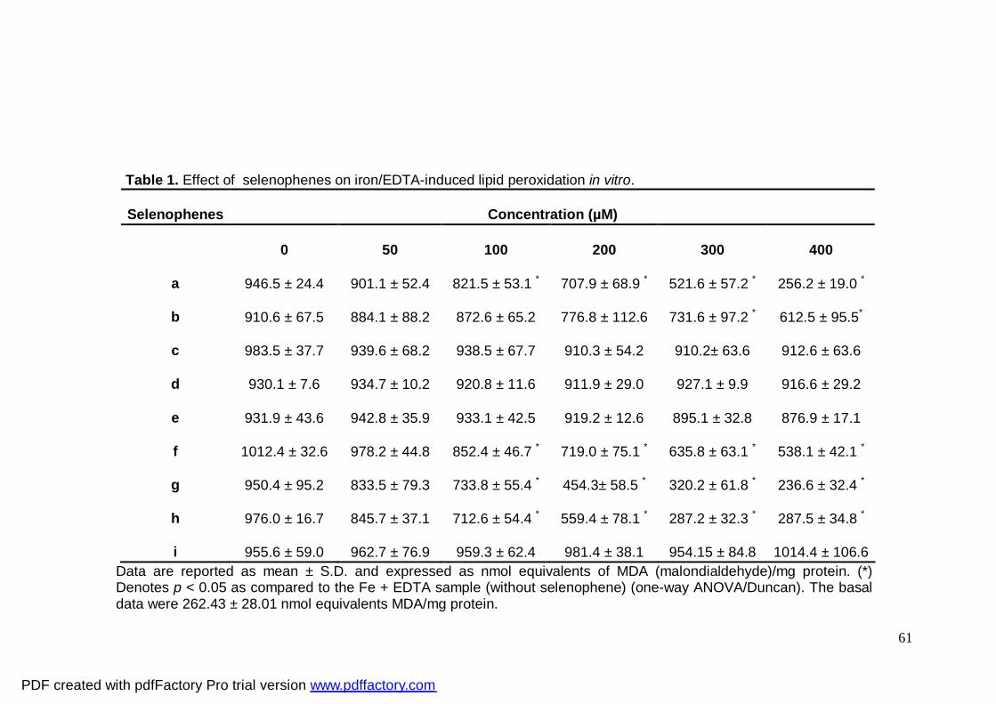
61
Table 1. Effect of selenophenes on iron/EDTA-induced lipid peroxidation in vitro.
Selenophenes
Concentration (µM)
0
50
100
200
300
400
a
946.5 ± 24.4
901.1 ± 52.4
821.5 ± 53.1 *
707.9 ± 68.9 *
521.6 ± 57.2 *
256.2 ± 19.0 *
b
910.6 ± 67.5
884.1 ± 88.2
872.6 ± 65.2
776.8 ± 112.6
731.6 ± 97.2 *
612.5 ± 95.5*
c
983.5 ± 37.7
939.6 ± 68.2
938.5 ± 67.7
910.3 ± 54.2
910.2± 63.6
912.6 ± 63.6
d
930.1 ± 7.6
934.7 ± 10.2
920.8 ± 11.6
911.9 ± 29.0
927.1 ± 9.9
916.6 ± 29.2
e
931.9 ± 43.6
942.8 ± 35.9
933.1 ± 42.5
919.2 ± 12.6
895.1 ± 32.8
876.9 ± 17.1 f
1012.4 ± 32.6
978.2 ± 44.8
852.4 ± 46.7 *
719.0 ± 75.1 *
635.8 ± 63.1 *
538.1 ± 42.1 *
g
950.4 ± 95.2
833.5 ± 79.3
733.8 ± 55.4 *
454.3± 58.5 *
320.2 ± 61.8 *
236.6 ± 32.4 *
h
976.0 ± 16.7
845.7 ± 37.1
712.6 ± 54.4 *
559.4 ± 78.1 *
287.2 ± 32.3 *
287.5 ± 34.8 *
i
955.6 ± 59.0
962.7 ± 76.9
959.3 ± 62.4
981.4 ± 38.1
954.15 ± 84.8
1014.4 ± 106.6
Data are reported as mean ± S.D. and expressed as nmol equivalents of MDA (malondialdehyde)/mg protein. (*) Denotes p < 0.05 as compared to the Fe + EDTA sample (without selenophene) (one-way ANOVA/Duncan). The basal data were 262.43 ± 28.01 nmol equivalents MDA/mg protein.
PDF created with pdfFactory Pro trial version www.pdffactory.com
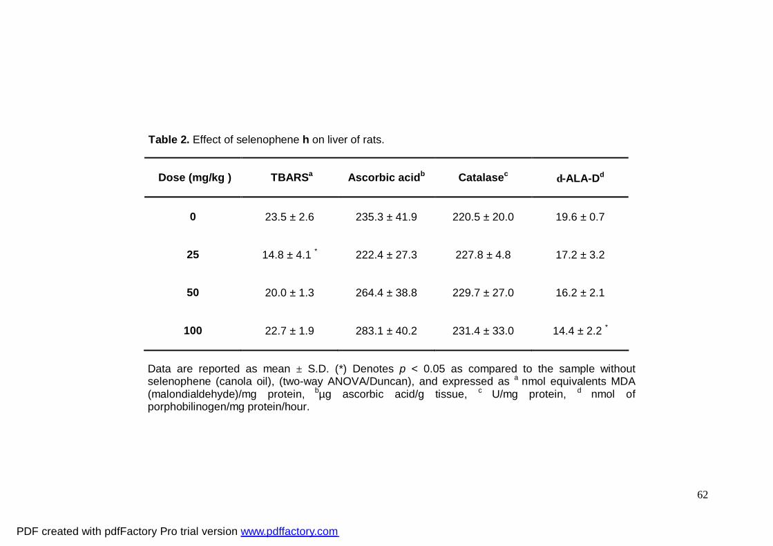
62
Table 2. Effect of selenophene h on liver of rats.
Data are reported as mean ± S.D. (*) Denotes p < 0.05 as compared to the sample without selenophene (canola oil), (two-way ANOVA/Duncan), and expressed as a nmol equivalents MDA (malondialdehyde)/mg protein, bµg ascorbic acid/g tissue, c U/mg protein, d nmol of porphobilinogen/mg protein/hour.
Dose (mg/kg )
TBARSa
Ascorbic acidb
Catalasec
δ-ALA-Dd
0
23.5 ± 2.6
235.3 ± 41.9
220.5 ± 20.0
19.6 ± 0.7
25
14.8 ± 4.1 *
222.4 ± 27.3
227.8 ± 4.8
17.2 ± 3.2
50
20.0 ± 1.3
264.4 ± 38.8
229.7 ± 27.0
16.2 ± 2.1
100
22.7 ± 1.9
283.1 ± 40.2
231.4 ± 33.0
14.4 ± 2.2 *
PDF created with pdfFactory Pro trial version www.pdffactory.com
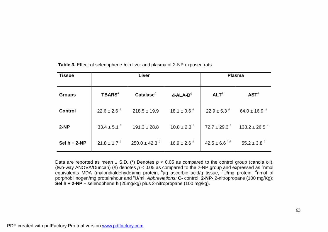
63
Table 3. Effect of selenophene h in liver and plasma of 2-NP exposed rats.
Data are reported as mean ± S.D. (*) Denotes p < 0.05 as compared to the control group (canola oil), (two-way ANOVA/Duncan) (#) denotes p < 0.05 as compared to the 2-NP group and expressed as anmol equivalents MDA (malondialdehyde)/mg protein, bµg ascorbic acid/g tissue, cU/mg protein, dnmol of porphobilinogen/mg protein/hour and eU/ml. Abbreviations: C- control; 2-NP- 2-nitropropane (100 mg/Kg); Sel h + 2-NP – selenophene h (25mg/kg) plus 2-nitropropane (100 mg/kg).
Tissue Liver Plasma
Groups
TBARSa
Catalasec
δ-ALA-Dd
ALTe
ASTe
Control
22.6 ± 2.6 #
218.5 ± 19.9
18.1 ± 0.6 #
22.9 ± 5.3 #
64.0 ± 16.9 #
2-NP
33.4 ± 5.1 *
191.3 ± 28.8
10.8 ± 2.3 *
72.7 ± 29.3 *
138.2 ± 26.5 *
Sel h + 2-NP
21.8 ± 1.7 #
250.0 ± 42.3 #
16.9 ± 2.6 #
42.5 ± 6.6 * #
55.2 ± 3.8 #
PDF created with pdfFactory Pro trial version www.pdffactory.com
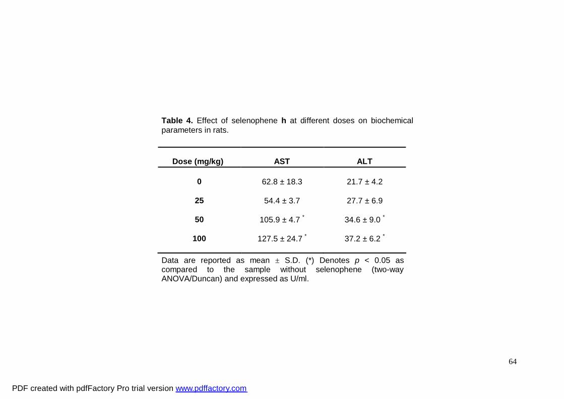
64
Table 4. Effect of selenophene h at different doses on biochemical parameters in rats.
Data are reported as mean ± S.D. (*) Denotes p < 0.05 as compared to the sample without selenophene (two-way ANOVA/Duncan) and expressed as U/ml.
Dose (mg/kg)
AST
ALT
0
62.8 ± 18.3
21.7 ± 4.2
25
54.4 ± 3.7
27.7 ± 6.9
50
105.9 ± 4.7 *
34.6 ± 9.0 *
100
127.5 ± 24.7 *
37.2 ± 6.2 *
PDF created with pdfFactory Pro trial version www.pdffactory.com
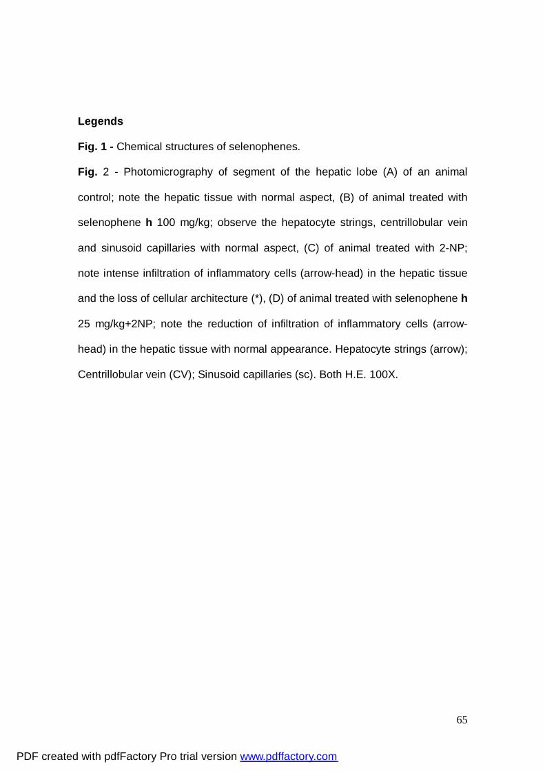
65
Legends
Fig. 1 - Chemical structures of selenophenes.
Fig. 2 - Photomicrography of segment of the hepatic lobe (A) of an animal
control; note the hepatic tissue with normal aspect, (B) of animal treated with
selenophene h 100 mg/kg; observe the hepatocyte strings, centrillobular vein
and sinusoid capillaries with normal aspect, (C) of animal treated with 2-NP;
note intense infiltration of inflammatory cells (arrow-head) in the hepatic tissue
and the loss of cellular architecture (*), (D) of animal treated with selenophene h
25 mg/kg+2NP; note the reduction of infiltration of inflammatory cells (arrow-
head) in the hepatic tissue with normal appearance. Hepatocyte strings (arrow);
Centrillobular vein (CV); Sinusoid capillaries (sc). Both H.E. 100X.
PDF created with pdfFactory Pro trial version www.pdffactory.com
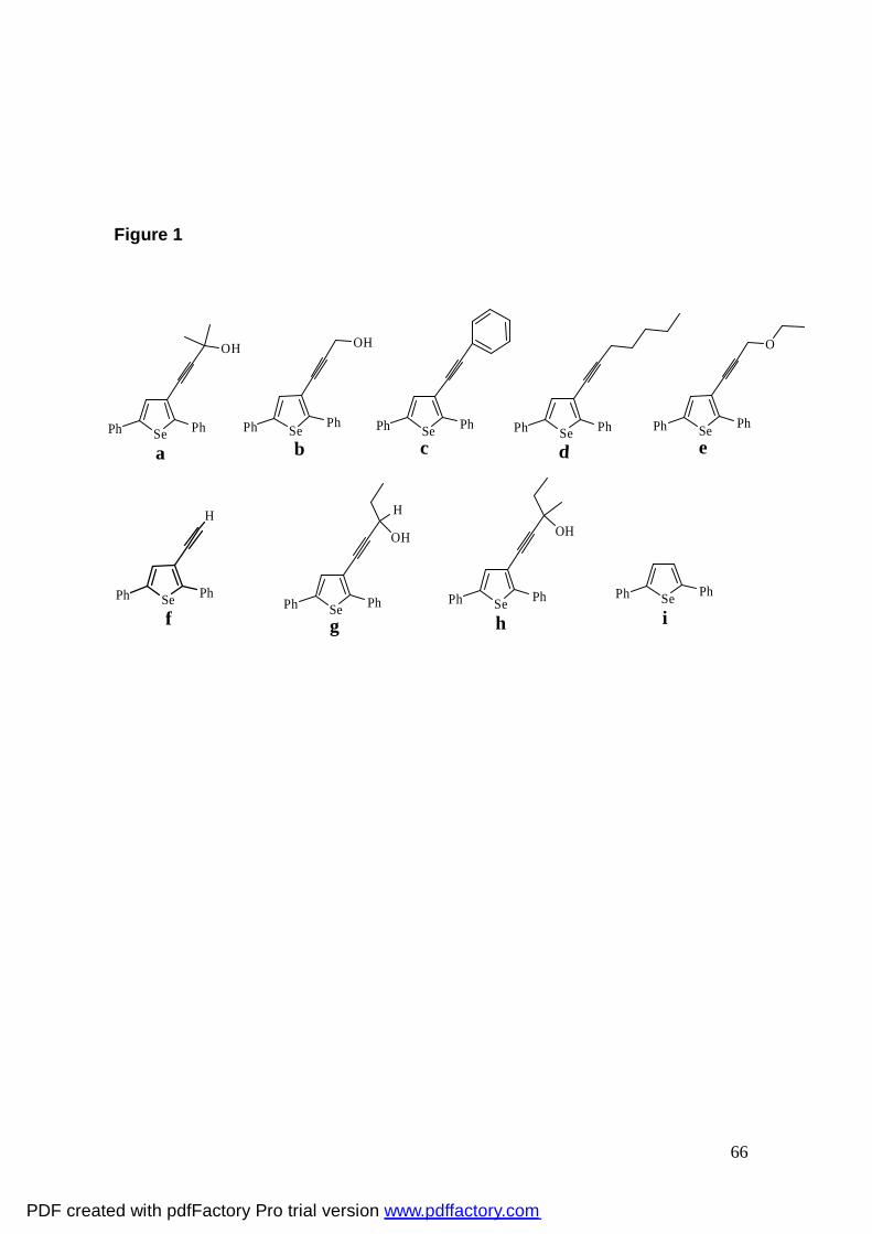
66
Figure 1
Se PhPh
OH
aSe PhPh
OH
bSe PhPh
cSe PhPh
dSe PhPh
O
e
Se PhPh
H
f Se PhPh
OH
H
gSe PhPh
OH
hSe PhPh
i
PDF created with pdfFactory Pro trial version www.pdffactory.com
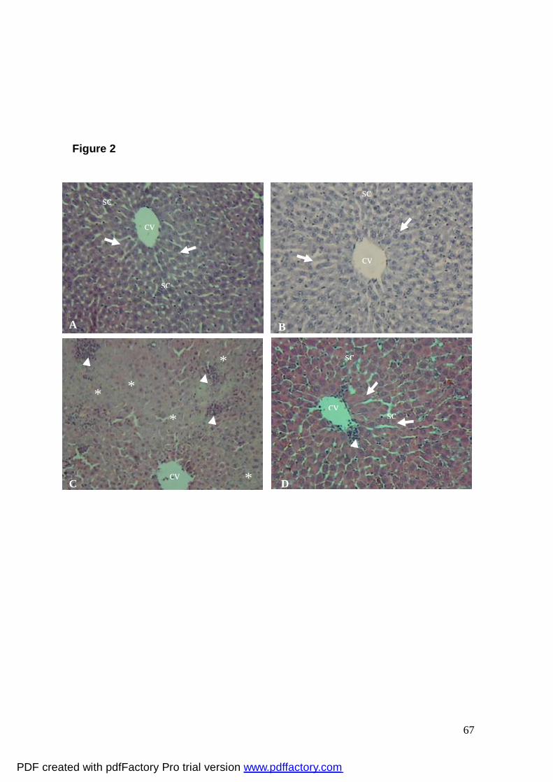
67
Figure 2
cv
* *
*
*
*
cv
sc
sc
sc
sc
cv
A
sc
sc
cv
B
C D
PDF created with pdfFactory Pro trial version www.pdffactory.com
68
4.2. Efeito hepatoprotetor do 3-alquinil selenofeno contra o dano no fígado
induzido pelo tetracloreto de carbono em ratos.
4.2.1. Manuscrito 2
HEPATOPROTECTIVE EFFECT OF 3-ALKYNYL SELENOPHENE AGAINST CARBON
TETRACHLORIDE-INDUCED LIVER DAMAGE IN RATS
(Sumetido à BMB Reports)
PDF created with pdfFactory Pro trial version www.pdffactory.com
69
Hepatoprotective effect of 3-alkynyl selenophene against carbon tetrachloride-induced
liver damage in rats
Ethel A. Wilhelm, Cristiano R. Jesse, Ricardo F. Schumacher, Cristina W. Nogueira *
Programa de Pós-Graduação em Bioquímica Toxicológica, Centro de Ciências
Naturais e Exatas, Universidade Federal de Santa Maria, Santa Maria, CEP
97105-900, RS, Brazil.
Abbreviated title: Hepatoprotective effect of selenophene in rats
*Correspondence should be sent to:
Cristina W. Nogueira
Programa de Pós-Graduação em Bioquímica Toxicológica, Centro de Ciências
Naturais e Exatas, Universidade Federal de Santa Maria, 97105-900, Santa
Maria, RS, Brazil.
Phone: 0-55-32208140
FAX: 0-55-32208978
E-mail: [email protected]
PDF created with pdfFactory Pro trial version www.pdffactory.com
70
Abstract
The aim of this study was to investigate the hepatoprotective effect of 1-
(2,5-diphenylselenophen-3-yl)-3-methylpent-1-yn-3-ol (selenophene h) on acute
liver injury induced by carbon tetrachloride in rats. In the first day of treatment,
animals received selenophene h (25 and 50 mg/kg; per oral, p.o.). In the
second day, rats received CCl4 (1 mg/kg; intraperitoneal, i.p.). Twenty-four
hours after CCl4 administration animals were euthanized and plasma and liver
were removed to the biochemical and histological analysis. Selenophene h
protected against the increase in aspartate aminotransferase (AST) and alanine
aminotransferase (ALT) activities induced by CCl4 administration. The
histological data showed extensive injury in liver from CCl4-treated rats, which
was attenuated by selenophene h. Selenophene h ameliorated lipid
peroxidation and ascorbic acid levels, catalase, δ-aminolevulinic dehydratase
(δ-ALA-D) and glutathione S-transferase (GST) activities altered by CCl4 .The
antioxidant effect of selenophene h on acute liver injury induced by CCl4 in rats
was demonstrated.
Keywords: selenium, CCl4, liver, hepatic damage.
PDF created with pdfFactory Pro trial version www.pdffactory.com
71
Introduction
The liver regulates many important metabolic functions. Hepatic injury is
associated with distortion of these metabolic functions (1). Additionally, it is the
key organ of metabolism that is continuously and variedly exposed to
xenobiotics because of its strategic placement in the body. The toxins absorbed
from the intestinal tract gain access first to the liver resulting in a variety of liver
ailments. Thus, liver diseases remain one of the serious health problems.
Modern medicines have little to offer for alleviation of hepatic diseases and
there are not much drugs available for the treatment of liver disorders (2).
CCl4 is a classical hepatotoxicant that causes rapid liver damage
progressing from steatosis to centrilobular necrosis. CCl4 requires bioactivation
by phase I cytochrome P450 system in liver to form reactive metabolic
trichloromethyl radical (CCl3•) and peroxy trichloromethyl radical (•OOCCl3).
These free radicals can bind with polyunsaturated fatty acid (PUFA) to produce
alkoxy (R•) and peroxy radicals (ROO•) (3). These free radicals can react with
sulfhydryl groups, such as glutathione (GSH) and protein thiols. The covalent
binding of trichloromethyl free radicals to cellular protein is considered to be the
initial step in a chain of events, eventually leading to membrane lipid
peroxidation and finally cell necrosis (4). Consequently, CCl4 is known to induce
reactive oxygen species (ROS) formation, deplete GSH of phase II enzyme,
and reduce antioxidant enzymes and substrates to induce oxidative stress that
is an important factor in acute and chronic liver injury. (4)
It is believed that ROS may injure cell membranes through lipid
peroxidation and modify or damage biomolecules, i.e., proteins, lipids,
PDF created with pdfFactory Pro trial version www.pdffactory.com
72
carbohydrates and DNA in vitro and in vivo (5). Significant cellular damage
occurs when the amount of produced free radicals exceeds the capacity of
endogenous cellular antioxidant defense system. According to the free radical
theory, blocking or retarding the chain reaction of oxidation is one of the
practicable strategies to preventing oxidative stress-induced hepatotoxicity.
The biochemistry and pharmacology of selenium is a subject of intense
current interest, particularly from the viewpoint of public health (6). Selenium,
long recognized as a dietary antioxidant, is now known to be an essential
component of the active sites of several enzymes, including glutathione
peroxidase and thioredoxin reductase, which catalyse reactions essential to the
protection of cellular components against oxidative damage (7). In addition to
their antioxidant property (8), selenium compounds were found to have neuro-
protective (9), anti-inflammatory (10) and hepatoprotective (11) properties.
Therefore, a number of novel pharmaceutical agents which are selenium-based
or which target specific aspects of selenium metabolism are under
development. Among these are the orally active antihypertensive agents,
anticancer, antiviral, immunosuppressive and antimicrobial agents, and
organoselenium compounds, which reduce oxidative tissue damage and edema
(12). The concept that selenium-containing molecules may be better
nucleophiles (and therefore antioxidants) than classical antioxidants, has led to
design synthetic organoselenium compounds (13).
Besides chalcogenophenes play an important role in organic synthesis
(14) they display antioxidant (8), antinociceptive and anti-inflammatory (15, 16)
properties. Based on the above considerations the objective of this study was to
PDF created with pdfFactory Pro trial version www.pdffactory.com
73
investigate the effect of 1-(2,5-diphenylselenophen-3-yl)-3-methylpent-1-yn-3-ol
(selenophene h), a selenophene, on acute liver injury induced by CCl4 in rats.
Results
Hepatic profile
A significant main effect of CCl4 (F1,40 = 15.27; p<0.0004) was observed.
Post hoc comparisons revealed that CCl4 increased ALT activity. Selenophene
h at the dose of 50 mg/kg prevented the increase of ALT activity induced by
CCl4 (Table 1).
Two-way ANOVA of plasma AST activity revealed a significant
selenophene h x CCl4 interaction (F2,40= 5.06; p<0.011). Post hoc comparisons
revealed that CCl4 (F1,40 = 79.88; p<0.0001) increased AST activity.
Selenophene h (F2,40 = 6.35; p<0.004) prevented significantly AST activity
increased by CCl4. (Table 1).
Selenophene h at both doses did not alter plasma ALT and AST activities
(Table 1).
Histological examination
Livers from CCl4-treated rats showed extensive injury, specifically
massive centrilobular necrosis, ballooning degeneration and cellular infiltration.
Selenophene h at both doses prevented pathological alterations caused by
CCl4 administration (Table 2; Figure 1).
Lipid peroxidation
PDF created with pdfFactory Pro trial version www.pdffactory.com
74
Two-way ANOVA of lipid peroxidation levels revealed a significant
selenophene h x CCl4 interaction (F2,40= 5.51; p<0.0077). Post hoc comparisons
showed that CCl4 exposure increased lipid peroxidation levels (F1,40 = 14.74;
p<0.0004) and selenophene h (25 and 50 mg/kg) significantly decreased lipid
peroxidation levels in liver of rats when compared to the CCl4 group (Table 1).
Lipid peroxidation levels were not altered in the liver of rats which
received selenophene h at both doses (Table 1).
Ascorbic acid
Two-way ANOVA of ascorbic acid levels revealed a significant
selenophene h x CCl4 interaction (F2,40= 7.82; p<0.0014). Post hoc comparisons
showed that selenophene h at the dose of 50 mg/kg significantly increased
ascorbic acid levels reduced by CCl4 (F1,40= 10.49; p<0.0024) in liver of rats
(Table 1).
Ascorbic acid levels were not altered in the liver of rats which received
selenophene h at both doses (Table 1).
Catalase activity
Two-way ANOVA of catalase activity revealed a significant selenophene
h x CCl4 interaction (F2,40= 22.35; p<0.0001). Post hoc comparisons showed
that selenophene h at the doses of 25 and 50 mg/kg significantly increased
catalase activity reduced by CCl4 (F1,40= 22.35; p<0.0001) in liver of rats (Table
1).
Catalase activity was not altered in the liver of rats which received
selenophene h at both doses (Table 1).
PDF created with pdfFactory Pro trial version www.pdffactory.com
75
GST activity
Two-way ANOVA of GST activity revealed a significant selenophene h x
CCl4 interaction (F2,40= 5.76; p<0.0063). Post hoc comparisons showed that
selenophene h at the dose of 25 and 50 mg/kg significantly decrease catalase
activity increased by CCl4 (F1,40= 6.16; p<0.0174) in liver of rats (Table 1).
GST activity was not altered in the liver of rats which received
selenophene h at both doses (Table 1).
δ- ALA-D activity
Two-way ANOVA of δ-ALA-D activity revealed a significant selenophene
h x CCl4 interaction (F2,40= 10.59; p<0.0002). Post hoc comparisons
demonstrated that CCl4 (F1,40= 79.46; p<0.0001) inhibited δ-ALA-D activity and
selenophene h at the 50 mg/kg (F2,50 = 0.55; p<0.5790) protected δ-ALA-D
inhibition induced by CCl4 (Table 1).
δ-ALA-D activity remained unaltered in the liver of rats which received
selenophene h at the both doses (Table 1).
Discussion
Several research groups have demonstrated that organoselenium
compounds have hepatoprotective effect (11, 26). In this study we reported the
hepatoprotective effect of selenophene h, an organoselenium compound, on
acute liver injury induced by CCl4 in rats. As expected, a single oral dose of
CCl4 showed significant hepatotoxicity, as evidenced by a dramatic elevation in
the plasma AST and ALT activities and an increased incidence of
PDF created with pdfFactory Pro trial version www.pdffactory.com
76
histopathological lesions in liver of rats. In addition, CCl4 exposure produced
oxidative damage, as evidenced by a significant increase in lipid peroxidation
levels and GST activity as well as a decrease in ascorbic acid levels, catalase
and δ-ALA-D activities, which suggest a role of oxidative stress in CCl4-induced
hepatotoxicity. Pretreatment with selenophene h showed a significant protective
effect against CCl4-induced acute oxidative damage and hepatotoxicity in rats.
Preventive action of liver damage induced by CCl4 has been widely used
as an indicator of liver protective activity of drugs in general (27). Since the
changes associated with CCl4-induced liver damage are similar to that of acute
viral hepatitis (28), CCl4-mediated hepatotoxicity was chosen as the
experimental model. In the present study, CCl4-expoused rats sustained
significant hepatic damage as represented by an increase in the ALT and AST
activities that are in accordance with others (29). These results were associated
with massive centrilobular necrosis, ballooning degeneration and cellular
infiltration of the liver observed in histopathological evaluation that is the
according to Plaa and Charbonneau (30). In this view, the ability of
selenophene h in normalizing the activity of transaminases and morphological
aspects of liver reflects the stabilization of plasma membrane as well as repair
of hepatic tissue damage caused by CCl4. These findings are in agreement with
the commonly accepted view that plasma levels of transaminases return to
normal levels with the healing of hepatic parenchyma and regeneration of
hepatocytes (31). Therefore, data on the literature have demonstrated that liver
damage is a therapeutic target of selenorganic compounds (12).
Oxidative stress is considered to play a prominent causative role in many
diseases including liver damage (32). Free radicals are capable of binding to
PDF created with pdfFactory Pro trial version www.pdffactory.com
77
proteins or lipids, or abstracting a hydrogen atom from an unsaturated lipid,
initiating lipid peroxidation and liver damage while playing a significant role in
the pathogenesis of diseases (33). Lipid peroxidation, usually measured
through its catabolites, such as MDA, is widely accepted to be one of the
principal causes of CCl4-induced liver injury (34). In this study we found an
increase in lipid peroxidation levels induced by CCl4 exposure; this result has
been reported by others (29). Selenophene h normalizes lipid peroxidation
levels, suggesting that the antioxidant activity is important in the protection
against CCl4-induced liver lesion (35). Accordingly, our research group has
reported that diphenyl diselenide, a well-known antioxidant and organoselenium
compound, has hepatoprotective effect (11, 26).
The body has an effective defense mechanism to prevent and neutralize
the free radical-induced damage. This is accomplished by a set of endogenous
antioxidant enzymes such as catalase and non-enzymatic antioxidant defenses
as ascorbic acid. Under oxidative stress, some endogenous protective factors
such as catalase and ascorbic acid are activated in the defense against
oxidative injury. In this study catalase activity and ascorbic acid levels were
decreased by CCl4 exposure in accordance with Jain et al. (36) and Ohta et al.
(37). Selenophene h was effective in protecting against this decrease,
reinforcing the antioxidant activity of this compound, since a reduction in these
parameters is associated with the accumulation of highly reactive free radicals
(38).
Besides, we observed a decrease in δ-ALA-D activity in rats that
received CCl4 and selenophene h was effective in restoring enzyme activity.
Since we have reported that δ-ALA-D activity is extremely sensitive to situations
PDF created with pdfFactory Pro trial version www.pdffactory.com
78
associated with oxidative stress (12), the beneficial effect of selenophene h on
this enzyme demonstrates the antioxidant capacity of this compound. Actually,
CCl4-induced inhibitory effect on hepatic δ-ALA-D activity can be tentatively
related to lipid peroxidation in this study. We can also infer that δ-ALA-D
inhibition participates, at least in part, in the toxicity manifestations caused by
CCl4.
In addition, the increase of GST activity can be due to the probable
response towards increased ROS generation and/or due the detoxification
mechanism. The effect of selenophene h in GST activity suggests a
compensatory mechanism to counteract lipid peroxidation induced by CCl4 in
liver of rats. Many powerful antioxidants, such as Vitamin E, GSH and
melatonin, show great ability in protection of experimental liver injuries (39, 40).
In conclusion, the results suggest that the selenophene h as an
antioxidant seems to be useful in therapy of CCl4 damage, since it has the
capability to alleviate many of the harmful effects of CCl4.
Experimental Procedure
Chemicals
1-(2,5-Diphenylselenophen-3-yl)-3-methylpent-1-yn-3-ol (selenophene h)
was prepared according to the literature method (14). Selenophene h and CCl4
were dissolved in canola oil. All chemicals were obtained from standard
commercial suppliers.
Animals
PDF created with pdfFactory Pro trial version www.pdffactory.com
79
Male adult Wistar rats, weighing 200-300g, were obtained from a local
breeding colony. The animals were kept in separate animal rooms, on a 12 h
light/dark cycle, in an air conditioned room (22 ± 2°C). Commercial diet
(GUABI, RS, Brazil) and tap water were supplied ad libitum. The animals were
used according to the guidelines of the Committee on Care and Use of
Experimental Animal Resources, the Federal University of Santa Maria, Brazil.
Exposure
Five to eight animals per group were usually tested in the experiments.
Rats received selenophene h (25 or 50 mg/kg of body weight; p.o.) (groups 2
and 3). The control group received only vehicle (canola oil, 5 ml/kg, p.o.) (group
1). Twenty-four hours later, animals received CCl4 (1 mg/kg of body weight; i.p.,
1:1) (17) (groups 4, 5 and 6). The protocol of rat treatments is given below:
Group (1) Canola oil (p.o.) plus canola oil (5 ml/kg, p.o.).
Group (2) Selenophene h (25 mg/kg, p.o.) plus canola oil (5 ml/kg, p.o.).
Group (3) Selenophene h (50 mg/kg, p.o.) plus canola oil (5 ml/kg, p.o.).
Group (4) Canola oil (5 ml/kg, i.p.) plus CCl4 (1 mg/kg; i.p.)
Group (5) Selenophene h (25 mg/kg, p.o.) plus CCl4 (1 mg/kg; i.p.)
Group (6) Selenophene h (50 mg/kg, p.o.) plus CCl4 (1 mg/kg; i.p.)
Twenty-four hours after CCl4 administration all rats were anesthetized for
blood collection by heart puncture (hemolyzed plasma was discharged). After
this procedure, rats were euthanized and the liver of animals was removed,
dissected and kept on ice until the time of assay. The samples of liver were
homogenized in 50 mM Tris-HCl, pH 7.4 (1/10, w/v), centrifuged at 2,400×g for
PDF created with pdfFactory Pro trial version www.pdffactory.com
80
15 min. The low-speed supernatants (S1) were separated and used for
biochemical assays.
Hepatic profile
Plasma AST and ALT activities were used as the biochemical markers
for the early acute hepatic damage (18), using a commercial kit (LABTEST,
Diagnostica S.A., Minas Gerais, Brazil). The enzymatic activities were
expressed as U/L.
Histopathology
To histological evaluation, all rats were subjected to a through necropsy
evaluation. Tissues were saved and fixed in 10% formalin. For light microscopy
examination, tissues were embedded in paraffin, sectioned at 5 µm and stained
with hematoxylin and eosin (HE). Liver sections were evaluated to sings of
toxicity characterized by the damage of liver cells around the central vein,
centrilobular necrosis, ballooning degeneration and cellular infiltration of the
liver cells. Pathological alterations were graded on a numeric score according to
their severity. The severity were graded from score 0 to 3, with 0 indicating
absent, 1 indicating low injury, 2 indicating moderate injury and 3 indicating
intense liver injury.
Lipid peroxidation
An aliquot of S1 (200 µl) from rats belonging to the experimental groups
was added to the reaction mixture containing 500 μl thiobarbituric acid (0.8%),
200 μl SDS (sodium dodecyl sulfate, 8.1%) and 500 μl acetic acid (pH 3.4), and
PDF created with pdfFactory Pro trial version www.pdffactory.com
81
was incubated for 2 hours at 95°C. TBARS were determined as described by
Ohkawa et al. (19). Malondialdehyde (MDA), formed as an end product of the
peroxidation of lipids, served as an index of the intensity of oxidative stress.
MDA reacts with thiobarbituric acid to generate a colored product that can be
measured optically at 532 nm. The lipid peroxidation was expressed as nmol
MDA equivalents/g tissue.
Ascorbic acid determination
Ascorbic acid levels are a non-enzymatic antioxidant defense that is
involved in protecting against the injurious effects of ROS. Ascorbic acid levels
determination was performed as described by Jacques-Silva et al. (20) with
some modifications. Briefly, S1 was precipitated in 10 % trichloroacetic acid
solution. An aliquot of S1 (300 µl) at a final volume of 575 µl of the solution was
incubated for 3 h at 38°C then 500 µl H2SO4 65% (v/v) was added to the
medium. The reaction product was determined using a color reagent containing
4.5 mg/ml dinitrophenyl hydrazine and CuSO4 (0.075 mg/ml) at 520 nm. The
content of ascorbic acid is related to tissue amount (µmol ascorbic acid/g
tissue).
Catalase activity
Catalase is an enzymatic antioxidant defense that is involved in
protecting against the injurious effects of ROS. Catalase activity was assayed
spectrophotometrically by the method of Aebi (21), which involves monitoring
the disappearance of H2O2 in the S1 presence at 240 nm. Enzymatic reaction
was initiated by adding an aliquot of 20 µl of S1 and the substrate (H2O2) to a
PDF created with pdfFactory Pro trial version www.pdffactory.com
82
concentration of 0.3 mM in a medium containing 50 mM phosphate buffer, pH
7.0. The enzymatic activity was expressed in as UI/mg protein.
Glutathione S-transferase (GST) activity
GST is an enzymatic antioxidant defense that protect against ROS-
induced injurious. GST activity was assayed through the conjugation of
glutathione with 1-chloro-2,4-dinitrobenzene (CDNB) at 340 nm as described by
Habig et al. (22). An aliquot of 100 μl of S1 was added in 0.1 M potassium
phosphate buffer, pH 7.4, with CDNB, as substrate, and 50 mM GSH. The
enzymatic activity was expressed in µmol/ min/ mg protein.
δ-Aminolevulinic dehydratase (δ-ALA-D) activity
δ-ALA-D, a sulfhydryl- and Zn2+-containing enzyme, is highly sensitive to
pro-oxidants and heavy metals (23). δ-ALA-D activity was assayed according to
the method of Sassa (24) by measuring the rate of product (porphobilinogen)
formation except that 100 mM sodium phosphate buffer pH 6.8 and 2.4 mM δ-
ALA were used. An aliquot of 200 µl of S1 tissue was incubated for 1 h at 37 ◦C.
The reaction product was determined using modified Erlich’s reagent at 555 nm.
The enzymatic activity was expressed as nmol PBG/mg protein/hour.
Protein Quantification
Protein concentration was measured by the method of Bradford (25),
using bovine serum albumin as the standard.
PDF created with pdfFactory Pro trial version www.pdffactory.com
83
Statistical analysis
Data were analyzed by using a two-way analysis of variance (ANOVA)
(selenophene h x CCl4), followed by Duncan’s Multiple Range Test when
appropriate. Main effects are presented only when the second order interaction
was non-significant. All data of experiments were expressed as means ± S.E.M.
Values of p<0.05 were considered statistically significant.
Acknowledgements
The financial support by UFSM, FAPERGS, CAPES and CNPQ is
gratefully acknowledged.
References
1. Wolf, P.L. (1999) Biochemical diagnosis of liver diseases. IJBC 14, 59-
90.
2. Chaterrjee, T.K. (2000) Medicinal Plants with Hepatoprotective
Properties. Herbal Options. Books and Applied Allied (P) Ltd., Calcutta,
143.
3. Hung, M.Y., Fu, T.Y., Shih, P.H., Lee, C.P., Du-Zhong, G.C.Y. (2006)
Eucommia ulmoides Oliv. leaves inhibits CCl4-induced hepatic damage
in rats. Food Chem. Toxicol. 44, 1424-1431.
PDF created with pdfFactory Pro trial version www.pdffactory.com
84
4. Brattin,W.J., Glende E.A.JJr., Recknagel, R.O. (1985) Pathological
mechanisms in carbon tetrachloride hepatotoxicity. Free Radic. Biol.
Med. 1, 27–38.
5. Halliwell, B. (1996) Antioxidants in human health and disease. Ann. Rev.
Nutr. 16, 33- 50.
6. May, S.W. (2002) Selenium-based pharmacological agents: an up date.
Exp. Opin. Invest. Drugs 11, 1261-1269.
7. Bock, A., Forchammer, J.H., Leinfelder, W., Sawers, G., Vepreck, B.,
Zinnia, F. (1991) Selenocysteine: the 21st amino acid. Mol. Microbiol. 5,
515-520.
8. Meotti, F.C., Stangherlin, E.C., Zeni, G., Nogueira, C.W., Rocha, J.B.T.
(2004) Protective role of aryl and alkyl diselenides on lipid peroxidation.
Environ. Res. 94, 276-282.
9. Porcíuncula, L.O., Rocha, J.B.T., Boeck, C.R., Vendite, D., Souza, D.O.
(2001) Ebselen prevents excitotoxicity provoked by glutamate in rat
cerebellar granule neurons. Neurosci. Lett. 299, 217-220.
10. Schewe, T. (1995) Molecular actions of ebselen–an antiinflammatory
antioxidant. Gen. Pharmacol. 26, 1153-1169.
11. Borges, L.P., Borges, V.C., Moro, A.V., Nogueira, C.W, Rocha, J.B.T.,
Zeni, G. (2005) Protective effect of diphenyl diselenide on acute liver
damage induced by 2-nitropropane in rats. Toxicology 210, 1-8.
12. Nogueira, C.W., Zeni, G., Rocha, J.B.T. (2004) Organoselenium and
organotellurium compounds: toxicology and pharmacology. Chem. Rev.
104, 6255- 6285.
PDF created with pdfFactory Pro trial version www.pdffactory.com
85
13. Arteel, G.E. and Sies, H. (2001) The biochemistry of selenium and the
glutathione system. Environ. Toxicol. Pharmacol. 10, 153-158.
14. Alves, D., Luchese, C., Nogueira, C.W., Zeni, G. (2007) Electrophilic
cyclization of (Z)-selenoenynes: Synthesis and reactivity of 3-
iodoselenophenes. J. Org. Chem. 18, 6726-6734.
15. Zeni, G., Nogueira, C.W., Panatieri, R.B., Silva, D.O., Menezes, P.H.,
Braga, A.L., Silveira, C.C., Stefani, H.A., Rocha, J.B.T. (2001) Synthesis
and anti-inflammatory activity of acetylenic thiopenes. Tetrahedron Lett.
42, 7921-7923.
16. Meotti, F.C., Silva, D.O., Santos, A.R.S., Zeni, G., Rocha, J.B.T.,
Nogueira, C.W. (2003) Thophenes and furans derivatives: a new class
of potential pharmacological agents. Environ. Toxicol. Pharmacol. 37,
37–44.
17. Ohta, Y., Sasaki, E., Nishida, K., Kongo, M., Hayashi, T., Nagata, M.,
Ishiguro, I. (1998) Inhibitory effect of Oren-gedoku-to (Huanglian-Jie-
Du-Tang) extract on hepatic triglyceride accumulation with the
progression of carbon tetrachloride-induced acute liver injury in rats. J.
Ethnopharmacol. 61, 75-80.
18. Reitman, S., Frankel, S. (1957) A colorimetric method for the
determination of serum glutamic oxalacetic and glutamic pyruvic
transaminases. Am. J. Clin. Pathol. 28, 56-63.
19. Ohkawa, H., Ohishi, N., Yagi, K. (1979) Assay for lipid peroxides in
animal tissues by thiobarbituric acid reaction. Anal. Biochem. 95, 351-
358.
PDF created with pdfFactory Pro trial version www.pdffactory.com
86
20. Jacques-Silva, M.C., Nogueira, C.W., Broch, L.C., Rocha, J.B.T. (2001)
Diphenyl diselenide and ascorbic acid changes deposition of selenium
and ascorbic acid in brain of mice. Pharmacol. Toxicol. 88, 119-125.
21. Aebi, H. (1984) Catalase in vitro. Meth. Enzymol. 105, 121-126.
22. Habig WH, Pabst MJ, Jakoby WB. Glutathione S-transferases. The first
enzymatic step in mercapturic acid formation. J. Biol. Chem. 1974; 249:
7130-7139.
23. Rocha, J.B.T., Pereira, M.E., Emanuelli, T., Christofari, R.S., Souza,
D.O. (1995) Effect of treatment with mercury chloride and lead acetate
during the second stage of rapid postnatal brain growth on delta-
aminolevulinic acid dehydratase (ALA-D) activity in brain, liver kidney
and blood of suckling rats. Toxicology 100, 27-37.
24. Sassa, S. (1982) Delta-aminolevulinic acid dehydratase assay. Enzyme
28, 133-145.
25. Bradford, M.M. (1976) A rapid and sensitive method for the quantitation
of microgram quantities of protein utilizing the principles of protein-dye
binding. Anal. Biochem. 72, 248-254.
26. Rocha, J.B.T., Gabriel, D., Zeni, G., Posser, T., Siqueira, L., Nogueira,
C.W., Folmer, V. (2005) Ebselen and diphenyl diselenide change
biochemical hepatic responses to overdosage with paracetamol.
Environ. Toxicol. Pharmacol. 19, 255-261.
PDF created with pdfFactory Pro trial version www.pdffactory.com
87
27. Clauson, G.A. (1989) Mechanism of carbon tetrachloride hepatotoxicity.
Pathol. Immunopathol. Res. 8, 104-112.
28. Rubinstein, D. (1962) Epinephrine release and liver glycogen levels after
carbon tetrachloride administration. Am. J. Physiol. 203, 1033-1037.
29. Wang, T., Sun, N., Zhang, W., Li, H., Lua, G., Yuan, B., Jiang, H., She,
J., Zhang, C. (2008) Protective effects of dehydrocavidine on carbon
tetrachloride-induced acute hepatotoxicity in rats. J. Ethnopharmacol.
117, 300-308
30. Plaa, G. L. and Charbonneau, M. (1989) Detection and evaluation of
chemically induced liver injury. Princ. Meth. Toxicol. 399-628.
31. Thabrew, M.I., Joice, P.D.T.M., Rajatissa, W.A. (1987) A comparative
study of the efficacy of Pavetta indica and Osbeckia octandra in the
treatment of liver dysfunction. Planta Med. 53, 239-241.
32. Kiso, Y., Tohkin Ino, H., Hatori, M., Sakamoto, T. and Namba, T. (1984)
Mechanism of antihepatotoxic activity of glycerrhizin: Effect of free
radical generation and lipid peroxidation. Planta Med. 50, 298-302.
33. Brattin,W.J., Glende E.A.JJr., Recknagel, R.O. (1985) Pathological
mechanisms in carbon tetrachloride hepatotoxicity. Free Radic. Biol.
Med. 1, 27-38.
34. Recknagel, R.O., Glende Jr., E.A., Dolak, J.A., Waller, R.L. (1989)
Mechanisms of carbon tetrachloride toxicity. Pharmacol. Ther. 43, 139-
154.
35. Castro, J.A., Ferrya, G.C., Castro, C.R., Sasamelt Fenos, O.M., Giltelte,
J.R. (1974) Prevention of CCl4 necrosis by inhibitors of drug
PDF created with pdfFactory Pro trial version www.pdffactory.com
88
metabolism. Further studies on metabolism of their action. Biochem.
Pharmacol. 23, 295-302.
36. Jain, A., Soni, M., Deb, L., Jain, A., Rout, S.P., Gupta, V.B., Krishna K.L.
(2008) Antioxidant and hepatoprotective activity of ethanolic and
aqueous extracts of Momordica dioica Roxb. Leaves. J.
Ethnopharmacol. 115, 61-66.
37. Ohta, Y., Kongo-Nishimura, M., Matsura, T., Yamada, K., Kitagawa, A.,
Kishikawa, T. (2004) Melatonin prevents disruption of hepatic reactive
oxygen species metabolism in rats treated with carbon tetrachloride. J.
Pineal Res. 36, 10-17.
38. Sheela, C.G., Angusti, K. (1995) Antiperoxide effects of S-allyl cystein
sulphoxide isolated from Allium sativum Linn and gugulipid in chlosterol
diet fed rats. Ind. J. Exp. Biol. 33, 337-341.
39. Wang, H., Wei, W., Wang, N.P., Gui, S.Y., Wu, L., Sun, W.Y., Xu, S.Y.
(2005) Melatonin ameliorates carbon tetrachloride-induced hepatic
fibrogenesis in rats via inhibition of oxidative stress. Life Sci. 77, 1902-
1915.
40. Murugesan, P., Muthusamy, T., Balasubramania, K., Arunakaran, J.
(2007) Effects of vitamins C and E on steroidogenic enzymes mRNA
expression in polychlorinated biphenyl (Aroclor 1254) exposed adult rat
Leydig cells. Toxicology 232, 170-182.
PDF created with pdfFactory Pro trial version www.pdffactory.com
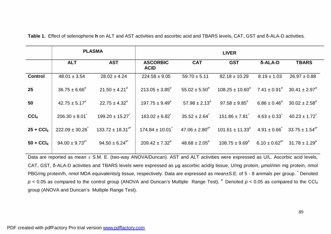
89
Table 1. Effect of selenophene h on ALT and AST activities and ascorbic acid and TBARS levels, CAT, GST and δ-ALA-D activities.
Data are reported as mean ± S.M. E. (two-way ANOVA/Duncan). AST and ALT activities were expressed as U/L. Ascorbic acid levels,
CAT, GST, δ-ALA-D activities and TBARS levels were expressed as μg ascorbic acid/g tissue, U/mg protein, μmol/min mg protein, nmol
PBG/mg protein/h, nmol MDA equivalents/g tissue, respectively. Data are expressed as mean±S.E. of 5 - 8 animals per group. * Denoted
p < 0.05 as compared to the control group (ANOVA and Duncan’s Multiple Range Test). # Denoted p < 0.05 as compared to the CCl4
group (ANOVA and Duncan’s Multiple Range Test).
ALT
AST ASCORBIC ACID
CAT GST δ-ALA-D TBARS
Control 48.01 ± 3.54 28.02 ± 4.24 224.58 ± 9.05 59.70 ± 5.11 82.18 ± 10.29 8.19 ± 1.03 26.97 ± 0.88
25 36.75 ± 6.66# 21.50 ± 4.21# 213.05 ± 3.85# 55.02 ± 5.50# 108.25 ± 10.60# 7.41 ± 0.91# 30.41 ± 2.97#
50 42.75 ± 5.17# 22.75 ± 4.32# 197.75 ± 9.49# 57.98 ± 2.13# 97.58 ± 9.85# 6.86 ± 0.46# 30.02 ± 2.58#
CCl4 206.30 ± 8.01* 199.20 ± 15.27* 163.02 ± 6.82* 35.52 ± 2.64* 151.86 ± 7.81* 4.63 ± 0.33* 40.23 ± 1.72*
25 + CCl4 222.09 ± 30.26* 133.72 ± 18.31#* 174.84 ± 10.01* 47.06 ± 2.80#* 101.61 ± 11.33# 4.91 ± 0.66* 33.75 ± 1.54#*
50 + CCl4 94.00 ± 9.73#* 94.50 ± 6.24#* 209.42 ± 7.32# 48.68 ± 2.05# 108.75 ± 9.69# 6.10 ± 0.62#* 31.78 ± 1.29#
PLASMA LIVER
PDF created with pdfFactory Pro trial version www.pdffactory.com
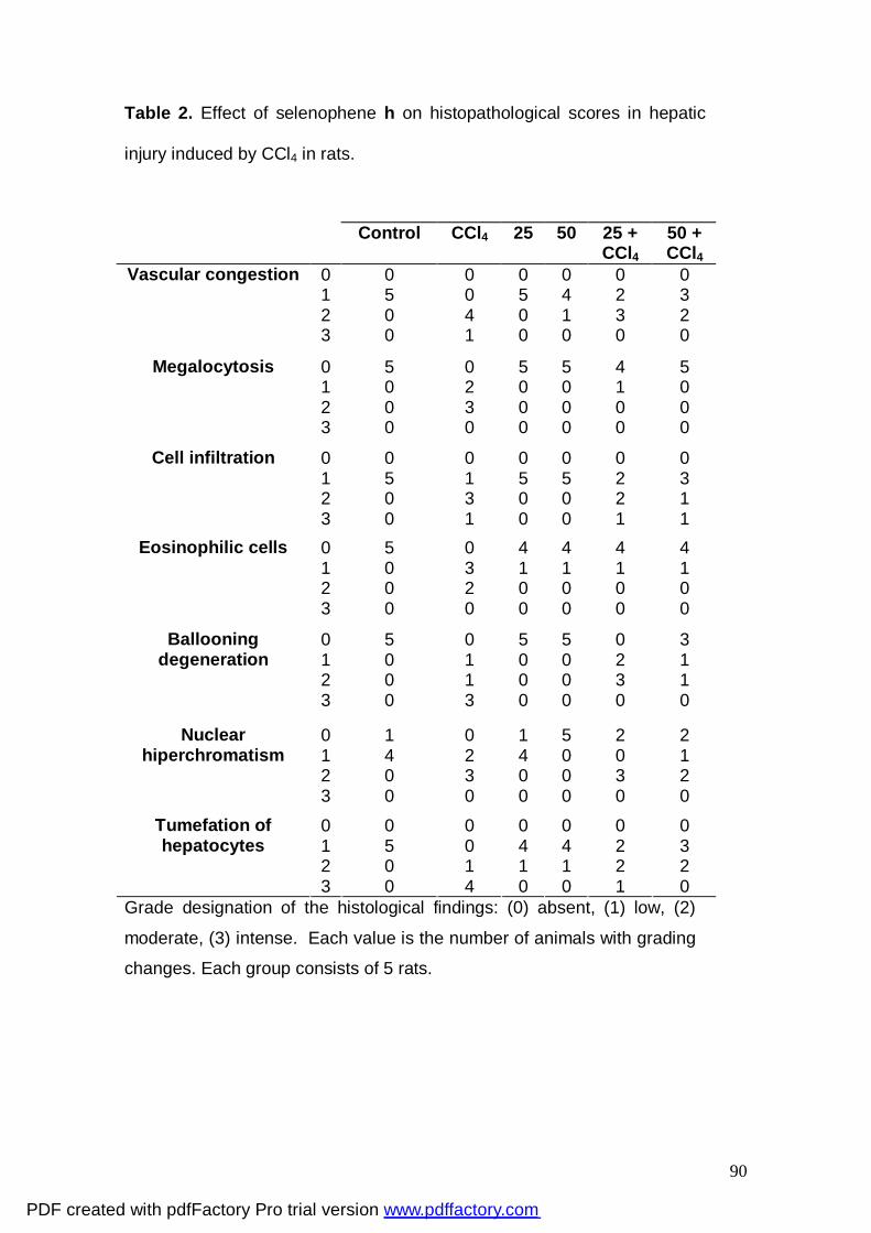
90
Table 2. Effect of selenophene h on histopathological scores in hepatic
injury induced by CCl4 in rats.
Grade designation of the histological findings: (0) absent, (1) low, (2)
moderate, (3) intense. Each value is the number of animals with grading
changes. Each group consists of 5 rats.
Control CCl4 25 50 25 + CCl4
50 + CCl4
0 0 0 0 0 0 0 1 5 0 5 4 2 3 2 0 4 0 1 3 2
Vascular congestion
3 0 1 0 0 0 0
0 5 0 5 5 4 5 1 0 2 0 0 1 0 2 0 3 0 0 0 0
Megalocytosis
3 0 0 0 0 0 0
0 0 0 0 0 0 0 1 5 1 5 5 2 3 2 0 3 0 0 2 1
Cell infiltration
3 0 1 0 0 1 1 0 5 0 4 4 4 4 1 0 3 1 1 1 1 2 0 2 0 0 0 0
Eosinophilic cells
3 0 0 0 0 0 0
Ballooning degeneration
0 1
5 0
0 1
5 0
5 0
0 2
3 1
2 0 1 0 0 3 1 3 0 3 0 0 0 0
Nuclear 0 1 0 1 5 2 2 hiperchromatism 1 4 2 4 0 0 1
2 0 3 0 0 3 2 3 0 0 0 0 0 0
0 0 0 0 0 0 0 1 5 0 4 4 2 3 2 0 1 1 1 2 2
Tumefation of hepatocytes
3 0 4 0 0 1 0
PDF created with pdfFactory Pro trial version www.pdffactory.com
91
Legends Figure 1 - Photomicrography of segment of the hepatic lobe (A) of a control
animal. Note the hepatocyte strings (arrow), the centrilobular vein (CV) and
sinusoid capillaries (*) with normal aspect, (B) of an animal treated with
CCl4, note intense ballooning degeneration (arrow) and (*) infiltration of
inflammatory cells, (C) of an animal treated with selenophene h (50 mg/kg),
note the hepatocyte strings (arrow), the centrilobular vein and some
inflammatory cells (*), (D) of an animal treated with CCl4 + selenophene h
(25 mg/kg); observe around the centrilobular vein some hepatocytes with
vacuolation (head-arrows), ballooning degeneration (arrow) and the
presence of inflammatory cells (*) in the sinusoid capillaries, (E) of an animal
treated with CCl4 + selenophene h (50 mg/kg). Observe the hepatocyte
strings (arrow), the centrilobular vein and sinusoid capillaries (*) with normal
aspect. H.E. 100X.
PDF created with pdfFactory Pro trial version www.pdffactory.com
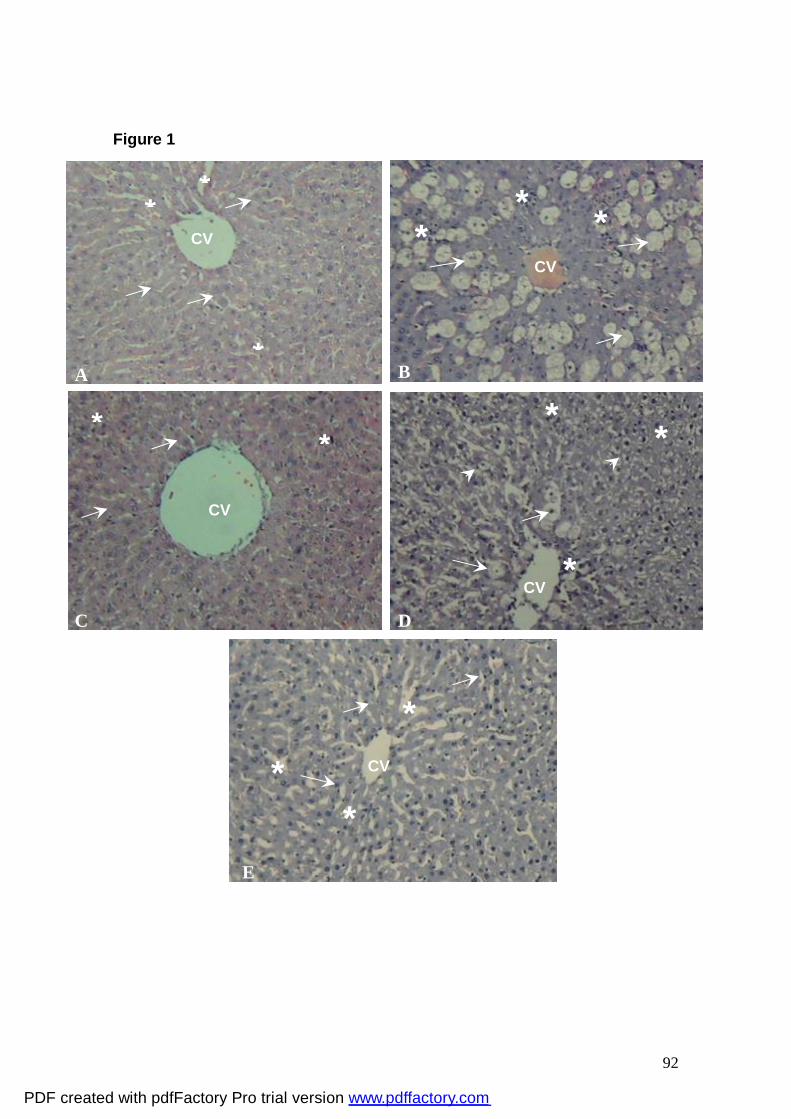
92
Figure 1
CV *
*
*
CV
* * *
B A
CV
* *
C D
E
* *
*
CV
*
*
*
CV
PDF created with pdfFactory Pro trial version www.pdffactory.com
93
5. DISCUSSÃO
O interesse por compostos naturais ou sintéticos que possam previnir,
reverter ou retardar o desenvolvimento de diversas patologias tem crescido
consideravelmente na comunidade científica nas últimas duas décadas. Neste
contexto, compostos heterocíclicos têm sido bastante estudados quanto as
suas atividades biológicas. De fato, estes compostos apresentam atividades
farmacológicas diversificadas, tais como: inibidor do HIV, antitumoral,
antifúngica, antinflamatória e analgésica, antiprotozoária, antiviral e
antioxidante (Barreiro et al., 2001; Zeni et al., 2001; Meotti et al., 2003, 2004;
Gonçales et al., 2005; Shiah et al., 2007; Juang et al., 2007). Dentre estes
compostos heterocíclicos destacam-se os que apresentam selênio em sua
estrutura como, por exemplo, o ebselen que apresenta importantes atividades
biológicas tais como: atividade catalítica e propriedades antioxidantes similares
à glutationa peroxidase (Parnhan e Graf, 1990), atividade neuroprotetora
(Osaki et al., 1997; Tan et al., 1997; Takasago et al., 1997; Kondoh et al., 1999;
Imai et al., 2001; Porciúncula et al., 2003) entre outras. Outro composto
heterocíclico que se destaca é o D-501036, um derivado de selenofeno, que
apresenta atividade anti-tumoral (Shiah et al., 2007). Entretanto, existem
poucos trabalhos na literatura que descrevem a atividade biológica desta
classe de compostos.
Baseando-se nestas perspectivas, um dos nossos objetivos foi
investigar a atividade antioxidante de 3-alquinil selenofenos em modelos de
dano oxidativo in vitro e ex vivo em ratos. Para isso testamos uma classe de
compostos 3-alquinil selenofeno, com diferentes substituições na ligação tripla
na posição 3 da molécula do selenofeno, com o objetivo de avaliar o perfil
antioxidante e o possível efeito tóxico in vitro. Como resultado, encontramos
compostos com atividade antioxidante, porém essa atividade foi dependente da
substituição na ligação tripla da molécula. De fato, Tiano e colaboradores
(2000) demonstraram que a estrutura química de compostos orgânicos de
selênio tem um importante papel no estabelecimento da atividade antioxidante.
Os resultados obtidos no Manuscrito 1 indicaram que os selenofenos
que apresentam um alquino terminal em sua estrutura ou que são facilmente
convertidos a alquinos terminais via eliminação de cetona (Shostakovskii et al.,
PDF created with pdfFactory Pro trial version www.pdffactory.com
94
1974) demonstraram uma melhor atividade antioxidante, o que sugere que a
presença de um alquino terminal na posição 3 do selenofeno é crucial para
este efeito.
Considerando as evidências que indicam que a enzima δ-ALA-D é
sensível a agentes pró-oxidantes (Nogueira et al., 2003; Fachinetto et al., 2006;
Luchese et al., 2007) e que a oxidação de enzimas sulfidrílicas é um dos
mecanismos pelo qual compostos orgânicos de selênio causam toxicidade em
mamíferos (Barbosa et al., 1998), o possível efeito tóxico dos 3-alquinil
selenofenos foi avaliado através da atividade desta enzima in vitro. Os
resultados obtidos demonstraram que nenhum dos 3-alquinil selenofenos
testados inibiu a atividade desta enzima, sugerindo que esta classe de
compostos não apresentou toxicidade sobre a atividade da δ-ALA-D.
Baseado nos resultados in vitro, o nosso próximo objetivo foi avaliar a
atividade hepatoprotetora do selenofeno h (que obteve melhor atividade
antioxidante in vitro) perante o modelo de indução de dano oxidativo pela
administração de 2-NP em ratos (ex vivo). A escolha da dose para os
experimentos ex vivo foi feita através de uma determinação da dose letal 50
(LD50), na qual a dose de 25 mg/kg deste composto não apresentou evidências
de toxicidade. Os resultados obtidos indicaram o efeito hepatoprotetor deste
composto contra o dano oxidativo induzido pelo 2-NP em ratos. De fato,
estudos têm demonstrado que compostos orgânicos de selênio podem atuar
como agentes terapêuticos em modelos de hepatotoxicidade (Nogueira et al.,
2004; Borges et al., 2005, 2006; Wilhelm et al., 2008).
O selenofeno h (25 mg/kg) protegeu contra o aumento dos marcadores
de dano hepático (AST e ALT) e do estresse oxidativo induzido pela
administração do 2-NP em ratos. Considerando a hepatotoxicidade do 2-NP,
este nitroalcano também induziu alterações microscópicas avaliadas por
inspeções histopatológicas. De fato, estudos revelaram que as alterações no
microambiente celular com destruição da integridade do hepatócito e liberação
do conteúdo intracelular para a circulação, antecedem as alterações
histopatológicas. Neste estudo, as avaliações histopatológicas demonstraram
extensiva injúria, caracterizada por intensa infiltração de células inflamatórias e
perda da arquitetura celular nos animais que receberam a administração de 2-
PDF created with pdfFactory Pro trial version www.pdffactory.com
95
NP. O selenofeno h protegeu o fígado desta injúria, enfatizando o efeito
hepatoprotetor deste composto.
Uma vez que o selenofeno h apresenta atividade antioxidante in vitro e
demostrou efeito protetor contra o aumento da peroxidação lipídica e a inibição
da atividade da enzima δ-ALA-D nos animais tratados com 2-NP, pode-se
sugerir que o efeito hepatoprotetor esteja intimamente ligado a atividade
antioxidante deste composto, diminuindo o estresse oxidativo possivelmente
por inativar os efeitos dos intermediários nocivos oriundos do metabolismo do
2-NP (IPHA e HAS)(Kohl et al., 1995). Tem sido demonstrado que a
administração de selênio reduz o estresse oxidativo, tal como o dano oxidativo
hepático (Kohl et al., 1995; Borges et al., 2005, 2006).
Sendo assim, os resultados do Manuscrito 1 demonstraram que o
selenofeno h apresenta efeito hepatoprotetor na dose de 25 mg/kg contra o
dano oxidativo induzido pelo 2-NP em ratos. Baseado nestes resultados, teve-
se como objetivo avaliar este efeito frente a outro indutor de hepatotoxicidade.
Utilizamos o modelo experimental de dano hepático induzido pela
administração de CCl4, composto conhecido pela sua toxicidade e com
mecanismo de ação bem descrito na literatura (Brattin et al., 1985; Basu, 2003;
Wang et al., 2005).
Os resultados obtidos no Manuscrito 2 demonstraram que o selenofeno
h protege contra o dano oxidativo induzido pelo CCl4 em ratos. A ação
preventiva contra o dano hepático induzido pelo CCl4 tem sido usada como
indicador de atividade protetora do fígado de drogas em geral (Clauson, 1989).
Uma única dose de CCl4 causou significante hepatotoxicidade, evidenciada por
elevação da atividade plasmática das enzimas AST e ALT, aumento da
incidência de lesões histopatológicas (necrose centrolobular, degeneração
balanosa, infiltração celular), aumento dos níveis de peroxidação lipídica e da
atividade da GST, bem como, diminuição dos níveis de ácido ascórbico e da
atividade das enzimas catalase e δ-ALA-D. Apartir dos resultados
demonstrados, verificamos que o selenofeno h protegeu de todas estas
alterações, confirmando o efeito hepatoprotetor deste composto em ratos.
A habilidade do selenofeno h de normalizar a atividade das
transaminases e os aspectos morfológicos do fígado reflete a estabilização da
membrana plasmática, bem como, o reparo do dano no tecido hepático
PDF created with pdfFactory Pro trial version www.pdffactory.com
96
causado pelo CCl4, dados estes corroborados por Plaa e Charbonneau (1989)
que sustentam a idéia de que os níveis plasmáticos das transaminases
retornam aos níveis normais com a cicatrização do parênquima hepático e a
regeneração dos hepatócitos. De acordo com estes achados, dados da
literatura tem demonstrado que o dano hepático é alvo terapêutico de
compostos orgânicos de selênio (Nogueira et al., 2004).
Os resultados obtidos com este modelo experimental corroboram com a
idéia obtida através dos resultados do Manuscrito 1 de que o selenofeno h
exerce efeito hepatoprotetor possivelmente devido a atividade antioxidante,
visto que neste modelo observamos uma proteção sobre a diminuição das
defesas antioxidantes enzimáticas (catalase, GST) e não enzimáticas (ácido
ascórbico) e da atividade da enzima δ-ALA-D causada pela exposição ao CCl4.
De fato, a redução destes parâmetros está associada com o acúmulo de
radicais livres altamente reativos (CCl3OOº, CCl3º) (Sheela and Angusti, 1995).
Adicionalmente, este composto protegeu contra a peroxidação lipídica induzida
por CCl4.
Em conclusão, os resultados dos Manuscritos 1 e 2 sugerem que o
selenofeno h, uma molécula com atividade antioxidante, pode ser uma útil
terapia contra o dano oxidativo induzido pelos hepatotoxicantes: 2-NP e CCl4,
considerando que este composto tem a habilidade de proteger dos efeitos
nocivos causados por estes. Além disso, podemos inferir que o selenofeno h
apresenta atividade hepatoprotetora pois demonstrou-se efetivo nos dois
modelos experimentais utilizados.
No Esquema 1, mostrado a seguir, é possível ter uma visão geral dos
efeitos do selenofeno h frente aos compostos hepatotóxicos (2-NP e CCl4 )
estudados neste trabalho.
PDF created with pdfFactory Pro trial version www.pdffactory.com
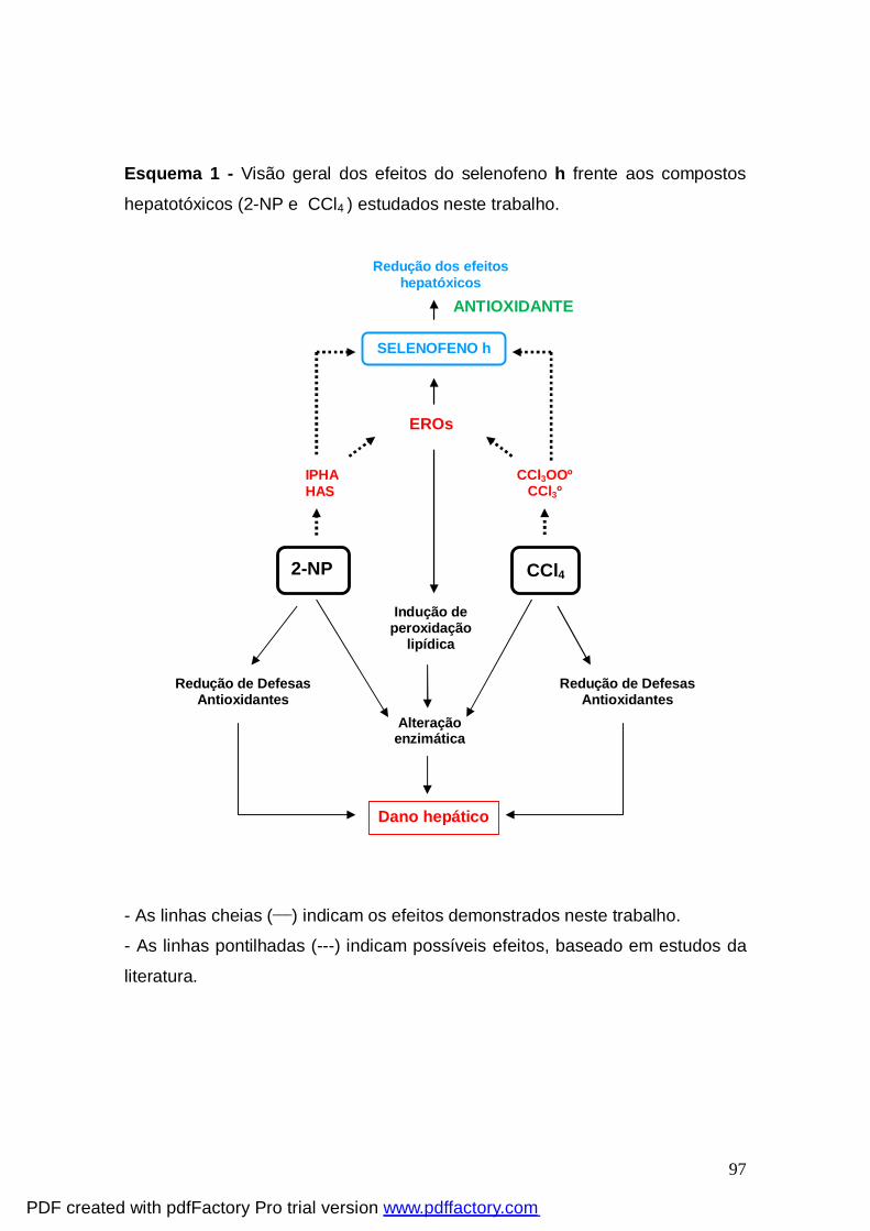
97
Esquema 1 - Visão geral dos efeitos do selenofeno h frente aos compostos
hepatotóxicos (2-NP e CCl4 ) estudados neste trabalho.
- As linhas cheias (___) indicam os efeitos demonstrados neste trabalho.
- As linhas pontilhadas (---) indicam possíveis efeitos, baseado em estudos da
literatura.
SELENOFENO h
CCl3OOº CCl3º
2-NP CCl4
Redução dos efeitos hepatóxicos
IPHA HAS
EROs
Indução de peroxidação
lipídica
Redução de Defesas Antioxidantes
Alteração enzimática
Redução de Defesas Antioxidantes
Dano hepático
ANTIOXIDANTE
PDF created with pdfFactory Pro trial version www.pdffactory.com
98
6. CONCLUSÕES
Com base nos resultados obtidos, nós podemos concluir que:
• O selenofeno h apresentou a melhor atividade antioxidante e não
demonstrou toxicidade in vitro (Manuscrito 1).
• O selenofeno h administrado pela via oral na dose de 25 mg/kg não
causou toxicidade em ratos (Manuscrito 1).
• O selenofeno h demonstrou-se efetivo em proteger contra o dano
oxidativo induzido pelo 2-NP (Manuscrito 1) e CCl4 (Manuscrito 2) em fígado
de ratos, sendo a atividade antioxidante deste composto um dos mecanismos
envolvidos neste efeito.
PDF created with pdfFactory Pro trial version www.pdffactory.com
99
7. REFERÊNCIAS BIBLIOGRÁFICAS ACHLIYA, G.S.; WADODKAR, S.G.; DORLE, A.K. Evaluation of
hepatoprotective effect of Amalkadi Ghrita against carbon tetrachloride-induced
hepatic damage in rats. J. Ethnopharmacol., 90, 229–232. 2004.
ALBERTS, B.; BRAY, D.; LEWIS, J.; RAFF, M.; ROBERTS, K.; WATSON, J. D.
Molecular Biology of the cell. 3ª ed, New York & London: Garland Publishing.
1994.
ALEXI, T.; HUGHES, P.E.; FAULL, R.L.M.; WILLIAMS, C.E. 3-Nitropropionic
acid’s lethal triplet: cooperative pathway of neurodegeneration. Neuro. Report.,
R57-R64. 1998.
ALVES, D.; REIS, J.S.; LUCHESE, C.; NOGUEIRA, C.W.; ZENI, G. Synthesis
of 3-alkynylselenophene derivatives by a cooper-free sonogashira cross-
coupling reaction. Eur. J. Org. Chem. 2, 377-382. 2008.
ARCADI, A.; CACCHI, S.; FABRIZI, G.; MARINELLI, F.; MORO, L. Synthesis
and in vitro and in vivo evaluation of the 2-(6'methoxy-3',4'-dihydro-1'-naphtyl)-
4H-3,1-benzoxazin-4- one as a new potent substrate inhibitor of human
leukocyte elastase. Bioorg. Med. Chem. Lett., 1432. 1999.
ARTEEL, G.E.; SIES, H. The biochemistry of selenium and the glutathione
system. Environ.Toxicol.Pharmacol., 10, 153-158. 2001.
BARBOSA, N.B.V.; ROCHA, J.B.T.; ZENI, G.; EMANUELLI, T.; BEQUE, M.C.;
BRAGA, A.L. Effect of organic forms of selenium on δ-Aminolevulinate
dehydratase from liver, kidney and brain of adult rats. Toxicol. Appl.
Pharmacol., 149, 243-253. 1998.
BARBOSA, N.B.V.; ROCHA, J.B.T.; WONDRACEK, D.C.; PEROTTONI, J.;
ZENI, G.; NOGUEIRA, C.W. Diphenyl diselenide reduces temporarily
PDF created with pdfFactory Pro trial version www.pdffactory.com
100
hyperglycemia: Possible relationship with oxidative stress. Chem. Biol. Interact., 163, 230-238. 2006.
BARBOSA, N.B.V.; NOGUEIRA, C.W.; GUECHEVA, T.N.; BELLINASO, M.L.;
ROCHA, J.B.T. Diphenyl diselenide supplementation delays the development of
N-nitroso-N-methylurea-induced mammary tumors. Arch. Toxicol., 82, 655-
663. 2008.
BARLUENGA, J.; TRINCADO, M.; RUBIO, E.; GONZALEZ, J. M. IPy2BF4-
promoted intramolecular addition of masked and unmasked anilines to alkynes:
direct assembly of 3-iodoindole cores. Angew. Chem., 42, 2406-2409. 2003.
BARREIRO, E. J.; FRAGA, C.A.F. Química Medicinal: As Bases Moleculares de ação de Fármacos, Artemed Editora, Porto Alegre, RS, 53. 2001.
BASU, S. Carbon tetrachloride-induced lipid peroxidation: eicosanoid formation
and their regulation by antioxidant nutrients. Toxicology, 189, 113-127. 2003.
BEHNE, D.; KYRIAKOPOULOS, A. Identification of type I iodothyronine 5’-
deiodinase as a selenoenzyme. Biochem. Biophys. Res. Co., 173, 1143-
1149. 1990.
BEM AF, FARINA M, PORTELLA RD, NOGUEIRA CW, DINIS TC,
LARANJINHA JA, ALMEIDA LM, ROCHA JB. Diphenyl diselenide, a simple
glutathione peroxidase mimetic, inhibits human LDL oxidation in vitro.
Atherosclerosis doi:10.1016/j.atherosclerosis.2008.02.030. 2008.
BERZELIUS, J.J. Afhandl. Fys. Krmi Mineralogi 6, 42. 1817.
BORGES, L.P.; BORGES, V.C.; MORO, A.V.; NOGUEIRA, C.W.; ROCHA,
J.B.T.; ZENI, G. Protective effect of diphenyl diselenide on acute liver damage
induced by 2-nitropropane in rats. Toxicology, 210, 1-8. 2005.
PDF created with pdfFactory Pro trial version www.pdffactory.com
101
BORGES, L.P., NOGUEIRA, C.W., PANATIERI, R.B., ROCHA, J.B.T., ZENI,
G. Acute liver damage induced by 2-nitropropane in rats: Effect of diphenyl
diselenide on antioxidant defenses. Chem. Biol. Interact., 160, 99-107. 2006.
BORGES, L.P.; BRANDÃO, R.; GODOI, B.; NOGUEIRA, C.W.; ZENI, G. Oral
administration of diphenyl diselenide protects against cadmium-induced liver
damage in rats. Chem. Biol. Interact., 171, 15-25. 2008.
BRATTIN, W.J.; GLENDE E.A.JJr.; RECKNAGEL, R.O. Pathological
mechanisms in carbon tetrachloride hepatotoxicity. Free Radic. Biol. Med. 1,
27-38. 1985.
BRUICE, YURKANIS, P. Quimica Orgânica .Ed. Pearson Prentice Hall. Sao
Paulo
4ª edição, vol. 2. 2006.
CAMERON J.R.; KARUNARATE W.A.E.. Carbon tetrachloride cirrhosis in
relation to liver regeneration. J. Pathol. Bacteriol., 92, 1-4, 1936.
CAO, X.; YOU, Q.; LI, Z.; LIU, X.; XU, D.; GUO, Q.; SHANG, J.; CHERN, J.;
CHEN, M. The design, synthesis and biological evaluation of 7-alkoxy-4-
heteroarylamino-3-cyanoquinolines as dual inhibitors of c-Src and Inos. Bioorg.
Med. Chem. Lett., 18, 6206-6209. 2008.
CHAN, G.F.Q.; TOWERS, G.H.N.; MITCHELL, J.C. Ultraviolet-mediated
antibiotic activity of thiophene compounds of Tagetes. Phytochemistry, 14,
2295-2296. 1975.
CLAUSON, G.A. Mechanism of carbon tetrachloride hepatotoxicity. Pathol.
Immunopathol. Res., 8, 104-112. 1989.
CLERCQ, E.D. Anti-HIV drugs: 25 compounds approved within 25 years after
the discovery of HIV. Int. J. Antimicrob. Agents.
doi:10.1016/j.ijantimicag.2008.10.010.
PDF created with pdfFactory Pro trial version www.pdffactory.com
102
DAWSON, V.L.; DAWSON, T.M. Free radicals and neuronal cell death. Cell. Death and Differentiation, 3, 71-78. 1996.
DUMONT, E.; VANHAECKE, F.; CORNELIS, R. Selenium speciation from food
source to metabolites: a critical review. Anall Bioanal. Chem., 385, 1304-1343.
2006.
FACHINETTO, R.; PIVETTA, L.A.; FARINA, M.; PEREIRA, R.P.; NOGUEIRA,
C.W.; ROCHA, J.B.T. Effects of ethanol and diphenyl diselenide exposure on
the activity of δ-aminolevulinate dehydratase from mouse liver and brain. Food
Chem. Toxicol., 44, 588-594. 2006.
FIALA, E.S.; CZERNIAK, R.; CASTONGUAY, A.; CONAWAY, C.C.;
RIVENSON, A. Assay of 1-nitropropane, 2-nitropropane, 1-azoxypropane and
2-azoxypropane for carcinogenicity by gavage in Sprague-Dawley rats.
Carcinogenesis 8, 1947-1949. 1987.
FIALA, E.S.; CONAWAY, C.C.; MATHIS, J.E. Oxidative DNA and RNA damage
in the livers of Sprague-Dawley rats treated with the hepatocarcinogen 2-
nitropropane. Cancer Res., 49, 5518-5522. 1989.
GAW, A.; COWAN, R.A.; REILLY, D.S.J.; STEWART, M.J.; SHEPERD, J.
Clinical Biochemistry: An ilustred colour text. Churchill livingstone. 2ed. 53-
55. 1999.
GHISLENI, G.; PORCIÚNCULA L.O.; CIMAROSTI, H.; ROCHA, J.B.T.;
SALBEGO, C.G.; SOUZA, D.O. Diphenyl diselenide protects rat hippocampal
slices submitted to oxygen–glucose deprivation and diminishes inducible nitric
oxide synthase immunocontent. Brain. Res., 986, 196-199. 2003.
GIANNI, P.; JAN, K.J.; DOUGLAS, M.J.; STUART, P.M.; TARNOPOLSKY, M.A.
Oxidative stress and the mitochondrial theory of aging in human skeletal
muscle. Exp. Geront., 39, 1391-1400, 2004.
PDF created with pdfFactory Pro trial version www.pdffactory.com
103
GONÇALES, C.E.P.; ARALDI, D.; PANATIERI, R.B.; ROCHA, J.B.T.; ZENI, G.;
NOGUEIRA, C.W. Antinociceptive properties of acetylenic thiophene and furan
derivatives: Evidence for the mechanism of action. Life Sci., 76, 2221-2234.
2005.
GONÇALVES, T.L.; ERTHAL, F.; CORTE, C.L.D.; MÜLLER, L.G.; PIOVEZAN,
C.M.; NOGUEIRA, C.W.; ROCHA, J.B.T. Involvement of oxidative stress in the
pre-malignant and malignant states of cervical cancer in women. Clin. Biochem., 38, 1071-1075. 2005.
GUO, N.; CONAWAY, C.C.; HUSSAIN, N.S.; FIALA, E.S. Sex and organ
differences in oxidative DNA and RNA damage due to treatment of Sprage-
Dawley rats with acetoxime or 2-nitropropane. Carcinogenesis, 11, 1659-1662.
1990.
HA, B.J.; LEE, S.H.; KIM, H.J.; LEE. J.Y. The role of Salicornia herbacea in
ovariectomy-induced oxidative stress. Biol. Pharm. Bull., 29, 1305-1309. 2006.
HABIG, W.H.; PABST, M.J.; JAKOBY, W.B. Glutathione-S-transferase, the first
enzymatic step in mercapturic acid formation. J. Biol. Chem., 249, 7130. 1974.
HADDAD, E.B.; MCCLUSKIE, K.; BIRRELL, M.A.; DABROWSKI, D.;
PECORARO, M.; UNDERWOOD, S.; CHEN, B.; SANCTIS, G.T.; WEBBER,
S.E.; FOSTER, M.L.; BELVISI, M.G. Differential effects of ebselen on neutrophil
recruitment, chemokine, and inflammatory mediator expression in a rat model of
lipopolysaccharide-induced pulmonary inflammation. J. Immun., 169, 974-982.
2002.
HALLIWELL, B., GUTTERIDGE, J. M. C. Free radicals in Biology and Medicine, 2ª ed, New York: Clerendon Press, 1989.
HALLIWELL, B. Reactive oxygen species and the central nervous system. J. Neurochem., 59, 1609-1623, 1992.
PDF created with pdfFactory Pro trial version www.pdffactory.com
104
HENRY, J.B. Clinical Diagnosis and Management by Laboratory Methods.
9ed. Sauders. p. 200-280. 1999.
HOFFMAN, J.L.; MCCONNELL, K.P. Periodate-oxidized adenosine inhibits the
formation of dimethylselenid and trimethylselenium ion in mice treated with
selenite. Arch. Biochem. Biophys., 254, 534-540. 1986.
HOLMGREN, A. Thioredoxin. Annu. Rev. Biochem., 54, 237-271. 1985.
HUDSON, J.B.; GRAHAM, E.A.; MIKI, N.; TOWERS, G.H.N.; HUDSON, L.L.;
ROSSI, R.; CARPITA, A.; NERI, D. Photoactive antiviral and cytotoxic activities
of synthetic thiophenes and their acetylenic derivatives. Chemosphere, 19,
1329-1343. 1989.
IARC. Monographs on the evaluation of the carcinogenic risk of chemicals to humans 29, 331-343. 1982.
IMAI, H.; MASYASU, H.; DEWAR, D.; GRAHAM, D.I.; MACRAE, I.M. Ebselen
protects both gray and White matter in a rodent model of focal cerebral
ischemia. Stroke, 32, 2149-2154. 2001.
JOSEPHY, P. D. Molecular Toxicology, New York: Oxford University Press,
1997.
JUANG, S.H.; LUNG, C.C.; HSU, K.S.; LI, Y.C.; HONG, P.C.; SHIAH, H.S.;
KUO, C.C.; HUANG, C.W.; WANG, Y.C.; HUANG, L.; CHEN, S.F.; FU, K.C.;
HSU, C.L.; LIN, M.J.; CHANG, C. J.; ASHENDEL, C. L.; CHAN, T. C. K.;
CHOU, K. M. CHANG, J. Y. D-501036, a novel selenophene-based
triheterocycle derivative, exhibits potent in vitro and in vivo antitumoral activity
which involves DNA damage and ataxia telangiectasia-mutated nuclear protein
kinase activation. Mol. Cancer Ther., 6, 193. 2007.
KALIL, A. N.; COELHO, J.; STRAUSS, E. Fígado e vias biliares: Clínica e cirurgia. Ed. Revinter. RJ. p3-60. 2001.
PDF created with pdfFactory Pro trial version www.pdffactory.com
105
KANDA, T.; ENGMAN, L.; COTGREAVE, I.A.; POWIS, G. Novel water-soluble
diorganyl tellurides with thiol peroxidase and antioxidant activity. J. Org. Chem., 64, 8161-8169. 1999.
KAWANISHI, S.; HIRAKU, Y.; MURATA, M.; OIKAWA, S. The role of metals in
site-specific DNA damage with reference to carcinogenesis. Free Rad. Biol. Medicin., 32, 822-832. 2002.
KIM, S. J.; REITER, J.R.; GARAY, R.V.M.; QI, W.; EL-SOKKARY, H.G.; TAN,
D. 2-Nitropropane-induced lipid peroxidation: antitoxic effects of melatonin.
Toxicology, 130, 183-190. 1998.
KLAYMAN, D.L.; GÜNTHER, W.H. Organic selenium compounds: their
chemistry and biology. New York: John Wiley and sons, 68 - 157. 1973.
KOHL, C.; MORGAN, P.; GESCHER, A. Metabolism of genotoxicant 2-
nitropropane to a nitric oxide species. Chem. Biol. Interact., 97, 175-184.
1995.
KONDOH, S.; NEGASAWA, S.; KAWABISHI, M.; YAMAGUSHI, K.;
KAJIMOTO, S.; OHTA, T. Effects of ebselen on cerebral ischemia and
reperfusion evaluated by microdialysis. Neurol. Res., 21, 682-686. 1999.
KONO, H.; ARTEEL, G.E.; RUSYN, I.; SIES, H.; THURMAN, R.G. Ebselen
prevents early alcohol-induced liver injury in rats. Free Rad. Biol. Med., 30,
403-411, 2001.
KOYANAGI, T.; NAKAMUTA, M.; ENJOJI, M.; IWAMOTO, H.; MOTOMURA, K.;
SAKAI, H.; NAWATA, H. The selenoorganic compound ebselen suppresses
liver injury induced by Propionibacterium acnes and lipopolysaccharide in rats.
Int. J. Mol. Med., 7, 321-327, 2001.
PDF created with pdfFactory Pro trial version www.pdffactory.com
106
LEE, K.S.; LEE, S.J.; PARK, H.J.; CHUNG, J.P.; HAN, K.H.; CHON, C.Y.; LEE,
S.I.; MOON, Y.N. Oxidative Stress Effect on the Activation of Hepatic Stellate
Cells. Yonsei Med. J., 42, 1-8, 2001.
LEWIS, T.R.; ULRICH, C.E.; BUSEY, W.M. Subchronic inhalation toxicity of
nitromethane and 2-nitropropane. J. Environ. Pathol. Toxicol., 2, 233-249.
1979.
LI, Q.-J.; BESSEMS, J.G.; COMMANDEUR, J.N.; ADAMS, B.; VERMEULEN,
N.P. Mechanism of protection of ebselen against paracetamol-induced toxicity
in rat hepatocytes. Biochem. Pharmacol., 48, 1631-1640, 1994.
LIMA, C.F.; FERNANDES-FERREIRA, M.; WILSON, C.P. Drinking of Salvia
officinalis tea increases CCl4-induced hepatotoxicity in mice. Food Chem.
Toxicol., 45, 456-464. 2007.
LOGUERCIO C.; FREDERICO A. Oxidative stress in viral and alcoholic
hepatitis. Free Radic. Biol. Med., 34, 1-10. 2003.
LUCHESE, C.; STANGHERLIN, E.C.; ARDAIS, A.P.; NOGUEIRA, C.W.;
SANTOS, F.W. Diphenyl diselenide prevents oxidative damage induced by
cigarette smoke exposure in lung of rat pups. Toxicology, 230, 189-186. 2007.
MARTIN, J.L., GERLACK, M.L. Selenium metabolism in animals. Ann. NY Acad. Sci., 192, 193-199. 1972.
MASUMOTO, H.; SIES, H. The reaction of ebselen with peroxynitrite. Chem. Res. Toxicol., 9, 262-267. 1996.
MCGREGOR D, LANG M. Carbon tetrachloride: genetic effects and other
modes of action. Mutat. Res. 366, 181-195.1996.
PDF created with pdfFactory Pro trial version www.pdffactory.com
107
MEOTTI, F.C.; BORGES, V.C.; ZENI, G.; ROCHA, J.B.T.; NOGUEIRA, C.W.
Potential renal and hepatic toxicity of diphenyl diselenide, diphenyl ditelluride
and ebselen for rats and mice. Toxicol. Lett., 143, 9-16. 2003.
MEOTTI, F.C.; STANGHERLIN, E.C.; ZENI, G.; NOGUEIRA, C.W.; ROCHA,
J.B.T. Protective role of aryl and alkyl diselenides on lipid peroxidation.
Environ. Res., 94, 276-282. 2004.
MOTTA, V.T. Bioquímica clínica: princípios e interpretações. 3 ed. POA.
Ed. Médica Missau. p215-232. 2000.
MOUSSAOUI, S.; OBINU, M.C.; DANIEL, N.; REIBAUD, M.; BLANCHARD, V.;
IMPERATO, A. The antioxidant ebselen prevents neurotoxicity and clinical
symptoms in a primate model of Parkinson’s disease. Experiment. Neurobiol.,
166, 235-245. 2000.
MOZIER, N.M.; MCCONNELL, K.P.; HOFFMAN, J.L. S-adenosyl-L-methionine:
thioether S-methyltransferase, a new enzyme in sulfur and selenium
metabolism. J. Biol. Chem., 263, 4527-4531. 1988.
MUGESH, G.; DU MONT, W.W.; SIES, H. Chemistry of biologically important
Organoselenium compounds. Chem. Rev., 101, 2125-2179. 2001.
NAVARRO-ALARCÓN, M.; LÓPEZ-MARTINEZ, M.C. Essentiality of selenium
in the human body: relationship with different diseases. Sci. Tot. Environ., 249,
347-371. 2000.
NOGUEIRA, C.W.; BORGES, V.C.; ZENI, G.; ROCHA, J.B.T.
Organochalcogens affect on delta-aminolevulinate dehydratase activity from
human erythrocytic cells in vitro. Toxicology 191, 169-178. 2003.
NOGUEIRA, C.W.; ZENI, G.; ROCHA, J.B.T. Organoselenium and
organotellurium compounds: Toxicology and pharmacology. Chem. Rev., 104,
6255-6285. 2004.
PDF created with pdfFactory Pro trial version www.pdffactory.com
108
NOWAK, P.M.; SALUK-JUSZCZAK, J.; OLAS, B.; KOLODZIEKCZYK, J.;
WACHOWICZ, B. The protective effects of selenoorganic compounds against
peroxynitrite-induced changes in plasma proteins and lipids. Cel. Mol. Biol. Lett., 11, 1-11. 2006.
OLDFIELD, J.E. The Two faces of selenium. J. Nutr., 117, 2002-2008. 1987.
OSAKI, M.; NAKAMURA, M.; TERAOKA, S.; OTA, K. Ebselen, a novel anti-
oxidant compound, protects the rat liver from ischemia-reperfusion injury.
Transpl. Int., 10, 96-102. 1997.
PACKER, L.; CADENAS, E. Handbook of Synthetic Antioxidants. New York,
Marcel Dekker, p24-28. 1997.
PARNHAM, M.J.; GRAF, E. Seleno-organic compounds and the therapy of
hydroperoxide-linked pathological conditions. Biochem. Pharmacol., 36, 3095-
3102. 1987.
PARNHAN, M.J. AND GRAF, E. Pharmacology of synthetic organic selenium
compounds. Prog. Drug Res., 36, 10-47. 1990.
PETRELLI, G.; SIEPI,G.; MILIGI, L.; VINEIS, P. Solvent in pesticides. Scand.
J. Work Environ. Health 19, 63-65. 1993.
PLAA, G. L.; CHARBONNEAU, M. Detection and evaluation of chemically
induced liver injury. Princ. Meth. Toxicol. Raven Press, 399-628. 1989.
PORCIÚNCULA, L.; ROCHA, J.B.T.; CIMAROSTI, H.; VINADÉ, L.; GHISLENI,
G.; SALBEGO, C.G.; SOUZA, D.O.G. Neuroprotective effect of ebselen on rat
hippocampal slices submitted to oxygen-glucose deprivation: correlation with
immunocontent of inducible nitric oxide synthase. Neurosci. Let., 346, 101-
104. 2003.
PDF created with pdfFactory Pro trial version www.pdffactory.com
109
RAUCY, J.L.; KRANER, J.C.; LASKER, J.M. Bioactivation of halogenated
hydrocarbons by cytochrome P4502E1. Crit. Rev. Toxicol., 23, 1-20. 1993.
RECKNAGEL, R.O. Carbon tetrachloride hepatotoxicity. Pharmacol. Rev., 19,
145. 1967.
RECKNAGEL, R.O.; GLENDE, J.R.; DOLAK, J.A.; WALLER, R.L. Mechanisms
of carbon tetrachloride toxicity. Pharmacol. Ther., 43, 139-154. 1989.
RECKNAGEL, R.O.; GLENDE, E.A.,; BRITTON, R.S. Free radical damage and
lipid peroxidation. In: Meeks, R.G., Harrison, S.D., Bull, R.J., (Eds),
Hepatotoxicology. CRC press, Florida, 401-436. 1991.
ROBBIANO, L.; MATTIOLI, F.; BRAMBILLA, G. DNA fragmentation by 2-NP in
rat tissues, and effects of the modulation of biotransformation process. Cancer Lett., 57, 61-66. 1991.
ROCHA, J.B.T.; GABRIEL, D.; ZENI, G.; POSSER, T.; SIQUEIRA, L.;
NOGUEIRA, C.W.; FOLMER, V. Ebselen and diphenyl diselenide change
biochemical hepatic responses to overdosage with paracetamol. Environ. Toxicol. Pharmacol., 19, 255-261. 2005.
ROSCHER, E.; ZIEGLER-SKYLAKAKIS, K.; ANDRAE, U. Involvement of
different pathways in the genotoxicity of nitropropanes in cultured mammalian
cells. Mutagen., 5, 375-380. 1990.
ROSSATO, J.; KELTZER, L. A.; CENTURIÃO, F. B. Antioxidant properties of
new chalcogenides against lipid peroxidation in rat brain. Neurochem. Res.,
318, 137-140. 2002.
SANTOS, F.W.; ORO, T.; ZENI, G.; ROCHA, J.B.T.; NASCIMENTO, P.C.;
NOGUEIRA, C.W. Cadmium induced testicular damage and its response to
administration of succimer and diphenyl diselenide in mice. Toxicol. Lett., 152,
255-263. 2004.
PDF created with pdfFactory Pro trial version www.pdffactory.com
110
SANTOS, F.W.; ZENI G.; ROCHA, J.B.T.; WEIS, S.N.; FACHINETTO, J.M.;
FAVERO, A.M.; NOGUEIRA, C.W. Diphenyl diselenide reverses cadmium-
induced oxidative damage on mice tissues. Chem-Biol. Interact., 151, 159-
165. 2005.
SAVEGNAGO, L.; TREVISAN, M.; ALVES, D.; ROCHA, J.B.T.; NOGUEIRA,
C.W.; ZENI, G. Antisecretory and antiulcer effects of diphenyl diselenide.
Environ. Toxicol. Pharmacol., 21, 86-92. 2006.
SAVEGNAGO, L.; PINTO L.G.; JESSE, C.R.; ALVES, D.; ROCHA, J.B.T.;
NOGUEIRA, C.W.; ZENI, G. Antinociceptive properties of diphenyl diselenide:
Evidences for the mechanism of action. Eur. J. Pharmacol., 555, 129-138.
2007a.
SAVEGNAGO, L.; JESSE, C.R.; PINTO, L.G.; ROCHA, J.B.T.; BARANCELLI,
D.A.; NOGUEIRA, C.W.; ZENI, G. Diphenyl diselenide exerts antidepressant-
like and anxiolytic-like effects in mice: involvement of L-arginine-nitric oxide-
soluble guanylate cyclase pathway in its antidepressant-like action. Pharmacol. Biochem. Behav., 88, 418-426. 2007b.
SCHATZKI, P.F. Rat liver adenosine triphosphatase changes following
experimental carbon tetra chloride administration. Am. Med. Assoc. Arch. Pathol., 75, 85. 1963.
SCHWARTZ, K.; FOLTSZ, P.J. Selenium as an integral part of factor 3 against
dietary necrotic liver degeneration. J. Am. Chem. Soc., 79, 200-214. 1957.
SHEELA, C.G., ANGUSTI, K. Antiperoxide effects of S-allyl cystein sulphoxide
isolated from Allium sativum Linn and gugulipid in chlosterol diet fed rats. Ind. J. Exp. Biol., 33, 337-341. 1995.
SHIAH, H.S.; LEE, W.S.; JUANG, S.H.; HONG, P.C.; LUNG, C.C.; CHANG,
C.J.; CHOU, K.M; CHANG, J.Y. Mitochondria-mediated and p53-associated
PDF created with pdfFactory Pro trial version www.pdffactory.com
111
apoptosis induced in human cancer cells by a novel selenophene derivative, D-
501036. Biochem. Pharmacol., 73, 610-619. 2007.
SHOSTAKOVSKII, M.F.; BOGDANOVA, A.V. In The Chemistry of Diacetylenes. Halsted: New York, 1974.
SIES, H. Ebselen, a selenoorganic compounds as glutathione peroxidase
mimic. Free Radic. Biol. Med., 14, 313-323. 1993.
SIES, H. Oxidative stress: oxidants and antioxidants. Exp. Physiol., 82, 291-
295. 1997.
SPERRY, J.B.; WRIGHT, D.L. Furans, thiophenes and related heterocycles in
drug discovery. Curr. Opin. Drug. Discov. Devel. 8, 723-740. 2005.
STADTMAN, T.C. Selenium-dependent enzymes. Annu. Rev. Biochem., 49,
93-110. 1980.
SUZUKI, K.T.; SOMEKAWA, L.; SUZUKI, N. Distribution and reuse of 76 Se-
selenoaçúcar in selenium-deficient rats. Toxicol. Appl. Pharmacol., 216, 303-
308. 2006.
TACHIBANA, S.; GOTOU, T.; NOMURA, M. Synthesis and physiological
activity of thiophenes and furans with 3- and 4-methoxyacetophenone
derivatives. J. Oleo Sci. 57, 107-113. 2008.
TRAN, J.A.; CHEN, C.W.; TUCCI, F.C.; JIANG, W.; FLECK, B.A.; CHEN, C.
Syntheses of tetrahydrothiophenes and tetrahydrofurans and studies of their
derivatives as melanocortin-4 receptor ligands. Bioorg. Med. Chem. Lett. 18,
1124-1130. 2008
TAKASAGO, T.; PETERS, E.E.; GRAHAM, D.L.; MASAYASU H.; MACRAE
I.M. Neuroprotective efficacy of ebselen, an anti-oxidant with anti-inflammatory
PDF created with pdfFactory Pro trial version www.pdffactory.com
112
actions, in a rodent model of permanent middle cerebral artery occlusion. Br. J. Pharmacol., 122, 1251-1256. 1997.
TAN, Y.X.; LI, W.H.; TAO, X.B.; JIANG, Y.Y.; CHEN, W.P.; ZHOU, B.
Protection of ebselen against anoxic damage of culture neurons of cerebral cortex. Chung. KuoYao Li Hsueh. Pao. 18, 201-203. 1997.
TIANO, L.; FEDELI, D.; SANTRONI, A.M.; VILLARINI, M.; ENGMAN, L.;
FALCIONI, G. Effect of three duaryl tellurides, and organoselenium compound
in trout erythrocytes exposed to oxidative stress in vitro. Mutat. Res., 464, 269-
277. 2000.
TIMBRELL, J.A. Principles of biochemical toxicology. 2ª edition. Washington
DC: Taylor e Francis London. 1991
TIMBRELL, J. Principles of biochemical toxicology, 3ª ed, London: Taylor &
Francis, 2000.
ULRICH, V.; HERMANN, G.; WEBER, P. Nitrite formation from 2-NP by
microssomal monooxygenases. Biochem. Pharmacol., 27, 2301-2304. 1978.
ULRICH, V.; WEBER, P.; MEISCH, F.; APPEN, F. Ebselen-binding equilibria
between plasma and target protein. Biochem. Pharmacol., 52, 15-19. 1996.
URSINI, F.; HEIM, S.; KIESS, M.; MAIORINO, M.; ROVERI, A.; WISSING, J.;
FLOHÉ, L. Dual function of the seleno-protein PHGPx during sperm maturation.
Science, 285, 1393-1396. 1990.
VALENTINE, J.L.; KANG, H.K.; SPIVEY, G.H. Selenium levels in human blood,
urine and hair in response to exposure via drinking water. Environ. Res., 17,
347-355. 1978.
VITAGLIONE, P.; MORISCO, F.; CAPORASO, V. Dietary antioxidant
compounds and liver health. Crit. Rev. Food Sci. Nutr., 44, 575-586. 2004.
PDF created with pdfFactory Pro trial version www.pdffactory.com
113
WALTHER, M.; HOLZHUTTER, H.; KUBAN, R.J.; WIESNER, R.; RATHMANN,
J.; KUHN, H. The inhibition of mammalian 15-lipoxygenase by the anti-
imflammatory drug Ebselen: Dual type mechanism involving covalent linkage
and alteration of the iron ligand sphere. J. Pharmac. Exp. Therap., 56, 196-
203. 1999.
WANG, H.; WEI, W.; WANG, N.P.; GUI, S.Y.; WU, L.; SUN, W.Y.; XU, S.Y.
Melatonin ameliorates carbon tetrachloride-induced hepatic fibrogenesis in rats
via inhibition of oxidative stress. Life Sci. 77, 1902-1915. 2005.
WASSER, S.; LIM, G.Y.; ONG, C.N.; TAN, C.E. Anti-oxidant ebselen causes
the resolution of experimentally induced hepatic fibrosis in rats. J. Gastroenterol. Hepatol., 16, 1244-1253, 2001.
WEBER, L.W.D.; BOLL M. Stampfl A. Hepatotoxicity and mechanism of action
of haloalkanes: carbon tetrachloride as a toxicological model. Crit. Rev. Toxicol., 33, 105-136. 2003.
WENDEL, A.; FAUSEL, M.; SAFAYI, H.; OTTER, R. A novel biologically active
seleno-organic compound II. Activity of PZ 51 in relation to glutathione
peroxidase. Biochem. Pharmacol., 33, 3241-3245. 1984.
WHANGER, P.D.; PEDERSEN, N.D.; HATFIELD, J.; WESWING, P.H.
Absorpion of selenite and selenomethionine from ligated digestive tract
segments in rats. Proc. Soc. Exp. Biol. Med., 153, 295-297. 1976.
WILBER, C.G. Toxicology of selenium: a review. Clin. Toxicol., 17, 171-230.
1980.
WILHELM, E.A.; JESSE, C.R.; SAVEGNAGO, L.; NOGUEIRA, C.W.;
Introduction of trifluoromethyl group into diphenyl diselenide molecule alters its
toxicity and protective effect against damage induced by 2-nitropropane in rats.
Exp. Toxicol. Phatol. 2008; p000.
PDF created with pdfFactory Pro trial version www.pdffactory.com
114
WILHELM, E.A.; JESSE, C.R.; LEITE, M.R.; NOGUEIRA, C.W. Studies on
preventive effects of diphenyl diselenide on acetaminophen-induced
hepatotoxicity in rats. Pathophysiology, p000. 2009.
WINGLER, K.; BRIGELIUS-FLOHÈ, R. Gastrointestinal glutathione peroxidase.
Biofactors, 10, 245-249. 1999.
XU, J.H.; HU, H.T.; LIU, Y.; QIAN, Y.H.; LIU, Z.H.; TAN, Q.R.; ZHANG, Z.J.
Neuroprotective effects of ebselen are associated with the regulation of Bcl-2
105 and Bax proteins in cultured mouse cortical neurons. Neurosci. Lett., 399,
210-214. 2006.
YAMAGUCHI, T.; SANO, K.; TAKAKURA, K.; SAITO, I.; SHINOHARA, Y.;
ASANO, T.; YASUHARA, H. Ebselen in acute ischemic stroke. A placebo-
controlled, doublé-blind clinical trial. Stroke, 29, 12-17. 1998.
YOUNG, I.S.; WOODSIDE, J.V. Antioxidants in health and disease. J. Clin. Pathol., 54, 176-186. 2001.
YUE, D.; YAO, T.; LAROCK, R. C. Syntheses of isochromenes and
naphthalenes by electrophilic cyclization of acetylenic arenecarboxaldehydes. J. Org. Chem., 71, 3381-3388. 2006.
ZENI, G.; PANATIERI, R.B.; LISSNER, E.; MENEZES, P.H.; BRAGA, A.L.;
STEFANI, H.A. Synthesis of polyacetylenic acids isolated from Heisteria
Acuminata. Org. Lett., 6, 819-821, 2001.
ZITTING, A.; SAVOLAINEN, H.; NICKELS, J. Acute effects of 2-nitropropane on
rat liver and brain. Toxicol. Lett. 9, 237-246. 1981.
PDF created with pdfFactory Pro trial version www.pdffactory.com


![Alkynyl-functionalised and linked bicyclo[1.1.1]pentanes of ...1 Electronic Supplementary Information Alkynyl-functionalised and linked bicyclo[1.1.1]pentanes of group 14 Timo Augenstein,](https://img.pdfslide.us/doc/110x75/60b6afdb534a927c0d375c0a/alkynyl-functionalised-and-linked-bicyclo111pentanes-of-1-electronic-supplementary.jpg)


![Tandem Bond-Forming Reactions of 1 Alkynyl Etherstminehan.com/Research/ACR2016.pdf[3,3]-Sigmatropic rearrangement of benzyl alkynyl ethers also takes place at temperatures ranging](https://img.pdfslide.us/doc/110x75/60ffad0c391d3760435bcb94/tandem-bond-forming-reactions-of-1-alkynyl-33-sigmatropic-rearrangement-of-benzyl.jpg)