Embed Size (px)
Citation preview
EFSUMB Course Book
Student Edition
Editors: Jan Tuma, Radu Badea, Christoph F. Dietrich
Ultrasound of the Hepatobiliary System
XinWu Cui, 1 Alina Popescu,
2 Ana Paula Barreiros,
3 Christoph F. Dietrich
1
1 Caritas-Krankenhaus, Bad Mergentheim Germany 2 Department of Gastroenterology, University of Medicine and Pharmacy Timişoara, Romania 3 Department of Internal Medicine 1, Johannes Gutenberg-University Mainz, Germany
Corresponding author: Prof. Dr. Christoph F. Dietrich Medizinische Klinik 2 Caritas-Krankenhaus Bad Mergentheim Uhlandstr. 7 97980 Bad Mergentheim Tel.: (+) 49 - 7931 – 58 – 2201 Fax: (+) 49 - 7931 – 58 – 2290 Email: [email protected]
Ultrasound of the biliary …. Cui 19.11.2013 13:32 2
Content
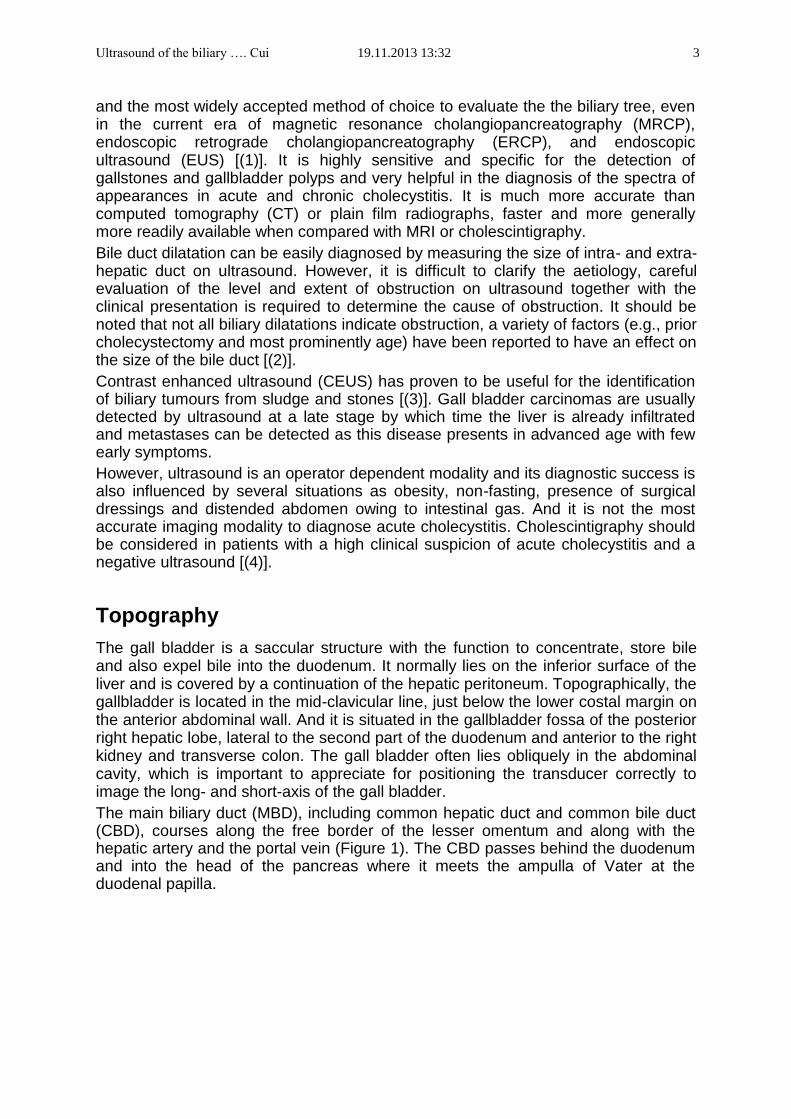
Content............................................................................................................................. ......... 1
Introduction ............................................................................................................................. .. 2
Topography .............................................................................................................................. 3
Anatomy ............................................................................................................................. ....... 5
Examination technique ........................................................................................................... 6
Patient preparation ............................................................................................................. . 6
Patient history and physical examination ........................................................................ 7
Transducer selection .......................................................................................................... 7
Patient position .................................................................................................................... 7
Ultrasound examination of the biliary system ................................................................. 7
Demonstration of gall bladder ....................................................................................... 7
Demonstration of extrahepatic bile duct .................................................................... 10
Demonstration of intrahepatic bile ducts ................................................................... 12
VIP: Very Important (and most frequent) Pathologies of gall bladder .......................... 13
Cholelithiasis ...................................................................................................................... 13
Biliary sludge ...................................................................................................................... 14
Acute cholecystitis ............................................................................................................. 15
Chronic cholecystitis ......................................................................................................... 19 Noninflammatory non-neoplastic gall bladder disorders : the hyperplastic
cholecystoses .................................................................................................................... 20
Adenomyomatous hyperplasia, ................................................................................... 20
Cholesterolosis .............................................................................................................. 20
Benign gall bladder tumours (polyps) ............................................................................ 21
Adenoma ........................................................................................................................ 22
Adenomyosis.................................................................................................................. 22
Cholesterol polyps ......................................................................................................... 22
Malignant gall bladder tumours ....................................................................................... 23
VIP: Very Important (and most frequent) Pathologies of bile ducts .............................. 24
Biliary cysts ........................................................................................................................ 24
Choledocholithiasis ........................................................................................................... 25
Cholangitis .......................................................................................................................... 26
Acute bacterial cholangitis ........................................................................................... 26
Primary sclerosing cholangitis (PSC) ......................................................................... 26
Secondary sclerosing cholangitis ............................................................................... 27
Cholangiocellular carcinoma (CCC) ............................................................................... 27
Ultrasound based diagnosis of jaundice ........................................................................ 28
References ............................................................................................................................ . 30
Introduction Biliary system diseases are a frequent pathology in medical practice. The diagnosis of biliary disease should be confirmed or excluded when a patient has pain in the right upper quadrant or suspicious jaundice. Conventional ultrasound is often the first
Ultrasound of the biliary …. Cui 19.11.2013 13:32 3
and the most widely accepted method of choice to evaluate the the biliary tree, even in the current era of magnetic resonance cholangiopancreatography (MRCP), endoscopic retrograde cholangiopancreatography (ERCP), and endoscopic ultrasound (EUS) [(1)]. It is highly sensitive and specific for the detection of gallstones and gallbladder polyps and very helpful in the diagnosis of the spectra of appearances in acute and chronic cholecystitis. It is much more accurate than computed tomography (CT) or plain film radiographs, faster and more generally more readily available when compared with MRI or cholescintigraphy. Bile duct dilatation can be easily diagnosed by measuring the size of intra- and extra-hepatic duct on ultrasound. However, it is difficult to clarify the aetiology, careful evaluation of the level and extent of obstruction on ultrasound together with the clinical presentation is required to determine the cause of obstruction. It should be noted that not all biliary dilatations indicate obstruction, a variety of factors (e.g., prior cholecystectomy and most prominently age) have been reported to have an effect on the size of the bile duct [(2)]. Contrast enhanced ultrasound (CEUS) has proven to be useful for the identification of biliary tumours from sludge and stones [(3)]. Gall bladder carcinomas are usually detected by ultrasound at a late stage by which time the liver is already infiltrated and metastases can be detected as this disease presents in advanced age with few early symptoms. However, ultrasound is an operator dependent modality and its diagnostic success is also influenced by several situations as obesity, non-fasting, presence of surgical dressings and distended abdomen owing to intestinal gas. And it is not the most accurate imaging modality to diagnose acute cholecystitis. Cholescintigraphy should be considered in patients with a high clinical suspicion of acute cholecystitis and a negative ultrasound [(4)].
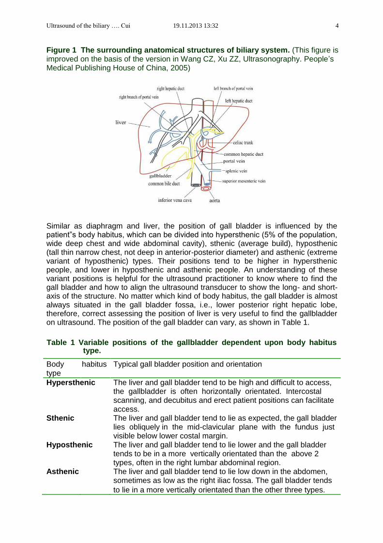
Topography The gall bladder is a saccular structure with the function to concentrate, store bile and also expel bile into the duodenum. It normally lies on the inferior surface of the liver and is covered by a continuation of the hepatic peritoneum. Topographically, the gallbladder is located in the mid-clavicular line, just below the lower costal margin on the anterior abdominal wall. And it is situated in the gallbladder fossa of the posterior right hepatic lobe, lateral to the second part of the duodenum and anterior to the right kidney and transverse colon. The gall bladder often lies obliquely in the abdominal cavity, which is important to appreciate for positioning the transducer correctly to image the long- and short-axis of the gall bladder. The main biliary duct (MBD), including common hepatic duct and common bile duct (CBD), courses along the free border of the lesser omentum and along with the hepatic artery and the portal vein (Figure 1). The CBD passes behind the duodenum and into the head of the pancreas where it meets the ampulla of Vater at the duodenal papilla.
Ultrasound of the biliary …. Cui 19.11.2013 13:32 4
Figure 1 The surrounding anatomical structures of biliary system. (This figure is improved on the basis of the version in Wang CZ, Xu ZZ, Ultrasonography. People’s Medical Publishing House of China, 2005)
Similar as diaphragm and liver, the position of gall bladder is influenced by the patient‟s body habitus, which can be divided into hypersthenic (5% of the population, wide deep chest and wide abdominal cavity), sthenic (average build), hyposthenic (tall thin narrow chest, not deep in anterior-posterior diameter) and asthenic (extreme variant of hyposthenic) types. Their positions tend to be higher in hypersthenic people, and lower in hyposthenic and asthenic people. An understanding of these variant positions is helpful for the ultrasound practitioner to know where to find the gall bladder and how to align the ultrasound transducer to show the long- and short-axis of the structure. No matter which kind of body habitus, the gall bladder is almost always situated in the gall bladder fossa, i.e., lower posterior right hepatic lobe, therefore, correct assessing the position of liver is very useful to find the gallbladder on ultrasound. The position of the gall bladder can vary, as shown in Table 1.
Table 1 Variable positions of the gallbladder dependent upon body habitus type.
Body habitus Typical gall bladder position and orientation
type
Hypersthenic The liver and gall bladder tend to be high and difficult to access, the gallbladder is often horizontally orientated. Intercostal scanning, and decubitus and erect patient positions can facilitate access. Sthenic The liver and gall bladder tend to lie as expected, the gall bladder lies obliquely in the mid-clavicular plane with the fundus just visible below lower costal margin. Hyposthenic The liver and gall bladder tend to lie lower and the gall bladder tends to be in a more vertically orientated than the above 2 types, often in the right lumbar abdominal region. Asthenic The liver and gall bladder tend to lie low down in the abdomen, sometimes as low as the right iliac fossa. The gall bladder tends
to lie in a more vertically orientated than the other three types.
Ultrasound of the biliary …. Cui 19.11.2013 13:32 5
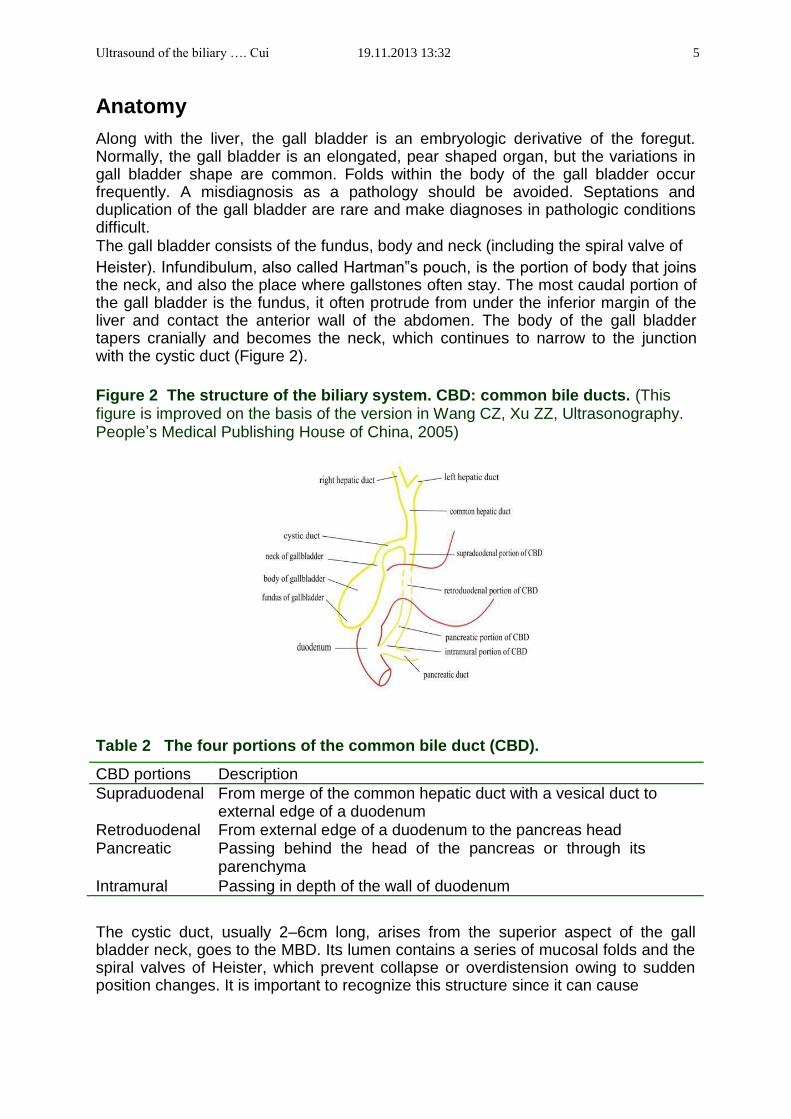
Anatomy Along with the liver, the gall bladder is an embryologic derivative of the foregut. Normally, the gall bladder is an elongated, pear shaped organ, but the variations in gall bladder shape are common. Folds within the body of the gall bladder occur frequently. A misdiagnosis as a pathology should be avoided. Septations and duplication of the gall bladder are rare and make diagnoses in pathologic conditions difficult. The gall bladder consists of the fundus, body and neck (including the spiral valve of Heister). Infundibulum, also called Hartman‟s pouch, is the portion of body that joins the neck, and also the place where gallstones often stay. The most caudal portion of the gall bladder is the fundus, it often protrude from under the inferior margin of the liver and contact the anterior wall of the abdomen. The body of the gall bladder tapers cranially and becomes the neck, which continues to narrow to the junction with the cystic duct (Figure 2).
Figure 2 The structure of the biliary system. CBD: common bile ducts. (This figure is improved on the basis of the version in Wang CZ, Xu ZZ, Ultrasonography. People’s Medical Publishing House of China, 2005)
Table 2 The four portions of the common bile duct (CBD).
CBD portions Description
Supraduodenal From merge of the common hepatic duct with a vesical duct to external edge of a duodenum
Retroduodenal From external edge of a duodenum to the pancreas head Pancreatic Passing behind the head of the pancreas or through its parenchyma
Intramural Passing in depth of the wall of duodenum The cystic duct, usually 2–6cm long, arises from the superior aspect of the gall bladder neck, goes to the MBD. Its lumen contains a series of mucosal folds and the spiral valves of Heister, which prevent collapse or overdistension owing to sudden position changes. It is important to recognize this structure since it can cause
Ultrasound of the biliary …. Cui 19.11.2013 13:32 6 acoustic shadowing and therefore may sometimes be mistaken for a gallstone in the neck. The cystic duct joins the common hepatic duct (3-4 cm long) which is merged by left and right hepatic ducts to form the common bile duct (CBD). The CBD is below the duodenal and divided into supraduodenal, retroduodenal, pancreatic, intramural parts (Table 2)(Figure 2). It passes into the head of the pancreas and meets the ampulla of Vater at the duodenal papilla. There are three variants of bond of the CBD and pancreatic duct: 1) CBD joins pancreatic duct to form a unifrorm duct and connect to the duodenum; 2) CBD and pancreatic duct are bridged in a duodenum wall; 3) CBD and pancreatic duct run into a duodenum separately. Bile is produced by hepatocytes and passes down through intra-hepatic bile ducts. Cholecystokinin that is in response to the ingestion of food can stimulate the smooth muscle of the gall bladder to contract and sphincter of Oddi in ampulla of Vater to relax, then the bile flow toward the duodenum. When the ampulla of Vater is closed, bile backs up into the cystic duct and into the gall bladder. Therefore, the measurement of gall bladder volume before and after a test meal can be used to assess gall bladder function to a limited degree. Contraction of >60% is regarded as normal. Normal contraction is a requirement before gallstone treatment with, for example, ursodeoxycholic acid. Cholecystomegaly in patients during long-standing fasting periods or with diabetes
mellitus may reveal gall bladder diameters of up to 15 6 cm without clinical relevance, while the clinically important hydrops sometimes shows right upper quadrant pain and occasionally fever. It is difficult to accurately measure the normal wall thickness of gall bladder since it is very thin. But the anterior wall of the gall bladder should always be measured as it is closer to the transducer and the normal gall bladder wall anteroposterior diameter is1–3 mm.
Examination technique
Patient preparation Ultrasound examination of the gall bladder and the biliary tree should be performed on fasting patients who should not eat or drink anything at least 6-8 hours before ultrasound examination for the following reasons: 1), the ingestion of food can stimulate the gall bladder to contract and expel bile into duodenum, which makes the gall bladder smaller and the wall thicker than normal, this may cause a misdiagnosis of pathological gall bladder wall thickening; in contrast, adequate fasting can distend the gallbladder; 2) the food residue and gas in the upper gastrointestinal (GI) tract can reduce image quality or preclude imaging of the gall bladder and biliary tree. Nevertheless, in emergency situations the examination can be also performed even if the gall bladder is partially contracted. It is also recommended to encourage a patient not to smoke for 6–8 h prior to the ultrasound examination, since there is some evidence that smoking can reduce image quality to scan the upper abdominal structures and some chemicals in tobacco may cause contraction of the gall bladder, even after fasting.
Ultrasound of the biliary …. Cui 19.11.2013 13:32 7
Patient history and physical examination Before the ultrasound examination, the clinical data should be known, it is recommended to take a short history of the patient and to palpate the abdomen.
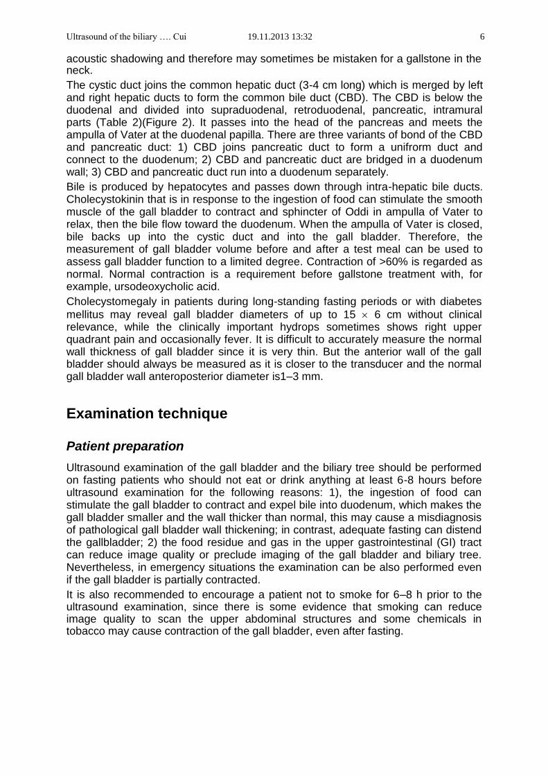
Transducer selection A convex multifrequency (1-5 MHz) transducer is often ideal for examination of the gallbladder. However, linear transducer (5-9 MHz) may be also used to evaluate the very superficial gall bladder to optimise image quality, for example, in very slim patients (Figure 3). The lower frequencies are often used in obese patients or when the gall bladder is deep.
Figure 3 The convex and linear transducers used for scanning biliary system.
Patient position Supine and left lateral decubitus are the most often used positions for the examination of gall bladder. The ultrasound examination can be started in a supine position and continued in a left posterior-oblique or left lateral decubitus, the latter two positions cause the liver and gall bladder to rotate anteromedially under the influence of gravity and this may optimise the use of the liver to image the gall bladder through an acoustic window or make the gall bladder more readily accessible below the thoracic cage. Prone or standing positions can be used to demonstrate the mobility of gall bladder sludge or stones.
Ultrasound examination of the biliary system
Demonstration of gall bladder The gall bladder can be scanned with right subcostal oblique, intercostals sections. The right subcostal section is often attempted first and it is also the most effective one. Placing the probe in longitudinal orientation and angling the probe superiorly, the scanning is performed following the ribs while the patient is asked to take a
Ultrasound of the biliary …. Cui 19.11.2013 13:32 8
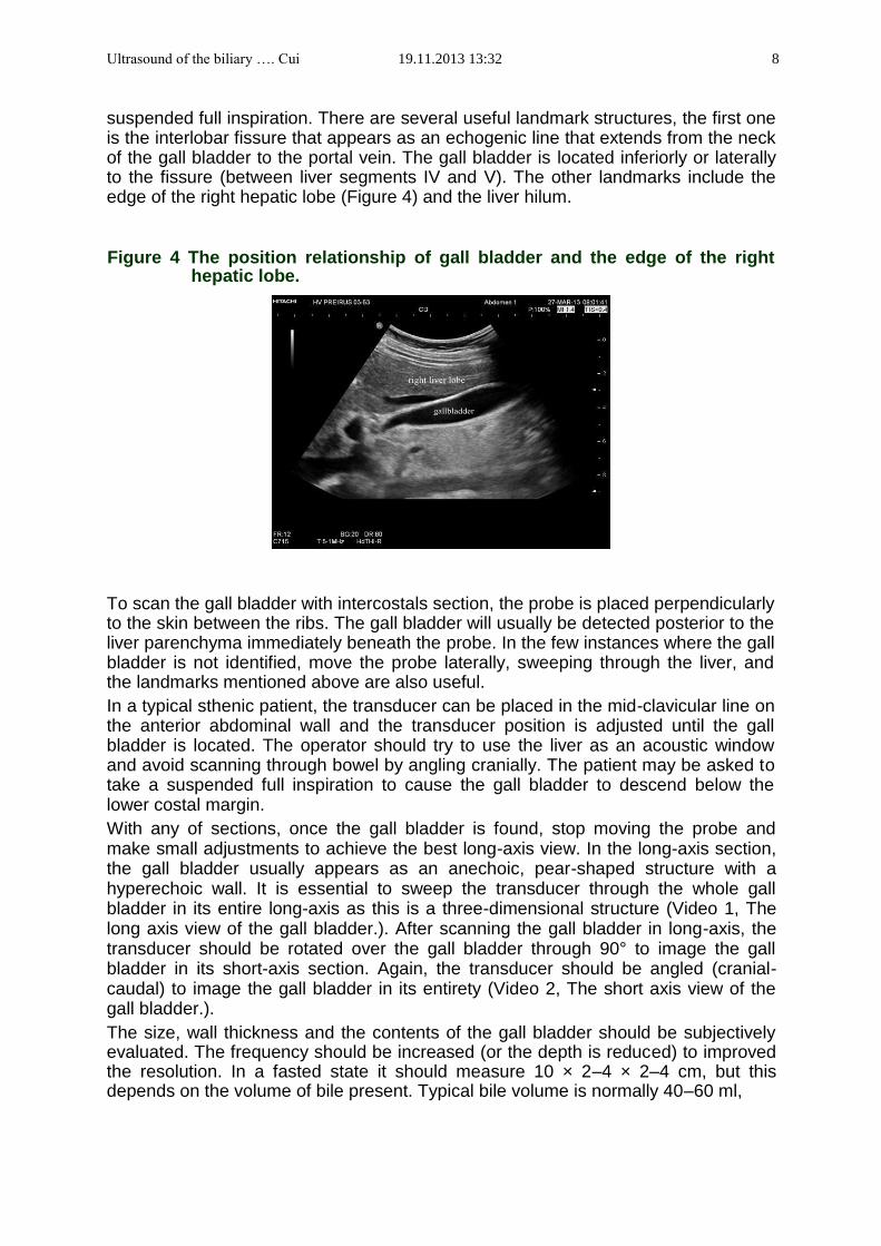
suspended full inspiration. There are several useful landmark structures, the first one is the interlobar fissure that appears as an echogenic line that extends from the neck of the gall bladder to the portal vein. The gall bladder is located inferiorly or laterally to the fissure (between liver segments IV and V). The other landmarks include the edge of the right hepatic lobe (Figure 4) and the liver hilum.
Figure 4 The position relationship of gall bladder and the edge of the right
hepatic lobe.
To scan the gall bladder with intercostals section, the probe is placed perpendicularly to the skin between the ribs. The gall bladder will usually be detected posterior to the liver parenchyma immediately beneath the probe. In the few instances where the gall bladder is not identified, move the probe laterally, sweeping through the liver, and the landmarks mentioned above are also useful. In a typical sthenic patient, the transducer can be placed in the mid-clavicular line on the anterior abdominal wall and the transducer position is adjusted until the gall bladder is located. The operator should try to use the liver as an acoustic window and avoid scanning through bowel by angling cranially. The patient may be asked to take a suspended full inspiration to cause the gall bladder to descend below the lower costal margin. With any of sections, once the gall bladder is found, stop moving the probe and make small adjustments to achieve the best long-axis view. In the long-axis section, the gall bladder usually appears as an anechoic, pear-shaped structure with a hyperechoic wall. It is essential to sweep the transducer through the whole gall bladder in its entire long-axis as this is a three-dimensional structure (Video 1, The long axis view of the gall bladder.). After scanning the gall bladder in long-axis, the transducer should be rotated over the gall bladder through 90° to image the gall bladder in its short-axis section. Again, the transducer should be angled (cranial-caudal) to image the gall bladder in its entirety (Video 2, The short axis view of the gall bladder.). The size, wall thickness and the contents of the gall bladder should be subjectively evaluated. The frequency should be increased (or the depth is reduced) to improved the resolution. In a fasted state it should measure 10 × 2–4 × 2–4 cm, but this depends on the volume of bile present. Typical bile volume is normally 40–60 ml,
Ultrasound of the biliary …. Cui 19.11.2013 13:32 9
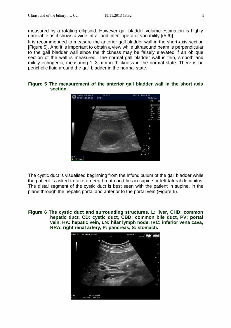
measured by a rotating ellipsoid. However gall bladder volume estimation is highly unreliable as it shows a wide intra- and inter- operator variability [(5;6)]. It is recommended to measure the anterior gall bladder wall in the short-axis section [Figure 5]. And it is important to obtain a view while ultrasound beam is perpendicular to the gall bladder wall since the thickness may be falsely elevated if an oblique section of the wall is measured. The normal gall bladder wall is thin, smooth and mildly echogenic, measuring 1–3 mm in thickness in the normal state. There is no pericholic fluid around the gall bladder in the normal state.
Figure 5 The measurement of the anterior gall bladder wall in the short axis section.
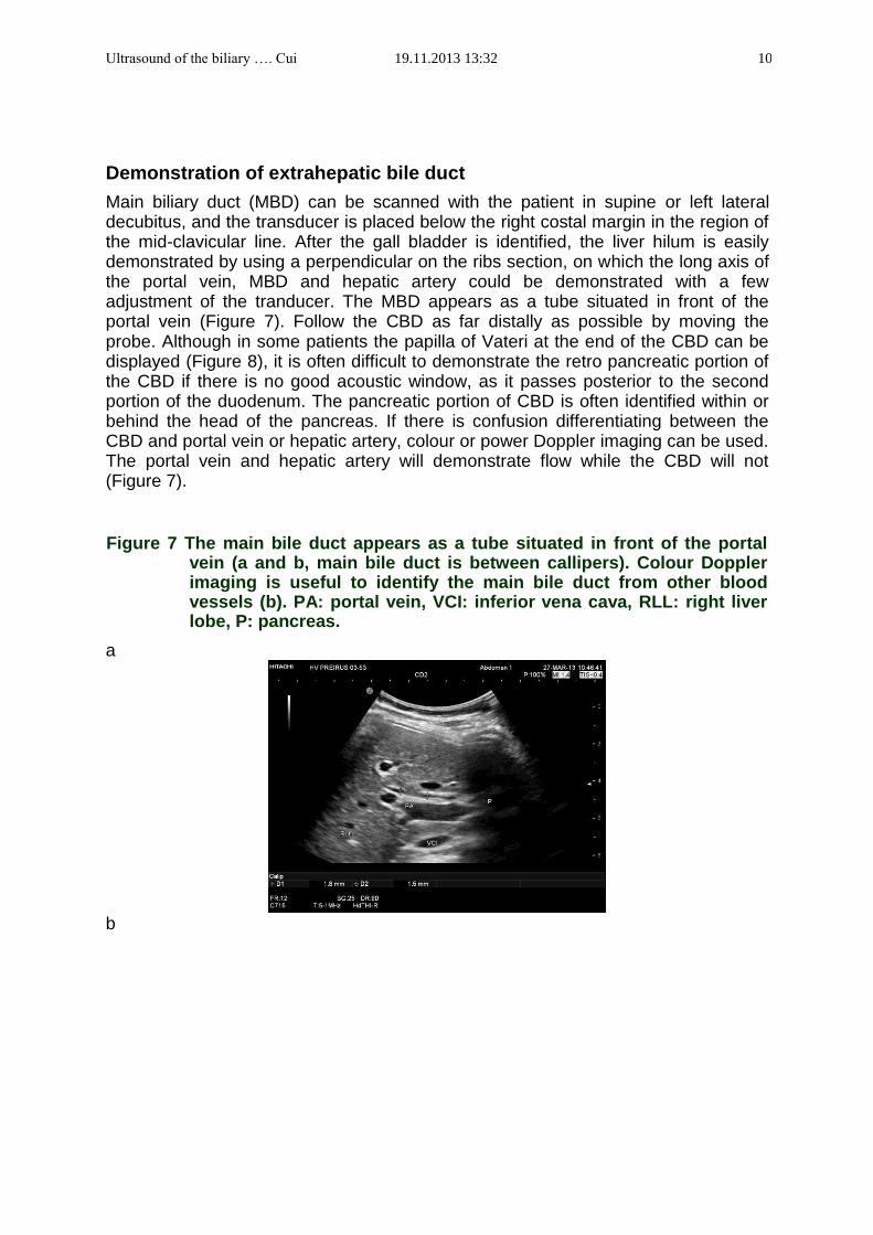
The cystic duct is visualised beginning from the infundibulum of the gall bladder while the patient is asked to take a deep breath and lies in supine or left-lateral decubitus. The distal segment of the cystic duct is best seen with the patient in supine, in the plane through the hepatic portal and anterior to the portal vein (Figure 6).
Figure 6 The cystic duct and surrounding structures. L: liver, CHD: common hepatic duct, CD: cystic duct, CBD: common bile duct, PV: portal vein, HA: hepatic vein, LN: hilar lymph node, IVC: inferior vena cava, RRA: right renal artery, P: pancreas, S: stomach.
Ultrasound of the biliary …. Cui 19.11.2013 13:32 10
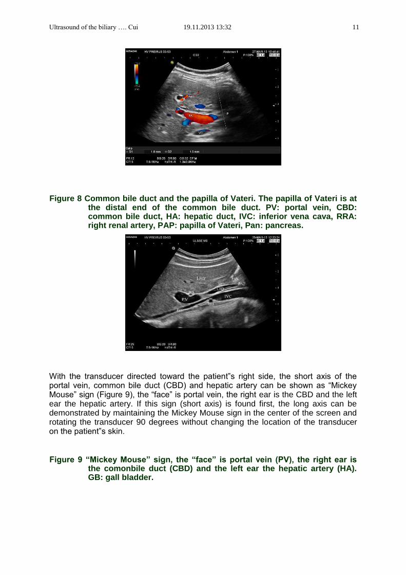
Demonstration of extrahepatic bile duct Main biliary duct (MBD) can be scanned with the patient in supine or left lateral decubitus, and the transducer is placed below the right costal margin in the region of the mid-clavicular line. After the gall bladder is identified, the liver hilum is easily demonstrated by using a perpendicular on the ribs section, on which the long axis of the portal vein, MBD and hepatic artery could be demonstrated with a few adjustment of the tranducer. The MBD appears as a tube situated in front of the portal vein (Figure 7). Follow the CBD as far distally as possible by moving the probe. Although in some patients the papilla of Vateri at the end of the CBD can be displayed (Figure 8), it is often difficult to demonstrate the retro pancreatic portion of the CBD if there is no good acoustic window, as it passes posterior to the second portion of the duodenum. The pancreatic portion of CBD is often identified within or behind the head of the pancreas. If there is confusion differentiating between the CBD and portal vein or hepatic artery, colour or power Doppler imaging can be used. The portal vein and hepatic artery will demonstrate flow while the CBD will not (Figure 7).
Figure 7 The main bile duct appears as a tube situated in front of the portal vein (a and b, main bile duct is between callipers). Colour Doppler imaging is useful to identify the main bile duct from other blood vessels (b). PA: portal vein, VCI: inferior vena cava, RLL: right liver lobe, P: pancreas.
a b
Ultrasound of the biliary …. Cui 19.11.2013 13:32 11
Figure 8 Common bile duct and the papilla of Vateri. The papilla of Vateri is at the distal end of the common bile duct. PV: portal vein, CBD: common bile duct, HA: hepatic duct, IVC: inferior vena cava, RRA: right renal artery, PAP: papilla of Vateri, Pan: pancreas.
With the transducer directed toward the patient‟s right side, the short axis of the portal vein, common bile duct (CBD) and hepatic artery can be shown as “Mickey Mouse” sign (Figure 9), the “face” is portal vein, the right ear is the CBD and the left ear the hepatic artery. If this sign (short axis) is found first, the long axis can be demonstrated by maintaining the Mickey Mouse sign in the center of the screen and rotating the transducer 90 degrees without changing the location of the transducer on the patient‟s skin.
Figure 9 “Mickey Mouse” sign, the “face” is portal vein (PV), the right ear is the comonbile duct (CBD) and the left ear the hepatic artery (HA). GB: gall bladder.
Ultrasound of the biliary …. Cui 19.11.2013 13:32 12
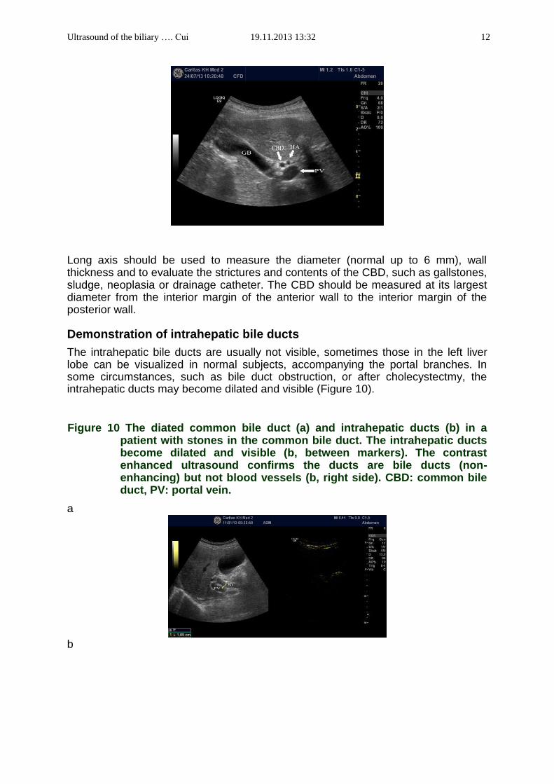
Long axis should be used to measure the diameter (normal up to 6 mm), wall thickness and to evaluate the strictures and contents of the CBD, such as gallstones, sludge, neoplasia or drainage catheter. The CBD should be measured at its largest diameter from the interior margin of the anterior wall to the interior margin of the posterior wall.
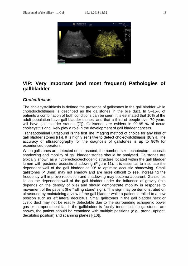
Demonstration of intrahepatic bile ducts The intrahepatic bile ducts are usually not visible, sometimes those in the left liver lobe can be visualized in normal subjects, accompanying the portal branches. In some circumstances, such as bile duct obstruction, or after cholecystectmy, the intrahepatic ducts may become dilated and visible (Figure 10).
Figure 10 The diated common bile duct (a) and intrahepatic ducts (b) in a patient with stones in the common bile duct. The intrahepatic ducts become dilated and visible (b, between markers). The contrast enhanced ultrasound confirms the ducts are bile ducts (non-enhancing) but not blood vessels (b, right side). CBD: common bile duct, PV: portal vein.
a b
Ultrasound of the biliary …. Cui 19.11.2013 13:32 13
VIP: Very Important (and most frequent) Pathologies of gallbladder
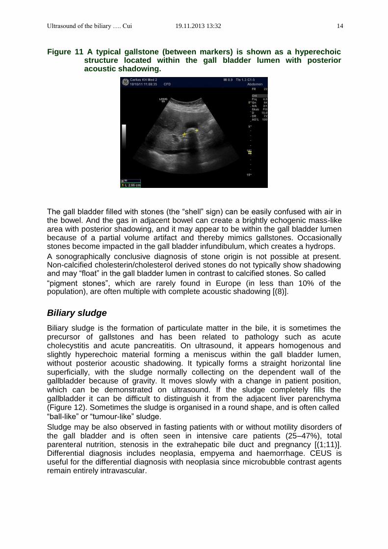
Cholelithiasis The cholecystolithiasis is defined the presence of gallstones in the gall bladder while choledocholithiasis is described as the gallstones in the bile duct. In 5–15% of patients a combination of both conditions can be seen. It is estimated that 10% of the adult population have gall bladder stones, and that a third of people over 70 years will have gall bladder stones [(7)]. Gallstones are evident in 90-95 % of acute cholecystitis and likely play a role in the development of gall bladder cancers. Transabdominal ultrasound is the first line imaging method of choice for any kind of gall bladder stones [(1)]. It is highly sensitive to detect cholecystolithiasis [(8;9)]. The accuracy of ultrasonography for the diagnosis of gallstones is up to 96% for experienced operators. When gallstones are detected on ultrasound, the number, size, echotexture, acoustic shadowing and mobility of gall bladder stones should be analysed. Gallstones are typically shown as a hyperechoic/echogenic structure located within the gall bladder lumen with posterior acoustic shadowing (Figure 11). It is essential to insonate the dependent wall of the gall bladder at 90° to optimise acoustic shadowing. Small gallstones (< 3mm) may not shadow and are more difficult to see, increasing the frequency will improve resolution and shadowing may become apparent. Gallstones lie on the dependent wall of the gall bladder under the influence of gravity (this depends on the density of bile) and should demonstrate mobility in response to movement of the patient (the “rolling stone” sign). This sign may be demonstrated on ultrasound by maintaining a view of the gall bladder while a patient is rolled to a new position such as left lateral decubitus. Small gallstones in the gall bladder neck or cystic duct may not be readily detectable due to the surrounding echogenic bowel gas or intraperitoneal fat. If the gallbladder is focally tender but no gallstones are shown, the patient should be examined with multiple positions (e.g., prone, upright, decubitus position) and scanning planes [(10)].
Ultrasound of the biliary …. Cui 19.11.2013 13:32 14
Figure 11 A typical gallstone (between markers) is shown as a hyperechoic structure located within the gall bladder lumen with posterior acoustic shadowing.
The gall bladder filled with stones (the “shell” sign) can be easily confused with air in the bowel. And the gas in adjacent bowel can create a brightly echogenic mass-like area with posterior shadowing, and it may appear to be within the gall bladder lumen because of a partial volume artifact and thereby mimics gallstones. Occasionally stones become impacted in the gall bladder infundibulum, which creates a hydrops. A sonographically conclusive diagnosis of stone origin is not possible at present. Non-calcified cholesterin/cholesterol derived stones do not typically show shadowing and may “float” in the gall bladder lumen in contrast to calcified stones. So called “pigment stones”, which are rarely found in Europe (in less than 10% of the population), are often multiple with complete acoustic shadowing [(8)].
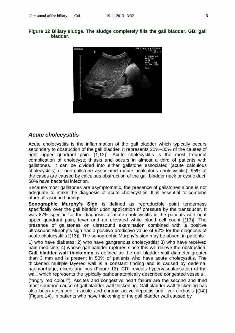
Biliary sludge Biliary sludge is the formation of particulate matter in the bile, it is sometimes the precursor of gallstones and has been related to pathology such as acute cholecystitis and acute pancreatitis. On ultrasound, it appears homogenous and slightly hyperechoic material forming a meniscus within the gall bladder lumen, without posterior acoustic shadowing. It typically forms a straight horizontal line superficially, with the sludge normally collecting on the dependent wall of the gallbladder because of gravity. It moves slowly with a change in patient position, which can be demonstrated on ultrasound. If the sludge completely fills the gallbladder it can be difficult to distinguish it from the adjacent liver parenchyma (Figure 12). Sometimes the sludge is organised in a round shape, and is often called “ball-like” or “tumour-like” sludge. Sludge may be also observed in fasting patients with or without motility disorders of the gall bladder and is often seen in intensive care patients (25–47%), total parenteral nutrition, stenosis in the extrahepatic bile duct and pregnancy [(1;11)]. Differential diagnosis includes neoplasia, empyema and haemorrhage. CEUS is useful for the differential diagnosis with neoplasia since microbubble contrast agents remain entirely intravascular.
Ultrasound of the biliary …. Cui 19.11.2013 13:32 15
Figure 12 Biliary sludge. The sludge completely fills the gall bladder. GB: gall
bladder.
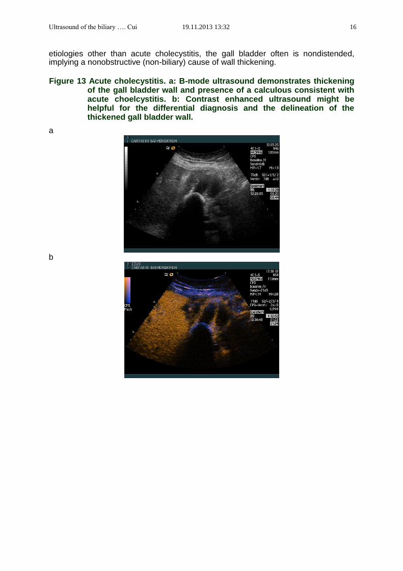
Acute cholecystitis Acute cholecystitis is the inflammation of the gall bladder which typically occurs secondary to obstruction of the gall bladder. It represents 20%–35% of the causes of right upper quadrant pain [(1;12)]. Acute cholecystitis is the most frequent complication of cholecystolithiasis and occurs in almost a third of patients with gallstones. It can be divided into either gallstone associated (acute calculous cholecystitis) or non-gallstone associated (acute acalculous cholecystitis). 95% of the cases are caused by calculous obstruction of the gall bladder neck or cystic duct. 50% have bacterial infection. Because most gallstones are asymptomatic, the presence of gallstones alone is not adequate to make the diagnosis of acute cholecystitis. It is essential to combine other ultrasound findings. Sonographic Murphy’s Sign is defined as reproducible point tenderness specifically over the gall bladder upon application of pressure by the transducer. It was 87% specific for the diagnosis of acute cholecystitis in the patients with right upper quadrant pain, fever and an elevated white blood cell count [(13)]. The presence of gallstones on ultrasound examination combined with a positive ultrasound Murphy‟s sign has a positive predictive value of 92% for the diagnosis of acute cholecystitis [(13)]. The sonographic Murphy‟s sign may be absent in patients 1) who have diabetes; 2) who have gangrenous cholecystitis; 3) who have received pain medicine; 4) whose gall baldder ruptures since this will relieve the obstruction. Gall bladder wall thickening is defined as the gall bladder wall diameter greater than 3 mm and is present in 50% of patients who have acute cholecystitis. The thickened multiple layered wall is a constant finding and is caused by oedema, haemorrhage, ulcers and pus (Figure 13). CDI reveals hypervascularisation of the wall, which represents the typically pathoanatomically described congested vessels (“angry red colour”). Ascites and congestive heart failure are the second and third most common cause of gall bladder wall thickening. Gall bladder wall thickening has also been described in acute and chronic active hepatitis and liver cirrhosis [(14)] (Figure 14). In patients who have thickening of the gall bladder wall caused by
Ultrasound of the biliary …. Cui 19.11.2013 13:32 16
etiologies other than acute cholecystitis, the gall bladder often is nondistended, implying a nonobstructive (non-biliary) cause of wall thickening.
Figure 13 Acute cholecystitis. a: B-mode ultrasound demonstrates thickening
of the gall bladder wall and presence of a calculous consistent with acute choelcystitis. b: Contrast enhanced ultrasound might be helpful for the differential diagnosis and the delineation of the thickened gall bladder wall.
a b
Ultrasound of the biliary …. Cui 19.11.2013 13:32 17
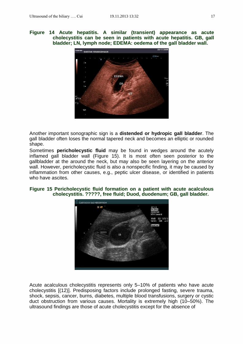
Figure 14 Acute hepatitis. A similar (transient) appearance as acute cholecystitis can be seen in patients with acute hepatitis. GB, gall bladder; LN, lymph node; EDEMA: oedema of the gall bladder wall.
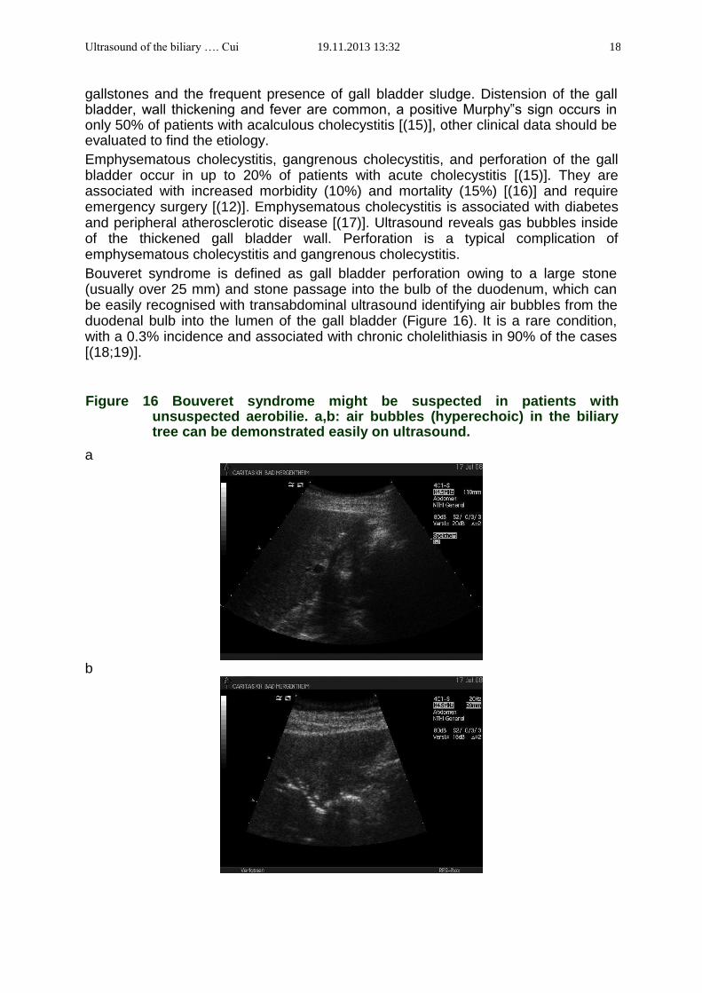
Another important sonographic sign is a distended or hydropic gall bladder. The gall bladder often loses the normal tapered neck and becomes an elliptic or rounded shape. Sometimes pericholecystic fluid may be found in wedges around the acutely inflamed gall bladder wall (Figure 15). It is most often seen posterior to the gallbladder at the around the neck, but may also be seen layering on the anterior wall. However, pericholecystic fluid is also a nonspecific finding, it may be caused by inflammation from other causes, e.g., peptic ulcer disease, or identified in patients who have ascites.
Figure 15 Pericholecystic fluid formation on a patient with acute acalculous
cholecystitis. ?????, free fluid; Duod, duodenum; GB, gall bladder.
Acute acalculous cholecystitis represents only 5–10% of patients who have acute cholecystitis [(12)]. Predisposing factors include prolonged fasting, severe trauma, shock, sepsis, cancer, burns, diabetes, multiple blood transfusions, surgery or cystic duct obstruction from various causes. Mortality is extremely high (10–50%). The ultrasound findings are those of acute cholecystitis except for the absence of
Ultrasound of the biliary …. Cui 19.11.2013 13:32 18
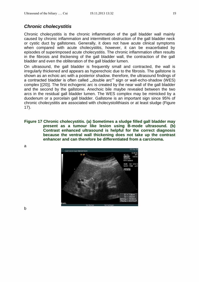
gallstones and the frequent presence of gall bladder sludge. Distension of the gall bladder, wall thickening and fever are common, a positive Murphy‟s sign occurs in only 50% of patients with acalculous cholecystitis [(15)], other clinical data should be evaluated to find the etiology. Emphysematous cholecystitis, gangrenous cholecystitis, and perforation of the gall bladder occur in up to 20% of patients with acute cholecystitis [(15)]. They are associated with increased morbidity (10%) and mortality (15%) [(16)] and require emergency surgery [(12)]. Emphysematous cholecystitis is associated with diabetes and peripheral atherosclerotic disease [(17)]. Ultrasound reveals gas bubbles inside of the thickened gall bladder wall. Perforation is a typical complication of emphysematous cholecystitis and gangrenous cholecystitis. Bouveret syndrome is defined as gall bladder perforation owing to a large stone (usually over 25 mm) and stone passage into the bulb of the duodenum, which can be easily recognised with transabdominal ultrasound identifying air bubbles from the duodenal bulb into the lumen of the gall bladder (Figure 16). It is a rare condition, with a 0.3% incidence and associated with chronic cholelithiasis in 90% of the cases [(18;19)].
Figure 16 Bouveret syndrome might be suspected in patients with unsuspected aerobilie. a,b: air bubbles (hyperechoic) in the biliary tree can be demonstrated easily on ultrasound.
a
b
Ultrasound of the biliary …. Cui 19.11.2013 13:32 19
Chronic cholecystitis Chronic cholecystitis is the chronic inflammation of the gall bladder wall mainly caused by chronic inflammation and intermittent obstruction of the gall bladder neck or cystic duct by gallstones. Generally, it does not have acute clinical symptoms when compared with acute cholecystitis, however, it can be exacerbated by episodes of superimposed acute cholecystitis. The chronic inflammation often results in the fibrosis and thickening of the gall bladder wall, the contraction of the gall bladder and even the obliteration of the gall bladder lumen. On ultrasound, the gall bladder is frequently small and contracted, the wall is irregularly thickened and appears as hyperechoic due to the fibrosis. The gallstone is shown as an echoic arc with a posterior shadow. therefore, the ultrasound findings of a contracted bladder is often called „„double arc‟‟ sign or wall-echo-shadow (WES) complex [(20)]. The first echogenic arc is created by the near wall of the gall bladder and the second by the gallstone. Anechoic bile maybe revealed between the two arcs in the residual gall bladder lumen. The WES complex may be mimicked by a duodenum or a porcelain gall bladder. Gallstone is an important sign since 95% of chronic cholecystitis are associated with cholecystolithiasis or at least sludge (Figure 17).
Figure 17 Chronic cholecystitis. (a) Sometimes a sludge filled gall bladder may present as a tumour like lesion using B-mode ultrasound. (b) Contrast enhanced ultrasound is helpful for the correct diagnosis because the ventral wall thickening does not take up the contrast enhancer and can therefore be differentiated from a carcinoma.
a b
Ultrasound of the biliary …. Cui 19.11.2013 13:32 20
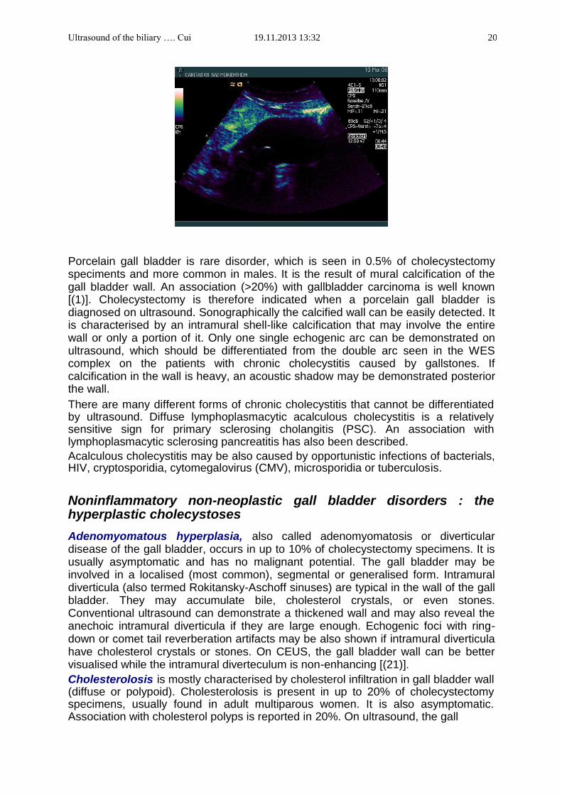
Porcelain gall bladder is rare disorder, which is seen in 0.5% of cholecystectomy speciments and more common in males. It is the result of mural calcification of the gall bladder wall. An association (>20%) with gallbladder carcinoma is well known [(1)]. Cholecystectomy is therefore indicated when a porcelain gall bladder is diagnosed on ultrasound. Sonographically the calcified wall can be easily detected. It is characterised by an intramural shell-like calcification that may involve the entire wall or only a portion of it. Only one single echogenic arc can be demonstrated on ultrasound, which should be differentiated from the double arc seen in the WES complex on the patients with chronic cholecystitis caused by gallstones. If calcification in the wall is heavy, an acoustic shadow may be demonstrated posterior the wall. There are many different forms of chronic cholecystitis that cannot be differentiated by ultrasound. Diffuse lymphoplasmacytic acalculous cholecystitis is a relatively sensitive sign for primary sclerosing cholangitis (PSC). An association with lymphoplasmacytic sclerosing pancreatitis has also been described. Acalculous cholecystitis may be also caused by opportunistic infections of bacterials, HIV, cryptosporidia, cytomegalovirus (CMV), microsporidia or tuberculosis.
Noninflammatory non-neoplastic gall bladder disorders : the hyperplastic cholecystoses Adenomyomatous hyperplasia, also called adenomyomatosis or diverticular disease of the gall bladder, occurs in up to 10% of cholecystectomy specimens. It is usually asymptomatic and has no malignant potential. The gall bladder may be involved in a localised (most common), segmental or generalised form. Intramural diverticula (also termed Rokitansky-Aschoff sinuses) are typical in the wall of the gall bladder. They may accumulate bile, cholesterol crystals, or even stones. Conventional ultrasound can demonstrate a thickened wall and may also reveal the anechoic intramural diverticula if they are large enough. Echogenic foci with ring-down or comet tail reverberation artifacts may be also shown if intramural diverticula have cholesterol crystals or stones. On CEUS, the gall bladder wall can be better visualised while the intramural diverteculum is non-enhancing [(21)]. Cholesterolosis is mostly characterised by cholesterol infiltration in gall bladder wall (diffuse or polypoid). Cholesterolosis is present in up to 20% of cholecystectomy specimens, usually found in adult multiparous women. It is also asymptomatic. Association with cholesterol polyps is reported in 20%. On ultrasound, the gall
Ultrasound of the biliary …. Cui 19.11.2013 13:32 21
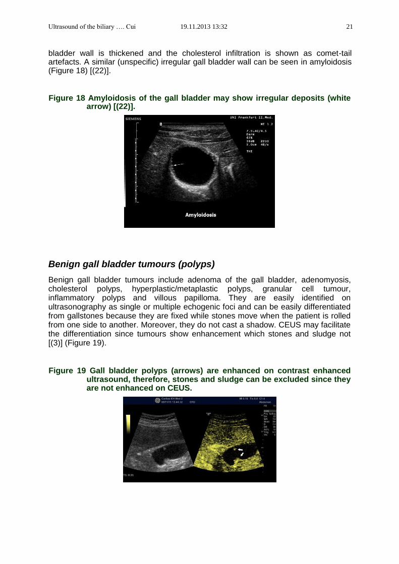
bladder wall is thickened and the cholesterol infiltration is shown as comet-tail artefacts. A similar (unspecific) irregular gall bladder wall can be seen in amyloidosis (Figure 18) [(22)].
Figure 18 Amyloidosis of the gall bladder may show irregular deposits (white
arrow) [(22)].
Benign gall bladder tumours (polyps) Benign gall bladder tumours include adenoma of the gall bladder, adenomyosis, cholesterol polyps, hyperplastic/metaplastic polyps, granular cell tumour, inflammatory polyps and villous papilloma. They are easily identified on ultrasonography as single or multiple echogenic foci and can be easily differentiated from gallstones because they are fixed while stones move when the patient is rolled from one side to another. Moreover, they do not cast a shadow. CEUS may facilitate the differentiation since tumours show enhancement which stones and sludge not [(3)] (Figure 19).
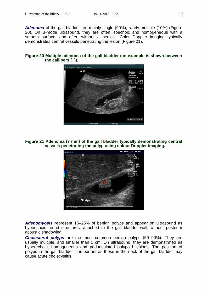
Figure 19 Gall bladder polyps (arrows) are enhanced on contrast enhanced ultrasound, therefore, stones and sludge can be excluded since they are not enhanced on CEUS.
Ultrasound of the biliary …. Cui 19.11.2013 13:32 22
Adenoma of the gall bladder are mainly single (90%), rarely multiple (10%) (Figure 20). On B-mode ultrasound, they are often isoechoic and homogeneous with a smooth surface, and often without a pedicle. Color Doppler imaging typically demonstrates central vessels penetrating the lesion (Figure 21).
Figure 20 Multiple adenoma of the gall bladder (an example is shown between the callipers (+)).
Figure 21 Adenoma (7 mm) of the gall bladder typically demonstrating central
vessels penetrating the polyp using colour Doppler imaging.
Adenomyosis represent 15–25% of benign polyps and appear on ultrasound as hypoechoic round structures, attached to the gall bladder wall, without posterior acoustic shadowing. Cholesterol polyps are the most common benign polyps (50–90%). They are usually multiple, and smaller than 1 cm. On ultrasound, they are demonstrated as hyperechoic, homogeneous and pedunculated polypoid lesions. The position of polyps in the gall bladder is important as those in the neck of the gall bladder may cause acute cholecystitis.
Ultrasound of the biliary …. Cui 19.11.2013 13:32 23
Growing gall bladder polyps or polyps larger than 10 mm should be surgically removed owing to the potential malignant transformation.
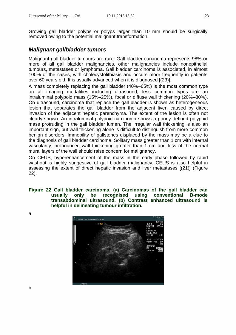
Malignant gallbladder tumors Malignant gall bladder tumours are rare. Gall bladder carcinoma represents 98% or more of all gall bladder malignancies, other malignancies include nonepithelial tumours, metastases or lymphoma. Gall bladder carcinoma is associated, in almost 100% of the cases, with cholecystolithiasis and occurs more frequently in patients over 60 years old. It is usually advanced when it is diagnosed [(23)]. A mass completely replacing the gall bladder (40%–65%) is the most common type on all imaging modalities including ultrasound, less common types are an intraluminal polypoid mass (15%–25%), focal or diffuse wall thickening (20%–30%). On ultrasound, carcinoma that replace the gall bladder is shown as heterogeneous lesion that separates the gall bladder from the adjacent liver, caused by direct invasion of the adjacent hepatic parenchyma. The extent of the lesion is often not clearly shown. An intraluminal polypoid carcinoma shows a poorly defined polypoid mass protruding in the gall bladder lumen. The irregular wall thickening is also an important sign, but wall thickening alone is difficult to distinguish from more common benign disorders. Immobility of gallstones displaced by the mass may be a clue to the diagnosis of gall bladder carcinoma. Solitary mass greater than 1 cm with internal vascularity, pronounced wall thickening greater than 1 cm and loss of the normal mural layers of the wall should raise concern for malignancy. On CEUS, hyperenhancement of the mass in the early phase followed by rapid washout is highly suggestive of gall bladder malignancy. CEUS is also helpful in assessing the extent of direct hepatic invasion and liver metastases [(21)] (Figure 22).
Figure 22 Gall bladder carcinoma. (a) Carcinomas of the gall bladder can usually only be recognised using conventional B-mode transabdominal ultrasound. (b) Contrast enhanced ultrasound is helpful in delineating tumour infiltration.
a
b
Ultrasound of the biliary …. Cui 19.11.2013 13:32 24
VIP: Very Important (and most frequent) Pathologies of bile ducts
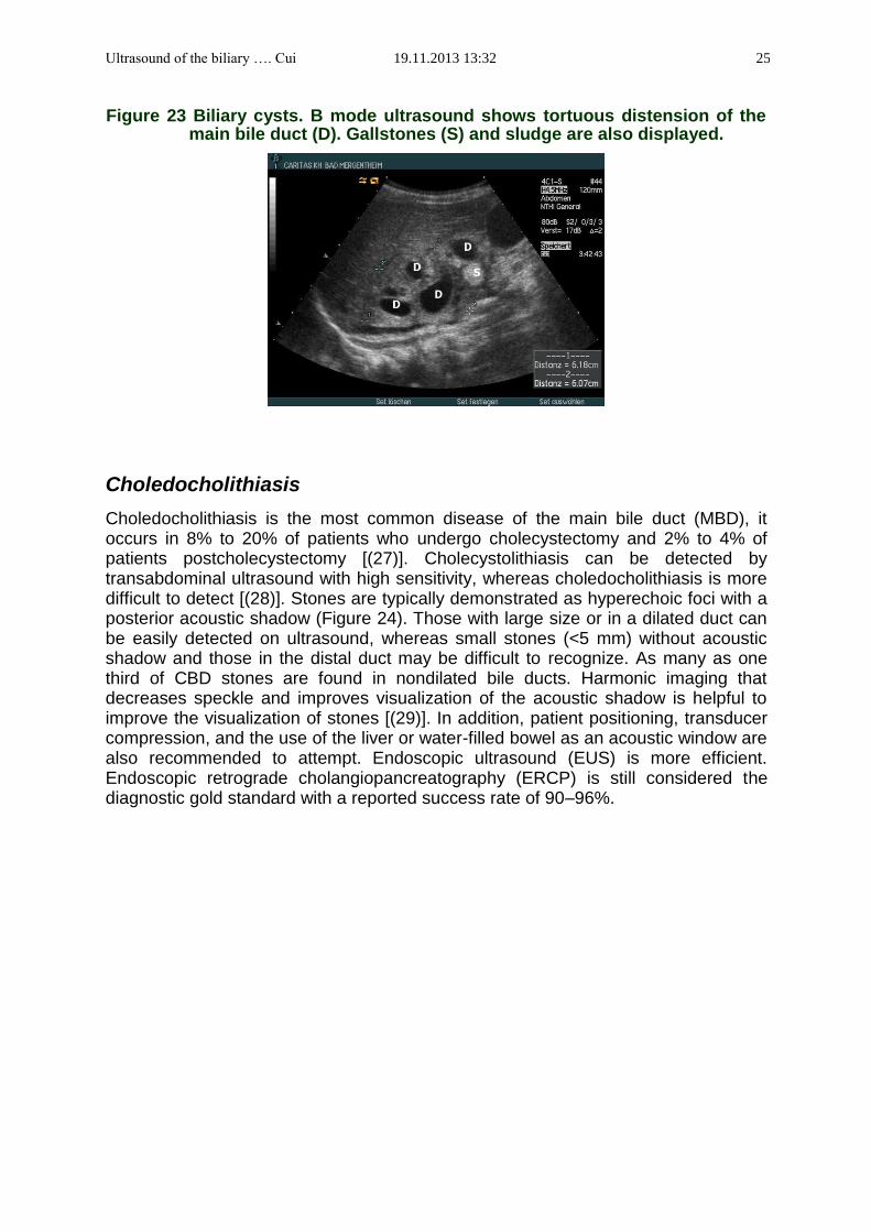
Biliary cysts Biliary cysts, originally termed choledochal cysts, are the most common cause of obstructive jaundice in infants, but they may be found at any age. They are not actually cysts but a dilatation of the biliary tree, which may secondarily obstruct other biliary ducts or the duodenum (Figure 23). Biliary cysts are associated with significant complications, such as ductal strictures, cholangitis, rupture, stone formation, and secondary biliary cirrhosis. Moreover, certain types of biliary cysts have a high risk to develop carcinoma. The Todani classification defines five types of biliary cyst (Table 3).
Table 3 Todani classification for biliary cysts [(24)]. Type Description Type 1 Characterised by a segmental or diffuse fusiform dilation of CBD (50–85%). Type 2 Diverticulum of CBD (2%) Type 3 Dilation of intraduodenal CBD (choledochocele) (1–5%) Type 4 Multiple cysts of extrahepatic bile ducts with (4a) or without (4b) cysts of
intrahepatic ducts (15-35%) Type 5 One or more cysts of intrahepatic ducts (Caroli‟s disease)(20%) CBD, common bile duct
The cystic mass can be shown on ultrasound in the right upper quadrant. However, the type III is difficult to demonstrate. Ultrasound has a sensitivity of 71- 97% percent for diagnosing biliary cysts [(25)]. Communication of the cystic lesion with the biliary tree must be demonstrated in order to differentiate biliary cysts from other cystic lesions. Ultrasound is able to show communication in 93% of patients in one report [(26)].
Ultrasound of the biliary …. Cui 19.11.2013 13:32 25
Figure 23 Biliary cysts. B mode ultrasound shows tortuous distension of the
main bile duct (D). Gallstones (S) and sludge are also displayed.
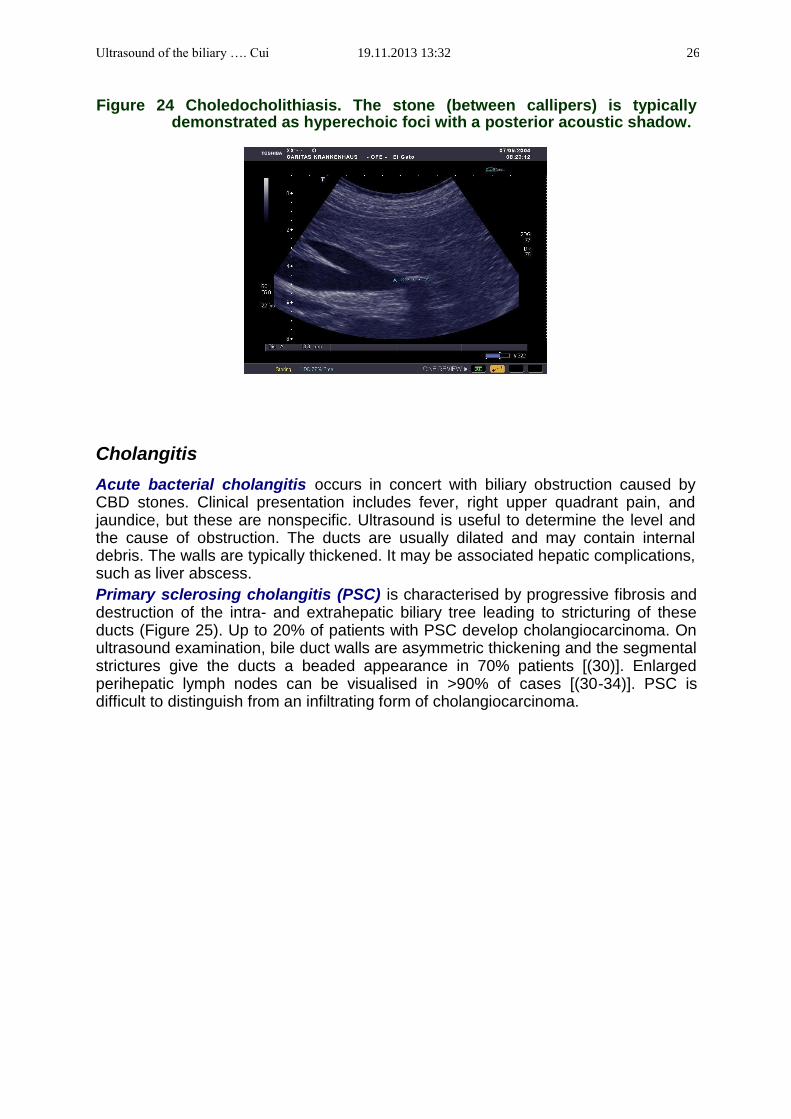
Choledocholithiasis Choledocholithiasis is the most common disease of the main bile duct (MBD), it occurs in 8% to 20% of patients who undergo cholecystectomy and 2% to 4% of patients postcholecystectomy [(27)]. Cholecystolithiasis can be detected by transabdominal ultrasound with high sensitivity, whereas choledocholithiasis is more difficult to detect [(28)]. Stones are typically demonstrated as hyperechoic foci with a posterior acoustic shadow (Figure 24). Those with large size or in a dilated duct can be easily detected on ultrasound, whereas small stones (<5 mm) without acoustic shadow and those in the distal duct may be difficult to recognize. As many as one third of CBD stones are found in nondilated bile ducts. Harmonic imaging that decreases speckle and improves visualization of the acoustic shadow is helpful to improve the visualization of stones [(29)]. In addition, patient positioning, transducer compression, and the use of the liver or water-filled bowel as an acoustic window are also recommended to attempt. Endoscopic ultrasound (EUS) is more efficient. Endoscopic retrograde cholangiopancreatography (ERCP) is still considered the diagnostic gold standard with a reported success rate of 90–96%.
Ultrasound of the biliary …. Cui 19.11.2013 13:32 26
Figure 24 Choledocholithiasis. The stone (between callipers) is typically
demonstrated as hyperechoic foci with a posterior acoustic shadow.
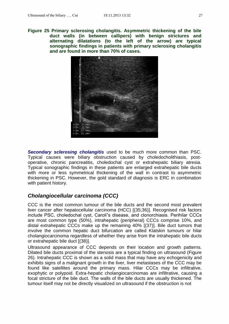
Cholangitis Acute bacterial cholangitis occurs in concert with biliary obstruction caused by CBD stones. Clinical presentation includes fever, right upper quadrant pain, and jaundice, but these are nonspecific. Ultrasound is useful to determine the level and the cause of obstruction. The ducts are usually dilated and may contain internal debris. The walls are typically thickened. It may be associated hepatic complications, such as liver abscess. Primary sclerosing cholangitis (PSC) is characterised by progressive fibrosis and destruction of the intra- and extrahepatic biliary tree leading to stricturing of these ducts (Figure 25). Up to 20% of patients with PSC develop cholangiocarcinoma. On ultrasound examination, bile duct walls are asymmetric thickening and the segmental strictures give the ducts a beaded appearance in 70% patients [(30)]. Enlarged perihepatic lymph nodes can be visualised in >90% of cases [(30-34)]. PSC is difficult to distinguish from an infiltrating form of cholangiocarcinoma.
Ultrasound of the biliary …. Cui 19.11.2013 13:32 27
Figure 25 Primary sclerosing cholangitis. Asymmetric thickening of the bile duct walls (in between callipers) with benign strictures and alternating dilatations (to the left of the arrow) are typical sonographic findings in patients with primary sclerosing cholangitis and are found in more than 70% of cases.
Secondary sclerosing cholangitis used to be much more common than PSC. Typical causes were biliary obstruction caused by choledocholithiasis, post-operative, chronic pancreatitis, choledochal cyst or extrahepatic biliary atresia. Typical sonographic findings in these patients are enlarged extrahepatic bile ducts with more or less symmetrical thickening of the wall in contrast to asymmetric thickening in PSC. However, the gold standard of diagnosis is ERC in combination with patient history.
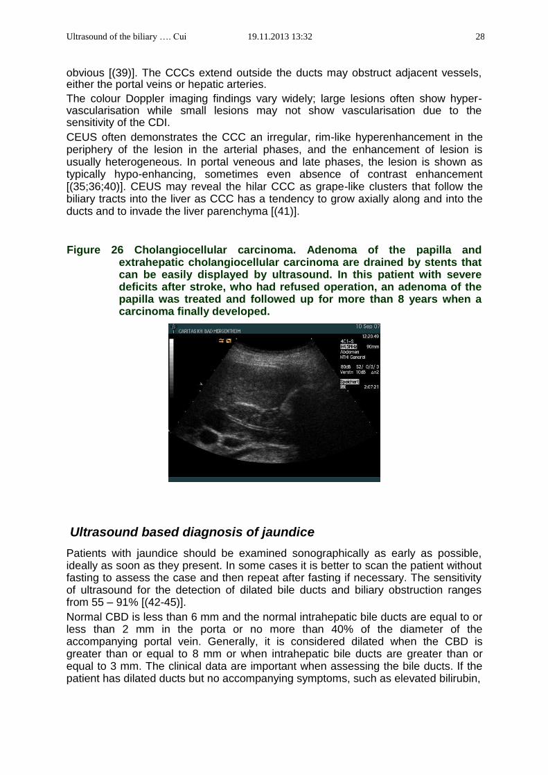
Cholangiocellular carcinoma (CCC) CCC is the most common tumour of the bile ducts and the second most prevalent liver cancer after hepatocellular carcinoma (HCC) [(35;36)]. Recognised risk factors include PSC, choledochal cyst, Caroli‟s disease, and clonorchiasis. Perihilar CCCs are most common type (50%), intrahepatic (peripheral) CCCs comprise 10%, and distal extrahepatic CCCs make up the remaining 40% [(37)]. Bile duct tumors that involve the common hepatic duct bifurcation are called Klatskin tumours or hilar cholangiocarcinoma regardless of whether they arise from the intrahepatic bile ducts or extrahepatic bile duct [(38)]. Ultrasound appearance of CCC depends on their location and growth patterns. Dilated bile ducts proximal of the stenosis are a typical finding on ultrasound (Figure 26). Intrahepatic CCC is shown as a solid mass that may have any echogenicity and exhibits signs of a malignant growth in the liver, liver metastases of the CCC may be found like satellites around the primary mass. Hilar CCCs may be infiltrative, exophytic or polypoid. Extra-hepatic cholangiocarcinomas are infiltrative, causing a focal stricture of the bile duct. The walls of the bile ducts are usually thickened. The tumour itself may not be directly visualized on ultrasound if the obstruction is not
Ultrasound of the biliary …. Cui 19.11.2013 13:32 28
obvious [(39)]. The CCCs extend outside the ducts may obstruct adjacent vessels, either the portal veins or hepatic arteries. The colour Doppler imaging findings vary widely; large lesions often show hyper-vascularisation while small lesions may not show vascularisation due to the sensitivity of the CDI. CEUS often demonstrates the CCC an irregular, rim-like hyperenhancement in the periphery of the lesion in the arterial phases, and the enhancement of lesion is usually heterogeneous. In portal veneous and late phases, the lesion is shown as typically hypo-enhancing, sometimes even absence of contrast enhancement [(35;36;40)]. CEUS may reveal the hilar CCC as grape-like clusters that follow the biliary tracts into the liver as CCC has a tendency to grow axially along and into the ducts and to invade the liver parenchyma [(41)].
Figure 26 Cholangiocellular carcinoma. Adenoma of the papilla and extrahepatic cholangiocellular carcinoma are drained by stents that can be easily displayed by ultrasound. In this patient with severe deficits after stroke, who had refused operation, an adenoma of the papilla was treated and followed up for more than 8 years when a carcinoma finally developed.
Ultrasound based diagnosis of jaundice Patients with jaundice should be examined sonographically as early as possible, ideally as soon as they present. In some cases it is better to scan the patient without fasting to assess the case and then repeat after fasting if necessary. The sensitivity of ultrasound for the detection of dilated bile ducts and biliary obstruction ranges from 55 – 91% [(42-45)]. Normal CBD is less than 6 mm and the normal intrahepatic bile ducts are equal to or less than 2 mm in the porta or no more than 40% of the diameter of the accompanying portal vein. Generally, it is considered dilated when the CBD is greater than or equal to 8 mm or when intrahepatic bile ducts are greater than or equal to 3 mm. The clinical data are important when assessing the bile ducts. If the patient has dilated ducts but no accompanying symptoms, such as elevated bilirubin,
Ultrasound of the biliary …. Cui 19.11.2013 13:32 29
sepsis, pain, or elevated liver enzymes, the dilated ducts are unlikely to be clinically relevant. After the dilated bile duct is detected on ultrasound, the level and the cause of obstruction should be assessed. Ultrasound is able to find cause in up to 71% of patients. The following points may be useful to find the cause of obstruction and the reason for jaundice. All intrahepatic ducts and extrahepatic duct are dilated. The level of obstruction should be at the lower end of the CBD. The most common benign and malignant causes are ductal gallstones and pancreatic cancer, respctively. Other causes include benign stricture secondary to pancreatitis, distal bile duct cholangiocarcionoma and periampullary cancer. Only intrahepatic bile ducts are dilated. The level of the obstruction is often at the hilus of the liver. The most common cause is hilar CCC. Other causes include PSC, Mirrizzi syndrome and gall bladder cancer. Only the common bile duct is dilated. The level of obstruction should be at the lower end of the CBD, but there is no sufficient time for the intrahepatic bile ducts to become dilated. Most common cause is distal bile duct stones, other causes include distal CBD tumours, such as CCC, or tumour from other origins but compress the CBD, such as pancreatic cancer. No bile duct is dialed. The cause of the jaundice is at a cellular level or involving microspcopic bile ducts too small to visualize on a scan. The causes include viral hepatitis, metabolic disorders, PSC, drug induced cholestasis or hepatitis, autoimmune hepatitis etc..
Ultrasound of the biliary …. Cui 19.11.2013 13:32 30
References
1. Nuernberg D, Ignee A, Dietrich CF. [Ultrasound in gastroenterology. Biliopancreatic system]. Med Klin (Munich) 2007; 102(2):112-126.
2. Horrow MM. Ultrasound of the extrahepatic bile duct: issues of size. Ultrasound Q 2010; 26(2):67-74.
3. Cui XW, Ignee A, Braden B, Woenckhaus M, Dietrich CF. Biliary papillomatosis and new ultrasound imaging modalities. Z Gastroenterol 2012; 50(2):226-231.
4. Shea JA, Berlin JA, Escarce JJ, Clarke JR, Kinosian BP, Cabana MD et al. Revised estimates of diagnostic test sensitivity and specificity in suspected biliary tract disease. Arch Intern Med 1994; 154(22):2573-2581.
5. Nuernberg D, Braden B, Ignee A, Schreiber-Dietrich DG, Dietrich CF. [Functional ultrasound in gastroenterology]. Z Gastroenterol 2008; 46(9):883-896.
6. Dietrich CF, Braden B. Sonographic assessments of gastrointestinal and biliary functions. Best Pract Res Clin Gastroenterol 2009; 23(3):353-367.
7. Freitas ML, Bell RL, Duffy AJ. Choledocholithiasis: evolving standards for diagnosis and management. World J Gastroenterol 2006; 12(20):3162-3167.
8. Cooperberg PL, Burhenne HJ. Real-time ultrasonography. Diagnostic technique of choice in calculous gallbladder disease. N Engl J Med 1980; 302(23):1277-1279.
9. Wermke W. [Ultrasonic diagnosis of bile duct calculi. A prospective study regarding the effects and the objective and subjective factors on accuracy in choledocholithiasis]. Ultraschall Med 1992; 13(6):246-254.
10. Hough DM, Glazebrook KN, Paulson EK, Bowie JD, Foster WL. Value of prone positioning in the ultrasonographic diagnosis of gallstones: prospective study. J Ultrasound Med 2000; 19(9):633-638.
11. Schirmer BD, Winters KL, Edlich RF. Cholelithiasis and cholecystitis. J Long Term Eff Med Implants 2005; 15(3):329-338.
12. Gore RM, Yaghmai V, Newmark GM, Berlin JW, Miller FH. Imaging benign and malignant disease of the gallbladder. Radiol Clin North Am 2002; 40(6):1307-23, vi.
13. Ralls PW, Colletti PM, Lapin SA, Chandrasoma P, Boswell WD, Jr., Ngo C et al. Real-time sonography in suspected acute cholecystitis. Prospective evaluation of primary and secondary signs. Radiology 1985; 155(3):767-771.
14. Kalliafas S, Ziegler DW, Flancbaum L, Choban PS. Acute acalculous cholecystitis: incidence, risk factors, diagnosis, and outcome. Am Surg 1998; 64(5):471-475.
15. Rubens DJ. Ultrasound imaging of the biliary tract. Ultrasound clin 2007; 2(2007):391-413.
16. Habib FA, Kolachalam RB, Khilnani R, Preventza O, Mittal VK. Role of laparoscopic cholecystectomy in the management of gangrenous cholecystitis. Am J Surg 2001; 181(1):71-75.
17. Konno K, Ishida H, Naganuma H, Sato M, Komatsuda T, Sato A et al. Emphysematous cholecystitis: sonographic findings. Abdom Imaging 2002; 27(2):191-195.
Ultrasound of the biliary …. Cui 19.11.2013 13:32 31
18. Duzgun AP, Ozmen MM, Ozer MV, Coskun F. Internal biliary fistula due to cholelithiasis: a single-centre experience. World J Gastroenterol 2007; 13(34):4606-4609.
19. Duzgun AP, Ozmen MM, Ozer MV, Coskun F. Internal biliary fistula due to cholelithiasis: a single-centre experience. World J Gastroenterol 2007; 13(34):4606-4609.
20. Rybicki FJ. The WES sign. Radiology 2000; 214(3):881-882. 21. Meacock LM, Sellars ME, Sidhu PS. Evaluation of gallbladder and biliary duct
disease using microbubble contrast-enhanced ultrasound. Br J Radiol 2010; 83(991):615-627.
22. Barreiros AP, Otto G, Ignee A, Galle P, Dietrich CF. Sonographic signs of amyloidosis. Z Gastroenterol 2009; 47(8):731-739.
23. Levy AD, Murakata LA, Rohrmann CA, Jr. Gallbladder carcinoma: radiologic-pathologic correlation. Radiographics 2001; 21(2):295-314.
24. Cheng SP, Yang TL, Jeng KS, Liu CL, Lee JJ, Liu TP. Choledochal cyst in adults: aetiological considerations to intrahepatic involvement. ANZ J Surg 2004; 74(11):964-967.
25. Fulcher AS, Turner MA, Sanyal AJ. Case 38: Caroli disease and renal tubular ectasia. Radiology 2001; 220(3):720-723.
26. Akhan O, Demirkazik FB, Ozmen MN, Ariyurek M. Choledochal cysts: ultrasonographic findings and correlation with other imaging modalities. Abdom Imaging 1994; 19(3):243-247.
27. Baron RL, Tublin ME, Peterson MS. Imaging the spectrum of biliary tract disease. Radiol Clin North Am 2002; 40(6):1325-1354.
28. Dietrich CF, Gouder S, Hocke M, Schuessler G, Ignee A. Endosonographie der Choledocholithiasis und ihrer Differentialdiagnosen. Endoskopie heute 2004; 17:160-166.
29. Ortega D, Burns PN, Hope SD, Wilson SR. Tissue harmonic imaging: is it a benefit for bile duct sonography? AJR Am J Roentgenol 2001; 176(3):653-659.
30. Hirche TO, Russler J, Braden B, Schuessler G, Zeuzem S, Wehrmann T et al. Sonographic detection of perihepatic lymphadenopathy is an indicator for primary sclerosing cholangitis in patients with inflammatory bowel disease. Int J Colorectal Dis 2004; 19(6):586-594.
31. Hirche TO, Russler J, Schroder O, Schuessler G, Kappeser P, Caspary WF et al. The value of routinely performed ultrasonography in patients with Crohn disease. Scand J Gastroenterol 2002; 37(10):1178-1183.
32. Hirche TO, Russler J, Braden B, Schuessler G, Zeuzem S, Wehrmann T et al. Sonographic detection of perihepatic lymphadenopathy is an indicator for primary sclerosing cholangitis in patients with inflammatory bowel disease. Int J Colorectal Dis 2004; 19(6):586-594.
33. Hirche TO, Russler J, Schroder O, Schuessler G, Kappeser P, Caspary WF et al. The value of routinely performed ultrasonography in patients with Crohn disease. Scand J Gastroenterol 2002; 37(10):1178-1183.
34. Cui XW, Jenssen C, Saftoiu A, Ignee A, Dietrich CF. New ultrasound techniques for lymph node evaluation. World J Gastroenterol 2013; 19(30):4850-4860.
35. Dietrich CF, Cui XW, Boozari B, Hocke M, Ignee A. Contrast-enhanced ultrasound (CEUS) in the diagnostic algorithm of hepatocellular and
Ultrasound of the biliary …. Cui 19.11.2013 13:32 32
cholangiocellular carcinoma, comments on the AASLD guidelines. Ultraschall Med 2012; 33 Suppl 1:S57-S66.
36. Dietrich CF, Maddalena ME, Cui XW, Schreiber-Dietrich D, Ignee A. Liver tumor characterization--review of the literature. Ultraschall Med 2012; 33 Suppl 1:S3-10.
37. DeOliveira ML, Cunningham SC, Cameron JL, Kamangar F, Winter JM, Lillemoe KD et al. Cholangiocarcinoma: thirty-one-year experience with 564 patients at a single institution. Ann Surg 2007; 245(5):755-762.
38. Bismuth H, Nakache R, Diamond T. Management strategies in resection for hilar cholangiocarcinoma. Ann Surg 1992; 215(1):31-38.
39. Hann LE, Greatrex KV, Bach AM, Fong Y, Blumgart LH. Cholangiocarcinoma at the hepatic hilus: sonographic findings. AJR Am J Roentgenol 1997; 168(4):985-989.
40. Tanaka S, Ioka T, Oshikawa O, Hamada Y, Yoshioka F. Dynamic sonography of hepatic tumors. AJR Am J Roentgenol 2001; 177(4):799-805.
41. Khalili K, Metser U, Wilson SR. Hilar biliary obstruction: preliminary results with Levovist-enhanced sonography. AJR Am J Roentgenol 2003; 180(3):687-693.
42. Pedersen OM, Nordgard K, Kvinnsland S. Value of sonography in obstructive jaundice. Limitations of bile duct caliber as an index of obstruction. Scand J Gastroenterol 1987; 22(8):975-981.
43. Pasanen PA, Partanen KP, Pikkarainen PH, Alhava EM, Janatuinen EK, Pirinen AE. A comparison of ultrasound, computed tomography and endoscopic retrograde cholangiopancreatography in the differential diagnosis of benign and malignant jaundice and cholestasis. Eur J Surg 1993; 159(1):23-29.
44. Lapis JL, Orlando RC, Mittelstaedt CA, Staab EV. Ultrasonography in the diagnosis of obstructive jaundice. Ann Intern Med 1978; 89(1):61-63.
45. Salem S, Vas W. Ultrasonography in evaluation of the jaundiced patient. J Can Assoc Radiol 1981; 32(1):30-34.