Embed Size (px)
Citation preview

EDITOR EMERITUS (1975–2005)
Claude Schulman, BelgiumEDITOR EMERITUS (2006–2013)
Francesco Montorsi, Italy
EDITOR-IN-CHIEF
James Catto, UK
Peter Albers, GermanyPeter Albertsen, USAAnders Bjartell, SwedenMichel Bolla, FranceChristopher Chapple, UKNoel Clarke, UKFirouz Daneshgari, USAJames Eastham, USAShin Egawa, JapanScott Eggener, USAMark Emberton, UKMatthew Galsky, USAMatthew Gettman, USA
Gianluca Giannarini, Switzerland
Inderbir Gill, USAMarkus Graefen, GermanyAxel Heidenreich, GermanyBrent Hollenbeck, USABrant Inman, USAPierre Karakiewicz, CanadaLou Kavoussi, USAAdam Kibel, USAYair Lotan, USASurena Matin, USAKevin McVary, USAMani Menon, USA
Rodolfo Montironi, ItalyJ. Kellogg Parsons, USAJens Rassweiler, GermanyClaus Roehrborn, USADan Sjoberg, USAArnulf Stenzl, GermanyAndrew Stephenson, USAChristian Stief, GermanyTullio Sulser, SwitzerlandGeorge Thalmann, SwitzerlandHouston Thompson, USAChristopher Wood, USAMichael Zelefsky, USAAlexandre Zlotta, Canada
CONSULTING EDITORS
EUROPEAN UROLOGY EDITORIAL OFFICEAcademic Urology Unit, University of Sheffi eld
The Medical SchoolBeech Hill Road
Sheffi eld S10 2RX, UK E-mail: [email protected]
Tel: +31 26 389 0680; Fax: +44 114 271 2268 Offi cial Journal of Societa Italiana di Urologia (SIU)
Offi cial Journal of the
ASSOCIATE EDITORS
SURGERY IN MOTION EDITOR
Alexander Mottrie, Belgium
DIGITAL MEDIA EDITOR
Alexander Kutikov, USA
MEDICAL ONCOLOGY EDITOR
Karim Fizazi, France
STATISTICAL EDITOR
Andrew Vickers, USA
NORTH AMERICAN EDITOR
Stephen Freedland, USAEAU-EBU UPDATE SERIES EDITOR
Oliver W. Hakenberg, Germany
MANAGING EDITOR
Cathy Pierce, USAEDITORIAL OFFICE ASSISTANT
Leila Ayandi, UKCOPY EDITOR
Samantha EnslenDragonfl y Editorial, USA
Christian Gratzke, GermanyMatthew Cooperberg, USA
Giacomo Novara, ItalyJean-Nicolas Cornu, France
Shahrokh Shariat, AustriaAnthony D’Amico, USA

EDITORIAL BOARD
EUROPEAN UROLOGY EDITORIAL OFFICEAcademic Urology Unit, University of Sheffi eld
The Medical SchoolBeech Hill Road
Sheffi eld S10 2RX, UK E-mail: [email protected]
Tel: +31 26 389 0680; Fax: +44 114 271 2268Offi cial Journal of Societa Italiana di Urologia (SIU)
Offi cial Journal of the
Firas Abdollah, ItalyHashim Ahmed, UKKarl-Erik Andersson, SwedenApostolos Apostolidos, GreeceMonish Aron, USARiccardo Autorino, ItalyMarko Babjuk, Czech RepublicAlexander Bachmann,
SwitzerlandRafael Badalyan, ArmeniaRiccardo Bartoletti, ItalyPatrick Bastian, GermanyRicarda Bauer, GermanyFrank Becker, GermanyJoaquim Bellmunt, SpainKarim Bensalah, FranceMichael Blute, USASteve Boorjian, USAAlberto Bossi, FranceAlberto Briganti, ItalyRichard Bryan, UKLukas Bubendorf, SwitzerlandJeffrey Cadeddu, USASteve Campbell, USAAbdullah Canda, TurkeyUmberto Capitanio, ItalyPeter Carroll, USARufus Cartwright, UKAndrea Cestari, ItalyJoseph Chin, CanadaLaurence Collette, BelgiumRenzo Colombo, ItalyElisabetta Costantini, ItalyFrancisco Cruz, PortugalGuido Dalbagni, USARocco Damiano, ItalyJean de la Rosette, NetherlandsCosimo De Nunzio, ItalyTheo de Reijke, NetherlandsJohn Denstedt, CanadaRoger Dmochowski, USAChristopher Eden, UKJason Efstathiou, USABehfar Ehdaie, USAMark Emberton, UK
Bernard Escudier, FranceVincenzo Ficarra, ItalyPaolo Fornara, GermanyClare Fowler, UKMatthew Galsky, USAJohn Gearhart, USAGianluca Giannarini,
SwitzerlandOfer Gofrit, IsraelChristian Gozzi, GermanyStavros Gravas, GreeceFrancesco Greco, GermanyJürgen Gschwend, GermanyBertrand Guillonneau, USAAxel Haferkamp, GermanyHarry Herr, USAPiet Hoebeke, BelgiumJacques Irani, FranceHendrik Isbarn, GermanyKazuto Ito, JapanAtes Kadioglu, TurkeyJeffrey Karnes, USAMichael Kattan, USAZiya Kirkali, TurkeyTobias Klatte, GermanyEric Klein, USAPilar Laguna, NetherlandsMassimo Lazzeri, ItalyEric Lechevallier, FranceEvangelos Liatsikos, GreeceWilliam Lowrance, USAStephan Madersbacher, AustriaMassimo Maffezzini, ItalyPadraig Malone, UKLuis Martínez-Piñeiro, SpainMani Menon, USAMartin Michel, NetherlandsAndrea Minervini, ItalyNicolas Mottet, FranceAlexander Mottrie, BelgiumMasaru Murai, JapanRichard Naspro, ItalyWillem Oosterlinck, BelgiumAnup Patel, UKJehonathan Pinthus, Canada
Guillaume Ploussard, FranceFrancesco Porpiglia, ItalyDavid Ralph, UKOliver Reich, GermanyMesut Remzi, AustriaMichael Rink, GermanyMorgan Rouprêt, FrancePaul Russo, USAKazutaka Saito, JapanAndrea Salonia, ItalyChristian Saussine, FranceVincenzo Scattoni, ItalyJack Schalken, NetherlandsThorsten Schlomm, GermanyMichael Seitz, GermanyMaurizio Serati, ItalyGiuseppe Simone, ItalyGuru Sonpavde, USACora Sternberg, ItalyJens-Uwe Stolzenburg,
GermanyUrs Studer, SwitzerlandNazareno Suardi, ItalySamir Taneja, USADerya Tilki, GermanyBertrand Tombal, BelgiumKarim Touijer, USAQuoc-Dien Trinh, CanadaLevent Türkeri, TurkeyRoderick van den Bergh,
NetherlandsTheo van der Kwast, CanadaHendrik Van Poppel, BelgiumBas van Rhijn, NetherlandsYoram Vardi, IsraelJochen Walz, FranceJohannes Witjes, NetherlandsWim Witjes, NetherlandsJean-Jacques Wyndaele, BelgiumEvanguelos Xylinas, FranceOfer Yossepowitch, IsraelRichard Zigeuner, AustriaAmnon Zisman, Israel

EURO
PEAN
URO
LOG
YSU
PPLE
MEN
TSCONTENTS
EUROPEAN UROLOGY SUPPLEMENTS, VOL. 13, NO. 3, SEPTEMBER 2014
11th Meeting of the EAU Robotic Urology Section (ERUS)17–19 September 2014, Amsterdam, The Netherlands
Welcome to the 11th Meeting of the EAU Robotic UrologySection (ERUS) . . . . . . . . . . . . . . . . . . . . . . . . . . . . . . . . . . . . . . . . v
Organisers . . . . . . . . . . . . . . . . . . . . . . . . . . . . . . . . . . . . . . . . . . . vi
Faculty . . . . . . . . . . . . . . . . . . . . . . . . . . . . . . . . . . . . . . . . . . . . . vi
Sponsor Acknowledgement . . . . . . . . . . . . . . . . . . . . . . . . . . . . . . vii
Floorplan venue . . . . . . . . . . . . . . . . . . . . . . . . . . . . . . . . . . . . . . viii
General Information . . . . . . . . . . . . . . . . . . . . . . . . . . . . . . . . . . . ix
Continuing Medical Education Accreditation . . . . . . . . . . . . . . . . . xi
Scientific Programme . . . . . . . . . . . . . . . . . . . . . . . . . . . . . . . . . . xivWednesday, 17 September . . . . . . . . . . . . . . . . . . . . . . . . . . . . . . . . . . xivThursday, 18 September . . . . . . . . . . . . . . . . . . . . . . . . . . . . . . . . . . . . xxiFriday, 19 September . . . . . . . . . . . . . . . . . . . . . . . . . . . . . . . . . . . . . . xxiv
Abstracts . . . . . . . . . . . . . . . . . . . . . . . . . . . . . . . . . . . . . . . . . . . . 1Oral Presentations . . . . . . . . . . . . . . . . . . . . . . . . . . . . . . . . . . . . . . . . 2
YAU-Junior ERUS – Poster abstracts (PYJ01–PYJ03) . . . . . . . . . . . . . . . . . 2YAU-Junior ERUS – Video abstracts (VYJ01–VYJ03) . . . . . . . . . . . . . . . . . 3ERUS – Poster abstracts (PE01–PE03) . . . . . . . . . . . . . . . . . . . . . . . . . . 4ERUS – Video abstracts (VE01–VE03) . . . . . . . . . . . . . . . . . . . . . . . . . . 6
Unmoderated Poster Presentations . . . . . . . . . . . . . . . . . . . . . . . . . . . . . 8YAU-Junior ERUS – Poster abstracts (PYJ04–PYJ27) . . . . . . . . . . . . . . . . . 8ERUS – Poster abstracts (PE04–PE89) . . . . . . . . . . . . . . . . . . . . . . . . . . 17
Unmoderated Video Presentations . . . . . . . . . . . . . . . . . . . . . . . . . . . . . . 50ERUS – Video abstracts (VE04–VE38) . . . . . . . . . . . . . . . . . . . . . . . . . . 50
About the Organisers . . . . . . . . . . . . . . . . . . . . . . . . . . . . . . . . . . . 61About the European Association of Urology (EAU) . . . . . . . . . . . . . . . . . . . . . 61About the EAU Robotic Urology Section (ERUS) . . . . . . . . . . . . . . . . . . . . . . 62
Author Index . . . . . . . . . . . . . . . . . . . . . . . . . . . . . . . . . . . . . . . . . 63

© 2014 European Association of Urology. Published by Elsevier B.V.
This journal and the individual contributions contained in it are protected under copyright, and the following terms and conditions apply to their use in addition to the terms of any Creative Commons or other user license that has been applied by the publisher to an individual article:
Photocopying Single photocopies of single articles may be made for personal use as allowed by national copyright laws. Permission is not required for photocopying of articles published under the CC BY license nor for photocopying for non-commercial purposes in accordance with any other user license applied by the publisher. Permission of the publisher and payment of a fee is required for all other photocopying, including multiple or systematic copying, copying for advertising or promotional purposes, resale, and all forms of document delivery. Special rates are available for educational institutions that wish to make photocopies for non-profi t educational classroom use.
Derivative Works Users may reproduce tables of contents or prepare lists of articles including abstracts for internal circulation within their institutions or companies. Other than for articles published under the CC BY license, permission of the publisher is required for resale or distribution outside the subscribing institution or company. For any subscribed articles or articles published under a CC BY-NC-ND license, permission of the publisher is required for all other derivative works, including compilations and translations.
Storage or Usage Except as outlined above or as set out in the relevant user license, no part of this publication may be reproduced, stored in a retrieval system or transmitted in any form or by any means, electronic, mechanical, photocopying, recording or otherwise, without prior written permission of the publisher.
PermissionsFor information on how to seek permission visit www.elsevier.com/permissions or call: (+44) 1865 843830 (UK) / (+1) 215 239 3804 (USA).
Author rights Author(s) may have additional rights in their articles as set out in their agreement with the publisher (more information at http://www.elsevier.com/authorsrights).
Notice No responsibility is assumed by the publisher for any injury and/or damage to persons or property as a matter of products liability, negligence or otherwise, or from any use or operation of any methods, products, instructions or ideas contained in the material herein. Because of rapid advances in the medical sciences, in particular, independent verifi cation of diagnoses and drug dosages should be made. Although all advertising material is expected to conform to ethical (medical) standards, inclusion in this publication does not constitute a guarantee or endorsement of the quality or value of such product or of the claims made of it by its manufacturer.
Advertising information: Advertising orders and enquiries can be sent to: USA, Canada and South America: Elsevier Inc., 360 Park Avenue South, New York, NY 10010-1710, USA; phone: (+1) (212) 633 3974; Europe and ROW: Advertising Sales: Elsevier Pharma Solutions, 32 Jamestown Road, London NW1 7BY, UK; phone: (+44) (0) 20 7424 4259; fax: (+44) (0) 20 7424 4433; e-mail: [email protected]. Commercial Reprint Sales, Greg Davies, Elsevier Ltd.; phone: (+44) 20 7424 4422; fax: (+44) 20 7424 4433; e-mail: [email protected].
The paper used in this publication meets the requirements of ANSI/NISO Z39.48-1992 (Permanence of Paper).
Abstracted/indexed in: BIOBASE, EMBASE, Medical Documentation, Current Contents – Clinical Medicine, Science Citation Index, Adis Clinical Trials Insight, SciVerse Scopus®. Full text available on SciVerse ScienceDirect®.
Orders, claims, and journal enquiries: please contact the Elsevier Customer Service Department nearest you:
St. Louis: Elsevier Customer Service Department, 3251 Riverport Lane, Maryland Heights, MO 63043, USA; phone: (800) 6542452 [toll free within the USA]; (+1) (314) 4478871 [outside the USA]; fax: (+1) (314) 4478029; e-mail: [email protected]
Oxford: Elsevier Customer Service Department, The Boulevard, Langford Lane, Kidlington, Oxford OX5 1GB, UK; phone: (+44) (1865) 843434; fax: (+44) (1865) 843970;e-mail: [email protected]
Tokyo: Elsevier Customer Service Department, 4F Higashi-Azabu, 1-Chome Bldg, 1-9-15 Higashi-Azabu, Minato-ku, Tokyo 106-0044, Japan; phone: (+81) (3) 5561 5037; fax: (+81) (3) 5561 5047;e-mail: [email protected]
Singapore: Elsevier Customer Service Department, 3 Killiney Road, #08-01 Winsland House I, Singapore 239519; phone: (+65) 63490222; fax: (+65) 67331510;e-mail: [email protected]
Printed by Henry Ling Ltd, Dorchester, Dorset, UK

v
Welcome to the 11th Meeting of the EAU Robotic UrologySection (ERUS)
Dear colleagues and friends,
It gives us great pride towelcome you to Amsterdam for three days of learning on robotic urologicalsurgery. The earlier editions of the annual ERUSmeeting showed the power of live surgery trainingfor both novice and more experienced robotic surgeons.
In Amsterdam, the Thursday and Friday will be packed with a variety of live surgical procedures.All procedures will follow the new regulations for live surgery from the EAU and they will allowfor plenty of interaction, being chaired by experienced robotic surgeons and an expert panel.
Developments in techniques and technologies will be presented and demonstrated in practice.With a live-satellite connection between Holland’s two largest cities, Amsterdam and Rotterdam,we consider the meeting a truly Dutch event with input from different institutes from this country.
The meeting venue is located in the heart of Amsterdam at just a walking distance from the RoyalPalace and the Amsterdam Canals.
Robotic surgery has grown from an experiment by few, to a useful tool for many. The broadspectrum of urological procedures for benign andmalignant diseases will be presented, illustratingthat robotic surgery has firmly landed in urology.
Now it is time to broaden the experience, improve quality, economize procedures, speed up thelearning curve and, above all look for ways to implement technological improvements in dailypractice.
In an ambience of interaction and learning, we hope you will discover new ways to improve yourpractice, make new friends, and enjoy your stay in Amsterdam!
Alex MottrieChairman EAU Robotic Urology Section Board

vi
Organisers
Scientific CommitteeClement-Claude Abbou, Vincennes (FR)Carl Magnus Annerstedt, Stockholm (SE)Walter Artibani, Verona (IT)Prokar Dasgupta, London (GB)Markus Graefen, Hamburg (DE)Ali Riza Kural, Istanbul (TR)Francesco Montorsi, Milan (IT)Alexandre Mottrie, Aalst (BE)Pierre-Thierry Piéchaud, Bordeaux (FR)Jens Rassweiler, Heilbronn (DE)Charles-Henry Rochat, Geneva (CH)Rafael Sanchez-Salas, Paris (FR)Henk Van Der Poel, Amsterdam (NL)
Local Organising CommitteeHarrie Beerlage, ’s-Hertogenbosch (NL)Willem de Blok, Amsterdam (NL)Sjoerd Klaver, Rotterdam (NL)Henk Van Der Poel, Amsterdam (NL)André Vis, Amsterdam (NL)Carl Wijburg, Arnhem (NL)
ERUS Congress OfficeCongress Consultants B.V.PO Box 300166803 AA ArnhemThe NetherlandsT: +31 (0)26 389 1751F: +31 (0)26 389 [email protected]
Faculty
Clement-Claude Abbou, Vincennes (FR)Ismail Acar, Ankara (TR)Thomas Ahlering, Orange (US)Waleed Alkhudair, Riyadh (SA)Carl Magnus Annerstedt, Stockholm (SE)Walter Artibani, Verona (IT)Jelle Barentsz, Nijmegen (NL)Harrie Beerlage, ’s-Hertogenbosch (NL)Sam Bhayani, St Louis (US)Maurizio Brausi, Modena (IT)Nicolo Buffi, Milan (IT)Abdullah Canda, Ankara (TR)James Catto, Sheffield (GB)Ben Challacombe, London (GB)Christopher Chapple, Sheffield (GB)Prokar Dasgupta, London (GB)John Davis, Houston (US)Georges De Boccard, Geneva (CH)Geert De Naeyer, Aalst (BE)Mihir Desai, Los Angeles (US)Vincenzo Ficarra, Padova (IT)Markus Graefen, Hamburg (DE)Khurshid Guru, Buffalo (US)Ashok Hemal, Winston Salem (US)Günter Janetschek, Salzburg (AT)Shamim Khan, London (GB)Sjoerd Klaver, Rotterdam (NL)Ali Riza Kural, Istanbul (TR)Francesco Montorsi, Milan (IT)Daniel Moon, Melbourne (AU)Alex Mottrie, Aalst (BE)Declan Murphy, Melbourne (AU)
Giacomo Novara, Padova (IT)Joan Palou, Barcelona (ES)Emmanouil Panagiotou, Athens (GR)Vito Pansadoro, Rome (IT)Vip Patel, Celebration (US)Pierre-Thierry Piéchaud, Bordeaux (FR)Dmitry Pushkar, Moscow (RU)Jens Rassweiler, Heilbronn (DE)Koon Ho Rha, Seoul (KR)John Rietbergen, Rotterdam (NL)Charles-Henry Rochat, Geneva (CH)Rafael Sanchez-Salas, Paris (FR)Christian Schwentner, Tübingen (DE)Ryoichi Shiroki, Aichi (JP)Prasanna Sooriakumaran, Oxford (GB)Michael Stöckle, Homburg (DE)Jens-Uwe Stolzenburg, Leipzig (DE)Nazareno Suardi, Milan (IT)Ashutosh Tewari, New York (US)Tom Tuytten, Heerlen (NL)Jean-Paul Van Basten, Nijmegen (NL)Ben Van Cleynenbreugel, Leuven (BE)Henk Van Der Poel, Amsterdam (NL)Fijs Van Leeuwen, Leiden (NL)Emmanuel Vander Poorten, Leuven (BE)André Vis, Amsterdam (NL)Alessandro Volpe, Novara (IT)Carl Wijburg, Arnhem (NL)Peter Wiklund, Stockholm (SE)Timothy Wilson, Duarte (US)Manfred Wirth, Dresden (DE)Jörn Witt, Gronau (DE)

vii
Sponsor Acknowledgement
The organisers respectfully acknowledge the following sponsors for providing unrestricted educational grantsand services to the 11th Meeting of the EAU Robotic Urology Section.
Platinum Sponsor
Other Sponsors and Exhibitors
ASTELLAS
ASTRAZENECA
BAYER
BIOMEDIC
BK MEDICAL
COLOPLAST
COVIDIEN
EDAP TMS
ELMED
MEDICAL DYNAMICS
MIMIC TECHNOLOGIES
OLYMPUS
ORSI
ROSWELL PARK CANCER INSTITUTE
SCANLAN INTERNATIONAL
SIMBIONIX USA CORPORATION
SURGICAL SPECIALTIES
SURGIQUEST

viii
Floorplan venue

ix
General Information
About AmsterdamFrom its earliest days, Amsterdam has been a bustling hub of commerce that welcomed other cultures with open arms. Learnmore about this lovely canal-side city, including the rich history and development of its tolerant society. Or jump straight tothe modern day and find out about the city’s architecture and its colourful neighbourhoods. If you’re feeling ambitious, youmight even pick up a fewwords of Dutch! A good website for information on Amsterdam is www.iamsterdam.com. The nationalcurrency of The Netherlands is the Euro (EUR or €).
AbstractsThe abstracts are available in this book.
Certificate of AttendanceA Certificate of Attendance for ERUS 2014 can be printed online as of Monday, 22 September on erus2014.uroweb.org.You will need your registration number (under the barcode on your badge) to print the Certificate of Attendance.
Cloakroom/luggageThe cloakroom is located in the main entrance area and is open during meeting hours. Please be sure to collect all personalbelongings at the end of the day.
Congress BagEach delegate can collect a congress bag in the registration area.
Congress DinnerThe congress dinner will take place on Thursday, 18 September at themagnificent National MaritimeMuseum: Het Scheepvaart-museum. The Museum shows how Dutch culture has been shaped by the sea. The museum has recently been renovated, but its350-year history and initial design as a naval arsenal shine through. The dinner will take place in the open courtyard of themuseum, featuring live music, a menu inspired by Dutch Golden Age trade routes, and an opportunity to mingle with colleagues.Entrance tickets can be purchased at the registration desk.
Disclosure links to IndustryIt is requested that all faculty disclose to the audience any links with the industry related to the topic of their lecture at thebeginning of their presentation(s). A link can be: being a member of the advisory board or having a consulting agreement with aspecific company.
EAU Policy on Live Surgery
The EAU established an official polidy on Live Surgery Events, offering organising centres a clear framework within whichto plan and perform live surgeries at any EAU congress or meeting. It outlines a set of guidelines in which the overridingprinciple is that patient safety must take priority over all other considerations in the conduct of live surgery. Read more on:http://www.uroweb.org/events/eau-live-surgery-events/
Emergency InformationEmergency phone number for police, fire brigade and ambulance service is 112. Contact the security or the organisationimmediately in case of an emergency in the congress venue.
ExhibitionA technical exhibition will be held jointly with the meeting in the exhibition hall on the ground floor.
Opening hours:Thursday 18 September 09.00–16.30Friday 19 September 09.00–16.00
First AidIn case of an emergency, contact a security guard or the organisation immediately.
InsuranceThe organisers do not accept responsibility for any personal damage. Participants are strongly recommended to arrange theirown personal insurance.
LanguageAll presentations during the meeting will be conducted in English. No translation will be provided.
Lost and FoundFound items should be returned to the registration desk. If you lose something, please report to this desk for assistance.

x G EN E R A L I N FORMAT I ON
Mobile PhonesThe sound and flashlight of mobile phones must be switched off during sessions.
PressJournalists can obtain free registration to the meeting. All media operators must show their credentials (press card dated2013/2014 and original assignment letter).
Registration areaThe registration area is located in the main entrance on the ground floor.
Opening hours:Wednesday 17 September 07.00–19.00Thursday 18 September 07.00–18.00Friday 19 September 07.30–18.00
SafetyAll bags may be subject to inspection. Security is present for your safety. Please take all personal effects with you when leavingthe session rooms.
Scientific PostersThe scientific posters are on display from 17 to 19 September in the poster area on the ground floor. It has been requested thatone of the authors is present to answer possible questions during the following poster viewing hours but this is not required.
Wednesday 17 September 11.00–11.2512.30–13.30
Thursday 18 September 10.00–10.3013.15–14.1515.30–16.00
Friday 19 September 10.00–10.3013.00–14.0015.30–16.00
Scientific VideosThe scientific videos are on digital display from 18 to 19 September in the exhibition area on the ground floor.
Smoking PolicySmoking is prohibited inside the congress venue.
Speaker Service Centre (SSC)All presentations should be handed in at the Speaker Service Centre (located on the first floor) at least three hours prior to thestart of the session.
Opening hours:Wednesday 17 September 07.00–19.00Thursday 18 September 07.00–18.00Friday 19 September 07.30–18.00
TransportationAmsterdam has an excellent public transport network (Openbaar Vervoer) including trams, busses, metros and ferries. It’s avery easy and affordable way to navigate the city. Public transport in Amsterdam is run by city transport company GVB andthere are a number of different ticket options for visitors and residents which basically depend on how long you plan to stay inAmsterdam and where you wish to go. Firstly, you should note that the OV-chip card (OV-chipkaart) is a smart card system usedfor all public transport in the Netherlands. This means when boarding/exiting your train/tram/bus/metro you need to check-inand check-out by holding your chip card against the card readers.
Venue AddressBeurs van BerlageDamrak 2431012 ZJ AmsterdamT: +31 (0) 20 530 [email protected]
WiFiFree wireless internet will be available in all areas and session rooms. Please search for the “ERUS2014” network and connect byentering the following:
Username: ERUS2014Password: ERUS2014

xi
Continuing Medical Education Accreditation
The 11th Meeting of the EAU Robotic Urology Section is accredited by the European Accreditation Council forContinuing Medical Education (EACCME) to provide the following CME activity for medical specialists. The EACCMEis an institution of the European Union of Medical Specialists (UEMS), www.uems.net.
The 11th Meeting of the EAU Robotic Urology Section is designated for a maximum of 15 hours of European externalCME credits. Each medical specialist should claim only those hours of credit that he/she actually spent in theeducational activity.
Through an agreement between the European Union of Medical Specialists and the American Medical Association,physicians may convert EACCME credits to an equivalent number of AMA PRA Category 1 Credits™. Information onthe process to convert EACCME credit to AMA credit can be found at www.ama-assn.org/go/internationalcme.
Live educational activities, occurring outside of Canada, recognized by the UEMS-EACCME for ECMEC credits aredeemed to be Accredited Group Learning Activities (Section 1) as defined by the Maintenance of CertificationProgram of The Royal College of Physicians and Surgeons of Canada.

SEEKING FOR PERFECTION IN ROBOTIC SURGERY?

SIGN UP TO THE UPCOMING UROLOGY TRAININGS!
Intensive Robotic Training - 6 days course- Max. 5 participants- This session contains:
• theoretical courses • Simulator training & dry lab• 2 days Animal training• 2 days Live surgery (with dual console)
- Price: 7.500 € pp excl. VAT
Please note there is a limited number of subscriptions per session allowed.
Intensive Procedure Specifi c Robotic Training on- Robot-Assisted Partial nephrectomy - Robot-Assisted Cystectomy & intracorporeal
neobladder - Robot-Assisted Radical Prostatectomy
- 2 or 3 days course- Max. 3 participants- This session contains:
• theoretical courses • Animal training• Live surgery (with dual console)
- Price: 3.500 € (2 day) pp excl. VAT 5.000 € (3 day) pp excl. VAT
Inclusive: training fee, lunch, didactical material, transport to and from training center.
We also offer you the possibility to prepare your own training! Our “CUSTOM-MADE TRAINING” makes it possible to adapt a program according the experiences and wishes of the participants!
You can fi nd extra information on the website: www.orsi-online.com
Contact: T: (+32) (0)9/334.69.26M: (+32) (0)474/[email protected]: OLV Robotic Surgery Institute Proefhoevestraat 12 I 9090 Melle
Structured training: 26-28/01 & 11-13/0223-25/02 & 11-13/02
Partial Nephrectomy:12-14/0116-17/0216-17/03
Cystectomy:2-4/02
RARP:2-4/031-3/04
Prof. Dr. Alex Mottrie Course DirectorDirector, OLV Robotic Surgery Instituteand Professor of Uro-Oncology,OLV Hospital, Aalst, Belgium
Prof. Dr. Vincenzo FicarraScientifi c Director, OLV Robotic Surgery Instituteand Professor of Urogoly,University of Padua, Italy
Dr. Giacomo NovaraScientifi c Director, OLV Robotic Surgery InstituteAssistant Professor of UrologyDepartment of Surgery, Oncology, and Gastroenterology - Urology Clinic University of Padua

xiv
Scientific Programme
WEDNESDAY, 17 SEPTEMBER
Young Academic Urologists-Junior ERUS Programme
Room: Effectenbeurszaal
08.30–08.40 Welcome and introduction new group YAU-JUNIOR ERUSN. Buffi, Milan (IT)C. Wijburg, Arnhem (NL)
08.40–09.25 Session 1 – Hot topics in robotic urologic surgeryModerators: R. Autorino, Cleveland (US)
N. Buffi, Milan (IT)C. Wijburg, Arnhem (NL)
08.40–08.55 Economics business case example cost-effectiviness.Robotics with the new Da Vinci Xi: Will the older systems still be supported?C. Wijburg, Arnhem (NL)
08.55–09.10 HOT courses: Results of a dry lab studyA.E. Canda, Ankara (TR)
09.10–09.25 Pilot study: Results from ERUS curriculumG. Novara, Padova (IT), A. Volpe, Novara (IT)
09.25–10.00 Session 2 – Challenging scenarios in robotic urologic surgeryModerators: G. De Naeyer, Aalst (BE)
N.D. Doumerc, Toulouse (FR)C. Schwentner, Tübingen (DE)
09.25–09.45 Robotic assisted radical prostatectomy: Step by step and difficult casesG. Pini, Stockholm (SE)
09.45–10.00 Nightmare session: How table side assistant can help to control a hemorrhagic event (Iliac artery and renalvein lesion)F. Annino, Arezzo (IT)
10.00–11.00 Session 3 – A look at the literatureModerators: F. Annino, Arezzo (IT)
A. Govorov, Moscow (RU)G. Pini, Stockholm (SE)
10.00–10.15 Learning curve in robotic surgery: Review of the literature (RALP, RAPN and RARC)G. De Naeyer, Aalst (BE)
10.15–10.30 Complications in robotic surgery: Review of the literature (RALP, RAPN and RARC)A. Wallerstedt, Stockholm (SE)
10.30–10.40 2014 Best papers in robotic prostatectomyN. Suardi, Milan (IT)
10.40–10.50 2014 Best papers in robotic renal surgeryR. Autorino, Cleveland (US)
10.50–11.00 2014 Best papers in robotic cystectomyC. Schwentner, Tübingen (DE)
11.00–11.25 Coffee break & poster viewing
11.25–12.10 Session 4 – Video and poster session. The 3 best abstracts and best 3 best videosModerators: N.D. Doumerc, Toulouse (FR)
D. Porres, Aachen (DE)A. Wallerstedt, Stockholm (SE)
11.25–11.32 PYJ03 Prevalence and predictors of thromboembolic events in patients undergoing lymph node dissectionduring radical prostatectomyS. Tyritzis, A.Wallerstedt, G. Steineck, T. Nyberg, J. Hugosson, A. Bjartell, U.Wilderäng, T. Thorsteinsdot-tir, S. Carlsson, J. Stranne, E. Haglind, N.P. Wiklund (Stockholm, Gothenburg, Lund, Sweden; Reykjavik,Iceland)

SC I ENT I F I C P R O G R AMME xv
11.32–11.39 PYJ02 Development and validation of the checklist based assessment tool for robot assisted radicalprostatectomyC. Lovegrove, G. Novara, K. Guru, A. Mottrie, B. Challacombe, J. Raza, H. Van Der Poel, J. Peabody,R. Popert, P. Dasgupta, K. Ahmed (London, United Kingdom; Padua, Italy; Buffalo, Detroit, United Statesof America; Aalst, Belgium; Amsterdam, The Netherlands)
11.39–11.46 PYJ01 A new intraoperative modular training system for the learning curve in robotic surgeryY. Al Salhi, S. Khorrami, M. De Angelis, T. Verdacchi, V. Giommoni, F. Annino (Arezzo, Italy)
11.46–11.53 VYJ02 Image guided robotic partial cystectomy using flexible cystoscopy and tile proA. Sridhar, S. Madhavan, S. Nathan (London, United Kingdom)
11.53–12.00 VYJ01 Robotic off-clamp zero ischemia partial nephrectomy in small, peripherally located, exophytic renalmass is safe and feasibleA.E. Canda, O.U. Cakici, K. Ener, A.F. Atmaca (Ankara, Turkey)
12.00–12.10 VYJ03 Robot-assisted radical nephroureterectomy in an ectopic pelvic kidneyC. Wagner, A. Schütte, J. Witt (Gronau, Germany)
12.10–12.15 Video and Poster session: The winners
12.15–12.30 Last years’ winner reports
12.30 Final remarksN. Buffi, Milan (IT)C. Wijburg, Arnhem (NL)
European Association of Urology Nurses (EAUN) Programme
Room: Berlage zaal
09.15–09.25 Opening EAUN-ERUSNurse specialist – J.E. Kinsella, London (GB)
09.25–09.40 New robots/new technologyUrologist – H. Van Der Poel, Amsterdam (NL)
09.40–10.00 Five prostatectomies a day, can it be done?OR Nurse – L. Söderkvist, Stockholm (SE)OR Nurse – E. Rundin, Stockholm (SE)
10.00–10.20 Cut the costs: Cost efficiency/effectiveness throughout the processUrologist – C-H. Rochat, Geneva (CH)
10.20–10.40 What kind of urological procedures can be done with the robot?Urologist – J.-P. Van Basten, Nijmegen (NL)
10.40–11.00 Nerve sparing, are there any standards?Urologist – V. Ficarra, Padua (IT)
11.00–11.30 Coffee break & poster viewing
11.30–12.30 Break-out session Ward nurses Room: Berlage zaalChair: J.E. Kinsella, London (GB)
11.30–11.50 QoL and symptom assessment in the Netherlands and the use of internetClinical nurse specialist – C. Tillier, Amsterdam (NL)
11.50–12.10 Erectile dysfunction, counseling and treatment: The nursing perspectiveOstomy and wound nurse – B. Jetten, Amsterdam (NL)
12.10–12.30 Transfer from OR – recoveryNurse – S. Van ’t Slot, Rotterdam (NL)Nurse – A. Van Houwelingen, Rotterdam (NL)
11.30–12.30 Break-out session OR nurses Room: Mendes da Costa kamerChair: L. Söderkvist, Stockholm (SE)
11.30–11.50 Positioning and draping, looking for a standard: Differences and similarities in the NetherlandsOR nurse – P. Kennedy, Amsterdam (NL)

xvi SC I ENT I F I C P R O G R AMME
11.50–12.10 How to teach student nurses in robotic surgeryOR Nurse/Educator – M. Landsbergen, Arnhem (NL)
12.10–12.30 Transfer from OR – recovery – ward: What standards need to be met?Recovery nurse – L. Thompson-Ritfeld, Rotterdam (NL)
12.30–13.30 Lunch break & poster viewing
13.30–15.00 Break-out session Ward nurses Room: Berlage zaalChair: W. De Blok, Amsterdam (NL)
13.30–14.00 Not at home after one day, a case storyClinical nurse specialist – W. De Blok, Amsterdam (NL)
14.00–14.30 RoboCare – It’s not all about the robotNurse coordinator – E. Birch, Melbourne (AU)
14.30–15.00 Emergency post-op situations on the wardUrologist – K. Ahmed, London (GB)
13.30–15.00 Break-out session OR nurses Room: KeurzaalChair: J.E. Kinsella, London (GB)
13.30–15.00 MIMIC simulation hands on trainingJ. Ostman, Seattle (US)
13.30–15.00 Simultaneous session OR Nurses during simulation session Room: Mendes da Costa kamerChair: L. Söderkvist, Stockholm (SE)
13.30–14.00 Emergency converting robotics to open operation: How to train the staffOR nurse – L. Juhl Hansen, Aalborg (DK)
14.30–15.00 First assistant’s role in Europe; and what about the training?OR nurse – J. Peterson, Aalborg (DK)
15.00–15.30 Coffee break & poster viewing
15.30–16.00 Plenary SessionChair: J.E. Kinsella, London (GB)
15.30–16.00 The patient’s perspective from the admission to dischargeInterview with patient – Mr. B. (NL)Interviewer – W. De Blok, Amsterdam (NL)
16.00–16.15 Closing remarks and take home messagesJ.E. Kinsella, London (GB)
European School of Urology (ESU) Courses
ESU Course 1 – Advanced course in Da Vinci prostatectomyRoom: Administratiezaal
13.30 IntroductionP-T. Piéchaud, Bordeaux (FR)
13.30–13.50 General principles of robotic radical prostatectomy
13.30–13.40 My way of access: How I place the portsW. Artibani, Verona (IT)
13.40–13.50 My way of access: How I place the portsP. Dasgupta, London (GB)
13.50–14.30 Step-by-step operative procedure
13.50–14.00 Bladder neck approach: Preservation or not: How do I do it?P. Dasgupta, London (GB)P.-T. Piéchaud, Bordeaux (FR)
14.00–14.10 Posterior dissection: Seminal vesicles complete dissectionW. Artibani, Verona (IT)

SC I ENT I F I C P R O G R AMME xvii
14.10–14.20 Posterior dissection seminal vesicles sparingP.-T. Piéchaud, Bordeaux (FR)
14.20–14.30 Lateral dissection: Anatomial reminders. Peri prostatic fascia, neuro vascular periprostatic structuresP. Dasgupta, London (GB)
14.30–15.00 Nerve sparing: Which space of dissection, how do I do it?
14.30–14.40 Interfascial dissectionP. Dasgupta, London (GB)
14.40–14.50 Interfascial antegrade dissectionW. Artibani, Verona (IT)
14.50–15.00 Intrafascial dissectionP.-T. Piéchaud, Bordeaux (FR)
15.00–15.30 Coffee break & poster viewing
15.30–16.00 Apex & DVC
15.30–15.40 First approachP. Dasgupta, London (GB)
15.40–15.50 Final approachW. Artibani, Verona (IT)P.-T. Piéchaud, Bordeaux (FR)
15.50–16.00 Special techniques for continence. Anterior fixation and Posterior fixation: Rocco techniqueW. Artibani, Verona (IT)
16.00–16.40 Anastomosis
16.00–16.10 Double half running sutureW. Artibani, Verona (IT)
16.10–16.20 Vloc sutureP. Dasgupta, London (GB)
16.20–16.30 Unique running sutureP.-T. Piéchaud, Bordeaux (FR)
16.30–16.40 Technique of extended lymphadenectomyW. Artibani, Verona (IT)
16.40–17.00 Specific situations
16.40–16.50 Bladder neck and median lobe. Previous prostatic surgery: TURP, AdenomectomyP.-T. Piéchaud, Bordeaux (FR)
16.50–17.00 Salvage prostatectomyP.-T. Piéchaud, Bordeaux (FR)
17.00–17.30 Postoperative complications
17.00–17.10 Prevention and management: actual standardP. Dasgupta, London (GB)
17.10–17.20 Anatomical and functional resultsW. Artibani, Verona (IT)
17.20–17.30 ConclusionsP.-T. Piéchaud, Bordeaux (FR)
17.30 Closure
ESU Course 2 – Advanced Course in Robotics in the Upper Urinary tractRoom: Veilingzaal
13.30–13.45 Introduction: Use of robot in upper urinary tract surgeryA. Mottrie, Aalst (BE)
13.45–14.00 Patient positioning, trocar positioning, trans- and retroperitoneal access in renal robotic surgeryD. Moon, Melbourne (AU)
xviii SC I ENT I F I C P R O G R AMME
14.00–14.15 Robotic pyeloplasty: Multichannel or single site techniqueB. Challacombe, London (GB)
14.15–14.30 Renal surgery: Nephrectomy and nephroureterectomy: How I do itC. Vaessen, Paris (FR)
14.30–14.40 Current indications for partial nephrectomy and nephrometry scoresD. Moon, Melbourne (AU)
14.40–15.00 Partial nephrectomy I
Step I: Isolation of renal hilumStep II: Mobilisation of the kidneyStep III: Clamping of renal pedicle: Different techniquesB. Challacombe, London (GB)D. Moon, Melbourne (AU)A. Mottrie, Aalst (BE)C. Vaessen, Paris (FR)
15.00–15.30 Coffee break & poster viewing
15.30–16.00 Partial nephrectomy II
Step IV: Different tumor resection techniquesB. Challacombe, London (GB)D. Moon, Melbourne (AU)A. Mottrie, Aalst (BE)C. Vaessen, Paris (FR)
16.00–16.30 Partial nephrectomy III
Step V: Different renorraphy techniquesB. Challacombe, London (GB)D. Moon, Melbourne (AU)A. Mottrie, Aalst (BE)C. Vaessen, Paris (FR)
16.30–16.40 Postoperative careB. Challacombe, London (GB)
16.40–17.00 Special techniques:
Which haemostatic agents are useful? Surgical bolster?B. Challacombe, London (GB)
Selective clamping or zeroischemia: What’s up, doc?C. Vaessen, Paris (FR)
New developmentsA. Mottrie, Aalst (BE)– Robotically applied Bulldogs– Fluorescence– Drop in ultrasound devices– Robotic suction/irrigator device
17.00–17.30 Partial nephrectomy IV: Special cases/complication managementB. Challacombe, London (GB)D. Moon, Melbourne (AU)A. Mottrie, Aalst (BE)C. Vaessen, Paris (FR)– Hilar tumours– Endophytic tumours– Cystic tumour– Limits of RAPN– Other
17.30 Closure

SC I ENT I F I C P R O G R AMME xix
ESU Course 3 – Advanced course in Da Vinci cystectomy and diversionRoom: Verwey kamer
13.30–13.50 IntroductionP. Wiklund, Stockholm (SE)
13.50–15.00 Cystectomy
13.50–14.07 Open radical cystectomy and lymphadenectomy in bladder cancerJ. Palou, Barcelona (ES)
14.07–14.24 Robotic-assisted cystectomy: Step-by-step maleK. Guru, New York (US)
14.24–14.40 Female cystectomyJ. Palou, Barcelona (ES)
14.40–15.00 LymphadenectomyC.M. Annerstedt, Stockholm (SE)
15.00–15.30 Coffee break & poster viewing
15.30–16.20 Diversion
15.30–15.40 Extracorporeal urinary diversionJ. Palou, Barcelona (ES)
15.40–16.10 Intracorporeal urinary diversion
Bricker conduitK. Guru, New York (US)
NeobladderC.M. Annerstedt, Stockholm (SE)P. Wiklund, Stockholm (SE)
16.10–16.20 Ways to proceed – Panel discussion
16.20–17.30 Results
16.20–16.45 Outcomes after robotic-assisted cystectomyP. Wiklund, Stockholm (SE)
16.45–17.10 ComplicationsC.M. Annerstedt, Stockholm (SE)
17.10–17.30 IRCC presentationK. Guru, New York (US)
17.30 Closure
ESU Course 4 – Robotics in Urogenital tumours: Where are we in 2014?Room: Rode kamer
13.30–13.35 IntroductionM. Stöckle, Homburg (DE)
13.35–15.00 Part one
13.35–14.00 Do the benefits of robotic prostatectomy justify the costs?M. Stöckle, Homburg (DE)
14.00–14.30 Present role of robotic prostatectomy in the UST. Ahlering, Orange (US)
14.30–15.00 Robotic surgery at the upper urinary tractJ. Stolzenburg, Leipzig (DE)
15.00–15.30 Coffee break & poster viewing
15.30–17.30 Part two
15.30–16.10 Proliferation of robotic surgery in non-Urological disciplines – US trendsT. Ahlering, Orange (US)

xx SC I ENT I F I C P R O G R AMME
16.10–16.50 Robotic cystectomy and urinary diversionM. Stöckle, Homburg (DE)
16.50–17.30 Risk stratified access: Transperitoneal versus extraperitoneal approach to prostate and kidneyJ. Stolzenburg, Leipzig (DE)
17.30 Closure
Course 5 – Advanced course in Da Vinci reconstructive surgeryRoom: Ontvangkamer
13.30–14.45 Session 1: Upper tract
13.30–13.45 UPJ Stenosis: Pyeloplasty step by stepA. Hemal, Winston Salem (US)
13.45–14.00 UPJ Stenosis: Single port pyeloplastyN. Buffi, Milan (IT)
14.00–14.15 Kidney: Special casesA.R. Kural, Istanbul (TR)
14.15–14.30 Stone surgery in upper and lower tractC-H. Rochat, Geneva (CH)
14.30–14.45 Discussion
14.45–15.45 Session 2: Ureter
14.45–15.00 Reconstructive ureteral surgery (benign)A. Hemal, Winston Salem (US)
15.00–15.30 Coffee break & poster viewing
15.30–15.45 Reconstructive ureteral surgery (malignant)A. Hemal, Winston Salem (US)
15.45–17.00 Session 3: Pelvis
15.45–16.00 Bladder diverticulectomyA.R. Kural, Istanbul (TR)
16.00–16.15 Urinary fistulaeA. Hemal, Winston Salem (US)
16.15–16.30 Urogenital prolapseC-H. Rochat, Geneva (CH)
16.30–16.45 Associated inguinal hernia repairC-H. Rochat, Geneva (CH)
16.45–17.00 Discussion
17.00–17.30 Session 4: External genitalia
17.00–17.20 Surgery for male infertilityG. De Boccard, Geneva (CH)
17.20–17.30 Discussion
17.30 Closure

SC I ENT I F I C P R O G R AMME xxi
THURSDAY, 18 SEPTEMBER
Plenary sessionsRoom: Grote zaal
All timings are subject to change due to live surgery
08.00–08.10 WelcomeEAU: C. Chapple, Sheffield (GB)ERUS: W. Artibani, Verona (IT)Host faculty: H. Van Der Poel, Amsterdam (NL)
08.10–08.45 State-of-the art lecturesModerator: W. Artibani, Verona (IT)Panellists: J. Palou, Barcelona (ES)
V. Pansadoro, Rome (IT)J. Rassweiler, Heilbronn (DE)
08.10–08.20 Outcomes of live surgery patients operated at ERUS ’13 StockholmP. Wiklund, Stockholm (SE)
08.20–08.30 Future of robotics in Japan and AsiaR. Shiroki, Nagoya (JP)
08.30–08.45 European UrologyG. Novara, Padova (IT)
08.45–10.00 Live Surgery IModerator: D. Murphy, Melbourne (AU)Panellists: S. Bhayani, St. Louis (US)
A.E. Canda, Ankara (TR)M. Desai, Los Angeles (US)F. Montorsi, Milan (IT)D. Pushkar, Moscow (RU)
Case presentationsM. Gan, Rotterdam (NL)
Partial nephrectomy with FireflyA. Mottrie, Aalst (BE)
Radical cystectomy with LND and intracorporeal ileal conduitC.M. Annerstedt, Stockholm (SE)K. Guru, Buffalo (US)
10.00–10.30 Coffee break & poster viewing
10.30–11.30 State-of-the-art lectures on indications for nerve sparing in RARPChairs: W. Artibani, Verona (IT)
M. Stöckle, Homburg (DE)
10.30–10.45 Nerve preservation and outcomeV. Ficarra, Padova (IT)
10.45–11.00 Grading nerve preservationA. Tewari, New York (US)
11.00–11.15 Patient selection for nerve preservationJ. Davis, Houston (US)
11.15–11.30 Lessons from open prostatectomyM. Graefen, Hamburg (DE)
11.30–12.00 Oral presentations of the 3 best abstractsChairman: C. Wijburg, Arnhem (NL)Co-chairmen: N. Buffi, Milan (IT)
C-H. Rochat, Geneva (CH)N. Suardi, Milan (IT)
10-min presentations of the 3 best abstracts

xxii SC I ENT I F I C P R O G R AMME
11.30–11.40 PE03 Improvement of fluorescence-based sentinel node detection during a combined sentinel node biopsy,extended pelvic lymph node dissection and robot-assisted radical prostatectomy procedureG. Kleinjan, N. Van Den Berg, O. Brouwer, C. Acar, E. Wit, E. Vegt, R. Valdés Olmos, F. Van Leeuwen,H. Van Der Poel (Leiden, Amsterdam, The Netherlands; Istanbul, Turkey)
11.40–11.50 PE02 Survival outcomes after robot-assisted radical cystectomy: Results from the international robotic cys-tectomyJ. Raza, S. Dibaj, G. Wilding, E. Field, J. Wing, A. Hosseini, A. Kibel, A. Mottrie, A. Weizer, A. Wagner,A. Hemal, D. Scherr, F. Schanne, F. Gaboardi, G. Wu, J. Peabody, J. Kaouk, J. Palou Redorta, K.H. Rha,L. Richstone, M.D. Balbay, M. Menon, M. Hayn, M. Stöckle, M. Woods, P. Wiklund, P. Dasgupta, R. Pruthi,R. Grubb, M.S. Khan, S. Siemer, T. Wilson, K. Guru, International Robotic Cystectomy Consortium (Buf-falo, St. Louis, Detroit, Boston, Winston-Salem, New York, Delaware, Rochester, Ohio, Portland, Wilm-ington, Duarte, United States of America; Stockholm, Sweden; Aalst, Belgium; Milan, Italy; Barcelona,Spain; Seoul, South Korea; Istanbul, Turkey; Homburg, Germany; London, United Kingdom)
11.50–12.00 PE01 Improved short-term renal function after robot-assisted partial nephrectomy with selective arterialclamping – a matched pair analysis for preoperative GFRN.N. Harke, F. Schiefelbein, G. Schoen (Gronau, Wuerzburg, Germany)
12.00–13.15 Live Surgery IIModerator: J. Davis, Houston (US)Panellists: T. Ahlering, Orange (US)
F. Montorsi, Milan (IT)D. Murphy, Melbourne (AU)H. Van Der Poel, Amsterdam (NL)
Case presentationsM. Gan, Rotterdam (NL)
Radical prostatectomyV. Patel, Orlando (US)
Robot assisted flexible ureterorenoscopyJ. Rassweiler, Heilbronn (DE)
13.15–14.15 Lunch & poster viewing
14.15–15.30 Round table on upcoming techniquesModerator: P. Dasgupta, London (GB)Panellists: W. Artibani, Verona (IT)
J. Barentsz, Nijmegen (NL)J. Palou, Barcelona (ES)T. Wilson, Duarte (US)
14.15–14.30 Update on ERUS training in robotic surgeryA. Volpe, Novara (IT)
14.30–14.45 MRI imaging and prostate cancer managementJ. Barentsz, Nijmegen (NL)
14.45–15.00 Future developments in surgical roboticsG. Janetschek, Salzburg (AT)
15.00–15.15 Industrial robots in operating theatre?E. Vander Poorten, Leuven (BE)
15.15–15.30 Discussion
15.30–16.00 Coffee break & poster viewing
16.00–17.15 Live surgery IIIModerator: H. Beerlage, ’s-Hertogenbosch (NL)Panellists: T. Ahlering, Orange (US)
W. Alkhudair, Riyadh (SA)W. Artibani, Verona (IT)J. Davis, Houston (US)C. Wijburg, Arnhem (NL)
Case presentationsM. Gan, Rotterdam (NL)

SC I ENT I F I C P R O G R AMME xxiii
Radical prostatectomy with 3D site table assistanceS. Klaver, Rotterdam (NL)
Sentinel lymphnode and extended radical prostatectomyH. Van Der Poel, Amsterdam (NL)P.-T. Piéchaud, Bordeaux (FR)
17.15–17.45 State-of-the-art lecturesModerator: J. Witt, Gronau (DE)Panellists: H. Beerlage, ’s-Hertogenbosch (NL)
N. Suardi, Milan (IT)T. Tuytten, Heerlen (NL)
17.15–17.30 State-of-the-art: Certification of robotic surgeonsW. Artibani, Verona (IT)
17.30–17.45 State-of-the-art: The role for lymph node dissection in advanced prostate cancerM. Wirth, Dresden (DE)
17.45 Closure
17.45–18.00 General Assembly ERUS Society – by invitation onlyA. Mottrie, Aalst (BE)

xxiv SC I ENT I F I C P R O G R AMME
FRIDAY, 19 SEPTEMBER
Plenary sessionsRoom: Grote zaal
All timings are subject to change due to live surgery
08.10–08.40 State-of-the-art lecturesModerator: M. Graefen, Hamburg (DE)Panellists: C. Abbou, Vincennes (FR)
W. Artibani, Verona (IT)A.R. Kural, Istanbul (TR)J-P. Van Basten, Nijmegen (NL)
08.10–08.20 Future of prostate cancer surgeryA. Tewari, New York (US)
08.20–08.30 The role of fluorescence in roboticsF. Van Leeuwen, Leiden (NL)
08.30–08.40 High risk PCa: A comparative analysis about open vs roboticF. Montorsi, Milan (IT)
08.40–10.00 Live surgery IVModerator: T. Wilson, Duarte (US)Panellists: J. Davis, Houston (US)
J. Palou, Barcelona (ES)R. Sanchez-Salas, Paris (FR)T. Tuytten, Heerlen (NL)C. Wijburg, Arnhem (NL)
Case presentationsM. Gan, Rotterdam (NL)
Radical cystectomy with ePLND and intracorporeal neobladderP. Dasgupta, London (GB)M.S. Khan, London (GB)P. Wiklund, Stockholm (SE)
Zero-ischemia partial nephrectomyM. Desai, Los Angeles (US)
10.00–10.30 Coffee break & poster viewing
10.30–11.30 Round table on complication prevention in robotic surgeryModerator: T. Ahlering, Orange (US)Panellists: P. Dasgupta, London (GB)
K. Rha, Seoul (KR)J.P Van Basten, Nijmegen (NL)B. Van Cleynenbreugel, Leuven (BE)
10.30–10.45 Entero-urethral anastomosisP. Sooriakumaran, Oxford (GB)
10.45–11.00 Bowel/stapling manipulation during cystectomychC. Wijburg, Arnhem (NL)
11.00–11.15 Partial nephrectomyB. Challacombe, London (GB)
11.15–11.30 Complications & RARPF. Montorsi, Milan (IT)
11.30–13.00 Live surgery VModerator: P-T. Piéchaud, Bordeaux (FR)Panellists: V. Ficarra, Padova (IT)
K. Guru, Buffalo (US)V. Pansadoro, Rome (IT)M. Stöckle, Homburg (DE)A.N. Vis, Amsterdam (NL)

SC I ENT I F I C P R O G R AMME xxv
Case presentationsM. Gan, Rotterdam (NL)
Single site pyeloplastyN. Buffi, Milan (IT)
Intracorporeal neobladderP. Wiklund, Stockholm (SE)
13.00–14.00 Lunch & poster viewing
14.00–14.15 ESOU-lecture: Neo-adjuvant chemotherapy in RARCM. Brausi, Modena (IT)
14.15–15.00 Pasedena consensus: Best practices on Robot Assisted Radical Cystectomy (RARC)Panellists: J. Catto, Sheffield (GB)
F. Montorsi, Milan (IT)T. Wilson, Duarte (US)
14.15–14.30 Overview and consensusJ. Catto, Sheffield (GB)T. Wilson, Duarte (US)
14.30–14.45 Meta-analysis of outcomesG. Novara, Padova (IT)
14.45–15.00 Surgical consensusK. Guru, Buffalo (US)P. Wiklund, Stockholm (SE)
15.00–15.30 Video sessionHead jury: G. De Naeyer, Aalst (BE)Jury: A. Vis, Amsterdam (NL)
P. Sooriakumaran, Oxford (GB)
10-min presentations of the 3 best videosAnnouncement Junior ERUS Simulator Olympics Contest
15.00–15.10 VE03 Robotic flexible ureteroscopy, safety, effectivity and a early resultsJ. Rassweiler, R. Saglam, A.S. Kabakci (Heilbronn, Germany; Ankara, Turkey)
15.10–15.20 VE02 Robot-assisted en-bloc radical cystectomy with nephroureterectomy and intracorporal urinary diver-sion by seven patients with muscle-invasive bladder cancer and upper urinary tract urothelial cell car-cinoma. Single-center experienceJ. Krude, A. Alexandrov, P. Lund, C. Hach, A. Goell (Essen, Germany)
15.20–15.30 VE01 Robot-assisted ureteral reimplantation using the psoas hitch technique – important surgical stepsM. Musch, J.L. Hohenhorst, M. Janowski, A. Pailliart, M. Vanberg, D. Kroepfl (Essen, Germany)
15.30–16.00 Coffee break & poster viewing
16.00–17.15 Live surgery VI: Special indicationsModerator: C-H. Rochat, Geneva (CH)Panellists: A. Hemal, Winston Salem (US)
A.R. Kural, Istanbul (TR)R. Shiroki, Nagoya (JP)M. Stöckle, Homburg (DE)
Case presentationsM. Gan, Rotterdam (NL)
Robot assisted adrenalectomyH. Beerlage, ’s-Hertogenbosch (NL)J. Rietbergen, Rotterdam (NL)
Simple prostatectomyJ.-U. Stolzenburg, Leipzig (DE)
17.15–17.35 State-of-the-art lecturesModerator: M. Stöckle, Homburg (DE)Panellists: V. Ficarra, Padova (IT)
E. Panagiotou, Athens (GR)B. Van Cleynenbreugel, Leuven (BE)

xxvi SC I ENT I F I C P R O G R AMME
17.15–17.25 Pelvic female robotic surgery – are we involved?M. Stöckle, Homburg (DE)
17.25–17.35 RARP vs external radiotherapyP. Sooriakumaran, Oxford (GB)
17.35–17.50 Closing remarks and presentation of ERUS ’15

E U RO P E AN U R O L O G Y SU P P L EMENT S 13 (2014) 1—60
Abstracts
Oral Presentations
Oral Presentations YAU-Junior ERUS – Poster abstracts (PYJ01–PYJ03)Wednesday 17 September 11.25–12.10
Oral Presentations YAU-Junior ERUS – Video abstracts (VYJ01–VYJ03)Wednesday 17 September 11.25–12.10
Oral Presentations ERUS – Poster abstracts (PE01–PE03)Wednesday 17 September 11.30–12.00
Oral Presentations ERUS – Video abstracts (VE01–VE03)Wednesday 17 September 15.00–15.30
Unmoderated Poster Presentations
YAU-Junior ERUS – Poster abstracts (PYJ04–PYJ27)
ERUS – Poster abstracts (PE04–PE89)
Unmoderated Video Presentations
ERUS – Video abstracts (VE04–VE38)
DisclaimerThe statements and the opinions published in this abstract book are solely those of the individual abstract authors and not of the organisers.The abstracts have been printed as submitted. For the consistency of this publication only a standard language spelling check was made on allabstracts; it is the decision of the organisers not to edit the abstracts in order not to change any context.
1569-9056/$ – see front matter © 2014 European Association of Urology. Published by Elsevier B.V. All rights reserved.
2 O R A L PRE S ENTAT IONS / E U RO P E AN U R O L O G Y SUP P L EMENT S 13 (2014) 1—60
Oral Presentations
YAU-Junior ERUS – Poster abstracts
PYJ01A new intraoperative modular training system for the learningcurve in robotic surgery
Y. Al Salhi, S. Khorrami, M. De Angelis, T. Verdacchi, V. Giommoni,F. Annino. San Donato Hospital, Dept. of Urology, Arezzo, Italy
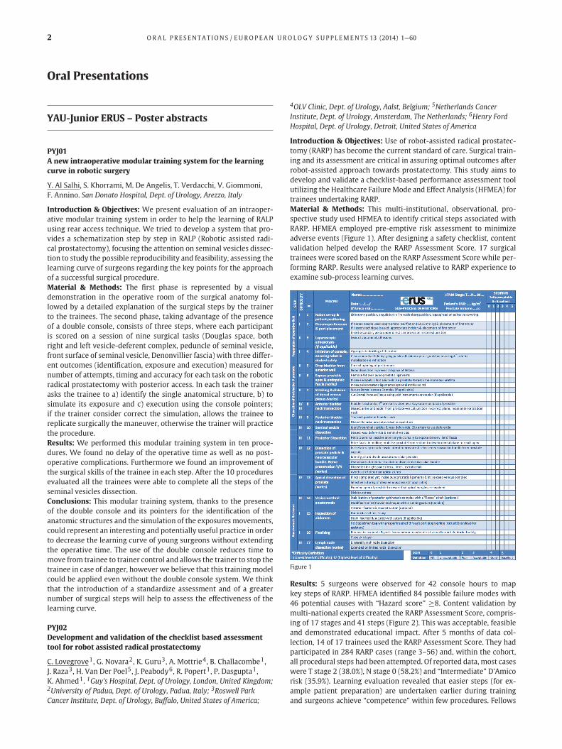
Introduction & Objectives: We present evaluation of an intraoper-ative modular training system in order to help the learning of RALPusing rear access technique. We tried to develop a system that pro-vides a schematization step by step in RALP (Robotic assisted radi-cal prostatectomy), focusing the attention on seminal vesicles dissec-tion to study the possible reproducibility and feasibility, assessing thelearning curve of surgeons regarding the key points for the approachof a successful surgical procedure.Material & Methods: The first phase is represented by a visualdemonstration in the operative room of the surgical anatomy fol-lowed by a detailed explanation of the surgical steps by the trainerto the trainees. The second phase, taking advantage of the presenceof a double console, consists of three steps, where each participantis scored on a session of nine surgical tasks (Douglas space, bothright and left vesicle-deferent complex, peduncle of seminal vesicle,front surface of seminal vesicle, Denonvillier fascia) with three differ-ent outcomes (identification, exposure and execution) measured fornumber of attempts, timing and accuracy for each task on the roboticradical prostatectomy with posterior access. In each task the trainerasks the trainee to a) identify the single anatomical structure, b) tosimulate its exposure and c) execution using the console pointers;if the trainer consider correct the simulation, allows the trainee toreplicate surgically the maneuver, otherwise the trainer will practicethe procedure.Results: We performed this modular training system in ten proce-dures. We found no delay of the operative time as well as no post-operative complications. Furthermore we found an improvement ofthe surgical skills of the trainee in each step. After the 10 proceduresevaluated all the trainees were able to complete all the steps of theseminal vesicles dissection.Conclusions: This modular training system, thanks to the presenceof the double console and its pointers for the identification of theanatomic structures and the simulation of the exposures movements,could represent an interesting and potentially useful practice in orderto decrease the learning curve of young surgeons without extendingthe operative time. The use of the double console reduces time tomove from trainee to trainer control and allows the trainer to stop thetrainee in case of danger, however we believe that this trainingmodelcould be applied even without the double console system. We thinkthat the introduction of a standardize assessment and of a greaternumber of surgical steps will help to assess the effectiveness of thelearning curve.
PYJ02Development and validation of the checklist based assessmenttool for robot assisted radical prostatectomy
C. Lovegrove1, G. Novara2, K. Guru3, A. Mottrie4, B. Challacombe1,J. Raza3, H. Van Der Poel5, J. Peabody6, R. Popert1, P. Dasgupta1,K. Ahmed1. 1Guy’s Hospital, Dept. of Urology, London, United Kingdom;2University of Padua, Dept. of Urology, Padua, Italy; 3Roswell ParkCancer Institute, Dept. of Urology, Buffalo, United States of America;
4OLV Clinic, Dept. of Urology, Aalst, Belgium; 5Netherlands CancerInstitute, Dept. of Urology, Amsterdam, The Netherlands; 6Henry FordHospital, Dept. of Urology, Detroit, United States of America
Introduction & Objectives: Use of robot-assisted radical prostatec-tomy (RARP) has become the current standard of care. Surgical train-ing and its assessment are critical in assuring optimal outcomes afterrobot-assisted approach towards prostatectomy. This study aims todevelop and validate a checklist-based performance assessment toolutilizing the Healthcare FailureMode and Effect Analysis (HFMEA) fortrainees undertaking RARP.Material & Methods: This multi-institutional, observational, pro-spective study used HFMEA to identify critical steps associated withRARP. HFMEA employed pre-emptive risk assessment to minimizeadverse events (Figure 1). After designing a safety checklist, contentvalidation helped develop the RARP Assessment Score. 17 surgicaltrainees were scored based on the RARP Assessment Score while per-forming RARP. Results were analysed relative to RARP experience toexamine sub-process learning curves.
Figure 1
Results: 5 surgeons were observed for 42 console hours to mapkey steps of RARP. HFMEA identified 84 possible failure modes with46 potential causes with “Hazard score” ≥8. Content validation bymulti-national experts created the RARP Assessment Score, compris-ing of 17 stages and 41 steps (Figure 2). This was acceptable, feasibleand demonstrated educational impact. After 5 months of data col-lection, 14 of 17 trainees used the RARP Assessment Score. They hadparticipated in 284 RARP cases (range 3–56) and, within the cohort,all procedural steps had been attempted. Of reported data, most caseswere T stage 2 (38.0%), N stage 0 (58.2%) and “Intermediate” D’Amicorisk (35.9%). Learning evaluation revealed that easier steps (for ex-ample patient preparation) are undertaken earlier during trainingand surgeons achieve “competence” within few procedures. Fellows

O R A L PRE S ENTAT IONS / E U RO P E AN U R O L O G Y SUP P L EMENT S 13 (2014) 1—60 3
Figure 2
failed to achieve competence in challenging critical steps, such asvesico-urethral anastomosis in the initial phase of the study.Conclusions: RARP Assessment Score based on HFMEA methodologyidentified critical hazardous steps specific to RARP and was used toassess and evaluate surgeons while performing RARP.
PYJ03Prevalence and predictors of thromboembolic events in patientsundergoing lymph node dissection during radical prostatectomy
S. Tyritzis1, A. Wallerstedt1, G. Steineck2, T. Nyberg2, J. Hugosson3,A. Bjartell4, U. Wilderäng2, T. Thorsteinsdottir5, S. Carlsson1,J. Stranne3, E. Haglind6, N.P. Wiklund1. 1Karolinska Institutet, Dept.of Molecular Medicine And Surgery, Section Of Urology, Stockholm,Sweden; 2Karolinska Institutet, Dept. of Clinical Cancer Epidemiology,Stockholm, Sweden; 3Sahlgrenska Academy at the University ofGothenburg, Dept. of Urology, Gothenburg, Sweden; 4Skåne UniversityHospital, Dept. of Urology, Lund, Sweden; 5University of Iceland, Dept.of Nursing, Reykjavik, Iceland; 6Sahlgrenska Academy at the Universityof Gothenburg, Dept. of Surgery, Gothenburg, Sweden
Introduction&Objectives: Lymph node dissection (LND) during rad-ical prostatectomy has been associated with increased risk of throm-boembolic events. We recorded the incidence and investigated thepredictors of deep venous thrombosis (DVT) and pulmonary em-bolism among other complications in patients undergoing or not un-dergoing LND during open (ORP) and robot-assisted laparoscopic rad-ical prostatectomy (RARP).Material & Methods: A total of 3544 patients were included be-tween 2008 and 2011. The cohort belongs to LAPPRO, a multi-institutional, prospective controlled clinical trial, conducted in Swe-den. Patient-completed questionnaires were used to gather data onadverse events. DVT and/or pulmonary embolism were the primaryoutcomes. Secondary outcomes were other types of 90-day compli-cations and re-admission causes. Logistic regression with forwardselection was used to identify the possible confounders for throm-boembolic events.Results: 547 (15.4%) patients underwent LND. Limited LND was per-formed in 266 patients (48.6%), while an extended LND was per-formed in 281 patients (51.4%). A robot-assisted extended LND wasperformed 3.2-fold more often compared to the open approach. Ir-respective of the type of LND, the robot-assisted LND had higherlymph node yields (21.5 vs. 18.3 nodes in the extended LND/9.2 vs. 6.1
nodes in the limited LND). Additionally, the extended LND resultedin higher overall detection of N1 patients compared to limited LND(11.1% vs. 1.2% in ORP/17.3% vs. 3.7% in RARP).LND exhibited an 8-fold and 6-fold higher risk of DVT and pulmonary embolism, respec-tively, compared to no-LND [RR 95% CI: 7.80 (3.51–17.32) and 6.29(2.11–18.73)]. A previous history of thrombosis, pT4 stage and Glea-son score ≥8 were identified as predictive factors for thromboem-bolic events. Low alcohol consumption was found to be protective.ORP with LND had a higher risk of DVT and/or pulmonary embolism[RR 95% CI: 11.22 (4.31–29.22) versus 6.61 (2.34–18.69) in RARP withLND]. In patients not undergoing LND, the open approach increased3.7-fold the risk for DVT or pulmonary embolism compared to therobot-assisted approach (95% CI 1.36–9.62). More wound, respira-tory, cardiovascular and neuromusculoskeletal complications wereencountered after LND compared to no-LND (14.6% vs. 6.3%). LNDwasalso associated with increased risk to undergo re-operation.Conclusions: We found that patients undergoing LND during radi-cal prostatectomy experienced more DVT and pulmonary embolismevents. Open surgery increased the risk for thrombosis more thanrobot-assisted surgery; this risk was significantly higher in patientsnot undergoing LND.
YAU-Junior ERUS – Video abstracts
VYJ01Robotic off-clamp zero ischemia partial nephrectomy in small,peripherally located, exophytic renal mass is safe and feasible
A.E. Canda1, O.U. Cakici2, K. Ener2, A.F. Atmaca1. 1Yildirim BeyazitUniversity, School of Medicine, Ankara Ataturk Training and ResearchHospital, Dept. of Urology, Ankara, Turkey; 2Ankara Ataturk Trainingand Research Hospital, Dept. of Urology, Ankara, Turkey
Introduction & Objectives: Renal hilar clamping decreases bleed-ing during performing robotic partial nephrectomy (RPN). However,particularly prolonged warm ischemia might have adverse effects onpostoperative renal function. Therefore, zero ischemia off-clamp RPNis increasingly being applied. We present a case of zero ischemia off-clamp RPN on a patient with a peripherally located small renal mass.Material &Methods: A 47 year-old male patient was evaluated com-plaining from recently onset headache and hypertension. He wasthen referred to our institution with the diagnosis of right adrenalmass of 5×4 cm size and concomitant 19×13 mm sized left renalmass with contrast enhancement on computerized tomography andmagnetic resonance imaging (MRI). Further work-up revealed a func-tioning right adrenal mass lesion with increased blood and urine cat-echolamines suggesting pheochromocytoma. We initially performeda right transperitoneal robotic adrenalectomy and pathology con-firmed benign pheochromocytoma. Threemonths afterwardswe per-formed a transperitoneal zero ischemia off-clamp RPN for left kidneymass lesion.Results: We used 5 abdominal ports including the 4th-robotic arm.Following early access to the renal pedicle, renal vein and renal artery

4 O R A L PRE S ENTAT IONS / E U RO P E AN U R O L O G Y SUP P L EMENT S 13 (2014) 1—60
were dissected and encircledwith vascular tapes. A laparoscopic bull-dog clamp was prepared to be used if needed. Then, renal mass wasisolated and without clamping the renal pedicle, zero ischemia RPNwas completed. We used a 3-0 V-Loc 180, 45 cm, 1/2 26 mm taperedneedle (Covidien™) to perform internal and external renorraphy. Inaddition, Lapra-Ty® clips (Ethicon Endo-Surgery)were used to anchorand secure each of a single strand of barbed suture on the renal cap-sule. No complication occurred. Intraoperative blood loss was 100 cc.Postoperative follow-up was uneventful and patient was dischargedon postoperative day-2. Histopathology demonstrated clear cell renalcell carcinoma, Fuhrman grade II, 17 mm in size with clear surgicalmargins. 6th-month abdominal MRI showed no recurrence or anyother lesion involving the right kidney. The patient stated that he isvery satisfiedwith the outcomes of the both robotic surgeries in addi-tion to the excellent abdominal cosmetic result that is also presentedat the end of the videowith an abdominal picture of the patient.Conclusions: Small, peripherally located and exophytic renal massesmight carry malignant tissue characteristics. These tumors might bedetected incidentally. Zero ischemia off-clamp RPN seems to be a safeand feasible surgical approach in the surgical management of thesetumors. This minimally invasive surgical approach has the advantageof avoiding complete renal ischemia and decrease in renal function.This approach might be particularly important in patients with un-derlying kidney disease.
VYJ02Image guided robotic partial cystectomy using flexiblecystoscopy and tile pro
A. Sridhar1, S. Madhavan2, S. Nathan1. 1University College LondonHospital NHS trust, Dept. of Urology, London, United Kingdom; 2TheLondon Clinic, Dept. of Urology, London, United Kingdom
Introduction & Objectives: Partial cystectomy is an option for soli-tary muscle invasive bladder lesions provided there is no concurrentCIS, and reasonable clearance can be obtained. Although the robotassisted laparoscopic approach for partial cystectomy is still experi-mental, its use in pelvic oncology as a whole has demonstrated lesserperioperative morbidity compared to the open approach. The chal-lenge for minimally invasive partial cystectomy has been adequatelocalization of the tumour as well as achieving adequate margins. Wedescribe a novel technique where we used concurrent flexible cys-toscopy during a robotic procedure for tumor localization as well asto guide excisional limits in order to achieve adequate clearance.Material & Methods: A 77 year old lady presented with bothersomelower urinary tract symptoms in the form of frequency, urgency andintermittent haematuria. A flexible cystoscopy showed a bladder le-sion at the dome of the bladder. She underwent rigid cystoscopy,transurethral bladder tumor resection and bladder mapping biopsiesunder general anesthetic, which showed a solitary muscle invasiveTransitional cell carcinoma without any evidence of CIS. Completionstaging with CT chest did not demonstrate any nodal or metastaticdisease. In view of a favorable histology, location of tumor and toavoid morbidity associated with a radical cystectomy, she was listedfor a Robotic partial cystectomy. For the procedure, the patient wasplaced in reverse Trendeleberg position. Standard 6 port configura-tion for cystectomy was used with a 12 mm supraumbilical cameraport, three 8 mm robotic ports, one 5 mm suction port and one 12mm air seal port. The bladder was identified and detached from theanterior abdominal wall. The pre peritoneal space was dissected lat-erally on either side down to the ureters in order to completely ex-pose the dome of the bladder. At this point an assistant inserted aflexible cystoscope and the tumour visualized. The cystoscopic im-age was projected onto the console using Tile Pro. The surgeon andassistant worked in tandem to identify excisional limits around thetumour, enough to provide a 2 cm limit of normal tissue. The surgeonindenting the water filled bladder with the robotic instrument, andconfirmed limits on the Tile Pro view. Once the limits were identified,
they were marked on the intraperitoneal surface using diathermy.The marked margins were incised to excise the lesion, and the speci-men bagged. Intraperitoneal spill of urine was kept to a minimum byminimal hydro distension during the flexible cystoscopy. A washoutwith sterile water was performed prior to closure of the bladder. Thebladder was closed in two layers using V-lock closure device.Conclusions: The above case report and attached video demonstratethe feasibility of Image guided Robotic partial cystectomy using flex-ible cystoscopy and Tile Pro. Using this technique we were able toachieve maximal bladder preservation (preservation of functionalstatus) with adequate clearance (as confirmed by histopathology).
VYJ03Robot-assisted radical nephroureterectomy in an ectopic pelvickidney
C. Wagner, A. Schütte, J. Witt. St. Antonius Hospital, Dept. of Urology,Gronau, Germany
Introduction & Objectives: During the past years, robot-assisted ap-proaches have become more and more common for many surgicalprocedures in urology, especially in the field of oncological surgery.However, the use of robot-assisted kidney surgery is still not thatcommon in comparison to prostate cancer surgery. Ectopic pelvickidneys are a quite rare condition, malignancies are even more rare.The use of robot-assisted surgery for radical nephroureterectomy inan ectopic pelvic kidney has (to our knowledge) so far not been de-scribed in a video.Material & Methods: We present a case of a 79 year old lady thatpresented with persistent gross hematuria and unspecific abdomi-nal pain. Ultrasound showed a left hydronephrotic ectopic pelvic kid-ney with a tumor approximately 7 cm in diameter. Because of sus-picious urothelial cells in the cytology from the kidney pelvis sam-ple, that was taken during RUPG and DJ Insertion, we decided to per-form a robot-assisted radical nephroureterectomy. CT scan showed aaberrant vascular supply from the contraleteral common iliac artery.The fact that the patient suffered from numerous medical conditions,such as IDDM, atrial fibrillaton with Warfarin Therapy, anemia (justto name a few), underlined the decision of choosing for a minimallyinvasive approach; furthermore, the robot-assisted technique allowsfor better visualisation and dexterity of the instruments.Results: Surgical Time was 155 minutes, EBL was 200 ml. Of note,intraoperatively vessels from the ipsilateal internal iliac artery wereencountered, that were not visible in the CT scan. The postoperativecourse was uneventful, final pathology showed a Clear Cell RCC pT1bG1 R0, with concomitant signs of chronic pyelonephritis due to hy-dronephrosis. Fortunately, no signs of TCC were found.Conclusions: In experienced hands, robot-assisted nephroureterec-tomy is feasible even in an ectopic pelvic kidney, however, due to thealtered anatomy, knowledge of vascular supply is mandatory. Intra-operative ultrasound can be helpful to identify additional vessels.
ERUS – Poster abstracts
PE01Improved short-term renal function after robot-assisted partialnephrectomy with selective arterial clamping – a matched pairanalysis for preoperative GFR
N.N. Harke1, F. Schiefelbein2, G. Schoen2. 1Prostate Center Northwest,St. Antonius-Hospital, Dept. of Urology, Pediatric Urology and UrologicOncology, Gronau, Germany; 2Missionsaerztliche Klinik, Dept. ofUrology, Wuerzburg, Germany
Introduction & Objectives: According to the international guidelinessmall renal masses should be treated with nephron-sparing surgery

O R A L PRE S ENTAT IONS / E U RO P E AN U R O L O G Y SUP P L EMENT S 13 (2014) 1—60 5
whenever technically feasible. Due to the growing possibilities espe-cially in robot-assisted partial nephrectomy (RPN) even large, hilaror intrarenal tumours can be removed. Nevertheless a short ischemiatime should be a major goal also in complex tumours. This can beachieved using super-selective clamping of the tumour feeding ves-sels based on fluorescence imaging.Material & Methods: 27 patients underwent RPN with selectiveclamping under the usage of indocyanine green (ICG). Intraopera-tively, the arterial branching can be tracked up to the hilum. After se-lective clamping of the tertiary or quaternary tumour feeding artery,administration of ICG confirms disrupted circulation and the tumourcan be excised. A matched-pair analysis for preoperative baselineeGFR (estimated glomerular filtration rate) could be employed for 23of these patients. Retrospective comparison was performed with 23matching partners out of a cohort of 167 patients where global is-chemia for renal artery control was used during RPN.Results: Comparing 23 patients in the selective clamping (1) and theglobal ischemia group (2), there were no significant differences inmean demographic data except for BMI (29.1 vs. 26.5 kg/m2, p=0.04).Clinical tumour size was 35.8 mm vs. 30.6 mm (p=0.153); in group 152% of the patients were found in the highest PADUA classificationrisk group vs. 26% in global ischemia group (p=0.189). Intraopera-tive parameter including operating time and blood loss did not dif-fer significantly in both groups. Similar results in postoperative dataincluding complications (n=3 in both groups) could be detected aswell as in histopathological findings with a malignant histotype in 12patients vs. 19 (p=0.17). Baseline kidney function as matching crite-ria was the same with a mean preoperative eGFR of 84.1 ml/min andcreatinine of 0.94 mg/dl in each group. In short-term renal functionoutcomes comparing baseline creatinine and eGFR with results be-fore discharge, a significantly reduced mean absolute eGFR loss (−5.9ml/min vs. −14.5 ml/min, p=0.033) and decreased change in creati-nine levels (+0.09 vs. +0.2 mg/dl, p=0.030) in the selective clampinggroup could be observed.Conclusions: Robotic partial nephrectomy with selective clampingof specific tumour feeding arterial branches using indocyanine greencan be performed safely even in complex tumour constellations. Theminimized ischemic trauma to the remaining parenchyma may leadto superior renal function preservation and significant reduced eGFRdecrease.
PE02Survival outcomes after robot-assisted radical cystectomy:Results from the international robotic cystectomy
J. Raza1, S. Dibaj1, G. Wilding1, E. Field1, J. Wing1, A. Hosseini2,A. Kibel3, A. Mottrie4, A. Weizer5, A. Wagner6, A. Hemal7,D. Scherr8, F. Schanne9, F. Gaboardi10, G. Wu11, J. Peabody12,J. Kaouk13, J. Palou Redorta14, K.H. Rha15, L. Richstone16,M.D. Balbay17, M. Menon12, M. Hayn18, M. Stöckle19, M. Woods20,P. Wiklund2, P. Dasgupta21, R. Pruthi20, R. Grubb3, M.S. Khan21,S. Siemer19, T. Wilson22, K. Guru1, International Robotic CystectomyConsortium. 1Roswell Park Cancer Inst., Dept. of Urology, Buffalo,United States of America; 2Karolinska Inst., Dept. of Urology, Stockholm,Sweden; 3Washington University in St Louis, Dept. of Urology, St. Louis,United States of America; 4Onze-Lieve-Vrouw Ziekenhuis, Dept. ofUrology, Aalst, Belgium; 5University of Michigan Health System, Dept. ofUrology, Detroit, United States of America; 6Beth Israel: HarvardMedical School, Dept. of Urology, Boston, United States of America;7Wake Forest University Baptist Medical Center, Dept. of Urology,Winston-Salem, United States of America; 8Weill Cornell MedicalCenter, Dept. of Urology, New York, United States of America; 9UrologicSurgical Associates of Delaware, Dept. of Urology, Delaware, UnitedStates of America; 10Luigi Sacco, Dept. of Urology, Milan, Italy;11University of Rochester Medical Center, Dept. of Urology, Rochester,United States of America; 12Henry Ford Health System, Dept. of Urology,Detroit, United States of America; 13Cleveland Clinic Foundation, Dept.
of Urology, Ohio, United States of America; 14Fundacio Puigvert, Dept. ofUrology, Barcelona, Spain; 15Yonsei Unv. Health System SeveranceHospital MIS/Robotic Center, Dept. of Urology, Seoul, South Korea; 16TheArthur Smith Institute for Urology, Dept. of Urology, New York, UnitedStates of America; 17Memorial Sisli Hospital, Dept. of Urology, Istanbul,Turkey; 18Maine Medical Center, Dept. of Urology, Portland, UnitedStates of America; 19University Clinics of Saarland, Dept. of Urology,Homburg, Germany; 20University of North Carolina, Dept. of Urology,Wilmington, United States of America; 21Guys Hospital, Dept. ofUrology, London, United Kingdom; 22City of Hope and BeckmanResearch Institute, Dept. of Urology, Duarte, United States of America
Introduction & Objectives: Data on the long-term oncological out-comes in patients undergoing robot-assisted radical cystectomy(RARC) is limited and mostly based on single institutional series. Wereport the clinical outcomes and associated prognostic factors in pa-tients who underwent RARC over 5 years ago.Material & Methods: In the IRCC database, 1586 patients underwentRARC for bladder cancer between 2004 and 2013. Only 315 patients(20%) had undergone surgery over five ormore years ago. Clinical andpathological data at the time of the latest follow-up were collected.Patients with<60 months of follow up were excluded from the anal-ysis. Recurrence free survival (RFS), cancer specific survival (CSS) andoverall survival (OS) were the outcomes of interest and plotted us-ing the Kaplan Meier Survival. Univariable and multivariable analy-ses were performed to identify factors associated with outcomes ofinterest.Results: 315 patients were included in the evaluation. 92 patientswere alive at the time of the analysis. Mean follow-up of patientsalive was 75 months. Mean age was 69 years, while 80% were men.48% patients had pathological non organ-confined disease. Soft tis-sue positive margins was 6%; median LNY was 16 with 29% positivelymph nodes. The median time to death and cancer specific deathwas 14 and 13 months respectively. The RFS, CSS and OS were 56%,61% and 39% respectively. On multivariable analysis, ASA, non- or-gan confined disease and LN positive disease were associated withpoorer RFS (HR1.43, 3.07 and 1.12 respectively), while age non-organconfined disease and positive lymph nodes affected both CSS and OS.Conclusions: The largest multi-institutional cohort of robot-assistedradical cystectomy present acceptable survival outcomes.
PE03Improvement of fluorescence-based sentinel node detectionduring a combined sentinel node biopsy, extended pelvic lymphnode dissection and robot-assisted radical prostatectomyprocedure
G. Kleinjan1, N. Van Den Berg1, O. Brouwer2, C. Acar3, E. Wit4,E. Vegt5, R. Valdés Olmos1, F. Van Leeuwen1, H. Van Der Poel4.1Leiden University Medical Centre, Dept. of Radiology, Leiden, TheNetherlands; 2Leiden University Medical Centre, Dept. of Urology,Leiden, The Netherlands; 3Acibadem University School of Medicine,Dept. of Urology, Istanbul, Turkey; 4The Netherlands Cancer Institute –Antoni Van Leeuwenhoek Hospital, Dept. of Urology, Amsterdam, TheNetherlands; 5The Netherlands Cancer Institute – Antoni VanLeeuwenhoek Hospital, Dept. of Nuclear Medicine, Amsterdam, TheNetherlands
Introduction & Objectives: Introduction and objectives: Robot-assisted radical prostatectomy (RARP) and sentinel node (SN) biopsycan be used to stage prostate cancer patients. Recently, the hy-brid tracer (ICG-99mTc-nanocolloid) was introduced for SN biopsy(van der Poel et al., Eur Urol 2011). Besides radioguidance, the flu-orescence signature of the hybrid tracer can be used to opticallyidentify the SN during surgery. To improve intraoperative opticalfluorescence-based fluorescence detection of SNs, we introduced adifferent tracer preparation and injection technique, and introduceda new fluorescence laparoscope.

6 O R A L PRE S ENTAT IONS / E U RO P E AN U R O L O G Y SUP P L EMENT S 13 (2014) 1—60
Material & Methods: Materials and Methods: 40 Patients with aBriganti nomogram-based risk >10% of lymph node (LN) metastaseswere included. After intraprostatic tracer injection, SN mapping wasperformed (lymphoscintigraphy and SPECT-CT). In groups 1 and 2 in-traoperatively SNs were pursued using a laparoscopic gamma probe(LGP) followed by fluorescence imaging (FI). In group 3 SNs were ini-tially located via FI. Compared to group 1, in groups 2 and 3 a newtracer-formulation was introduced with a reduced total injected vol-ume (2.0 mL vs. 3.2 mL), but increased particle concentration. Forgroups 1 and 2 the Tricam SL II + D-light C laparoscopic fluorescenceimaging (LFI) system was used. In group 3 the LFI-system was up-graded to an Image 1 HUB HD + D-light P system.Results: Results: SPECT-CT images were taken as a starting point forSN biopsy. In group 1 we identified amean of 50% of the SNs based ontheir fluorescent signature. Improvement of the hybrid tracer prepa-ration and the injection technique resulted in a visualization rate of64% in group 2. The introduction of the new fluorescence detectionsystem increased the visualization rate to 93% in group 3. A Kruskal-Wallis test showed a significant difference in the fluorescence visu-alization rate between the groups (p=0.004). Biochemical recurrenceoccurred in three patients in group 1 and 2 and in zero patients ingroup 3, suggesting that better nodal staging was achieved in group3 with the optimized SN identification protocol.Conclusions: Conclusion: With different steps of improvement, thein vivo fluorescence detection in SNs during RARP + SN biopsy couldbe significantly enhanced. Although we showed a high fluorescencevisualization rate in group 3, still SPECT-CT is used for the rough lo-calization of the SNs in prostate cancer patients.
ERUS – Video abstracts
VE01Robot-assisted ureteral reimplantation using the psoas hitchtechnique – important surgical steps
M. Musch, J.L. Hohenhorst, M. Janowski, A. Pailliart, M. Vanberg,D. Kroepfl. Kliniken Essen-Mitte, Dept. of Urology, Essen, Germany
Introduction & Objectives: We present the most important stepsof robot-assisted ureteral reimplantation using the psoas hitch tech-nique (RAURI). In addition, the results of our RAURI series are de-scribed.Material & Methods: Between October 2009 and May 2013 RAURIwas performed in 14 patients (in combination with a Boari flap in 5patients) with benign ormalign lesions of the distal part of the ureter.The patient and surgical data were collected prospectively. Follow-upwas accomplished using standardized questionnaires that were sentto the patients.Results: Resection of the distal ureter was necessary due to urothe-lial carcinoma in 5 patients, ureteric stricture caused by advancedprostate cancer in 2 patients, ureteric stricture caused by an inflam-matory conglomerate tumour of the adnexa in 1 patient, and iatro-genic ureteric stricture following gynecologic or urologic surgery in 6patients. RAURI can be divided into the following important steps,which are illustrated in our video: 1) mobilization of the bladder,2) preparation of the ureter, 3) fixation of the bladder on the psoasmuscle, 4) opening of the bladder, 5) formation of a submucosal tun-nel, 6) tension-free vesico-ureteric anastomosis and ureteric stent-ing, 7) closure of the bladder. The median operative duration (in-cluding docking and undocking of the robot) was 227.5 min. Therewere no intraoperative complications. Postoperative complicationsaccording to the Clavien-Dindo classification occurred in 10 patientswithin 90 days after surgery; 9 patients experienced minor compli-cations (grade I–II), and 1 patient a major complication (grade IIIb).The median postoperative length of stay was 8 days. All patients re-
mained asymptomatic during amedian follow-up of 13.42months. In1 patient an asymptomatic short stricture on the site of ureterovesicalanastomosis was identified radiographically and subsequently un-derwent successful endoscopic incision.Conclusions: RAURI showed good surgical results in our patient pop-ulation and thus seems to be a reliableminimal invasive alternativeto open surgery. We feel that following the general principles of opensurgery during important surgical steps of RAURI was a major factorfor these successful outcomes.
VE02Robot-assisted en-bloc radical cystectomy withnephroureterectomy and intracorporal urinary diversion byseven patients with muscle-invasive bladder cancer and upperurinary tract urothelial cell carcinoma. Single-center experience
J. Krude, A. Alexandrov, P. Lund, C. Hach, A. Goell. Alfried KruppKrankenhaus, Dept. of Urology, Essen, Germany
Introduction & Objectives: In this video we describe step-by-stepour technique of robot-assisted en-bloc radical cystectomy withnephroureterectomy and intracorporal urinary diversion in seven pa-tients with the diagnosis of muscle-invasive bladder cancer and syn-chronius upper urinary tract urothelial cell carcinoma. We also showimportant bench-marks of this procedure. This video shows in detailall steps of the procedure from port placement (seven-port transperi-toneal approach) over the ablative steps (bladder and kidney re-section and pelvic lymph node dissection) to the fully intracorporalrobot-assisted reconstruction.Material & Methods: After informed consent, we assessed 7 patientsundergoing an en-bloc radical cystectomy and nephroureterectomyat Alfried Krupp Hospital, Germany. All procedures were performedby one surgeon. We primarily assessed surgery and console duration,conversion rate, blood loss, intraoperative transfusion, and resectionstatus. Secondary endpoints were postoperative transfusions as wellas intra- and postoperative complications.Results: Three of the patients weremale. Median agewas 75.14 years(64–84), the mean BMI was 27.98 kg/m2 (22–34). Two of the patientswere ASA 2, four ASA 3 and one ASA 4. The indication for an en-blocradical cystectomy and nephroureterectomy was diagnosis of a si-multaneous transitional cell cancer of the lower and upper urinarytract in three patients. The other four patients had a muscle-invasivebladder cancer and a functionless kidney. Urinary diversion was re-constructed as intracorporal ureterocutaneostomy in six and as ex-tracorporal ileum conduit in one patient. Surgery lasted 296.85 min,thereof 199.71 min console time on average. There was no need ofa conversion to open surgery. The mean blood loss was 238 ml andnone of the patients required blood transfusions. Resection marginswere tumor-free in six cases. In the patient with positive resectionmargins the tumor stage was pT4a. Postoperative complications oc-curred in two of the patients: paralytic Ileus (Clavien-Dindo class 1(CDC 1) in one patient and port hernia (CDC 3b) in the other patient.Conclusions: Robot-assisted en-bloc radical cystectomy andnephroureterectomy could be performed with acceptable surgerydurations with a high intraoperatively and postoperatively safety.Further surgeries have to be performed to evaluate functional andoncologic results.
VE03Robotic flexible ureteroscopy, safety, effectivity and a earlyresults
J. Rassweiler1, R. Saglam2, A.S. Kabakci3. 1SLK-Kliniken HeilbronnGmbH, Dept. of Urology, Heilbronn, Germany; 2Medicana InternationalAnkara Hospital, Dept. of Urology, Ankara, Turkey; 3HacettepeUniversity, Dept. of Bioengineering, Ankara, Turkey
Introduction & Objectives: Flexible ureterorenoscopy (FURS) or ret-rograde intrarenal surgery are rapidly evolving in the last decade

O R A L PRE S ENTAT IONS / E U RO P E AN U R O L O G Y SUP P L EMENT S 13 (2014) 1—60 7
based on significant improvements of the armamentarium. This ap-proach has become a viable alternative to extracorporeal shock wavelithotripsy and percutaneous nephrolithotomy even for larger renalcalculi. Despite the progress in the design of the ureterorenoscopesand accessories for flexible ureterorenoscopy, the surgeon has to per-form this procedure in a standing position with suboptimal ergon-omy, which may not only result in orthopaedic complaints amongendourologists. It may be one of the reasons for imperfect perfor-mance of flexible ureterorenoscopy, resulting in the need of secondsession and frequent repair of the endoscopes.Material & Methods: The company ELMED from Turkey designedand developed a new robotic manipulator to remotely control all ofthe functions of the flexible ureterorenoscope. This robot may sig-nificantly reduce the learning curve of the procedure. The AvicennaRoboflex consists of surgeons console, and manipulator of flexibleureterorenoscope. After introducing the access sheath in a usualmethod, the robot with draped arm comes to the operation area. Theflexible ureterorenoscope is introduced in to the sheath manuallyand stabilized by the proximal support. Then, the hand piece of thescope is locked to the robotic arm. Finally, the laser fiber driver isattached and laser probe introduced. The entire docking manoeuvretakes less the 60 seconds. The right hand control enables deflection. Acentral wheel enables fine adjustment of the deflection useful duringsystematic dusting of the stone. The left joystick allows rotation aswell as advancing and retracting the scope. The speed of rotation andadvancement can be regulated at the screen of the console. The laser
fiber can be remotely moved forward and backward. Integrated wa-ter pump can be adjusted also to increase or decrease the flow ratefor optimal endoscopic vision.Results: In the meantime, we treatedmore than eighty kidney stonesby seven different urologists. An access sheath was used in the ma-jority of patients (n=72). In six girls and in cases of narrow ureters(n=3), the ureterorenoscope was inserted directly following rigidureteroscopy and placement of a guide-wire. Mean time to dock therobot was 59.6 (35–124) seconds, which decreased after 42 casesto a mean of 45.9 seconds. Mean fragmentation time was 46 (15–118) minutes corresponding to a mean fragmentation speed of 29.1(18–46) mm3 per minute increasing to 32.7 mm 3/min after 42cases. Mean console time was 53 (23–135) minutes and mean oper-ative time including placement of the access sheath and DJ-stent 74(40–182) minutes. Complete stone disintegration was accomplishedin 79 patients (96%). Based on plain-X-ray and ultrasound evalua-tion, after 3 months 65 patients (80%) were stone-free, whereas 16(20%) showed clinically insignificant residual fragments. Based ona questionnaire, we found a there is a significant difference whencomparing the ergonomics of classical versus robot-assisted flexibleureteroscopy (total score 31.3 vs. 5.6; p<0.01) and this is true for alldomains.Conclusions:We could demonstrate safe and efficacious applicationof the device with a short learning curve by seven experienced en-dourologists. Future studies have to evaluate the impact of the deviceon clinical outcome of FURS.

8 UNMOD E R A T E D PO S T E R PRE S ENTAT IONS / E U RO P E AN U R O L O G Y SUP P L EMENT S 13 (2014) 1—60
Unmoderated Poster Presentations
YAU-Junior ERUS – Poster abstracts
PYJ04Preliminary learning curve of robot-assisted laparoscopicintrafascial radical prostatectomy of a single surgeon withexperience in more than 1,000 laparoscopic urologic surgeries:A comparative analysis with recently performed 100 laparoscopicradical prostatectomies
H.K. Ha1, H.K. Seo2, S.D. Kim3, T.K. Jern1. 1Pusan National UniversityHospital, Dept. of Urology, Busan, South Korea; 2National Cancer Center,Dept. of Urology, Ilsan, South Korea; 3Dong A Medical Center, Dept. ofUrology, Busan, South Korea
Introduction & Objectives: Despite many analytic reports about thelearning curve transition from open to robot assisted intrafascial rad-ical prostatectomy (RARP), a few comparative results with laparo-scopic intrafascial radical prostatectomy (LRP) were reported. There-fore, we evaluated operative and postoperative outcomes of RARPwith simultaneously performed 100 LRPs.Material & Methods: A single surgeon (HKH) experienced in morethan 1,000 laparoscopic operations including 415 cases of radicalnephrectomy, 85 radical cystectomy, 369 radical prostatectomy and212 other urologic tumors from 2009. We evaluated the operative(operation time, intraoperative transfusion and complication, hos-pital stay, margin status, pathologic stage, Gleason score) and post-operative parameters (continence and erectile function) of initial 50cases of RARP without tutoring compared with recently performed100 LRPs.Results:Despite all LRPswere performed via 4-ports extra-peritonealapproach, RARPs were also done extra-peritoneally except first 5cases. Mean age (p=0.205) and prostate volume (p=0.520) of RARPand LRP were 64.6±6.9 yr and 66.3±6.0 yr, 34.2±14.9 cc and 32.3±9.9cc, respectively. Mean preoperative PSA (p=0.403) was 10.1±10.4 and12.7±15.9 in RARP and LRP. Mean Gleason score and percentage ofpathologically localized cancer were 7.0 and 7.1, 73.3% and 56.0% inRARP and LRP. Mean operation time (p=0.003) and length of hospitalstay (p=0.721) of RARP and LRP were 145.5±43.6 min (90–240) and118.1±39.1 min, 6.4±0.9 day (5–9) and 6.6±1.1 day (5–11). Mean op-eration time of RARP of 1st to 5th RARP, 6th to 10th, 11th to 15th,16th to 20th, after 20th was 177, 162, 158, 130 and 107 min, re-spectively. After 17 cases, the mean operation time of RARP showedsimilar to LRP (less than 2 hrs). There was no intraoperative trans-fusion and complications during both approaches. Positive surgicalmargin in localized cancer (p=0.825) was shown in 12% and 8% inRARP an LRP, respectively. At postoperative 6 weeks, sexual inter-course (p=0.308) was reported in 12% and 8%, pad-free continence(p=0.056) in 68% and 33% in patients with RARP and LRP, respec-tively. At postoperative 3 months, sexual intercourse (p=0.216) wasreported in 13.3% and 12%, pad-free continence (p<0.001) in 96.7%and 81% in patients with RARP and LRP, respectively.Conclusions: The previous large volume experience of LRPs mayshorten the learning curve of RARP in relation to oncologic and func-tional outcomes. Additionally, previous experiences of laparoscopymay help improved functional outcomes of RARP.
PYJ05Standardized reporting of the overall success of robot-assistedradical cystectomy with totally intracorporeal neobladderdiversion: The neobladder (nb) pentafecta concept
S. Tyritzis1, J. Collins1, P. Sooriakumaran2, T. Nyberg3, M. Jonsson1,C. Adding1, M. Schumacher1, N.P. Wiklund1, A. Hosseini1.1Karolinska Institutet, Dept. of Molecular Medicine And Surgery, Sectionof Urology, Stockholm, Sweden; 2University of Oxford, Dept. of SurgicalSciences Nuffield, London, United Kingdom; 3Karolinska Institutet, Dept.of Clinical Cancer Epidemiology, Stockholm, Sweden
Introduction & Objectives: To quantify the oncological and func-tional outcome of robot-assisted radical cystectomy (RARC) with to-tally intracorporeal neobladder, utilizing a PENTAFECTA tool.Material & Methods: Between 2003 and 2012 in a tertiary referralcenter, 70 patients underwent RARCwith totally intracorporeal mod-ified Studer ileal neobladder formation. The neobladder (nb) PENTA-FECTA was achieved when all 5 elements were achieved in an indi-vidual patient: 1. Negative surgical margins, 2. No recurrence andcancer-specific death at 24 months, 3. No 90-day complications ≥Clavien grade 3, 4. Daytime continence (0–1 pad/day) at 12 monthsand 5. Satisfactory sexual activity or potency at 12 months. Wealso estimated the (nb) QUADRAFECTA, by excluding from the (nb)PENTAFECTA definition the absence of complications.Results: 31 patients were included in the analysis. Median follow-up of the cohort was 30.3 months (IQR: 12.7–35.6). The (nb) PENTA-FECTA was achieved in 32.3% of the entire cohort and 42.9% of themale patients who underwent a nerve-sparing RARC with neoblad-der formation. The (nb) QUADRAFECTA rates were 54.8% and 66.7%,respectively. We recorded negative surgical margins in 29/31 pa-
Figure 1

UNMOD E R A T E D PO S T E R PRE S ENTAT IONS / E U RO P E AN U R O L O G Y SUP P L EMENT S 13 (2014) 1—60 9
tients (93.5%). Overall, 21 adverse events were recorded in 14 pa-tients (45.2%). Three Clavien grade I-II complications occurred in 3patients at 90 days (9.7%). Eighteen Clavien grade III-V complicationswere recorded in 11 patients (35.5%). The most common complica-tion was symptomatic lymphocele (4/31 patients). 19 male (90.5%)and 2 out of 3 (66.7%) female patients were continent (0–1 pad/day)at 12 months. 16 out of 21 (76.2%) of the nerve-spared patients werepotent with or without PDE5 medication at 12 months (Figure 1).Conclusions: In our series, which included learning curve cases, the(nb) PENTAFECTA rates were very encouraging. Postoperative com-plications is the measure that most affected the overall PENTAFECTArate. By using the (nb) PENTAFECTA, our aim is to create a standard-ized reference for reporting the patient’s global health and the opera-tion’s overall success. This combined outcomemeasuremay be usefulfor assessing service development and future regulation.
PYJ06Clinical short term effects of Silodosin and Naftopidil on lowerurinary tract symptoms after robot-assisted laparoscopic radicalprostatectomy
T. Kim1, S. Lee2, J. Bae3. 1Donga university, Dept. of Urology, Busan,South Korea; 2Hanyang University, Dept. of Urology, Seoul, SouthKorea; 3Korea University, Dept. of Urology, Seoul, South Korea
Introduction & Objectives: There are limited evidences on lowerurinary tract symptoms (LUTS) persisted after robot-assisted laparo-scopic radical prostatectomy. This study evaluated clinical effects ofα-blocker on LUTS in patients with robot-assisted laparoscopic radi-cal prostatectomy (RARP).Material & Methods: From January 2010, 120 patients were per-formed to RARP. 67 male patients of them received Silodosin 8mg/day or Naftopidil 75 mg/day for 8 weeks. The internationalprostate symptom score and quality of life index (QOL) were sur-veyed before and at the end of 8 week administration in all subjects.Results: Total international prostate symptom score (IPSS) associatedwith voiding symptoms and storages symptoms were significantlydecreased at 8 weeks compared with baseline (P<0.01). In a Silo-dosin group, voiding symptomwas improved and other improved thestorage symptoms. QOL index was significantly improved with bothgroups.Conclusions: LUTS detected in patients performed RARP weremarked improved with administration of Silodosin and Naftopidil.These symptoms could represent a novel target formedical treatmentby understanding of the voiding dysfunction physiology.
PYJ07Does preoperative storage symptom influence postoperativerecovery of storage symptoms after robot assisted laparoscopicradical prostatectomy?
S.H. Lee, W.S. Ham, B.H. Chung. Yonsei University College of Medicine,Dept. of Urology, Seoul, South Korea
Introduction & Objectives: Incontinence after radical prostatectomyaffects patient’s quality of life regardless of pathology results. Ob-structive symptoms could be relieved after robot assisted laparo-scopic radical prostatectomy (RALRP) due to the removal of theprostate. However, storage symptomsmay be not. This study investi-gated the effect of preoperative storage symptom on the recovery ofpostoperative storage symptom in patients who underwent RALRP.Material & Methods: From 2007 to 2013 who underwent RALRP in359 patients. Among them, 231 patients were available for analysis ofmedical records. There are 3 categories in the international prostatesymptoms scores (IPSS) that evaluate storage symptoms (IPSS ques-tion No. 2, 4, 7). Patients were divided into two groups: the storagesymptom group (storage symptom score ≥6) and the non storagesymptom group (storage symptom score <6). ’Worsening of blad-der irritation’ was defined as higher postoperative storage symptom
score than preoperative symptom score. “Recovery of bladder irrita-tion” was defined as same or lower storage symptom score than pre-operative symptom score.Results: The mean age of patients was 65.9±9.7 years (48–75 years)and mean PSA was 6.48±2.5 ng/dl (2.9–31.7). Before surgery, themean IPSS score was 11.5±2.6, the mean obstructive symptom scorewas 6.9±1.3, and the mean storage symptom score was 4.6±0.8. Ofthe 231 patients, 77 (33.3%) belonged to the storage storage symp-tom group and 154 (66.7%) belonged to the non-storage symptomgroup. Worsening of bladder irritation was significantly higher in thestorage symptom group after RALRP (79% vs. 55%, p=0.02). The periodof recovery to the preoperative status was also significantly longer inthe storage symptom group (11.2 months vs. 8.3 months, p<0.01).Conclusions: Storage symptoms after RALRPweremore severe in pa-tients with bladder irritation preoperatively. The recovery of storagebladder symptoms after RALRP was also longer than that of non stor-age symptom group. Our results suggest that preoperative storagebladder symptom score may be used as a predicting factor of postop-erative storage symptoms.
PYJ08Does anterior periprostatic fat tissue removed during robotassisted laparoscopic radical prostatectomy contain any lymphnodes?
A.F. Atmaca1, A.E. Canda1, M. Keske2, M.E. Arslan2, D. Kamaci2,E. Alkan3, M.D. Balbay3. 1Yildirim Beyazit University, School ofMedicine, Ankara Ataturk Training and Research Hospital, Dept. ofUrology, Ankara, Turkey; 2Ankara Ataturk Training and ResearchHospital, Dept. of Urology, Ankara, Turkey; 3Memorial Sisli Hospital,Dept. of Urology, Istanbul, Turkey
Introduction & Objectives:We investigated if anterior periprostaticfat tissue removed during robot assisted laparoscopic radical prosta-tectomy (RARP) contain any lymph nodes (LNs).Material & Methods: Anterior periprostatic fat tissues removed dur-ing RARP in 88 patients were sent separately for histopathologic eval-uation. Correlation with postoperative pathologic stage was made.Patients with a history of previous prostate or bladder surgery andradiation therapy were excluded.Results: Mean patient age, serum prostate specific antigen (PSA),prostate weight and body mass index (BMI) were 62.3±5.7 years(range, 46–74), 9.2±6.4 ng/mL (range, 0.3–37), 57.9±28.0 grams(range, 11–180) and 26.8±1.7 kg/m2 (range, 20–30), respectively.Overall, LNs in anterior periprostatic fat tissues were detected in8 (9.1%) patients with a mean LN yield of 1.4±0.7 LNs (range, 1–3). Of those, no metastatic LN was detected. Postoperative patho-logic stages included pT0 (n=1, 1.1%), pT2a (n=19, 21.6%), pT2b (n=11,12.5%), pT2c (n=37, 42.0%), pT3a (n=15, 17.0%) and pT3b (n=5, 5.7%).Overall, positive surgical margins (PSM) were detected in 24 (27.3%)patients. Of the patients with pT2 disease (n=67), PSM rate was 13.4%(n=9). Of the patients with pT3 disease (n=20), PSM rate was 70.0%(n=14). Of the patients with pT2a (n=19), pT2c (n=37) and pT3a(n=15) disease, LNs in anterior periprostatic fat tissues were detectedin 1 (5.3%), 6 (16.2%) and 1 (6.7%) patient in each group, respec-

10 UNMOD E R A T E D PO S T E R PRE S ENTAT IONS / E U RO P E AN U R O L O G Y SUP P L EMENT S 13 (2014) 1—60
tively. No LN was detected in anterior periprostatic fat tissue in pa-tients with pT0 (n=1), pT2b (n=11) and pT3b (n=5) disease. Of thepatients with postoperative Gleason score (GS) 3+3=6 (n=41) and GS4+3=7 (n=12) diseases, LNs in anterior periprostatic fat tissues weredetected in 7 (17.1%) and 1 (8.3%) patient, respectively. No LN wasdetected in anterior periprostatic fat tissue in patients with postop-erative GS of 3+4=7 (n=23), 4+4=8 (n=6), 5+3=8 (n=1), 3+5=8 (n=1)and 4+5=9 (n=4). We did not have any patient with postoperative GSof 5+4=9 or 5+5=10. Patients were classified according to BMI (kg/m2)as follows: 30 obese (n=1). Of the patients, LNs in anterior prostaticfat tissues were detected in 0 (0%), 2 (25%), 5 (62.5%) and 1 (12.5%)patients of underweight, optimal weight, overweight and obese pa-tients due to BMI, respectively.Conclusions: LNs might be present in the anterior periprostatic fattissue and in our series, LNs was detected in almost 10% of the pa-tients. Therefore, this fat should be removed and should be sent forpathologic evaluation, not pushed back during RARP. Presence ofmetastatic LNs might have an impact on the oncologic outcomes ofthe patients that warrants further research.
PYJ09Conversion from robotic to open surgery: Translating knowledgefrommotor racing to healthcare
F. Zattoni, A. Guttilla, A. Crestani, F. Cattaneo, A. Iannetti, L. Angelini,A. De Gobbi, F. Dal Moro, F. Zattoni. University of Padua, Dept. ofUrology, Padua, Italy
Introduction & Objectives: In cases of emergency, the conversionfrom robotic to an open surgery is not trivial and requires an ade-quate training of all the members of the team. The aim of our workwas to investigate the possible problems of a robotic conversion incases of emergency, share a job distribution flowchart and exercisedoctors and nurses, as a team of Formula One, to perform as quicklyas possible a procedure which can save patient’s life.Material & Methods: By performing an analogue in proactive plan-ning, active management and post hoc learning in the motor racingpits stop we examined our robotic conversion to open surgery in caseof emergency. In the last 2months we simulated several emergenciesduring a robotic surgery that required a conversion to open surgery.An ordinary atmosphere of a robotic room was recreated. A humandummywas placed on the operating table and a Da Vinci robot lockedto it. All surgeons, anaesthesiologists, nurses and paramedical staffwere involved. Each type of professional was recognized through adifferent cap to be able to trace the movements of each person dur-ing the conversion. All the simulations were timed and filmed. Thevideos at the and were analysed in order to understand the problemsand complications during the procedure.Results: After several simulations our average conversion time was93±25 (57–183) seconds. At the and of our training we reduced theconversion time by a mean of 45 seconds. In this simulations wefound different kind of problems. We tried to improve these in thefollowing ways: 1) proactive learning with briefings before surgeryand checklists to prevent errors; 2) at the beginning of the surgeryestablish who will give the orders in case of need; 3) clearly definedtasks assigned to every member of the team; 4) give clear messagewith a key code; 5) find the correct placement of instrument, tableand equipment in the operating room for more safe ad quick move-ments; 6) prepare gloves and scrubs for the chief surgeon on the in-strument table from the beginning of robotic surgery; 7) do not re-move the trocars during conversion; pneumoperitoneum can reducethe bleeding; 8) make sure the robot is well disconnected to the tro-car and distant from the body of the patient; sudden movements ofthe da Vinci patient-side cart can cause serious problems to the pa-tient’s limbs; 9) do not take any sacks outside the operative room.Wepropose our flowchart for the jobs distribution in the team.Conclusions: We proposed a generic conversion model that can beapplied to each robotic group. Though it is impossible to to establish
a universally accepted conversion model because each robotic groupis composed of different person with different habits and differentways of working. The geometry of the operating room is very impor-tant and an accurate preliminary study for spaces is necessary. Thelessons from motor racing can be applied to healthcare for proactiveplanning, active management and post hoc learning. Standardisationof working practices, interpersonal communication, consistency andcontinuous development is fundamental for success. The applicationof these concepts would result in improvements in the quality andsafety in the conversion process from robotic surgery to open. Wehope that this study will be the beginning of courses and tests foreach robotic team in order to improve, in cases of emergency, in theconversion from robotic to open surgery.
PYJ10Robotic cystectomy is associated with an early transition tointracorporeal urinary diversion which is sustained for patientsreceiving orthotopic neobladder
A. Sridhar, E. Zacharakis, G. Basnett, P. Cathcart, S. Nathan, J. Hines,T. Briggs, J. Kelly. University College London Hospital NHS trust, Dept. ofUrology, London, United Kingdom
Introduction & Objectives: Intracorporeal reconstruction followingrobotic cystectomy is feasible but technically challenging. In thisstudy we report the transition to intracorporeal urinary diversionwithin an established robotic surgery program.Material & Methods: Demographics, performance status, intraoper-ative, post operative events and cancer follow up data were main-tained prospectively for all patients undergoing cystectomy.Results: 79 patients underwent robotic cystectomy and urinary di-version over 36 months by two surgeons. Of these 28 had a neoblad-der (7 extracorporeal and 21 intracorporeal) and 51 had ileal con-duits (21 extracorporeal, 30 intracorporeal). Cancer and patient char-acteristics were similar between extracorporeal and intracorporealgroups. Initially reconstruction was performed extracorporeal withmigration to intracorporeal diversionwithin 5 cases for each surgeon.A preference for intracorporeal reconstruction was maintained for allpatients receiving orthotopic neobladder and 75% receiving conduit.Outcomes were similar regardless of approach with median opera-tive time for conduit being 270minutes and for neobladder being 360minutes. 8/79 patients required blood transfusion. Themedian lengthof stay was 10 days. Complications included urosepsis/pelvic collec-tion (14/79), benign stricture (2/79), malignant stricture (4/79), ileus8/79 and urine leak (5/79). One patient died as a result of MI within30 days of surgery. 11 patients had extravesical disease on post op-erative pathology. At median followup of 18 months, there were 5cancer deaths within 1 year of surgery and 4 recurrences.Conclusions: The transition to intracorporeal urinary diversion is anearly event for experienced surgeons performing robotic cystectomy.The intracorporeal approach is maintained for orthotopic diversionand likely to be adopted as the standard approach for patients elect-ing to have robotic cystectomy and continent diversion.
PYJ11Robotic-assisted bladder diverticulectomy: Challenges andsolutions
R.M. Oliveira Soares, G. Athanasiadis, K. Patil. Royal Surrey CountyHospital, Dept. of Urology, Guildford, United Kingdom
Introduction & Objectives: Bladder diverticulae are herniations ofthe bladder that can be congenital but are more often secondary tooutflow obstruction. Even after resolution of the obstruction, diver-ticulae may cause incomplete emptying of the bladder and, conse-quently, infections, stones and even cancer. Excision of the diverticu-lum significantly improves their complaints.Material & Methods: We report the case of a 60-year old man that,despite a transurethral resection of the prostate, was needingmanual

UNMOD E R A T E D PO S T E R PRE S ENTAT IONS / E U RO P E AN U R O L O G Y SUP P L EMENT S 13 (2014) 1—60 11
pressure to empty his 10-cm bladder diverticulum. We describe thetechnique we used for his robotic-assisted bladder diverticulectomywith concomitant cystoscopy, highlighting the most difficult steps ofthe surgery and discussing the different tricks that canmake it easier.Results: The procedure was uneventful and the patient was dis-charged after 3 days with no significant post-void residual aftercatheter removal.Conclusions: Robotic-assisted bladder diverticulectomy is a safe andeffective procedure, provided we keep in mind the importance ofcareful identification of the diverticulum and surrounding structures.
PYJ12Comparison in perioperative results between the use of avalveless trocar system and the standard insufflation techniqueduring robot-assisted radical prostatectomy
P. Salwa, M. Addali, N.N. Harke, C. Wagner, A. Schütte, J. Witt. St.Antonius-Hospital, Academic Teaching Hospital Affiliated to theUniversity Münster, Dept. of Urology, Gronau, Germany
Introduction & Objectives: The primary aim of the study was to in-vestigate if the use of the valveless AirSeal™ insufflation system im-pairs the lung function of patients less than a conventional system inpatients undergoing robot-assisted radical prostatectomy (RARP). Asecond purpose of the study was to investigate the influence of thedifferent gas insufflator systems (AirSeal™ and conventional [StrykerPneumosure 45L]) on early perioperative results.Material & Methods: A dual center prospective randomized inter-ventional clinical trial was performed on a cohort of 200 patients whounderwent RARP from January to April 2014. In our institution werandomized 100 patients (50 each arm). We are reporting about ourperioperative surgical results regarding average skin-skin time, cal-culated blood loss, CO2 volume used, shoulder pain, time to first fla-tus, bowel peristaltic on the 1st postoperative day and positive surgi-cal margins rates.Results: In our cohort we did not observe a significant difference inany of the measured perioperative parameter (Table 1), but found atendency of a higher CO2 consumption, a longer skin-skin time and ahigher rate of shoulder pain in the Airseal™ group.
Table 1
Airseal™ Conventional insufflation
Age (years) 62.0 61.7Weight (kg) 91.8 87.2Height (cm) 180.5 180.0Median iPSA-Value (ng/ml) 7.8 8.3Average skin-skin time (min) 205.2 190.3 (p=0.11)Calculated blood loss (ml) 765.1 739,9 (p=0.74)CO2 used (ml) 726.4 646 (p=0.083)Shoulder pain presence (%) 6% 2% (p=0.289)Time to first flatus (h) 23.4 23.9 (p=0.71)Bowel peristaltic 1st post-Op day (%) 94% 94%Positive surgical margin (%) 9% 4% (p=0.35)
Conclusions: Using the valveless Airseal™ insufflation system com-pared with conventional insufflation showed a reduced CO2 use anda shorter average skin-skin time in one study [1]. Inconsistently withthese data we did not found significant differences concerning thisparameter. Especially regarding the CO2 use there was a tendency fora higher CO2 consumption in the Airseal™ group. A possible expla-nation for these results could be the characteristic of our center (highvolume, >1100 RARP’s per year, high standardized process, special-ized teammembers). Therefore the possible advantages of the valve-less system (less pressure undulation, fewer lenses fogging with thereduced need for lens cleaning) may be without relevance in our set-ting.1. Use of the valveless trocar system reduces carbon dioxide ab-sorption during laparoscopy when compared with standard trocars.Reference:[1] Herati AS, Andonian S, Rais-Bahrami S, Atalla MA, Srinivasan AK,
Richstone L, Kavoussi LR. Urology. 2011 May;77(5):1126–32.
PYJ13Robot-assisted adrenalectomy – single-institutional results
K.A. Probst, M. Janssen, C.-H. Ohlmann, S. Siemer, M. Stöckle.Saarland University Medical Center, Dept. of Urology, Homburg/Saar,Germany
Introduction & Objectives: Minimal-invasive resection of adrenalmasses becomes more accessible to urologists with the increasingexperience in robot assisted surgery of renal anomalies. Typical Indi-cations include the hormone-active and hormone-inactive adenomaas well as metastases of other tumour entities and the rare primaryadrenal malignancies. We present the operative and oncological re-sults of all RALA (Robot-assisted laparoscopic adrenalectomy) proce-dures at our institution since the introduction of robotic surgery.Material & Methods: We analyzed perioperative data of 43 patientswho underwent RALA at our institution between May 2008 and May2014. Evaluation focused on operative performance (total time andblood loss) as well as the rate of intraoperative complications and thefrequency of malignant histopathological findings. We also lookedat the rate of conversion and blood transfusion and evaluated thechanges in operating time between the first half (2008–2011) andsecond half (2012–2014) of procedures.Results: Median blood loss was 50 ml with a mean overall operat-ing time of 137 min. We evaluated n=19 procedures between 2008and 2011 (set 1) and n=24 procedures between 2012 and 2014 (set2). Comparing the changes between the two sets we found a medianoperating time of 148 min for set 1 and a median of 127.5 min forset 2. Mean blood loss changed from 122 ml (set 1) to 67 ml (set 2).The mean tumour size was 5.32 cm (range 1–10 cm) with an averageweight of 34.0 g (range 2.5–210 g). Intraoperative complications oc-curred in n=9 cases of which n=4 were classified as Clavien 1, n=3 asClavien 2 and n=2 as Clavien 3. In 2 cases conversion to open surgerywas necessary due to excessive bleeding and large tumour size re-spectively. This leads to a conversion rate of 4.6%; however both con-versions occured during the first 15% of procedures. Blood transfusionwas necessary in only 1 case early in the series where the tumourwasadherent to the spleen. We identified n=20 hormone-active tumours(mainly adrenal cortical adenoma), n=7 inactive tumours and n=16malignancies (predominantly metastases of other tumour entities).There were 2 cases of adrenal cortical carcinoma as well as 1 case ofmalignant pheochromocytoma.Conclusions: Our data supports robotic surgery as a reliable and safeapproach in the treatment of adrenal gland masses. Conversion aswell as blood transfusions were more likely to occur in the earlierstage of our series. Operating time and blood loss decreased overtime, even without taking the introduction of new surgeons into ac-count. Similar to the conditions for robotic renal procedures limita-tions arise from intraoperative complications like bleeding as well asvery large masses, but otherwise RALA is a feasible treatment optionin patients who qualify for laparoscopic surgery.
PYJ14Perioperative, oncological and functional outcomes andcomplications of robot-assisted radical prostatectomy andextended pelvic lymph node dissection after prior abdominalsurgery: A single surgeon’s experience
G.B. Di Pierro1, P. Grande2, H. Danuser3, A. Mattei3. 1LuzernerKantosspital - Sapienza University of Rome, Dept. of Urology, Lucerne,Switzerland; 2Sapienza University of Rome, Dept. of Urology, Rome,Italy; 3Luzerner Kantosspital, Dept. of Urology, Lucerne, Switzerland
Introduction & Objectives: To assess the impact of prior abdominalsurgery on perioperative, oncological and functional outcomes andcomplications in patients undergoing transperitoneal robot-assistedradical prostatectomy (RARP) and extended pelvic lymph node dis-section (ePLND).Material & Methods: From November 2008 to October 2012, a total
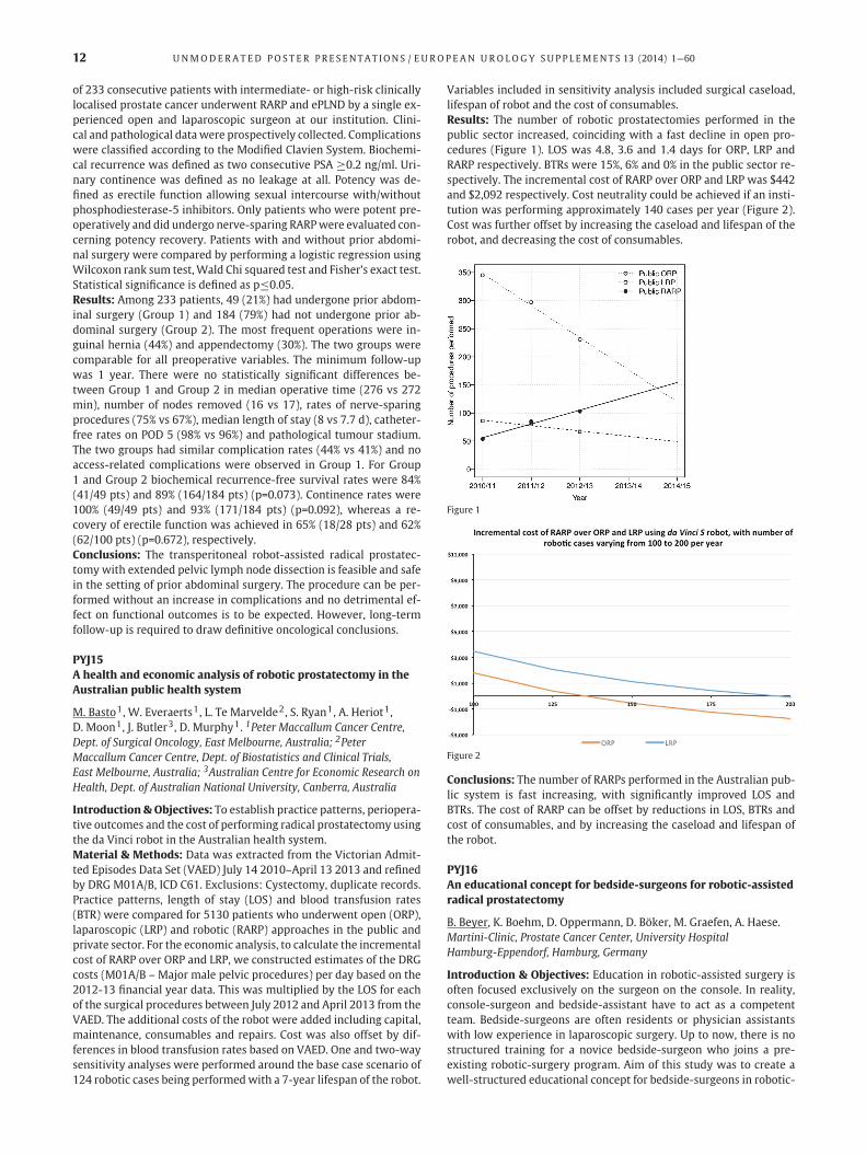
12 UNMOD E R A T E D PO S T E R PRE S ENTAT IONS / E U RO P E AN U R O L O G Y SUP P L EMENT S 13 (2014) 1—60
of 233 consecutive patients with intermediate- or high-risk clinicallylocalised prostate cancer underwent RARP and ePLND by a single ex-perienced open and laparoscopic surgeon at our institution. Clini-cal and pathological datawere prospectively collected. Complicationswere classified according to the Modified Clavien System. Biochemi-cal recurrence was defined as two consecutive PSA ≥0.2 ng/ml. Uri-nary continence was defined as no leakage at all. Potency was de-fined as erectile function allowing sexual intercourse with/withoutphosphodiesterase-5 inhibitors. Only patients who were potent pre-operatively and did undergo nerve-sparing RARPwere evaluated con-cerning potency recovery. Patients with and without prior abdomi-nal surgery were compared by performing a logistic regression usingWilcoxon rank sum test,Wald Chi squared test and Fisher’s exact test.Statistical significance is defined as p≤0.05.Results: Among 233 patients, 49 (21%) had undergone prior abdom-inal surgery (Group 1) and 184 (79%) had not undergone prior ab-dominal surgery (Group 2). The most frequent operations were in-guinal hernia (44%) and appendectomy (30%). The two groups werecomparable for all preoperative variables. The minimum follow-upwas 1 year. There were no statistically significant differences be-tween Group 1 and Group 2 in median operative time (276 vs 272min), number of nodes removed (16 vs 17), rates of nerve-sparingprocedures (75% vs 67%), median length of stay (8 vs 7.7 d), catheter-free rates on POD 5 (98% vs 96%) and pathological tumour stadium.The two groups had similar complication rates (44% vs 41%) and noaccess-related complications were observed in Group 1. For Group1 and Group 2 biochemical recurrence-free survival rates were 84%(41/49 pts) and 89% (164/184 pts) (p=0.073). Continence rates were100% (49/49 pts) and 93% (171/184 pts) (p=0.092), whereas a re-covery of erectile function was achieved in 65% (18/28 pts) and 62%(62/100 pts) (p=0.672), respectively.Conclusions: The transperitoneal robot-assisted radical prostatec-tomy with extended pelvic lymph node dissection is feasible and safein the setting of prior abdominal surgery. The procedure can be per-formed without an increase in complications and no detrimental ef-fect on functional outcomes is to be expected. However, long-termfollow-up is required to draw definitive oncological conclusions.
PYJ15A health and economic analysis of robotic prostatectomy in theAustralian public health system
M. Basto1, W. Everaerts1, L. Te Marvelde2, S. Ryan1, A. Heriot1,D. Moon1, J. Butler3, D. Murphy1. 1Peter Maccallum Cancer Centre,Dept. of Surgical Oncology, East Melbourne, Australia; 2PeterMaccallum Cancer Centre, Dept. of Biostatistics and Clinical Trials,East Melbourne, Australia; 3Australian Centre for Economic Research onHealth, Dept. of Australian National University, Canberra, Australia
Introduction &Objectives: To establish practice patterns, periopera-tive outcomes and the cost of performing radical prostatectomy usingthe da Vinci robot in the Australian health system.Material & Methods: Data was extracted from the Victorian Admit-ted Episodes Data Set (VAED) July 14 2010–April 13 2013 and refinedby DRG M01A/B, ICD C61. Exclusions: Cystectomy, duplicate records.Practice patterns, length of stay (LOS) and blood transfusion rates(BTR) were compared for 5130 patients who underwent open (ORP),laparoscopic (LRP) and robotic (RARP) approaches in the public andprivate sector. For the economic analysis, to calculate the incrementalcost of RARP over ORP and LRP, we constructed estimates of the DRGcosts (M01A/B – Major male pelvic procedures) per day based on the2012-13 financial year data. This was multiplied by the LOS for eachof the surgical procedures between July 2012 and April 2013 from theVAED. The additional costs of the robot were added including capital,maintenance, consumables and repairs. Cost was also offset by dif-ferences in blood transfusion rates based on VAED. One and two-waysensitivity analyses were performed around the base case scenario of124 robotic cases being performedwith a 7-year lifespan of the robot.
Variables included in sensitivity analysis included surgical caseload,lifespan of robot and the cost of consumables.Results: The number of robotic prostatectomies performed in thepublic sector increased, coinciding with a fast decline in open pro-cedures (Figure 1). LOS was 4.8, 3.6 and 1.4 days for ORP, LRP andRARP respectively. BTRs were 15%, 6% and 0% in the public sector re-spectively. The incremental cost of RARP over ORP and LRP was $442and $2,092 respectively. Cost neutrality could be achieved if an insti-tution was performing approximately 140 cases per year (Figure 2).Cost was further offset by increasing the caseload and lifespan of therobot, and decreasing the cost of consumables.
Figure 1
Figure 2
Conclusions: The number of RARPs performed in the Australian pub-lic system is fast increasing, with significantly improved LOS andBTRs. The cost of RARP can be offset by reductions in LOS, BTRs andcost of consumables, and by increasing the caseload and lifespan ofthe robot.
PYJ16An educational concept for bedside-surgeons for robotic-assistedradical prostatectomy
B. Beyer, K. Boehm, D. Oppermann, D. Böker, M. Graefen, A. Haese.Martini-Clinic, Prostate Cancer Center, University HospitalHamburg-Eppendorf, Hamburg, Germany
Introduction & Objectives: Education in robotic-assisted surgery isoften focused exclusively on the surgeon on the console. In reality,console-surgeon and bedside-assistant have to act as a competentteam. Bedside-surgeons are often residents or physician assistantswith low experience in laparoscopic surgery. Up to now, there is nostructured training for a novice bedside-surgeon who joins a pre-existing robotic-surgery program. Aim of this study was to create awell-structured educational concept for bedside-surgeons in robotic-

UNMOD E R A T E D PO S T E R PRE S ENTAT IONS / E U RO P E AN U R O L O G Y SUP P L EMENT S 13 (2014) 1—60 13
assisted radical prostatectomy (RARP), which is adaptable by othersurgical subspecialties.Material & Methods: An education concept for novice bedside-surgeons was developed and introduced to our surgical team. There-fore an expert panel, which included experienced console surgeons,bedside-surgeons, and surgical nurses was formed. The step stones ofthe operation were defined and the requirements for assisting werediscussed. In the OR the educational process was conducted by an ex-perienced bedside-surgeon and surgical nurse.Results:Our concept contains 6 phases applicable in one week. Phase0: “Know your environment”: Presentation of the bedside-surgeonthe day prior first surgery. Introduction to all the components ofthe robotic-surgical system and explanation of the principles of pa-tient’s positioning. Phase 1: “Know your operation”: Active observa-tion of RARP performed by an experienced bedside-surgeon, Learn-ing of instruments and their use in surgery. Phase 2: “Orientation inthe body”: Camera operating during trocar placement, suction and re-traction with laparoscopic instruments during the first steps of RARP.Phase 3: “Applying of your basic skills”: Trocar placement and apply-ing laparoscopic skills. Assisting the operation as long as the noviceassistant and surgeon feel comfortable. Phase 4: “Deepen your keyskills”: Assisting the key steps of RARP like seminal vesicle prepara-tion and nerve-sparing. Phase 5: “Act independently”: Assisting thewhole operation. Intervention of the experienced bedside surgeononly in case of emergency.Conclusions: This work presents the first concept exclusively dedi-cated to novice bedside-surgeons. It provides an easy access to basiclaparoscopic skills and sufficient assistance to robotic-assisted radicalprostatectomy. Further research is needed to improve and validateour bedside-surgeon education program.
PYJ17Rectal injury during robot-assisted radical prostatectomy (RARP)with and without bowel preparation: Perioperative results
M. Godes, N.N. Harke, M. Addali, A. Schütte, C. Wagner, J. Witt.Prostate Center Northwest, St. Antonius-Hospital, Department ofUrology, Pediatric Urology and Urologic Oncology, Gronau, Germany
Introduction & Objectives: Fast track in surgery is a multimodal,multidisciplinary perioperative concept to optimize the periopera-tive management with potentially faster recovery after a surgicaltreatment. In this concept, a bowel preparation (BP) is not recom-mended as a routine prior to elective surgery. The aim of this studywas to evaluate the rate of rectum injury and perioperative results ofRARP with and without BP.Material & Methods: Data of 5543 RARP patients were analyzed ret-rospectively including 2651 patients without BP and 2892 with pre-operative BP with emphasis on patients with an intraoperatively de-tected rectal injury.Results: A total of 11 (0.2%) rectal injury cases were identified dur-ing RARP, in 5 cases with BP and in 6 cases without BP. The mean agewas 63.6 and 69.3 years, BMI 25.7 and 24.8 kg/m2, initial PSA was
Abstract PYJ17 – Table 1. Perioperative results of rectal injury during RARP
Pt No. Bowel preparation Cumulative surgical Age BMI Prostate weight Gleason Organ-confined disease PSM Nerve-sparing Cath. time(no/yes) experience (years) (kg/m2) (g) score (no/yes) (no/yes) (no/yes) (days)
Clinic Surgeon
1 yes 29 29 49 24.7 40 3+5 no yes yes 82 yes 369 265 57 27.7 61 3+3 yes no yes 63 yes 1160 80 74 25.8 130 3+4 yes no no 54 yes 2389 6 70 26.4 85 3+3 yes no yes 55 yes 2886 306 68 24 106 4+3 no no no 56 no 3015 324 57 24.8 58 3+3 yes no yes 57 no 3325 21 72 23.9 30 4+3 no no yes 58 no 3370 49 69 22.5 35 4+3 yes no yes 59 no 3609 1970 72 24.2 113 4+4 yes yes yes 510 no 4089 250 69 29.4 36 5+4 no yes no 511 no 4516 2347 77 23.9 67 4+4 no no yes 5
14.6 and 24.8 ng/ml, mean prostate weight 84.4 g and 56.5 g corre-spondingly in patients with and without bowel preparation. Mediancatheter time was with 5 days identical for both groups. All rectal in-juries were identified intraoperatively. 2-layer closure technique wasused. No colostomy was performed. In one patient in the group with-out bowel preparation an open revision for an infected hematomawas necessary. No association of rectal injury with any periopera-tive parameter, pathological finding or surgeon experiencewas found(see Table 1).Conclusions: A rectal injury during RARP is a rare complication. Thetypical postoperative course is not affected after intraoperative re-pair. Abdication of bowel preparation does not increase the rate ofrectal injuries.
PYJ18Robot-assisted laparoendoscopic single-site inguinallymphadenectomy: Initial investigation in a cadaver model
R. Autorino1, H. Laydner1, H. Zargar2, E. Cherullo1, J. Kaouk2.1University Hospitals Case Medical Center, Dept. of Urology, Cleveland,United States of America; 2Cleveland Clinic, Dept. of Urology, Cleveland,United States of America
Introduction & Objectives: Aim of the present study was to deter-mine the feasibility of robot assisted laparoendoscopic single sitesurgery (R-LESS) inguinal lymphadenectomy (ILND) in a cadavermodel.Material & Methods: Two fresh male cadavers were used and placedin supine frog leg position. A 6-cm incision was made at the level ofthe apex of the femoral triangle and a Gelpoint™ port preloaded withfour ports was introduced. The da Vinci Si robotic system in a 3-armconfiguration was docked from the opposite side at about a 45° angle.Borders of dissection were the inguinal ligament superiorly, the mid-point of the sartorius muscle laterally, and adductor longus musclemedially. Bilateral ILND was performed with inclusion of the superfi-cial and deep inguinal nodes and identification, ligation and section ofgreater saphenous vein, and its branches at its insertion into femoralvein. After undocking the robot, specimen was removed through theAlexis wound retractor by removing the GelSeal cap.Results: Time for setup, including positioning, multichannel port in-sertion, robot docking, and insertion of instruments, was 30 minutes.The dissection was accomplished by having the non-dominant handlifting the packet and the monopolar scissors in the dominant dis-secting. Both right (n=2) and left (n=2) side procedures were com-pleted successfully without the addition of extra ports and each sidetook about 120 min to be completed. It was not deemed necessary tocross the robotic instruments in a chopstick configuration. No tear-ing of the multichannel platform and no significant gas leakage wasnoticed. No injuries to major vessels occurred. The pathology reportshowed eight nodes removed on the right side and nine on the leftside.Conclusions: R-LESS ILND is technically feasible, allowing theremoval of inguinal lymph nodes within the same boundaries of con-

14 UNMOD E R A T E D PO S T E R PRE S ENTAT IONS / E U RO P E AN U R O L O G Y SUP P L EMENT S 13 (2014) 1—60
ventional surgical dissection. By involving one single incision, it holdsthe potential of reducing surgical morbidity, while carrying a limitedincrease in technical demands. Further investigation is warrantedto determine whether the adoption of this novel robot assisted ap-proach could translate into an actual clinical benefit for selected pe-nile cancer patients.
PYJ19Robot-assisted laparoscopic partial nephrectomy in patients withprevious abdominal surgery
R. Autorino1, H. Zargar2, W. Isac3, H. Laydner1, G.-P. Haber2,R. Stein2, J. Kaouk2. 1University Hospitals Case Medical Center, Dept. ofUrology, Cleveland, United States of America; 2Cleveland Clinic, Dept. ofUrology, Cleveland, United States of America; 3Louisiana StateUniversity, Dept. of Urology, Shreveport, United States of America
Introduction & Objectives: The aims of this study was to determinethe impact of history of previous abdominal surgery (PAS) on the out-comes of robotic partial nephrectomy (RPN).Material & Methods: The medical records of consecutive patientswho underwent RPN for a single renal mass from 2006 to 2013 werereviewed. Patients were divided in two groups: those who had his-tory of PAS and those with no previous abdominal surgery (NPAS).With respect to location of PAS and its proximity to the site of RPN,four sub-groups of patients were considered: (a) patients with re-mote site of PAS in relation to RPN; (b) patients with history of PAS inthe proximity of RPN site; (c) patients with history of previous um-blical hernia/abdominal hernia mesh repair.; (d) cases with high like-lihood of widespread abdominal adhesions [major PAS (MPAS)].Results: From the 670 consecutive patients undergoing RPN duringthe study period, 627 met our inclusion criteria. Three hundred andtwenty one patients had history of PAS (51.2%). On univariable andmultivariable analyses, history of PAS or history of MPAS were notpredictors of development of overall complications while CCI, EBLand tumor size were the only significant factors.Conclusions: RPN can be safely performed in patients with history ofPAS with surgical outcomes comparable to those obtained in patientswithout history of PAS. Moreover, the extent of PAS does not seemto affect these outcomes. Sound surgical technique in gaining access,placing the ports and performing meticulous adhesiolysis representkey factors to minimize the risk of complication in this challengingsetting.
PYJ21Salvage robotic prostatectomy after brachytherapy: Our initialexperience
M. Fajardo Paneque, C.B. Congregado Ruiz, J.M. Conde Sánchez,C. Corchuelo Maillo, I. Osmán Sánchez, J.F. Villegas Osorio, J.M. PenaOuteiriño, R.A. Medina López. Virgen Del Rocío Universitary Hospital,Dept. of Urology, Seville, Spain
Introduction & Objectives: Salvage radical prostatectomy has be-come a feasible option for patients with biochemical relapse afterlow-dose rate brachytherapy for localized prostate cancer. The aimof this study is to review our serie of salvage radical prostatectomiesafter low-dose rate brachytherapy with curative intent, in patients
Abstract PYJ21 – Table 1
Persistence disease: PSA≥0.1 ng/mL pT stage Gleason score Pathologic Positive surgical margin Seminal vesicle Extra-capsular extensionand adjuvant hormonal therapy node status (p=0.01) invasion (p=0.04)
SRP (n=8) 1 (12.5%) 7T2c 5 (3+3) N+1 1 (12.5%) 0 1 (12.5%)1T3a 2 (3+4) N-1
1 (4+5) Nx6
sRALP (n=7) 3 (42.9%) 2T2c 1 (3+3) N+0 6 (85.7%) 2 (33.3%) 5 (71.4%)3T3a 2 (3+4) N-42T3b 3 (4+3) Nx3
1 (4+4)
with low risk localized prostate carcinoma, and to compare oncologicoutcomes, functional and surgical complications between open sal-vage radical prostatectomy (SRP) and robotic-assisted laparoscopicprostatectomy (sRALP).Material & Methods: Descriptive and comparative study of 15 pa-tients who underwent salvage radical prostatectomy between De-cember 2009 and May 2014. Recurrence was confirmed by biopsy.Metastatic disease was discard prior to surgery. 8 patients underwentSRP and 7 patients sRALP.We analyzed clinical and oncologic param-eters at diagnosis and at relapse, peri-operative complications, hos-pital stay and functional data, and pathologic characteristics (Table).Results: Median follow-up: 27.25 months in SRP, 11 months insRALP.Both groups were homogeneous in clinical stage, PSA andGleason at diagnosis and at relapse. Clavien I-II complications were37.5% in SRP and 28.5% in sRALP. Clavien III were 12.5% and 0 respec-tively. Anastomotic leak rate was 37.5% in first group and 28.6% inthe second one. Median length of stay was 5 days in SRP and 4 daysin sRALP. Functional data (urinary continence and sexual function)was similar in both groups.None PSA recurrence has been detected. There has been no exitus inthis cohort until date.Conclusions: We observed a higher percentage of locally advanceddisease in robotic surgery patients, associated with an increase inpositive surgical margins in this group (p=0.01).A trend to a greaterrate of complications was seen in open surgery.
PYJ22The robot alone is not enough – how to to provide acomprehensive enhanced recovery service
N. Campain1, J. Mcgrath1, L. Jackson1, N. Batchelor2, M. Daugherty2,E. Waine1. 1Royal Devon & Exeter Hospital, Dept. of Urology, Exeter,United Kingdom; 2Royal Devon & Exeter Hospital, Dept. of Anaesthesia,Exeter, United Kingdom
Introduction&Objectives: To describe experiencewith an enhancedrecovery protocol (ERP) for patients undergoing robotically-assistedlaparoscopic radical prostatectomy (RALP) in a UK cancer centre.Material & Methods: An ERP for patients undergoing RALP was usedsince inception of a robotic programme in December 2012. All pa-tients were entered onto the ERP regardless of age and stage of dis-ease. Key elements included pre-op counseling, carbohydrate load-ing, specific anaesthetic and analgesic regimes coupled with earlymobilisation in a unit where embedded ER practice was already inplace for radical cystectomy. Data was collected prospectively on theBAUS complex surgery database. Data included age, stage, grade, PSAat diagnosis and time to discharge. Re-admissions were screened forretrospectively.Results: 300 patients with a mean age of 64.9 years (range 47–74yrs) underwent RALP. The median PSA at diagnosis was 8.0 ng/ml(range 0.9–42). Pre-operative gleason scoring confirmed that 9%, 82%and 9% of patients had low, intermediate and high grade disease. 61%had clinical stage T2 disease. A total of 9 patients (4%) had positivelymph nodes. 2 patients required peri-operative blood transfusion.Complications included sepsis (2), haemorrhage (2) and woundinfection (1). Mean length of stay was 1.2 days with a median/modeof 1 day. 240 patients (80%) were discharged to their own home on

UNMOD E R A T E D PO S T E R PRE S ENTAT IONS / E U RO P E AN U R O L O G Y SUP P L EMENT S 13 (2014) 1—60 15
Figure 1. Length of stay (LOS) for UK Cancer Centres. Key: Yellow – our institution;Red – examples of other institutions with surgical robot; Grey line – national average(mean) length of stay.
or before the first post-operative day. This compares favorably withcurrent lengths of stay in the UK (Figure 1).Conclusions: An ERP for RALP allows the majority of patients to haveovernight-stay surgery. Length of stay in our institution is in the topdecile nationally, and can only be achieved by incorporating minimalaccess surgery (RALP) within an established and continuously evolv-ing care pathway. Introducing technology alone (the daVinci surgicalrobot) is not enough to maximise the benefits of enhanced recovery.
PYJ23Predictive value of surgical margins for biochemical progressionthree years after RARP: Location, length and focality
E. Collette1, M. Kliffen2, M. Gan1, R. Engel1, D. Van Den Ouden1,D. De Lange1, O.S. Klaver1. 1Maasstad Hospital, Dept. of Urology,Rotterdam, The Netherlands; 2Maasstad Hospital, Dept. of Pathology,Rotterdam, The Netherlands
Introduction & Objectives: Various prognostic variables for bio-chemical progression (BCR) after robot assisted radical prostatec-tomy (RARP) are known. Less well known is the predictive value ofcharacteristics of surgical margins, like location, length and focality.Material &Methods: Prospective registration and retrospective anal-ysis. From January 2009 until March 2011 420 patients underwentRARP in our hospital because of clinical localized prostate carcinoma.The procedures were performed by one surgeon. All patients wereanalyzed at exact 36 months of follow-up. The variables pTumor-stadium, pGleason score and surgical margins were evaluated. Posi-tive surgical margins were analyzed; location (apex vs. other), length(short 0.1–3.0 mm vs. long>3.0 mm) and focality (unifocal vs. multi-focal). Definition of limited surgical margin is 0.1–3.0 mmAND unifo-cal, definition of extensive surgical margin is>3.0 mmORmultifocal.The end variable BCR is defined as PSA>0.2 ng/ml.Results: Within 36 months BCR occurred in 115/420 (27%) of pts.Three years BCR outcomes are listed in the table. In univariable anal-ysis no significant difference was seen regarding location and presen-tation of BCR; apex 20/40 (50%) pts vs. other 17/38 (45%) pts (p=0.64).We did see a significant difference regarding length; short 5/27 (19%)pts vs. long 32/51 (63%) pts (p<0.001). Also focality showed a signif-icant difference; unifocal 21/54 (39%) pts vs. multifocal 16/24 (67%)pts (p=0.02). There was a significant difference seen between limitedsurgical margin 5/25 (20%) pts vs. extensive surgical margin 32/53(60%) pts (p=0.001). Inmultivariable logistic regression analysis therewas corrected for chronological order of surgery, initial PSA, prostatevolume, BMI, age, pGleason score and pTumor-stadium. With nega-tive surgical margins as reference, a limited surgical margin appearedas no predictor for BCR (p=0.39) and an extensive surgical margin ap-peared as a strong significant predictor for BCR (p=0.02; OR 2.385;95% CI 1.128–5.043).Conclusions: Three years after surgery 73% of pts is free of biochem-
Table 1. Post-RALP biochemical recurrence risk table after 36 months follow-up
Pathological Pathological Surgical margin Surgical margin Surgical marginGleason score tumor-stadium negative limited extensive
pGl3+3 pT2ab 5% (2/41) N.A. 0% (0/1)pT2c 7% (6/88) 0% (0/7) 25% (1/4)pT3a 20% (2/10) N.A. 33% (1/3)pT3b 0% (0/1) N.A. N.A.
pG3+4 pT2ab 13% (2/16) N.A. N.A.pT2c 20% (15/76) 14% (1/7) 33% (2/6)pT3a 21% (3/14) 33% (1/3) 29% (2/7)pT3b 33% (3/9) 0% (0/1) 0% (0/1)
pGl4+3 pT2ab 57% (4/7) N.A. 0% (0/1)pT2c 20% (3/15) 0% (0/1) 100% (1/1)pT3a 27% (3/11) 50% (1/2) 100% (3/3)pT3b 75% (3/4) N.A. 100% (4/4)
pGl8-9-10 pT2ab 50% (2/4) 0% (0/1) 0% (0/1)pT2c 61% (11/18) N.A. 67% (4/6)pT3a 50% (4/8) N.A. 80% (4/5)pT3b 75% (15/20) 67% (2/3) 100% (10/10)
Total 23% (78/342) 20% (5/25) 60% (32/53)
ical recurrence, despite of the first 150 pts concerning the oncolog-ical learning curve of the surgeon (previously presented) with 41%(174/420) of pts presenting ≥pT3a and/or ≥pGl7b. After correctionfor seven prognostic variables, a limited surgical margin appeared asno predictor for BCR and an extensive surgical margin appeared asa strong significant predictor for BCR. When counselling on salvagetherapy, one might take this into account.
PYJ24“Same day discharge” RALP – the ultimate form of enhancedrecovery
N. Campain1, J. Mcgrath1, L. Jackson1, N. Batchelor2, M. Daugherty2,E. Waine1. 1Royal Devon & Exeter Hospital, Dept. of Urology, Exeter,United Kingdom; 2Royal Devon & Exeter Hospital, Dept. of Anaesthesia,Exeter, United Kingdom
Introduction & Objectives: Robotically-assisted radical prostatec-tomy (RALP) is now the predominant approach for prostate cancersurgery, with over 70% of all patients in the UK currently undergoinga robotic procedure.It is usual practice in all UK cancer centres for pa-tients to stay in hospital for at least 1 night following RALP. Enhancedrecovery after surgery (ERAS) care pathways allow earlier dischargefrom hospital. We describe our experience with “same-day surgery”for patients undergoing RALP in a UK cancer centre.Material & Methods: Patients undergoing RALP were identified pre-operatively by the operating surgeon and anaesthetic team to besuitable for potential planned “same-day surgery” (on the basis ofmedical assessment and social circumstances). Suitable patientswereplaced first on the operating list and underwent pre-op counseling,carbohydrate loading, specific anaesthetic and analgesic regimes cou-pled with early mobilisation. Data was collected prospectively, in-cluding age, stage, grade, PSA at diagnosis and time to discharge. Re-admissions were screened for retrospectively.Results: 10 patients underwent “same-day” surgery for RALP with amean age of 65 years (range 56–70). Median PSA at diagnosis was 8.9ng/ml (range 4.8–22.8). Two patients had high-grade disease accord-ing to pre-operative Gleason scoring. All patients were ASA grade 1 or2 and all operations had<300 mls blood loss. Median operating timewas 2–2.5 hours. All patients lived within 60 minutes of the hospi-tal. All patients were discharged home on the same day with basicanalgesia and 28 days of low-molecular weight heparin. There wereno re-admissions within 30 days. Trial-without-catheter (TWOC) wassuccessful in all patients.Conclusions: This is the first demonstration that “same-day surgery”for robotically-assisted laparoscopic prostatectomy can be success-fully achieved and represents the ultimate goal of enhanced recovery(ERAS). Robotic radical surgery for prostate cancer can be plannedpre-operatively so that appropriately selected patients do not require

16 UNMOD E R A T E D PO S T E R PRE S ENTAT IONS / E U RO P E AN U R O L O G Y SUP P L EMENT S 13 (2014) 1—60
admission to hospital overnight and can be safely discharged homeon the same operative day.
PYJ25The initial 56 robot cystectomies of an experienced roboticsurgeon: Trends in perioperative parameters and complications
E. Collette, M. Gan, R. Engel, D. Van Den Ouden, D. De Lange,O.S. Klaver.Maasstad Hospital, Dept. of Urology, Rotterdam, TheNetherlands
Introduction & Objectives: Radical cystectomy is performed robotassisted in our hospital since 2012, including the intracorporal con-struction of a Bricker urine deviation or a Studer neobladder. Thisprocedure is performed by one surgeon who already had experiencewith 700 robot prostatectomies.Material &Methods: Prospective registration and retrospective anal-ysis. From January 2012 until May 2014 56 patients underwentrobot cystectomy. Ten functional cystectomies were performed and46 radical tumor cystectomies. 30 patients received neo-adjuvantchemotherapy. Four neobladders and 52 brickers were conducted.Results: 50 procedures were conducted intracorporeal and 6 pro-cedures extracorporeal, of which 3 concerning conversions and 1neobladder. Mean age is 67 year and BMI is 26. ASA score I n=2,II n=36 and III n=18. We observed a significant difference in meanoperative time concerning the first half of the RARC-Bricker cohort(n=22, min=321) and the second half (n=21, min=280) (p=0.004)and the mean intra-operative blood loss also showed a significantdecrease between the first (n=22, ml=695) and second half (n=21,ml=450) (p=0.03). Hospital stay showed a decreasing non-significanttrend between the first (n=22, days=13.5) and second half of thecohort (n=21, days=11.9) (p=0.33). Six (6/56=11%) patients stayed>1 night at the ICU. Half of pts (23/46) showed <pT2 and half ofpts (23/46) showed ≥pT2, including 5 N+ pts. Two patients experi-enced a positive surgical margin (both ypT4B). We observed a signifi-cant difference in mean lymph node harvest concerning the first half(n=21, Node=12) and the second half of the oncological cohort (n=22,Node=17) (p<0.05). Therewas a significant reduction observed in thenumber of complications: the first half presented 71% (20/28) and thesecond half 43% (12/28) (p=0.03). Low grade complications were seenin 45% (25/56) of pts; Clavien grade 1 n=7, grade 2 n=18. A high gradecomplication was observed in 13% (7/56) of pts; grade 3a n=2, grade3b n=4, grade 5 n=1 (12 days after Salvage surgery).
Figure 1. Significant reduction of complications (p=0.03).
Conclusions: RARC including IC-Bricker derivation is feasible, butnot without obstacles, even after extensive robotic experience (700–1,000 RARP). It is safe according to early oncological results. The re-sults of this initial cohort of robot cystectomy show an increase inlymph node harvest and a decrease in hospital stay. We observeda significant reduction in operative time, intra-operative blood lossand complications. We present only 13% high grade complications,despite the learning curve.
PYJ26“Natural” dry lab training in Da Vinci surgery. A new originalsetting
S. Khorrami, Y. Al Salhi, F. Annino, M. De Angelis. San Donato HospitalAUSL 8, Dept. of Urology, Arezzo, Italy
Introduction & Objectives: To propose an economic and repro-ducible dry lab exercises. Our idea is to develop easy simulation exer-
cises, in order to help the trainee in the learning of the robotic surgicalskills, that could Simulate tissues and parenchyma as an alternativeto common dry lab.Material &Methods:We evaluate specific skills such as delicato, dis-section, section and suture and intra-abdominal spatial orientation.As subjects of training we identified, regarding the softness, the re-moval of flower petals, for the dissection, we introduce the peelingof a tomato, regarding the section and suture of parenchyma andurethra we used a hot dog and finally for the spatial orientation weplayed chess. The exercises have been reproduced ten times for eachtrainee and had been evaluated by an expert robotic surgeon whogave a score from one to five for each exercise.Results: We found a very rapid improvement of the skills of thetrainee, starting from an average score of 2.1 for each exercise reach-ing an average score of 4.3 at the tenth attempt. Furthermore thetrainer, after several simulations for each exercise, found this kindof training enough realistic such as the texture of materials and themovements of the robotic arms.Conclusions: We think that this robotic training represents a valid,low cost and easily achievable simulation for the young surgeon thatwants to start practicing robotic surgery. We expect that it will allowthe trainees to manipulate tissues that mimic some characteristic ofhuman tissues better than the common dry lab usually utilized. Fur-ther studies will provide the possibility of an improvement of surgicalskills and if it could represent a valid tool for the learning curve of thetrainee. Abstract submit for junior erus section.
PYJ27Patient’s experience of the Robotic Assisted Laparoscopic radicalProstatectomy (RALP) learning curve
V. Hanchanale, C. Fragkopoulou, E. Jehan, G.M. Nandwani, R. Singh,R. Chahal, S.K. Addala. Bradford Teaching Hospitals, Dept. of Urology,Bradford, United Kingdom
Introduction & Objectives: Robotic Assisted Laparoscopic radicalProstatectomy (RALP) is widely used as gold standard treatment forlocalised prostate cancer. The objective of this study was to under-stand the impact of a surgical team’s learning curve on the patient’sapprehensions, expectations and experience of undergoing an inno-vative surgical technique.Material & Methods: Prospective data was collected about patientexperience during the RALP learning curve. Patient questionnaireswere posted to the first 120 patients who underwent robotic assistedlaparoscopic prostatectomy (RALP) at our centre. The questions weredivided into 4 domains; (a) Anxiety and expectations about the oper-ation, (b) quality of preoperative information, (c) patients experienceof the RALP and (d) views on expensive new surgical techniques. Wecompared the outcomes in 3 groups divided into three equal cohortsalong the learning curve.Results: Seventy-five patients (62.5%) returned the completed ques-tionnaires. They were divided into three equal groups of 25 patientsto assess the variation in the parameters as the surgeons gainedmoresurgical experience. Pre–operative anxiety and apprehensions aboutthe operation decreased over time (Table 1). Similarly, expected hos-pital stay and actual hospital stay was shorter in the 51–75 cohortcompared to 1–25 cohort. The number of patients (∼50%) who ex-pected that they would need morphine in the post-operative phase
Table 1
1 to 25 26 to 50 51 to 75
Mean anxiety level (range 1–10) 5.64 4.96 4.8Expected hospital stay in days 2.4 2.7 2.1Actual hospital stay in days 2 2.2 1.8Expected minor complications (%) 56 68 56Actual minor complications (%) 24 16 12Expected major complications (%) 24 20 28Actual major complications (%) 8 4 0

UNMOD E R A T E D PO S T E R PRE S ENTAT IONS / E U RO P E AN U R O L O G Y SUP P L EMENT S 13 (2014) 1—60 17
did not change with progression on the learning curve. Overall Sixtyand twenty-four percent of the patients expected to have minor andmajor complications respectively compared to 17% and 4% of patientswho actually experienced these events. The incidence of minor andmajor complications as reported by the patient, decreased with in-creasing surgical experience. The patient’s expectations about returnto physical activity and normal continence were concordant withtheir actual recovery. All patients rated their overall robotic experi-ence as excellent or good with 100% of the patients expressing confi-dence in recommending robotic surgery to their friends and relativesand supported the extra cost of robotic surgery.Conclusions: Patients were not unduly apprehensive about roboticsurgery during the early surgical learning curve and this improvedover the course of learning curve. The incidence of minor and ma-jor complications experienced by patients improved with increasingsurgical experience.
ERUS – Poster abstracts
PE04Robot-assisted retroperitoneal lymph node dissection formetastatic testicular germ cell tumors: Safety and feasibility inthe early phases of the learning curve
O. Acar1, T. Esen2, F. Cezayirli1, A. Musaoglu1. 1Vkf AmericanHospital, Dept. of Urology, Istanbul, Turkey; 2Koc University, School ofMedicine, Dept. of Urology, Istanbul, Turkey
Introduction & Objectives: Retroperitoneal lymph node dissec-tion (RPLND) is performed in patients with advanced germ cell tu-mors (GCT) and residual retroperitoneal masses that persist post-chemotherapy. Minimally invasive approaches represent an attrac-tive alternative to the well established open surgical technique.Herein, we aimed to document the safety and feasibility of robot-assisted RPLND in the initial phases of the learning curve.Material & Methods: After the adaptation of robotic technology inour instution, as of May 2010, a total of 10 open and 8 robot-assistedRPLNDs were performed in patients with clinical stage IIA-III GCT.Charts were retrospectively reviewed in order to document clini-cal characteristics, perioperative findings and postoperative outcomeand investigate any relevant differences between the study groups.Results: Mean patient age was 27.9±9.6 and 31.1±11.8 years in theopen and robotic groups, respectively (p=0.532). Four patients in theopen surgery group had primary retroperitoneal disease whereas onepatient in the robotic group had bilateral testicular tumor with en-larged retroperitoneal lymph nodes. 60% of the patients who weremanaged by open surgery had clinical stage IIIA and higher diseasewhile 75% of the patients who were operated robotically had clini-cal stage II disease. All except two patients in the robotic group wereoperated due to residual masses post-chemotherapy. Maximal diam-eter of the retroperitoneal mass was 8.7±7.8 cm (range = 2–28) and2.8±1.4 cm (range = 1–5) on average in the open and robot-assistedsurgery groups, respectively (p=0.043). The difference between thegroups was statistically insignificant in terms of operative duration,estimated blood loss amount and length of hospitalization. Open con-version ratewas 12.5% (1/8) in the robotic RPLND group. Two patientsin the open surgery group and a single patient in the robotic groupsuffered from clavien grade 2 and higher complications in the post-operative period. Excised lymph nodes did not harbor viable tumorcells in all except one patient in the robotic surgery group. Matureteratoma was the most common eventual histopathologic diagnosis.After a mean follow-up of 9.3±4.9 months none of the patients devel-oped recurrent disease.Conclusions:Open RPLNDmay have been preferred for largermassesand higher stage diseases. Perioperative findings, morbidity and early
oncological outcome of open RPLND is similar to that of its roboticcounterpart. Despite the technically challenging nature, RPLND canbe performed safely and efficiently via the robot-assisted laparo-scopic route even early in the learning curve.
PE05Nurses teaching doctors – an on-site training program created bydaVinci coordinators
L. Birch Moeller. Herlev Hospital, Dept. of Anesthesiology, Herlev,Denmark
Introduction & Objectives: The presentation will demonstrate howdaVinci Coordinators at the Center for Robotic Surgery at Herlev Hos-pital, have created their own internal training program for doctorsand nurses aspiring to the daVinci team. In January 2012, Depart-ment of Anesthesiology reorganized and created Center for RoboticSurgery, covering robotic assisted surgery in urology, gynecology andgeneral surgery. This reorganization gave scrub nurses in Center forRobotic Surgery the opportunity to indulge in robotic surgery andthe perioperative nursing concerning this. All of the nurses were sentto a training facility in Strasbourg for basic training, 3 were trainedas daVinci Coordinators. After a year, the idea of an internal train-ing program occurred. The reasons for that was a growing lack oftrained staff as the quantity of robotic surgery grew and on-site train-ing seemed to be a good alternative to sending more teams off toStrasburg for training.Material & Methods: The training program consists of two modules.First module is a one-day Dry Lab including a review of the systemfrom a to z, trouble shooting, emergency procedures, and simula-tor training. The second module is a two-day Wet Lab on a porcinemodel. It was decided that two of the daVinci Coordinators shouldact as instructors together with one senior doctor with considerablerobotic experience. With amixture of both nurses and doctors as par-ticipants, considerations of the content and structure of the trainingprogram were many and challenging as well.Results: At present time, we have completed 3 courses and expects tocontinue running approximately 3–4 courses per year. The outcome,in terms of staff trained on-site instead of off-site, has had a benefi-cial effect on the team spirit for nurses and doctors in the Center forRobotic Surgery. Furthermore, with the on-site training program, it isnow much more accessible to get staff trained and able to work withthe daVinci system. In addition to this, it gave the daVinci coordina-tors the opportunity to make use of their skills, by being part of theinstructor team on the training program.Conclusions: Eachmodule was completedwith oral evaluation of thecontent and structure. Evaluation showed a greater need to learn andtrain emergency undocking rather than e.g. positioning of the patient.We have now after evaluation changed the program to consist of amore thorough training of emergency procedures and troubleshoot-ing. Overall, the on-site training program at Herlev Hospital has beena great success both for each participants, for the daVinci coordina-tors and for the Center for Robotic Surgery at Herlev Hospital.
PE06Robot-assisted bladder sparing operations for malignant tumorsof the bladder with the lesion of the bladder neck
A. Teplov, S. Berelavichus, S. Pyanikin, N. Vetsheva, P. Davydenko,N. Yashina. Vishnevsky Surgeon Institute, Dept of Urology, Moscow,Russia
Introduction & Objectives: Robot-assisted bladder sparing opera-tions of patients with invasive bladder cancer are presented by singleobservations, mainly in case of the tumor in diverticulum of the blad-der and tumors of urachus. Methods of robot-assisted resection of thebladder tumor in the area of the bladder neck have been developed.Material & Methods: 5 robot-assisted operations have been made:for invasive bladder cancer pT2a-bN0M0G2 with the localization in

18 UNMOD E R A T E D PO S T E R PRE S ENTAT IONS / E U RO P E AN U R O L O G Y SUP P L EMENT S 13 (2014) 1—60
the bladder neck – in case of 4 patients and pheochromocytoma withthe lesion of the bladder – 1 patient. Criteria for an inclusion of pa-tients: a non-multiple bladder cancer localized in the bladder neckwith the level of invasion of the tumorwithin themuscle layer T2a–b,with the N0, G1–2 level of differentiation. Three robotic manipulatorsperformed this operationwith the use of bipolar clip, monopolar scis-sors, clip and needle holder. The first stage: cystoscopy and stentingof the ureters. The second stage: robot-assisted resection with trans-bladder light illumination. The third stage: pelvic lymphadenectomy.The defect of the rear wall was taken in by the thread V-Loc, 3-0 withthe one- row continuous suture. Resection of the mouth of the ureterwith the formation of mucoso-muscle cuff on the ureter-stent – 2 pa-tients.Results: Median observations of patients ranged from 11 to 16months (13.3 months). Recurrence of the tumor and metastases havenot been revealed. Functional results in Table 1.
Table 1. Results of the robotic treatment of the bladder cancer and pheochromocy-toma with the lesion of the bladder
Sex Age Diagnosis Duration of Volume of Complications Duration ofof the blood loss hospital stay
operation (ml) (days)
f 62 Pheochromocytoma 2 h 30 min 50 none 10of the bladder
m 65 Bladder cancer 2 h 40 min 80 none 8pT2aN0M0G2
m 69 Bladder cancer 3 h 20 min 100 none 9pT2bN0M0G2
f 67 Bladder cancer 2 h 15 min 60 none 12pT2bN0M0G2
m 57 Bladder cancer 2 h 30 min 100 none 7pT2aN0M0G2
Conclusions: Bladder sparing operations in the form of robotic re-section of the bladder neck are the operations of choice for muscle-invasive bladder cancer and pheochromocytoma with the lesion ofthe bladder neck, which allow to remove radically the tumor, to re-sect the ureter with one-stage the formation of mucoso-muscle cuffon the ureter-stent, to perform the pelvic lymphadenectomy, and topreserve the urinary bladder in a large amount compared with theopen resection and TUR of the bladder.
PE07Short-term results after robot-assisted laparoscopic radicalprostatectomy compared to open surgery
A. Wallerstedt1, S. Tyritzis1, T. Thorsteinsdottir2, S. Carlsson1,J. Stranne3, O. Gustafsson4, J. Hugosson3, A. Bjartell5,U. Wilderäng2, P. Wiklund1, G. Steineck2, E. Haglind6. 1KarolinskaInstitutet, Dept. of Molecular Medicine and Surgery, Section of Urology,Stockholm, Sweden; 2Sahlgrenska Academy, Dept. of Clinical CancerEpidemiology, Department of Oncology, Institute of Clinical Sciences,Gothenburg, Sweden; 3Sahlgrenska Academy, Dept. of Urology, Instituteof Clinical Sciences, Gothenburg, Sweden; 4Karolinska Institutet, Dept.of Molecular Department of Clinical Science, Intervention andTechnology, Stockholm, Sweden; 5Lund University, Dept. of Urology,Skåne University Hospital, Lund, Sweden; 6Sahlgrenska Academy, Dept.of Surgery, Institute of Clinical Sciences, Gothenburg, Sweden
Introduction & Objectives: Robot-assisted laparoscopic radicalprostatectomy has become awidespread technique without random-ized trials showing clear benefits in outcomes over open prostatec-tomy. The objective of this study was to evaluate patient-reportedoutcomes three months after surgery in robot-assisted laparoscopiccompared to open retropubic radical prostatectomy.Material & Methods: Data derived from LAPPRO; a prospective con-trolled trial where data was collected with validated patient ques-tionnaires and by health-care professionals from all men diagnosedwith prostate cancer and planned for radical prostatectomy at 14 par-ticipating centers. Difference in outcome between the two treatment
groups were analyzed using logistic regression, with and without ad-justment for possible confounders.Results: Questionnaires were received from 2517 (94%) patients. Therobotic group had lower perioperative bleeding, P=<0.001, shortertime in recovery unit, P=0.05, and shorter hospital stay, P=<0.001.Operating time was shorter with the open technique, P=<0.001. Re-operation during initial hospital stay was more frequent after opensurgery, after adjusting for tumour factors and lymph-node dissec-tion, OR 0.31 (95% CI 0.11–0.90). Men operated with open techniquewere more likely to seek health-care, within 90 days after surgery,compared to men operated by the robotic technique, P=0.003. It wasmore common to seek health-care for cardiovascular reasons in theopen group after adjusting for non-tumour and tumour-specific con-founders, OR 0.63 (95% CI 0.42–0.94). Gastrointestinal and psycholog-ical reasons for seeking health care were more common in the opengroup but was affected by adjusting for non-tumour and tumour spe-cific factors. The readmittence rate was not statistically different be-tween the groups. A limitation of the study is the lack of a standard-ized tool for the assessment of the adverse events.Conclusions: Patient-reported data at three months shows that therobot-assisted technique improved some short-term outcomes butresulted in longer OR time compared to open surgery. Reoperationduring initial hospital stay was significantly more frequent in theopen group after adjusting for tumour related factors and lymph-node dissection.
PE08Early and late complications after robot-assisted radicalcystectomy with totally intracorporeal urinary diversion
A. Hosseini, C. Adding, O. Laurin, P. Wiklund. Karolinska UniversityHospital, Dept. of Urology, Stockholm, Sweden
Introduction & Objectives: During the last years more centers per-formed radical cystectomy robotically. The aim of this report is todescribe our results for early- and late complications after robot-assisted radical cystectomy (RARC) with totally intracorporeal uri-nary diversion.Material & Methods: Between December 2003 and June 2013, a to-tal of 164 bladder cancer patients (127 male, 37 female) underwentRARC with totally intracorporeal urinary diversion. Mean age was64 years (range 37–87). Neoadjuvant Cisplatine-based chemotherapywas administrated in 33% of the patients. 29 patients (20%) had BCGtreatment prior surgery. A total of 86 (52%) patients received a conti-nent urinary diversion with intracorporeal neobladder formation and78 (48%) an ileal conduit. Conversion to open surgery was necessaryin 5 patients.Results: Patients with Ileal conduit had more frequent early compli-cations with higher Median operating time was 382 minutes (range177–760). On surgical pathology, 48% of patients had pT1 or less dis-ease, 25% had pT2, and 27% had pT3/PT4 tumor. Patients who hadneoadjuvant chemotherapy were found to have pT0 disease in 49%.Themean number of lymph nodes removedwas 21 (range 0–60). 23%of patients had lymph node positive disease. Positive surgicalmarginsoccurred in 6 cases (3.6%). Median follow-up was 18 months (range3–117 months). We recorded a total of 102 early complications (30days) were recorded of which 22 patients (13%) had Clavien grade 3or higher. Three patients (2%) died within 90 days after surgery, onefrompulmonary embolism and two from cardiac arrest. Clavien gradethan patients with neobladder; however, the difference was not sta-tistically significant. Whereas patients with orthotopic neobladderhad more late complications than the patients with ileal conduit(p<0.05). We found no relation between neoadjuvant treatment andhistory of BCG-treatment in relation to postoperative early and latecomplications.Conclusions: RARC with intracorporeal urinary diversion is a com-plex surgical procedure with post-operative morbidity. However themajority of complications are low grade (Clavian ≤2) and compari-
UNMOD E R A T E D PO S T E R PRE S ENTAT IONS / E U RO P E AN U R O L O G Y SUP P L EMENT S 13 (2014) 1—60 19
son with open cystectomy series will have to await further studies. Ahistory of BCG- and neoadjuvant treatment in these patients was notassociated with a higher complication rate.
PE10Open versus robotic nephron-sparing surgery: 4 year results anddeterminants of decision making
O. Acar1, T. Esen2, M. Vural3, A. Musaoglu1. 1VKF American Hospital,Dept. of Urology, Istanbul, Turkey; 2Koc University, School of Medicine,Dept. of Urology, Istanbul, Turkey; 3VKF American Hospital, Dept. ofRadiology, Istanbul, Turkey
Introduction & Objectives: Robotic technology has enabled asmooth transition from open to minimally invasive nephron-sparingsurgery. Herein, we aimed to compare the outcomes of openand robot-assisted nephron-sparing surgeries performed within therobotic era of our institution.Material &Methods: After the installation of robotic hardware in ourhospital, as of May 2010, a total of 64 open and 53 robotic NSSs havebeen carried out by a single surgeon. Charts were retrospectively re-viewed in order to document the clinical characteristics, perioper-ative findings, postoperative outcome and investigate any relevantdifferences between the groups.Results: Mean patient age was 55.9±11.1 and 50.7±13.5 years inthe open and robotic groups, respectively (p=0.026). Forty patientsin each group were male. Mean ASA score was significantly higherin the open surgery group (1.6±0.7 vs. 1.4±0.5, respectively). Meantumor size did not differ significantly between the study groups(4.1±1.9 vs. 3.4±2.5, p=0.085). However, patients in the open surgerygroup had more complicated tumors with higher mean R.E.N.A.L.score, mean P.A.D.U.A. score and lower c-index value. Mean oper-ative time was 103.7±33 and 143.02±48.2 minutes in the open androbotic groups, respectively (p=0.0001). Estimated blood loss was in-significantly higher in the open group (184.4±126.2 vs. 180.9±149.3ml, p=0.890). A total of 28 (43.7%) and 19 (35.8%) patients in theopen and robotic groups respectively, were managed under warm-ischemic conditions and mean WIT was significantly higher in therobotic group (17.5±7.08 vs. 22.4±5.6 minutes, p=0.014). Length ofhospitalization was significantly higher in the open surgery group(4.5±1.7 vs. 3.9±1.2 days, p=0.027). Mean postoperative eGFRwas sig-nificantly lower than themean preoperative eGFR in the open surgerygroup (74.5±18.6 vs. 83.8±20.4 ml/min/1.73 m2, p=0.008) which wasnot the case for robotic NSS (83.3±17.2 vs. 89.5±16.8 ml/min/1.73m2,p=0.065). Eight patients in open and robotic groups, respectively suf-fered from a total of 18 and 13 clavien grade≥2 complications in theperioperative period. Two patients in each group developed local re-currence after a mean duration of 22.2 months. Only one of these pa-tients had an indeterminate surgical margin status while the 3 othershad clear margins. One of them with additional systemic metastaseswas managed with targeted therapy and the other three underwentradical nephrectomy. None were lost due to a kidney-cancer relatedproblem.Conclusions:More complicated kidney tumors, older andmore prob-lematic patients were handled via open NSS. Operative durationand warm-ischemia time were longer in the robotic surgery groupwhereas eGFR decline was higher in the open surgery group. Periop-erative complications and oncologic outcome were similar betweenthe groups during the follow-up period.
PE11Role of robotic radical prostatectomy in hormonal therapyfor high-risk prostate cancer: A propensity score-adjustedanalysis
W.S. Ham, S.H. Lee, K.C. Koo. Yonsei University College of Medicine,Dept. of Urology and Urological Science Institute, Seoul, South Korea
Introduction & Objectives: To evaluate the role of robotic radical
prostatectomy (RRP) in hormonal therapy (HT) for high-risk prostatecancer (PC).Material &Methods:We performed a propensity score–based analy-sis of 372 consecutive patients [group I (RRP with adjuvant HT): 209,group II (primary HT): 163] treated for high-risk PC. After adjustingfor propensity scores, a Cox proportional hazard model (COX) wasemployed to test the influence of the respective treatment on post-operative hormone refractory PC (HRPC) progression, overall deathand PC-specific death (PCSD).Results: Group II patients had an older age andmore severe high-riskcharacteristics. During a median of 30.0 mo of follow-up, 9 men pro-gressed into HRPC, 4 and 14 men died from PC and other causes ingroup I, vs. 23, 20 and 24 men, respectively, in group II. In group I,the 10-year HRPC progression-free survival rate was 87.0% and the10-year overall survival rate and 10-year PC-specific survival ratewere 71.3% and 95.2%, respectively, vs. 21.1%, 21.4% and 30.4%, re-spectively, in group II. There were significant differences betweentwo groups with regards to the Kaplan-Meier estimates of these end-points. After adjusting for the propensity to receive RRP with HT orprimary HT, a regression analysis of survival based on COX revealedpredictive influences of selection of treatmentmodality on these end-points.Conclusions: RRP with adjuvant HT may prolong the time to HRPCprogression, overall death and PCSD compared to primary HT. There-fore, RRP may have a significant role as a local therapy in treatinghigh-risk patients.
PE12Robot-assisted retroperitoneal lymph node dissection forpost-chemotherapy non-seminomatous germ cell tumor
S.H. Jeon, D.S. Kim, S.H. Lee, S.-G. Chang. Kyung Hee UniversityHospital, Dept. of Urology, Seoul, South Korea
Introduction & Objectives: The advancement in medical robot tech-nology has allowed urologists the benefit of a wider selection ofchoices when choosing the right modality for laparoscopic surgery.We report here a case of robot assisted laparoscopic retroperitoneallymph node dissection for post chemotherapy non-seminomatousgerm cell tumor in Korea.Material & Methods: An 18-year-old-patient male was diagnosedwith stage IIIb (T1N2M0S2, β-HCG 23,245.16mIU/L, AFP 169.5 ng/ml,LD 644 U/L) mixed germ cell type testis cancer after radical orchiec-tomy of a left testicular mass and had undergone 3 cycles of BEP(bleomycis, etoposide, and cisiplatine) chemotherapy. Re-evaluationof the patient after chemotherapy showed normalization of tumormarkers but remnant left para-aortic, aortocaval and right retrocrurallymph nodes. His parents were counseled for retroperitoneal lymphnode dissection (RPLND) and offered robot retroperitoneal lymphnode dissection (R-RPLND). They consented and left ipsilateral nervesaving R-RPLND using left modified template was partaken.Results: The patient had favorable body size for operation but wasvery slim due to chemotherapy (height 174.1 cm, weight 49.3 kg, BMI16.3). He was put in a right lateral decubitus position and a 12 mmperiumbilical camera port, three 8-mm robotic ports (onemidline be-low the xyphoiod, the second midline above the pubis and the thirdmedial to the left anterioir superior iliac spine) and two additionalassistant ports (5 mm, 12 mm Rt upper and lower quadrants) wereplaced. First, the remnant right spermatic cord was dissected out andtaken down to the point marked at previous orchiectomy. Next dis-section was done inferiorly until the iliac crossing of the ureter wasseen. Dissecting out the ureter allowed for proper retraction using thethird Davinci arm to avoid ureteral injury was done. Lateral paraorticlymph nodes packages were dissected and visualization of the com-mon iliac artery and aorta. Moderate left template was used; superi-orly the renal hilum, inferiorly the iliac crossing of the ureter, lateralythe ureter and medially the lateral border of the aorta up until the in-feriormesecteric artery level. Ultimately, the common iliac, pericaval,

20 UNMOD E R A T E D PO S T E R PRE S ENTAT IONS / E U RO P E AN U R O L O G Y SUP P L EMENT S 13 (2014) 1—60
and interaortocaval LNs were taken. The console time was time was255 minutes, with 300 mL estimated blood loss (EBL). No immedi-ate post-operative complications were observed and the patient wasdischarged on post-operative day 4. Pathology results showed mixedgerm cell tumor in both aortocaval (1/13) and para-aortic (1/6) lymphnodes. He is currently on his second cycle of adjuvant chemotherapy(etoposide, and cisiplatine) and we plan to follow up very carefully atthe outpatient department.Conclusions: Robot RPLND is a feasible surgical method, and in thehands of a seasoned surgeon results in little post-operative compli-cations. Further studies on refinement of the surgical technique willbe needed.
PE13Initial experience of robotic assisted retroperitoneoscopic partialnephrectomy
M.C. Schrempf, T. Horn, M. Heck, T. Maurer, M. Thalgott,M. Autenrieth, J.E. Gschwend, H. Kübler. Klinikum Rechts Der Isar DerTechnischen Universität München, Dept. of Urology, München, Germany
Introduction & Objectives: Nephron sparing partial nephrectomyhas become the gold standard for renal masses smaller than 4 cmand should be favored in renal masses up to 7 cm. With the increas-ing availability of the da Vinci surgical system, robotic assisted la-paroscopic partial nephrectomy (RALPN) is being increasingly uti-lized for the management of small renal masses. Althoughmost casesof RALPN are still performed via a transperitoneal (TP) approach, theretroperitoneal (RP) technique is gaining popularity especially for thetreatment of posteriorly located renal masses. The RP technique hasbeen shown to result in earlier return of bowel function and reducedoperative time compared to the TP approach. It provides an easy ac-cess to the renal hilum and posterior surface of the kidney with-out the need for bowel mobilization thereby minimizing the risk forbowel injury. The objective of this study was to report the initial ex-perience and to assess the outcome of the first cases of robotic as-sisted retroperitoneoscopic partial nephrectomy at our institution.Material & Methods: Between May 2013 and March 2014 we per-formed the first 13 RP RALPN cases at our institution using a previ-ously described technique. All cases were performed by two roboticsurgeons who had extensive experience with TP RALPN.Results: All patients had a posteriorly located renal mass. The meanpatient age was 64 years, the mean R.E.N.A.L nephrometry scorewas 6.3. Mean operative time (OT) including docking time was 166min and warm ischemia time (WIT) was 16 min. Mean estimatedblood loss (EBL) was 136 ml and positive margin rate (PMR) was 0%.Three patients suffered a complication that required further inter-vention. Two of them experienced a postoperative hemorrhage dueto pseudoaneurysms and required embolization; one patient had aurine leak. All three subjects who experienced a complication hadnephrometry scores between 7 and 10; one of them received ther-apeutic anticoagulation because of atrial fibrillation.Conclusions: This limited data of an initial series shows that RPRALPN represents an excellent alternative to the TP approach fornephron sparing surgery. Surgeons trained with TP RALPN can switchto RP RALPN and achieve excellent results regarding WIT, PMR, EBLand OT. The absence of distinct anatomic landmarks and a smallerworking space in comparison to the TP access can be challenging andcareful patient selection is important when transferring from the TPto the RP approach in order to avoid complications. These early re-sults encouraged us to apply the RP technique to posteriorly and lat-erally located renal masses and hold on to TP RALPN for anteriorlylocated renal masses.
PE14Validation of a nurse-led robotic prostatectomy care pathway
E. Birch1, W. Everaerts1, N. Papa2, S. Van Rij1, M. Basto1,K. Schubach1, N. Lawrentschuk1, J. Goad1, D. Moon1, D. Murphy1.1Peter MacCallum Cancer Centre, Dept. of Surgical Oncology,Melbourne, Australia; 2The Austin Hospital, Dept. of Surgical Oncology,Melbourne, Australia
Introduction & Objectives: As part of the Peter MacCallum CancerCentre (PMCC) “Robocare” program, we recently set up a Nurse LedRobotic Prostatectomy Care Pathway (NLRPCP) to help men preparefor their robotic-assisted radical prostatectomy (RARP) and managethe side-effects and oncological follow up in a multidisciplinary fash-ion. This NLRPCP (Figure 1) aims to enhance patient care by pro-viding support, ensuring safety and optimizing efficiency in termsof decreased length of stay and minimizing hospital follow up vis-its. The objective of this study is to assess our standardized NLRPCPin terms of patient satisfaction, coordination of care between disci-plines, length of stay and readmission rates.Material & Methods: We retrospectively analysed the medicalrecords of all patients that underwent RARP between 07/2012 and03/2013 at PMCC and were managed in the NLRPCP. In our analysiswe looked at Pathway Compliance, Length of Stay and PostoperativeCourse. Moreover, a patient satisfaction questionnairewas sent to pa-tients undergoing RARP over an 18 month period.Results: Between 07/2012 and 03/2013 124 patients underwent aRARP at PMCC. Eighty-five percent of patients were discharged day1 post-op and mean length of stay was 1.3 days. In the peri-operativeperiod 98% of patients received at least one phone call between dis-
Figure 1

UNMOD E R A T E D PO S T E R PRE S ENTAT IONS / E U RO P E AN U R O L O G Y SUP P L EMENT S 13 (2014) 1—60 21
charge and trial of void (TOV) and 95% received at least one call be-tween their TOV and 2-month post-op visit. After their 2-month post-op visit, 74 (60%) patients were followed up in the nurse’s telephoneclinic. Of the remaining 50 patients, 23 (19%) were followed up else-where (private urologist, different hospital), 23 (20%) were follow inthe outpatient clinic for medical reasons (oncological follow up, com-plication management) and 4 patients (3%) were lost to follow up. Intotal 29 complications were picked up by the phone clinic, of which11 (40%) could be resolved over the phone, 8 (28%) complicationswere managed at the outpatient clinic and 9 patients (31%) neededreadmission. In our NLRPCP 18 patients (15%) were referred to thepsychologist, 44 (35%) to the sexual health/erectile dysfunction clinicand 44 (35%) to the physiotherapist. Seventy-four patients filled outthe patient satisfaction questionnaire. Seventy-one patients (96%) feltwell or very well supported in the NLRPCP in general. Thirty-seven(50%) patients found well or very well supported by the telephonecalls in the perioperative phase.Conclusions: This study shows that the NLRPCP at Peter MacCallumCancer Centre is a safe plan of care with high patient satisfaction. Thispathway limits the post-operative length of stay and readmissionrates as well as the outpatient follow up. As such, this study demon-strates the validity of a model of nurse-led shared care that createsa true multidisciplinary approach which betters care, improves out-comes for RARP patients and lowers financial impact on hospital re-sources.
PE15Comparison of the the learning curve of classical and roboticflexible ureteroscopy
R. Saglam1, Z. Tokatli1, I. Gokce2, O. Bas3, G. Preminger4,J. Rassweiler5, AVROG (Avicenna Roboflex Group). 1MedicanaInternational Ankara Hospital, Dept. of Urology, Ankara, Turkey;2Ankara University, Dept. of Urology, Ankara, Turkey; 3Training andResearch Hospital, Dept. of Urology, Ankara, Turkey; 4Duke University,Dept. of Urology, Durham-North Carolina, United States of America;5SLK Kliniken, Dept. of Urology, Heilbronn, Germany
Introduction & Objectives: Although we know that learning curveof robotic assisted flexible ureteroscopy (RAFURS), is shorter than thelearning curve of da Vinci, current literature lacks studies on learningcurves of RAFURS, in this study we aimed to compare learning curvesof manual and RAFURS procedures in experts and trainees.Material & Methods: A unique kidney model contains open calicesto insert a standard 500 mm3 artificial stones and 2 mm holes onthe bottom of it, is used to overcome the shortcomings of in-vivostudies such as respiration movement, different anatomy and stonecomposition. Experts group (n=5) consisted of urologists with an ex-perience of greater than 50 cases of FURS. Participants in the traineesgroup (n=5) had no prior experiencewith FURS. Each participant frag-mented artificial stones with a volume of 500 mm3 using 230 micronlaser fiber, with energy of 800 mJ and frequency of 8 Hz. and the timerecorded. They repeated the procedure either robot assisted or man-ually at least 5 times each.Results:Mean duration of the first and fifth manual procedure in theexpert group were 24 and 20.8 minutes while mean duration of firstand fifth robotic procedures were 34.6 and 26.2 minutes respectively.In the trainees group, mean duration of first and fifth manual proce-dures were 43.6 and 31.6 minutes while they were 37 and 25.4 min-utes with Roboflex respectively.Conclusions:Duration of fragmentation seems to decrease even afterthe second procedure, both for experts and trainees. Additionally du-ration of the fragmentation seems to be shorter with Roboflex thanwith manual FURS in the trainees group. RAFURS appears to lessenthe learning curve for flexible ureteroscopic procedures.
PE16Robotic-assisted radical prostatectomy: The largest Russianexperience
D. Pushkar, K. Kolontarev, A. Govorov, P. Rasner.Moscow StateMedico Stomatological University, Hospital 50, Dept. of Urology,Moscow, Russia
Introduction & Objectives: Despite the presence of different treat-ment options for localised prostate cancer, radical prostatectomystill remains the “gold standard” for managing this disease. Robot-assisted radical prostatectomy (RARP) is a rapidly evolving techniquefor the treatment of localized prostate cancer. We report the largestRussian experience with the use of the da Vinci S robot for RARP atour department.Material & Methods: This was a nonrandomized prospective studyevaluating 600 patients (mean age 66 years) who underwent RARPfrom November 2008 till September 2013 performed by a single sur-geon, without previous laparoscopic training, but with extensive ex-perience in open retropubic prostatectomy. International Consulta-tion on Incontinence Questionnaire-Urinary Incontinence Short Form(ICIQ-UI SF) and the International Index of Erectile Function (IIEF-6)were used to evaluate the functional outcomes. Postoperative conti-nence was defined as the use of no pads; potency was defined as theability to achieve and maintain satisfactory erections for sexual in-tercourse >50% of times, with or without the use of oral phosphodi-esterase type 5 inhibitors; Biochemical recurrence (BCR) was definedas two consecutive PSA levels of>0.2 ng/mL after RARP.Results: The mean blood loss was 180 mL (35–2000). The mean op-erating time was 165 minutes (97–295). In our series an overall com-plication rate was 23% (Clavien I, 3%; Clavien II, 12.3%; Clavien IIIb,7.7%). Twelve patients required a blood transfusion. We had conver-sion to open procedures in 5 cases and incidental bladder opening inone case. There were no other intraoperative complications. The pos-itive margin rate was 17% for all patients. Three and 12 months aftersurgery, 83.2% and 91% patients were continent and 43.8 and 75.4%were potent respectively.Conclusions: Our experience with da Vinci prostatectomy is promis-ing. RARP offers good results when performed by a single surgeonwith wide experience in open surgery.
PE17Hydrodissection technique of neurovascular bundle preservationduring robotic radical prostatectomy
D. Pushkar, K. Kolontarev, A. Govorov, P. Rasner.Moscow StateMedico Stomatological University, Hospital 50, Dept. of Urology,Moscow, Russia
Introduction&Objectives: Preservation of the neurovascular bundleduring radical prostatectomy is extremely important for postopera-tive erectile function.We examined whether hydrodissection of the neurovascular bundleduring da Vinci radical prostatectomywould result in improved erec-tile function postoperatively.Material &Methods: Sixty-three patients (mean age 64.4 years) whounderwent nerve sparing radical prostatectomy were randomly as-signed to a standard neurovascular bundle dissection (n=30) or hy-drodissection of the neurovascular bundles using ErbeJet 2 equip-ment (n=33). All procedures were done by a single high volume sur-geon. In all men erectile function was evaluated by the InternationalIndex of Erectile Function (IIEF-6) score preoperatively, and 6 weeksand 3 months postoperatively.Results: There was no significant difference in IIEF-6 score preoper-atively in either group. In men with bilateral neurovascular bundlepreservation mean International Index of Erectile Function scores inthe hydrodissection group were higher than in the standard dissec-tion group by 1.8 at 6 weeks and by 2.8 at 3months (p<0.05). In menwith unilateral partial neurovascular bundle resection there was also

22 UNMOD E R A T E D PO S T E R PRE S ENTAT IONS / E U RO P E AN U R O L O G Y SUP P L EMENT S 13 (2014) 1—60
significant improvement between the hydrodissection and standarddissection groups at 6 weeks and 3 months (p<0.05).Conclusions: Hydrodissection of the neurovascular bundle during daVinci radical prostatectomy improves postoperative International In-dex of Erectile Function scores. Longer follow-up is needed to evalu-ate direct impact of hydrodissection on erectile function in patientsafter da Vinci prostatectomy.
PE18Functional and oncological results of da Vinci radicalprostatectomy in obese patients in Russia
K. Kolontarev, A. Govorov, P. Rasner, D. Pushkar.Moscow StateMedico Stomatological University, Hospital 50, Dept. of Urology,Moscow, Russia
Introduction & Objectives: In several studies obesity has been sug-gested as a risk factor for worse perioperative outcomes in the daVinci radical prostatectomy. However, the real impact of obesity onperioperative outcomes has not yet been well estimated for roboticradical prostatectomy (RRP).We report our experience with the RRP in obese patients at our de-partment.Material & Methods: Results of 605 da Vinci radical prostatectomycases were analyzed starting from November 2008. First hundredcases were excluded from final analysis due to the learning curve pe-riod. Results of 505 cases of robotic prostatectomy by a single sur-geon were prospectively analyzed. Obesity was defined as having abody mass index (BMI) greater than 30. Patients with BMI >30 werecompared to those with BMI <30. Specific comparators between thegroups were: age, total operating time, estimated blood loss, totalprostate specific antigen (PSA), specimen weight, pathological stage,grade and margin, complications, and functional outcomes. We alsoprovide with some details of techniques for surgeons, which may op-timize results of the procedure in obese patients.Results:Ninty-sevenmenwere identified as obese. When comparingthe two groups, no statistically significant difference (p>0.05) wasnoted in blood loss (205 versus 180 ml in patients with BMI>30 and30 and 0.05). There was no difference in positive margin rates in pT2and pT3 stages in both groups. Pad-free results were slightly betterin non-obese group (67% versus 58% in obese patients in 3 monthsfollow-up). The continence rate in patients with a BMI >30 was 87and 91% in patients with a BMI<30 in 32months of follow-up period.Nerve-sparing technique was used in 70% cases in non-obese groupand 55% cases in obese patients. The erectile function rate in patientswith a BMI >30 was 68% and 64% in patients with a BMI <30 in 32months of follow-up period.Conclusions: Robotic prostatectomy is not associated with increasedmorbidity in the obese patient. There were no statistically significantdifferences noted in oncological or functional outcomes between thetwo groups in 32 months of follow-up period. We believe that daVinci prostatectomy could be recommended to those obese patientswith previously delayed open radical surgery due to the high risk ofpossible complications. Based on our personal experience we formedsome practical recommendations which may help surgeons to avoidunnecessary mistakes and will allow them to achieve better resultsin robotic surgery of obese patients.
PE19Prospective non-randomized trial comparing perioperative dataof patients treated by robot-assisted radical prostatectomy andradical retropubic prostatectomy
D. Pushkar, A. Govorov, K. Kolontarev, P. Rasner, V. Diakov,A. Bernikov.Moscow State Medico Stomatological University, Hospital50, Dept. of Urology, Moscow, Russia
Introduction & Objectives: The first robot-assisted radical prostate-ctomy (RARP) in Russia was performed in 2007. Since then the De-
partment of Urology of MSMSU has acquired more experience in per-forming RARP procedures than any other institution in the RussianFederation. The aim of our study was to continue the assessment ofthe perioperative outcomes of two contemporary series of patientstreated by RARP or radical retropubic prostatectomy (RRP) in a singleinstitution an to present the final data of this trial. The interim datawas presented on ERUS 2012.Material & Methods: We performed a prospective non-randomizedstudy comparing the perioperative data of 100 patients who had un-dergone RARP, and 100men after RRP at our institution fromNovem-ber 2010 to July 2012. The two groups were comparable for variousclinical and pathological variables including median age (66.7 years),mean PSA value (6.4 ng/ml), clinical stage, Gleason score, Charlsonscore, Body Mass Index etc. The mean prostate volume was signif-icantly smaller in the RALP group (37 vs 53 cc, p<0.05), as well asthe mean International Index of Erectile Function (IIEF) score (10.4 vs22.4, p<0.05). All RARP cases were performed by a laparoscopically-naive surgeon having vast surgical experience in RRP.Results: Several perioperative characteristics are summarized in Ta-ble 1.
Table 1. Perioperative characteristics of patients treated by RARP or RRP
RARP RRP p
Median operative time, min 165 104 <0.05Median intraoperative blood loss, ml 185 490 <0.05Blood transfusions, % of patients 7 21 <0.05Median in-hospital stay, days 5 10 <0.05Foley catheter removed (median days) 7 14 <0.05
We observed perioperative complications in 10 (10%) patients under-going RARP and in 15 (15%) men undergoing RRP (p>0.05).Conclusions: The final data of our study showed better results forRARP compared to RRP in terms of median operative time, intra-operative blood loss, blood transfusion rates, in-hospital stay andcatheterization time. The positive surgical margin rates and compli-cation rates did not differ significantly between the two types of pro-cedures. The shift from an open to a robotic approach was facilitatedby previous substantial experience in RRP that – at least in our opin-ion – minimized the learning curve of RARP before obtaining profi-ciency.
PE20Safety and effectivity of a new robot for flexible ureteroscopy
J. Rassweiler1, R. Saglam2, O. Traxer3, D. Hoenig4, G. Preminger5,A. Muslumanoglu6, K. Sarica7, A. Armagan8, T. Caskurlu9, AVROG(Avicenna Roboflex Group). 1SLK-Kliniken Heilbronn GmbH, Dept. ofUrology, Heilbronn, Germany; 2Medicana International AnkaraHospital, Dept. of Urology, Ankara, Turkey; 3Paris University TenonHospital, Dept. of Urology, Paris, France; 4Smiths Urology Clinic, Dept. ofUrology, New York, United States of America; 5Duke University, Dept. ofUrology, Durham-North Carolina, United States of America; 6BagcilarTraining and Research Hospital, Dept. of Urology, Istanbul, Turkey;7Kartal Training and Research Hospital, Dept. of Urology, Istanbul,Turkey; 8Bezmialem University, Dept. of Urology, Istanbul, Turkey;9Medeniyet University, Dept. of Urology, Istanbul, Turkey
Introduction & Objectives: Flexible ureterorenoscopy (FURS) rep-resents a technically challenging procedure requiring specific en-dourologic skills. Based on preclinical studies, we present early clin-ical experience with treatments performed by 7 different experi-enced endourologists (IDEAL phase 2) who have used the RoboflexAvicenna™ device (ELMED, Turkey) developed for remote controlledflexible ureteroscopy. We searched the safety and effectivity of thisnew device.Material &Methods: After Ethical approval, a total 81 patients (meanage 42, range 6–68) were treated. The stone volumes were calculatedby CT software, and single and multiple stones with a maximal totalvolume of 3000 mm3 were accepted in the study. The patients with

UNMOD E R A T E D PO S T E R PRE S ENTAT IONS / E U RO P E AN U R O L O G Y SUP P L EMENT S 13 (2014) 1—60 23
urinary tract infection and previous ureteral and kidney surgery wereexcluded. We used Roboflex Avicenna. After locking the flexible en-doscope, all functions could be controlled remotely from the console,out of the radiation exposure field, without wearing a lead apron. Inaddition, in and out of the laser fiber and the flow rate could be con-trolled through the touch screen. Operation time and pulverisationwere time recorded. After one and three months from the operationwe checked the residual fragments by KUB and ultrasonography. Werequested from all 7 surgeons to fill out the validated questionnaire.Results: All patients had renal calculi with a mean CT calculatedvolume of 1296 mm3. Access sheath was used in majority of pa-tients (n=72). In six girls and in cases of narrow ureters (n=3), theuretero-renoscope was inserted directly following rigid ureteroscopyand placement of a guide-wire. Mean time to dock the robot was 59.6(35–124) seconds, which decreased after 42 cases to a mean of 45.9seconds. Mean time to identify the stone including complete inspec-tion of renal collecting system was 3.7 (2–8) minutes. Mean frag-mentation time was 46 (15–118) minutes corresponding to a meanfragmentation speed of 29.1 (18–46) mm3 per minute increasing to32.7 mm3 per minute after 42 cases. Mean console time was 53 (23–135) minutes and mean operative time including placement of ac-cess sheath and DJ-stent 74 (74–182) minutes. Complete stone dis-integration was accomplished in 79 patients (96%). After 3 months65 patients (80%) were stone-free. Robotic FURS showed significantlybetter ergonomics compared to classical FURS.Conclusions: Robotic-assisted flexible ureterorenoscopy using theAvicenna Roboflex™ provides a suitable, safe and effective platformfor FURS with significant improvement of ergonomics. Future stud-ies will also evaluate the impact of the device on clinical outcome ofFURS.
PE21Health related quality of life outcomes following robot-assistedradical cystectomy
M. Goldstraw, A. Sridhar, P. Cathcart, T. Briggs, S. Nathan, J. Kelly.University College London Hospital, Dept. of Urology, London, UnitedKingdom
Introduction & Objectives: The objectives of the study were to eval-uate Quality of life (QoL) in patients with invasive bladder cancerwho received an orthotopic neobladder or an ileal conduit followingrobot-assisted radical cystectomy (RARC).Material & Methods: The European Organization for Research andTreatment of Cancer QLQ-C30 and the QLQ-muscle-invasive bladdercancer module (QLQ BLM-30) questionnaire were administered toall patients undergoing RARC at a single institute. Study period in-cluded patients operated on between July 2011 to December 2013.Telephone reminders were sent out to non-responders at 4 weeks.Results: 73 patients, aged 30–86, underwent RARC and ileal conduit(n=48) or an orthotopic neobladder (n=25). Neobladder patients hada lower median age (55) compared to ileal conduits (67) and had alower BMI at 23 (22–29) versus 27 (23–32) for ileal conduit. Patientresponse rateswere good at 61%with a noticeable improvement in alldomain scores for those>6months follow-up in comparison to thosewith Overall quality of life was good with 73% of responders scoring≥5 of 7 (neobladder = ileal conduit). Overall health scores reported77% of neobladders and 64% of ileal conduits scoring ≥5 of 7 reflect-ing the healthier population group in the former. Patient perceptionof body image was markedly different in the two groups with 90% ofneobladders either “not at all” or “a little” dissatisfied with their bodyin comparison to 58% of ileal conduits. This figure was mirrored bythe question of whether they felt physically less attractive as a resultof their illness (90% vs 66%). None of the patients undergoing ilealconduit urinary diversion reported interest in sex as opposed to 50%of the neobladders reporting either “quite a bit” or “very interested”.Unfortunately this transferred poorly to sexual activity with 24% re-porting “quite a bit” or “very much”.
Conclusions: It appears that patients undergoing ileal conduit orneobladder have a good functional recovery at ≥6 months followingRARC. Overall quality of life scores are similar in the two groups al-though neobladder patients appear to have a higher overall healthscore, perhaps reflecting the healthier patient cohort. Patient per-ception of body image is markedly different in the two groups withneobladder patients scoring highly. Unfortunately, sexual activity ap-pears to be low.
PE22Oncological outcomes following robot-assisted radicalcystectomy
M. Goldstraw1, E. Zacharakis2, S. Ashwin2, P. Cathcart2, S. Nathan2,T. Briggs2, J. Kelly2. 1University College London Hospital, Dept. ofUrology, London, United Kingdom; 2Urology, Dept. of Urology, London,United Kingdom
Introduction & Objectives: Radical cystectomy is the gold standardtreatment for muscle-invasive bladder cancer and offers survival ad-vantages for high risk superficial disease. Neoadjuvant chemotherapymay also offer a survival advantage in suitably selected patients. Atpresent, the long-term oncological outcomes for robot-assisted rad-ical cystectomy (RARC) are unclear. We report the oncological out-comes of large series from a single centre performing this operationsince July 2011. Furthermore, a significant proportion (42%) of thisgroup have undergone neoadjuvant chemotherapy.Material &Methods: Prospective data collectionwas performed overthe period July 2011–June 2014 using our Uro-oncology databasewith retrospective analysis. Classification of surgical complicationswas undertaken using the Clavien grading system. Statistical analysiswas performed using Stata© software and KaplanMeyer curves for T-stage undertaken to report disease-specific survival and recurrence-free survival.Results: 73 consecutive RARC patients with a median age of 66(30–86) were included for analysis with 48 (66%) intracorporealileal conduits and 25 (34%) neobladders. Median follow-up was 11.8months (3–32). Neobladder patients had a lower BMI at 23 (22–29)in comparison to 27 (23–32) for ileal conduits. Male to female ra-tio was 3:1.Common complications included blood transfusion- 9.6%,ileus- 14.0% and pelvic collection- 6.8%. 30-day readmission rate was8.0%.Overall 33 patients (42%) received neoadjuvant chemotherapyand 13 were downstaged to T0 at cystectomy. 32.4% of patients werestage pT3 or higher disease or were N1. Cancer specific survival wascalculated at 6.2%, 8.7% and 18.1% at 180, 360 and 720 days respec-tively. Disease-free survival was 16.5%, 21.5% and 21.5% at 180, 360and 720 days.71% of patients who recurred had detectable diseasewithin 6 months. Furthermore, of the patients who progress to death86% progress within 12 months from surgery.Conclusions: Survival data appears to correlate directly with T-stageand those patients who are ≥T3 or N1 do particularly badly. Theimpact of neoadjuvant chemotherapy appears to significantly down-stage a number of patients but some patients do progress during this12 week treatment course. Identifying which patients will benefitmost from this treatment is a critical next step to move forward.
PE23A pilot prospective single centre randomised controlled trial ofopen, robotic and laparoscopic (CORAL) radical cystectomy
C. Gan1, F. Ismail1, K. Ahmed1, J. Watkins1, T. O’Brien1,P. Rimington2, P. Dasgupta1, M.S. Khan1. 1Guy’s Hospital, Dept. ofUrology, London, United Kingdom; 2Eastbourne District GeneralHospital, Dept. of Urology, East Sussex, United Kingdom
Introduction & Objectives: Laparoscopic and robotic approaches inradical cystectomy have become increasingly popular, but a highlevel of evidence comparing these to open radical cystectomy is lack-ing. We report results of a pilot 3-arm randomised controlled trial

24 UNMOD E R A T E D PO S T E R PRE S ENTAT IONS / E U RO P E AN U R O L O G Y SUP P L EMENT S 13 (2014) 1—60
Abstract PE23 – Table 1
ORC (n=20) RaRC (n=20) LRC (n=19) P value
30 day complications (any Clavien grade) 14/20 (70%) 11/20 (55%) 5/19(26%) 0.02 (ORC vs LRC, p<0.01)30 day complications (Clavien ≥3) 4/20 (20%) 6/20 (30%) 1/19 (5%) 0.1590 day complications (any Clavien grade) 14/20 (70%) 11/20 (55%) 6/19 (32%) 0.0790 day complications (Clavien≥3) 4/20 (20%) 7/20 (35%) 2/19 (11%) 0.19Operative time (mins) 293 389 301 0.001 (RaRC vs ORC and LRC, p<0.001)Estimated blood loss (mL) 808 585 460 0.07Time to oral solids (days) 7.5 4 4 0.03 (ORC vs RaRC, p=0.05; ORC vs LRC, p=0.01)Length of stay (days) 14.4 11.9 9.7 0.02 (ORC vs LRC, p=0.02)Postive resection margins 2/20 (10%) 3/20 (15%) 1/19 (5%) 0.86Lymph nodes retrieved 19 16 16 0.541 year recurrence rate 2/19 (11%) 5/19 (26%) 3/17 (18%) 0.431 year overall mortality 0/19 (0%) 1/20 (5%) 3/18 (17%) 0.111 year disease specific mortality 0/19 (0%) 0/20 (0%) 2/18 (11%) 0.11
comparing open (ORC), robotic (RARC) and laparoscopic radical cys-tectomy (LRC) with extracorporeal urinary diversion.Material & Methods: From March 2009 to July 2012, 164 patientswith muscle invasive or high risk bladder cancer were seen. Of these93 were suitable for inclusion in the trial; 60 (64.5%) agreed and 33declined. 71 others were not eligible. Primary endpoints were 30 and90 day complications, and secondary endpoints were peri-operativeclinical and pathological, and quality of life (QOL) analyses. Resultswere analysed by intention to treat.Results: 60 patients were randomised equally into 3 groups. 1 patientin the LRC group was withdrawn due to change in diagnosis. Therewere no significant differences in patient demographics between the3 groups. ORC was associated with a higher incidence of 30-day com-plications of any Clavien grade compared to LRC only. Mean opera-tive time for RARC was longer than for the other 2 approaches, butORC resulted in slower return to oral solids and longer hospital staysthan the minimally invasive approaches. There were no differencesin pathological, oncological or QOL outcomes.Conclusions: Radical cystectomy is a morbid procedure with highcomplication rates, whichever surgical approach is used. Randomi-sation into surgical trials is feasible, but recruitment from a singlecentre is slow and logistically challenging. We propose multicentre,multinational trials for such procedures.
PE24Perioperative and long-term postoperative success rates ofAnderson-Hynes robot-assisted pyeloplasty (RAP): A single centerexperience
M. Traumann1, L.A. Kluth1, M. Schmid1, C. Meyer1, B. Schwaiger1,C. Rosenbaum1, P. Schriefer1, M. Fisch1, R. Dahlem1, D. Seiler2,S. Ahyai1, A. Haese3, F. Chun1. 1University Medical CenterHamburg-Eppendorf, Dept. of Urology, Hamburg, Germany;2Kantonsspital Aarau, Dept. of Urology, Aarau, Switzerland;3Martini-Clinic, University Medical Center Hamburg-Eppendorf, Dept.of Prostate Cancer Center, Hamburg, Germany
Introduction & Objectives: To investigate perioperative and long-term postoperative success rates of Anderson-Hynes robot-assistedpyeloplasty (RAP) at a single center.Material & Methods: We retrospectively reviewed our RAP experi-ence of 61 patients performed by two surgeons between 2004 and2013 regarding operating time, length of hospital stay, perioperativecomplication, and success. Overall success was measured on neces-sary redo pyeloplasty. We also identified patients with temporarystent placement due to symptomatic hydronephrosis or with furtherobstruction in diuretic renography.Results:Median age, operating time, and follow-upwas 33 years, 195minutes and 64 months. No conversion to open procedure was nec-essary. The success rate was 98% (n=60) with one patient undergoingopen redo pyeloplasty due to a recurrent stenosis. Temporary stentplacement was reported in 3 patients due to pyelonephritis and di-latation.
Conclusions: Satisfying long-term success rates including low com-plication rates of RAP can be reported in this study. RAP presents asafe and standardised procedure for symptomatic ureteropelvic junc-tion obstruction.
PE25Paradoxical lower positive margin rates seen in patients withD’Amico high risk prostate cancer compared to low/intermediaterisk patients in robotic prostatectomy series
R. Weston, B. Hickerton, S. Jose, S. Obediat, P. Cornford. The RoyalLiverpool University Hospital, Dept. of Urology, Liverpool, UnitedKingdom
Introduction & Objectives: Robotic assisted radical prostatectomy(RARP) was introduced 3 years ago, in a relatively PSA screening-naïve population. We describe the short term oncological outcomesin 245 consecutive RARP from a single surgeon in patients with high-risk disease, compared with low and intermediate-risk disease.Material & Methods: Mean age was 61 years (47–74). The averagepre-operative PSA was 8.8 ng/ml. Median length of stay was 1 day.Patients were risk stratified according to the D’Amico criteria. 76% ofpatients were low/intermediate risk and 24% were in the high-riskgroup. All pathology was centrally reviewed. Degree of nerve sparingand local extent of excision was determined by pre-operative param-eters including D’Amico stratification.Results: The overall positive surgical margin (PSM) rate was 17.6%.In the high-risk group the PSM rate was 15.4% and 20.4% in thelow/intermediate-risk group. In the high-risk group 5 of 65 patients(7.5%) had a detectable post op PSA (>0.1). Two of 185 patients (1.1%)in the low/intermediate-risk group had a detectable post op PSA. Twoof the seven PSA failures had a PSM.
Risk group Number of Pathological Percentage of patients Post Oppatients staging (N) with positive margin PSA>0.1
High 60 T2 (37) 16% 3T3 (23) 13% 2
Intermediate/Low 185 T2 (140) 16% 0T3 (45) 33% 2
Conclusions: These short-term results demonstrate that robotic rad-ical prostatectomy gives acceptable short term oncological outcomesin a high-risk group. The table demonstrates that the T2 margin ratesare similar in both groups however the unexpected T3 disease in thelow/intermediate group results in a significantly higher PSM rate. Theparadoxical lower positive margin rate in the high risk group is likelyto be due to wider excision in this patient cohort. D’Amico stratifica-tion is far more predictive of biochemical failure then PSM rate.

UNMOD E R A T E D PO S T E R PRE S ENTAT IONS / E U RO P E AN U R O L O G Y SUP P L EMENT S 13 (2014) 1—60 25
PE26Multicenter study of outcomes of robotic partial nephrectomyafter major abdominal surgery
R. Barod1, H. Rahbar1, S. Marshall2, H. Zargar3, M. Ball4, J. Larson5,M. Allaf4, M. Stifelman2, S. Bhayani5, J. Kaouk3, C. Rogers1. 1HenryFord Hospital, Dept. of Vattikuti Urology Institute, Detroit, United Statesof America; 2New York University, Dept. of Urology, New York, UnitedStates of America; 3Cleveland Clinic, Dept. of Urology, Cleveland, UnitedStates of America; 4Johns Hopkins University, Dept. of Urology,Baltimore, United States of America; 5Washington University, Dept. ofUrology, St Louis, United States of America
Introduction & Objectives: To evaluate outcomes of robotic partialnephrectomy (RPN) after major abdominal surgery through an openipsilateral/midline abdominal incision.Material & Methods: 607 consecutive RPN surgeries were identifiedin five academic centers. A total of 57 patients had previously un-dergone major abdominal surgery, defined as having an open uppermidline/ipsilateral incision. Perioperative outcomes were comparedto 550 patients who had no previous major abdominal surgery.Results: The most common types of open upper midline/ipsilateralsurgeries included exploratory laparotomy – 13 (23%), open cholecys-tectomy – 10 (18%), and abdominal aortic aneurysm repair – 5 (9%).
Table 1. Patient characteristics and perioperative outcomes of 607 RPN patients withand without prior major open abdominal surgery
Variable Major abdominal surgery No previous surgery P-valueN=57 N=550
Age 60.5±11.6 58.1±11.5 0.128BMI 30.7±6.9 29.8±5.9 0.309ASA 2.6±0.6 2.4±0.6 0.067Tumor size (cm) 2.93±1.71 3.13±1.63 0.385Tumor location
Anterior 19 (33%) 148 (27%)Posterior 11 (20%) 154 (28.0)
Nephrometry score 7.40±1.86 7.12±1.87 0.330OR time (min) 197.3±74.9 184.4±58.1 0.134EBL (ml) 193.4±175.2 175.1±166.4 0.433Transfusion 3 (5.3%) 16 (2.9%) 0.331Warm ischemia time 20.6±9.3 19.2±7.9 0.219Intraoperative complication 2 (3.5%) 4 (0.7%) 0.043Postoperative complication 11 (19.3%) 69 (12.5%) 0.151Clavien I 6 (10.5%) 37 (6.7%)Clavien II 4 (7.0%) 22 (4.0%)Clavien III 1 (1.8%) 7 (1.3%)Clavien IV 0 (0.0) 3 (0.5%)LOS 2.78±1.12 2.67±1.42 0.574
1–2 Days 28 (51%) 307 (58%) 0.0533 Days 13 (24%) 157 (29%)>3 Days 14 (25%) 72 (13%)
Comparing prior surgery and no prior surgery groups (Table 1), therewas no significant difference in patient or tumor characteristics suchas age (60 vs. 58 yrs), BMI (30.8 vs. 29.9), or ASA (2.6 vs. 2.5),nephrometry score (7.40 vs 7.12), or tumor size (2.93 vs 3.13 cm). Themajor abdominal surgery group had a trend to higher EBL, OR time,and warm ischemia time that did not achieve statistical significance.The prior surgery group had a higher rate of intraoperative compli-cation (3.5% vs 0.7%), but only one complication was related to theprior surgery (enterotomy during lysis of adhesions repaired robot-ically). There was no difference in postoperative complications andthere was no difference in hospital stay. A retroperitoneal approachwas used in 9% of cases of major abdominal surgery group vs 4% inthe no prior surgery group (p=0.10).Conclusions: RPN is feasible and safe in select patients after majorabdominal surgery through an open ipsilateral/midline abdominal in-cision. The higher intraoperative complication rate noted in our studydid not appear to be related to the adhesions.
PE27Use of the Satinsky clamp in robotic kidney surgery
R. Barod1, H. Rahbar1, J. Larson2, H. Zargar3, S. Marshall4, M. Ball5,M. Allaf5, J. Kaouk3, M. Stifelman4, S. Bhayani2, C. Rogers1. 1HenryFord Hospital, Dept. of Vattikuti Urology Institute, Detroit, United Statesof America; 2Washington University, Dept. of Urology, St Louis, UnitedStates of America; 3Cleveland Clinic, Dept. of Urology, Cleveland, UnitedStates of America; 4New York University, Dept. of Urology, New York,United States of America; 5Johns Hopkins University, Dept. of Urology,Baltimore, United States of America
Introduction & Objectives: The Satinsky clamp is an alternative tobulldog clamps for renal hilar control during robotic partial nephrec-tomy (RPN), although potential concerns include the possibility ofcollisions. We evaluate the use of the Satinsky clamp during RPN us-ing a multicenter database and compare outcomes to RPN performedwith bulldog clamps.Material & Methods: A multicenter study of RPN at 5 academic in-stitutions included 793 patients with information available aboutmethod of hilar clamping: Satinsky clamp, n=82 (10.3%) and bull-dog clamp, n=711 (89.7%). Patient baseline characteristics, tumor fea-tures, and peri-operative outcomeswere compared between Satinskyand bulldog clamp groups. The Satinsky clamp was passed through adedicated second assistant port for en-bloc hilar clamping.Results: Outcomes of the Satinsky and bulldog clamp groups areshown in Table 1.
Table 1. Comparison of patient characteristics and perioperative outcomes of 793RPN patients based on type of hilar clamping
Variable Satinsky (N=82) Bulldog (N=711) P-value
Tumor size 3.09±1.43 3.04±1.52 0.779Nephrometry score 7.41±1.60 7.45±1.82 0.884Tumor location 0.032
Anterior 36 (44%) 205 (29%)Posterior 18 (22%) 216 (30%)Other 28 (34%) 290 (41%)
Tumor location 0.001Polar 37 (45%) 337 (47%)Interpolar 25 (31%) 85 (12%)Other 20 (24%) 288 (41%)
Hilar tumor 8 (38.1) 69 (14.1) 0.003Multiple tumor 5 (6.1%) 44 (6.2%) 0.178Vessel anomaly 17 (22.7%) 122 (24.9) 0.669Baseline CKD (GFR<60) 14 (17.1%) 98 (13.8%) 0.427Operative time (min) 196.7±41.2 187.7±62.9 0.240Warm ischemia time (min) 19.9±6.9 20.0±7.4 0.925Estimated blood loss (ml) 257.3±200.4 182.6±208.1 0.002Transfusion 11 (13.4) 36 (5.1) 0.002Intraoperative complications 3 (3.7) 14 (2.0) 0.317Post-operative complications 18 (22.0) 106 (14.9) .096Clavien 1 4 (4.9) 39 (5.5)Clavien 2 10 (12.2) 41 (5.8)Clavien 3 2 (2.4) 17 (2.4)Clavien 4 2 (2.4) 9 (1.3)Length of stay (days) 3.62±1.61 2.84±1.62 0.001Positive surgical margins 1 (1.2) 18 (2.5) 0.602
There were no significant differences in patient characteristics be-tween groups for age, BMI or ASA score. Patients in the Satinsky groupwere more likely to have an anterior tumor (44% vs 29%), interpolartumor (31% vs 12%) and hilar tumor (38% vs 14%), but there was nodifference in tumor size, nephrometry score, multiple tumors, mul-tiple vessels, or baseline GFR<60. Patients in the Satinsky group hada higher estimated blood loss (257 vs 183 ml), transfusion rate (13%vs 5%) and length of stay (3.6 vs 2.8 days) but no difference in in-traoperative complications, postoperative complications, or warm is-chemia time. The three intraoperative complication in Satinsky groupincluded: renal vein injury, small bowel serosal tear and epigastricvessel injury.Conclusions: In a multicenter study of RPN, the Satinsky clampwas used for hilar clamping in a small percentage of cases, particu-larly for anterior, interpolar/hilar tumors. These cases had a higher
26 UNMOD E R A T E D PO S T E R PRE S ENTAT IONS / E U RO P E AN U R O L O G Y SUP P L EMENT S 13 (2014) 1—60
blood loss, which could make bulldog clamps more challenging, butthere was no increase in intraoperative or postoperative complica-tions.
PE28Intraoperative frozen section of the prostate to reduce the risk ofpositive margin whilst ensuring nerve sparing in patients withintermediate and high-risk prostate cancer during roboticradical prostatectomy – first UK centre experience
N. Vasdev, A. Soosainathan, T. Kanzara, T. Lane, G. Boustead,J. Adshead. Lister Hospital, Dept. of Robotic Urology, Department ofUrology, Stevenage, United Kingdom
Introduction & Objectives: To evaluate whether intraoperativefrozen section analysis (FS) of the prostate surface might provide sig-nificant information ensuring nerve-sparing and minimizing positivemargin rates.Material & Methods: Between 11/2011 and 04/2014, 20 patientswith intermediate and high risk prostate cancer treated with roboticradical prostatectomy (RRP) received intraoperative whole surface FSanalysis of the prostate. The apex and base were circumferenciallydissected as well as the whole posterolateral tissue correspondingto the neurovascular bundles (NVB). Multiple perpendicular sectionswere cut systematically for FS analysis.Results: Frozen Section analysis was performed in 20 patients whounderwent a RRP and 40 corresponding FS analysis were performed.Tumour was identified in 8/40 (20%) of FS and the ipsilateral nervebundle was excised. On analysis of the nerve bundle, 5 proven T3 can-cer foci were found within the NVB at the matched point. No tumourwas found in the remaining 3 bundles. On final histology all patientswith T2 (n=15) disease had negative surgical margins and T3 (5) hadtumour present in 2/5 patients (40%). Our positive surgical rate in our500 cases performed so far has dropped to 0% from a previous 17% us-ing this technique. On the commencement of intraoperative FS tech-nique the mean operative time increase by 17 minutes (range 11–47minutes). Biochemical relapse data not long enough yet but all PSAs<0.05 @ 3 months median follow up.Conclusions: In our series there was a reduction in the PSM for T2disease with bilateral nerve spare from 17.8% to 0% on the com-mencement of intraoperative frozen section analysis. Our T3 positivemargin rate remained high at 40% but both men are potent. Our tech-nique has been cautious to start but improving. Larger patient num-bers and longer follow is required to validate these results, but ourinitial results are very encouraging.
PE29Developing a robotic prostatectomy service and a roboticfellowship programme – our first 500 cases
N. Vasdev, T. Kanzara, M.S. Gowrie, T. Lane, G. Boustead, J. Adshead.Lister Hospital, Dept. of Robotic Urology, Department of Urology,Stevenage, United Kingdom
Introduction & Objectives: Robotic radical prostatectomy (RRP) isan established treatment for prostate cancer in selected centres withappropriate expertise.We studied our single-centre experience of de-veloping a RRP service and subsequent training of 2 additional sur-geons by the initial surgeon and the introduction of United Kingdom’sfirst nationally accredited robotic fellowship training programme.We assessed the learning curve of the 3 surgeons with regard to peri-operative outcomes and oncological results.Material & Methods: Five hundred consecutive patients underwentRRP between November 2008 and April 2014. Patients were dividedinto 3 equal groups (Group 1, case 1–100; Group 2, case 101–200;Group 3, case 201–300; Group 4 301–400 and Group 5 401–500).Age, ASA score, preoperative co-morbidities and indications for la-paroscopic radical prostatectomy were comparable for all 5 patientgroups. Peri-operative and oncological outcomes were compared
across all 5 groups to assess the impact of the learning curve forlaparoscopic radical prostatectomy. All surgical complications wereclassified using the Clavien-Dindo system.Results: The mean age was 60.7 years (range 41–74). There was asignificant reduction in the mean console time (p<0.001), operatingtime (p<0.001), mean length of hospital stay (p<0.001) and durationof catheter (p<0.001) between the 5 groups as the series progressed.The two most important factors predictive of positive surgical mar-gins (PSM) at RRP were the initial prostate specific antigen (PSA) andtumor stage at diagnosis. The overall PSM rate was 22.7%. The inci-dence of major complications i.e. grade Clavien-Dindo system score≤III was 2% (10/500).Conclusions: RRP is a safe procedure with low morbidity. As sur-geons progress through the learning curve peri-operative parametersand oncological outcomes improve. This learning curve is not affectedby the introduction of a fellowship-training programme. Using a care-fully structured mentored approach, RRP can be safely introduced asa new procedure without compromising patient outcomes.
PE30New technique for robotic assisted nephrectomy for central renaltumors with intra-operative evaluation of tumour histology
R. Barod, V. Kapoor, A. Tapper, C. Rogers. Henry Ford Hospital, Dept. ofVattikuti Urology Institute, Detroit, United States of America
Introduction & Objectives: We describe a technique for robotic as-sisted nephrectomy for central renal tumors of uncertain histologythat allows for intraoperative pathological specimen assessment andmodification of surgical approach to nephroureterectomy in the rareinstances that transitional cell carcinoma is found.Material & Methods: Eight patients had central renal masses ofuncertain histology that were not amenable to nephron-sparingsurgery. All patients were offered diagnostic ureteroscopy, but re-quested a single-stage diagnostic procedure at the time of nephrec-tomy for practical considerations. Patients underwent a modifiedfour-arm robotic radical nephrectomy technique using a GelPOINT®
access port for the assistant port. Following hilar ligation, the ureterwas divided between two closely placed hem-o-lok clips. The kidneywas immediately placed in an endocatch bag and extracted throughthe GelPOINT for frozen section analysis. If frozen section confirmedtransitional cell carcinoma, a completion ureterectomy with bladdercuff excision was performed.Results: Eight patients underwent the modified nephrectomy tech-nique. Three patients had transitional cell carcinoma on frozen sec-tion analysis and underwent completion ureterectomy and lymphnode dissection. All patients had negative margins. One patient de-veloped hematuria requiring cystoscopic fulguration postoperatively(Clavien 3). No recurrences were documented at median 8.3 monthsfollow up.Conclusions: We describe the safety and feasibility of a robotic as-sisted nephrectomy technique for patients with central tumors notamenable to nephron sparing surgery and of uncertain histology thatfacilitates on-table frozen section analysis to determine if transitionalcell carcinoma is present and modification of surgical approach tonephroureterectomy in these cases.
PE31The results of 2013 survey to evaluate laparoscopic and roboticpartial nephrectomy practice in the United Kingdom
N. Vasdev1, S. Mafeld1, T. Lane1, G. Boustead1, J. Adshead1,N. Soomro2. 1Lister Hospital, Dept. of Robotic Urology, Department ofUrology, Stevenage, United Kingdom; 2Freeman Hospital, Dept. ofRobotic Urology, Department of Urology, Newcastle Upon Tyne, UnitedKingdom
Introduction & Objectives: With the increase of routine ultrasoundand cross-sectional imaging there has been an increase in the number

UNMOD E R A T E D PO S T E R PRE S ENTAT IONS / E U RO P E AN U R O L O G Y SUP P L EMENT S 13 (2014) 1—60 27
of small renal masses worldwide Due to this trend there is migrationon the diagnosis of smaller renal masses that localized lesions, whichhas allowed for urologists to pursue nephron-sparing approaches totreatment, including partial nephrectomy and targeted in situ abla-tion.Material & Methods: In the UK there is a variation in the numberof LPN and RPN being performed throughout the country. To estab-lish current practice of either technique we developed a question-naire, whichwas sent to all Urological cancer units in the UK inMarch2013 with a request of all participating urological surgeons to returnthe survey within 3 months of receipt. Prior to sending the question-naire each cancer centre was individually telephone by both the firstand second authors of this paper. A 73% response was received fromnational experts in the UK performing either LPN (Group 1) or RPN(Group 2). All data collected included selection criteria, pre-operativescoring, details of surgical technique, average warm ischemia timeand approximate complication rate.Results: The results of the survey indicated that surgeons in Group 1(LPN) consisted of 19 Surgeons who have performed a total of 465LPN from June 2002 until November 2012. All surgeons select tu-mour <4 cm in size which are at least 50% exophytic. Ten percentof surgeons use the RENAL scorings system. The control of the renalhilum was with the lap bulldog (50%), lap satinsky (20%), rummellloop (10%) and no clamp (10%). The mean warm ischemia time was16.6 minutes (range 0–30). The mean incidence of urinoma was 2.4%(range 2–15), AV Fistula 0.6% (range 0–3) and emergency nephrec-tomy was 0.5% (0–4). Twenty percent of surgeons perform Retroperi-toneal LPN.In Group 2 (RPN) there were only 7 surgeons which in-dicates that RPN is only being performed by a select few centres inthe UK and is a relatively new procedure who have performed a to-tal of 227 RPN from January 2008 until November 2012. All surgeonsselected tumour <5 cm which were technically feasible. Fifty sevenpercent of surgeons use a scorings system i.e. RENAL or PADUA. Themean warm ischemia time was 15.3 minutes (range 0–27). The con-trol of the renal hilum was with the lap bulldog (100%). The mean in-cidence of urinomawas 1 (range 0–5), AV Fistula 0.5% (range 0–5) andemergency nephrectomywas 0. None of surgeons perform Retroperi-toneal RPN.The results of our questionnaire indicate that the numberof RPN in the UK is increasing gradually and is associated with a com-parable WIT to LPN. The numbers of centres performing RPN are fewwhen compared to LPN, but the volume of cases per surgeon is higherthan LPN. Only one surgeon who participated in our survey performsretroperitoneal RPN.Conclusions: In conclusion the results of our questionnaire indicatethat RPN is a new technique in the UK which is being performed ina few select centres in the UK. The technique is being safely per-formed with only one surgeon performing the technique using theretroperitoneal approach. Early data indicates that the major compli-cation rates of RPN cases are lower nationally in comparison to LPN.However longer follow up is required to validate these results.
PE32Augmented reality in robotic partial nephrectomy utilizingintraoperative ultrasound
A. Hughes-Hallett1, P. Pratt2, A. Darzi1, J. Vale1, E. Mayer1. 1ImperialCollege London, Dept. of Surgery and Cancer, London, United Kingdom;2Imperial College London, Hamlyn Centre For Robotic Surgery, London,United Kingdom
Introduction & Objectives: The use of preoperative imaging to cre-ate reconstructions of a patient’s anatomy for image guidance hasbeen widely published and successfully translated into the operat-ing theatre. Although the use of preoperative imaging for guidance isefficacious it can only ever be used as a roadmap, lacking the levelsof accuracy in deformation and registration to offer a sufficiently ac-curate platform to guide in tumor resection. This study assesses thefeasibility of a novel approach to image guidance utilizing freehand,
registered, 3D ultrasound to create an augmented reality-operatingenvironment.Material & Methods: Initially a porcine kidney was injected with anagar-based tumor phantom with similar echogenic properties to tu-mor, this tumor phantom had been previously validated. The kidney(and tumor) was then scanned with a tracked ultrasound probe, uti-lizing a methodology previously outlined by our group. From this,using a novel algorithm, a 3D tumor volume was created and over-laid onto the operative scene. In addition to this simple overlay a 3Dscene reconstruction was also created using the stereo camera of theda Vinci classic system, allowing the scene to be manipulated andviewed from a multitude of different viewpoints. This process can beperformed quickly allowing on-the-fly viewing of reconstructions.Results: Figure 1 shows the progression from standard 2D ultrasoundto the proposed augmented reality image guidance system. The sys-tem allows for reconstructions to be made fast enough to have littleimpact on the operative workflow.
Figure 1. a) Conventional US view. b) & c) Creation of freehand ultrasound recon-struction using multiple ultrasound slices. d) Ultrasound reconstruction viewed inconjunction with stereo scene reconstruction.
Figure 2 shows the tablet-based system that the surgeon can use tomanipulate his view of the operative scene, allowing him to view thereconstruction from numerous angles.
Figure 2. Tablet interface for manipulating and viewing image guidance.
Conclusions: This initial study suggests that the use of augmentedreality guidance for tumor resection in partial nephrectomy, utiliz-ing tracked ultrasound, is feasible. More work is planned to establishwhether this approach to image guidance improves resection qualityover traditional intraoperative ultrasound.

28 UNMOD E R A T E D PO S T E R PRE S ENTAT IONS / E U RO P E AN U R O L O G Y SUP P L EMENT S 13 (2014) 1—60
PE33Side docking for robot assisted vesicovaginal fistula resection
I. Katafigiotis, A. Ploumidis, G. Garaganis, A.-F. Spinoit, M. Thanou,A. Ploumidis. Athens Medical Center, Dept. of Urology, Athens, Greece
Introduction & Objectives: Vesicovaginal fistula (VF) is a rare com-plication of hysterectomy. We describe the advantages of side dock-ing during robot-assisted vesicovaginal resection (RAVR).Material & Methods: A 34 years-old female with a history of hys-terectomy with bilateral salpingo-oophorectomy and adjuvant radi-ation therapy due to uterine cancer was submitted later on for leftreimplantation of the ureter due to iatrogenic obstruction. Since thehysterectomy, she has been complaining for high volume urine leak-age from the vagina. AnMRI was performed depicting a VF of high in-sertion. A RAVR of the fistula with use of the 4 arms was decided. Thepatient was placed in a dorsal lithotomy position and a cystoscopywas performed. Two ureteric catheters were inserted to both uretersexiting from the urethra while an additional ureteric catheter wasplaced in the opening of the fistula and exiting through the vagina inorder to mark the route of the fistula. The dorsal lithotomy positionwas maintained and a sided docking of the da Vinci system was cho-sen for possible instrumentation through the vagina and bladder. Ex-travesical dissection between the bladder and vagina was performedwith the aid of a uterine manipulator that was placed externally fromthe assistant. Finally the bladder was opened and the fistula wasresected. The opening of the bladder and vagina were closed withcontinuous single layer suture and omentum was placed in betweenin order to avoid recurrence. The ureteric catheters were removedimmediately postoperatively while the indwelling catheter was re-moved two weeks later.Results: The console time was 55 min and the estimated bloodloss was minimal. After removal of the catheter immediate con-tinence was achieved. Six months postoperatively the patient re-mained symptom free.Conclusions: RAVR in post-radiation cases even though challengingis feasible and efficient. Side docking of the robot with the patient inthe lithotomy position gives the ability for simultaneous cystoscopy ifneeded, while stenting of the ureters and endoscopically identifyingthe VF provides maximal safety.
PE34Laparoscopic versus robotic partial nephrectomy byretroperitoneal access in PADUA score 8 or more
S.W. Lee, J.Y. Jeong, S.B. Jung, H.H. Sung, S.S. Jeon, S.I. Seo. SamsungMedical Center, Sungkyunkwan University School of Medicine, Dept. ofUrology, Seoul, South Korea
Introduction & Objectives: To study perioperative clinicopatho-logic outcome of laparoscopic versus robotic retroperitoneal partialnephrectomywhich weremore challenging preoperative aspects anddimensions used for anatomic (PADUA) score 8 or more.Material & Methods: We retrospectively analyzed clinicopatho-logic data of our hospital who underwent laparoscopic or roboticretroperitoneal partial nephrectomy by single surgen with medicalrecord and imaging from October 2007 to May 2013, and selected thedata which PADUA scores were 8 or more and divided two (laparo-scopic and robotic) groups. Baseline characteristics (age, sex, tumorside and size, eGFR and etc.), Perioperative clinical outcome (ischemictime, operative time, complication, hospital stay, percent change ineGFR and etc.), postoperative pathologic outcomes (surgical marginand etc.) were compared in two groups.Results: Overall 56 patients (13 were in laparoscopic group and 43in robotic group) were selected. Baseline characteristics and preop-erative tumor data of two groups were not significantly different, ex-cept mean age (p<0.05). Mean ischemic time, and mean operativetime of two groups were not significantly different (p>0.05). Periop-erative complication rates of laparoscopic group (30.8%) were signifi-
cantly higher than robotic group (4.6%) (p<0.05) (Table 1). In laparo-scopic group, A-V fistula and intraoperative bleeding were 2 (15.4%)cases respectively, whereas in robotic group, 1 (2.3%) case respec-tively. Clavien grade were also significantly higher in laparoscopicgroup (p<0.05). Pathologic surgical margin positive rate and meanthickness of safety margin were not statistically different (p>0.05).Conclusions: Our data showed that retroperitoneal robotic partialnephrectomy is more safe than retroperitoneal pure laparoscopicpartial nephrectomy in case of PADUA score 8 or more.
PE35A two-stage approach to the management of renal tumors withcaval and atrial extension: A novel hybrid technique withrobotic-assistance
D. Dalela, F. Penna, L. Hsu, R. Barod, A. Dwivedi, A. Abdelmalak,A. Khan, J. Morgan, C. Rogers, M. Menon. Henry Ford Hospital, Dept. ofUrology, Detroit, United States of America
Introduction &Objectives:We report a novel technique formanage-ment of renal cell carcinoma (RCC) with cavo-atrial tumor thrombususing a two-stage approach: 1) robotic- assistance for kidney mobi-lization and inferior vena cava (IVC) dissection with overnight recov-ery, followed by 2) open completion nephrectomywith IVC and atrialthrombectomy. We hypothesize that an initial minimally-invasiveapproach for the kidney could decrease bleeding from the nephrec-tomy bed and make the open atrial thrombectomy easier to plan.Material & Methods: Robot-assisted kidney mobilization was per-formed using a midline Gelpoint for assistance. The kidney was com-pletely freed except for its attachment to the renal vein. After repo-sitioning and re-docking, the IVC and renal veins were dissected. Thepatient recovered overnight and was taken back to surgery the nextday with extension of the midline incision to a median sternotomy,allowing immediate progression to bypass and circulatory arrest forcompletion nephrectomy and thrombectomy.Results: The first stage robotic surgery was uneventful with an esti-mated blood loss of 200 mL and console time <4 hours. For the sec-ond stage, bypass was initiated within 1 hour of incision; durationsof cardiopulmonary bypass and circulatory arrest were 159 and 25minutes respectively. There was no bleeding from the nephrectomybed despite heparinization and hypothermia.Conclusions: A staged approach for RCC with atrial thrombus usingrobotic mobilization of the kidney followed by a delayed open com-pletion nephrectomy and thrombectomy is feasible with potentialto reduce bleeding from the nephrectomy bed and facilitate surgicalplanning between the urology, cardiothoracic surgery, and anesthe-sia teams.
PE36Functional outcomes after robot-assisted radical prostatectomyin patients with severe vs. mild and moderate LUTS
N.N. Harke, M. Godes, M. Addali, A. Schütte, C. Wagner, J. Witt.Prostate Center Northwest, St. Antonius-Hospital, Dept. of Urology,Pediatric Urology and Urologic Oncology, Gronau, Germany
Introduction & Objectives: Postoperative incontinence remains amajor patients’ concern after robot-assisted radical prostatectomy(RARP). Especially patients with preoperative micturition difficultiesare believed to have a higher rate of postoperative sequelae. Thisstudy compares the functional outcomes in patients with mild andmoderate LUTS (lower urinary tract symptoms) vs. severe symptoms.Material & Methods: Retrospective analysis of 4870 patients afterRARP was performed with division in two groups according to the In-ternational Prostate Symptoms Score (IPSS): in group 1 patients wereallocated with mild to moderate symptoms (0–19, n=4524), in group2 those with severe symptoms (20–35, n=346) could be found.Results: Mean preoperative IPS-score in group 1 was 7.2 and23.6 in group 2 (p<0.001). No significant differences in mean age

UNMOD E R A T E D PO S T E R PRE S ENTAT IONS / E U RO P E AN U R O L O G Y SUP P L EMENT S 13 (2014) 1—60 29
(64.2 vs. 64.5 years), initial PSA as well as BMI and postoperativecomplications according to the Calvien-Dindo-classification. Meantransurethral catheter indwelling time was shorter in group 1 (5.6vs. 6 days, p=0.046) while the prostate weight was higher in group 2(52 g vs. 61 g, p<0.001). After three months 79% and 67% of the pa-tients were continent (no or safety pad use), p <0.001, with furtherprogress after 12 months to 92% in group 1 and 90% in patients withsevere preoperative LUTS, p=0.21. Urine loss less than two to threetimes weekly could be found in 87% and 77% in group 2 (p=0.004).Mean IPSS after 12 months was 4.9 in group 1 vs. 8.1 in group 2(p<0.001).Conclusions: Postoperative continence shows improvement in thefirst year. Three months postoperatively, a superior continence canbe found in patients with mild to moderate preoperative LUTS. How-ever in the long term course, similarly good results can be observedin both groups. Especially patients with severe LUTS benefit from apronounced symptoms’ improvement.
PE37Robot-assisted radical prostatectomy – comparison of operative,functional and oncological results in the youngest and oldestpatients
N.N. Harke, M. Godes, M. Addali, C. Wagner, H. Zecha, J. Witt. ProstateCenter Northwest, St. Antonius-Hospital, Dept. of Urology, PediatricUrology and Urologic Oncology, Gronau, Germany
Introduction & Objectives: Despite the ongoing discussion concern-ing therapeutic alternatives beyond surgery especially in the elderlypatients, a major proportion of these patients with prostate cancerwill be treated with radical prostatectomy. This study investigatesthe results in the group of the youngest and oldest 25% percent ofthe patients after robot-assisted radical prostatectomy (RARP).Material &Methods:Data of 5100 patients after RARPwere analyzedretrospectively. In this collective, 1250 patients could be found in thelower (group 1) and 1250 patients in the upper quartile (group 2),respectively.Results: Mean age in group 1 was 55.2 vs. 72.3 years. Mean preop-erative IIEF-5 was 18.6 vs. 10.3 in group 2 with an IPSS of 7.3 vs. 9.0(p<0.001). PSA was 10.4 and 10.6 ng/ml, respectively (p<0.001). Nosignificant differences could be observed in blood loss, transurethralcatheter indwelling time and complications according to the Clavien-Dindo classification. In 92% vs. 82%, postoperative Gleason sum was≤7 with a pT-stage ≥3a in 23% and 36% (p<0.001). After a meanfollow-up of 30.6 months, recurrence occurred in 8.5% vs. 6.9%(p=0.16) with a cancer-specific survival in 99.8% and 99.6% (p=0.5).After 24 months, 95% vs. 91% of the patients were continent (definedas no pad or safety pad use); in each group 2%were strongly impairedby the urine loss (p=1.0).With amean IIEF-5 of 5.6, 22% of the patientsin the upper quartile could have sexual intercourse.Conclusions: Both groups achieved similar results after RARP includ-ing perioperative parameters and oncological follow-up. An individ-ual life expectancy above ten years should be respected to ensure thatthe patient can benefit from the long term oncological advantages.Comparable functional results proofs that RARP should be discussedas a treatment option also in elderly patients according to their indi-vidual health condition.
PE38Introduction of a “dedicated prostate surgical planning meeting”increases the frequency of nerve-sparing in men undergoingrobotic prostatectomy for prostate cancer while maintainingoncological safety
A. Sridhar, M. Goldstraw, G. Basnett, N. Ramachandran, M. Walkden,A. Kirkham, C. Allen, J. Hines, S. Nathan, J. Kelly, T. Briggs, P. Cathcart.UCLH NHS Foundation Trust, Dept. of Urology, London, United Kingdom
Introduction & Objectives: Nerve-Sparing (NS) surgery has been
demonstrated to be independently associated with increased risk of aPositive Surgical Margin (PSM) following Radical Prostatectomy (RP)for prostate cancer when patient selection has not been performed.We examine if the introduction of a “dedicated prostate surgical plan-ningmeeting” enabled us to select patients for nerve sparingwith outincreasing the PSM rate in men undergoing RP.Material &Methods: Patients included in the studywere grouped ac-cording to whether they had undergone RP before (group 1, 390men)or after (Group 2, 237 men) the introduction of a “dedicated prostatesurgical planning meeting”. The outcome of the surgical planningmeeting’ was a graphical surgical plan individualised the patientsdisease and pre-surgical functional status. Post op data collected in-cluded oncological and functional status via a calidated questionnaire(IIEF5, ICIQ-SF).The accuracy of preop staging, the incidence of nervespare and the PSM rate were used as end points for comparision withp=0.05 set as significance.Results:Median age and serum PSA for the entire patient cohort were59 years and 10.4 ng/ml respectively while pathological stages werepT2c in 43%, pT3a in 40% and pT3b in 17% of patients. No significantdifference was noted in disease characteristics between Group 1 andGroup 2. No significant difference in presurgical potency status wasfound between Group 1 and Group 2. Nerve-sparing was performedin 32% (125/390) of men in Group 1 compared to 46% (109/237) ofmen in Group 2 (p=0.02). No significant difference was noted in thePSM rate between Group 1 and Group 2 (23% versus 14%).There wasno statistical significant difference between the accuracy of stagingbetween the two groups although the specificity and positive pre-dictive value of staging when discussed in a dedicated meeting werefound to be higher (specificity group 1 38.5% vs 49.5% in group 2; pos-itive predictive value 63.6% in group 1 vs 70.6 in group 2).Conclusions: The introduction of a “dedicated prostate surgical plan-ning meeting” was associated with a significant increase in the fre-quency of nerve-sparing in men with high risk prostate cancer whilemaintaining oncological safety.
PE39Use of 3D HD auxiliary monitor by bedside assistant results inshorter console-time and ischemia-time in robot-assistedlaparoscopic partial tumor-nephrectomy
M. Alamyar1, H. Bouma2, W. Goossens1, F. Wieringa2, B. Kroon1,P. Eendebak2, C. Wijburg1, G. Smits1. 1Rijnstate Hospital, Dept. ofUrology, Arnhem, The Netherlands; 2TNO, Dept. of Scientific Research,Eindhoven, The Netherlands
Introduction & Objectives: Recently, we have shown that con-necting live three-dimensional (3D) monitors to all three availableDa Vinci robot® (Intuitive) generations improved the impression ofshared perception for the whole surgical team. Standardized dry labexperiments revealed that delicate teamwork was faster (up to 40%)when using the 3Dmonitor. We now studiedwhether introduction of3D auxiliary monitor for the assistants at the operating table indeedhad a positive impact for robot-assisted laparoscopic partial tumor-nephrectomies (RAL-PN). In order to exclude possible learning curveeffects, we examined the data of 26 procedures of 1 experienced sur-geon and team members where in every case the same standardizedsurgical procedure was performed.Material & Methods: Skin-to-skin time, console time, warm is-chemia time and blood loss data of the last 13 subsequent partialnephrectomies using the conventional 2D auxiliary monitor werecompared with the first 13 using the 3D auxiliary monitor. The pe-rioperative outcomes were collected by retrospective review of themedical records. To display the 3D image, the digital YPbPr video sig-nals for Left and Right images where connected to amedical grade 3Dmonitor utilizing passive rotational polarization glasses (Sony LMD-2451MT, Tokyo, Japan) via two video converters (AJA video, HD10A-R0).All RAL-PNs were performed trans-peritoneally with identicalrobot (4) and assistant (2) port placements. In all patients the same

30 UNMOD E R A T E D PO S T E R PRE S ENTAT IONS / E U RO P E AN U R O L O G Y SUP P L EMENT S 13 (2014) 1—60
standardized procedureswere used in every step of the operation, e.g.dissection of the vascular hilus, clamping of the vessels, the tumor-enucleo-resection, closure in 2 layers and unclamping after closureof the first layer. Between both groups no significant differences werenoted for patient or tumor characteristics. Analysis was performed byStudent’s t-test.Results: The skin-to-skin time, the console time and the warm is-chemia time were significantly reduced. In contrast, blood loss andnon-console time were not significantly reduced. In the table figuresare shown as average ± standard deviation.
Table 1
Blood loss Skin-skin Console Ischemiaml ± std min ± std min ± std min ± std
2D N=13 200±191 180±35 145±29 16.8±5.33D N=13 142±134 153±30 115±22 13.0±3.5Change % −29 −15 −20 −22P (sign.) 0.19 0.02 0.004 0.02
Conclusions: We demonstrated that, at least for RAL-PNs, applica-tion of a 3D-monitor for the assistants resulted in clinically signifi-cant shorter warm ischemia times and console operating times whencompared to the conventional 2D view. This is the first clinical studythat shows a crucial relevance in optimizing shared spatial percep-tion, which is in accordance with our previously published test re-sults of 2D vs 3D in a lab setting.
PE40Prospective comparison of the EPIC urinary function subscale tothe AUAsi and urinary QOL as a metric of “urinary outcomes”following robotic assisted radical prostatectomy
T. Ahlering1, K. Osann2, A. Gordon1, H. Dhaliwal1, B. Morales1,T. Van1, D. Skarecky1. 1University of California, Irvine, Dept. ofUrology, Orange, United States of America; 2University of California,Irvine, Dept. of Medicine, Orange, United States of America
Introduction & Objectives: Radical prostatectomy impacts urinaryQOL negatively due to incontinence and favorably due to relief ofobstruction. The EPIC “Urinary Function” (UF) score represents thestandard by which “Urinary Outcomes” have been measured in highprofile publications. The EPIC UF was validated in men with prostatecancer whereas AUAsi/QOLwas validated in “normal”menwith vary-ing degrees of LUTS. This is the first study in RARP men to prospec-tively compare and contrast “urinary outcomes” via the EPIC UF andAUAsi/QOL.Material & Methods: 121 men undergoing RARP by one surgeonfilled out self-administered questionnaires: EPIC, AUAsi/QOL preop-eratively and 3, 9 and 12 months (M). Continence was defined as NOpad usage and available for 114/121 subjects (94%). The AUAsi andthe urinary QOL were transformed to the same scale as the EPIC UF,i.e. maximum 100 and worst 0. Statistical significance was tested us-ing paired t-tests.Results: Baseline age was 60.4 (range 43–76) and all menwere conti-nent. Pad free rates at 3 and 12 M were 71.7% and 95.5%. The EPIC UFbaseline was 96.3 compared to Urinary QOL of 68.5. The baseline uri-nary QOL was much lower because of LUTS. By one year the EPIC UFdeclined but returned by 12M to 91.3 (5% below baseline) due to theimpact of incontinence. The AUAsi showed persistent improvementas it is not affected by incontinence only obstructive and irritativesymptoms. The Urinary QOL which is impacted by incontinence andobstruction/irritative symptoms showed a similar pattern as the EPICUF however by 12M the score was 80.6 (17% improved over base-line). The improvement of long term of urinary QOL following RARPindicates that relief of LUTS is more impactful than incontinence.Conclusions: Both the EPIC and urinary QOL measure HRQOL how-ever results in this study were widely disparate as the EPIC UFdemonstrated urinary function following RARP that did not return
to baseline. In distinction, the urinary QOL which is sensitive to bothLUTS and incontinence demonstrated in the same patients a signif-icant improvement of 17% above baseline. Even though the EPIC UFwas validated in men with prostate cancer this study indicates theUrinary QOL is a better measure of “urinary outcomes” followingRARP as it is sensitive to LUTS and incontinence.
PE41Long term outcomes in severe lower urinary tract symptoms inmen following RARP
A. Gordon, D. Skarecky, T. Ahlering. University of California, Irvine,Dept. of Urology, Orange, United States of America
Introduction&Objectives: Few studies have reported short and longterm urinary outcomes of RARP in men with severe lower urinarytract symptoms (LUTS), independently from mild or moderate LUTS.Material & Methods: RARP was performed on 665 patients by onesurgeon from 2002 to 2007. Men returned pre and post-operativeself-reported AUAss, urinary QOL and continence (pad usage) ques-tionnaires. Men with preoperative severe LUTS, AUAss ≥20, (n=53,8%) were observed longitudinally for a mean of 4.0 years (range 1.6–9.4 years) and compared to men with mild/moderate LUTS (AUAss≤19, n=612, 92%).Results: In men with severe LUTS, baseline average AUAss and QOLscores were 24.3 and 4.0. Long term AUAss improved by 70% (17points, p<0.001), specifically 59% had AUAss drop to <8, 35% to 8–18 and 6% remained at ≥20. All individual AUA scores on averagesignificantly improved (p<0.001) postoperatively, and decreased themost at 3 months except Nocturia p<0.05. The mean QOL scores de-clined from 4.0 to 2.0 (p<0.05). Preoperatively, 73% (38/52) had aQOL score of 4–6 compared to just 18% (6/33, p<0.001) long termfollowing RARP. Overall pad free status is 71% versus 89%.Conclusions: RARP significantly improved urinary symptoms andQOL scores in men with severe LUTS, with an overall pad free statusof 71%. Specifically, thesemen should be counseled that RARP confersa significant short and long term benefit with regard to relief of theirobstructive and irritative symptoms.
PE42Urinary continence after robotic prostatectomy: Results from arandomized, double-blind multi-center phase 4 clinical trialevaluating Solifenacin Succinate versus placebo
T. Ahlering1, D. Abala2, L. Belkoff3, B. Miles4, J. Peabody5, W. He6,J. Brandt6, G. Haas6, F. Bianco7. 1University of California, Irvine, Dept.of Urology, Orange, United States of America; 2United MedicalProfessionals, Dept. of Urology, Syracuse, United States of America;3Urologic Consultants of Southeastern, Dept. of Urology, Bala Cynwyd,United States of America; 4Houston Methodist Research Institute, Dept.of Urology, Houston, United States of America; 5Henry Ford Hosptial,Dept. of Urology, Detroit, United States of America; 6Astellas, Dept. ofUrology, Northbrook, United States of America; 7Urological ResearchNetwork, Dept. of Urology, Miami, United States of America
Introduction & Objectives: Deleterious bladder effects after rad-ical prostatectomy such as reduced compliance, decreased capac-ity, and hyperactivity influence recovery of urinary continence.NCT01371994 (Vanguard Trial) a multi-center, randomized, double-blind study assessed the effects of postoperative Solifenacin succinate(SS) vs placebo on early return to continence after Robot Assisted, La-paroscopic Radical Prostatectomy (RALP).Material &Methods: Continence outcomes after RALP were assessedby the Vanguard Trial. Primary and secondary outcomes were col-lected daily with a smartphone device (SPH) provided to patients theday of Foley catheter removal. Digitally recorded, encrypted patientanswers were transmitted to dedicated servers. The study had an ini-tial 21 day screening phase. Those voiding spontaneously during thelast week of screening – but reporting urinary incontinence that re-

UNMOD E R A T E D PO S T E R PRE S ENTAT IONS / E U RO P E AN U R O L O G Y SUP P L EMENT S 13 (2014) 1—60 31
quired 2 to 10 pads inclusive per day (PPD) – were eligible for 1:1randomization ratio to either 5 mg SS daily or placebo. Randomizedpatients kept the SPH for another 12 weeks and answered daily in-quiries, such as the number of PPD used over the prior 24 hours. Theprimary endpoint was the time interval from randomization to con-tinence – defined as 0 pads use or a security pad completely dry for 3consecutive days. Average PPD change from baseline to each visit wasa secondary endpoint and the number of patients who reached 0–1PPD use on any day of study was also analyzed as a post-hoc analysis.Results: Of the 1125 screened patients, 1086 had SPH data and837 (77.1%) wore one or less PPD by 15 weeks after RALP. 640 pa-tients met randomization criteria and 622 patients had completepost-baseline SPH data. There was no significant difference in thecontinence time interval – primary endpoint (p=0.17). Mean changefrom baseline to end of treatment in average daily pad use was −2.9and −3.2, for placebo and SS, respectively (p=0.033). By study end,202/309 (65.4%) in placebo and 233/313 (74.4%) in SS reported 0–1PPD use (p=0.0137). Dry mouth was the onlycommon adverse event:0.6% and 6.1% of placebo and SS, respectively.Conclusions: Solifenacin succinate did not significantly affect timeto continence following RALP, but was significantly associated withreaching the 0–1 PPDmilestone by the end of the study. Among 1086screened subjects with SPH data 77% reach the 0–1 PPD milestone 15weeks after RALP (Level 1–B evidence).
PE43Tablet based image guided robotic surgery – first in man
A. Gavazzi1, A. Belba1, S. Tazzioli1, B. Willis2, A. Grogan2,D. Wallace2, S. Chew3, P. Amoroso3, I. Gill4, P. Dasgupta5. 1CentroOncologico Fiorentino, Dept. of Urology, Florence, Italy; 2TranslucentMedical, Dept. of Urology, San Francisco, United States of America; 3TheLondon Clinic, Dept. of Urology, London, United Kingdom; 4USC, Dept. ofUrology, Los Angeles, United States of America; 5Guy’s Hospital, Dept. ofUrology, London, United Kingdom
Introduction & Objectives: Preoperative MR and CT images containlarge amounts of anatomical data, but this powerful information isunder-utilised during actual interventional procedures. The aim ofthis study is to report the feasibility of using a tablet/iPad based 3Dimaging device for the first 2 cases of robotic assisted radical prosta-tectomy (RARP).Material & Methods: The Translucent Medical system is comprisedof a tablet computer with a touch screen display, a tracking systemhoused in a portable cart, amagnetic field generator and position sen-sors. Proprietary software uses the tracking data from the sensors topresent 3D patient images in alignment with the patient’s anatomy.As the tablet computer display ismoved, the system software updatesimage data over 20 times per second to show the patient’s internalanatomy in motion on the tablet display. After extensive laboratoryand cadaveric testing it was used in two RARP patients with informedconsent. The tumours were colour coded for accurate visualisation.Results: The average operative time was 120 minutes. Both patientshad negative margins – the first had two Gleason 6 cancers, one sus-pected T3 at the apex, the second patient had a Gleason 7 anteriortumour at the bladder neck. Both achieved continence, at 3 monthsand 2 weeks respectively and are undergoing penile rehabilitation.Conclusions: This new tablet-like device is small, portable and easyto use. The 3D images align to the position of the patient on the op-erating table as well as during the movement of the prostate and itsneighbouring structures during RARP. The device is undergoing fur-ther clinical testing based on IDEAL principles within the frameworkof a health technology assessment.
PE44Setting up a new robot assisted radical cystectomy service
G. Athanasiadis1, R. Soares1, M. Swinn2, M. Perry3, C. Jones1,K. Patil1. 1Royal Surrey County Hospital, Dept. of Urology, Guildford,United Kingdom; 2East Surrey Hospital, Dept. of Urology, Redhill, UnitedKingdom; 3St George’s Hospital, Dept. of Urology, London, UnitedKingdom
Introduction & Objectives: Radical cystectomy is the treatment ofchoice for muscle invasive bladder cancer in fit patients. However, itis associatedwith significantmorbidity andmortality of 48% and 4.2%respectively at 90 days. Median length of stay in UK is 13 days. A care-fully designed service combining minimally invasive surgery with anEnhanced Recovery Programme may reduce length of stay, compli-cations and mortality. We describe our experience in setting up andimplementing such a service.Material & Methods: A new regional robot assisted radical cystec-tomy service started in April 2013. Between April 2013 and June2014, 23 patients (19 men and 4 women), of average age 72 yearsold (range: 56–81) and median ASA grade 2 (range: 1–3), under-went robot assisted radical cystectomy with extended pelvic lymphnode dissection. 21 of the patients had ileal conduit and 2 orthotopicneobladder formation. As part of our Enhanced Recovery Programme,patient education, pre-operative carbohydrate drinks, spinal analge-sia, goal-directed fluid therapy, early feeding (with nutritional sup-plementation) and intensive earlymobilisationwere applied and opi-oid analgesics and nasogastric tube insertion were avoided.Results: Median surgical time was 6.5 h (range: 5 h 30 min–8 h53 min), median blood loss was 223 ml (range: 30–500 ml) and me-dian length of stay was 8 days (range: 5–29 days). One death wasrecorded (Clavien 5), one patient underwent laparotomy (Clavien3b), one patient required readmission for transfusion and drainage ofpelvic haematoma (Clavien 3a) and one more patient was transfused(Clavien 2). There were no other significant complications.Conclusions: A well designed Enhanced Recovery Programme incombination with minimally invasive surgery can dramatically im-prove complications and length of stay compared to the nationalstandards, even from its beginning. Long term results are awaited.
PE45Simultaneous robotic partial nephrectomy and laparoscopichemicolectomy: Report of a case
A. Gavazzi1, A. Belba1, C. Urena2, P. Trevisan2. 1Centro OncologicoFiorentino, Dept. of Urology, Florence, Italy; 2Centro OncologicoFiorentino, Dept. of General Surgery, Florence, Italy
Introduction&Objectives: Primary cancermay occur synchronouslyin different organs. Synchronous renal cell carcinoma in patients withcolorectal carcinoma is reported in various percentages ranging from0.03 to 4.85%. When surgical treatment is indicated, usually two sep-arate operations are planned for resection. To our knowledge this isthe first report about a synchronous Robotic left partial nephrectomywith contralateral laparoscopic hemicolectomy.Material & Methods: A 65-year-old male was studied with an US ofthe abdomen due to strong urinary symptoms. The US showed a leftrenal mass of 4 cm. The abdominal CT confirmed the tumor of theleft kidney and showed a syncronous tumor of the right colon ascen-dens with lymphadenopathy. The patient subsequently underwentsynchronous Robotic resection of the renal tumor and laparoscopicresection of the colon.Results: Total operation time and blood loss were 210 minutes and100 milliliter. The operation time needed for laparoscopic tumornephrectomy was 110 minutes, and blood loss was 50 milliliter. Forthe hemicolectomy, operative time and blood loss were 100 minutesand 50 milliliter, respectively. The procedure was uncomplicated. Onpostoperative day 2 the wound drain was removed, and on day 4 thepatient had flatus and the first bowel motion. The patient was dis-
32 UNMOD E R A T E D PO S T E R PRE S ENTAT IONS / E U RO P E AN U R O L O G Y SUP P L EMENT S 13 (2014) 1—60
charged 8 days postoperatively. There were no postoperative com-plications.Conclusions: This abstract demonstrates the feasibility of this newapproach with less complications. The interaction of Robotic and la-paroscopic surgery can offer new opportunity to treat simultaneousmultiorgan disease.
PE46Surgical margins less than 1 mm have no effect on biochemicalrecurrence after robotic radical prostatectomy
G. D’Elia, P. Emiliozzi, A. Iannello, A. Cardi. AZ Ospedaliera SanGiovanni, Dept. of Urology, Rome, Italy
Introduction & Objectives: Positive surgical margins (SM) are uni-versally acknowledged as an independent predictor of biochemicalrecurrence after open radical prostatectomy. However, it is not clearwhether tumor distance less than 1 mm from the surgical marginmight affect biochemical failure. We assessed the impact on bio-chemical recurrence in men with robotic radical prostatectomy spec-imens having negative SM, positive SM and SM less than 1 mm.Material & Methods: A consecutive series of 400 men undergo-ing robotic radical prostatectomy with a minimum follow up of 24months was divided into 3 groups based on margin status: negative,positive and less than 1 mm. Biochemical recurrence was defined asPSA greater than 0.2 ng/ml on 2 consecutive tests. Cox regressionmodels were constructed to evaluate predictors of biochemical re-currence.Results: A total of 40 patients (10%) had margins less than 1 mm, 60patients (15%) had positivemargins (8% of T2 and 26% of pT3) and 312patients (75%) had negative margins. Preoperative PSA, pathologicalstage, Gleason score, and margin status were independent predictorsof biochemical recurrence. Patients with negative SM and those witha SM less than 1 mm had similar rates of biochemical recurrence (logrank test p=0.18).Conclusions: Surgical margins less than 1 mm seem to have no ef-fect on biochemical recurrence after robotic radical prostatectomy.Longer followup is necessary for confirmation of this finding.
PE47Perioperative, pathological and functional outcomes in roboticradical prostatectomy patients with prostate weight more than100 gr
G. D’Elia, P. Emiliozzi, A. Iannello, A. Cardi. AZ Ospedaliera SanGiovanni, Dept. of Urology, Rome, Italy
Introduction & Objectives: This study examines perioperative,pathological and functional outcomes as well as complications ofrobotic radical prostatectomy patients with prostate weight morethan 100 g.Material & Methods: Out of 500 consecutive robotic radical prosta-tectomy patients, 40 had a prostate specimen weight more than 100gr. Perioperative data and functional and pathological results wereprospectively collected. Perioperative outcome measures included:operative time, estimated blood loss, transfusion rate, complicationrate according to modified Clavien system, median hospital stay,mean catheterization time. Pathologic outcome measures encom-passed positive surgical margin rate and biochemical recurrence freesurvival (PSA<0.2). Return of continence was evaluated at 1, 3, 6 and12 months (continent 0 pads; incontinent 1 or more pads). Return ofpotency was evaluated at 1, 3, 6 and 12 months with IIEF-5 scores in32 out of 40 patients who underwent a nerve-sparing procedure.Results: Mean age was 67.1 years (58–71). Mean body mass index(BMI) was 28.6. Median preoperative PSA level was 9.9 ng/ml. Medianprostate weight was 118 gr (100–188). Mean operative time was 160minutes (110–220min). Mean estimated blood loss was 200 cc. Bloodtransfusion was needed in 1 patient. Median hospital stay was 4 days,mean catheterization time was 9.4 days. According to the modified
Clavien system, grade III complication rate was 2.5% (rectal injury),whereas minor complication rate was 20%. Positive surgical marginrate was 13.3% for pT2 disease (4 of 30 pts) and 30% for pT3 disease(3 of 10 pts). Overall biochemical recurrence free survival is 87.5%at mean follow up of 16.9 months. Complete continence at 1, 3, 6,and 12 months was 48%, 80%, 88% and 92%, respectively. Mean ageof the 32 patients who underwent a nerve-sparing procedure was 64years (range 58–70). At 1, 3, 6 and 12 months return of potency (IIEF-5 >21) with or without the use of oral medications was achieved in3.1%, 9.3%, 31% and 58.9%, respectively.Conclusions: Robotic radical prostatectomy in prostate specimenweight more than 100 gr has a low perioperative complication rateand acceptable pathological and functional outcomes.
PE48Perioperative, pathological and functional outcomes of roboticradical prostatectomy: 500 consecutive cases with a minimum 12month follow-up
G. D’Elia, P. Emiliozzi, A. Iannello, A. Cardi. AZ Ospedaliera SanGiovanni, Dept. of Urology, Rome, Italy
Introduction & Objectives: This study examines perioperative,pathological and functional outcomes as well as complications ofrobotic radical prostatectomy at a large community-setting centerwith a quality assurance program.Material & Methods: Perioperative data and functional and patho-logical results of 500 consecutive patients who underwent roboticradical prostatectomy were prospectively collected. Perioperativeoutcome measures included: operative time, estimated blood loss,transfusion rate, complication rate according tomodified Clavien sys-tem,median hospital stay,mean catheterization time. Pathologic out-come measures encompassed positive surgical margin rate and bio-chemical recurrence free survival (PSA <0.2). Return of continencewas evaluated at 1, 3, 6 and 12 months (continent 0 pads; inconti-nent 1 or more pads). Return of potency was evaluated at 1, 3, 6 and12monthswith IIEF-5 scores in 402 patientswho underwent a nerve-sparing procedure (mean age 61 years; range 36–70).Results: Mean age was 64.1 years (36–73). Mean body mass index(BMI) was 26.6. Median preoperative PSA level was 6.9 ng/ml. Meanoperative time was 146 minutes. Mean estimated blood loss was 160cc. Blood transfusion was needed in 8 patients. Median hospital staywas 3 days, mean catheterization time was 8.1 days. According tothe modified Clavien system, grade III complication rate was 2.6%,whereas minor complication rate was 17%. Positive surgical marginrate was 12.8% for pT2 disease and 29% for pT3 disease. Overall bio-chemical recurrence free survival is 95%. Complete continence at 1,3, 6, and 12 months was 57%, 88%, 94% and 98%, respectively. Meanage of the 402 patients who underwent a nerve-sparing procedurewas 61 years (range 36–69). At 1, 3, 6 and 12 months return of po-tency (IIEF-5 >21) with or without the use of oral medications wasachieved in 6%, 22%, 51% and 68%, respectively.Conclusions: Robotic radical prostatectomy has a low perioperativecomplication rate and acceptable outcomes in terms of positive surgi-cal margins andmaintenance of urinary continence and erectile func-tion.
PE49Robot-Assisted Radical Cystectomy (RARC) results in lower costsand less complications as compared to Open Radical Cystectomy(ORC)
P. Stelwagen, L. Roovers, P. Weijerman, G. Smits, M. Van Balken,C. Wijburg. Rijnstate Hospital, Dept. of Urology, Arnhem, TheNetherlands
Introduction & Objectives: Despite the increasing application of theda Vinci robot in radical cystectomies, little is known about the cost-effectiveness. We have analyzed all costs and complications up to 90

UNMOD E R A T E D PO S T E R PRE S ENTAT IONS / E U RO P E AN U R O L O G Y SUP P L EMENT S 13 (2014) 1—60 33
days after cystectomy and compared themwith the open radical cys-tectomy group.Material &Methods: All patients who underwent radical cystectomybetween January 2010 and September 2013 in our center were in-cluded. The reason for choosing January 2010 is that total costs couldbe retraced up until this date. Up until November 2010 only openradical cystectomies (ORC) were performed. Starting from Novem-ber 2010 all patients underwent robot-assisted radical cystectomy(RARC), unless during the first 20 RARCs they had a cT3–4 tumor orprevious extensive abdominal surgery. After the first 20 RARCs theopen approach was only used with tetraplegia and when laparoscopywas impossible. This selection of cases was analyzed separately (S-ORC). Only oncological cystectomies were included and all were per-formed intracorporeal (including neobladders). The perioperative pa-rameters, complications (Clavien) and total costs up to 90 days afterradical cystectomy were analyzed. Total costs included all registeredcosts up to 90 days post-operative and all extra costs for materials,readmissions, transfusions and purchasing/maintenance of the robot.Results: The ORC-group was comprised of 18, the S-ORC-group of 15and the RARC-group of 56 patients. Mean operating time (minutes,skin to skin) was 325 for ORC, 368 for S-ORC and 339 for RARC. Meanblood loss was 211 cc, 1750 cc and 2111 cc for RARC, ORC and S-ORC respectively. Length of stay was shorter for RARC with 13.6 dayscompared to 24 days for ORC and 21 days for S-ORC. The incidence ofsevere complications (Clavien 3–5) was lower for RARC at 11% versus21.4% for S-ORC and 44.4% for ORC. Total costs were compared withORC (100%). Total costs of S-ORC were 99% and costs of RARC were93.5%. The RARC group was divided in groups of 20. Total costs of thelast group were 77.8% of ORC.Conclusions: This retrospective analysis of costs shows that, includ-ing the extra costs of robotic surgery, RARC is not more expensivethan ORC, possibly even cheaper. Besides that, RARC seems to haveless complications and shorter length of stay as compared to ORC. Awell-designed cost-effectiveness study is needed tot confirm theseresults.
PE50Robotic prostatectomy as a part of multimodal therapy for locallyadvanced prostate cancer – biochemical recurrence free survivalat 3 years
A. Sridhar, E. Zacharakis, M. Goldstraw, G. Basnett, J. Hines, T. Briggs,S. Nathan, J. Kelly. UCLH NHS Foundation Trust, Dept. of Urology,London, United Kingdom
Introduction & Objectives: There has been a paradigm shift in therole of surgery as a primary treatment option, either alone or in con-junction with adjuvant therapy, for high risk prostate cancer. Surgeryin this setting has shown to not only to provide definitive staging ofthe disease, but has also reduced the need for adjuvant therapy whendown staging/downgrading occurs. Limited data is available on thebiochemical recurrence free survival in patientswho undergo Roboticprostatectomy as a part of multimodal therapy for high risk prostatecancer. We present this data from our centre with a median followup of 24 months.Material & Methods: 651 patients underwent robotic prostatectomyfor prostate cancer over a 52 month period. 65 of these patientsunderwent Robotic prostatectomy as a part of multimodal therapy(i.e., had adjuvant therapy prior to biochemical recurrence), with amedian follow up of 24 months. Decision for multimodal therapywas made either preoperatively or immediate post operatively withhistopathological data at a multidisciplinary team meeting. Patientdemographics, cancer characteristics and oncological outcomes wereobtained prospectively. PSA was measured at 3 monthly intervals forthe first two years, and thenwent on to 6monthly PSAmeasurementsprovided no rise in PSA beyond nadir was noted. A Kaplan Meier sur-vival curve was generated for patients to obtain the biochemical re-currence free survival over the follow up period.
Results: The median age, PSA, gleason score and stage of the cohortof patients were 64 (49–71), 32 (15–73), 7 (7–10) and T3a (T3a-T4)respectively. 43/65 patients had T3b or above disease (66.2%). Out ofthe 65 patients undergoing Robotic prostatectomy as a part of multi-modal therapy for prostate cancer, 6 patients (9.2%) had a biochem-ical recurrence despite adjuvant therapy. No patient has had a def-inite disease recurrence seen on imaging till date. The biochemicalrecurrence (BCR) free survival for this group of patients is as shownin the survival graph below. The BCR free survival at 3, 6, 12, 24 and36 months post-surgery were 100%, 96.7%, 94.4%, 86.8 and 77.2% re-spectively.Conclusions: Robotic prostatectomy is a viable option as a primarystep in the multimodal therapy of prostate cancer offering a BCR freesurvival of 77.2% at 36 months and a disease free survival of 100%with a median follow up of 24 months.
PE51A novel mathematical model to predict severity of postoperativefunctional reduction before partial nephrectomy: An importanceof calculating Resected And Ischemized Volume (RAIV)
T.Y. Shin1, C. Komninos2, H.B. Jung1, Y.G. Lee1, K.S. Bang3,H.J. Jeong4, W.K. Han5, S.J. Hong5, B.H. Jung5, S.K. Lim6, S.K. Lee1,W.K. Lee1, K.H. Rha5. 1Hallym University College of Medicine, Dept. ofUrology, Chuncheon, South Korea; 2General Hospital of Nikaia “St.Panteleimon”, Dept. of Urology, Athens, Greece; 3Hallym University,Dept. of General Education, Chuncheon, South Korea; 4Johns HopkinsBloomberg School of Public Health, Dept. of Health Policy andManagement, Baltimore, United States of America; 5Yonsei UniversityCollege of Medicine, Dept. of Urology, Seoul, South Korea; 6ChangiGeneral Hospital, Dept. of Urology, Singapore, Singapore
Introduction & Objectives: Preoperative prediction for postopera-tive kidney function is an essential step to achieve improved renalfunction and prevent chronic kidney disease. We introduce the novelformula specialized for calculating resected and ischemized volume(RAIV) before partial nephrectomy and examine whether RAIV hasthe value to predict postoperative renal function.Materials & Methods: We did a retrospective cohort study of 210patients who underwent robotic partial nephrectomy (PN) betweenSeptember 2006 and October 2013 at a tertiary cancer care center.Based on abdominal-pelvis CT and MRI scan, RAIV was calculated bythe novel mathematical formula using integral calculus. Comparativeanalysis between RAIV and current nephrometries was undertakenfor its degree of association and predictability regarding severity ofpostoperative functional reduction.Results: In multivariable analysis, RAIV shows a superior associa-tion (B: 6.5; p=0.005/B: 6.35; p=0.009, respectively) with absolute

34 UNMOD E R A T E D PO S T E R PRE S ENTAT IONS / E U RO P E AN U R O L O G Y SUP P L EMENT S 13 (2014) 1—60
change of eGFR/percent change of eGFR. Area under ROC curve showsan accurate predictability of RAIV on the stratified event of absolutechange of eGFR/event of percent change of eGFR comparedwith threeof representative nephrometries. The calibration plot of this modelwas excellent (close to the 45-degree line) within the whole range ofpredicted probabilities.Conclusions:We demonstrated the method of preoperative calcula-tion of RAIV with novel formula and it has a superior correlation withabsolute and percent change of eGFR compared to current nephrome-tries and the prediction model achieved strong correlation for abso-lute and percent change of eGFR.
PE52Initial experience of robotic adrenalectomy by a single opensurgeon: Early outcomes and learning curve
J. Ha Bum, L. Yong Seong, K. Ki Kyung, L. Young Goo. HallymUniversity College of Medicine, Dept. of Urology, Seoul, South Korea
Introduction & Objectives: This study aimed to analyze the initialexperience of robotic adrenalectomy in an open surgeon who wasnaïve to laparoscopic surgery, and to investigate the outcomes ofrobotic adrenalectomy and the change of operative time.Materials & Methods: From July 2008 to October 2012, 20 consec-utive patients (15 women, 5 men) underwent robotic, unilateral,transperitoneal adrenalectomy. All surgeries were performed by asingle open surgeon. The data of patients were analysed retrospec-tively.Results: The mean operative time was 216.8 (145–355) min andthe mean tumor size was 3.4 (1.7–8.0) cm. The mean change inhemoglobin levels was −1.8 (−0.2 to −6.7) g/d. The mean durationof hospital stay was 5.1 (1–16) days. Neither conversion nor mortal-ity occurred. As operative complications, one case of intraoperativetransfusion, one case of postoperative transfusion, one case of pro-longed abdominal discomfort, and one case of prolonged drainagewere reported. Benign tumors including eight Cushing’s adenomas(40%) were most frequently observed. In a simple linear regressionanalysis, the changes in the operative time of robotic adrenalectomywere statistically insignificant, whereas the mean operation time ofthe last 5 robotic adrenalectomies was significantly shorter than thatof the first 5 cases.

UNMOD E R A T E D PO S T E R PRE S ENTAT IONS / E U RO P E AN U R O L O G Y SUP P L EMENT S 13 (2014) 1—60 35
Figure 1. The trend-line in overall operative time of robotic adrenalectomy.
Figure 2. The trend-line in overall operative time of robotic radical prostatectomy.
Figure 3. The trend-line in overall operative time of robotic radical nephrectomy.
Conclusions: Robotic adrenalectomy is a good option even foropen surgeons to treat adrenal tumors, considering the feasible andsafe outcomes in this study. However, for open surgeons, roboticadrenalectomy can be challenging in the initial learning period.
PE53Hautmann Ileal neobladder with chimney modification duringrobotic radical cystectomy: Assessment of functional outcomesand complications?
K.K. Kim, Y.G. Lee. Hallym University College of Medicine, Dept. ofUrology, Seoul, South Korea
Introduction & Objectives: Orthotopic neobladder reconstruction isa common method for urinary diversion following radical cystec-tomy. We report on the functional outcomes and complications ofHautmann ileal neobladder with chimney modification.Materials & Methods: Between 2006 and 2012, 30 patients with in-vasive bladder cancer were included in this study. All underwentrobotic radical cystectomy with orthotopic neobladder reconstruc-tion by a Hautmann ileal neobladder with chimney modification.Neobladder reconstruction was performed with open surgical tech-nique. Functional outcomes and complications were evaluated usinguroflowmetry, a questionnaire, and reviewing medical records retro-spectively.
Results: The mean (range) follow–up period was 19.4 (12–43)months. 13 early complications developed in 9 patients, whereas fivelate complications in 5 patients. The most common early complica-tions were wound infection and ileus. The most common late com-plicationswere uretero-neobladder stricture and urethra-neobladderstricture. 90% of patients voided spontaneously without CIC assis-tance. 67% of patients were continent. 23% of patients used 1–2 pads,3% of patient used 3–4 pads and 7% of patients used 5≥ pads per 24h. Daytime and night-time continence were achieved in 83% and 66%of patients. 70% of patients reported mucus leakage every day. 57% ofpatients voided spontaneously without CIC and were continent with-out a pad. The mean (range) maximum NB capacity, voided volume,PVR, and maximum flow rate were 414.6 (267–695) mL, 374.1 (230–677) mL, 40.4 (9–128) mL, and 20.8 (8.3–39.0) mL/s, respectively.
Conclusions: The technique of Hautmann ileal neobladder withchimney modification under robotic radical cystectomy provides ac-

36 UNMOD E R A T E D PO S T E R PRE S ENTAT IONS / E U RO P E AN U R O L O G Y SUP P L EMENT S 13 (2014) 1—60
ceptable results in both aspects of complication rates and functionaloutcomes.
PE54Clinical values of zero ischemia technique in robotic partialnephrectomy
T.Y. Shin1, S.K. Lim2, B.H. Jung3, K.H. Rha3. 1Chuncheon SacredHospital, Hallym Medical College, Dept. of Urology, Chuncheon, SouthKorea; 2Changi General Hospital, Dept. of Urology, Singapore,Singapore; 3Yonsei University College of Medicine, Dept. of Urology,Seoul, South Korea
Introduction &Objectives: In the era of robotic partial nephrectomy,several efforts on improved renal functional outcome have been re-ported. Zero-ischemia is a novel technique that eliminates global is-chemia, the clinical value of which needs to be demonstrated. Tocompare the postoperative functional outcomes of patients who un-derwent zero-ischemia and total-clamping robotic partial nephrec-tomy (RPN).Materials & Methods: From February 2009 to October 2012, adatabase of 126 consecutive patients who underwent RPN were ret-rospectively analyzed, 117 patients met our inclusion criteria andwere stratified into two groups, 20 patients underwent zero-ischemiaRPN and 97 patients underwent total-clamping RPN. Post hoc poweranalysis was subsequently performed for calculation of sufficientsample size. Demographics/tumor characteristics, functional out-comes, and complications were analyzed.Results: All zero-ischemia RPN cases were successfully performed.Mean tumor size was 3.4 cm (standard deviation [SD]: ±1.4), meanRENAL nephrometry score was 7.3 (SD: ±2.0) and no Clavien-DindoIII-V complications were recorded. Zero-ischemia RPN group hada significantly lower percentage decrease in the post-operativeestimated glomerular filtration rate at 1 week (1.8 versus 20.8ml/min/1.73 m2, p=0.001) and 3 months (0 versus 9.9ml/min/1.73m2, p=0.032) when compared with the total-clamping RPN group.
There were no significant differences in surgical margin and compli-cation rates.Conclusions: Zero-ischemia confers improved renal functional out-comes in comparison to total clamping RPN, with acceptable compli-cations and oncological outcomes even in large and complex tumors.
PE55Laparoendoscopic single-site robotic partial nephrectomy wasless painful than conventional approach without disadvantage offunctional outcomes: 6-Month follow up
T.Y. Shin1, S.K. Lim2, K.H. Rha3. 1Chuncheon Sacred Hospital, HallymMedical College, Dept. of Urology, Chuncheon, South Korea; 2ChangiGeneral Hospital, Dept. of Urology, Singapore, Singapore; 3YonseiUniversity College of Medicine, Dept. of Urology, Seoul, South Korea
Introduction & Objectives: To reduce morbidities and scarring as-sociated with surgical interventions, laparoendoscopic single-site(LESS) surgery have been introduced and increasingly being adoptedby urologists worldwide. In the present study, we compared longterm functional outcomes and pain scale of patients who underwentLESS-RPN comparing to conventional RPN (C-RPN).Materials & Methods: A total of 165 consecutive patients who hadRPN were identified from our institutional review board approvedcomputerized database between September 2006 to July 2012. Pa-tients were stratified into two groups: 80 patients underwent C-RPNand 79 patients underwent LESS-RPN.Results: LESS-RPN group had not significant differences in number ofpatients with negative for surgical margin (p=0.194), absolute changeof postoperative renal function (p=0.899) and postoperative compli-cation rate (p=0.279) compared to C-RPN group. However, LESS-RPNgroup had a longer mean warm ischemia time (p=0.001), total oper-ation time (p=0.033) and lower VAPS at discharge (p=0.048).
Conclusions: Long term functional outcomes in LESS-RPN were notinferior compared to C-RPN. Furthermore, Visual analog pain scale atdischarge was reduced and better aesthetic advantage also could beexpected in LESS-RPN.

UNMOD E R A T E D PO S T E R PRE S ENTAT IONS / E U RO P E AN U R O L O G Y SUP P L EMENT S 13 (2014) 1—60 37
PE56Comparable number of dissected lymph nodes during radicalprostatectomy in open and robotic-assisted approach in ahigh-volume center
B. Beyer, P. Tennstedt, K. Boehm, J. Schiffmann, A. Haese, M. Graefen.Martini-Clinic, Prostate Cancer Center, University HospitalHamburg-Eppendorf, Hamburg, Germany
Introduction & Objectives: Pelvic lymph node dissection (pLND)during radical prostatectomy (RP) for prostate cancer is a stagingtool with clear impact on potential adjuvant treatment. Furthermore,some studies suggest a possible curative role of pLND. Since differ-ent surgical approaches such as robotic assisted radical prostatec-tomy (RARP) and open radical prostatectomy (ORP) are routinely per-formed it is important to investigate whether differences in lymphnode yield adherent to the respective surgical technique exist. Sincelymph node yield and indication for pLND heavily depend on treat-ment pattern in the respective institution and surgical experience it isimportant to investigate the effect of the respective approach in a set-ting in which both techniques are routinely offered as a high-volumeprocedure.Material & Methods: Retrospective assessment of 2152 men treatedwith ORP (79.4%) or RARP (20.6%) including pLND for clinically lo-calized PCa at a tertiary care center in one year (March 2012–March2013). A 1 to 1 propensity score matching was performed for 804patients accounting for different patient characteristic between thegroups. Propensity score was matched for age, BMI, Gleason score,clinical Tumor stage and preoperative PSA. A multivariate regressionanalysis was performed. Stratification into low-, intermediate- andhigh-risk group was performed according to D’Amico and compari-son of number of dissected lymph nodes.Results: No differences in baseline characteristics (age, BMI, Glea-son score, preoperative PSA, clinical T-stage) between both matchedgroups were detected (p>0.05). Number of resected lymph nodeswas comparable (median 8 (IQR 4–14) RARP vs. median 8 (IQR 4–15)ORP). The frequency of patients with more than 10 resected lymphnodes varied between D’Amico risk groups (low: 19.5%; intermedi-ate: 41.2%; high: 59.3%), but not between surgical techniques (RARPvs. ORP for low: 19.8%/19.3%, intermediate: 38.7%/43.8%, and high:59.3%/59.3%). Multivariate logistic regression analysis including sur-gical technique, age at surgery, BMI, clinical stage, Gleason score andpreoperative PSA showed that only biopsy Gleason score had a signif-icant influence on LN yield (p<0.0001). In a comparison of 3 high vol-ume surgeons who offer both techniques no difference in the numberof resected lymph nodes was seen.Conclusions: Our data show no differences between lymph nodeyield and the surgical technique in a high-volume center. Variousfactors influence number of taken lymph nodes but not surgical ap-proach itself. For experienced surgeons there are no drawbacks re-garding pLND when RARP is used.
PE57Radical robotic assisted laparoscopic prostatectomy: A day caseprocedure
S. Goonewardene1, E. Rowe2. 1Guys Hospital, Kings College London,Dept. of Urology, London, United Kingdom; 2Bristol SouthmeadHospital, Urology, Bristol, United Kingdom
Introduction & Objectives: The length of stay in our unit has beenreduced following the move from open to robotic radical prostate-ctomy in 2008, from 4 days to 1.2 days. Anecdotally, many patientsappeared to be well enough, to be discharged on the same day. It hasbeen shown that daycase laparoscopic prostatectomy is feasible byRoy et al, 2009 [1]. We therefore commissioned an audit to examinethe need for medical intervention, within the first 24 hours followingRALP, in order to ascertain its potential safety as a daycase procedure.Material & Methods: 100 consecutive patients were identified. The
procedure used was a 6 port trans-peritoneal technique, with useof the four-arm Da Vinci robot S system. Patients were monitoredovernight and discharged the next morning as per our standard pro-tocol. All identified complications were classified according to theClavien-Dindo system [2]. Any requirement for medical and nursinginterventions were noted.Results: The mean age of patients undergoing a RALP was 63.8 years(range 50–77 years). The mean length of stay was 1.4 days (range 1–6 days). There were no intraoperative complications. There were noconversions to open procedures. No patients required intraoperativeor postoperative transfusions. No patients required a return to the-atre. Only one patient required medical review by a doctor withinthe 24 hours following his surgery. A second patient developed a lowgrade temperature that settled without medical treatment.Conclusions: This audit data suggests that RALP could be safely per-formed as a daycase procedure. We plan to develop this within ourdepartment.
PE58Impact of an enhanced recovery program on totallyintracorporeal robotic-assisted radical cystectomy in a highvolume centre
A. Hosseini1, J. Collins1, O. Laurin1, T. Nyberg2, R. Sanchez-Salas1,C. Adding1, P. Wiklund1. 1Karolinska University Hospital, Dept. ofUrology, Stockholm, Sweden; 2Karolinska University Hospital, Dept. ofClinical Cancer Epidemiology, Stockholm, Sweden
Introduction & Objectives: To describe our experience with the im-plementation and refinement of an enhanced recovery program (ERP)for totally intracorporeal RARC. To assess the impact on length of stay(LOS), complication and readmission rates.Material & Methods: In total 181 patients underwent totally intra-corporeal RARC between December 2003 and December 2013. ERPwas introduced in October 2012 and continues to evolve. We com-pare outcomes between 87 consecutive patients undergoing totallyintracorporeal RARC before ERP and 94 consecutive patients after im-plementation. Outcomes recorded included LOS, complication rateswithin the first 30 days (Clavien classification) and readmission rates.Results: Since implementation of ERP our patient demographics havechanged with patients more likely to be older and to undergo an ilealconduit urinary diversion. In the most recent 50 patients median LOSwas 8 days (IQR 6–10) for ileal conduit patients and 7 days (IQR 6–10)for neobladder patients.
Before After Statisticalimplementation implementation significance
ERP ERP
Number of patients 87 94Male to female ratio 72:15 66:28 P<0.05Median age (IQR) 66 (59–71) 70 (61–74) P<0.05Neobladder to ileal conduit ratio 48:39 25:69 P<0.0001Median length of stay in days for
patients with ileal conduit (IQR) 9 (9–22) 8 (7–11) P<0.001Median length of stay for patients
with neobladder (IQR) 9 (8–12) 7 (6–10) P<0.001Clavien 30-day complication rates for ileal conduits (%)
None 14 (36%) 36 (52%) P<0.05Clavien 1–2 13 (33%) 18 (28%)Clavien>3 12 (31%) 14 (20%)
Clavien 30-day complication rates for neobladders (%)None 23 (48%) 9 (36%) NS*Clavien 1–2 17 (35%) 9 (36%)Clavien>3 8 (17%) 7 (28%)
Readmission rates post ileal conduit (%) 11 (28%) 17 (25%) NS*Readmission rates post neobladder (%) 15 (32%) 11 (44%) NS*30-day mortality rate (%)
Ileal conduit 1 (2.5%) 1 (1.5%) NS*Neobladder 0 0
*NS = not significant.
Conclusions: Despite changing patient demographics within this
38 UNMOD E R A T E D PO S T E R PRE S ENTAT IONS / E U RO P E AN U R O L O G Y SUP P L EMENT S 13 (2014) 1—60
time period resulting inmore elderly patients, we have seen a contin-ued gradual decrease in LOS. Recovery following totally intracorpo-real RARC is enhanced with our ERP protocol. ERP is a safe approachpromoting standardization of post-operative care and resulting in de-creased LOS and decreased variability in LOS.
PE59Early experience with the MIM symphony software registrationfor MRI-targeted transperineal prostate biopsies
W. Akhter, M. Khan, O. Karim, H. Motiwala, M. Laniado. Heatherwood& Wexham Park Hospital NHS Trust, Dept. of Urology, Slough, Berkshire,United Kingdom
Introduction & Objectives: The MIM Symphony software registra-tion system offers novel features unavailable in other MRI/TRUS fu-sion systems that increase the accuracy of MRI-targeted transper-ineal biopsies compared to standard visual or software-registrationfusion systems. These features include (1) a virtual rectal probe thatcorrects for the rotation of the prostate when hips are flexed at thetime of biopsy compared to the supine position used for mpMRI (2) asoft water-filled balloon to cover the transrectal probe that maintainsthe prostate shape unchanged for transperineal biopsies comparedto the deformation that occurs when a transrectal probe is used topress against the prostate on transrectal MRI/USS fusion biopsies. Toreport the incidence and prostate cancer characteristics of men un-dergoing MRI targeted biopsies using the MIMS software registrationsystem and the robotic prostatectomy pathology relating to upgrad-ing or downgrading.Material & Methods: Prospective data collection of 13 patients pre-senting to a specialist urologist. All men underwent multiparametricMRI scanning of the prostate before biopsy on a 3Tesla MR SiemensMRI scanner. MRI lesions were graded using a Likert scale based onthe Pi-RADS system by specialist uroradiologists. Insignificant cancerwas defined as Gleason score 6 and maximum cancer core length of3 mm; Significant cancer was defined as primary Gleason pattern 4or maximum cancer core length ≥6 mm. All histopathology was re-ported by dedicated specialist uropathologists.Results: There were 13 patients with characteristics (mean) age62 years, PSA 6.2 ug/L, prostate volume 43 ml, PSA density 0.17ng/ml/cm3, 17% free PSA normal:abnormal feel (69%:31%), Caucasian92%: Asian 8%, 17% family history of prostate cancer. The median Lik-ert risk was 4 and cancer was found in 85% of men with median pri-mary grade 4, secondary grade 3 andmedianmaximum cancer lengthof 4.5 mm. No cancer or insignificant cancer was found in 30%, equiv-ocal risk cancer in 15% and significant cancer in 55%. Seven patientsunderwent radical prostatectomy. Rather than upgrading, there wasdowngrading of 3 of the 7 men from higher to lower Gleason scores(4+3 to 3+4).Conclusions: The unique properties of theMIMS Symphony softwaresystem that allow accurate fusion of the MRI and ultrasound imagesin the same plane without deformation result in highly accurate tar-geting of abnormal MRI lesions and more accurate risk grading.
PE60Open versus totally intracorporeal robotic radical cystectomy,bilateral extended pelvic lymph node dissection and Studerurinary diversion for bladder cancer
A.F. Atmaca1, A.E. Canda1, B. Gok2, Z. Akbulut1, S. Altinova2,M.D. Balbay3. 1Yildirim Beyazit University, School of Medicine, AnkaraAtaturk Training and Research Hospital, Dept. of Urology, Ankara,Turkey; 2Ankara Ataturk Training and Research Hospital, Dept. ofUrology, Ankara, Turkey; 3Memorial Sisli Hospital, Dept. of Urology,Istanbul, Turkey
Introduction & Objectives: We retrospectively compared outcomesof open (n=42) versus totally intracorporeal robotic (n=32) radicalcystectomy (RARC), bilateral extended pelvic lymph node dissec-
tion (BEPLND) and Studer urinary diversion in bladder cancer pa-tients.Material & Methods: Groups were compared in terms of patient de-mographics, operative parameters, postoperative parameters, patho-logic parameters, complications (due to modified Clavien classifica-tion) and functional outcomes (urinary continence and erectile func-tion).Results: Groups had similar patient demographics (male/female ra-tio, mean patient age, mean body mass index and previous abdom-inal surgery history) (p>0.05). Groups had similar pT stages (or-gan confined disease and local extravesical disease) (p>0.05). All pa-tients in robotic group underwent BEPLND. In open group, 71.4% and28.6% of patients underwent BEPLND and standard PLND, respec-tively. Mean operative time was similar in open (9.2 hours) versusrobotic (9.8 hours) approach (p>0.05). Mean estimated blood losswas significantly lower in robotic group (412.5 versus 1314.3 mL,p=0.000). Similar (+) surgical margin rates were detected betweenopen (2.4%) versus robotic (6.3%) groups (p>0.05). Mean lymph node(LN) yield was significantly higher in robotic group (25.4 versus17.2, p=0.005). When standard PLND dissection patients were ex-cluded in open group, a higher trend was detected regarding meanLN yield rate in the robotic group (25.4 versus 20.4, p=0.118). Sig-nificantly more patients had bilateral neurovascular bundle (NVB)sparing surgery (93.7% versus 64.3%, p=0.004) and BEPLND (100% ver-sus 71.4%, p=0.001) in robotic group. Mean duration of hospital staywas similar in open (18.8 days) versus robotic (17.4 days) approach(p>0.05). Mean time to liquid diet, mean time to regular diet, meantime to ambulation and mean abdominal drain removal time weresimilar between groups (p>0.05). Minor and major complicationsand readmission rates due to major complications during perioper-ative period (0–30 days) and postoperative 31–90 days were similar.However, number of postoperative readmissions due to minor com-plications was lower in robotic group (n=0 versus n=7, p=0.017). Rateof patients with day-time continence of no pad use was higher inrobotic group (84.6% versus 75%, p>0.05). Rate of patients with se-vere day-time incontinence was lower in robotic group (8.3% versus16.6%, p>0.05). Postoperativemean IIEF scores were similar betweengroups (p>0.05).Conclusions: Decreased blood loss, better preserving NVBs, de-creased rates of hospital readmissions due to minor complicationsseem to be the significant advantages of robotic surgery. In addition,a better trend for improved day-time continence and LN yield werealso detected in robotic approach when compared to open surgery.
PE61Early experience with robot assisted laparoscopic partialnephrectomy in a single institution: Results of our first 153patients
C. Holzhauer, M. Gan, O.S. Klaver, D. De Lange.Maasstad Ziekenhuis,Dept. of Urology, Rotterdam, The Netherlands
Introduction & Objectives: The increasing use and quality of ab-dominal imaging technologies have led to a growing detection ofasymptomatic, small renal masses. The current standard of care forthe treatment of localized renal tumors is partial nephrectomy (PN).We present the results of our initial series of robot assisted partialnephrectomies (RAPNs).Material & Methods: A total of 153 RAPNs were performed between2009 and January 2014 at our institution. Data was retrieved fromour database to assess demographics, perioperative variables, post-operative complications and outcomes of RAPN. Complications weregraded according to the Clavien-Dindo classification. Renal func-tion was estimated using the Modification of Diet in Renal Disease(MDRD) Study equation.Results: The median diameter of the tumors was 34 mm (IQR 17–68.5) and the R.E.N.A.L.-score 6 (IQR 4–10). Median operative timewas 135 min (IQR 84–218.5), blood loss 100 ml (IQR 0–787.5) and

UNMOD E R A T E D PO S T E R PRE S ENTAT IONS / E U RO P E AN U R O L O G Y SUP P L EMENT S 13 (2014) 1—60 39
warm ischemia time 17 min (IQR 10–35). 8 Cases (4.8%) were con-verted to a radical nephrectomy, there were no conversions to anopen procedure. The median length of stay was 3 days (IQR 2–5).28 Patients had Clavien grade I and II complications (20.5%). Claviengrade III and IV complications were seen in 14 cases (9.2%). Grade IVcomplication included 4 cases of postoperative hemorrhage, whichall were successfully treated endovascular; 1 patient suffered froman perforation of the ileum, which required laparotomy and resec-tion; 1 patient had intoxication of opiates, a one day observation atthe IC-unit was required. Pathology showed 106 malignant tumors(69.7%). In nine of these (6.1%, N=9), a positive surgical margin wasfound. After an median follow-up of 12 months (IQR 1–48), one re-currence was detected. This patient underwent a robot assisted rad-ical nephrectomy. The mean preoperative eGFR was 93 ml/min/1.73m2 (SD ±37.6) which decreased significantly to 71 ml/min/1.73 m2
(SD ±21.3) 6 months after surgery. 77.9% Of the patients had a post-operative eGFR of>60 ml/min/1.73 m2.Conclusions: RAPN is feasible with acceptableWIT, blood loss, lengthof stay and complication-rate. We found a postoperative decline inrenal function, although the majority of patients preserve a goodrenal function. These result are comparable with known literatureabout the RAPN.
PE62Laparoscopic partial nephrectomy versus robot-assisted partialnephrectomy for renal cell carcinoma: A multicenter analysis offunctional outcomes
S.H. Hong1, T.H. Kim2, J.H. Bae3, S.W. Lee4, S.H. Lee5. 1The CatholicUniversity of Korea, Dept. of Urology, Seoul, South Korea; 2Dong-AUniversity, Dept. of Urology, Busan, South Korea; 3Korea University,Dept. of Urology, Seoul, South Korea; 4Hanyang University, Dept. ofUrology, Seoul, South Korea; 5Yonsei University, Dept. of Urology, Seoul,South Korea
Introduction & Objectives: Robot-assisted partial nephrectomy is anemerging technique as an alternative modality for a small renal mass.The oncologic outcomes of robot-assisted partial nephrectomy werecomparable with laparoscopic partial nephrectomy. Still, functionaloutcomes were not evaluated yet. We present the recent comparisonof functional outcomes between robot-assisted partial nephrectomyand laparoscopic partial nephrectomy with 5 multicenter results.Material & Methods: Data on patients who underwent partialnephrectomy for renal cell carcinoma between 2003 and 2011 atmultiple institutions were collected. Patient characteristics includ-ing preoperative estimated glomerular filtration rate (EGFR) wereinvestigated, and postoperative results were evaluated. Functionaloutcome change was evaluated with variations in pre- and post-operative EGFR. Student-t test and chi-square test were used to com-pare differences between different surgical procedures.Results: A total of 1061 patients were included for evaluation; 223patients underwent laparoscopic partial nephrectomy, while 838patients underwent robot-assisted partial nephrectomy. The meanfollow-up period was 31.72±25.8 months (range, 12–120 months).Preoperative patient’s age, body mass index, gender distribution be-tween robot-assisted partial nephrectomy and laparoscopic partialnephrectomy were equivalent. Preoperative EGFR in laparoscopicpartial nephrectomy was higher than robot-assisted partial nephrec-tomy (p<0.001). Operative results with operative time (214 vs. 152min, p<0.001), ischemic time (34 vs. 23 min, p<0.001) were signif-icantly shorter in robot-assisted laparoscopic partial nephrectomy.Tumor size between two groups (2.26 vs. 2.56 cm, p=0.005) wassignificantly different. Subset analysis based on tumor location re-vealed that robot-assisted partial nephrectomy was done in tech-nically difficult cases. But, estimated blood loss (EBL) data betweentwo groups (358 vs. 277 cc, p<0.001) revealed robot-assisted partialnephrectomy is technically more feasible. Surgical margin rate wassimilar between laparoscopic partial nephrectomy (1%) and robot-
partial nephrectomy (1.6%) (p=0.573). Postoperative renal functionalchange was remarkable in laparoscopic partial nephrectomy (15.46ml/min/1.73 m2) than robot-assisted partial nephrectomy (13.41ml/min/1.73 m2) (p=0.014).Conclusions: Robot-assisted partial nephrectomy is equivalent inoncologic outcomes. And the functional outcome in robot-assistedpartial nephrectomy is superior to laparoscopic partial nephrec-tomy. The renal function can be preserved even the tumor is tech-nically difficult. In addition, operative parameters for robot-assistedpartial nephrectomy appear to be less affected by tumor com-plexity compared to laparoscopic partial nephrectomy. But, robot-assisted laparoscopic procedure has few disadvantages such as cost-effectiveness, and assistant control of renal hilum. Additional ran-domized studies are needed.
PE63Outcomes of robot-assisted laparoscopic radical prostatectomyin high-risk prostate cancer patients: Experience in 34 patientswith oncologic and functional outcomes
A.E. Canda1, A.F. Atmaca1, O.U. Cakici2, B. Gok2, M. Arslan2,S. Altinova2, Z. Akbulut1, M.D. Balbay3. 1Yildirim Beyazit University,School of Medicine, Ankara Ataturk Training and Research Hospital,Dept. of Urology, Ankara, Turkey; 2Ankara Ataturk Training andResearch Hospital, Dept. of Urology, Ankara, Turkey; 3Memorial SisliHospital, Dept. of Urology, Istanbul, Turkey
Introduction &Objectives:We report outcomes of robot-assisted la-paroscopic radical prostatectomy (RARP) and extended pelvic lymphnode (LN) dissection (ePLND) in high-risk prostate cancer (HRPCa)classified due to D’Amico risk group with minimum 1-year follow-up.Material & Methods: A total of 34 patients who had at least onepreoperative HRPCa feature underwent RARP were included. LN dis-section was performed in patients who had >5% of LN involvementprobability due to Partin’s tables. Mean patient age and preopera-tive serum PSA were 62.6±6.4 years and 12.2±9.1 ng/ml, respectively.Preoperatively, 2 (5.8%), 1 (2.9%), 11 (32.3%), 3 (8.8%) and 17 (50%)patients had prostate biopsy Gleason scores (GS) 5+4, 4+5, 4+4, 3+5and <8, respectively. No patient had GS 5+5 or 5+3 disease. Bilat-eral neurovascular bundle (NVB) sparing, unilateral NVB-sparing andnon-NVB-sparing surgery were performed in 16 (%47), 5 (%15) and13 (%38) patients, respectively.Results: Mean console time, intraoperative blood loss, duration ofhospital stay and urethral catheter removal time were 162.1±64.4minutes, 232.2±255.1 cc, 4.1±2.1 days and 12.6±6.2 days, respec-tively. During perioperative period (0–30 days), 4 minor (prolongedileus [n=1], prolonged anastomotic leakage [n=1], prolonged lym-phatic drainage [n=1] and urinary tract infection [n=1]) and 2 ma-jor (intraoperative bladder injury [n=2] that were repaired intraop-eratively) complications occurred due to modified Clavien classifica-tion. No complication was detected during postoperative 31–90 days.Postoperative pathologic stages included pT0, pT2a, pT2b, pT2c, pT3aand pT3b disease in 2 (5.8%), 5 (14.7%), 3 (8.8%), 6 (17.6%), 10 (29.4%)and 8 (23.5%) patients, respectively. Postoperative GS included 3+3=6(n=7, 20.5%), 3+4=7 (n=10, 29.4%), 4+3=7 (n=6, 17.6%), 4+4=8 (n=2,%5.8), 5+3=8 (n=1, 2.9%), 3+5=8 (n=1, 2.9%), 4+5=9 (n=3, 8.8%), 5+4=9(n=1, 2.9%) and 5+5=10 (n=1, 2.9%). No malignancy was detected in 2(5.8%) patients’ pathologic specimens. Positive surgical margin (SM)rate was 32.3% (2.9% in pT2 and 29.4% in pT3 disease). Of the patients,17 (50%) underwent ePLND. Mean LN yield was 11.8±8.3 (range, 3–37). One patient had LNmetastasis. Of the patients, 4 (11.8%) receivedadjuvant radiotherapy therapy (ART) alone, 4 (11.8%) received hor-mone therapy (HT) alone and 4 (11.8%) received ART+HT postoper-atively. Mean follow-up was 27.8±11.1 months. Biochemical recur-rence was detected in 9 (26.4%) patients. Of the 34 patients with 1-year follow-up, 17 (50%) were fully continent (0 pad/day), 6 (17.7%)wore a safety pad/day, 6 (17.7%) wore 1 pad/day, 3 (8.8%) wore 2

40 UNMOD E R A T E D PO S T E R PRE S ENTAT IONS / E U RO P E AN U R O L O G Y SUP P L EMENT S 13 (2014) 1—60
pads/day and 2 (5.9%) wore >2 pads/day. Of the 24 patients withno preoperative erectile dysfunction (ED), 15 (44.1%) had no ED ata mean follow-up of 1-year. Trifecta and pentafecta rates were 38%and 26%, respectively.Conclusions: Due to our experience, RARP and ePLND in HRPCa is asafe procedure with satisfactory oncologic and functional outcomes.
PE64Postoperative complications of radical prostatectomyapproached by robot-assisted, conventional laparoscopic,perineal and retropubic method in patients with prostate cancer:A prospective and non-randomized comparison study
J. Jaeyong, S.W. Lee, S.B. Jung, D.K. Choi, H.H. Sung, H.G. Jeon,B.C. Jung, S.I. Seo, H.M. Lee, H.Y. Choi, S.S. Jeon. Samsung MedicalCenter, Dept. of Urology, Seoul, South Korea
Introduction & Objectives: Robot-assisted laparoscopic radicalprostatectomy has been rapidly replaced other surgical methods.However, there have been little data to prospectively compare on-cologic, and functional results among surgical approaches. The aimof this study was to compare postoperative complications of radicalprostatectomy approached by robot-assisted, conventional laparo-scopic, perineal and retropubic method.Material & Methods: From June 2011 to May 2013, patients whounderwent radical prostatectomy for prostate cancer were prospec-tively included to evaluate postoperative complications. Patientswere non-randomly divided into four groups of surgical type. Inter-national index of erectile function (IIEF) was used to investigate sex-ual function, and normal sexual function was defined as IIEF-5 score>17. Sexual function was compared among patients who were pre-operatively able to intercourse and underwent nerve sparing pro-cedure. Continence was defined as pad use of one or less per day.Postoperative complications of in-hospital, postoperative 1, 3 and 12month were investigated using Clavien classification.Results: Robotic (n=96), conventional laparoscopic (n=27), retropu-bic (n=46) and perineal (n=20) approach were followed-up for 12months. There were no differences among four groups regardingage, PSA, prostate size, Gleason score, clinical stage, NCCN classifi-cation, hospital stay, pathologic stage, positive margin rate and rateof nerve sparing were not different among four groups (p>0.05).Robotic group had lower overall complication rate and less pain thanretropubic approach during hospital stay (p<0.05, Table 1), howeverthis differences were not significant in the postoperative 1, 3 and 12month. Robotic approach have less wound complication than retrop-ubic and perineal approach (p<0.05). The 3.2% of all patients (n=6)experienced inguinal hernia, and there was no difference among thegroups. Urinary leakage at the anastomosis site were not differentamong groups (p>0.05). Continent rate at postoperative 1, 3 and 12month were not different among the groups. The rate of intercoursewere 26.1%, 20.0%, 20.0% and 20.0%, respectively (p>0.05).
Table 1. Complications in OP field and hospital stay
RALRP LRP RPP RRP P-value
EBL, ml* 326±161.2 278±143.4 512±312.8 755±581.8 <0.001Transfusion, %† 1.0 0 5.0 20.0 <0.001No. of complication 1.6±1.2 1.7±1.4 1.8±1.5 2.3±1.7 0.066Pain score (NRS) 4.9±1.8 5.3±2.1 5.4±2.0 5.6±2.7 0.387Wound problem, % 0 3.7 10.0 8.7 0.029Hernia, % 3.3 4.5 6.3 2.8 0.923
*Post hoc analysis, RALRP had smaller EBL than RRP; †Post hoc analysis, there is sig-nificant difference between RRP and RALRP.
Conclusions: Robotic approach had lower overall complication rateand less pain than retropubic method during hospital stay. However,there were no more differences among the four groups at postopera-tive 1, 3 and 12 month.
PE65Urinary diversion after radical cystectomy – our initialexperience comparing outcomes of extracorporeal andintracorporeal technique
Y. Thyavihally, A. Patil, N. Dharmadhikari, N. Gulavani, H. Rao,A. Pednekar. Kokilaben Dhirubhai Ambani Hospital, Dept. ofUro-oncology, Mumbai, India
Introduction & Objectives: Robotic assisted radical cystectomy forbladder cancer (RARC) has been reported with potential for improve-ment in peri-operative morbidity compared to the open approachand is gaining popularity. Yet, the concomitant urinary diversion istypically performed extracorporeally at most centers, primarily be-cause intracorporeal diversion is technically complex and type ofurinary diversion contributes to majority of complications. We re-port our initial experience and compare outcomes of intracorporealurinary diversion (ICUD) to extracorporeal urinary diversion (ECUD)techniques done in our institute by the author.Material & Methods: We analyzed the prospectively collected dataof 30 patients who underwent RARC and ileal urinary diversion atour institution from June 2012 to May 2014. Mean age was 59 years(47–78 years). There were 24 male patients and 6 female patients.Twenty patients had ICUD out of which ileal conduit was done in 18patients (14 males, 4 females) and studers ileal orthotopic neoblad-der was done in 2 male patients. Ten patients underwent ECUD outof which ileal conduit was done in 8 patients (6 males, 2 females) andstuders ileal orthotopic neobladder was done in 2 male patients. Op-erative data and short-term outcomes between the two groups wereassessed.Results:Mean age of ICUD group was 60 years and ECUD group was62 years and therewas no significant differenceswere noted betweenthe groups in terms of patient age, BMI, performance status, priorsurgery, or pathologic stage. Mean operating time for ICUD was160mts (110–200) which was longer than ECUD group – 110 mts (90–180) (p=0.04). There were no 30 day mortality and one patient ofECUD had 90 day mortality due to recurrent intestinal obstruction.One patient of ICUD developed adhesive intestinal obstruction man-aged conservatively and another patient aged 78 years had pneumo-nia on 8th day treated with antibiotics. Time to oral feeds was betterwith ICUD of 24 hours compared to 72 hours in ECUD. Requirement ofpain medications, early ambulance, tolerance to oral feeds was betterICUD group. There were no incidence of intestinal anastomotic leak,urinary leak, ureteric stricture in either group. One patient of ECUDhad stomal hernia after 6 months which was repaired laparoscopi-cally.Conclusions: Urinary diversion after RARC is a significant contribu-tor to complications. Complication rates were similar in patients whohad whether ICUD or ECUD and also there were no increase compli-cations in neobladder patients in our series which is comparable toliterature. Robot-assisted intracorporeal urinary diversion can be ac-complished safely with acceptable operative times even during earlyexperience. Larger studywithmore patients and longer follow upwillbe required draw further conclusion.
PE66Trifecta results of robot-assisted partial nephrectomy (RPN) –retrospective analysis of 70 initial patients
V. Zimmermanns, I. Paramythelli, S. Lahme. Siloah St. TrudpertHospital, Dept. of Urology, Pforzheim, Germany
Introduction & Objectives: Based on the EAU guidelines open par-tial nephrectomy is considered to be the standard of care whennephron sparing partial nephrectomy is indicated. Laparoscopic orrobot-assisted laparoscopic partial nephrectomy are possible alter-natives, which may provide advantages. An analysis of the initial 70own patients was performed to evaluate possible benefits.Material & Methods: 70 RPN were performed in 67 patients. Mean

UNMOD E R A T E D PO S T E R PRE S ENTAT IONS / E U RO P E AN U R O L O G Y SUP P L EMENT S 13 (2014) 1—60 41
age was 65.5 years. Two thirds of the patients weremale. A transperi-toneal approach with a 4 arm daVinci Standard System was used inall cases. The Trifecta result was calculated in order to assess the out-come. As different definitions of Trifecta have been used previously astrict specification for Trifecta (warm ischemia time (WIT)<20 min-utes, no complications, R0 resection) and a less strict one (WIT <25minutes, no urological complications, R0 resection) were used andcompared.Results: RENAL Score averaged out 7 (range 4–12) with a mean tu-mour size of 3.35 cm (0.7–8.5 cm). Amalignant histology was presentin 70%. In 5 cases RPN had to be changed to robot-assisted radicalnephrectomy. In two cases because of positive frozen sections, intwo cases because of technically impossible RPN, in one case becauseof ureteral tumour involvement. No conversion to open surgery wasnecessary. Mean operating time accounted for 223 (112–335) min-utes and the mean console time for 172 (60–285) minutes. MeanWITwas 14.5 (9–29) minutes. In 15 cases a no clamping technique with-out ischemia could be carried out. Postoperative transfusions werenecessary in 6 cases (8.5%). The mean decrease in haemoglobin con-centration was 3 g/dl and the rise in serum creatinine 0.15 mg/dl,which represents a decrease in estimated glomerular filtration rate(GFR) of 9.5 ml/min/1.73 m2 (−47.5 to +28.5). Overall complicationrate was 27%. In 10 times grade Clavien I, 6 times Clavien II and 3times Clavien III. Depending on a strict or less strict specification, aTrifecta result could be achieved in 46% or 84% respectively.Conclusions: RPN proofed to be a safe procedure of high oncologi-cal efficacy. WIT was in the lower range of published series with anequivalently moderate loss of renal function. In line with other publi-cations there is a tendency to a higher Trifecta rate in comparison tolaparoscopic series. This fact and the quite high rate of zero ischemiaresections may indicate a possible advantage of RPN. There is an ob-vious need for a standard definition of Trifecta to provide a valid basefor comparison of results.
PE67Early oncologic failure after robot-assisted radical cystectomy:Results from the international robotic cystectomy consortium
M. Saar1, J. Syed2, K. Guru2, S. Dibaj2, E. Field2, A. Khan2, A. Kibel3,A. Mottrie4, A. Weizer5, A. Wagner6, A. Hemal7, D. Scherr8,F. Schanne9, F. Gaboardi10, G. Wu11, J. Peabody12, J. Kaouk13,J. Palou Redorta14, K.H. Rha15, L. Richstone16, M.D. Balbay17,M. Menon12, M. Hayn18, M. Woods19, P. Wiklund20, P. Dasgupta21,R. Pruthi19, R. Grubb22, M.S. Khan21, S. Siemer1, T. Wilson23,G. Wilding2, M. Stöckle1. 1University Clinics of Saarland, Dept. ofUrology, Homburg, Germany; 2Roswell Park Cancer Institute, Dept. ofUrology, Buffalo, United States of America; 3Washington UniversitySchool of Medicine, Dept. of Urology, St. Louis, United States of America;4Onze-Lieve-Vrouw Ziekenhuis, Dept. of Urology, Aalst, Belgium;5University of Michigan, Dept. of Urology, Ann Arbor, United States ofAmerica; 6Beth Israel Deaconess Medical Center, Dept. of Urology,Boston, United States of America; 7Wake Forest University BaptistMedical Center, Dept. of Urology, Winston-Salem, United States ofAmerica; 8Weill Cornell Medical Center, Dept. of Urology, New York,United States of America; 9Urologic Associates of Delaware, Dept. ofUrology, Newark, United States of America; 10Luigi Sacco, Dept. ofUrology, Milan, Italy; 11University of Rochester Medical Center, Dept. ofUrology, Rochester, United States of America; 12Henry Ford HealthSystem, Dept. of Urology, Detroit, United States of America; 13ClevelandClinic Foundation, Dept. of Urology, Cleveland, United States of America;14Fundacio Puigvert, Dept. of Urology, Barcelona, Spain; 15YonseiUniversity Health System Severance Hospital, Dept. of Urology, Seoul,South Korea; 16The Arthur Smith Institute For Urology, Dept. of Urology,New York, United States of America; 17Memorial Sisli Hospital, Dept. ofUrology, Istanbul, Turkey; 18Maine Medical Center, Dept. of Urology,Portland, United States of America; 19University of North Carolina, Dept.of Urology, Wilmington, United States of America; 20Karolinska
University Hospital, Dept. of Urology, Stockholm, Sweden; 21Guy’sHospital and King’s College London School of Medicine, Dept. of Urology,London, United Kingdom; 22Washington University School of Medicine,Dept. of Urology, Wilmington, United States of America; 23City of Hopeand Research Institute, Dept. of Urology, Duarte, United States ofAmerica
Introduction & Objectives: Despite being performed for over adecade with satisfactory early oncologic outcomes, there are stillconcern regarding the induction of local recurrence and port sitemetastasis following Robot-assisted Radical Cystectomy (RARC). Toour knowledge no major clinical study evaluating early oncologicalfailure (EOF) with long-term follow-up has been presented regardingRARC. We attempt to assess the oncological safety of RARC with em-phasis on rapid local recurrence and port site metastasis in a multi-institutional cohort.Material & Methods: The IRCC database comprising of 1586 patientsfrom 37 institutions in 13 countries performing RARC was queriedfor EOF. EOF was defined as presence of rapid local spread dispropor-tional to the primary stage within onemonth and port site metastasisafter surgery. The lead surgeons from each institutionwere contactedto confirm any reports of early failure. Each incidence was analyzedto identify common variables which may be associated with EOF.Results: In the entire database of 1586 patients, EOF was reported in6 patients (0.4%). Three male and 3 female patients, had mean ASAof 2.8. Mean age was 72 years (range 61–83 years). No inadvertentspillage of urine was reported. No major postoperative complicationwas noted in all of the patients. Patients had high grade disease, withnegative margins. The data base identified 3 cases of port site metas-tasis (0.2%). All of these patients had >T2 final pathological staging,with 2/12 and 1/8 positive lympho nodes in 2 cases. Specimen re-trieval was performed using the standard lap-bag, without any re-ported urine spillage. Mean time to port site metastasis was 327 days(range 63–180 days). Two metastases were at port site, while 1 casehad additional lung lesions.Conclusions: Early oncological failure and port site metastasis arerare but a significant outcome after RARC. Further prospective col-lection of factors associated with early failure, can help in a betterunderstanding of this rare yet significant outcome.
PE68Perioperative outcomes of neoadjuvant chemotherapy versusradical cystectomy in patients treated with robot-assisted radicalcystectomy: Results from the International Robotic CystectomyConsortium (IRCC)
M. Woods1, J. Syed2, S. Dibaj2, E. Field2, A. Khan2, A. Mottrie3,A. Weizer4, A. Wagner5, A. Hemal6, D. Scherr7, F. Schanne8,F. Gaboardi9, G. Wu10, J. Peabody11, J. Kaouk12, J. Palou Redorta13,K.H. Rha14, L. Richstone15, M. Menon11, M. Stöckle16, P. Wiklund17,P. Dasgupta18, R. Grubb19, M.S. Khan18, S. Siemer16, T. Wilson20,G. Wilding2, K. Guru2, R. Pruthi1. 1University of North Carolina, Dept.of Urology, Wilmington, United States of America; 2Roswell Park CancerInstitute, Dept. of Urology, Buffalo, United States of America;3Onze-Lieve-Vrouw Ziekenhuis, Dept. of Urology, Aalst, Belgium;4University of Michigan, Dept. of Urology, Ann Arbor, United States ofAmerica; 5Beth Israel Deaconess Medical Center, Dept. of Urology,Boston, United States of America; 6Wake Forest University BaptistMedical Center, Dept. of Urology, Winston-Salem, United States ofAmerica; 7Weill Cornell Medical Center, Dept. of Urology, New York,United States of America; 8Urologic Surgical Associates of Delaware,Dept. of Urology, Newark, United States of America; 9Luigo Sacco, Dept.of Urology, Milan, Italy; 10University of Rochester Medical Center, Dept.of Urology, Rochester, United States of America; 11Henry Ford HealthSystem, Dept. of Urology, Detroit, United States of America; 12ClevelandClinic Foundation, Dept. of Urology, Cleveland, United States of America;13Fundacio Puigvert, Dept. of Urology, Barcelona, Spain; 14Yonsei

42 UNMOD E R A T E D PO S T E R PRE S ENTAT IONS / E U RO P E AN U R O L O G Y SUP P L EMENT S 13 (2014) 1—60
University Health System Severance Hospital, Dept. of Urology, Seoul,South Korea; 15The Arthur Smith Institute For Urology, Dept. of Urology,New York, United States of America; 16University Clinics of Saarland,Dept. of Urology, Homburg, Germany; 17Karolinska University Hospital,Dept. of Urology, Stockholm, Sweden; 18Guy’s Hospital and King’sCollege London School of Medicine, Dept. of Urology, London, UnitedKingdom; 19Washington University School of Medicine, Dept. ofUrology, St. Louis, United States of America; 20City of Hope andResearch Institute, Dept. of Urology, Duarte, United States of America
Introduction & Objectives: Neoadjuvant chemotherapy (NAC) in pa-tients with muscle-invasive bladder cancer remains underutilized.Morbidity and mortality associated with NAC has been consideredas one of the main obstacles. To compare perioperative outcomes be-tween patients receiving NAC and those treated with Robot-assistedradical Cystectomy RARC alone.Material & Methods: Between 2004 and 2014, a total of 1247 pa-tients who underwent RARC were enrolled in IRCC with completeddata on chemotherapy. 179 patients (14.4%) had undergone NAC.Clinical, pathological and perioperative data up to their latest follow-up were assessed. Patients whowere lost to follow-up were censoredat the excluded. Perioperative outcomes such as operative time, hos-pital stay and complications, overall (OS), cancer specific (CS) andrecurrence-free survivals (RFS) were the primary outcomes and plot-ted using the Kaplan Meir Survival. Univariate and multivariate anal-yses were performed to identify prognostic factors after NAC.Results:Overall, 179 (14.4%) patients received NAC. Patients who un-derwent NAC had higher ASA score (51 vs. 45%), tumor grade (93 vs.86.7%). Meanwhile age and gender were similar.
Conclusions: Neoadjuvant chemotherapy is associated with longerlymph node dissection times but not with higher morbidity or mor-tality. Use of neoadjuvant chemotherapy is encouraged in patientswho undergo Robot-assisted radical cystectomy.
PE69Measuring surgical competency in urethro-vesical anastomosisduring robot-assisted surgery: Development and validation ofRobotic Anastamosis Competence Evaluation (RACE) score
K. Guru1, J. Syed1, E. Field1, M. Sharif1, C. Jay1, D. Eun2, M. Fumo3,J. Hu4, D. Lee5, Z. Mehboob1, A. Khan1, J. Nyquist6, J. Peabody7,R. Sarle8, H. Stricker7, Z. Yang1, G. Wilding1, J. Mohler1. 1RoswellPark Cancer Institute, Dept. of Urology, Buffalo, United States ofAmerica; 2Temple University School of Medicine, Dept. of Urology,Philidelphia, United States of America; 3University of Illinois College ofMedicine At Rockford, Dept. of Urology, Rockford, United States ofAmerica; 4University of California, Dept. of Urology, Los Angeles, UnitedStates of America; 5Penn Presbyterian Medical Center, Dept. of Urology,Philidelphia, United States of America; 6University at Buffalo, Dept. ofUrology, Buffalo, United States of America; 7Henry Ford Health System,Dept. of Urology, Detroit, United States of America; 8Wayne StateUniversity Urology Department, Dept. of Urology, Detroit, United Statesof America
Introduction & Objectives: Subjective and generic assessment ofsurgical competency is inadequate in objectively evaluating technicalcompetence. A surgical competency measurement tool may be use-ful for robot-assisted surgery training and certification. We presentthe development and validation of a novel measurement instrumentfor urethra-vesical anastomosis (UVA) during Robot-assisted surgery(RAS).Material &Methods: A panel of 5 experienced robotic surgeons (Del-phi methodology without inter-expert consultations) developed anovel 6 domain scoring system, for assessing surgical skills at per-forming urethra-vesical anastomosis. Each domain comprised of a 5point score (1: least optimal to 5: best). Higher score (range 5–30) re-flects better performance. The final scoring systemwas construct val-idated using three participant groups; Expert (>100 robotic cases),Intermediate (<25 robotic console hours) and Novice (no roboticconsole hour) surgeons. All participants performed the UVA on aninanimate model, using the da-Vinci Surgical System™. The videorecordings of the performances were evaluated by 4 blinded experi-enced robotic surgeons. Comparisons were made between the expertand intermediate groups and expert and novice groups respectivelyto establish the construct validity of the scoring system. Wilcoxon-Rank sum test were used utilized to compare outcomes.Results: Following two rounds of Delphi methodology performed todevelop the novel scoring system 100% consensus was achieved onthe assessment, language and content. 7 experts, 10 intermediatesand 10 novices participated in our validation study. The mean overallscore for the expert group was significantly higher than the interme-diate and novice groups (27.3 vs. 19.5 and 13.6, p=0.004 and p<0.001respectively). Additionally, significant differences were noted in themean overall scores between the intermediate and the novice groups(19.5 vs. 13.6, p=0.03).
Conclusions: Robotic Aanastomosis Competency Evaluation (RACE)

UNMOD E R A T E D PO S T E R PRE S ENTAT IONS / E U RO P E AN U R O L O G Y SUP P L EMENT S 13 (2014) 1—60 43
score is a novel construct validatedmeasurement tool which assessestechnical surgical skill and can identify surgical competence in UVAduring RAS.
PE70Robot-assisted radical prostatectomy after previous prostatesurgery: Clinical and functional outcomes
V. Tugcu1, A. Atar1, S. Sahin1, T. Kargi1, I. Comez2, A.I. Tasci1.1Bakirkoy Dr. Sadi Konuk Training Hospital, Dept. of Urology, Istanbul,Turkey; 2Canakkale State Hospital, Dept. of Urology, Canakkale, Turkey
Introduction & Objectives: The objective of this study was to clar-ify the effect of previous TURP or open prostatectomy (OP) on surgi-cal, oncological, and functional outcomes after robot assisted radicalprostatectomy (RARP).Material & Methods: The records of 380 patients who underwentRARP between August 2009 andMarch 2013 were retrospectively re-viewed. A total of 25 men had undergone surgery for primary blad-der outlet obstruction (20 TURP and 5 OP) before RARP (Group 1). Amatch-paired analysis was performed using our database to identify36 additional men without a history of prostate surgery with equiv-alent clinicopathologic characteristics to serve as a control group(Group 2). Patients characteristics, complications and functional out-comes followed up for 12 months were assessed.Results: Both groups were similar with respect to peroperative char-acteristics as age, BMI, PSA, prostate volume, clinical stage, Glea-son score, D’amico risk, ASA, IPSS, continence and potency status.RARP resulted in longer console time and higher blood loss comparedto surgery naive patients. No difference were found in the patho-logic stage, positive surgical margin and nerve sparing procedure be-tween the groups. We noted a greater rate of urinary leakage (pelvicdrainage >4 days) in Group 1 (12% vs 2.8%). Biochemical recurrencedeveloped in 12% and 11.1% of patients, respectively. No significantdifferencewas found in the anastomotic stricture, continence and po-tency rates.Conclusions: RARP after TURP or open prostatectomy is a challeng-ing but oncologically promising procedure with a longer dissectiontime, and greater blood loss. Patients with incidental prostate cancerfound at the time of TURP or had previously undergone TURP as wellas OP should be considered for RARP.
PE71Robotic assisted radical cystectomy with intracorporeal ilealconduit; results from a single center study
A. Witjens, P. Weijerman, G. Smits, C. Wijburg. Rijnstate Hospital,Dept. of Urology, Arnhem, The Netherlands
Introduction & Objectives: Since the introduction of robotic assistedminimally invasive surgery, complicated operations are feasible aftera relatively short learning curve. Also totally intracorporeal roboticassisted radical cystectomy (RARC) has become an acceptable alter-native to open cystectomy. The authors present the results of RARCwith intracorporeal ileal conduit (ICIC) diversion in our center.Material &Methods: FromNovember 2010 until March 2014, all per-formed RARC-ICIC cases were evaluated with a postoperative followup for at least 3 months. The procedures were performed by 3 sur-geons. Perioperative parameters, 30-day readmission rate and 30-daycomplications (Clavien classification) were analyzed.Results: In 69 patients a RARC-ICIC was performed. In 5 patients onlya ICIC was performed because of unresectable malignancy at the timeof the operation. Thesewere not included in the analysis. In the RARC-ICIC group in total 15 women and 54 men. In 68 cases was an onco-logical indication for surgery, these patient underwent a pelvic lymphnode dissection (PLND). In 1 case no PLND was performed, the indi-cation was therapy resistant interstitial cystitis. Mean (skin to skin)operation time was 318 minutes. Mean blood loss was 218 cc. Meanhospital stay was 13 days. The 30-day readmission rate was 13%,
mostly because of fever by a urinary tract infection. Complication rate(Clavien grade 1–5) was 55%, in 43% a grade 1 or 2 complication, in12% a serious complication (Clavien grade 3–5), in which 0% grade 5complications.Conclusions: RARC with ICIC diversion is complex surgery but withthis results we can conclude that it is a save procedure, even in thelearning curve. A complete minimally invasive RARC-ICIC seems tohave advantages, however, prospective randomized controlled trialscomparing RARC-ICIC with open cystectomy with identical perioper-ative protocols are required to proof the benefits of minimally inva-sive surgical techniques.
PE72Body Mass Index (BMI) has no effect on perioperative parametersin robot assisted partial nephrectomy patients
I. Tufek1, O.B. Argun2, M. Keskin1, R. Coskuner1, A. Sahin1,A.R. Kural1. 1Acibadem University, Dept. of Urology, Istanbul, Turkey;2Acibadem Maslak Hospital, Dept. of Urology, Istanbul, Turkey
Introduction & Objectives: Robot-assisted partial nephrectomy(RAPN) is a promising, minimally-invasive treatment method for T1renal masses. Although robotic assistance eases the procedure pa-tient characteristics may affect perioperative parameters. The aim ofthis study is to evaluate the effect of body mass index (BMI) on peri-operative parameters in robotic partial nephrectomy patients.Material & Methods: Between April 2008 and May 2014 98 patientsunderwent RAPN operation at our institution by a single surgeonwithextensive prior robotic experience. Perioperative parameters of thepatients with BMI 30 (group II). There were 61 patients in group Iand 37 patients in group II. Transperitoneal approach was used in allpatients.Results: Mean operative and warm ischemia time was 135 and 19minutes in group I and 146 and 20 minutes in group-II, respectively.Mean estimated blood loss was 192 ml in group-I and 260 ml ingroup-II. One patient in group I (2%) and 2 patients in group II (5%)had blood transfusions. Mean hospitalization time was 3.9 days ingroup I and 4.1 days in group II. No patients had positive surgical mar-gins in both groups. There were 3 complications in group I (3 Claviengrade IIIB) and 4 complications in group II (1 Clavien grade I, 1 gradeII, 1 grade IIIA and 1 grade IVA). None of the differences between thegroups were statistically significant.Conclusions: Surgical expertise can overcome patient related diffi-culties and provide optimal perioperative results in RAPN patients.
PE73Psa Recurrence in patients treated with robot assisted radicalprostatectomy and extended pelvic lymph node dissection
S. Keskin1, I. Tufek1, O.B. Argun2, H. Ozveri1, A. Sahin1, A.R. Kural1.1Acibadem University, Dept. of Urology, Istanbul, Turkey; 2AcibademMaslak Hospital, Dept. of Urology, Istanbul, Turkey
Introduction & Objectives: To analyze the factors for PSA recurrencein patients treated with Robot Assisted Radical Prostatectomy (RARP)and extended pelvic lymph node dissection (ePLND).Material &Methods: BetweenMarch 2005 andMay 2014, 878 RARPswere performed in our department. Patients with intermediate orhigh risk factors according to D’Amico criteria had undergone pelviclymph node dissection (PLND). Extended template was used afterJune 2007. The patients with limited PLNDwere excluded from study.Extended PLND was performed in 381 patients. There were 323 pa-tients with a minimum follow up of 12 months. Age, preoperativePSA, postoperative Gleason score, positive surgical margin rate andtumor volume were analyzed for PSA progression. Two consecutivePSA values above 0.2 ng/ml were defined as PSA recurrence.Results: The patient characteristics are summarized in Table 1.ThePSA recurrence rate was %15 (49/323). Therewere 43 (13.3%) patientswith lymph node invasion (LNI). The age was similar in both groups.

44 UNMOD E R A T E D PO S T E R PRE S ENTAT IONS / E U RO P E AN U R O L O G Y SUP P L EMENT S 13 (2014) 1—60
Table 1
Patients with Patients with no pPSA recurrence PSA recurrence
(n=49) (n=2749
Age 62.96 62.45 0.4PSA 12.36 10.10 0.003Pathology Gleason score 7.47 6.92 <0.001Positive surgical margins 20 (40.8%) 49 (17.8%) <0.001Lymph node invasion 26 (53%) 16 (5.8%) <0.001Tumor volume (cc) 12.79 3.43 <0.001
PSA level was higher in the PSA recurrence group (12.36 vs 10.10ng/ml p=0.003). Pathology Gleason score (7.47 vs 6.92 P<0.0001),positive surgical margin rate (40.8% vs 17.8% P<0.0001), Lymph nodeinvasion rate (53% vs 5.8% P<0.0001) and timor volume (12.79 vs3.43 cc P<0.0001) were higher in the PSA recurrence group.Conclusions: PSA level, lymph node invasion rate, positive surgicalmargin rate and tumor volume are important for PSA recurrence.
PE74Is robotic partial nephrectomy safe for T1b tumours?A comparison of the functional and oncological outcomes for T1aand T1b tumours at a single centre
A. Kadirvelarasan, T. Malthouse, S. Froghi, M. Puglisi, G. Cozzi,M. Brown, K. Ahmed, B. Challacombe. Guy’s and St Thomas’ NhsFoundation Trust, Dept. of Urology, London, United Kingdom
Introduction & Objectives: Partial nephrectomy remains the goldstandard definitive treatment of T1a renal tumours, with robotic par-tial nephrectomy (RPN) gaining popularity. We assessed whetheroutcomes achieved using RPN in T1a tumours could be successfullyreproduced in T1b tumours.Material & Methods: Using a prospective database of 123 electivecases, the peri-operative, oncological, and functional outcomes of 101T1a tumours were compared with 22 T1b tumours including a singleT2a.Results: Mean age was 55.6 years (T1a) and 55.1 years (T1b), meantumour size 2.61 cm vs. 5.03 cm, ASA scores 1.79 vs. 1.7, BMI 28.7 vs.27.5 andmean PADUA scores 7.43 (T1a) vs. 8.53 (T1b) (p<0.05).Therewere no significant differences in operative times, 179 mins (T1a) vs.194 mins (T1b), warm ischaemic times (17.9 vs. 19.7 mins) or hos-pital stay (3.4 days and 3.6 days).Estimated blood loss was signifi-cantly decreased in the T1a group (145 ml vs. 244 ml), with no signif-icant difference in subsequent haemoglobin drop (1.42 vs. 1.47 g/dl).There were 2 positive margins early in the T1a group but no radiolog-ical recurrences at mean 16 months. There was 1 conversion to rad-ical nephrectomy in the T1a group but no transfusions or open con-versions. Both groups had 1 Clavien grade IIIa complication (angio-embolisation) with 1 Clavien IIIb (ureteric stent) in the T1a group.Serum creatinine rose by 5.13 mol/l (T1a) and 14.09 mmol/l (T1b).75/101 T1a’s and 16/22 T1b’s were performed for malignancy.Conclusions: In the elective setting RPN can be performed safelyon selected T1b tumours achieving equivalent oncological and func-tional results to those seen with T1a tumours, potentially extendingindications for RPN. It is possible the advantages offered by the DaVinci Surgical systemmay overcome the limitations previously posedby the larger T1b tumours with LPN.
PE75Robotic intracorporeal Vescica Ileale Padovana (VIP):Perioperative outcomes
R. Papalia1, S. Guaglianone1, M. Ferriero1, R. Mastroianni2,G. Simone1, M. Gallucci1. 1Regina Elena National Cancer Institute ofRome, Dept. of Urology, Rome, Italy; 2Campus Bio-Medico University,Dept. of Urology, Rome, Italy
Introduction & Objectives: Robotic radical cystectomy (RC) withintracorporeal neobladder reconstruction is currently considered a
challenging procedure characterized by a long operative time, lead-ing to concern about its wide reproducibility. The aim of our study isto illustrate perioperative outcomes of our technique for robotic, in-tracorporeal, orthotopic, Padua neobladder, using staplers to entirelyreplicate established open principles of reservoir configuration.Material & Methods: From August 2012 to December 2013, 41 pa-tients underwent robotic intracorporeal Padua stapled neobladderat a single tertiary cancer center. We performed robotic RC, ex-tended lymphadenectomy, and totally intracorporeal Padua neoblad-der. Baseline demographics, pathology data, complications, and func-tional outcomes were assessed.Results: Robotic intracorporeal urinary diversion was successfullyperformed in 41 patients with a minimum 90-d follow-up. Mean ageand body mass index were 64.7 yr (SD 7.2) and 27.7 kg/m2, (3.6) re-spectively. Median operative time (console time) was 4.2 h (range:4–6). Mean estimated blood loss was 210 ml (SD 60), mean time toregular diet was 6 d (range: 5–21 d), mean hospital stay was 9 d(range: 6–45 d), and 30- and 90-d complications were Clavien grade1–2 (n=15 and 0), Clavien grade 3–5 (n=12 and 9), respectively. Thisstudy is limited by small sample size and short follow-up period.Conclusions: Robot-assisted orthotopic neobladder (VIP) is feasi-ble and safe. The partially stapled neeobladder we presented couldshorten operative time for totally intracorporeal urinary diversion.
PE76Robot assisted laparoscopic ureteric reimplantation: Feasibilityand our initial experience
Y. Thyavihally, A. Pednekar, H. Rao, A. Patil, N. Gulavani,N. Dharmadhikari. Kokilaben Dhirubhai Ambani Hospital, Dept. ofUro-oncology, Mumbai, India
Introduction & Objectives: The da Vinci robot is being used foran increasing variety of reconstructive procedures. Robotic-assistedlaparoscopic ureteric reimplantation has been shown to have sim-ilar efficacy and safety profiles to open surgery in various stud-ies. Robot-assisted surgery offers intraoperative advantages, includ-ing three-dimensional visualization with magnification and wristedmovements with additional degrees of freedom. Objective of this pre-sentation is to describe the feasibility and operative techniques usedduring daVinci® robot-assisted laparoscopic re-implantation and re-port our initial experience and short-term outcome of such proce-dures.Material & Methods: Between July 2012 and December 2013, 4 pa-tients underwent robot-assisted ureteric re-implantations becauseof various surgical conditions. We present a description of proce-dure, the incidence of perioperative complications and the results offollow-up examination. The data were collected retrospectively us-ing the patients’ records. Two patients had tumor in the bladder di-verticulum and robotic diverticulectomy was performed along withexcision of lower ureter as ureters were close to diverticlum. Onepatient had diverticulum with atonic bladder secondary to chronicoutlet obstruction who underwent diverticulectomy with reductioncystoplasty and other patient had lower ureteric stricture secondaryto repeated ureteroscopy. All four had extravesical ureteric reimplan-tation with psoas hitch and one required baori flap.Results: The median console time was 140 mts (110–190). Therewere no intraoperative complications but there was one conversionto open surgery. One patient who had pelvic lymph node dissec-tion as well developed lymphocele formation which required pig-tail drainage (Clavien-Dindo – III) and there was no conversion toopen method. The median hospital stay after surgery was 5 days.Urethral catheters were removed on 5th post operative day. At a me-dian follow-up of 12 months, all patients were doing well and wereasymptomatic.Conclusions: Robot-assisted reconstructive surgery of the distalureter is feasible and can be used without compromising the gener-ally accepted principles of open surgical procedures. The functional

UNMOD E R A T E D PO S T E R PRE S ENTAT IONS / E U RO P E AN U R O L O G Y SUP P L EMENT S 13 (2014) 1—60 45
outcome was good in short-term follow-up and severe postopera-tive complications were rare in our initial experience. Robot-assistedlaparoscopic surgery can be safely used for ureteral reimplantationwith or without psoas hitch or boari flap.
PE77Robot–Assisted laparoscopic anatrophic nephrolithotomy withice-slush for Staghorn stone
H.W. Lee1, J.H. Cho1, W.S. Ham2, Y.T. Kim1, T.Y. Lee1, S.Y. Park1.1Hanyang University, Dept. of Urology, Seoul, South Korea; 2YonseiUniversity, Dept. of Urology, Seoul, South Korea
Introduction&Objectives:We report the initial experience of robot-assisted laparoscopic anatrophic nephrolithotomy (RLAN) with ice-slush for staghorn stone.Material & Methods: The patient was a 55-year-old woman whopresented to the urology department with abdominal pain and grosshematuria. Computed tomography revealed a large renal stone mea-suring 3.3×2.0×6.6 cm and mild hydronephrosis in the left side. Af-ter the ureteral catheter was inserted, the patient underwent leftRLAN, which was performed transperitonealy via 4 ports in the leftflank position. After superior retraction of the spleen and medial mo-bilization of the colon, the renal pedicle was exposed. After entrap-ment of kidney with entrapment bag, ice-slush was inserted throughthe 12 mm assistant port. 20 min before inducing kidney ischemia,12.5 g of mannitol was infused. The renal artery and vein wereclamped using a bulldog clamp. A nephrotomy incision was madeon the Brodel line. The stone was extracted from the abdominal cav-ity using an endo-bag. The collecting system was irrigated with nor-mal saline, and the collecting system was closed with 3-0 Monocrylrunning suture. The cortex was closed using 2-0 Monocryl by slidingknots technique.Results: Total operative time was 210 minutes, and Total consoletime is 165 minutes. blood loss was 300 ml and cold ischemia timewas 41 minutes. There are no transfusion and intraoperative compli-cation. 2 days after operation, urethral foley catheter and Hemovacdrain were removed. Hospital stay was 5 days. Postoperative CTshowed mild post-operative perirenal hematoma and tiny residualstones at 9 days after operation.Conclusions: RLAN is a feasible alternative for patients with staghornstone who are candidates for open surgery.
PE78The rate of positive surgical margins is not affected byintrafascial bilateral nerve preservation during robot-assistedradical prostatectomy
M. Godes, N.N. Harke, M. Addali, C. Wagner, A. Schütte, J. Witt.Prostate Center Northwest, St. Antonius-Hospital, Dept of Urology,Pediatric Urology and Urologic Oncology, Gronau, Germany
Introduction & Objectives: Preserved potency after radical prostate-ctomy is one of the major wishes of most patients with prostate can-cer undergoing surgery. For intraoperative preservation of the neu-rovascular structures, several quality grades of nerve-sparing weredescribed: the extrafascial preparation without respect to the bun-dles, the interfascial approach between the prostatic and levator fas-cia as well as the intrafascial nerve sparing with dissection on or inthe plane of the prostatic fascia. The aim of this study was to investi-gate if the rate of positive surgical margins is influenced by the indi-vidual grade of the nerve-sparing technique.Material & Methods: Data of 5158 patients who underwent robot-assisted radical prostatectomy (RARP) between 2007 and 2013 wereanalyzed retrospectively. Nerve-sparing was performed bilaterallyeither intra- or interfascial or in a combination with unilateral intra-and interfascial neurovascular bundle preservation. The impact ofnerve preservation on PSM rates was analyzed using the multivari-ate logistic regression, adjusted for known predictors of PSM.
Results: In 939 patients (18.2%), extrafascial preparation was per-formed, 2677 patients (51.9%) underwent RARPwith a bilateral nervepreservation, either intra- or interfascially. Overall PSM rates were8.5% for interfascial, 6.6% for the intrafascial nerve-sparing and 5.2%for the combined intra-/interfascial procedure. 20.0% positive surgi-cal margins could be observed in the group without preservation ofthe bundles. Intrafascial bilateral nerve-sparing had no influence onPSM rates in the multivariate logistic analysis. Cumulative surgicalexperience with RARP was a factor for the reduction of the PSM rate.Locally advanced prostate cancer and higher initial PSA were the in-dependent predictive factors for PSM (see table).
Table 1. Predictors of positive surgical margins: multivariate logistic analysis
Potential predictive factor OR 95.0% CI P value
Organ confined prostate cancer (yes/no) 9.80 7.46–12.87 0.000Initial PSA (ng/ml) 1.01 1.00–1.01 0.003Nerve sparing:
No NS Reference – –Interfascial NS 0.86 0.64–1.17 0.336Inter/intrafascial NS 0.71 0.49–1.02 0.061Intrafascial NS 1.16 0.73–1.85 0.537
Cumulative surgical experience:Reference –101–500 operations 0.77 0.58–1.04 0.090501–1500 operations 0.63 0.44–0.92 0.016>1501 operations 0.46 0.31–0.67 0.000
Conclusions: Despite close preparation in the prostatic fascia, the in-trafascial preservation of the neurovascular bundles is not a risk fac-tor for a higher rate of PSM in RARP. To achieve these results, a care-ful selection of the patients has to be done with respect to the inter-national guidelines and contraindications for nerve-sparing to avoidhazard to the oncological outcome.
PE79A novel surgical technique for preserving bladder neck duringrobotic-assisted laparoscopic radical prostatectomy; preliminaryresults
L. Tunc1, H. Gumustas1, Y. Akin2, S. Atkin3, T. Peker4, O. Erdem5,I. Bozkirlli1. 1Gazi University School of Medicine, Dept. of Urology,Ankara, Turkey; 2Harran University School of Medicine, Dept. ofUrology, Sanliurfa, Turkey; 3Gaziantep State Hospital, Dept. of Urology,Gaziantep, Turkey; 4Gazi University School of Medicine, Dept. ofAnatomy, Ankara, Turkey; 5Gazi University School of Medicine, Dept. ofPathology, Ankara, Turkey
Introduction & Objectives: Robotic-assisted radical prostatectomy(RALP) and laparoscopic radical prostatectomy (LRP) have becomestandards of minimally invasive surgical (MIS) treatment for organconfined prostate cancer (PCa). Although superior functional out-comes have been described in the literature for MIS superiority interms of early continence, outcomes are still controversial. Addition-ally, expensive equipment is still problem for RALP. However, thereare some surgical techniques for early recovery and continence afterRALP, there has not been introduced a surgical procedure for urinarycontinence at time of catheter removal after surgery. The primarypurpose of present studywas to describe our novel surgical techniqueincluding dissection technique for preserving bladder neck, duringRALP. Additionally, we introduced a novel anatomical area betweenbladder neck and prostate.Material & Methods: Between, December 2012 and May 2014, 52RALP surgeries were performed in our institute. Demographic, peri-operative and postoperative data were recorded. Quality of life (QoL)scores were assessed before RALP, after urethral catheter removal, atthe 1st month of RALP. Fatty connective tissue between bladder neckand prostate was introduced and circular muscle fibres of internalsphincter was seen, in all patients. Complications were classified ac-cording to Clavien-Dindo classification. Statistical analyses were per-formed.

46 UNMOD E R A T E D PO S T E R PRE S ENTAT IONS / E U RO P E AN U R O L O G Y SUP P L EMENT S 13 (2014) 1—60
Results:Mean follow-upwas 9.6±5.2months, mean agewas 61.1±6.5years. Our novel technique for preserving bladder neck was per-formed in 52 patients and they were continent after catheter removalas mean duration of catheter was 9.4±1.4 days. However, there wassignificant difference in QoL before RALP and after catheter removal,but there was no statistically difference between before and 1 monthafter RALP (respectively; p<0.001, p=0.5). Furthermore, there was nocomplication related with bladder neck such as bladder neck stric-ture, acute/chronic urinary retention as well as no Clavien 3, 4 and5 complications. Additionally, conventional laparoscopy and/or opensurgery was not needed in any of RALP case.Conclusions: Our novel technique provided very early continence astime of catheter removal after RALP, within short-term follow-up.These can help early recovery and develop QoL scores after RALP.
PE80Does robotic partial nephrectomy carry a lower risk ofreadmission than open partial nephrectomy? A contemporarynational analysis of 5276 cases from the NSQIP database
R. Autorino1, H. Zargar2, S. Butler3, H. Laydner1, E. Cherullo1,J. Kaouk2. 1University Hospitals Case Medical Center, Dept. of Urology,Cleveland, United States of America; 2Cleveland Clinic, Dept. of Urology,Cleveland, United States of America; 3Cleveland Clinic, Dept. ofQuantitative Health Sciences, Cleveland, United States of America
Introduction & Objectives: The aim of this study was to explore fac-tors associated with readmission following nephrectomy proceduresusing a contemporary large US national database.Material & Methods: A validated, national surgical outcomesdatabase, the American College of Surgeons NSQIP registry wasqueried for data on all patients undergoing open partial nephrectomy(OPN), minimally invasive (laparoscopic + robotic) partial nephrec-tomy (MIPN), and minimally invasive radical nephrectomy in 2011and 2012 at over 350 participating institutions. Patients undergoingthese procedures were identified using the Current Procedural Ter-minology codes. The primary outcome was unplanned 30-day hospi-tal readmission. Preoperative variables included demographic data,baseline medical co-morbidities, and main surgical outcomes. A mul-tivariable logistic regression model was constructed to assess for fac-tors independently associated with the primary outcome.Results: Overall, 5276 cases were identified and included in the anal-ysis: 1411 OPN (26.7%), 2210 MIPN (41.8%), and 1655 LRN (31.3%).Overall, 30-day readmission rate was 5.9% (7.8% for OPN, 4.5% forMIPN, 6.1% for LRN). On multivariable analysis, the odds for 30-dayreadmission for MIPN was approximately 70% that of OPN (p=0.012).The odd for 30-day readmission for 2012 was about 80% of that of2011 (p<0.001). The odd of re-admission for diabetics was 1.6 timesthat of non-diabetics (p<0.001) and the odd of a patient with a sur-gical site infection being re-admitted was approximately 5 times thatof one without (p<0.001). History of steroid use, history of bleedingdisorder and occurrence of postop transfusion increase the odds ofreadmission by approximately 2 (p=0.005, p=0.038, and p<0.001, re-spectively), whereas a postoperative urinary infection increased theodds of re-admission by 5.5 (p<0.001).Conclusions: An analysis of a large national validated datasetsuggests that contemporary 30-day readmission rates followingnephrectomy are influenced by patients’ characteristics as well spe-cific postoperative adverse events. Moreover, MIPN carries lowerodds of readmission than OPN, regardless the confounding effects ofother variables. Despite the limitations of this analysis, such findingsindirectly support the expanding role of robotic technology in thefield of NSS.
PE81Robotic single port radical prostatectomy: First clinicalinvestigation using a novel robotic system
R. Autorino1, J. Kaouk1, G.-P. Haber1, V. Flamand2, A. Ouzzane2,S. Crouzet3, A. Villers2. 1Cleveland Clinic, Dept. of Urology, Cleveland,United States of America; 2CHU Lille, University Lille Nord De France,Dept. of Urology, Lille, France; 3Edouard Herriot Hospital, Dept. ofUrology, Lyon, France
Introduction & Objectives: The aim of the present investigation wasto determine the clinical feasibility and safety of robotic single-portradical prostatectomy (RP) using a novel robotic surgical system.Material & Methods: This was a prospective, IRB approved, phase 1study. After enrollment, patients underwent RP over a 3-week periodin July 2010 and then followed for 3 years postoperatively. The pro-cedures were performed using the da Vinci SP Surgical System, whichis intended to provide the same core clinical capabilities as the exist-ing multi-port da Vinci system, except that 3 articulating endoscopicinstruments and an articulating endoscopic camera are inserted intothe patient through a single robotic port. The main outcomes werethe technical feasibility of the procedures (as measured by the rateof conversions) and the safety of the procedures (as measured by theincidence of perioperative complications). Secondary endpoints con-sisted of evaluating other key surgical perioperative outcomes as wellas mid-term functional and oncological outcomes.Results: A total of 11 patients underwent robotic single port RP.There were no conversions to alternative surgical approaches. Over-all, two major (Clavien grade III b) postoperative complications wereobserved. At 1 year follow-up, one patient experienced biochemi-cal recurrence, which was successfully treated with salvage radiationtherapy. Study limitations include the small sample and the lack of acontrol group.Conclusions: Robotic single-port RP was successfully completed us-ing a novel robotic platform without posing any new risks to patientsafety.
PE82Robotic single port nephrectomy procedures: First clinicalinvestigation using a novel robotic system
J. Kaouk1, R. Autorino1, G.-P. Haber1, V. Flamand2, A. Ouzzane2,S. Crouzet3, A. Villers2. 1Cleveland Clinic, Dept. of Urology, Cleveland,United States of America; 2CHU Lille, University Lille Nord De France,Dept. of Urology, Lille, France; 3Eduard Herriot Hospital, Dept. ofUrology, Lyon, France
Introduction & Objectives: The aim of this study was to determinethe clinical feasibility and safety of robotic single-port nephrectomy(radical, simple, partial) procedures by using a novel robotic surgicalsystem.Material & Methods: This was a prospective, IRB approved, phase1 study. After enrollment, patients underwent a robotic single-portnephrectomy procedure over a 3-week period in July 2010 and thenfollowed for 3 years postoperatively. The procedures were done us-ing the da Vinci SP Surgical System, which is intended to provide thesame core clinical capabilities as the existing multi-port da Vinci sys-tem, except that 3 articulating endoscopic instruments and an artic-ulating endoscopic camera are inserted into the patient through asingle robotic port. The main outcomes were the technical feasibil-ity of the procedures (as measured by the rate of conversions) andthe safety of the procedures (as measured by the incidence of periop-erative complications). Secondary endpoints consisted of evaluatingother key surgical perioperative outcomes as well as mid-term func-tional and oncological outcomes.Results: A total of 8 subjects underwent nephrectomy procedures(partial nephrectomy = 4; radical nephrectomy = 2; simple nephrec-tomy = 2). There were no conversions to alternative surgical ap-proaches and no major postoperative complications. The median

UNMOD E R A T E D PO S T E R PRE S ENTAT IONS / E U RO P E AN U R O L O G Y SUP P L EMENT S 13 (2014) 1—60 47
warm ischemia time for three of the partial nephrectomies was 38minutes. At 3-year follow-up all patients presented a preserved renalfunction and none had tumor recurrence. Study limitations includethe small sample and the lack of a control group.Conclusions: Robotic single port nephrectomy procedures were suc-cessfully carried out using a novel purpose-built robotic platformwithout posing additional risks to patient safety.
PE83Robotic simple prostatectomy: A large multi-institutionalmulti-national analysis
R. Autorino1, R. Sotelo2, R. Sanchez-Salas3, D. Matei4, O. Castillo5,M. Aron6, J.K. Parsons7, A. Vora8, D. Sutherland9, L. NúñezBragayrac2, Y. Ahallal3, D. Amparore10, M. Ferro4, S. Chopra6,J. Hwang8, A. Volpe11, I. Derweesh7, S. Samavedi12, G. Bandi13,J. Jensen14, N. Patel7, N. Muruve15, O. De Cobelli4, X. Cathelineau3,V. Patel12, F. Porpiglia10, A. Mottrie11. 1University Hospitals CaseMedical Center, Dept. of Urology, Cleveland, United States of America;2La Floresta Medical Institute, Dept. of Urology, Caracas, Venezuela;3Institut Montsouris, Dept. of Urology, Paris, France; 4EuropeanInstitute of Oncology, Dept. of Urology, Milan, Italy; 5Clinica Indisa,Universidad Andres Bello, Dept. of Urology, Santiago, Chile; 6Universityof Southern California, Dept. of Urology, Los Angeles, United States ofAmerica; 7University of California San Diego Health System, Dept. ofUrology, San Diego, United States of America; 8Washington HospitalCenter, Dept. of Urology, Washington, United States of America;9MultiCare Health System, Dept. of Urology, Tacoma, United States ofAmerica; 10University of Turin, “San Luigi Gonzaga” Hospital, Dept. ofUrology, Orbassano, Italy; 11O.L.V. Clinic, Dept. of Urology, Aalst,Belgium; 12Global Robotics Institute, Dept. of Urology, Orlando, UnitedStates of America; 13Georgetown University, Dept. of Urology,Washington, United States of America; 14Marshall University, Dept. ofUrology, Huntington, United States of America; 15Cleveland ClinicFlorida, Dept. of Urology, Weston, United States of America
Introduction & Objectives: The aim of the present study was to re-port a large multi-institutional series of robot assisted simple prosta-tectomy (RASP).Material & Methods: Consecutive cases of RASP done between 2008and November 2014 at participating institutions were included inthis retrospective analysis. Each group carried out the procedure ac-cording to its own protocols, criteria, and techniques. Demographicdata, main perioperative outcome parameters, and information re-lated to the surgical technique were gathered and analyzed.Results: Overall, 456 cases (mean age 68 yo; mean BMI 28 kg/mq;mean Charlson score 2) were included in the analysis. In 24% of casespatients had history of previous abdominal surgery. in 31% they hada prostatec biopsy and in 18% they presented an indwelling Foleycatheter. Baseline mean functional parameters were as follows: IPSS23, Qmax 8 ml/sec, PVR 236 ml. Mean preop PSA was 8 ng/dl. Meanprostate volume was 119 ml. In the majority of cases (68%) a Millintechnique was used. An the approach was most frequently (82%)transperitoneal. Mean operative time was 144 min and estimatedblood loss was 241 ml. An intraoperative complication was recordedin 3.3% of cases and an intraoperative transfusion was used in 5.9% ofcases. Mean hospital stay was 3 days and mean time to catheter re-moval was 8 days. A postoperative complication was oberved in 14%of cases, being mostly of low grade. On pathology mean weight ofremoved adenoma was 82 cc. Mean postoperative PSA was 2 ng/dl.Mean postoperative IPSS was 7 with a mean Qmax of 25 ml/sec.Conclusions: This study confirms that RASP is a safe and efficaciousminimally invasive treatment for BPH. The procedure is successfullyperformed in a variety of healthcare settingwhere robotic technologyis already in place for other more common urological indications.
PE84Comparing outcomes of laparoscopic and robotic radicalnephrectomy for localized renal cell carcinoma. Single centreand single surgeon experience
Y. Thyavihally, A. Pednekar, N. Gulavani, H. Rao, A. Patil,N. Dharmadhikari, H. Pokharkar. Kokilaben Dhirubhai AmbaniHospital, Dept. of Uro-oncology, Mumbai, India
Introduction & Objectives: Laparoscopic radical nephrectomy iswidely practiced surgical treatment for renal tumors. Since the in-troduction of robotic surgery in the uro-oncology, many procedureshave been done robotically which were regularly dealt with laparo-scopic approach. We prospectively evaluated the safety, feasibility,and efficiency of robotic radical nephrectomy (RRN) [Group 1] andcompared this with laparoscopic radical nephrectomy (LRN) [Group2] in the management of renal cell carcinoma.Material &Methods: From June 2012 toMay 2014, 45 robotic radicalnephrectomies have been done by single surgeon for renal cell carci-noma. Perioperative and follow up data was collected prospectivelyand compared with retrospective data of 72 pure laparoscopic radicalnephrectomies done from 2008 to 2014 by the same surgeon. Meanage of the patients was 51 years (29–69) in group 1 and 53 years (28–65). Stage wise both groups were comparable.Results: There was no statistical difference between the two groupsin terms of blood loss, operating time, conversion to open surgery,transfusion rates, hospital stay and intraoperative complications(p>0.05). There was significant difference between lymph node yieldin group 1 (mean 13 nodes in 21 patients) compared to group 2 (mean6 nodes in 28 patients). Surgical difficulty was better in group 1 incases of multiple vessels (n=11 group 1, n=12 group 2), large tumormore than 8–9 cm (n=15 group 1, n=10 group 2) and upper polar tu-mor compared to group 2. Total cost to patient was 30–40% higherin robotic group compared to laparoscopic group. With mean followup of 14 months (range-3–23) in group 1 and 48 months (range 6–72months) there were no recurrence in group 1 and in group 2 therewere 2 local recurrences and 19 developed distant metastases. Therewere no incidences of port site recurrences in either group.Conclusions: Robotic radical nephrectomy is a safe and techni-cally feasible option for performing radical nephrectomy for RCC.Both groups (RRN and LRN) had comparable intra-operative, peri-operative, post-operative and oncological outcomes. RRN had bet-ter lymph node yield, technically easy for large upper polar tumorsand tumors with multiple arteries with additional cost to the pro-cedure. In this comparative study, the benefits of RRN observed arefewer compared to LRN. However this may be an option for selectcases or may be advisable if the cost of robotic surgery comes down.Only large prospective comparative study will confirm these clinicalobservations.
PE85Early unclamping technique during robotic partial nephrectomycan minimize warm ischemia without increasing morbidity
B. Peyronnet1, H. Baumert2, F. Bruyère3, S. Droupy4, A. De LaTaille5, N. Doumerc6, J.-C. Bernhard7, C. Vaessen8, M. Rouprêt8,K. Bensalah1, French Consortium for Robotic Partial Nephrectomy(FC-RPN). 1CHU Rennes, Dept. of Urology, Rennes, France; 2HopitalSaint-Joseph, Dept. of Urology, Paris, France; 3CHU Tours, Dept. ofUrology, Tours, France; 4CHU Nimes, Dept. of Urology, Nimes, France;5Hopital Mondor, Dept. of Urology, Créteil, France; 6CHU Toulouse,Dept. of Urology, Toulouse, France; 7CHU Bordeaux, Dept. of Urology,Bordeaux, France; 8Hopital Pitié Salpétrière, Dept. of Urology, Paris,France
Introduction & Objectives: Early unclamping of the renal pedi-cle has been reported to decrease warm ischemia time (WIT) dur-ing laparoscopic partial nephrectomy. Our objective was to com-pare peri-operative outcomes of early unclamping (EU) versus stan-

48 UNMOD E R A T E D PO S T E R PRE S ENTAT IONS / E U RO P E AN U R O L O G Y SUP P L EMENT S 13 (2014) 1—60
dard unclamping (SU) during robotically assisted partial nephrec-tomy (RPN).Material & Methods: A retrospective multi-institutional study wasconducted at eight French academic centres between 2009 and 2013.Patients who underwent RPN for a renal mass were included in thestudy. Patients without vascular clamping or for whom the deci-sion to perform a radical nephrectomy was taken before unclamp-ingwere excluded. Peri-operative outcomeswere compared usingχ2
test and Fisher exact tests for discrete variables and Mann-Whitneytest for continuous variables. Predictors of WIT and estimated bloodloss (EBL) were assessed using multiple linear regression analy-sis.Results: There were 430 patients: 222 in the EU group and 208 in theSU group. Tumors were larger (35.8 vs. 32.3 mm, p=0.02) and morecomplex (RENAL Score: 6.9 vs. 6.1, p<0.0001) in the EU group butsurgeons were more experienced (>50 procedures: 12.2% vs. 1.4%,p<0.0001). MeanWIT was shorter (16.7 min vs. 22.3 min, p<0.0001)and EBL was higher (369.5 mL vs. 240 mL, p=0.001) in the EU groupwith no significant difference regarding complications or transfusionrates. Those results remained the same when analyzing subgroups ofcomplex renal tumors (RENAL score ≥7) or RPN performed by low-experienced surgeons (<20 procedures). In multivariable analysis,EUwas predictive of decreasedWIT (β=−0.34; p<0.0001) but was notassociated with EBL (β=−0.09, p=0.16).Conclusions: EU can reduceWIT during RPNwithout increasingmor-bidity even for complex renal tumours or when being performed bylow-experienced surgeons.
PE86No proven benefit of postoperative drainage after robotic partialnephrectomy
B. Peyronnet1, Z. Khene1, G. Verhoest1, C. Vaessen2, J.-C. Bernhard3,N. Doumerc4, M. Rouprêt2, H. Baumert5, S. Droupy6, A. De LaTaille7, F. Bruyère8, K. Bensalah1, French Consortium for RoboticPartial Nephrectomy (FC-RPN). 1CHU Rennes, Dept. of Urology,Rennes, France; 2Hopital La Pitié Salpétrière, Dept. of Urology, Paris,France; 3CHU Bordeaux, Dept. of Urology, Bordeaux, France; 4CHUToulouse, Dept. of Urology, Toulouse, France; 5Hopital Saint-Joseph,Dept. of Urology, Paris, France; 6CHU Nimes, Dept. of Urology, Nimes,France; 7Hopital Mondor, Dept. of Urology, Créteil, France; 8CHU Tours,Dept. of Urology, Tours, France
Introduction & Objectives: Despite little evidence in the literature,most surgeons leave a drain postoperatively after robotic partialnephrectomy (RPN). The aim of this study was to assess the impactof a post-operative drainage after RPN.Material & Methods: A retrospective multicentric study was con-ducted including all the RPN from 8 centers between 2009 and 2013.Two centers have stopped using postoperative drainage after a fewdozen of cases. Preoperative characteristics, complication rates (ac-cording to Clavien score), the need for postoperative imaging or pro-cedure (surgical or radiological) and length of hospital stay werecompared between the groups postoperative drainage (D) and nopostoperative drainage (ND) using χ2 test for discrete variables andMann-Whitney test for continuous variables.Results: Among 521 RPN included, 105 were done without postop-erative drainage (20%). In the ND group, surgeons, surgeons weremore experienced (>50 procedures: 53% vs. 1%; p<0.0001) but tu-mors were more complex (RENAL SCORE: 7.5 vs. 6.4; p<0.0001).Patients in both groups had similar complication rates (22.9% vs.21.7%; p=0.79) and major complication rates (Clavien ≥3: 9.5% vs.9.6%; p=0.38). The omission of postoperative drainage did not in-crease the risk of needing a CT scan (RR=0.9; 95% CI: 0.5–1.5). Con-versely, length of hospital stay was shorter in the ND group (4.3 vs.5.8 days; p=0.0005). There were 6 urinary fistula, 4 in the D group(1%) and 2 in the ND group (1.9%; p=0.42) but CT scan was needed toconfirm this diagnosis in each cases in both groups. Reoperation or
Table 1
Drainage No drainage P valueN=416 N=105
Renal score 6.4 (4–11) 7.5 (4–11) <0.0001*Surgeon’s experience <0.0001*
<20 cases 63.2% 32.4%20–50 cases 35.3% 14.3%>50 cases 1.5% 53.3%
Complications 21.7% 22.9% 0.79Major complications (Clavien≥3) 9.5% 9.6% 0.38Opening of urinary tract 43% 42% 0.88Postoperative CT scan 13.3% 14.9% 0.68Length of stay (days) 5.8 (2–45) 4.3 (2–14) 0.0005*
radiological/endoscopic procedures were needed in 100% of urinaryfistulae (none was cured only by the post-operative drain).Conclusions: The omission of postoperative drainage does not in-crease the risk of postoperative complication, CT scan or procedures.Conversely, it could increase length of stay.
PE87Toxic perirenal fat: The ultimate enemy of robotic partialnephrectomy
B. Peyronnet, Z. Khene, T. Fardoun, R. Mathieu, S. Vincendeau,A. Manunta, N. Rioux-Leclercq, G. Verhoest, K. Bensalah. CHU Rennes,Dept. of Urology, Rennes, France
Introduction & Objectives: Toxic perirenal fat (TPF) could be definedas an inflammatory perirenal fat adhering to the parenchyma andwhich dissection is difficult and bloody. The objective of this studywas to evaluate the impact of TPF on the technical difficulty of robotassisted partial nephrectomy (RPN).Material & Methods: We analyzed data from 170 patients undergo-ing RPN for renal tumors. Patients were divided into two groups ac-cording to the existence or not of TPF diagnosed peroperatively. Pe-rioperative data were compared between the two groups. Predictorsof TPF were sought by univariate and multivariate analysis (binarylogistic regression).Results: A TPF was observed in 69 patients (40.6%). RENALNEPHROMETRY score, surgeon’s experience and ASA score were sim-ilar between both groups. The operative time in the TPF group wason average 40 min longer than in the non-TPF group (191 min vs. 154min, p <0.0001). The blood loss was 2 times higher in the TPF group(739 ml vs. 334 ml, p <0.0001). The TPF was associated with an in-
Table 1
n=170 Toxic perirenal fat Non toxic perirenal fat p value(n=69) (n=101)
Operative time (ml) 191 (90–310) 154 (70–300) <0.0001*Warm ischemia time (min) 17 (0–37) 16.2 (0–43) 0.59Estimated blood loss (ml) 736 (100–3500) 334 (0–1400) <0.0001*Conversion to radical
nephrectomy 3 (4.3%) 0 0.03*Conversion to open surgery 8 (11.6 %) 1(1%) 0.002*Transfusions 13 (18.8%) 7 (6.9%) 0.02*Complications 17 (25%) 22 (21.7%) 0.62Major complications
(Clavien≥3) 6 (8.7%) 11 (10.9%) 0.76Positive surgical margins 4 (6%) 7 (7%) 0.79
Table 2
OR 95% CI p value
Age 0.96 0.90–1.01 0.18Male Gender 23.2 5.45–143.51 <0.001*BMI 1.2 1.13–1.32 0.03*HTA 2.76 0..83– 9.67 0.09Dyslipidemia 2.71 0.76–10.18 0.12Smoking 0.43 0.1–1.67 0.23Alcoolism 7.96 0.59–217.29 0.11Diabetes 2 0.35–12.8 0.43
OR: Odds ratio; CI: Confidence interval.

UNMOD E R A T E D PO S T E R PRE S ENTAT IONS / E U RO P E AN U R O L O G Y SUP P L EMENT S 13 (2014) 1—60 49
creased risk of conversion to open surgery (11.6% vs. 1%, p=0.0002)and to radical nephrectomy (4.3% vs. 0%, p=0.03). In univariate anal-ysis, male gender, obesity, hypertension, dyslipidemia, smoking, dia-betes and alcoholism were significantly associated with the presenceof TPF. In multivariate analysis, male sex and obesity were the onlypredictors of TPF.Conclusions: The presence of TPF is associated with increased op-erative time and blood loss and a higher risk of conversion to opensurgery and to radical nephrectomy. Male gender and obesity arema-jor risk factors for TPF.
PE88Robotic simulator feasibility testing
M. Bloch, L. Leist, H. Segal. Simbionix, Research and development,Cleveland, United States of America
Introduction & Objectives: The demand for surgeons to implementrobotic surgery programs in their hospitals has led to the need fortraining solutions. Simbionix has developed trainingmodules for sur-geons and residents to experience robotic skills and procedures forthe da Vinci Skills Simulator. Simbionix has set out to provide an ad-ditional training platform for robotic surgery.Materials &Methods: The simulator prototype hardware utilized thefollowing core technologies and components: Stereoscopic view wasintegrated in the simulation and displayed using 3-D glasses. A head-rest was provided to stabilize the user’s head and a customized tablewas used as an arm rest and to define a workspace. High precisiontracking technology was connected to master controllers, and a pedalset was used for camera and clutching. A simulator processing unitwith Virtual Reality software was used, including the Vaginal CuffClosure and Fundamentals of Robotic Surgery (FRS) tasks. The pro-totype was tested by physicians and industry representatives withthe goal of validating the concept, the core technology, the softwareand the acceptance of the suggested solution. 19 user-tests were per-formed during the CREOG & APGO 2014 Annual Meeting and duringthe AUA 2014 Annual Meeting.Results: The technology conceptwaswell received. People liked largework space and having no limitation in hand motion as movementwas not restricted by a mechanical system. Feedback was also col-lected on the quality, realism and usefulness of the simulation soft-ware. Video fidelity and responsiveness was found to be good. Theneed for procedure based simulation was identified, and the vaginalcuff closure module was found to be useful for practicing this chal-lenging task.Conclusions: Testing the training platform allowed validating thetechnology and direct continued development towards the goal ofproviding a robust training system. The next development stage is theplatform design to include: Ergonomic adjustments, a complete pedalset; in-simulation educational aids to teach elements such asworkingin the correct workspace. Additional software modules will be devel-oped to provide a complete educational offering. The robotic simula-tor will be integrated into an online curricula management system,MentorLearn™, to help training programs incorporate the simulatorinto the robotic curriculum and provide performance data reports.We expect that the future direction of integrating simulation basedtraining and curriculum will expand and Simbionix will continue to
work in collaboration with institutions and societies to fill in needs inrobotic simulation, including possible future development of proce-dural tasks and complete procedure simulation for complex surgicalprocedures. The end result in all of the above examples lends to im-proved patient safety.
PE89Robotic surgery: Driving a shift in prostate cancer patients’treatment preferences
V. Hanchanale, M. Elmamoun, S. Addala, R. Chahal, R. Singh. BradfordTeaching Hospitals NHS Trust, Dept. of Urology, Bradford, UnitedKingdom
Introduction & Objectives: Robotic assisted laparoscopic radicalprostatectomy (RALRP) as a minimally invasive modality for thetreatment of localised prostate cancer was introduced in our unitin September 2012. Prior to this, laparoscopic radical prostatectomy(LRP) and open radical prostatectomy (ORP) were offered as surgi-cal options. By 2013 there appeared to be a shift in the treatmentpathway of patients towards surgery despite the number of cancerdiagnoses and non-surgical treatments remaining constant. A retro-spective study was designed to assess patient treatment preferencespre and post the introduction of RALRP in our tertiary referral centre.Materials & Methods: All newly diagnosed localised prostate can-cers cases were collated between January 2011 and December 2013.Data was collected from our prostate cancer register. We comparedpatient treatment choices in 2011, when LRP was the only minimallyinvasive surgical option, to those in 2013, when RALRP became anestablished treatment modality alongside laparoscopic and open. Pa-tient treatment categories were as follows; active surveillance (AS),surgery (subdivided into robotic, laparoscopic and open) and radio-therapy.Results: A total of 669 patients were included in the 3 year period.The median age was 61. In 2011, when LRP was the only minimallyinvasive treatment available, 32% of overall patients opted for surgery(5% of patients opting for LRP and 27% undergoing open procedures).The remaining patients underwent radiotherapy or were placed inan AS programme. By 2013, when robotic prostatectomy was es-tablished, the overall number of patients opting for surgery had in-creased to 51%. RALRP was performed in 50% of patients. Only 2 pa-tients (0.9%) underwent open procedures, as robotic surgery was notfeasible due to previous open colorectal resections. No patients un-derwent LRP in 2013. Comparing the two periods, by 2013, there wasa 51% increase in the number of patients opting for surgery overall.The number of patients opting for active surveillance during the sameperiod dropped by 28%.Conclusions: The introduction of robotic methods has dramaticallyincreased the proportion of patients opting for surgery to treat theirprostate cancer. This increase is offset by a drop in the number of pa-tients opting for active surveillance. While laparoscopic surgery wasavailable as a minimally invasive option, it wasn’t until the introduc-tion of robotic surgery that a significant shift in patients’ preferencesaway from active surveillance and towards surgery had occurred. In2013, 100% of minimally invasive prostatectomies were performedwith robotic assistance, leading to LRP no longer being offered to pa-tients.

50 UNMOD E R A T E D V I D E O PRE S ENTAT IONS / E U RO P E AN U RO L O G Y SUP P L EMENT S 13 (2014) 1—60
Unmoderated Video Presentations
ERUS – Video abstracts
VE04Bladder preserving robotic excision of leiomyoma: Transvesicalvs extravesical approach
A.N. Sridhar1, M. Goldstraw1, S. Madhavan2, G. Basnett1,S. Nathan1, J.D. Kelly1. 1University College London Hospital NHS trust,Dept. of Urology, London, United Kingdom; 2The London Clinic, Dept. ofUrology, London, United Kingdom
Introduction & Objectives: Leiomyoma is a mesenchymal tumourand is the most common benign tumour of the bladder. They nor-mally present with irritative or obstructive symptoms, and are occa-sionally associated with haematuria. MRI of the bladder can clearlydelineate the origin of the tumour from the bladder wall ratherthan urothelium. Enucleation has been described an option forlarge biopsy confirmed benign lesions. We describe two approachesfor Robotic bladder sparing enucleation. Decision on approach waslargely based on location and size of the tumour.Material &Methods: Case 1: Intravescial approachA 32 year oldmalepresented with a single episode of retention of urine. An ultrasoundscan of the bladder showed a spherical mass near the left uretericorifice. An MRI scan showed a 4 cm tumour arising from the muscle.An excision biopsy (enucleation) was planned. The bladder is takendown and the prostatovesical junction is identified. Fat over the junc-tion is removed. A retraction stitch is put on the anterior bladder wallto elevate it. The anterior bladder wall is incised to identify the tu-mour. Themucosa is incised after locating the ureteric orifices (whichwere stented prior to procedure). The tumour was enucleated cir-cumferentially. Haemostasis was ascertained and the mucosal defectclosed. The wall of the bladder was closed in two layers using V-lockclosure device. Case 2: Extravesical approachA 64 year old male pre-sented with obstructive LUTS andmicroscopic haematuria associatedwith loin pain. An ultrasound KUB showed a spherical mass involvingmost of the bladder lumen. Further CT and MRI dilneated the massto be arising from the muscle, obstructing the left ureteric orificeand causing obstructive nephropathy requiring nephrostomy. Percu-taneous biopsy confirmed the diagnosis of spindle cell Leiomyoma.Patient was listed for a robotic enucleation/partial cystectomy. Theperitoneum is dissected of the mass, commencing on the lateral sur-face. The ureter is identified and the dissection is maintained medialto the ureter to avoid the bladder pedicle. The peritoneum is inciseddown to the enucleation plane circumferentiually. Blood vessels en-countered are clipped and divided. As on MRI the tumour appearedto be completely obliterating the left ureteric orifice, and in order toserve its integrity during the enucleation, the ureter was clipped anddivided to be re-implanted at the end of the procedure. The tumourwas enucleated and bagged. The remnant ureteric stump was closedon the bladder side. Approximating the mucosa to bladder musclecloses the submucosal space. The ureter is mobilized and fed througha defect in the muscle in order to be re-implanted. He peritoneumis closed over the suture line to support the re-implantation. Post oppathology on both patients showed specimen confined tumour, com-pletely excised. Length of stay post procedure was 3 days in bothpatients. Cystogram at 3 weeks did not demonstrate any leak andcatheter was removed.Conclusions: Bladder leiomyomas can cause severe obstructivesymptoms and enucleation using a minimally invasive approach isfeasible while maintaining bladder neck integrity. Both transvesical
and extravesical approaches have been described. Small to mediumsized tumours can be excised transvesically, whereas the extravesicalapproach is optimal for large tumours.
VE05Impact of a novel complete reconstruction of the posteriorurethral support (CORPUS) on urinary continence recovery afterrobotic radical prostatectomy
F. Dal Moro, A. Crestani, C. Valotto, F. Zattoni. University of Padova,Dept. of Urology, Padova, Italy
Introduction & Objectives: Although many intra-operative tech-nical modifications to prevent side-effects after radical prostatec-tomy (RP) without compromising oncological adequacy have beenreported, post-operative urinary incontinence remains a frustratingsituation/problemwhich significantly impairs patients’ quality of life.To determinewhether a novel intra-operative technique of COmpleteReconstruction of the Posterior Urethral Support (CORPUS) improvesrate and time of continence recovery after robotic radical prostatec-tomy (RARP).Material & Methods: In this prospective study, between January andJune 2013, 36 consecutive patients suitable for not nerve-sparingRARP were alternately assigned to either CORPUS technique orRocco’s standard reconstruction. In the CORPUS group, fibres of thebilateral portions of the puborectalis muscle were used to create asort of posterior hammock for the urethra. ICIQ-SF and IPSS were col-lected for all patients pre-operatively, and then ICIQ-SF at 1 day andboth tests at 30 days and every 3 months until 1 year after catheterremoval after RARP. Intra-/post-operative complications were eval-uated. Pearson’s χ2 test compared urinary continence according toICIQ-SF at 1, 30 days and 1 year. To compare the interval before thereturn of urinary continence between the groups, the Kaplan-Meiermethod was used with the log-rank test to analyse differences be-tween curves. Statistical significance was set at p<0.05.Results: 50% of CORPUS patients were continent immediately aftercatheter removal, 83% after 30 days and 94.4% after 1 year. In con-trols, the respective rates were 16%, 61% and 94.4%. The differenceswere statistically significant in both cases. The Kaplan-Meier curves[see Figure 1] showed that complete continence was achieved fasterin the CORPUS group than in controls, although the percentage wasthe same after 1 year (log-rank test, p=0.0661). IPSS at 30 days and12 months did not reveal obstructive problems in either group. Onelimitation of this study is the low number of cases, “super-selected”to evaluate the true effect of CORPUS reconstruction.
Figure 1
Conclusions: The early continence rate and time to continence re-covery of CORPUS patients were significantly improved, comparedwith those of patients undergoing the standard Rocco technique. Fur-

UNMOD E R A T E D V I D E O PRE S ENTAT IONS / E U RO P E AN U RO L O G Y SUP P L EMENT S 13 (2014) 1—60 51
ther studies extending the inclusion criteria are needed to confirmthe impact of the new CORPUS technique in a more heterogeneousgroup.
VE06Laparoscopic robot-assisted upper pole heminephrectomy forfunctionless part of duplex kidney. Case report
B. Kassabov1, A. Alexandrov2, M. Lamche1, P. Schrameck1, S. Buse2.1Barmherzige Brüder Hospital, Dept. of Urology and Andrology, Vienna,Austria; 2Alfried Krupp Hospital, Dept. of Urology, Essen, Germany
Introduction & Objectives: To describe step-by-step our techniqueof robot-assisted upper pole heminephrectomy in a 39-year oldwoman with functionless part of duplex kidney.Material & Methods:We performed the procedure through a robot-assisted laparoscopic approach (3-arm da Vinci Si-HD) over 5 ports.For the nephrectomy, we used 1 assistant port (12 mm) and a suc-tion port (5 mm) in addition to 1 camera port and 2 robot ports. Portswere placed to allow access to the kidney and distal ureter. After in-traoperative repositioning of the patient, we performed the excisionof the bladder cuff using three of the ports for nephrectomy and 2additional ports: 1 camera port (12 mm) and 1 robot port (8 mm).Results: Surgery lasted 228 min, thereof 138 min console time. Esti-mated blood loss was less than 110 ml. There were no postoperativecomplications. The catheter were removed on the third day, and thepatient was discharged on the fifth day.Conclusions: Laparoscopic robot-assisted upper pole heminephrec-tomy of duplex kidney with reduced number of ports could be per-formed with acceptable surgery durations. and was intraoperativelyand postoperatively safe. Further procedures using this techniquehave to be performed to evaluate functional outcomes and to confirmsafety.
VE07Laparoscopic, robot-assisted end-to-end anastomosis of theureter in a patient with ureteral stenosis
P. Klumpen, J. Krude, A. Alexandrov, S. Buse. Alfried Krupp Hospital,Dept. of Urology and Urologic Oncology, Essen, Germany
Introduction & Objectives: To describe our technique of robot-assisted end-to-end anastomosis of the ureter in patients withureteral stenosis.Material & Methods: The video shows a robot-assisted end-to endanastomosis in a 70-year old woman with a right-sided ureteralstenosis after an iatrogenic ureteral injury. The patient presentedwith right-sided flank pain and fever two years after undergoingureterorenoscopy for stone removal. Sonography showed dilatationof the right renal pelvis and we excluded other pathologies by ct-scan. Retrograde urography revealed a midureteral stenosis that wasconfirmed by ureteroscopy. The placement of a double-j-stent wasnot possible and we inserted a nephrostomy tube. We then con-ducted a laparoscopic, robot-assisted procedure (4-arm da Vinci Si-HD) over 5 ports (One camera port, two robot ports, one 12 mm as-sistant port and a 5 mm suction port). To obtain good exposure ofthe surgical site, we first sutured and elevated the right ovar towardsthe anterior abdominal wall. After opening retroperitoneal space, weprepared the right ureter and exposed it over 5 cm. After visualiza-tion of the stenotic part of the ureter bymethylene blue injection intothe nephrostomy tube, we proceeded to ureterotomy and excisionof the stenotic portion of the ureter (length ca. 1.5 cm).We inserteda double-j-stent into both resection ends using a terumo guidewireand we reestablished ureteral continuity by end-to-end anastomosisusing interrupted monifil sutures (Monocryl 4-0 and 5-0).Results: Surgery lasted 140 min, thereof 93 min console time. Es-timated blood loss was 50 ml. The patient recovered from surgerywithout any complications. The nephrostomy tube was removed onthe sixth postoperative day and the patient was discharged. We re-
moved the double-j-stent five weeks after the the procedure, and weexcluded restenosis by retrograde urography.Conclusions: We describe a rapid and simple technique for laparo-scopic robot-assisted end-to-end anastomosis of the ureter. This pro-cedure may represent a treatment option in the management ofureteral stenosis.
VE08Off clamp robotic partial nephrectomy
I. Evren, F. Aksun, M.G. Yenice, I. Yigitbası, V. Tugcu. Bakırköy Dr. SadiKonuk Education and Research Hospital, Dept. of Urology, Istanbul,Turkey
Introduction &Objectives: Renal cell carcinomas are 2–3% of all can-cers. Nowadays partial nephrectomy is the standard treatment for thetumors less than 4 cm. Over the past decade robot-assisted partialnephrectomy has played an ever-increasing role in the technique ofpreserving renal function by retaining the unaffected kidney. We re-port a case of off-clamp robot-assisted partial nephrectomy.Material & Methods: A 36-year-old patient without any complaintwas admitted to our clinic. A renal mass was incidentally detectedon urinary ultrasound. Abdominal MRI revealed the presence of a re-nal tumour 7.5×12.5 mm diameter localized in the right lower pole.Off-clamp robot-assisted partial nephrectomy was planned.Results: The operation time was approximately 185 minutes. Theestimated blood loss was 230 cc and there was not any postopera-tive complication. The drainage catheter was removed on the secondpost-operative day.Conclusions: Robot-assisted partial nephrectomy without hilarclamping in patients with complex renal lesions has been showed tobe safe and reliable in experienced hands.
VE09Robot assisted laparoscopic adrenalectomy: Our initialexperience and video demonstration of surgical steps
Y. Thyavihally, A. Patil, N. Gulavani, N. Dharmadhikari, A. Pednekar,H. Rao, H. Pokharkar. Kokilaben Dhirubhai Ambani Hospital, Dept. ofUro-oncology, Mumbai, India
Introduction & Objectives: Minimally invasive adrenalectomy hasbecome the standard of care for benign adrenal neoplasms and isbeing increasingly utilized for malignant disease. Robotic assistanceoffers unique advantages in visualizing and dissecting the adrenalgland, especially considering its challenging vasculature. Recent ev-idence supports the use of robotic surgery for the minimally inva-sive surgical management of adrenal masses. Objective of this paperis to present our early experience of robotic adrenalectomy (RA) high-lighting the surgical steps in the video.Material & Methods:We reviewed the medical records of 6 consec-utive patients who underwent RA performed by a single surgeon be-tween August 2012 and March 2014. Four were right sided and twowere left sided. Surgical steps of transperitoneal RA include lateralposition, port placement similar to kidney surgery, exposure of theadrenal gland, identification and control of the adrenal vein, circum-ferential dissection of the adrenal gland and specimen retrieval.Results: Median tumor size on computed tomography (CT) was 4.5cm (range 2.3–6.8), console time of 90mts (60–130), estimated bloodloss of 60 ml (30–100) and hospital stay of 3 days (2–5). There wereno intraoperative and postoperative complications and there was noconversion to either pure laparoscopy or open method. Blood pres-sure fluctuation in pheochromocytoma cases was minimal withoutany adverse outcomes. Histopathology revealed pheochromocytomain 4 cases, adrenal adenoma and carcinoma in one each.Conclusions: Over the last decade, robot-assisted adrenalectomy hasbeen included in the surgical armamentarium for the management ofadrenal masses. It can be performed safely and effectively with oper-ative time and complications rates similar to laparoscopic adrenalec-

52 UNMOD E R A T E D V I D E O PRE S ENTAT IONS / E U RO P E AN U RO L O G Y SUP P L EMENT S 13 (2014) 1—60
tomy. Also, it can provide potential advantages of faster learningcurve, a more precise dissection, a shorter hospital stay, less bloodloss, and lower occurrence of postoperative complications. Thesefindings seem to support the use of robotics for the minimally inva-sive surgical management of adrenal masses as seen with our initialexperience.
VE10Robot assisted laparoscopic colposacropexy: Our technique stepby step
G. Cochetti, F. Barillaro, K. Ioannidou, S. Pohja, A. Boni, E. Mearini.University of Perugia, Dept. of Urological Andrological and MinimallyInvasive Techniques, Terni, Italy
Introduction & Objectives: To present a “step-by-step” video ofrobot-assisted laparoscopic colposacropexy with uterus preserva-tion. We performed an original technique including the applicationof a “d reversed” shaped prolene meh. The size of the mesh was mod-ified depending on the size of anterior or posterior vaginal fascial de-fect that should be corrected.Material & Methods: After having performed 30 laparoscopic col-posacropexy of which 10 with uterus preservation, from September2012 to March 2013 we carried out 6 robot assisted laparoscopic col-posacropexy and 9 hysterocolposacropexy for the treatment of uro-genital prolapse thatwas higher than the II degree according to BadenWalker Classification. Regardless of the type, the prolene mesh wasconfigured to “d reversed” and folded so as to be fixed on the sideof both the anterior and posterior of the vaginal wall (or uterineisthmus). The size of the mesh was modified depending on the typeand extent of vaginal fascial defect, and then the meh was attachedto the sacrum. All patients underwent diagnostic management in-cluding medical history, urogynecological examination, urodynamicexam, abdominal ultrasound scan and dynamic ultrasound scan ofpelvic organ prolapse. The follow up included urogynecological ex-amination at 1, 3, 6 and 12months after surgery and a dynamic ultra-sound scan of pelvic organ prolapse at 12 months after surgery.Results: The mean operative time was 100 minutes (ranged from 60to 150). No conversion to open or laparoscopic surgery was needed.The estimated intraoperative mean blood loss was 70 ml (rangedfrom 20 to 100). The mean length of hospital stay was 4 days (rangedfrom 3 to 7). No peri-operative or post-operative complications oc-curred. At mean 14 months follow-up (ranged from 12 to 28), nomesh erosion or prolapse recurrence occurred with the exception oftwo patients with preoperative IV degree cystocele and hysterocelewho presented asymptomatic I-II degree cystocele. De novo stressurinary incontinence occurred in one patient which needed only 1pad/day.Conclusions: Colposacropexy is the gold standard treatment for sin-gle or multiple pelvic organ prolapse. The robotic approach allowsfor effective solidification between the vaginal wall and the meshthrough placement of sutures not causing ischemia of the vaginalwall in order to reduce the risk of mesh erosion. The magnificationof operative field reduces the microscopic lesions of the vaginal wallfacilitating the mesh erosion, that have not occurred with presentedtechnique. The prolene mesh “d reversed” shaped that was config-ured depending on the size of vaginal fascial defect and the effec-tive solidification between the vaginal wall and the mesh without is-chemia of vaginal wall make our technique a safe, effective and easilyrepeatable surgical solution for treatment of pelvic organ prolapse.
VE11Robot assisted laparoscopic ureterolysis and omental wrappingin patients with idiopathic retroperitoneal fibrosis
A. Vis, E. De Vries, J. Nieuwenhuizen, E. Meuleman. VUmc, Dept. ofUrology, Amsterdam, The Netherlands
Introduction & Objectives: Idiopathic retroperitoneal fibrosis is a
rare disease, characterized by the presence of a fibro-inflammatorytissue that surrounds the abdominal aorta and the iliac arteries, andoften entraps the ureters. Glucocorticoids are the mainstay of treat-ment of idiopathic retroperitoneal fibrosis, but they often have sub-stantial toxic effects. We analyzed the outcomes of robot assistedlaparoscopic ureterolysis and omental wrapping in patients withretroperitoneal fibrosis with obstructive uropathy.Material & Methods: ln this study we enrolled 6 patients, aged 47–78 years, who underwent a robot assisted laparoscopic ureterolysisand omental wrapping because of idiopathic retroperitoneal fibrosis.A total of 9 obstructed renal collecting systems were operated; threepatients underwent bilateral surgery. Operation time, length of hos-pital stay and perioperative complications were recorded. Trifecta ofa successful procedure was defined as a patient with no drains (noJJ stent, no nephrostomy), free of anti-inflammatory medication andwithout complaints.Results: Mean follow up time was 13 months (range 2–37 months).In 8 of 9 (88.9%) collecting systems, ureterolysis was successful and in5 of 6 (83.3%) patients trifecta was achieved. Median operation timeand hospital stay were 208 (range, 157–266) minutes and 4 (range,1–7) days respectively. In 1 patient, the ureter was not found; there-fore ureterolysis could not be performed. One urinary tract infec-tion (Clavien-Dindo II) and one drainage of an asymptomatic urinoma(Clavien-Dindo IIIa) were recorded.Conclusions: Robot assisted laparoscopic ureterolysis and omentalwrapping in patients with idiopathic retroperitoneal fibrosis is de-manding but feasible and should be considered in patients who re-main symptomatic or dependent on intra-urinary drains, despiteconservative treatment.
VE12Robot assisted radical cystectomy and Intra corporeal ilealconduit – video demonstrating steps of the procedure
Y. Thyavihally, A. Pednekar, N. Dharmadhikari, N. Gulavani, A. Patil,H. Pokharkar, H. Rao. Kokilaben Dhirubhai Ambani Hospital, Dept. ofUro-oncology, Mumbai, India
Introduction & Objectives: Minimally invasive surgical treatmentfor muscle invasive bladder cancer has gained popularity in recenttimeswith potential benefit compared to traditional open radical cys-tectomy. Urinary diversion is usually done by extracorporeal method.We evaluated preoperative outcomes, feasibility and safety of intra-corporeal ileal conduit (ICIC) in patients treated with robot-assistedradical cystectomy (RARC) and highlighting the salient steps of theprocedure in the video segments.Material & Methods: Thirty patients underwent RARC and ileal uri-nary diversion at our institution from June 2012 to May 2014. Meanage was 59 years (47–78 years). Among these, 26 patients had ilealconduit urinary diversion and out of these patients, 18 were intra-corporeal and 8 were extracorporeal urinary diversions. Out of 18 in-tracorporeal ileal conduit, 14 were male patients and 4 were femalepatients. Surgery was performed by using Da Vinci Si robotic surgi-cal system (Intuitive Surgicals, USA). In brief surgical steps include,mild Trendlenberg position, isolation of 15 cms of ileum, restoringthe bowel continuity by side to side stapled anastomosis, uretericanastomosis by bricker’s method (16 patients) or Wallace method (2Patients), ureters were stented by infant feeding tube and stoma iscreated. Operative data and short-term outcomes were assessed.Results:Mean age of the patients was 62 (51–78) years; Mean oper-ating time for ICICwas160mts (110–200). Therewere no 30 daymor-tality and one patient of ICUD developed adhesive intestinal obstruc-tion managed conservatively. Time to oral feeds was 24 hours andmean hospitalization was 9 days (7–15 days). One patient aged 78years had pneumonia on 8th day treated with antibiotics. There wereno incidence of intestinal anastomotic leak, urinary leak, uretericstricture, stoma related complications in the group after mean followup of 16 months (3–24).

UNMOD E R A T E D V I D E O PRE S ENTAT IONS / E U RO P E AN U RO L O G Y SUP P L EMENT S 13 (2014) 1—60 53
Conclusions: Robotic assisted laparoscopic radical cystectomy withintracorporeal ileal conduit has been reported with potential ben-efits compared to the traditional open method. With time roboticsurgeons are becoming more familiar with intracorporeal urinary di-version especially ileal conduit. As show in our small series, this canbe achieved safely with no increase in complications rates. Howevermore number of patients with more data is necessary to confirmthese findings.
VE13Robot assisted total intracorporeal augmentationileo-cystoplasty- Video demonstration of surgical technique
Y. Thyavihally, A. Pednekar, A. Patil, H. Rao, N. Gulavani,H. Pokharkar. Kokilaben Dhirubhai Ambani Hospital, Dept. ofUro-oncology, Mumbai, India
Introduction & Objectives: Robotic enterocystoplasty provides aminimally invasive approach to bladder augmentation in patientswith small capacity overactive bladder (OAB). Augmentation cysto-plasty (AC) historically has been reserved for patient’s refractory toconservative management. We report our initial experience high-lighting technique of complete intracorporeal robot assisted laparo-scopic enterocystoplasty.Material & Methods: From September 2012 to March 2014, we haveperformed completely intracorporeal augmentation cystoplasty inthree male patients. Two patients had received intravesical BCG forlow and intermediate grade transitional cell carcinoma and had de-veloped small capacity bladder with severe irritative voiding symp-toms not responding to conservative management including anti-tubercular treatment. One patient had small capacity bladder aftertransurethral resection of prostate. We used 4-arm Da Vinci Si HD(Intuitive surgicals, USA) system and standard port technique. Crit-ical steps of the procedure is presented in the accompanying videowhich includes opening of bladder, isolating 15 cms of ileum, re es-tablishing ileal continuity, suturing detubalrized segment to bladder.Results: The ages of the patients were 73, 65 and 58 years. MeanBlood loss was 120 ml. Average duration of surgery was 190 mts.In brief steps of the procedure include steep trendlenberg position,horizontal vesicostomy, selecting 12–15 cm segment of the ileum,restoring ileal continuation, detubularization and intracorporeal su-turing to urinary bladder which is opened at its dome in inverted U-shaped configuration. There were no intra operative complications oropen conversion. One patient developed adhesive intestinal obstruc-tion post operatively requiring exploration. On follow up this patientdid not show any improvement in symptoms and underwent opensimple cystectomy with ileal conduit after 2 months. Other two pa-tients are doing well after 7 and 4 months of follow up and voidingwell without the need of self catheterization.Conclusions: Robotic enterocystoplasty is an advanced procedurethat is technically feasible and safe. We perform the entire operationintracorporeally and our experience is encouraging giving minimalinvasive benefit to patients. Additional experience may result in rou-tine robotic urinary augmentation in select group of patients.
VE14Robot assisted ureteral reconstruction in 3 patients
O.B. Argun1, I. Tufek2, S. Keskin2, A. Sahin2, A.R. Kural2. 1MaslakAcibadem Hospital, Dept. of Urology, Istanbul, Turkey; 2AcibademUniversity, Dept. of Urology, Istanbul, Turkey
Introduction & Objectives: Robot-assisted surgery has been com-monly used as a minimally invasive approach for reconstructive uri-nary tract procedures in the last decade. In this video, 3 recon-structive ureteral procedures (2 ureteroneocystostomy and 1 ureter-oureterostomy) are presented.Material & Methods: First patient is a 48-year-old woman who hadthermal injury of right distal ureter during robotic hysterectomy.
Psoas hitch and ureteroneocystostomy was performed by using ex-travesical Lich-Gregoir technique. Second patient is a 74-year oldman who had left distal ureteral stricture occurred as a result oftransurethral resection of bladder tumor around the ureteric ori-fice. Following unsuccessful ureteral catheterization, robot assistedureteroneocystostomy was performed. Last patient is a 70-year oldwoman who had right ureteral ligature during open hysterectomy.Following percutaneous drainage for 3 weeks, she underwent robotassisted exploration. The ureter was isolated proximally and distally.It was obstructed by a metallic clip at the level of the iliac bifurca-tion. The clip was removed, the obstructed segment was resected androbot assisted ureteroureterostomywas performed. Double J ureteralstent was placed for 4 weeks in all patients.Results: Postoperative period was uneventful in all 3 patients. Noneof them required blood transfusion. Intravenous pyelogram and di-uretic renogram revealed no obstruction and well functioning kid-neys in all patients.Conclusions: Robot assisted ureteral reconstruction is a safe and ef-fective method. Recognition of ureteral injury during any roboticsurgery allows early reconstruction with the advantage of vision onthe console and robotic instruments.
VE15Robot-assisted enucleation of 12 cm angiomyolipoma
F. Barillaro, G. Cochetti, S. Pohja, A. Boni, E. Lepri, E. Mearini.University of Perugia, Dept. of Urological Andrological and MinimallyInvasive Techniques, Terni, Italy
Introduction & Objectives: We present the Robot-Assisted enucle-ation of a 12 cm renal mass suspicious for angiomiolypoma of the leftkidney. The robotic approach can be considered as a valid alternativeto the laparoscopic approach of renal masses.Material & Methods: A 54 years old female patient came to us refer-ring left lumbar pain and intestinal sub-occlusion. CT scan showed a12 cm esophitic mass located at the median part of the left kidneysuspicious for angiomyolipoma. The renal scan showed a total GFRof 30 ml/min. Preoperative serum creatinine (CrS) was 0.67 mg/dL.The day before surgery a superselective embolization was performedin order to decrease eventual intraoperative bleedings. A transperi-toneal approach was preferred. The left colon was mobilized andthe mass was clearly visible. The lateral limit of the aorta was fol-lowed cranially up to the left renal vein. The renal artery was identi-fied. A vessel loop was passed around the artery and a bulldog clampwas placed on it. A tumour enucleation was performed. Great atten-tion was paid to find the good cleavage plane between the mass andhealthy renal tissue in order to preserve as much as possible the re-nal parenchyma. At the end of the procedure the specimen was posi-tioned into an endobag and removed.Results: Global ischemia time was 14 minutes. Operative time was240 minutes; estimated blood loss was less than 50 cc. Patient wasdischarged on postoperative day 3. Final histopathological exam con-firmed the mass to be benign angiomyolipoma. At 1 month postopCrS is 0.75 mg/dL.Conclusions: Huge renal angiomyolipomas represent a potentialproblem due to the tendency to spontaneous rupture and the lifethreatening risk of hemorrhage. Size of the tumor, angiogenic com-ponent and concomitant tuberous sclerosis can be related with thehemorrhagic event. Robot Assisted Laparoscopic Partial Nephrec-tomy (RALPN) has been proposed as conservative treatment of be-nign andmalignant disease of the kidney. Robotic enucleation of hugeangiomyolipomas is a feasible surgical strategy. The robotic excisionallows fine dissection and accurate clamping of all tumor vessels.The robotic approach offers typical advantages of minimally invasivesurgery like postoperative painless course and short convalescence.

54 UNMOD E R A T E D V I D E O PRE S ENTAT IONS / E U RO P E AN U RO L O G Y SUP P L EMENT S 13 (2014) 1—60
VE16Robot-assisted laparoscopic resection of bladder diverticula
J.L. Hohenhorst, D. Kroepfl, A. Pailliart, H. Loewen, M. Musch. KlinikenEssen-Mitte, Dept. of Urology, Essen, Germany
Introduction & Objectives: Diverticulectomy is indicated for largediverticula (DI) associated with incomplete emptying, recurrent uri-nary tract infection, bladder calculi, or endoscopically non-treatabletumors. Evolution of minimally invasive technology facilitated la-paroscopic management of bladder DI. We present robot-assistedlaparoscopic diverticulectomy (RADIX) with focus on surgical tech-nique (i.e. identification of, approach to and resection of the DI, andclosure of the bladder), complications and outcome.Material & Methods: Between 2009 and 2013 14 consecutive pa-tients underwent RADIX in our clinic. Data were collected retrospec-tively using the patient’s charts and questionnaires that were sent tothe patients.Results: The surgical technique of RADIX is illustrated and describedin our video. First, cystoscopy is performed for ureteral catheteriza-tion and to determine the relationship between ureteral orifice andDI mouth. Then, following port placement, the peritoneum is incisedmedially to the medial umbilical ligament and both the ductus defer-ens and the ureter are identified. For visualization of the DI the blad-der is filled via the transurethral catheter. The DI neck is identified,opened and transected. In case of close proximity of the DI mouth tothe ureteral orifice, a transvesical incision superior or even contralat-erally to the DI mouth allows easier identification and preservationof the ureteral orifice, and could be especially useful if ureteral reim-plantation is necessary. At the end of the procedure the bladder isclosed with running sutures in two layers. There were no intraoper-ative complications. Major postoperative complications according tothe Clavien-Dindo classification occurred in 2 patients within 90 daysof surgery (1× grade IIIa, 1× grade IVa). The median postoperativehospital stay was 8 days. At a median follow-up of 20.8 months, allpatients remained without signs of DI recurrence and were asymp-tomatic.Conclusions: In our hands RADIX is the treatment of choice for pa-tients in whom diverticulectomy is indicated. We feel that in case ofclose proximity of the DI mouth to the ureteral orifice, a transvesicalapproach allows easier identification and preservation of the ureteralorifice, and could be especially useful if ureteral reimplantation isnecessary.
VE17Robot-assisted laparoscopic YV-plasty in patients with refractorybladder neck contracture
M. Musch, J.L. Hohenhorst, A. Pailliart, H. Loewen, D. Kroepfl. KlinikenEssen-Mitte, Dept. of Urology, Essen, Germany
Introduction & Objectives: In most cases bladder neck contracture(BNC) in the adult male patient is iatrogenic following transurethralresection of the prostate, radical prostatectomy or pelvic irradiation.While there is general agreement on initial treatment comprising en-doscopic procedures like bladder neck incision, bladder neck resec-tion and balloon dilation, there is little evidence on the managementof complex refractory BNC. We present a case series of patients withrefractory BNC treated with robot-assisted laparoscopic YV-plasty ofthe bladder neck (RAYV).Material & Methods: Between 01/2013 and 03/2014 4 consecutivemale patients underwent RAYV in our clinic. In 3 cases BNC emergedafter transurethral resection of the prostate and in 1 case after retrop-ubic simple prostatectomy. Each patient had 3 unsuccessful previousendoscopic treatments. Data were collected retrospectively using thepatient’s charts and questionnaires sent to the patients.Results: The surgical technique of RAYV is illustrated and describedin our video. All procedures were performed using a transperitonealsix-port approach (four-arm robotic setting) with the patients in a
steep Trendelenburg position. First, cystoscopy is performed to de-termine the relationship between BNC and urethral sphincter. Then,the peritoneum is incised medially and the bladder is approached asfor cystostomy. To avoid bleeding from the dorsal venous complex arunning suture is performed at the level of the prostatic apex. Afterremoval of the prevesical fat the bladder neck is identified. Then theY incision is performed through all layers with cold scissors. There-after, interrupted sutures are placed in the way that the apex of theV-flap is brought to the base of the Y incision, so that a wide blad-der neck is accomplished. At the end of the procedure a suprapubicbladder catheter and pelvic drainage are placed. There were no intra-operative complications. Furthermore, no major postoperative com-plications according to the Clavien-Dindo classification occurred. 2patients experienced minor complications within 90 days of surgery(1× grade I, 1× grade II). The postoperative hospital stay ranged from8 to 14 days. During follow-up (range 0.5–14.6 months) 3 of the 4patients maintained a subjectively unimpaired voiding function. Nocase of refractory BNC or de novo stress urinary incontinence wasobserved. 1 patient with preexisting stress urinary incontinence un-derwent a successful transobturator male sling procedure 4 monthsafter RAYV.Conclusions: To our knowledge, this is the first report on RAYV forrefractory BNC. In our case series RAYV was successful in all but 1 pa-tient during short follow up. At the same time, no intraoperative ormajor postoperative complications were observed.
VE18Robot-assisted salvage extended pelvic lymph node dissection inprostate cancer
K. Decaestecker, C. Van Praet, N. Lumen. Ghent University Hospital,Department of Urology, Ghent, Belgium
Introduction & Objectives: To present the case of a patient who un-derwent robot-assisted salvage pelvic lymph node dissection (PLND)for prostate cancer.Material & Methods: A 70-year old patient had a rising prostate-specific antigen (PSA) up to 1.12 ng/mL 3 years after robot-assistedradical prostatectomy (without PLND) and adjuvant radiotherapy fora pT3a Gleason 4+3 prostate cancer. Choline PET-CT identified 1 sus-picious pelvic lymph node next to the left external iliac vessels. Thepatient underwent robot-assisted transperitoneal extended PLND.Results: In the video we present surgical technique of PLND (only theright side). Operation time was 175 minutes, estimated blood losswas 30 mL. The patient suffered from transient penoscrotal edema,which recuperated after 7 days. Of 19 lymph nodes removed, oneharbored prostate cancer. Within 4 weeks of surgery PSA droppedto<0.03 ng/mL and remains undetectable 12 months after surgery.Conclusions: In selected patients salvage PLND can induce a durablePSA response.
VE19Robotic assisted radical cystectomy and extended pelvic lymphnode dissection
G. Simone, M. Ferriero, R. Papalia, S. Guaglianone, M. Gallucci. ReginaElena National Cancer Institute, Dept. of Urology, Rome, Italy
Introduction & Objectives: Robot assisted radical cystectomy isgaining population among urologists. The objective of this video is tohighlight surgical steps of radical cystectomy with an extended “sep-arate packages” lymph node dissection in a male patient.Material & Methods: Surgical steps of radical cystectomy were: iso-lation of both ureters, Douglas incision and dissection of posteriorplane, isolation and control of bladder and prostatic pedicles, liga-tion and section of distal ureters for frozen section, access to Retziusspace, management of dorsal vein complex, section of urethra andurethral catheter closure. A step by step lymph node disection wasdemonstrated, starting from cava vein and aortic bifurcation, pro-

UNMOD E R A T E D V I D E O PRE S ENTAT IONS / E U RO P E AN U RO L O G Y SUP P L EMENT S 13 (2014) 1—60 55
ceeding on lateral aspect of right common iliac artery, then complet-ing the right external iliac and the obturaror node dissection. Subse-quently the presacral nodes were removed from the aortic bifurca-tion, toward the hypogastric nodes. The left common iliac dissectionwas perormed before deflecting medially the sigma and completingthe left pelvic node dissection as done for the right side. Packageswere marked with different size hem-o-locks and removed into twoseparate endocatch bags.Results: Intraoperative blood loss was 250 ml, operative time was 55and 85minutes for cystectomy and pelvic lymphadenectomy, respec-tively.Conclusions: The video demonstrates the feasibility of robot assistedradical cystectomy with a meticulous extended pelvic lymph nodedissection.
VE20Robotic assisted re-do partial nephrectomy and partialadrenalectomy in a patient with von Hippel Lindau disease
P. Sharma, K. Janosek-Albright, R. Barod, C. Rogers. Henry FordHospital, Dept. of Urology, Detroit, United States of America
Introduction & Objectives: We present the case of a simultaneousrobotic assisted redo partial nephrectomy and partial adrenalectomyin a patient with von Hippel Lindau disease.Material & Methods: A 32 year old female with von Hippel Lin-dau disease presented with a new onset 2.8 cm hypervascular rightadrenal mass consistent with pheochromocytoma and an enlarg-ing 2.8 cm right inferior pole renal mass. Her past medical historyincluded cerebellar hemangioblastoma and a previous right partialnephrectomy for a G2pT1a clear cell RCC with negative margins. HerBMI was 44 and pre-operative eGFR was 110.Results: This video shows our technique of simultaneous robotic as-sisted redo right partial nephrectomy with partial adrenalectomy.Surgery was performed under alpha blockade with doxazosin. Thepatient had an uneventful postoperative course and was dischargedon day 3. Pathology of the renal mass showed a G2pT1a clear cellRCC with negative margins. Pathology of the adrenal mass showed apheochromocytoma with negative margins.Conclusions: To our knowledge, we demonstrate the first case of si-multaneous robotic redo partial nephrectomy and adrenalectomy ina patient with von Hippel Lindau disease, showing the technique isboth feasible and effective.
VE21Robotic fistulectomy and advancement flap for rectal-neobladderfistula
R. Ballestero Diego1, M. Gómez2, D. Truán1, S. Zubillaga1,E. Mediavilla2, J. Fuentes1, C. Carrión1, G. Velilla1, J.I. Del Valle1,J.A. Portillo1, M. Correas1, E. Ramos1, M. Dominguez1, F. Campos1,J.L. Gutiérrez1. 1Hospital Universitario Marqués de Valdecilla, Dept. ofUrology, Santander, Spain; 2Hospital Universitario Marqués deValdecilla, Dept. of General Surgery, Santander, Spain
Introduction & Objectives: Rectal injury is a severe complication af-ter prostatectomy or bladder cancer surgery. Most of rectal injurieswill behave as delayed fistulas. Although several techniques havebeen described, there is no consensus on how to solve them. Objec-tive: We present the robotic transanal approach with excision of thefistula and an advancement full thickness flap to resolve it.Material & Methods: A 55 years old man with a cistectomy withStuder neobladder presents rectal-neobladder fistula after surgery 4years ago. He had a good tolerance until his renal function worsened.Cistography and cistoscopy showed a small 5 mm fistula, 4 cm fromthe anal margin. We describe the patient position, Da Vinci robot andtrocars position and tips-and-tricks in dissection and suture tech-nique. All the surgery is performed through a robot transanal device.Conclusions: Transanal minimally invasive surgery (TAMIS) is a valid
alternative to treat vesicorectal fistulas. This technique is effectiveand reproductible and the robotic approach facilitates its perfor-mance.
VE22Robotic ileocistoplasty combined with supratrigonal cystectomy
R. Ballestero Diego, S. Zubillaga, D. Truán, J. Del Valle, J. Portillo,M. Correas, E. Ramos, M. Dominguez, C. Aguilera, F. Campos,E. Mediavilla, J. Fuentes, C. Carrión, G. Velilla, J. Gutiérrez. HospitalUniversitario Marqués de Valdecilla, Dept. of Urology, Santander, Spain
Introduction & Objectives: Enteric bladder augmentation is the pre-ferred option of treatment in cases of diminished bladder capacitycausing renal function impairment.Material & Methods: We present a rare case of a 60-year-old malewho was admitted in our hospital with history of nephrolithiasis, bi-lateral hidronephrosis and a thick bladder wall. Repeated urine cul-tures including mycobacteriae where negatives, as well as urine cy-tology and bladder biopsy. He curses with acute renal insufficiency,bilateral grade 2 hidronephrosis, thickened bladder wall and high leftureteral stenosis. Bilateral nephrostomy was performed with sub-sequent normalization of renal function. Posteriorly, we decided tocarry out a supratrigonal cystectomy with combined Camey II typesubstitution ileocystoplasty. We explain patient position, Da Vincirobot and trocars position and tips-and-tricks in dissection and su-ture technique.Conclusions: In the last years, robotic assisted surgery has emergedas an interesting option for urologists to perform different typesof urinary diversion. Improvements in ergonomy, vision and suturetechnique provide extra help for complex cases as intracorporealileocystoplasty.
VE23Robotic inferior vena cava thrombectomy
R. Papalia1, R. Mastroianni2, G. Simone1, S. Guaglianone1,M. Ferriero1, M. Gallucci1. 1Regina Elena National Cancer Institute,Dept. of Urology, Rome, Italy; 2Campus Bio-Medico University, Dept. ofUrology, Rome, Italy
Introduction & Objectives: The surgical approach of inferior venacava (IVC) thrombus is usually open. At selected institutions, therobotic approach is being explored. We present here a case of roboticIVC thrombectomy.Material & Methods: The video shows the robotic approach of IVCthrombus in a 82 years old man with right kidney cancer extended tothe vena cava.Results: Thrombectomy time was 160 min, IVC clamp time was 55min and nephrectomy time was 110 min. Thrombus Size was 3 cm.Estimated blood loss was 800 cc. No intraoperative and postopera-tive transfusions were necessary. POD1 Haemoglobin was 12.7 andsCreatinine 1.88. Patient was discharged 3 days after surgery.Conclusions: At selected institutions robotic surgery for selectedlevel I and II caval thrombi is feasible. Further, clinical experience isnecessary to determine the appropriate place of robotic surgery inmanaging these complex patients with caval involvement.
VE24Robotic Millin prostatectomy
G. Simone, M. Ferriero, R. Papalia, S. Guaglianone, M. Gallucci. ReginaElena National Cancer Institute, Dept. of Urology, Rome, Italy
Introduction & Objectives: This video highlights surgical steps of arobotic Millin prostatectomy for benign prostatic hyperplasia.Material & Methods: With patient in steep Trendelemburg positiona 5 trocar transperitoneal access was performed. Once prepared theRetzius space a transverse incision of the ventral prostate was per-formed and the dissection plane was developed anteriorly and pro-

56 UNMOD E R A T E D V I D E O PRE S ENTAT IONS / E U RO P E AN U RO L O G Y SUP P L EMENT S 13 (2014) 1—60
gressively laterarly. Once transected the prostatic urethra the ure-thral catheter was clip ligated and a traction was applied to thecatheter to expose the posterolateral aspect of the lobes. The bladderneck was finally transected and the posterior dissection was com-pleted. Hemostasis was selectively performed in the prostatic fossawith monopolar coagulation. Bladder neck trigonization and anteriorapproximation of bladder neck with ventral prostate completed theprocedure.Results:Operative timewas 75minutes and intraoperative blood losswas 200 mL. Patient was discharged on first postoperative day andthe urethral catheter removed on third postoperative day.Conclusions: Robotic Millin prostatectomy is a feasible and effectiveprocedure for large BPH.
VE25Robotic prostatectomy following neoadjuvant TAK-700 andleuprorelin acetate akin to salvage radical prostatectomy: Initialexperience from the NEPTUNE study
A. Sridhar, M. Goldstraw, G. Basnett, J. Hines, S. Nathan, T. Briggs,J. Kelly, P. Cathcart. UCLH NHS Foundation Trust, Dept. of Urology,London, United Kingdom
Introduction & Objectives: The NEPTUNE study is a National CancerResearch Institute (NCRI) and Cancer Research UK endorsed random-ized phase II study of neoadjuvant TAK-700 and Leuprorelin acetateversus surgery alone for intermediate and high risk prostate cancer.In this abstract we report the technical nuances of robotic prostatec-tomy in this clinical trial setting.Material & Methods: A significant proportion of men undergo-ing radical prostatectomy for high risk prostate cancer experiencebiochemical failure requiring subsequent adjuvant therapy in theform of radiation therapy or endocrine therapy. The developmentof more effective testosterone targeted therapy such as TAK-700 hasprompted a re-evaluation of use of these agents in the neoadjuvantsetting prior to radical prostatectomy in an attempt to reduce theneed for adjuvant therapy. Within the the NEPTUNE study, patientsopting to undergo surgery for intermediate and high risk prostatecancer are randomised to undergo either immediate robotic radicalprostatectomy or alternatively 24 weeks of therapy with TAK-700(300 mg twice daily) and leuprorelin acatate (3.75 mg every 28 days)following by robotic radical prostatectomy.Results: Following TAK-700 and leuprorelin acatate therapy, tissueplans around the prostate are very similar to those experiencedwhenperforming a “salvage radical prostatectomy”. The endopelvic fasciais densely adherent to the prostate. In addition, dissection of the sem-inal vesicles demonstrates marked fibrosis while the layers of thedenonvilliers fascia are fused to both the prostate and the rectummaking dissection difficult. Despite these changes, full bilateral nervesparing remains feasible. Pathological findings following adminis-tration of TAK-700 demonstrate marked therapy affect with down-staging of tumour volume.Conclusions: Robotic radical prostatectomy with bilateral nervesparing is possible following neoadjuvant administration TAK-700 –an example of a new era testosterone suppression therapy. However,treatment effect results in a markedly more difficult dissection akinto that of “salvage prostatectomy”.
VE26Robotic pyeloplasty and synchronous removal of many kidneystones: A piece of cake with robotic surgery
A.E. Canda1, O.U. Cakici2, A.F. Atmaca1, M.E. Arslan2, D. Kamaci2.1Yildirim Beyazit University, School of Medicine, Ankara AtaturkTraining and Research Hospital, Dept. of Urology, Ankara, Turkey;2Ankara Ataturk Training and Research Hospital, Dept. of Urology,Ankara, Turkey
Introduction&Objectives:Wepresent a challenging case of uretero-
pelvic junction (UPJ) obstruction with its renal pelvis and every calixfull of small sized many soft stones that we did not identify preoper-atively and which was a very surprising intraoperative finding.Material & Methods: A 35 year-old female patient was referred withthe diagnosis of right UPJ obstruction. She had right flank pain for 6months and urinary tract infection. She did not have history of uri-nary tract stone disease, abdominal or other surgery. Urine analysisrevealed >50 leukocytes/HPF. Urine culture was sterile. Abdominalultrasound (USG) revealed right hydronephrosis with extremely di-lated calices and presence of 4 or 5 stones in 5 mm size in right re-nal pelvis. Intravenous urography (IVU) showed no renal opacity butright hydronephrosis, extremely dilated calices and a huge right kid-ney. Nuclear renal scanwith diuretic washout demonstrated obstruc-tive right kidneywith T1/2>20minutes. Abdominal CTwas not donepreoperatively.
Results: We performed right transperitoneal robotic pyeloplasty.When we excised the obstructed UPJ and opened the renal pelvis, wewere very surprised to see that almost the whole renal pelvis and allof the renal calices were full of 5 mm sized many soft kidney stones.By using the advantage of the robotic wristed hand movements, wecleared all the stones in the renal pelvis and easily entered every calixand removed all of the small sized stones that we think is almost im-possible to achieve with standard laparoscopy. Dilated renal pelviswas excised, a JJ stent was inserted and anastomosis with the spat-ulated ureter was completed successfully without any complication.Operation time was 1.5 hours. Blood loss was 50 cc. Postoperativefollow-up was uneventful. Patient was discharged on postoperativeday-2. 3rd-month nuclear scan showed non-obstructive right kidney.CT urography showed stone-free state.Conclusions: Robotic pyleloplasty is a safe minimally invasive pro-cedure that has excellent surgical and functional outcomes. Preoper-ative USG and IVU can miss most of the small non-opaque and softstones as happened in our case that might be a surprising intraop-erative situation to handle for the surgeon. Therefore, we think thatpreoperative CT is also essential in all patients for detecting partic-ularly non-opaque and small stones. Surgical robot allows precise,gentle and quick tissue handling with 3D magnified image capabil-ity, higher grades of wristed hand movements and decreased handtremor that gives the opportunity of easily entering every distal calixand removal of all small sized stones leading to a stone-free state.In addition, robotic approach has the advantages of enabling quickertissue dissection, reconstruction, intracorporeal suturing, antegradedouble-J stenting and excellent ergonomics for the console surgeon.
VE27Saphenous vein sparing robot assisted video endoscopic inguinallymphadenectomy (R-VEIL): Steps as video demonstration
Y. Thyavihally, A. Pednekar, N. Gulavani, H. Pokharkar, H. Rao,N. Dharmadhikari, A. Patil. Kokilaben Dhirubhai Ambani Hospital,Dept. of Uro-oncology, Mumbai, India
Introduction & Objectives: Groin lymph node dissection (GND) incarcinoma of penis is associated with high incidence of wound re-lated complications like flap necrosis, infection etc. R-VEIL is a surgi-

UNMOD E R A T E D V I D E O PRE S ENTAT IONS / E U RO P E AN U RO L O G Y SUP P L EMENT S 13 (2014) 1—60 57
cal techniquewith the same template of the open GND but performedwith robotic instruments under video guidance to decrease proce-dure related morbidity while maintaining good oncological results.We present our experience with Saphenous vein sparing R-VEIL, aminimally invasive approach to avoid complications highlighting theimportant surgical steps while maintaining good oncological princi-ples.Material & Methods: From Aug 2012 to May 2014, we have per-formed 11 R-VEIL in 9 patients. Seven patients had unilateral, 2 pa-tients had bilateral surgery and 2 had Saphenous vein sparing surgeryto minimize the edema of leg. Seven had N0 groin and two had N1groin. Technique of R-VEIL involves a 2-cm mid-thigh incision anddeveloping a plane just deep to Camper’s (fatty) fascia by using fin-ger dissection. After creating sufficient working space 3 robotic portsand 1 assistant port are placed, and the robotic device (Da Vinci SiHD) was docked. Inguinal triangle was dissected to include both su-perficial and deep lymph nodes in the dissection template. The greatsaphenous vein was preserved after taking all its tributaries in twopatients with N0 groin.Results:Mean age of the patients was 57 years (range 48–66). Meanconsole time was 138 mts (range 110–210) for each groin, bloodloss 70 ml (range 30–100 ml). There were no intraoperative compli-cations. None of the patient had wound related complications likenecrosis, infection. Average time for lymphorrheoa to stop was 14days. Two patients had lymph drainage for 20 and 23 days. Seven pa-tients had edema of leg upto 3–4 weeks which settled in 4 patientsand 3 patients have long term mild to moderate lymphedema. Twopatients who had saphenous sparing surgery did not develop anyedema. With mean follow up of 13 months none of the patient hadlocal recurrence. One developed para-aortic lymph node metastasisand died of disease.Conclusions: R-VEIL is safe and technically feasible option in manag-ing N0 and N1 inguinal lymph nodes in carcinoma penis avoiding flaprelated complicationswhilemaintaining oncological principles. Spar-ing great saphenous vein may reduce post operative venous edemaas shown in open surgery literature. However longer follow up withmore number of cases is required to assess the oncological safety.
VE28Simultaneous bilateral robotic partial nephrectomy: Videopresentation and critical evaluation of the technique
C. Giberti, M. Schenone, F. Gallo. San Paolo Hospital, Dept. of Surgery,Division of Urology, Savona, Italy
Introduction & Objectives: In the last few years, robot-assisted par-tial nephrectomy (RAPN) has become a promising procedure ableto bridge the technical difficulties of laparoscopic partial nephrec-tomy, permitting a broader diffusion of laparoscopic treatment ofrenal masses. More recently, the expanded role of robot-assistedsurgery has also included the simultaneous treatment of bilateralrenal tumors. This type of procedure, which is certainly fascinating,still needs to be well defined regarding the indications and the tech-nique. We report our first case of simultaneous bilateral robotic par-tial nephrectomy (RAPN) in order to show the feasibility of our tech-nique and critically discuss both the advantages and disadvantages ofthis procedure.Material & Methods: A 69-year old male patient visited our depart-ment due to incidental finding of bilateral mesorenal small renalmasses (2.5 cm on the right and 3.5 cm on the left). The procedurewas performed using a 3-arms Da Vinci robot. We started from theright side with the patient in flank position. Port placement: a 12-mm periumbilical camera port, two 8-mm robotic ports in a wide “V”configuration centered on the renal tumor, an additional 12 mm as-sistant port on themidline between the umbilicus and symphysis pu-bis. A right RAPN without hilar clamping was performed. Renorraphywas performed with running outside-in monocryl sutures preloadedwith Hem-o-lok clips including haemostatic agents. After complet-
ing the right RAPN, the robot was undocked and the trocars wereremoved. Without interrupting the anesthesia, the patient was re-ported in supine position and, after a 180 degrees rotation of the sur-gical bed, was newly placed in contralateral flank position. Using boththe previous periumbilical and midline ports, two other robotic tro-cars were placed centered on the left renal tumor. The robot was thenredocked and a left RAPNwithout hilar clamping was also performed.Results: Total operation time: 285 minutes (total console time: 240minutes). Estimated blood losses:150 cc. Postoperative period: un-eventful. Pathological examination: bilateral renal cell carcinoma(pT1aG1), negative surgical margins.Conclusions: Very few papers have been reported in literature con-cerning simultaneous bilateral RAPNs probably due to the low inci-dence of bilateral renal tumors but also due to the difficulties of thistype of surgery. In this setting, the procedure should be planned ap-propriately in order to maximize the benefits and minimize the risks.We think that our technique is noteworthy for some aspects.Wemin-imize the number of the ports (six overall). The disposition of the op-eratory room allows the rotation of the patient’s bed and the quickredock of the robot. The operative time was acceptable for a bilateralprocedure. The unclamping technique decreased the risk of postoper-ative renal insufficiency which is more frequent after a bilateral pro-cedure. The cost for two nephrectomies was decreased because wedidn’t shut the robot down and used the same instruments. In con-clusion, our experience was encouraging and confirmed the feasibil-ity and the safety of this procedure. Furthermore, the planning of ourtechnique was time and cost effective with a cosmetic benefit for thepatient. However, we think that an appropriate selection of the pa-tients and a skill in robotic renal surgery are really advisable beforeapproaching this type of surgery.
VE29Single site robotic ureteropieloplasty in an inverted kidney
R. Ballestero Diego, D. Truán, S. Zubillaga, F. Campos, E. Mediavilla,J. Fuentes, C. Carrión, G. Velilla, J.I. Del Valle, J.A. Portillo, M. Correas,E. Ramos, M. Dominguez, J.L. Gutiérrez. Hospital UniversitarioMarqués de Valdecilla, Dept. of Urology, Santander, Spain
Introduction & Objectives: 3 decades ago, right renal grafts wereplaced in an inverted position to provide an anterior renal pelvis, eas-ier to approach in case of urologic complications.Material & Methods: We present an infrequent case of a 51-year-old female transplanted 24 years before. The graft had been placed inan inverted position in the right iliac fossa. In the last 2 years shesuffered two episodes of renal failure due to an ureteral obstruct-ing loop resolved with an antegrade doble J. Complementary studiesdemonstrated an ascendent loop in a right inverted graft. The scintig-raphy shows a delayed pelvic kidney with preserved function. Giventhese results we decided to perform a single site robotic modifiedureteropyeloplasty without transecting neither the renal pelvis northe ureter. We present the patient positioning, single port placement,robot docking, and intraoperative dissection and suturing in this case.Conclusions: Robotic technology has become available to urologiststo facilitate pyeloplasty, especially in difficult cases such as trans-planted kidneys. We present our experience with the first single siterobotic surgery which we think it will became the future of roboticsurgery.
VE30Step-by-step technique description of robot-assisted radicalcystectomy and fully intracorporal ileal conduit diversion
A. Alexandrov1, B. Kassabov2, J. Krude1, P. Klumpen1, C. Hach1,A. Goell1, S. Buse1. 1Alfried Krupp Krankenhaus, Dept. of Urology andUrologic Oncology, Essen, Germany; 2Barmherzige Brüder Hospital,Dept. of Urology and Andrology, Vienna, Austria
Introduction & Objectives: To describe our technique of robot-

58 UNMOD E R A T E D V I D E O PRE S ENTAT IONS / E U RO P E AN U RO L O G Y SUP P L EMENT S 13 (2014) 1—60
assisted intracorporal radical cystectomy, lymph node dissection andfully intracorporal ileal conduit reconstruction that we apply in pa-tients with muscle-invasive urothelial urinary bladder cancer. Thevideo shows in detail all steps of the procedure from port placement(seven-port transperitoneal approach) over the ablative steps (blad-der and prostate resection and pelvic lymph node dissection) to thefully intracorporal robot-assisted ileal conduit reconstruction.Material & Methods: The robotic cystectomy is performed via a six-port transperitoneal approach. We used 1 camera port (12 mm), 2robot ports (8 mm), 1 assistant port (12 mm), 1 hybrid port (1 robotport (8 mm) inserted within an assistant port (12mm)), and a suctionport (5 mm).Results: In this case of our series, surgery lasted 297 min, thereof2001 min console time. The blood loss was 238 ml. Resection mar-gins were tumor-free. There were no intra- or postoperative compli-cations.Conclusions: Robotic-assisted radical cystectomy could be per-formed with acceptable surgery durations and without intraopera-tive and postoperative complications. Further procedures have to beperformed to evaluate functional and oncologic safety.
VE31Surgical technique, surgical benchmarks and impact of teamconstancy on surgical duration in robot-assisted isolatedsacrocolpopexy and sacrocolpopexy combined with modifiedburch colposuspension procedure
A. Alexandrov, J. Krude, P. Lund, C. Hach, A. Goell, S. Buse. AlfriedKrupp Krankenhaus, Dept. of Urology, Essen, Germany
Introduction & Objectives: In this video we describe step-by-step our technique and intra- and postoperative bench-marks of robot-assisted isolated sacrouteropexy/sacrocolpopexy andsacrouteropexy/sacrocolpopexy combined with modified Burch col-posuspension procedure. This video shows all steps of the procedurein detail: From port placement (five-port transperitoneal approach)over dissection of vesicovaginal and rectovaginal spaces and suture aY-shaped piece of synthetic mesh to the vagina and promontory.Material & Methods: We enrolled 101 consecutive patients in thisobservational study. The patients underwent surgery between Au-gust 2009 and May 2014 at Alfried Krupp Hospital, Essen, Germany.All procedures were performed by one surgeon. We assessed surgeryduration, console duration, blood loss, intra- and postoperative com-plications. We described frequencies as number and/or percent andcontinuous data as mean (standard deviation, SD) as appropriate. Wealso calculated the correlation between the various time duration andexperience by linear regression.Results: The mean age was 69 (SD 11) years, the mean BMI 26.8(SD 5.2). Sixty-nine (68.3%) patients were ASA 1–2, 31 (31.7%) ASA3. Seventy-seven (76.2%) women had undergone previous abdominalsurgery. Most of them (81.8%) had a previous history of hysterectomy(abdominal or vaginal). All procedures could be conducted withoutconversion to open surgery. Intraoperative complications occurred insix cases (5.9%): A superficial lesion (serosa) of the colon and caecumin three cases and lesion of the bladder in other 3 cases. The overallmean surgery duration was 105minutes (SD 30), thereof 72 (SD 26.8)minutes console time. The mean blood loss amounted to 31 ml (SD40). The mean console duration was 58 minutes (SD 22) in patientswith isolated sacrouteropexy/sacrocolpopexy and 85minutes (SD 23)in combined sacrouteropexy/sacrocolpopexy andmodified Burch col-posuspension procedure (p=0.003). There was a significant correla-tion between duration and centre experience for total surgery time(R 0.610, p<0.001) and console time (R 0.666, p<0.001). In contrast,nonconsole time did not decrease with increasing number of cases (R0.22, p=0.154). Postoperative complications included Clavien-Dindoclassification 1 (abdominal complains) in three patients, CDC 2 (post-operative transfusion of two RCC) in one patient, and CDC 3b (porthernia) in other patient.
Conclusions: Both robot-assisted isolated sacrouteropexy/sacro-colpopexy and combined sacrouteropexiy/sacrocolpopexy modifiedBurch colposuspension were safe procedures, resulted in minimalblood loss and were feasible within moderate surgery duration.Surgery duration reduction in our centre solely depended on con-sole surgeon experience. Varying team members in the nonconsolepart of the procedure resulted in a stagnation of duration. These re-sults underline the importance of team constancy to reduce surgerydurations. However, this observation has to be put in relation to theteaching mission of the institution.
VE32Technical aspects of robot-assisted laparoscopic prostatectomy inlarge prostates (≥75 g)
W. Everaerts1, M. Basto2, R. Graves1, J. Kucharczyk2, J. Peters1,D. Moon2, A. Costello1, D. Murphy2. 1Royal Melbourne Hospital, Dept.of Urology, Melbourne, Australia; 2Peter Maccallum Cancer Institute,Dept. of Cancer Surgery, Melbourne, Australia
Introduction & Objectives: Robot-assisted radical prostatectomy(RARP) in patients with large prostate glands is known to be challeng-ing. The increased size of some prostates decreases theworking spacein the pelvis, creating impaired visibility and limiting the mobility ofthe prostate. Moreover, it is often difficult to perform a bladder necksparing procedure and bladder neck reconstruction is required. Theobjective of this video is show specific technical tips on how to dealwith large prostate glands (≥75 g) during RARP and to evaluate theimpact of prostate weight on surgical and pathological outcomes dur-ing RARP.Material & Methods: We performed a retrospective analysis ofall men undergoing RARP at our centre between October 2003and September 2013. All perioperative and pathological data wereprospectively collected in our database. Men were divided into threegroups depending on the weight of the pathological prostate speci-men: small (<50 g), medium (50–74 g) and large (≥75 g) prostates.The parameters analysed included age, psa, clinical staging, biopsyGleason score, D’Amico risk stratification, operative time, estimatedblood loss, hospital stay, pathological staging and Gleason score andcomplication rate. Based on our personal experience along with thevideo files available for most of the performed RARPs, we created avideo compilation that illustrates how to deal with the different as-pects of large prostate glands.Results: In our centre, 2042 consecutive men underwent RARP fromOctober 2003 tot September 2013. Based on the criteria above,1178 radical prostatectomy specimens were classified a small, 86 asmedium sized and 181 as large. In this video we how to approachthe apex, bladder neck, including large median lobes, neurovascu-lar bundle, seminal vesicles and urethrovesical anastomosis in largeprostates. In our series, patients with large prostates were signifi-cantly older and had a higher pre-op PSA values than patients withsmaller glands (median age 65 vs 62 vs 59 years, median PSA 8.0 vs6.7 vs 5.8 ng/ml for large, medium and small glands respectively). Asdescribed in literature, operation time and estimated blood loss weresignificantly elevated in the large prostate group (median operationtime 185 vs 171 vs 165 min and median blood loss 250 vs 200 vs200 ml for large, medium and small glands respectively). Transfusionrates were, however, not significantly different. Also peri-operativecomplication rates (for all Clavien scores) and hospital stay were sim-ilar in the three groups. In the large prostate group, there were signif-icantly less positive surgical margins compared to small prostates forpathological for pT2 tumours, with a similar trend in pT3 specimens.(positive margins 7.7%, 9.7% and 15.3% for pT2 tumours and 42.4%,45.6% and 49.2% in large, medium and small tumours respectively.Conclusions: RARP in men with large prostates can be technicallychallenging, as illustrated by longer operation times and higher peri-operative blood loss. Nevertheless, several surgical techniques can beapplied to facilitate surgery on large glands, allowing the operation

UNMOD E R A T E D V I D E O PRE S ENTAT IONS / E U RO P E AN U RO L O G Y SUP P L EMENT S 13 (2014) 1—60 59
to be performed with similar complication rates and at least similaroncological efficacy.
VE33The effect of bladder neck sparing versus bladder neckreconstruction on early return of continence
A. Sridhar, M. Goldstraw, G. Basnett, S. Nathan, J. Hines, J. Kelly,T. Briggs, P. Cathcart. UCLH NHS Foundation Trust, Dept. of Urology,London, United Kingdom
Introduction & Objectives: A number of studies have demonstrateda significant improvement in the early return of continence withBladder Neck Sparing (BNS) compared to Bladder Neck Reconstruc-tion (BNR) following robotic prostatectomy. However, no study todate has used a standard technique for assessing the size of the blad-der neck prior to anastomosis. We describe a novel technique usingthe volume of a urethral catheter balloon to calculate the size of thebladder neck prior to anastomosis. Furthermore, we evaluate the im-pact of Bladder Neck Sparing versus Bladder Neck Reconstruction onearly return of urinary continence.Material & Methods: 326 patients underwent robotic prostatectomyover a 1-year period by four surgeons. In 115 out of these patients,the volume of the bladder neck prior to anastomosis was measuredintra-operatively using the balloon of the urethral catheter to calcu-late bladder neck size. A bladder neck sparing procedure was consid-ered to have been performed if the bladder neck was no larger than 1ml. If the bladder neckwas calibrated as larger than 1ml – the bladderneck was reconstructed using lateral plication sutures. Patient demo-graphic, cancer characteristics, oncological and functional outcomeswere obtained prospectively. The ICIQ-SF questionnaire was used tomeasure patient continence at 6 weeks, 3 months, 6 months and 1year. A multivariate analysis was performed considering factors suchas age, ethnicity, BMI, comorbidities, gland size, cancer stage, gradeof nerve spare and surgeon.Results: At 6 weeks, 43% of patients that had undergone BNS werecontinence compared to 18% that had BNR (OR 0.3, 95% CI 0.08–1.03,P=0.05). At 3, 6 and 12 months, continence rates were similar for thetwo approaches (3M: 54% versus 44%, P=0.67, 6M: 71% versus 67%,P=0.76, 12M 91% versus 81%, P=0.33). No difference was noted in thebasal positive margin rates (BNS: 3.7% versus BNR: 2.6%, P=0.76).Conclusions: The volume of a catheter balloon can be reliably usedto calibrate the size of the bladder neck prior to performing the anas-tomosis at the time of robotic radical prostatectomy. Bladder necksparing as opposed to bladder neck reconstruction was found to beassociated with improved 6 week continence, thereafter, no differ-ence in continence between the two approaches was noted.
VE34Totally intracorporeal robot assisted Vescica Ileale Padovana(VIP): A step by step technique
R. Papalia1, S. Guaglianone1, R. Mastroianni2, M. Ferriero1,G. Simone1, M. Gallucci1. 1Regina Elena National Cancer Institute ofRome, Dept. of Urology, Rome, Italy; 2Campus Bio-Medico University,Dept. of Urology, Rome, Italy
Introduction & Objectives: Robotic radical cystectomy (RC) withintracorporeal neobladder reconstruction is currently considered achallenging procedure characterized by a long operative time, lead-ing to concern about its wide reproducibility. We present here ourtechnique of Robotic Totally Intracorporeal Vescica Ileale Padovana(VIP) using staplers to configure part of the reservoir.Material & Methods: From August 2012 to December 2013, we per-formed robotic totally intracorporeal stapled VIP in 41 patients. Thevideo shows step by step our surgical technique.Results: Robotic totally intracorporeal urinary diversionwas success-fully performed in all the patients. Mean age and body mass indexwere 64.7 yr (SD 7.2) and 27.7 kg/m2, (3.6)respectively. Median op-
erative time (console time) was 4.2 h (range: 4–6). Mean estimatedblood loss was 210 ml (SD 60), mean time to regular diet was 6 d(range: 5–21 d), mean hospital stay was 9 d (range: 6–45 d), and 30-and 90-d complications were Clavien grade 1–2 (n=15 and 0), Claviengrade 3–5 (n=12 and 9), respectively.Conclusions: Robot-assisted orthotopic neobladder (VIP) is feasi-ble and safe. The partially stapled neobladder we presented couldshorten operative time for totally intracorporeal urinary diversion.
VE35Video based FAQs: A novel way to disseminate patientinformation on robotic assisted surgery
A. Alenezi, G. Nana, H. Motiwala, M. Laniado, O. Karim.Wexham ParkHospital, Dept. of Urology, Slough, United Kingdom
Introduction & Objectives: For several years, we have providedprinted patient information literature to patients prior to surgery.With the increase use of digital technology and the advent of thesmartphone and tablet computer or iPad we thought it appropriateto provide patients with downloadable digital versions of this infor-mation based on a list of common frequently asked questions (FAQs).Material & Methods: We compiled a list of common FAQs posedby patients prior to robotic assisted prostatectomy. Video sequenceswere created based on these questions using an iMac and Fi-nal Cut video editing software and audio recordings were cre-ated in Garage Band (Apple, Cupertino, CA). Images were eitherfrom our own recording or purchased commercially (Shutterstock,www.shutterstock.com).Results: We have created over 30 video sequences approximately1 minute in length patients FAQs. These videos are now online anddownloadable by patients to iOS or Android devices. In this video,we show some examples of our patient information videos which arebased on pre, per and post operative questions. Patient feedback hasconsistently been, very positive.Conclusions:Webelieve this video format is appropriate in amoderndigital age. In addition, we believe that the format we have adoptedis applicable to other operations and specialties. For patients with-out access to digital technology, we provide an iPad to view the videosequences in clinic.
VE36Y’ pouch neobladder – a simplified method of intracorporealneobladder after robotic cystectomy
A. Sim, T. Todenhöfer, J. Mischinger, O. Halalsheh, J. Boettge,S. Rausch, S. Aufderklamm, A. Stenzl, G. Gakis, C. Schwentner.Eberhard-Karls University Tuebingen, Dept. of Urology, Tuebingen,Germany
Introduction & Objectives: In the recent years, robotic-assisted radi-cal cystectomy (RARC) has shown similar oncological outcomes com-pared to the gold standard open radical cystectomy with added ben-efit of less blood loss and shorter hospital stay. Robotic cystectomywith intracorporeal ileal neobladder is a complex surgical procedureand is usually done in experienced centers. We propose robotic cys-tectomy with intracorporeal neobladder using the “Y” pouch previ-ously described in open radical cystectomy. The objective is to havestreamlined intracorporeal neobladder diversion to shorten operat-ing time and minimize complications.Material &Methods:Our patient is a 67 years old gentlemanwith di-agnosis ofmuscle invasive bladder carcinoma. The patient underwentrobotic cystectomy, pelvic lymph node dissection and intracorporeal“Y” pouch neobladder. The key surgical steps will be shown in the ac-companied video. The key surgical steps of intracorporeal “Y” pouchneobladder are: 1) bowel identification and isolation, 2)urethro-ilealanastomosis, 3)central pouch detubularization and reconstruction, 4)uretero-ileal anasotomosis.Results: There is no perioperative complication. The post-operative

60 UNMOD E R A T E D V I D E O PRE S ENTAT IONS / E U RO P E AN U RO L O G Y SUP P L EMENT S 13 (2014) 1—60
IVU and cystogram showed an intact neobladder with no upper tractdilatations.Conclusions: We think that the “Y” pouch is easier to perform thanconventional spherical pouches without compromising on functionaloutcomes and will be a good alternative for patients undergoingRARC with intracorporeal diversion.
VE37Zero ischemia robotic partial nephrectomy for tumors with highnephrometry score
G. Simone, M. Ferriero, R. Papalia, S. Guaglianone, M. Gallucci. ReginaElena National Cancer Institute, Dept. of Urology, Rome, Italy
Introduction & Objectives: Zero ischemia robotic partial nephrec-tomy is a challenging procedure, requiring advanced surgical skills.This video highlights main surgical steps of this procedure.Material & Methods: The first case was a 4.3 cm right renal tumorwith predominant endophytic growth in a solitary kidney, the sec-ond one was a 3.5 cm totally endophytic renal tumor. The first pro-cedure was an imperative partial nephrectomy. In both cases hilarvessels were neither isolated nor clamped. Tumor enucleation undercontrolled hypotensive anaesthesia was performed in both cases.Results: In both cases procedure was successfully completed withoutperioperative complications. Pathological report confirmed a pT1band pT1a clear cell RCC, respectively, with negative surgical margins.Conclusions: The video demonstrates feasibility and effectiveness ofzero ischemia robotic partial nephrectomy for challenging cases. Keysteps of a successful enucleation were a blunt dissection combinedwith a selective control of small feeding arteries.
VE38Zero-ischemia robot assisted partial nephrectomy for highnephrometry score tumors
R. Papalia1, G. Simone1, R. Mastroianni2, M. Ferriero1,S. Guaglianone1, M. Gallucci1. 1Regina Elena National Cancer Instituteof Rome, Dept. of Urology, Rome, Italy; 2Campus Bio-Medico University,Dept. of Urology, Rome, Italy
Introduction & Objectives: Zero ischemia robotic partial nephrec-tomy (PN) is considered a challenging procedure. We present here avideo to explore the feasibility of off-clamp PN in high nephrometryscore renal masses.Material & Methods: A 40 years old woman was admitted to our de-partment for a 6 cm right renal tumor predominantly endophytic.Padua Nephrometry score was 12. ASA score was 2. PreoperativesCreatinine was 0.87, preoperative eGFR was 77. The patient under-went robotic PN without hilar clamping. A transperitoneal approachwas used. The right kidney was isolated and flipped in order to al-low circumferential exposure of the tumor. A cuff of fatty tissue waspreserved to facilitate exposure of the mass during resection. Renalparenchyma was incised and a dissection plane between healthy re-nal parenchyma and the tumor was found. During dissection specificvascular branches supplying the tumor were identified and treatedwith electrocautery without any need of clip ligation. A point specifichemostasis was performed and defined areas of bleeding were iden-tified and sutured.Results: The procedure was successfully completed. Operative timewas 120 minutes. Estimated blood loss was 300 cc. No intraoperativeand postoperative transfusions were necessary. The patient was dis-charged 3 days after surgery. sCreatinine and eGFR at discharge were1 mg/dl and 67 ml/min, respectively.Conclusions: Off-clamp PN in high nephrometry score renal massesis feasible in expert hands. Further studies are necessary to evaluatethe easy reproducibility of this procedure.

61
About the Organisers
About the European Association of Urology (EAU)
A vibrant network of urological professionalsFounded in 1972, the European Association of Urology (EAU) is now entering its fourth decade, a period marked by growth in itsmembership, thanks to the efforts made in the mid-1990s to modernise the EAU’s structure and widen its activities. With the goal tocreate a dynamic network of medical professionals, membership has been extended and is now open to urologists-in-training, urologicalscientists and to related disciplines in Europe and abroad. Moreover, the EAU has increased the number of EAU activities that could beof benefit to other medical professionals. Today, the estimated number of practicing urologists in Europe is at 16,000, a significant andfast-growing medical community in which the EAU aims to be a leading partner in discussions that impact on global urological affairs.
Facilitating growthWith the crucial goal to enhance patient care, the EAU’s cores mission is to act as the representative body for all European urologists,thus facilitating the continuous development of urology and all its subspecialties. In order to maintain the high standards of urologicalcare throughout Europe, the EAU stimulates urological research and helps disseminate the results. Another key goal is promotingcontributions by its members to medical and scientific literature, thereby highlighting European urological achievements. The EAUalso focuses on establishing training and urological practice standards and help contribute in defining European urological health carepolicies.
A centralised structureThe EAU’s governing structure is the EAU Board composed of an Executive Committee (EC) and the chairs of the EAU Offices. Chairedby the Secretary General Per Anders Abrahamsson and together with the other EC members, the EC oversees the implementation ofall programmes and activities. Constituting the current EC are Hein van Poppel, Manfred Wirth and Walter Artibani who all lend theirsupport to the EAU Secretary General.
Committed involvementOver one hundred European urologists are involved in the boards of the EAU Offices and Committees who all meet periodically to assessthe strategies and plans mapped out within the EAU. As administrative body, the EAU Central Office, supports the EC and the EAU offices.An Executive Management team supervises the EAU Central Office with the Operational Manager (Jacqueline Roelofswaard) directingand organising all operational affairs of the EAU Central Office and the Business Manager (Maurice Schlief) implementing the financialand business plans. Located in Arnhem, the Netherlands, the EAU Central Office employs approximately 55 staff.
Active representationThe General Assembly, held annually as the official meeting for EAU members coincides with the Annual EAU Congress. All active EAUmembers can exercise their vote at the General Assembly where decisions are made by a majority of votes from all who are present. TheGeneral Assembly also votes or approves new and honorary members of the EAU, elect members of the EC and nominates new boardmembers.
Fulfilling key tasksEducation and postgraduate training are essential tasks of the EAU. With the aim to promote quality urological education across Europe,the EAU’s education programmes are easily accessible and affordable to all European urologists and urologists-in-training. Strategies andgoals for education are developed, organised and supervised by the European School of Urology (ESU), the EAU’s official education office.The ESU organises courses during the Annual EAU Congress and in collaboration with the European National Urological Associations.
A key task of the EAU is to support scientific activities. The Scientific Congress Office prepares the scientific programme of the Annual EAUCongress and its aim is to ensure a high quality level programme. Research fellowship programmes are funded through the EuropeanUrological Scholarship Programme (EUSP). Recently, the EAU has also set up a Foundation for Urological Research which aims to serveas a dynamic link between the industry on one hand and scientific and medical research communities on the other hand.
Communicating achievementsProviding effective communication links to promote and disseminate scientific results and information amongst European urologistsremains vital. European Urology is the EAU’s official scientific journal, widely disseminated and highly regarded by readers. The EAUVideo Committee is the editorial body responsible for the European Urology Video Journal, which distributes selected new videos onurological diseases and techniques.
The official EAU newsletter, European Urology Today, publishes a range of information on European urology and activities as wellas specialised information provided by affiliated European urological associations and organisations. Finally, the EAU maintainsdedicated Internet sites such as Uroweb (www.uroweb.org), which provides general information resources to members, and Urosource(www.urosource.com) which offers a wide database of urological and scientific information.
Joining the EAUAll registered European urologists; European urologists-in-training and medical professionals in affiliated fields are eligible for EAUmembership. We are not only on the lookout for innovative talent but our doors are also open to interested non-European urologists.Joining the EAU is not only about European urology; it is also about enhancing and ensuring the future of our speciality with the ultimategoal to provide the best patient care.
To learn more about the EAU and its membership, visit www.uroweb.org.

62 A B O U T T H E ORGAN I S E R S
About the EAU Robotic Urology Section (ERUS)
As the most recent addition to the EAU’s sections, the EAU Robotic Urology Section (ERUS) represents the latest field in urologicalsurgery, robotics. Through regular meetings, masterclasses and academic cooperation, we aim to bring together opinion leaders in thefield of urologic surgery, with a focus on robot-assisted surgery.
In 2004, 20 physicians convened in France to discuss the newly emerging field of robot-assisted urology. ERUS has since become ascientific platform for urologists interested in the cutting edge of robotic surgery development. Recent annual meetings have attractedover 700 participants. In 2011, ERUS joined the EAU as a section.
What do we do?ERUS organises regular scientific meetings to promote discussion and give demonstrations through live surgery sessions. These areannual section meetings, as well as EBU-accredited masterclasses that offer training at various levels. We gather, exchange and spreadscientific information, and are developing standards for robotic surgery training.
Who is this section for?This section is for urologists who are specialising in robotic surgery, on all levels. With our Junior ERUS session, participants under theage of 40 can familiarise themselves with the basics of robotic surgery before learning the complexities of the ever-changing subject.
ERUS MasterclassERUS regularly collaborates with the European School of Urology to offer 1 or 2-day intensive classes on one specific topic of roboticurology, such as prostatectomy, cystectomy or surgery on the upper urinary tract. These masterclasses are accredited by the EuropeanBoard of Urology through the EU-ACME programme, and feature international expert speakers and live surgery sessions.
More information: http://www.uroweb.org/sections/robotic-urology-erus/

E U RO P E AN U R O L O G Y SUP P L EMENT S 13 (2014) 63—66
Author Index
Abala, D., 30 (PE42)Abdelmalak, A., 28 (PE35)Acar, C., 5 (PE03)Acar, O., 17 (PE04), 19 (PE10)Addala, S.K., 16 (PYJ27), 49 (PE89)Addali, M., 11 (PYJ12), 13 (PYJ17), 28 (PE36),
29 (PE37), 45 (PE78)Adding, C., 8 (PYJ05), 18 (PE08), 37 (PE58)Adshead, J., 26 (PE28, PE29, PE31)Aguilera, C., 55 (VE22)Ahallal, Y., 47 (PE83)Ahlering, T., 30 (PE40, PE41, PE42)Ahmed, K., 2 (PYJ02), 23 (PE23), 44 (PE74)Ahyai, S., 24 (PE24)Akbulut, Z., 38 (PE60), 39 (PE63)Akhter, W., 38 (PE59)Akin, Y., 45 (PE79)Aksun, F., 51 (VE08)Al Salhi, Y., 2 (PYJ01), 16 (PYJ26)Alamyar, M., 29 (PE39)Alenezi, A., 59 (VE35)Alexandrov, A., 6 (VE02), 51 (VE06, VE07),
57 (VE30), 58 (VE31)Alkan, E., 9 (PYJ08)Allaf, M., 25 (PE26, PE27)Allen, C., 29 (PE38)Altinova, S., 38 (PE60), 39 (PE63)Amoroso, P., 31 (PE43)Amparore, D., 47 (PE83)Angelini, L., 10 (PYJ09)Annino, F., 2 (PYJ01), 16 (PYJ26)Argun, O.B., 43 (PE72, PE73), 53 (VE14)Armagan, A., 22 (PE20)Aron, M., 47 (PE83)Arslan, M.E., 9 (PYJ08), 39 (PE63), 56 (VE26)Ashwin, S., 23 (PE22)Atar, A., 43 (PE70)Athanasiadis, G., 10 (PYJ11), 31 (PE44)Atkin, S., 45 (PE79)Atmaca, A.F., 3 (VYJ01), 9 (PYJ08), 38 (PE60),
39 (PE63), 56 (VE26)Aufderklamm, S., 59 (VE36)Autenrieth, M., 20 (PE13)Autorino, R., 13 (PYJ18), 14 (PYJ19), 46 (PE80,
PE81, PE82), 47 (PE83)AVROG (Avicenna Roboflex Group), 21 (PE15),
22 (PE20)
Bae, J.H., 9 (PYJ06), 39 (PE62)Balbay, M.D., 5 (PE02), 9 (PYJ08), 38 (PE60),
39 (PE63), 41 (PE67)Ball, M., 25 (PE26, PE27)Ballestero Diego, R., 55 (VE21, VE22),
57 (VE29)Bandi, G., 47 (PE83)Bang, K.S., 33 (PE51)Barillaro, F., 52 (VE10), 53 (VE15)
Barod, R., 25 (PE26, PE27), 26 (PE30),28 (PE35), 55 (VE20)
Bas, O., 21 (PE15)Basnett, G., 10 (PYJ10), 29 (PE38), 33 (PE50),
50 (VE04), 56 (VE25), 59 (VE33)Basto, M., 12 (PYJ15), 20 (PE14), 58 (VE32)Batchelor, N., 14 (PYJ22), 15 (PYJ24)Baumert, H., 47 (PE85), 48 (PE86)Belba, A., 31 (PE43, PE45)Belkoff, L., 30 (PE42)Bensalah, K., 47 (PE85), 48 (PE86, PE87)Berelavichus, S., 17 (PE06)Bernhard, J.-C., 47 (PE85), 48 (PE86)Bernikov, A., 22 (PE19)Beyer, B., 12 (PYJ16), 37 (PE56)Bhayani, S., 25 (PE26, PE27)Bianco, F., 30 (PE42)Birch, E., 20 (PE14)Birch Moeller, L., 17 (PE05)Bjartell, A., 3 (PYJ03), 18 (PE07)Bloch, M., 49 (PE88)Boehm, K., 12 (PYJ16), 37 (PE56)Boettge, J., 59 (VE36)Böker, D., 12 (PYJ16)Boni, A., 52 (VE10), 53 (VE15)Bouma, H., 29 (PE39)Boustead, G., 26 (PE28, PE29, PE31)Bozkirlli, I., 45 (PE79)Brandt, J., 30 (PE42)Briggs, T., 10 (PYJ10), 23 (PE21, PE22),
29 (PE38), 33 (PE50), 56 (VE25), 59 (VE33)Brouwer, O., 5 (PE03)Brown, M., 44 (PE74)Bruyère, F., 47 (PE85), 48 (PE86)Buse, S., 51 (VE06, VE07), 57 (VE30),
58 (VE31)Butler, J., 12 (PYJ15)Butler, S., 46 (PE80)
Cakici, O.U., 3 (VYJ01), 39 (PE63), 56 (VE26)Campain, N., 14 (PYJ22), 15 (PYJ24)Campos, F., 55 (VE21, VE22), 57 (VE29)Canda, A.E., 3 (VYJ01), 9 (PYJ08), 38 (PE60),
39 (PE63), 56 (VE26)Cardi, A., 32 (PE46, PE47, PE48)Carlsson, S., 3 (PYJ03), 18 (PE07)Carrión, C., 55 (VE21, VE22), 57 (VE29)Caskurlu, T., 22 (PE20)Castillo, O., 47 (PE83)Cathcart, P., 10 (PYJ10), 23 (PE21, PE22),
29 (PE38), 56 (VE25), 59 (VE33)Cathelineau, X., 47 (PE83)Cattaneo, F., 10 (PYJ09)Cezayirli, F., 17 (PE04)Chahal, R., 16 (PYJ27), 49 (PE89)Challacombe, B., 2 (PYJ02), 44 (PE74)Chang, S.-G., 19 (PE12)
Cherullo, E., 13 (PYJ18), 46 (PE80)Chew, S., 31 (PE43)Cho, J.H., 45 (PE77)Choi, D.K., 40 (PE64)Choi, H.Y., 40 (PE64)Chopra, S., 47 (PE83)Chun, F., 24 (PE24)Chung, B.H., 9 (PYJ07)Cochetti, G., 52 (VE10), 53 (VE15)Collette, E., 15 (PYJ23), 16 (PYJ25)Collins, J., 8 (PYJ05), 37 (PE58)Comez, I., 43 (PE70)Conde Sánchez, J.M., 14 (PYJ21)Congregado Ruiz, C.B., 14 (PYJ21)Corchuelo Maillo, C., 14 (PYJ21)Cornford, P., 24 (PE25)Correas, M., 55 (VE21, VE22), 57 (VE29)Coskuner, R., 43 (PE72)Costello, A., 58 (VE32)Cozzi, G., 44 (PE74)Crestani, A., 10 (PYJ09), 50 (VE05)Crouzet, S., 46 (PE81, PE82)
D’Elia, G., 32 (PE46, PE47, PE48)Dahlem, R., 24 (PE24)Dal Moro, F., 10 (PYJ09), 50 (VE05)Dalela, D., 28 (PE35)Danuser, H., 11 (PYJ14)Darzi, A., 27 (PE32)Dasgupta, P., 2 (PYJ02), 5 (PE02), 23 (PE23),
31 (PE43), 41 (PE67, PE68)Daugherty, M., 14 (PYJ22), 15 (PYJ24)Davydenko, P., 17 (PE06)De Angelis, M., 2 (PYJ01), 16 (PYJ26)De Cobelli, O., 47 (PE83)De Gobbi, A., 10 (PYJ09)De La Taille, A., 47 (PE85), 48 (PE86)De Lange, D., 15 (PYJ23), 16 (PYJ25), 38 (PE61)De Vries, E., 52 (VE11)Decaestecker, K., 54 (VE18)Del Valle, J.I., 55 (VE21, VE22), 57 (VE29)Derweesh, I., 47 (PE83)Dhaliwal, H., 30 (PE40)Dharmadhikari, N., 40 (PE65), 44 (PE76),
47 (PE84), 51 (VE09), 52 (VE12), 56 (VE27)Di Pierro, G.B., 11 (PYJ14)Diakov, V., 22 (PE19)Dibaj, S., 5 (PE02), 41 (PE67, PE68)Dominguez, M., 55 (VE21, VE22), 57 (VE29)Doumerc, N., 47 (PE85), 48 (PE86)Droupy, S., 47 (PE85), 48 (PE86)Dwivedi, A., 28 (PE35)
Eendebak, P., 29 (PE39)Elmamoun, M., 49 (PE89)Emiliozzi, P., 32 (PE46, PE47, PE48)Ener, K., 3 (VYJ01)

64 A U T H O R I N D E X / E U RO P E AN U R O L O G Y SU P P L EMENT S 13 (2014) 63—66
Engel, R., 15 (PYJ23), 16 (PYJ25)Erdem, O., 45 (PE79)Esen, T., 17 (PE04), 19 (PE10)Eun, D., 42 (PE69)Everaerts, W., 12 (PYJ15), 20 (PE14),
58 (VE32)Evren, I., 51 (VE08)
Fajardo Paneque, M., 14 (PYJ21)Fardoun, T., 48 (PE87)Ferriero, M., 44 (PE75), 54 (VE19), 55 (VE23,
VE24), 59 (VE34), 60 (VE37, VE38)Ferro, M., 47 (PE83)Field, E., 5 (PE02), 41 (PE67, PE68), 42 (PE69)Fisch, M., 24 (PE24)Flamand, V., 46 (PE81, PE82)Fragkopoulou, C., 16 (PYJ27)French Consortium for Robotic Partial
Nephrectomy (FC-RPN), 47 (PE85),48 (PE86)
Froghi, S., 44 (PE74)Fuentes, J., 55 (VE21, VE22), 57 (VE29)Fumo, M., 42 (PE69)
Gaboardi, F., 5 (PE02), 41 (PE67, PE68)Gakis, G., 59 (VE36)Gallo, F., 57 (VE28)Gallucci, M., 44 (PE75), 54 (VE19), 55 (VE23,
VE24), 59 (VE34), 60 (VE37, VE38)Gan, C., 23 (PE23)Gan, M., 15 (PYJ23), 16 (PYJ25), 38 (PE61)Garaganis, G., 28 (PE33)Gavazzi, A., 31 (PE43, PE45)Giberti, C., 57 (VE28)Gill, I., 31 (PE43)Giommoni, V., 2 (PYJ01)Goad, J., 20 (PE14)Godes, M., 13 (PYJ17), 28 (PE36), 29 (PE37),
45 (PE78)Goell, A., 6 (VE02), 57 (VE30), 58 (VE31)Gok, B., 38 (PE60), 39 (PE63)Gokce, I., 21 (PE15)Goldstraw, M., 23 (PE21, PE22), 29 (PE38),
33 (PE50), 50 (VE04), 56 (VE25), 59 (VE33)Gómez, M., 55 (VE21)Goonewardene, S., 37 (PE57)Goossens, W., 29 (PE39)Gordon, A., 30 (PE40, PE41)Govorov, A., 21 (PE16, PE17), 22 (PE18, PE19)Gowrie, M.S., 26 (PE29)Graefen, M., 12 (PYJ16), 37 (PE56)Grande, P., 11 (PYJ14)Graves, R., 58 (VE32)Grogan, A., 31 (PE43)Grubb, R., 5 (PE02), 41 (PE67, PE68)Gschwend, J.E., 20 (PE13)Guaglianone, S., 44 (PE75), 54 (VE19),
55 (VE23, VE24), 59 (VE34), 60 (VE37,VE38)
Gulavani, N., 40 (PE65), 44 (PE76), 47 (PE84),51 (VE09), 52 (VE12), 53 (VE13),56 (VE27)
Gumustas, H., 45 (PE79)Guru, K., 2 (PYJ02), 5 (PE02), 41 (PE67, PE68),
42 (PE69)Gustafsson, O., 18 (PE07)Gutiérrez, J., 55 (VE22)Gutiérrez, J.L., 55 (VE21), 57 (VE29)Guttilla, A., 10 (PYJ09)
Ha, H.K., 8 (PYJ04)Ha Bum, J., 34 (PE52)Haas, G., 30 (PE42)Haber, G.-P., 14 (PYJ19), 46 (PE81, PE82)Hach, C., 6 (VE02), 57 (VE30), 58 (VE31)
Haese, A., 12 (PYJ16), 24 (PE24), 37 (PE56)Haglind, E., 3 (PYJ03), 18 (PE07)Halalsheh, O., 59 (VE36)Ham, W.S., 9 (PYJ07), 19 (PE11), 45 (PE77)Han, W.K., 33 (PE51)Hanchanale, V., 16 (PYJ27), 49 (PE89)Harke, N.N., 4 (PE01), 11 (PYJ12), 13 (PYJ17),
28 (PE36), 29 (PE37), 45 (PE78)Hayn, M., 5 (PE02), 41 (PE67)He, W., 30 (PE42)Heck, M., 20 (PE13)Hemal, A., 5 (PE02), 41 (PE67, PE68)Heriot, A., 12 (PYJ15)Hickerton, B., 24 (PE25)Hines, J., 10 (PYJ10), 29 (PE38), 33 (PE50),
56 (VE25), 59 (VE33)Hoenig, D., 22 (PE20)Hohenhorst, J.L., 6 (VE01), 54 (VE16, VE17)Holzhauer, C., 38 (PE61)Hong, S.H., 39 (PE62)Hong, S.J., 33 (PE51)Horn, T., 20 (PE13)Hosseini, A., 5 (PE02), 8 (PYJ05), 18 (PE08),
37 (PE58)Hsu, L., 28 (PE35)Hu, J., 42 (PE69)Hughes-Hallett, A., 27 (PE32)Hugosson, J., 3 (PYJ03), 18 (PE07)Hwang, J., 47 (PE83)
Iannello, A., 32 (PE46, PE47, PE48)Iannetti, A., 10 (PYJ09)International Robotic Cystectomy
Consortium, 5 (PE02)Ioannidou, K., 52 (VE10)Isac, W., 14 (PYJ19)Ismail, F., 23 (PE23)
Jackson, L., 14 (PYJ22), 15 (PYJ24)Jaeyong, J., 40 (PE64)Janosek-Albright, K., 55 (VE20)Janowski, M., 6 (VE01)Janssen, M., 11 (PYJ13)Jay, C., 42 (PE69)Jehan, E., 16 (PYJ27)Jensen, J., 47 (PE83)Jeon, H.G., 40 (PE64)Jeon, S.H., 19 (PE12)Jeon, S.S., 28 (PE34), 40 (PE64)Jeong, H.J., 33 (PE51)Jeong, J.Y., 28 (PE34)Jern, T.K., 8 (PYJ04)Jones, C., 31 (PE44)Jonsson, M., 8 (PYJ05)Jose, S., 24 (PE25)Jung, B.C., 40 (PE64)Jung, B.H., 33 (PE51), 36 (PE54)Jung, H.B., 33 (PE51)Jung, S.B., 28 (PE34), 40 (PE64)
Kabakci, A.S., 6 (VE03)Kadirvelarasan, A., 44 (PE74)Kamaci, D., 9 (PYJ08), 56 (VE26)Kanzara, T., 26 (PE28, PE29)Kaouk, J., 5 (PE02), 13 (PYJ18), 14 (PYJ19),
25 (PE26, PE27), 41 (PE67, PE68),46 (PE80, PE81, PE82)
Kapoor, V., 26 (PE30)Kargi, T., 43 (PE70)Karim, O., 38 (PE59), 59 (VE35)Kassabov, B., 51 (VE06), 57 (VE30)Katafigiotis, I., 28 (PE33)Kelly, J., 10 (PYJ10), 23 (PE21, PE22),
29 (PE38), 33 (PE50), 50 (VE04),56 (VE25), 59 (VE33)
Keske, M., 9 (PYJ08)Keskin, M., 43 (PE72)Keskin, S., 43 (PE73), 53 (VE14)Khan, A., 28 (PE35), 41 (PE67, PE68),
42 (PE69)Khan, M.S., 5 (PE02), 23 (PE23), 38 (PE59),
41 (PE67, PE68)Khene, Z., 48 (PE86, PE87)Khorrami, S., 2 (PYJ01), 16 (PYJ26)Ki Kyung, K., 34 (PE52)Kibel, A., 5 (PE02), 41 (PE67)Kim, D.S., 19 (PE12)Kim, K.K., 35 (PE53)Kim, S.D., 8 (PYJ04)Kim, T., 9 (PYJ06)Kim, T.H., 39 (PE62)Kim, Y.T., 45 (PE77)Kirkham, A., 29 (PE38)Klaver, O.S., 15 (PYJ23), 16 (PYJ25), 38 (PE61)Kleinjan, G., 5 (PE03)Kliffen, M., 15 (PYJ23)Klumpen, P., 51 (VE07), 57 (VE30)Kluth, L.A., 24 (PE24)Kolontarev, K., 21 (PE16, PE17), 22 (PE18,
PE19)Komninos, C., 33 (PE51)Koo, K.C., 19 (PE11)Kroepfl, D., 6 (VE01), 54 (VE16, VE17)Kroon, B., 29 (PE39)Krude, J., 6 (VE02), 51 (VE07), 57 (VE30),
58 (VE31)Kübler, H., 20 (PE13)Kucharczyk, J., 58 (VE32)Kural, A.R., 43 (PE72, PE73), 53 (VE14)
Lahme, S., 40 (PE66)Lamche, M., 51 (VE06)Lane, T., 26 (PE28, PE29, PE31)Laniado, M., 38 (PE59), 59 (VE35)Larson, J., 25 (PE26, PE27)Laurin, O., 18 (PE08), 37 (PE58)Lawrentschuk, N., 20 (PE14)Laydner, H., 13 (PYJ18), 14 (PYJ19), 46 (PE80)Lee, D., 42 (PE69)Lee, H.M., 40 (PE64)Lee, H.W., 45 (PE77)Lee, S., 9 (PYJ06)Lee, S.H., 9 (PYJ07), 19 (PE11, PE12), 39 (PE62)Lee, S.K., 33 (PE51)Lee, S.W., 28 (PE34), 39 (PE62), 40 (PE64)Lee, T.Y., 45 (PE77)Lee, W.K., 33 (PE51)Lee, Y.G., 33 (PE51), 35 (PE53)Leist, L., 49 (PE88)Lepri, E., 53 (VE15)Lim, S.K., 33 (PE51), 36 (PE54, PE55)Loewen, H., 54 (VE16, VE17)Lovegrove, C., 2 (PYJ02)Lumen, N., 54 (VE18)Lund, P., 6 (VE02), 58 (VE31)
Madhavan, S., 4 (VYJ02), 50 (VE04)Mafeld, S., 26 (PE31)Malthouse, T., 44 (PE74)Manunta, A., 48 (PE87)Marshall, S., 25 (PE26, PE27)Mastroianni, R., 44 (PE75), 55 (VE23),
59 (VE34), 60 (VE38)Matei, D., 47 (PE83)Mathieu, R., 48 (PE87)Mattei, A., 11 (PYJ14)Maurer, T., 20 (PE13)Mayer, E., 27 (PE32)Mcgrath, J., 14 (PYJ22), 15 (PYJ24)Mearini, E., 52 (VE10), 53 (VE15)

A U T H O R I N D E X / E U RO P E AN U R O L O G Y SU P P L EMENT S 13 (2014) 63—66 65
Mediavilla, E., 55 (VE21, VE22), 57 (VE29)Medina López, R.A., 14 (PYJ21)Mehboob, Z., 42 (PE69)Menon, M., 5 (PE02), 28 (PE35), 41 (PE67,
PE68)Meuleman, E., 52 (VE11)Meyer, C., 24 (PE24)Miles, B., 30 (PE42)Mischinger, J., 59 (VE36)Mohler, J., 42 (PE69)Moon, D., 12 (PYJ15), 20 (PE14), 58 (VE32)Morales, B., 30 (PE40)Morgan, J., 28 (PE35)Motiwala, H., 38 (PE59), 59 (VE35)Mottrie, A., 2 (PYJ02), 5 (PE02), 41 (PE67,
PE68), 47 (PE83)Murphy, D., 12 (PYJ15), 20 (PE14), 58 (VE32)Muruve, N., 47 (PE83)Musaoglu, A., 17 (PE04), 19 (PE10)Musch, M., 6 (VE01), 54 (VE16, VE17)Muslumanoglu, A., 22 (PE20)
Nana, G., 59 (VE35)Nandwani, G.M., 16 (PYJ27)Nathan, S., 4 (VYJ02), 10 (PYJ10), 23 (PE21,
PE22), 29 (PE38), 33 (PE50), 50 (VE04),56 (VE25), 59 (VE33)
Nieuwenhuizen, J., 52 (VE11)Novara, G., 2 (PYJ02)Núñez Bragayrac, L., 47 (PE83)Nyberg, T., 3 (PYJ03), 8 (PYJ05), 37 (PE58)Nyquist, J., 42 (PE69)
O’Brien, T., 23 (PE23)Obediat, S., 24 (PE25)Ohlmann, C.-H., 11 (PYJ13)Oliveira Soares, R.M., 10 (PYJ11)Oppermann, D., 12 (PYJ16)Osann, K., 30 (PE40)Osmán Sánchez, I., 14 (PYJ21)Ouzzane, A., 46 (PE81, PE82)Ozveri, H., 43 (PE73)
Pailliart, A., 6 (VE01), 54 (VE16, VE17)Palou Redorta, J., 5 (PE02), 41 (PE67, PE68)Papa, N., 20 (PE14)Papalia, R., 44 (PE75), 54 (VE19), 55 (VE23,
VE24), 59 (VE34), 60 (VE37, VE38)Paramythelli, I., 40 (PE66)Park, S.Y., 45 (PE77)Parsons, J.K., 47 (PE83)Patel, N., 47 (PE83)Patel, V., 47 (PE83)Patil, A., 40 (PE65), 44 (PE76), 47 (PE84),
51 (VE09), 52 (VE12), 53 (VE13),56 (VE27)
Patil, K., 10 (PYJ11), 31 (PE44)Peabody, J., 2 (PYJ02), 5 (PE02), 30 (PE42),
41 (PE67, PE68), 42 (PE69)Pednekar, A., 40 (PE65), 44 (PE76), 47 (PE84),
51 (VE09), 52 (VE12), 53 (VE13),56 (VE27)
Peker, T., 45 (PE79)Pena Outeiriño, J.M., 14 (PYJ21)Penna, F., 28 (PE35)Perry, M., 31 (PE44)Peters, J., 58 (VE32)Peyronnet, B., 47 (PE85), 48 (PE86, PE87)Ploumidis, A., 28 (PE33)Pohja, S., 52 (VE10), 53 (VE15)Pokharkar, H., 47 (PE84), 51 (VE09),
52 (VE12), 53 (VE13), 56 (VE27)Popert, R., 2 (PYJ02)Porpiglia, F., 47 (PE83)Portillo, J.A., 55 (VE21, VE22), 57 (VE29)
Pratt, P., 27 (PE32)Preminger, G., 21 (PE15), 22 (PE20)Probst, K.A., 11 (PYJ13)Pruthi, R., 5 (PE02), 41 (PE67, PE68)Puglisi, M., 44 (PE74)Pushkar, D., 21 (PE16, PE17), 22 (PE18, PE19)Pyanikin, S., 17 (PE06)
Rahbar, H., 25 (PE26, PE27)Ramachandran, N., 29 (PE38)Ramos, E., 55 (VE21, VE22), 57 (VE29)Rao, H., 40 (PE65), 44 (PE76), 47 (PE84),
51 (VE09), 52 (VE12), 53 (VE13),56 (VE27)
Rasner, P., 21 (PE16, PE17), 22 (PE18, PE19)Rassweiler, J., 6 (VE03), 21 (PE15), 22 (PE20)Rausch, S., 59 (VE36)Raza, J., 2 (PYJ02), 5 (PE02)Rha, K.H., 5 (PE02), 33 (PE51), 36 (PE54,
PE55), 41 (PE67, PE68)Richstone, L., 5 (PE02), 41 (PE67, PE68)Rimington, P., 23 (PE23)Rioux-Leclercq, N., 48 (PE87)Rogers, C., 25 (PE26, PE27), 26 (PE30),
28 (PE35), 55 (VE20)Roovers, L., 32 (PE49)Rosenbaum, C., 24 (PE24)Rouprêt, M., 47 (PE85), 48 (PE86)Rowe, E., 37 (PE57)Ryan, S., 12 (PYJ15)
Saar, M., 41 (PE67)Saglam, R., 6 (VE03), 21 (PE15), 22 (PE20)Sahin, A., 43 (PE72, PE73), 53 (VE14)Sahin, S., 43 (PE70)Salwa, P., 11 (PYJ12)Samavedi, S., 47 (PE83)Sanchez-Salas, R., 37 (PE58), 47 (PE83)Sarica, K., 22 (PE20)Sarle, R., 42 (PE69)Schanne, F., 5 (PE02), 41 (PE67, PE68)Schenone, M., 57 (VE28)Scherr, D., 5 (PE02), 41 (PE67, PE68)Schiefelbein, F., 4 (PE01)Schiffmann, J., 37 (PE56)Schmid, M., 24 (PE24)Schoen, G., 4 (PE01)Schrameck, P., 51 (VE06)Schrempf, M.C., 20 (PE13)Schriefer, P., 24 (PE24)Schubach, K., 20 (PE14)Schumacher, M., 8 (PYJ05)Schütte, A., 4 (VYJ03), 11 (PYJ12), 13 (PYJ17),
28 (PE36), 45 (PE78)Schwaiger, B., 24 (PE24)Schwentner, C., 59 (VE36)Segal, H., 49 (PE88)Seiler, D., 24 (PE24)Seo, H.K., 8 (PYJ04)Seo, S.I., 28 (PE34), 40 (PE64)Sharif, M., 42 (PE69)Sharma, P., 55 (VE20)Shin, T.Y., 33 (PE51), 36 (PE54, PE55)Siemer, S., 5 (PE02), 11 (PYJ13), 41 (PE67,
PE68)Sim, A., 59 (VE36)Simone, G., 44 (PE75), 54 (VE19), 55 (VE23,
VE24), 59 (VE34), 60 (VE37, VE38)Singh, R., 16 (PYJ27), 49 (PE89)Skarecky, D., 30 (PE40, PE41)Smits, G., 29 (PE39), 32 (PE49), 43 (PE71)Soares, R., 31 (PE44)Soomro, N., 26 (PE31)Sooriakumaran, P., 8 (PYJ05)Soosainathan, A., 26 (PE28)
Sotelo, R., 47 (PE83)Spinoit, A.-F., 28 (PE33)Sridhar, A., 4 (VYJ02), 10 (PYJ10), 23 (PE21),
29 (PE38), 33 (PE50), 50 (VE04),56 (VE25), 59 (VE33)
Stein, R., 14 (PYJ19)Steineck, G., 3 (PYJ03), 18 (PE07)Stelwagen, P., 32 (PE49)Stenzl, A., 59 (VE36)Stifelman, M., 25 (PE26, PE27)Stöckle, M., 5 (PE02), 11 (PYJ13), 41 (PE67,
PE68)Stranne, J., 3 (PYJ03), 18 (PE07)Stricker, H., 42 (PE69)Sung, H.H., 28 (PE34), 40 (PE64)Sutherland, D., 47 (PE83)Swinn, M., 31 (PE44)Syed, J., 41 (PE67, PE68), 42 (PE69)
Tapper, A., 26 (PE30)Tasci, A.I., 43 (PE70)Tazzioli, S., 31 (PE43)Te Marvelde, L., 12 (PYJ15)Tennstedt, P., 37 (PE56)Teplov, A., 17 (PE06)Thalgott, M., 20 (PE13)Thanou, M., 28 (PE33)Thorsteinsdottir, T., 3 (PYJ03), 18 (PE07)Thyavihally, Y., 40 (PE65), 44 (PE76),
47 (PE84), 51 (VE09), 52 (VE12),53 (VE13), 56 (VE27)
Todenhöfer, T., 59 (VE36)Tokatli, Z., 21 (PE15)Traumann, M., 24 (PE24)Traxer, O., 22 (PE20)Trevisan, P., 31 (PE45)Truán, D., 55 (VE21, VE22), 57 (VE29)Tufek, I., 43 (PE72, PE73), 53 (VE14)Tugcu, V., 43 (PE70), 51 (VE08)Tunc, L., 45 (PE79)Tyritzis, S., 3 (PYJ03), 8 (PYJ05), 18 (PE07)
Urena, C., 31 (PE45)
Vaessen, C., 47 (PE85), 48 (PE86)Valdés Olmos, R., 5 (PE03)Vale, J., 27 (PE32)Valotto, C., 50 (VE05)Van, T., 30 (PE40)Van Balken, M., 32 (PE49)Van Den Berg, N., 5 (PE03)Van Den Ouden, D., 15 (PYJ23), 16 (PYJ25)Van Der Poel, H., 2 (PYJ02), 5 (PE03)Van Leeuwen, F., 5 (PE03)Van Praet, C., 54 (VE18)Van Rij, S., 20 (PE14)Vanberg, M., 6 (VE01)Vasdev, N., 26 (PE28, PE29, PE31)Vegt, E., 5 (PE03)Velilla, G., 55 (VE21, VE22), 57 (VE29)Verdacchi, T., 2 (PYJ01)Verhoest, G., 48 (PE86, PE87)Vetsheva, N., 17 (PE06)Villegas Osorio, J.F., 14 (PYJ21)Villers, A., 46 (PE81, PE82)Vincendeau, S., 48 (PE87)Vis, A., 52 (VE11)Volpe, A., 47 (PE83)Vora, A., 47 (PE83)Vural, M., 19 (PE10)
Wagner, A., 5 (PE02), 41 (PE67, PE68)Wagner, C., 4 (VYJ03), 11 (PYJ12), 13 (PYJ17),
28 (PE36), 29 (PE37), 45 (PE78)Waine, E., 14 (PYJ22), 15 (PYJ24)

66 A U T H O R I N D E X / E U RO P E AN U R O L O G Y SU P P L EMENT S 13 (2014) 63—66
Walkden, M., 29 (PE38)Wallace, D., 31 (PE43)Wallerstedt, A., 3 (PYJ03), 18 (PE07)Watkins, J., 23 (PE23)Weijerman, P., 32 (PE49), 43 (PE71)Weizer, A., 5 (PE02), 41 (PE67, PE68)Weston, R., 24 (PE25)Wieringa, F., 29 (PE39)Wijburg, C., 29 (PE39), 32 (PE49), 43 (PE71)Wiklund, N.P., 3 (PYJ03), 8 (PYJ05)Wiklund, P., 5 (PE02), 18 (PE07, PE08),
37 (PE58), 41 (PE67, PE68)Wilderäng, U., 3 (PYJ03), 18 (PE07)Wilding, G., 5 (PE02), 41 (PE67, PE68),
42 (PE69)
Willis, B., 31 (PE43)Wilson, T., 5 (PE02), 41 (PE67, PE68)Wing, J., 5 (PE02)Wit, E., 5 (PE03)Witjens, A., 43 (PE71)Witt, J., 4 (VYJ03), 11 (PYJ12), 13 (PYJ17),
28 (PE36), 29 (PE37), 45 (PE78)Woods, M., 5 (PE02), 41 (PE67, PE68)Wu, G., 5 (PE02), 41 (PE67, PE68)
Yang, Z., 42 (PE69)Yashina, N., 17 (PE06)Yenice, M.G., 51 (VE08)Yigitbası, I., 51 (VE08)Yong Seong, L., 34 (PE52)
Young Goo, L., 34 (PE52)
Zacharakis, E., 10 (PYJ10), 23 (PE22),33 (PE50)
Zargar, H., 13 (PYJ18), 14 (PYJ19), 25 (PE26,PE27), 46 (PE80)
Zattoni, F., 10 (PYJ09), 50 (VE05)Zecha, H., 29 (PE37)Zimmermanns, V., 40 (PE66)Zubillaga, S., 55 (VE21, VE22), 57 (VE29)


![April 2018 C-7A Caribou Association Page 1 C-7AC-7A ...- Ron Lester [459, 67] Newsletter Editor Emeritus - Pat Hanavan [535,68] Newsletter Editor Emeritus - Dave Hutchens [459, 69]](https://img.pdfslide.us/doc/110x75/5f8a93040571051cac6c4021/april-2018-c-7a-caribou-association-page-1-c-7ac-7a-ron-lester-459-67-newsletter.jpg)