Embed Size (px)
Citation preview

Edinburgh Research Explorer
A zebrafish model for nevus regeneration
Citation for published version:Richardson, J, Zeng, Z, Ceol, C, Mione, M, Jackson, I & Patton, EE 2011, 'A zebrafish model for nevusregeneration' Pigment cell & melanoma research, vol 24, no. 2, pp. 378-381. DOI: 10.1111/j.1755-148X.2011.00839.x
Digital Object Identifier (DOI):10.1111/j.1755-148X.2011.00839.x
Link:Link to publication record in Edinburgh Research Explorer
Document Version:Publisher's PDF, also known as Version of record
Published In:Pigment cell & melanoma research
Publisher Rights Statement:Copyright © 2011 John Wiley & Sons A/SRe-use of this article is permitted in accordance with the Creative Commons Deed, Attribution 2.5, which doesnot permit commercial exploitation.
General rightsCopyright for the publications made accessible via the Edinburgh Research Explorer is retained by the author(s)and / or other copyright owners and it is a condition of accessing these publications that users recognise andabide by the legal requirements associated with these rights.
Take down policyThe University of Edinburgh has made every reasonable effort to ensure that Edinburgh Research Explorercontent complies with UK legislation. If you believe that the public display of this file breaches copyright pleasecontact [email protected] providing details, and we will remove access to the work immediately andinvestigate your claim.
Download date: 15. Jun. 2018
A zebrafish model for nevus regeneration
Jennifer Richardson1,2, Zhiqiang Zeng1,2,
Craig Ceol3, Marina Mione4, Ian J. Jackson1
and E. Elizabeth Patton1,2
1Institute of Genetics and Molecular Medicine, MRC Human
Genetics Unit, The University of Edinburgh, Edinburgh, UK2Edinburgh Cancer Research UK Centre, The University of
Edinburgh, Edinburgh, UK3Programs in Molecular Medicine and Cell Dynamics, University of
Massachusetts Medical School, Worcester, MA, USA4IFOM, the FIRC Institute of Molecular Oncology Foundation, Via
Adamello, Milan, Italy
Correspondence E. E. Patton, e-mail: [email protected]
doi: 10.1111/j.1755-148X.2011.00839.x
Re-use of this article is permitted in accordance with the Terms
and Conditions set out at http://wileyonlinelibrary.com/onlineopen#
OnlineOpen_Terms
Dear Editor,
Nevi are senescent and benign tumors of melano-
cytes, some of which can progress to melanoma (Gray-
Schopfer et al., 2007). BRAFV600E is the most frequent
mutation in human nevi and melanoma, and promotes
senescence in human melanocytes (Gray-Schopfer
et al., 2007). The functional activity of BRAFV600E has
been validated in both zebrafish and mouse animal mod-
els (Damsky and Bosenberg, 2010; Patton et al., 2010).
Both models display nevus-like melanocytic hyperplasia;
however, the focus has been on the malignant transfor-
mation of these nevi to melanoma and not the nevi
themselves.
In zebrafish, the transgenic expression of BRAFV600E
from the mitfa promoter can promote fish-nevus
development, but an additional genetic mutation, for
example, in p53 is required to promote progression to
malignancy (Patton et al., 2005). BRAFV600E nevi develop
in the young adult fish, and once formed remain static,
and do not continue to grow for the remainder of the life
of the fish. Thus, like in humans, fish-nevi appear to have
limited growth potential, most likely due to oncogenic
BRAF-induced senescence pathways. Even in BRAFV600E
animals that are deficient for p53, only some fish-nevi
progress to melanoma (Patton et al., 2005), suggesting
that the constraints on fish-nevus growth are robust and
that multiple cellular changes are required to promote
transformation to melanoma.
We were able to exploit the regenerative capacity of
the zebrafish to explore the self-renewal potential of the
fish-nevus. The zebrafish pigmentation pattern consists
of three pigment cell types: the melanocytes, the irido-
phores, and the xanthophores (Kelsh et al., 2009).
Partial amputation of the fin tissue has previously been
studied to dissect the genetic pathways responsible for
melanocyte regeneration (Rawls and Johnson, 2000,
2001). Following microinjection of the BRAFV600E trans-
gene into the single-cell embryo (mosaic transgenics),
nevi occur randomly, with a proportion in the caudal fin
(Appendix S1). Zebrafish fin pigmentation patterns are
highly stereotyped, and zebrafish-nevi are clearly distin-
guishable from normal patterning by ectopic dark, dif-
fusely pigmented and often larger melanocytes. This
allowed us to ask whether the constraints on fish-nevus
growth are maintained in the context of the regenerat-
ing fin tissue. The distal portion of the caudal fin was
amputated, removing between one-quarter and one-half
of the nevus, and the regrowth of the tail and fish-nevus
was recorded. We reasoned that there could be four
possible outcomes: nevus regrowth with the regenerat-
ing tail fin, enhanced nevus regrowth with the regener-
ating tail fin, no nevus growth with the regenerating tail
fin, or regression of the remaining nevus.
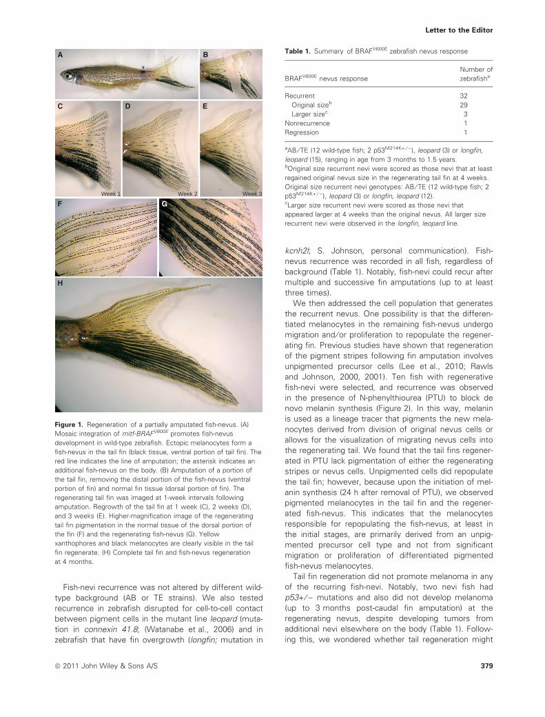
Thirty-four zebrafish displaying fish-nevi within the
caudal fin underwent partial amputation (Figure 1B and
Table 1). Zebrafish were imaged initially as a reference
image, immediately following partial nevus removal (Fig-
ure 1A, B) and subsequently at 1-week intervals for at
least 3 weeks post-surgery (Figure 1C–G). Four different
outcomes were observed (Table 1). The most frequent
outcome was complete regrowth of the nevus (regener-
ate; n = 32; Figure 1C–G). Regenerating fin nevus tissue
carried the mitfa-BRAFV600E transgene cells, as con-
firmed by genotyping of the regenerate fin tissue (data
not shown). However, because the fish are mosaic
transgenics, non-nevus tail fin tissue also carried and
expressed the transgene preventing us from determin-
ing the origin of the repopulating fish-nevus melano-
cytes. Fin regeneration without regrowth of the nevus
was also observed (n = 1; non-regenerate; Figure S1),
as was one case of nevus regression in which the
remaining segment of nevus appeared to regress leav-
ing the original stripe pattern evident (regression; Fig-
ure S2). Rates of fish-nevus regrowth could vary, but all
fish-nevi could be clearly seen to begin recurrence
within 1 week. Most fish-nevi had recurred by 3 weeks,
although one fish required up to 10 weeks for complete
regrowth. Three fish showed enhanced regrowth along
the length of the tail fin (Figure S3). Thus, we find that
most fish-nevi have the potential for recurrence within
the context of the regenerating tail fin tissue.
378 ª 2011 John Wiley & Sons A/S
Pigment Cell Melanoma Res. 24; 378–381 LETTER TO THE EDITOR
Fish-nevi recurrence was not altered by different wild-
type background (AB or TE strains). We also tested
recurrence in zebrafish disrupted for cell-to-cell contact
between pigment cells in the mutant line leopard (muta-
tion in connexin 41.8; (Watanabe et al., 2006) and in
zebrafish that have fin overgrowth (longfin; mutation in
kcnh2l; S. Johnson, personal communication). Fish-
nevus recurrence was recorded in all fish, regardless of
background (Table 1). Notably, fish-nevi could recur after
multiple and successive fin amputations (up to at least
three times).
We then addressed the cell population that generates
the recurrent nevus. One possibility is that the differen-
tiated melanocytes in the remaining fish-nevus undergo
migration and ⁄ or proliferation to repopulate the regener-
ating fin. Previous studies have shown that regeneration
of the pigment stripes following fin amputation involves
unpigmented precursor cells (Lee et al., 2010; Rawls
and Johnson, 2000, 2001). Ten fish with regenerative
fish-nevi were selected, and recurrence was observed
in the presence of N-phenylthiourea (PTU) to block de
novo melanin synthesis (Figure 2). In this way, melanin
is used as a lineage tracer that pigments the new mela-
nocytes derived from division of original nevus cells or
allows for the visualization of migrating nevus cells into
the regenerating tail. We found that the tail fins regener-
ated in PTU lack pigmentation of either the regenerating
stripes or nevus cells. Unpigmented cells did repopulate
the tail fin; however, because upon the initiation of mel-
anin synthesis (24 h after removal of PTU), we observed
pigmented melanocytes in the tail fin and the regener-
ated fish-nevus. This indicates that the melanocytes
responsible for repopulating the fish-nevus, at least in
the initial stages, are primarily derived from an unpig-
mented precursor cell type and not from significant
migration or proliferation of differentiated pigmented
fish-nevus melanocytes.
Tail fin regeneration did not promote melanoma in any
of the recurring fish-nevi. Notably, two nevi fish had
p53+ ⁄ ) mutations and also did not develop melanoma
(up to 3 months post-caudal fin amputation) at the
regenerating nevus, despite developing tumors from
additional nevi elsewhere on the body (Table 1). Follow-
ing this, we wondered whether tail regeneration might
Week 1
A B
C D E
F
H
GWeek 2 Week 3
Figure 1. Regeneration of a partially amputated fish-nevus. (A)
Mosaic integration of mitf-BRAFV600E promotes fish-nevus
development in wild-type zebrafish. Ectopic melanocytes form a
fish-nevus in the tail fin (black tissue, ventral portion of tail fin). The
red line indicates the line of amputation; the asterisk indicates an
additional fish-nevus on the body. (B) Amputation of a portion of
the tail fin, removing the distal portion of the fish-nevus (ventral
portion of fin) and normal fin tissue (dorsal portion of fin). The
regenerating tail fin was imaged at 1-week intervals following
amputation. Regrowth of the tail fin at 1 week (C), 2 weeks (D),
and 3 weeks (E). Higher-magnification image of the regenerating
tail fin pigmentation in the normal tissue of the dorsal portion of
the fin (F) and the regenerating fish-nevus (G). Yellow
xanthophores and black melanocytes are clearly visible in the tail
fin regenerate. (H) Complete tail fin and fish-nevus regeneration
at 4 months.
Table 1. Summary of BRAFV600E zebrafish nevus response
BRAFV600E nevus response
Number of
zebrafisha
Recurrent 32
Original sizeb 29
Larger sizec 3
Nonrecurrence 1
Regression 1
aAB ⁄ TE (12 wild-type fish; 2 p53M214K+ ⁄ )), leopard (3) or longfin,
leopard (15), ranging in age from 3 months to 1.5 years.bOriginal size recurrent nevi were scored as those nevi that at least
regained original nevus size in the regenerating tail fin at 4 weeks.
Original size recurrent nevi genotypes: AB ⁄ TE (12 wild-type fish; 2
p53M214K+ ⁄ )), leopard (3) or longfin, leopard (12).cLarger size recurrent nevi were scored as those nevi that
appeared larger at 4 weeks than the original nevus. All larger size
recurrent nevi were observed in the longfin, leopard line.
Letter to the Editor
ª 2011 John Wiley & Sons A/S 379
stimulate tumor formation in tumor prone BRAFV600E
p53 lines in which all melanocytes carry the BRAFV600E
transgene. We repeated our tail regeneration assay in
five stable transgenic BRAFV600E ⁄ V600E p53) ⁄ ) zebrafish
and found no progression to melanoma at the tail fin
(followed up to 4 weeks post-amputation; data not
shown). Likewise, amputation of the tail fins in the
highly tumor prone RASV12 stable lines (Santoriello
et al., 2010) also did not stimulate tumorigenesis (fol-
lowed up to 3 weeks post-amputation; n = 24; data not
shown). Thus, in the proliferative environment of the
regenerating tail fin, sufficient cellular controls are main-
tained to prevent tumorigenesis in BRAFV600E- and
RASV12-expressing melanocytes.
In conclusion, otherwise growth-restricted zebrafish
fish-nevi have the potential to repopulate large portions
of a nevus from an unpigmented precursor cell type,
without promoting tumorigenicity. UV light exposure
and BRAF mutations contribute to nevus initiation in
humans but the maintenance, recurrence, and regres-
sion of nevi are not well understood. Nevi often regress
in older people (Tucker et al., 2002), and a proportion of
patients display nevi recurrence following removal by
surgical curettage or dermabrasion (King et al., 2009).
These recurrent nevi are not tumorigenic but can often
resemble a dysplastic nevus or melanoma (pseudomel-
anoma). The source of melanocytes that repopulate a
recurrent nevus is unknown, but it has been postulated
that the melanocytes may be derived from nearby mela-
nocyte stem cells or residual nevus melanocytes at the
site of removal (King et al., 2009). In our model, the
regenerative nevi appear to be derived from an unpig-
mented precursor population, at least during the first
11 days of regeneration. The potential for differing
regenerative outcomes of the fish-nevi suggests that
fish-nevi may actively regulate and sustain their growth.
While we do not know how human nevus maintenance
compares with the zebrafish-observed nevus outcomes,
this model provides a novel platform to study funda-
mental questions about nevus maintenance, regrowth,
and regression.
Acknowledgements
We are grateful to Corina Anastasaki (Edinburgh, UK), Natalie
Reynolds (Edinburgh, UK), and Dr. James Lister (Virginia, USA) for
sharing plasmid reagents, Professor David Harrison (Edinburgh, UK)
and Dr. Val Doherty (Edinburgh, UK) for helpful discussions, and
Dr. Karthika Paranthaman for excellent fish husbandry. This work
was funded by the Medical Research Council (J.R., E.E.P., I.J.), the
Association for International Cancer Research (Z.Z., E.E.P.), the
Wellcome Trust (E.E.P.), Medical Research Scotland (H.I., E.E.P.),
the Associazione Italiana per la Ricerca sul Cancro (M.M.), and the
European Union FP7 ZF-CANCER project (E.E.P.)
References
Damsky, W.E. Jr, and Bosenberg, M. (2010). Mouse melanoma
models and cell lines. Pigment Cell Melanoma Res. 23, 853–859.
Gray-Schopfer, V., Wellbrock, C., and Marais, R. (2007). Melanoma
biology and new targeted therapy. Nature 445, 851–857.
Kelsh, R.N., Harris, M.L., Colanesi, S., and Erickson, C.A. (2009).
Stripes and belly-spots -a review of pigment cell morphogenesis
in vertebrates. Semin. Cell Dev. Biol. 20, 90–104.
King, R., Hayzen, B.A., Page, R.N., Googe, P.B., Zeagler, D., and
Mihm, M.C. Jr (2009). Recurrent nevus phenomenon: a clinico-
pathologic study of 357 cases and histologic comparison with
melanoma with regression. Mod. Pathol. 22, 611–617.
Lee, Y., Nachtrab, G., Klinsawat, P.W., Hami, D., and Poss, K.D.
(2010). Ras controls melanocyte expansion during zebrafish fin
stripe regeneration. Dis. Model Mech. 3, 496–503.
Patton, E.E., Widlund, H.R., Kutok, J.L. et al. (2005). BRAF muta-
tions are sufficient to promote nevi formation and cooperate with
p53 in the genesis of melanoma. Curr. Biol. 15, 249–254.
Patton, E.E., Mitchell, D.L., and Nairn, R.S. (2010). Genetic and
environmental melanoma models in fish. Pigment Cell Melanoma
Res. 23, 314–337.
Rawls, J.F., and Johnson, S.L. (2000). Zebrafish kit mutation reveals
primary and secondary regulation of melanocyte development
during fin stripe regeneration. Development 127, 3715–3724.
Rawls, J.F., and Johnson, S.L. (2001). Requirements for the kit
receptor tyrosine kinase during regeneration of zebrafish fin
melanocytes. Development 128, 1943–1949.
A B
C D
E F
Figure 2. Regenerating fish-nevi develop from an undifferentiated
precursor. (A) A zebrafish tail fin with a fish-nevus (asterisks) was
partially amputated and regrown in the presence of phenylthiourea
(PTU) for 11 days (B). PTU blocks new melanin synthesis (Rawls
and Johnson, 2000). Red boxes indicate areas of normal (top) and
fish-nevus (bottom) regenerating fin. In the normal and fish-nevus
(C and E), the fin regenerates in PTU without pigmentation and the
blood vessels are clearly visible. (D and F) After 24 h in fresh water
(PTU washout), the melanocytes are clearly visible (red arrows).
Letter to the Editor
380 ª 2011 John Wiley & Sons A/S

Santoriello, C., Gennaro, E., Anelli, V., Distel, M., Kelly, A., Koster,
R.W., Hurlstone, A., and Mione, M. (2010). Kita driven expression
of oncogenic HRAS leads to early onset and highly penetrant
melanoma in zebrafish. PLoS ONE 5, e15170.
Tucker, M.A., Fraser, M.C., Goldstein, A.M., Struewing, J.P., King,
M.A., Crawford, J.T., Chiazze, E.A., Zametkin, D.P., Fontaine,
L.S., and Clark, W.H. Jr (2002). A natural history of melanomas
and dysplastic nevi: an atlas of lesions in melanoma-prone fami-
lies. Cancer 94, 3192–3209.
Watanabe, M., Iwashita, M., Ishii, M., Kurachi, Y., Kawakami, A.,
Kondo, S., and Okada, N. (2006). Spot pattern of leopard Danio
is caused by mutation in the zebrafish connexin41.8 gene.
EMBO Rep. 7, 893–897.
Supporting information
Additional Supporting Information may be found in the
online version of this article:
Figure S1. A nonrecurrent fish-nevus.
Figure S2. A regressive fish-nevus.
Figure S3. A recurrent fish-nevus with enhanced
regrowth.
Appendix S1. Materials and Methods.
Please note: Wiley-Blackwell are not responsible for
the content or functionality of any supporting materials
supplied by the authors. Any queries (other than missing
material) should be directed to the corresponding author
for the article.
Letter to the Editor
ª 2011 John Wiley & Sons A/S 381


![Toronto SCC epigenetics and aginginteresting skin lighteners on melanocytes looking atinteresting skin lighteners on melanocytes looking at Tyrosinase [TYR] and Ferritin [FTH1] gene](https://img.pdfslide.us/doc/110x75/602d4f8f53f48f1d883bdfdb/toronto-scc-epigenetics-and-aging-interesting-skin-lighteners-on-melanocytes-looking.jpg)