Embed Size (px)
Citation preview
Edinburgh Research Explorer
Intratubular germ cell neoplasia of the human testis
Citation for published version:Mitchell, RT, E Camacho-Moll, M, Macdonald, J, Anderson, R, Kelnar, CJH, O'Donnell, M, Sharpe, RM,Smith, LB, Grigor, KM, Wallace, WHB, Stoop, H, Wolffenbuttel, KP, Donat, R, Saunders, PT & Looijenga,LH 2014, 'Intratubular germ cell neoplasia of the human testis: heterogeneous protein expression andrelation to invasive potential', Modern Pathology. https://doi.org/10.1038/modpathol.2013.246
Digital Object Identifier (DOI):10.1038/modpathol.2013.246
Link:Link to publication record in Edinburgh Research Explorer
Document Version:Peer reviewed version
Published In:Modern Pathology
Publisher Rights Statement:This is a PDF file of an unedited manuscript that has been accepted for publication. The publisher version isavailable at: http://dx.doi.org/10.1038/modpathol.2013.246
General rightsCopyright for the publications made accessible via the Edinburgh Research Explorer is retained by the author(s)and / or other copyright owners and it is a condition of accessing these publications that users recognise andabide by the legal requirements associated with these rights.
Take down policyThe University of Edinburgh has made every reasonable effort to ensure that Edinburgh Research Explorercontent complies with UK legislation. If you believe that the public display of this file breaches copyright pleasecontact [email protected] providing details, and we will remove access to the work immediately andinvestigate your claim.
Download date: 03. Jul. 2020
Rod Mitchell 1
Intratubular germ cell neoplasia of the human testis: heterogeneous protein 1
expression and relation to invasive potential 2
3
Mitchell RT1,3, Camacho-Moll M1, Macdonald J1, Anderson RA1, Kelnar CJH3, O’Donnell M2, Sharpe RM1, 4
Smith LB1, Grigor KM2, Wallace WHB3, Stoop H4, Wolffenbuttel KP4, Donat R5, *Saunders PTK1 and 5
*Looijenga LHJ4. 6
7
1 MRC Centre for Reproductive Health, The University of Edinburgh, Queen's Medical Research Institute, 47 Little 8
France Crescent, Edinburgh, EH16 4TJ, Scotland, UK 9
2 Department of Pathology, Western General Hospital, Crewe Road, Edinburgh, EH4 2XU, Scotland, UK 10
3 Edinburgh Royal Hospital for Sick Children, 9 Sciennes Road, Edinburgh, EH9 1LF, Scotland, UK 11
4 Department of Pathology (LHJL, HS) and Pediatric Urology (KPW), Erasmus MC-University Medical Center 12
Rotterdam, Josephine Nefkens Institute, Room 430b, P.O. Box 1738, 3000 DR Rotterdam, The Netherlands 13
5Department of Urology, Western General Hospital, Crewe Road, Edinburgh, EH4 2XU, Scotland, UK 14
* These authors contributed equally to this study. 15
16
17
PDF compression, OCR, web optimization using a watermarked evaluation copy of CVISION PDFCompressor
Rod Mitchell 2
Running Title: Proliferation in intratubular germ cell neoplasia 18
19
Correspondence and reprint requests 20
Rod Mitchell, MRC Centre for Reproductive Health, The University of Edinburgh, The Queen's Medical 21
Research Institute, 47 Little France Crescent, Edinburgh EH16 4TJ, Scotland, UK. FAX: 44 131 242 6197. 22
e-mail: [email protected] 23
24
PDF compression, OCR, web optimization using a watermarked evaluation copy of CVISION PDFCompressor
Please cite this article as: Mitchell, RT, E Camacho-Moll, M, Macdonald, J, Anderson, RA, Kelnar, CJH, E Camacho-Moll, M, Sharpe, RM, Smith, LB, Grigor, KM, Wallace, WHB, Stoop, H, Wolffenbuttel, KP, Donat, R, Saunders, PT & Looijenga, LH, 'Intratubular germ cell neoplasia of the human testis: heterogeneous protein expression and relation to invasive potential', Modern pathology : an official journal of the United States and Canadian Academy of Pathology (2014), http://dx.doi.org/10.1038/modpathol.2013.246
Rod Mitchell 3
Abstract: Testicular germ cell cancer develops from pre-malignant intratubular germ cell neoplasia, 25
unclassified cells that are believed to arise from failure of normal maturation of fetal germ cells from 26
gonocytes (OCT4+/ MAGEA4-) into pre-spermatogonia (OCT4-/MAGEA4+). Intratubular germ cell neoplasia 27
cell subpopulations based on stage of germ cell differentiation have been described, however the importance 28
of these subpopulations in terms of invasive potential has not been reported. We hypothesised that cells 29
expressing an immature (OCT4+/ MAGEA4-) germ cell profile would exhibit an increased proliferation rate 30
compared to those with a mature profile (OCT4+/ MAGEA4+). Therefore, we performed triple 31
immunofluorescence and stereology to quantify the different intratubular germ cell neoplasia cell 32
subpopulations, based on expression of germ cell (OCT4, PLAP, AP2γ, MAGEA4, VASA) and proliferation 33
(Ki67) markers, in testis sections from patients with pre-invasive disease, seminoma and non-seminoma. We 34
compared these subpopulations with normal human fetal testis and with seminoma cells. Heterogeneity of 35
protein expression was demonstrated in intratubular germ cell neoplasia cells with respect to gonocyte and 36
spermatogonial markers. It included an embryonic/fetal germ cell subpopulation lacking expression of the 37
definitive intratubular germ cell neoplasia marker OCT4, that did not correspond to a physiological (fetal) 38
germ cell subpopulation. OCT4+/MAGEA4- cells showed a significantly increased rate of proliferation 39
compared with the OCT4+/MAGEA4+ population (12.8 v 3.4%, p<0.0001) irrespective of histological tumour 40
type, reflected in the predominance of OCT4+/MAGEA4- cells in the invasive tumour component. 41
Surprisingly, OCT4+/MAGEA4- cells in patients with pre-invasive disease showed significantly higher 42
proliferation compared to those with seminoma or non-seminoma (18.1 v 10.2 v 7.2%, p<0.05 respectively). 43
In conclusion, this study has demonstrated that OCT4+/MAGEA4- cells are the most frequent and most 44
proliferative cell population in tubules containing intratubular germ cell neoplasia, which appears to be an 45
important factor in determining invasive potential of intratubular germ cell neoplasia to seminomas. 46
47
Keywords and Topic Category: 48
Testicular germ cell tumours, Cell differentiation, Cell proliferation, Germ cells, Carcinoma in situ 49
50
51
PDF compression, OCR, web optimization using a watermarked evaluation copy of CVISION PDFCompressor

Rod Mitchell 4
Introduction 52
Testicular germ cell cancer is the most common malignancy in young men and the incidence of these tumours 53
is increasing worldwide [1,2]. The tumours are classified as seminoma or non-seminoma with a distinct cell of 54
origin and pathogenesis compared with spermatocytic seminoma of late adulthood [1]. These tumours result 55
from transformation, usually in young adulthood, of pre-invasive intratubular germ cell neoplasia (also known 56
as carcinoma in situ) cells that arise during fetal life [3,4]. Intratubular germ cell neoplasia cells are believed to 57
be germ cells that have failed to undergo normal maturation during fetal or early postnatal life. 58
59
In humans, during fetal life, primordial germ cells migrate into the developing gonad at around 5 weeks of 60
gestation and become gonocytes [5]. These cells express proteins associated with pluripotency (e.g. OCT4 and 61
NANOG) [6,7] and a number of other embryonic markers (e.g. AP2γ and PLAP) [8,9]. During the remainder 62
of fetal life and into the early postnatal period these cells begin to express germ cell specific proteins (e.g. 63
VASA and MAGEA4) during their transition from gonocytes into spermatogonia and this is associated with a 64
loss of the gonocyte protein markers [7]. This transition occurs in an asynchronous manner such that cells at 65
different stages of development may be present in an individual seminiferous cord during this period and some 66
of these cells may co-express both gonocyte and a spermatogonial markers [10]. 67
68
Intratubular germ cell neoplasia cells express many of the same proteins as gonocytes (e.g. OCT4, PLAP, 69
AP2γ) and these are often used in conjunction with histological evaluation to diagnose the condition in 70
testicular biopsies [11]. It is also recognised that intratubular germ cell neoplasia cells may express proteins 71
indicative of spermatogonia (e.g. MAGEA4, VASA, TSPY) [3,12,13]. The clinical significance of the 72
differing protein expression profiles amongst intratubular germ cell neoplasia cells is not known. 73
74
Proliferation of pre-invasive cells is important for the development of an invasive tumour and proliferation has 75
been shown to occur in intratubular germ cell neoplasia cells prior to the development of an invasive tumour 76
[12]. However, proliferation in the different sub-populations of intratubular germ cell neoplasia cells, based on 77
germ cell differentiation profile, has not previously been investigated. 78
79
The aim of this study was to characterise the heterogeneous protein expression profiles of intratubular germ 80
cell neoplasia cells using co-localisation of multiple proteins simultaneously and to compare this to the 81
PDF compression, OCR, web optimization using a watermarked evaluation copy of CVISION PDFCompressor
Rod Mitchell 5
expression profiles of normal germ cells in the human fetal testis. In addition we aimed to quantify the 82
different intratubular germ cell neoplasia sub-populations associated with different testicular germ cell cancer 83
histological types and to investigate whether the protein expression profile of intratubular germ cell neoplasia 84
cells is related to proliferation of these cells and hence to their invasive potential. 85
86
Materials and Methods 87
Tissue collection 88
Human intratubular germ cell neoplasia/testicular germ cell cancer tissue: Ethical approval was obtained for 89
the use of archived human testicular tissue from the Pathology Departments at the Western General Hospital in 90
Edinburgh (REC Reference number - 10/S1402/33) and Erasmus MC-University Medical Center, Rotterdam 91
(Institutional review board - MEC 02.981 and CCR2041). Samples were randomly selected from the testicular 92
germ cell tumour database and analysed by light microscopy for the presence of intratubular germ cell 93
neoplasia cells. The diagnosis included pre-invasive disease (childhood, n=4; adulthood, n=7), seminoma 94
(n=9) and non-seminoma (n=8). Patient details are described in Table 1. The specimens had been fixed in 95
formalin for 24 hours. 96
97
Human Fetal Testes: Human fetal testes were obtained following termination of pregnancy during 2nd 98
trimester (14-19 weeks, n=5). Women gave consent in accordance with national guidelines, and ethical 99
approval was obtained from the Local Research Ethics Committee (Reference number – LREC08/S1101/1). 100
No terminations were due to fetal abnormalities. Gestational age was determined initially by ultrasound, 101
followed by measurement of foot length. Testes were fixed for 2h in Bouins, transferred into 70% ethanol and 102
then embedded in paraffin. Sections of 5�m thickness were prepared. 103
104
Immunohistochemistry 105
Details of antibodies, dilutions and requirement for antigen retrieval are shown in Table 2. Sections were 106
dewaxed in xylene, rehydrated in graded alcohols and washed in tap water. Antigen retrieval involved pressure 107
cooking in 0.01M citrate (pH 6.0) buffer as described previously [14]. Sections were treated with 3% (v/v) 108
H2O2 in methanol for 30 min and washed in water, followed by Tris-buffered saline (TBS, 0.05M Tris and 109
0.85% NaCl, pH 7.6) for a further 5 min. Endogenous biotin was blocked using an avidin/biotin blocking kit 110
(Vector Laboratories, Peterborough, UK), according to the manufacturers instructions. Sections were 111
PDF compression, OCR, web optimization using a watermarked evaluation copy of CVISION PDFCompressor
Rod Mitchell 6
incubated in appropriate normal serum (diluted 1:5 with TBS containing 5% (w/v) bovine serum albumin 112
(BSA) (Sigma, Poole, Dorset, UK) for 30 min. Sections were incubated overnight with primary antibody 113
diluted in serum at 4oC in a humidified chamber. Sections were washed in TBS (2x5min) and incubated for 30 114
min with the appropriate biotinylated secondary antibody (swine anti-rabbit, rabbit anti-mouse; both Dako, 115
Ely, UK or rabbit anti-goat; Vector Laboratories), diluted in normal serum. This was followed by two further 5 116
min washes in TBS and incubation for 30 min with Streptavidin-HRP at 1:1000 (Dako), diluted in TBS. 117
118
Visualisation was performed using 3,3-diaminobenzidine tetrahydrochloride (DAB) (Dako) and sections were 119
counterstained with haematoxylin, dehydrated in graded alcohols, immersed in xylene and mounted in Pertex 120
medium (CellPath, Hemel Hempstead, UK). For each experiment a negative control (primary antibody 121
replaced with the appropriate normal serum) was included. Images were captured using an Olympus Provis 122
microscope (Olympus, London, UK) and Canon DS126131 camera with Canon EOS image capture software 123
(Canon, Woodhatch, Surrey, UK). 124
125
Immunofluorescence 126
Sections were initially treated as described for single staining as far as the primary antibody stage, with 127
Phosphate Buffered Saline (PBS; Sigma) washes between each step. Antigen retrieval was required for all 128
experiments. Details of antibodies, serum and visualisation method are listed in Table 2. 129
130
Following overnight incubation with primary antibody in serum, sections were incubated with secondary 131
antibody for 30 min, followed by fluorescently labelled Tyramide (1:50; Perkin Elmer, Cambridge, UK) in 132
dilution buffer. For this and subsequent steps, the sections were kept in darkness. Sections were then 133
microwaved in 0.01M citrate (pH 6.0) for 2.5 min and left to cool for 30 min, before being washed in water 134
and PBS for 5 min. Sections were incubated for 30 min in serum. They were incubated with secondary 135
antibody for 30 min, followed by the labelled Tyramide (1:50) using a different fluorescent label. After the 136
second visualisation sections were microwaved again as described above and incubated with the third 137
secondary antibody for 30 min, followed by the third labelled Tyramide (1:50). DAPI (Sigma) was applied to 138
the sections at 1:1000 in PBS for 10 min and the slides were mounted using Permafluor (Immunotech, 139
Marseille, France). Images were captured using an LSM 510 Confocal microscope (Carl Zeiss, Hertfordshire, 140
UK). 141
PDF compression, OCR, web optimization using a watermarked evaluation copy of CVISION PDFCompressor
Rod Mitchell 7
142
Quantification of germ cell differentiation and proliferation 143
Quantification of germ cell subpopulations and proliferation indices were performed for the triple-stained 144
sections as previously described [10]. For each sample, a minimum of 10 randomly selected fields with tubules 145
containing intratubular germ cell neoplasia were counted and included an average of 1000 cells per section. 146
Images were obtained using an Axiovert 200M microscope with attached Axiocam HRc camera and 147
Axiovision 4.6 software (all Carl Zeiss). All germ cells within each section were manually counted and 148
quantified according to their protein expression profile and proliferation status by marking cells in layered 149
images using Adobe Photoshop 7.0 (Adobe, San Jose, CA, USA). 150
151
Statistics 152
Statistical analysis was performed using Graphpad Prism 5 software (La Jolla, CA, USA). Groups were 153
compared using Students t-test. Multiple groups were analysed using one-way analysis of variance (ANOVA). 154
Statistical significance was set at P<0.05. 155
156
Results 157
In order to characterise the heterogeneity of expression of germ cell proteins in putative intratubular germ cell 158
neoplasia cells we first compared the expression of a range of germ cell-specific proteins in testicular tissue 159
from patients with testicular germ cell cancer (including tubules containing intratubular germ cell neoplasia 160
cells and those with apparently normal spermatogenesis) with that of the normal human fetal testis. 161
162
Expression of gonocyte markers in human fetal testis, intratubular germ cell neoplasia and 163
spermatogonia 164
OCT4, AP2γ and PLAP were expressed in germ cells (gonocytes) in the human fetal testis. In sections from 165
patients with testicular germ cell cancer these proteins were also expressed in intratubular germ cell neoplasia 166
cells; however none of the proteins were expressed in spermatogonia in tubules that contained active 167
spermatogenesis (Fig. 1A-I). 168
169
Expression of spermatogonial markers in human fetal testis, intratubular germ cell neoplasia and 170
spermatogonia 171
PDF compression, OCR, web optimization using a watermarked evaluation copy of CVISION PDFCompressor
Rod Mitchell 8
MAGEA4 and VASA were expressed in germ cells (pre-spermatogonia) in the human fetal testis. These 172
proteins were also expressed in intratubular germ cell neoplasia cells. There was also expression of these 173
proteins in germ cells (MAGEA4 in spermatogonia and early spermatocytes, VASA in all germ cells) of 174
tubules that contained active spermatogenesis in patients with testicular germ cell cancer (Fig 1J-O). 175
176
Co-expression of gonocyte and spermatogonial markers in intratubular germ cell neoplasia cells and 177
normal testis 178
For the identification of intratubular germ cell neoplasia cells and to attempt to distinguish these cells from 179
normal germ cells, AP2γ and VASA co-expression was investigated (Fig 2). In tubules with normal-appearing 180
spermatogenesis there was expression of VASA in the cytoplasm of germ cells, but no expression of AP2γ 181
(Fig 2A). In tubules containing a mixture of germ cells characteristic of either intratubular germ cell neoplasia 182
or normal spermatogonia the putative intratubular germ cell neoplasia cells (located on the basement 183
membrane) were identified as AP2γ+/VASA-, whilst a small proportion of these cells were AP2γ+/VASA+ (Fig 184
2B). AP2γ-/VASA+cells, located nearer the lumen were also identified in intratubular germ cell neoplasia 185
tubules. These putative spermatocytes were also identified in tubules in which the majority of the cells were 186
intratubular germ cell neoplasia cells (AP2γ+/VASA-; Fig 2C). Similar populations of germ cells were also 187
identified within the human fetal testis, based on co-staining for OCT4 and VASA (Supp. Fig. 1; [7]). 188
189
Heterogeneity of expression of ‘classical’ intratubular germ cell neoplasia markers in patients with 190
testicular germ cell cancer 191
In order to demonstrate the heterogeneity of expression of gonocyte proteins in intratubular germ cell 192
neoplasia, co-localisation of OCT4, AP2γ and PLAP was undertaken. OCT4 and AP2γ were always co-193
expressed and were localised to the nuclei of intratubular germ cell neoplasia cells (Supp. Fig. 2). A similar 194
pattern of co-expression was demonstrated for OCT4 and PLAP, with co-expression of OCT4 (nuclear) and 195
PLAP (cytoplasm +/- nuclear) in the majority of cells (OCT4+/PLAP+) within these tubules, however there 196
were also germ cells that were OCT4+/PLAP- (Fig. 3A). These two populations were also identified in the 197
human fetal testis (Fig. 3B). However within the intratubular germ cell neoplasia containing tubules we also 198
identified a rare population of cells that were OCT4-/PLAP+ (Fig. 3C), whilst this population of germ cells was 199
not identified in any of the human fetal testis sections (Fig. 3B). 200
PDF compression, OCR, web optimization using a watermarked evaluation copy of CVISION PDFCompressor
Rod Mitchell 9
201
The expression of spermatogonial markers in putative intratubular germ cell neoplasia cells 202
To further characterise the germ cells within intratubular germ cell neoplasia containing tubules that either did 203
or did not express OCT4, triple immunofluorescence staining was undertaken for MAGEA4/VASA, OCT4 204
and PLAP (Fig. 4). In tubules containing intratubular germ cell neoplasia cells, the majority of germ cells 205
expressed nuclear OCT4 and most of these were co-stained with PLAP (cytoplasmic +/- nuclear). A small 206
proportion of presumptive spermatocytes expressed VASA only (Fig. 4A). However there were infrequent 207
germ cells which expressed VASA and PLAP, but which did not express OCT4 (Fig. 4A), suggesting that a 208
proportion of the VASA expressing cells (VASA+/OCT4-) are not ‘normal’ spermatogonia/spermatocytes and 209
may represent gonocytes that have downregulated OCT4 and begun to express VASA but retain PLAP 210
expression (VASA+/OCT4-/PLAP+). Similar sub-populations were found when the co-expression of MAGEA4 211
with OCT4 and PLAP was undertaken (Figure 4B). 212
213
Quantification of intratubular germ cell neoplasia phenotypes depending on histological testicular germ 214
cell cancer type 215
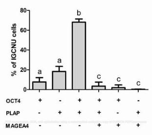
The proportion of intratubular germ cell neoplasia cells (identified by expression of OCT4 and/or PLAP) with 216
different expression profiles based on OCT4/PLAP/MAGEA4 co-staining was determined (Fig. 5). By far the 217
most common phenotype was OCT4+/PLAP+/MAGEA4-, which was found in a significantly higher proportion 218
(68%) of cells compared with the other phenotypes (Fig. 5; b versus a,c). A smaller proportion (7.7%) of 219
intratubular germ cell neoplasia cells were OCT4+/PLAP-/MAGEA4-. Overall 82% of intratubular germ cell 220
neoplasia cells expressed OCT4 with the remaining 18% of putative intratubular germ cell neoplasia cells 221
expressing PLAP (but no detectable OCT4). In terms of spermatogonial markers, MAGEA4 expression was 222
found in 6% of putative intratubular germ cell neoplasia cells (defined by expression of OCT4 and/or PLAP; 223
Fig. 5) and this represented a significantly lower proportion of cells compared to those not expressing 224
MAGEA4 (Fig 5; c versus a,b). There was a shift towards an increasing proportion of these MAGEA4+ cells 225
from pre-invasive (child) to pre-invasive (adult) and seminoma, whilst very few putative intratubular germ cell 226
neoplasia cells in non-seminoma expressed this protein profile, however the differences in expression were not 227
significant (Supp. Fig. 3). 228
229
PDF compression, OCR, web optimization using a watermarked evaluation copy of CVISION PDFCompressor

Rod Mitchell 10
Proliferation of intratubular germ cell neoplasia cells according to germ cell expression profile and 230
histological testicular germ cell cancer type 231
In order to investigate whether the germ cell expression profile of the putative intratubular germ cell neoplasia 232
cells might determine their proliferation rate, which might in turn affect their invasive potential, triple 233
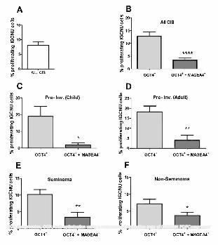
immunofluorescence for OCT4/MAGEA4/Ki67 was performed (Supp. Fig. 4). Overall, the proportion of 234
proliferating (Ki67+/OCT4+) intratubular germ cell neoplasia cells was 8.1% (Fig. 6A). There was a shift 235
towards increased proliferation in intratubular germ cell neoplasia cells from patients with pre-invasive disease 236
compared with those with a seminoma or non-seminoma, but this was not statistically significant (Supp. Fig. 237
5). However, when intratubular germ cell neoplasia (identified by expression of OCT4+) cell proliferation was 238
analysed according to whether or not the cells also expressed MAGEA4, a significantly higher proliferation 239
rate was found for the OCT4+/MAGEA4- population compared to OCT4+/MAGEA4+ intratubular germ cell 240
neoplasia cells (12.8 v 3.4%, p<0.0001; Fig. 6B). Moreover, the significant difference in proliferation rate 241
between the two intratubular germ cell neoplasia phenotypes was consistent when the same analysis was 242
performed according to whether the intratubular germ cell neoplasia cells were from patients with pre-invasive 243
disease (child or adult), seminoma or non-seminoma (Fig. 6C-F). Furthermore, when the proliferation rates in 244
the two sub-populations of intratubular germ cell neoplasia cells (OCT4+/MAGEA4- or OCT4+/MAGEA4+) 245
were compared according to the histology of the adjacent testicular germ cell cancer, there was a significantly 246
higher proliferation rate in the OCT4+/MAGEA4- cells in pre-invasive disease compared with these cells in 247
seminoma and non-seminoma (Fig. 7A); in contrast, there was no difference in the proportion of 248
OCT4+/MAGEA4+ that were proliferating for the different tumour types (Fig. 7B). 249
250
Proliferation of seminoma cells according to germ cell expression profile 251
Given that the OCT4+/MAGEA4- population of intratubular germ cell neoplasia cells were more proliferative 252
than the OCT4+/MAGEA4+ population, we investigated MAGEA4, OCT4 and PLAP expression in seminoma 253
cells in order to determine whether this results in a predominance of OCT4+/MAGEA4- cells in the resulting 254
tumours. Indeed we found that in the majority of intra-tubular seminoma cells MAGEA4 was not expressed 255
and that the majority of cells were OCT4+/PLAP+/MAGEA4- (Fig. 8A). MAGEA4 positive cells were seen in 256
tubules with normal appearance adjacent to areas of invasive seminoma (Fig. 8B), however MAGEA4 257
expression was not seen in the invasive seminoma cells (Fig. 8C). The OCT4+/MAGEA4- seminoma cells 258
PDF compression, OCR, web optimization using a watermarked evaluation copy of CVISION PDFCompressor
Rod Mitchell 11
were highly proliferative, whilst a smaller proportion of OCT4+/MAGEA4- intratubular germ cell neoplasia 259
cells were proliferative. In contrast MAGEA4 expressing cells were rarely proliferative (Supp. Fig. 6). 260
261
Discussion 262
The present study has characterised the heterogeneity of germ cell protein expression in the human testis based 263
on co-expression of germ cell proteins involved in differentiation from gonocyte to pre-spermatogonia. We 264
have demonstrated an infrequent population of cells within intratubular germ cell neoplasia containing tubules 265
with an expression profile distinct from germ cells in the normal human fetal testis. We have also 266
demonstrated that the most common sub-population in intratubular germ cell neoplasia containing tubules, 267
which displays a ‘gonocyte’ expression profile (OCT4+/MAGEA4-), is associated with an increased 268
proliferation rate compared to the subpopulation expressing a ‘pre-spermatogonial’ profile 269
(OCT4+/MAGEA4+), and that this (OCT4+/MAGEA4-) population represents the true intratubular germ cell 270
neoplasia cell and precursor for invasive seminoma and non-seminoma. The findings of the present study are 271
summarised in Fig 9. 272
273
Intratubular germ cell neoplasia cells are thought to originate in fetal life from abnormally differentiated 274
gonocytes [3,15]. This is supported by similarities in morphological, immunohistochemical and genetic 275
profiles [3,16,17]. Intratubular germ cell neoplasia cells share expression of a variety of proteins that are 276
involved in pluripotency and early germ cell fate, such as OCT4 [18-20], PLAP [9,21] and AP2γ [8]. As 277
gonocytes differentiate into spermatogonia during fetal life these markers have been shown to be 278
downregulated [3,6,7,20,22,23]. OCT4, AP2γ and PLAP are described as ‘classical’ markers of intratubular 279
germ cell neoplasia cells in adulthood and persistence of expression of these proteins is routinely used for 280
diagnostic purposes for patients at risk of, or with suspected, testicular germ cell cancer [11]. These markers 281
are considered highly sensitive and specific for intratubular germ cell neoplasia cells. Whilst we found co-282
expression of AP2γ and OCT4 in intratubular germ cell neoplasia cells with no cells expressing a single 283
marker alone, there was heterogeneity in the co-expression of PLAP and OCT4 in intratubular germ cell 284
neoplasia cells. Expression of OCT4 has been reported to be expressed by all intratubular germ cell neoplasia 285
cells [19], whilst PLAP has been reported to be expressed in 83-99% of intratubular germ cell neoplasia cells 286
[9]. The present co-localisation studies demonstrate that the majority of the cells expressing OCT4 also 287
PDF compression, OCR, web optimization using a watermarked evaluation copy of CVISION PDFCompressor
Rod Mitchell 12
express PLAP. Overall co-localisation was seen in 68% of cells in tubules with intratubular germ cell 288
neoplasia. Both of these sub-populations are also present in the normal human fetal testis during the transition 289
from gonocyte to spermatogonia. However, our co-localisation studies have demonstrated the presence of a 290
sub-population of intratubular germ cell neoplasia cells with a protein expression profile distinct from the 291
germ cells in the normal human fetal testis. This OCT4-/PLAP+ sub-population represented 18% of the total 292
cells in tubules with intratubular germ cell neoplasia. PLAP is expressed in most of the germ cells in a first 293
trimester fetal testis, but is rare by the start of the second trimester [23]. OCT4 is also present in most of the 294
germ cells of the first trimester, but is downregulated later in gestation in comparison to PLAP [24]. This study 295
has shown that an OCT4-/PLAP+ population can be identified in tubules with intratubular germ cell neoplasia, 296
whilst our results confirm that PLAP is not expressed without co-expression of OCT4 in the normal human 297
fetal testis [23]. As these cells do not occur as part of normal germ cell development they may represent 298
impaired maturation of gonocytes with loss of OCT4 and retention of PLAP expression as a result of an 299
altered germ cell niche. 300
301
In addition to the proteins that are found in undifferentiated germ cells, intratubular germ cell neoplasia cells 302
have also been reported to express proteins characteristic of differentiated germ cells such as VASA [25] and 303
MAGEA4 [26]. These markers begin to be expressed in germ cells during fetal life in increasing proportions 304
as the cells differentiate [24,26-28]. We have shown that the majority of intratubular germ cell neoplasia cells 305
(based on the expression of OCT4 and/or PLAP) do not express the spermatogonial proteins MAGEA4 and 306
VASA. We have quantified the expression of these differentiated germ cell markers in putative intratubular 307
germ cell neoplasia for the first time and shown that MAGEA4 is only expressed in 6% of OCT4 and/or 308
PLAP-expressing cells and therefore is not a common phenotype for putative intratubular germ cell neoplasia 309
cells. Heterogeneous expression of MAGEA4 in intratubular germ cell neoplasia has been described 310
previously, however VASA expression was reported to be expressed in all intratubular germ cell neoplasia 311
cells [3]. We found that VASA was expressed heterogeneously in a similar proportion of putative intratubular 312
germ cell neoplasia cells as those expressing MAGEA4. 313
314
Previous studies have indicated that differentiated germ cells (e.g. spermatogonia) may be present within 315
intratubular germ cell neoplasia containing tubules [12,22]. Co-staining for OCT4 and VASA/MAGEA4 316
identified cells that had an OCT4-/VASA+ phenotype [22]. These cells would be considered differentiated 317
PDF compression, OCR, web optimization using a watermarked evaluation copy of CVISION PDFCompressor
Rod Mitchell 13
germ cells rather than intratubular germ cell neoplasia cells. We have described similar populations in our 318
samples, however triple co-localisation has demonstrated that some VASA or MAGEA4 expressing cells that 319
do not express OCT4, express PLAP and therefore may not represent ‘normal’ spermatogonia. As a result it is 320
likely that only cells expressing neither OCT4 nor PLAP may represent normally matured germ cells that have 321
not undergone pre-invasive change. The OCT4-/VASA+/PLAP+ or OCT4-/MAGEA4+/PLAP+ populations may 322
represent pre-invasive germ cells that have undergone a degree of maturation towards pre-spermatogonia (due 323
to downregulation of OCT4 and expression of VASA/MAGEA4), alternatively they may represent pre-324
sprematogonia that have aberrantly retained PLAP expression following the downregulation of OCT4. In order 325
to determine whether these populations could represent intratubular germ cell neoplasia cells we investigated 326
expression during the development of invasive disease. OCT4 (without MAGEA4) was expressed in all intra-327
tubular and invasive seminomas, indicating that intratubular germ cell neoplasia cells with invasive potential 328
express OCT4 and do not express MAGEA4. Therefore we conclude that the OCT4-/MAGEA4+/PLAP+ cells 329
do not represent intratubular germ cell neoplasia cells with malignant potential and are more likely to be a 330
separate population of abnormally differentiated germ cells that are present in intratubular germ cell neoplasia 331
containing tubules. 332
333
Uncontrolled proliferation of cells is a hallmark of invasive tumours [29]. Previous studies have demonstrated 334
proliferation in intratubular germ cell neoplasia and overt testicular germ cell cancer [12,30-32], and a 335
previous study has shown that Ki67 expression is found in intratubular germ cell neoplasia cells in 14/16 non-336
seminomas and 14/17 seminomas, although the proportion of intratubular germ cell neoplasia cells that were 337
proliferating was not quantified [31]. A detailed analysis of proliferation in the various intratubular germ cell 338
neoplasia sub-populations in relation to the underlying tumour type has not previously been performed. 339
Intratubular germ cell neoplasia cells have previously been reported to proliferate at a relatively high rate. In a 340
study of sections taken from patients with testicular germ cell cancer, using Ki67 as a marker of proliferation, 341
17.42% of intratubular germ cell neoplasia cells were found to be Ki67 positive [30]. Overall we found that 342
8.1% of intratubular germ cell neoplasia cells were positive for Ki67, however we have shown that the 343
proliferation rate is dependent on which intratubular germ cell neoplasia sub-population is investigated. We 344
have shown that PLAP is not expressed in ~20% of intratubular germ cell neoplasia (OCT4+) cells and this 345
may partially explain the differences seen between proliferation of intratubular germ cell neoplasia cells in the 346
present study compared to previous studies which relied on PLAP expression to identify intratubular germ cell 347
PDF compression, OCR, web optimization using a watermarked evaluation copy of CVISION PDFCompressor
Rod Mitchell 14
neoplasia cells [12,30]. The OCT4+/MAGEA4+ (and also OCT4-/MAGEA4+, not shown) populations are less 348
proliferative than those expressing OCT4+/MAGEA4- which provides further evidence supporting the view 349
that the OCT4+/MAGEA4- cells have more invasive potential than those expressing a more mature phenotype. 350
This hypothesis is supported by our finding of little or no expression of MAGEA4 in the OCT4+ seminoma 351
cells of an invasive tumour. We therefore propose that the OCT4+/MAGEA4- population of intratubular germ 352
cell neoplasia cells give rise to the invasive tumour, whilst the OCT4+/MAGEA4+ population has a lower 353
capacity to progress to invasiveness and may represent germ cells that are arrested in the transition from 354
gonocyte to spermatogonia and do not contribute to the invasive tumour. OCT4-/MAGEA4+ cells are 355
occasionally seen within the seminomatous component but are likely to represent spermatogonia that have 356
become enclosed in the invasive tumour. 357
358
Differences in the proliferation of intratubular germ cell neoplasia cells have recently been investigated with 359
respect to one of the key regulators of the mitosis-meiosis switch, DMRT1 [12]. This study demonstrated that 360
intratubular germ cell neoplasia cells expressing DMRT1 were significantly less proliferative than those 361
intratubular germ cell neoplasia cells that did not express DMRT1 and that progression from ‘early-stage’ 362
(Ki67-) intratubular germ cell neoplasia cell to invasive disease (Ki67+) is associated with a down-regulation 363
of DMRT1. In order to test the hypothesis that certain subpopulations of intratubular germ cell neoplasia cells 364
display differences in invasive potential, future studies involving isolation of the different intratubular germ 365
cell neoplasia sub-populations followed by germ cell transplantation or xenografting may be performed. 366
367
The present study has demonstrated the presence of proliferating intratubular germ cell neoplasia cells in testis 368
tissue from patients with pre-invasive disease, seminoma and non-seminoma with a higher rate of proliferation 369
in the OCT4+/MAGEA4- population in pre-invasive samples compared to those with an invasive tumour 370
(either seminoma or non-seminoma). The finding of higher rates of proliferation of intratubular germ cell 371
neoplasia cells in pre-invasive disease compared to tumours samples might be considered surprising given 372
previous reports of low rates of proliferation in pre-invasive intratubular germ cell neoplasia cells [12]. In 373
adults with pre-invasive disease this may be explained by an increase in proliferation around the time of 374
progression to invasive disease, however this would not explain the proliferation rate for intratubular germ cell 375
neoplasia cells in the pre-invasive childhood patients in which it might be expected that these cells are 376
relatively quiescent. However, we have demonstrated previously that a higher proportion of OCT4+ germ cells 377
PDF compression, OCR, web optimization using a watermarked evaluation copy of CVISION PDFCompressor
Rod Mitchell 15
in the second trimester human fetal testis are proliferating compared with the MAGEA4+ population and that 378
the rates of proliferation in the present study are similar to those in the normal human fetal testis for each sub-379
population [10], indicating that the proliferation in the germ cell sub-populations in children with pre-invasive 380
disease may simply reflect the proliferation rates in the normal human fetal testis. 381
In conclusion, we have described in detail the heterogeneity of germ cell protein expression in cells within 382
intratubular germ cell neoplasia tubules. We have demonstrated sub-populations of OCT4- cells that do not 383
correspond to an equivalent stage of normal human fetal germ cell differentiation, suggesting that these cells 384
may have lost expression of proteins that may determine their malignant potential. We have also demonstrated 385
that a more undifferentiated/pluripotent expression profile is associated with an increased proliferation rate 386
compared with a differentiated phenotype. These results indicate that germ cells expressing an 387
OCT4+/MAGEA4- phenotype are those that will ultimately lead to tumour formation. 388
389
Disclosure/Conflict of Interest: The authors have no conflicts of interest to disclose. 390
391
Acknowledgements 392
We are grateful to Sheila Macpherson for her assistance during the present study and to Ronnie Grant for his 393
assistance with illustrations. The MAGEA4 antibody was a kind gift from Dr. Giulio Spagnoli. This study was 394
supported by the Wellcome Trust (Grant Code - 098522) and the Medical Research Council (Career 395
Development Fellowship). 396
397
Supplementary information is available at Modern Pathology’s website 398
399
References 400
1. McGlynn KA, Devesa SS, Sigurdson AJ, et al. Trends in the incidence of testicular germ cell tumors in 401
the United States. Cancer 2003; 97: 63-70. 402
2. Richiardi L, Bellocco R, Adami HO, et al. Testicular cancer incidence in eight northern European 403
countries: secular and recent trends. Cancer Epidemiol Biomarkers Prev 2004; 13: 2157-2166. 404
3. Rajpert-De Meyts E. Developmental model for the pathogenesis of testicular carcinoma in situ: genetic 405
and environmental aspects. Hum Reprod Update 2006; 12: 303-323. 406
4. Skakkebaek NE. Possible carcinoma-in-situ of the testis. Lancet 1972; 2: 516-517. 407
PDF compression, OCR, web optimization using a watermarked evaluation copy of CVISION PDFCompressor
Rod Mitchell 16
5. Waters BL, Trainer TD. Development of the human fetal testis. Pediatr Pathol Lab Med 1996; 16: 9-23. 408
6. Hoei-Hansen CE, Almstrup K, et al. Stem cell pluripotency factor NANOG is expressed in human fetal 409
gonocytes, testicular carcinoma in situ and germ cell tumours. Histopathology 2005; 47: 48-56. 410
7. Mitchell RT, Cowan G et al. Germ cell differentiation in the marmoset (Callithrix jacchus) during fetal 411
and neonatal life closely parallels that in the human. Hum Reprod 2008; 23: 2755-2765. 412
8. Hoei-Hansen CE, Nielsen JE et al. Transcription factor AP-2gamma is a developmentally regulated 413
marker of testicular carcinoma in situ and germ cell tumors. Clin Cancer Res 2004; 10: 8521-8530 414
9. Jorgensen N, Rajpert-De Meyts E, Graem N, et al. Expression of immunohistochemical markers for 415
testicular carcinoma in situ by normal human fetal germ cells. Lab Invest 1995; 72: 223-231 416
10. Mitchell RT, Saunders PT, Childs AJ, et al. Xenografting of human fetal testis tissue: a new approach to 417
study fetal testis development and germ cell differentiation. Hum Reprod 2010; 25: 2405-2414. 418
11. Oosterhuis JW, Looijenga LH. Testicular germ-cell tumours in a broader perspective. Nat Rev Cancer 419
2005; 5: 210-222. 420
12. Jorgensen A, Nielsen JE, Almstrup K, et al. Dysregulation of the mitosis-meiosis switch in testicular 421
carcinoma in situ. J Pathol 2013; 229: 588-598. 422
13. Kersemaekers AM, Honecker F, Stoop H, et al. Identification of germ cells at risk for neoplastic 423
transformation in gonadoblastoma: an immunohistochemical study for OCT3/4 and TSPY. Hum Pathol 424
2005; 36: 512-521. 425
14. Norton AJ, Jordan S, Yeomans P. Brief, high-temperature heat denaturation (pressure cooking): a simple 426
and effective method of antigen retrieval for routinely processed tissues. J Pathol 1994; 173: 371-379. 427
15. Skakkebaek NE, Berthelsen JG, Giwercman A, et al. Carcinoma-in-situ of the testis: possible origin from 428
gonocytes and precursor of all types of germ cell tumours except spermatocytoma. Int J Androl 1987; 10: 429
19-28. 430
16. Almstrup K, Hoei-Hansen CE, Wirkner U, et al. Embryonic stem cell-like features of testicular 431
carcinoma in situ revealed by genome-wide gene expression profiling. Cancer Res 2004; 64: 4736-4743. 432
17. Looijenga LH, Gillis AJ, Stoop HJ, et al. Chromosomes and expression in human testicular germ-cell 433
tumors: insight into their cell of origin and pathogenesis. Ann N Y Acad Sci 2007; 1120: 187-214. 434
18. Jones TD, Ulbright TM, Eble JN, et al. OCT4: A sensitive and specific biomarker for intratubular germ 435
cell neoplasia of the testis. Clin Cancer Res 2004; 10: 8544-8547. 436
PDF compression, OCR, web optimization using a watermarked evaluation copy of CVISION PDFCompressor
Rod Mitchell 17
19. Looijenga LH, Stoop H, de Leeuw HP, et al. POU5F1 (OCT3/4) identifies cells with pluripotent potential 437
in human germ cell tumors. Cancer Res 2003; 63: 2244-50. 438
20. Rajpert-De Meyts E, Hanstein R, Jorgensen N, et al. Developmental expression of POU5F1 (OCT-3/4) in 439
normal and dysgenetic human gonads. Hum Reprod 2004; 19: 1338-1344. 440
21. Hustin J, Collette J, Franchimont P. Immunohistochemical demonstration of placental alkaline 441
phosphatase in various states of testicular development and in germ cell tumours. Int J Androl 1987; 10: 442
29-35. 443
22. Cools M, van Aerde K, Kersemaekers AM, et al. Morphological and immunohistochemical differences 444
between gonadal maturation delay and early germ cell neoplasia in patients with undervirilization 445
syndromes. J Clin Endocrinol Metab 2005; 90: 5295-5303. 446
23. Honecker F, Stoop H, de Krijger RR, et al. Pathobiological implications of the expression of markers of 447
testicular carcinoma in situ by fetal germ cells. J Pathol 2004; 203: 849-857. 448
24. Gaskell TL, Esnal A, Robinson LL, et al. Immunohistochemical profiling of germ cells within the human 449
fetal testis: identification of three subpopulations. Biol Reprod 2004; 71: 2012-2021. 450
25. Rajpert-De Meyts E, Bartkova J, Samson M, et al. The emerging phenotype of the testicular carcinoma in 451
situ germ cell. APMIS 2003; 111: 267-278; discussion 278-269. 452
26. Aubry F, Satie AP, Rioux-Leclercq N, et al. MAGEA4, a germ cell specific marker, is expressed 453
differentially in testicular tumors. Cancer 2001; 92: 2778-2785. 454
27. Castrillon DH, Quade BJ, Wang TY, et al. The human VASA gene is specifically expressed in the germ 455
cell lineage. Proc Natl Acad Sci U S A 2000; 97: 9585-9590. 456
28. Zeeman AM, Stoop H, Boter M, et al. VASA is a specific marker for both normal and malignant human 457
germ cells. Lab Invest 2002; 82: 159-166. 458
29. Albers P, Ulbright TM, Albers J, et al. Tumor proliferative activity is predictive of pathological stage in 459
clinical stage A nonseminomatous testicular germ cell tumors. J Urol 1996; 155: 579-586. 460
30. Almstrup K, Nielsen JE, Mlynarska O, et al. Carcinoma in situ testis displays permissive chromatin 461
modifications similar to immature foetal germ cells. Br J Cancer 2010; 103: 1269-1276. 462
31. Datta MW, Renshaw AA, Dutta A, et al. Evaluation of cyclin expression in testicular germ cell tumors: 463
cyclin E correlates with tumor type, advanced clinical stage, and pulmonary metastasis. Mod Pathol 2000; 464
13: 667-672. 465
PDF compression, OCR, web optimization using a watermarked evaluation copy of CVISION PDFCompressor

Rod Mitchell 18
32. Chen YT, Cao D, Chiu R, et al. Chromosome X-encoded Cancer/Testis antigens are less frequently 466
expressed in non-seminomatous germ cell tumors than in seminomas. Cancer Immun 2013; 13: 10. 467
468
PDF compression, OCR, web optimization using a watermarked evaluation copy of CVISION PDFCompressor
Rod Mitchell 19
Figure Legends 469
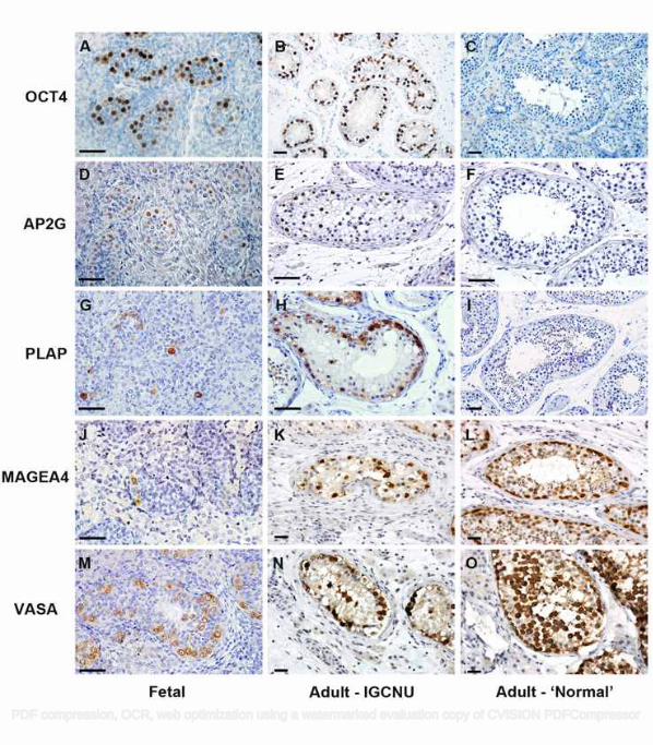
Figure 1 - Expression of gonocyte markers (OCT4, AP2γ and PLAP; A-I) and spermatogonial markers 470
(MAGEA4 and VASA; J-O) in human fetal testis, intratubular germ cell neoplasia containing tubules (Adult – 471
intratubular germ cell neoplasia) and tubules from adult testis with active spermatogenesis (Adult – ‘Normal’). 472
Gonocyte proteins are detected in human fetal germ cells and intratubular germ cell neoplasia cells, but are 473
absent from the germ cells in tubules with apparently normal spermatogenesis; whilst spermatogonial proteins 474
are expressed in germ cells in all tissue types. Human fetal samples are 14 (A,D), 16 (J) and 18 weeks (G,M) 475
gestation. Scale bar = 50µm. 476
477
Figure 2 - Representative image for expression of VASA (green) and AP2γ (red) in tubules from adult 478
patients with testicular germ cell cancer. A) Tubule with apparently normal spermatogenesis: VASA is 479
expressed in the germ cells with no expression of the intratubular germ cell neoplasia/gonocyte protein AP2γ. 480
B) Tubule with abnormal spermatogenesis: VASA expression is seen in the presumptive spermatocytes 481
towards the lumen (white arrowhead), however germ cells along the basement membrane express VASA 482
(yellow arrowhead) or AP2γ (yellow arrow). A small proportion of cells co-express VASA and AP2γ (white 483
arrow). C) Intratubular germ cell neoplasia tubule: The majority of cells express the intratubular germ cell 484
neoplasia protein AP2γ (yellow arrow) with a small number of cells adjacent to the basement membrane 485
expressing VASA (presumptive spermatogonia; pink arrow). A small number of germ cells expressing VASA 486
are located towards the lumen (presumptive spermatocytes; white arrowhead). 487
488
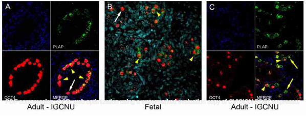
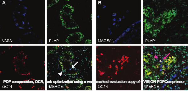
Figure 3 - Expression of OCT4 (red) and PLAP (green) in intratubular germ cell neoplasia containing tubules 489
from men with testicular germ cell cancer (A,C) and in normal human fetal testis tissue (B). The majority of 490
intratubular germ cell neoplasia cells co-express OCT4 and PLAP (A,C; yellow arrowheads). Occasional 491
OCT4 positive intratubular germ cell neoplasia cells are negative for PLAP expression (A; white arrow). 492
Similar sub-populations of OCT4+/PLAP- (B, white arrow) and OCT4+/PLAP+ (B, yellow arrowheads) cells 493
are also identified in the human fetal testis. In tubules with intratubular germ cell neoplasia, occasional OCT4-494
/PLAP+ cells are identified (C; yellow arrows), however no similar population is seen in the human fetal testis. 495
Counterstain (DAPI; blue). 496
497
PDF compression, OCR, web optimization using a watermarked evaluation copy of CVISION PDFCompressor

Rod Mitchell 20
Figure 4 - Representative image for expression of OCT4 (red), VASA (A; blue), MAGEA4 (B; blue) and 498
PLAP (green) in intratubular germ cell neoplasia containing tubules from patients with testicular germ cell 499
cancer. A) VASA expression is demonstrated in putative ‘spermatogenic’ germ cells that are negative for 500
intratubular germ cell neoplasia cell proteins PLAP and OCT4 (white arrow). A small proportion of the 501
VASA+ cells that are negative for OCT4 express PLAP (white arrowhead). B) The majority of cells co-express 502
OCT4 and PLAP without MAGEA4, other sub-populations are identified including PLAP+/OCT4+/MAGEA4+ 503
(pink arrow) and PLAP+/OCT4-/MAGEA4- (pink arrowhead). Counterstain (DAPI; pale blue) in merged 504
panels. 505
506
Figure 5 - Quantification of putative intratubular germ cell neoplasia phenotypes. Expression (+) of OCT4, 507
PLAP and MAGEA4 for intratubular germ cell neoplasia containing tubules (n=9; pre-invasive, seminoma and 508
non-seminoma; n=3 each). Bars with different letters are significantly different from each other (p<0.05). 509
Mean +/- SEM. 510
511
Figure 6 - Proliferation in putative intratubular germ cell neoplasia cells. A) Overall proliferation in all 512
intratubular germ cell neoplasia cells. B) Proliferation (Ki67+) of intratubular germ cell neoplasia (OCT4+) 513
cells based on the co-expression with MAGEA4 in tubules from all patients (B), children with pre-invasive 514
disease (C; Pre-Inv. Child; n=4), adults with pre-invasive disease (D; Pre-Inv. Adult; n=6), seminoma (n=7) 515
and non-seminoma (n=8). Mean +/- SEM. * p<0.05, ** p<0.01, **** p<0.0001. 516
517
Figure 7 - Proliferation of intratubular germ cell neoplasia cells based on diagnosis of pre-invasive disease 518
(PRE INV; n=7), seminoma (SEM; n=7) or non-seminoma (NON-SEM; n=8). A) Proliferation of OCT4+ 519
intratubular germ cell neoplasia cells. B) Proliferation of OCT4+/MAGEA4+ intratubular germ cell neoplasia 520
cells. Mean +/- SEM. * p<0.05, ** p<0.01, in comparison with pre-invasive intratubular germ cell neoplasia. 521
522
Figure 8 - Representative images for expression of OCT4 (red), PLAP (green) and MAGEA4 (blue) in testis 523
sections from patients with A) Intra-tubular seminoma, B) Seminoma with surrounding ‘normal’ (*) tubules 524
and C) Seminoma. Note the lack of expression of MAGEA4 in both intra-tubular seminoma and invasive 525
seminoma. Counterstain (DAPI; pale blue) in merged panels (bottom right). 526
527
PDF compression, OCR, web optimization using a watermarked evaluation copy of CVISION PDFCompressor
Rod Mitchell 21
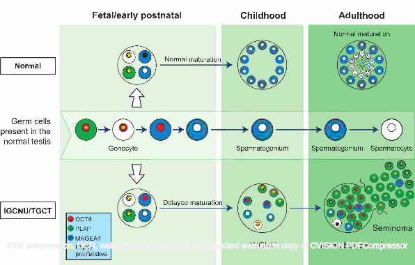
Figure 9 - Schematic for germ cell maturation and proliferation in germ cells during transition from 528
gonocyte to intratubular germ cell neoplasia and testicular germ cell cancer (bottom). Germ cell maturation 529
from gonocyte to initiation of spermatogenesis is represented in the testis during the different stages of life 530
(middle). For comparison, germ cell differentiation in the normal testis is also shown (top). Germ cells in the 531
fetal testis may exhibit delayed maturation with persistence of gonocyte markers through childhood. A 532
variety of germ cell protein profiles are present in the intratubular germ cell neoplasia tubule, however it is 533
the cells expressing exclusively gonocyte proteins (with no spermatogonial proteins) that are more 534
proliferative and contribute to the majority of the cells in intratubular seminoma and subsequently invasive 535
seminoma. Cells expressing spermatogonial proteins are occasionally seen in the tubule or resultant tumour 536
but exhibit low proliferation rates. Expression of OCT4 (red), PLAP (green) and MAGEA4 (blue) is shown 537
for individual cells and cells with high rates of proliferation are indicated (yellow asterisk).538
PDF compression, OCR, web optimization using a watermarked evaluation copy of CVISION PDFCompressor
Rod Mitchell 22
Supplementary Figures 539
Figure S1 540
Expression of OCT4 (red) and VASA (green) in a seminiferous cord from a 14 week gestation human fetal 541
testis. Sub-populations of gonocytes (OCT4+/VASA-; white arrowhead), pre-spermatogonia (OCT4+/VASA+; 542
white arrow) and spermatogonia (OCT4-/VASA+; yellow arrowhead) are present within the tubule. 543
544
Figure S2 545
Representative image for expression of AP2γ (B; green) and OCT4 (C; red) in an intratubular germ cell 546
neoplasia containing tubule from patients with testicular germ cell cancer. Intratubular germ cell neoplasia 547
cells co-express both proteins in all cells (D; yellow). Nuclear counterstain with DAPI (A; blue). 548
549
Figure S3 550
Proportion of putative intratubular germ cell neoplasia cells expressing MAGEA4 in pre-invasive disease in 551
childhood (PRE-INV. Child; n=4) and adulthood (PRE-INV. Adult; n=6), seminoma (SEM; n=9) and non-552
seminoma (NON-SEM; n=8). Mean +/- SEM. 553
554
Figure S4 555
Example of triple immunofluorescence used for quantification of proliferation in sub-populations of 556
intratubular germ cell neoplasia cells. Expression of Ki67 (green), OCT4 (red) and MAGEA4 (blue) in 557
tubules containing intratubular germ cell neoplasia from a patient with testicular germ cell cancer. 558
Arrowheads indicate proliferating OCT4+/MAGEA4- intratubular germ cell neoplasia cells, whilst the arrow 559
indicates a proliferating OCT4+/MAGEA4+ intratubular germ cell neoplasia cell. 560
561
Figure S5 562
Proliferation (Ki67+) in MAGEA4 expressing intratubular germ cell neoplasia (OCT4+) cells in children with 563
pre-invasive disease (C; Pre-Inv. Child; n=4), adults with pre-invasive disease (D; Pre-Inv. Adult; n=6), 564
seminoma (n=7) and non-seminoma (n=8). Mean +/- SEM. 565
566
Figure S6 567
PDF compression, OCR, web optimization using a watermarked evaluation copy of CVISION PDFCompressor

Rod Mitchell 23
Representative image of Ki67 (green), OCT4 (red) and MAGEA4 (blue) expression in seminoma (n=9). 568
Seminoma cells express OCT4 (but not MAGEA4) and a large proportion also express Ki67, whilst the 569
OCT4+ intratubular germ cell neoplasia cells (*) are less proliferative. MAGEA4+ cells are present in 570
adjacent tubules (#) and are Ki67-. MAGEA4+/OCT4- cells are occasionally seen interspersed between the 571
seminoma cells (arrow; inset). Counterstain (DAPI; pale blue) in merged panel (bottom right). 572
573
574
PDF compression, OCR, web optimization using a watermarked evaluation copy of CVISION PDFCompressor
PDF compression, OCR, web optimization using a watermarked evaluation copy of CVISION PDFCompressor

PDF compression, OCR, web optimization using a watermarked evaluation copy of CVISION PDFCompressor
PDF compression, OCR, web optimization using a watermarked evaluation copy of CVISION PDFCompressor
PDF compression, OCR, web optimization using a watermarked evaluation copy of CVISION PDFCompressor
PDF compression, OCR, web optimization using a watermarked evaluation copy of CVISION PDFCompressor
PDF compression, OCR, web optimization using a watermarked evaluation copy of CVISION PDFCompressor

PDF compression, OCR, web optimization using a watermarked evaluation copy of CVISION PDFCompressor

PDF compression, OCR, web optimization using a watermarked evaluation copy of CVISION PDFCompressor
PDF compression, OCR, web optimization using a watermarked evaluation copy of CVISION PDFCompressor
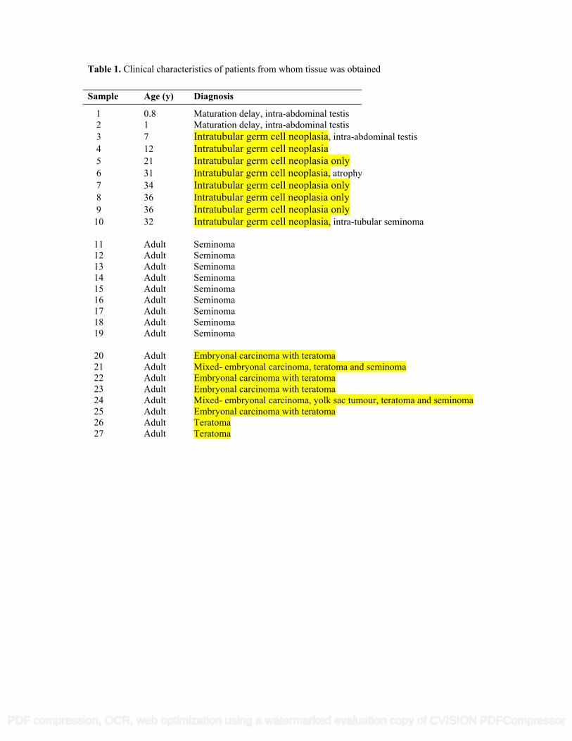
Table 1. Clinical characteristics of patients from whom tissue was obtained
1 0.8 Maturation delay, intra-abdominal testis 2 1 Maturation delay, intra-abdominal testis 3 7 Intratubular germ cell neoplasia, intra-abdominal testis 4 12 Intratubular germ cell neoplasia 5 21 Intratubular germ cell neoplasia only 6 31 Intratubular germ cell neoplasia, atrophy 7 34 Intratubular germ cell neoplasia only 8 36 Intratubular germ cell neoplasia only 9 36 Intratubular germ cell neoplasia only 10 32 Intratubular germ cell neoplasia, intra-tubular seminoma 11 Adult Seminoma 12 Adult Seminoma 13 Adult Seminoma 14 Adult Seminoma 15 Adult Seminoma 16 Adult Seminoma 17 Adult Seminoma 18 Adult Seminoma 19 Adult Seminoma 20 Adult Embryonal carcinoma with teratoma 21 Adult Mixed- embryonal carcinoma, teratoma and seminoma 22 Adult Embryonal carcinoma with teratoma 23 Adult Embryonal carcinoma with teratoma 24 Adult Mixed- embryonal carcinoma, yolk sac tumour, teratoma and seminoma 25 Adult Embryonal carcinoma with teratoma 26 Adult Teratoma 27 Adult Teratoma
Sample Age (y) Diagnosis
PDF compression, OCR, web optimization using a watermarked evaluation copy of CVISION PDFCompressor
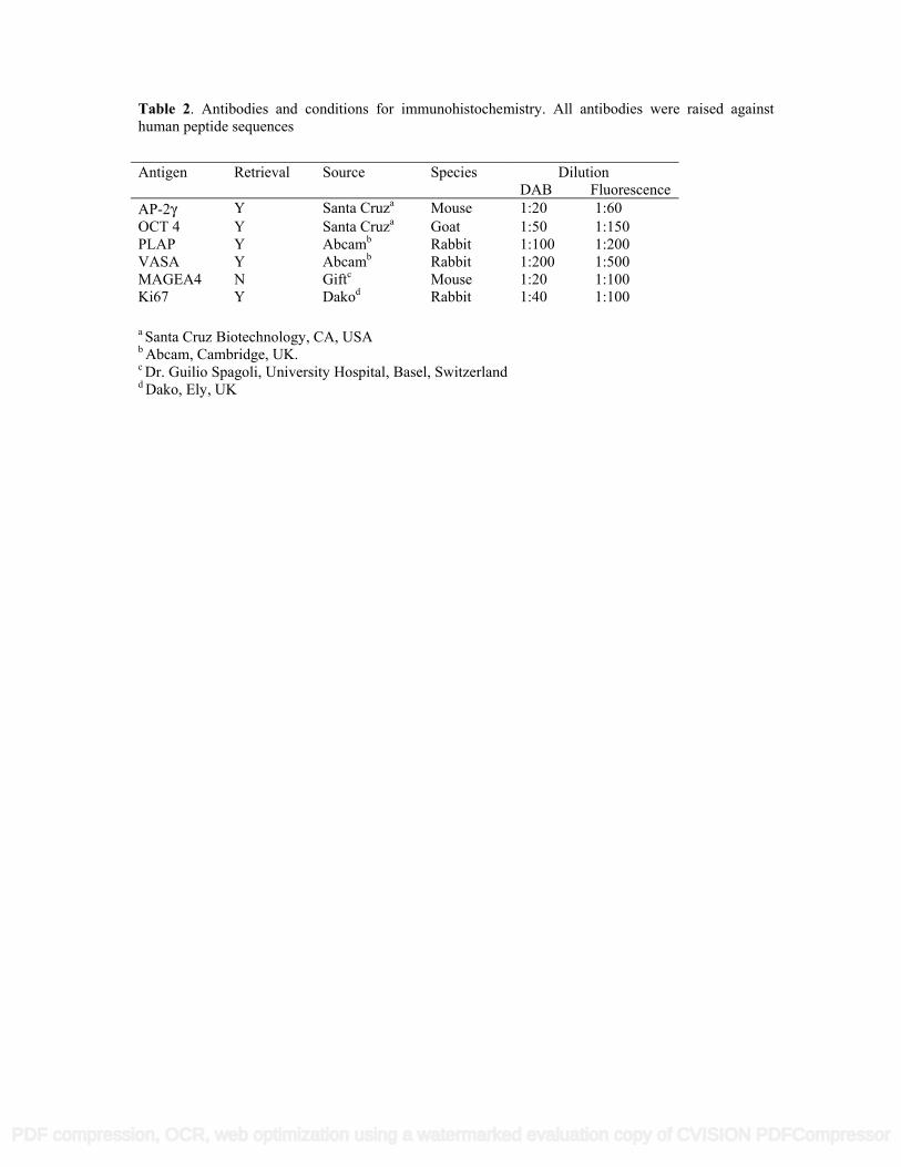
Table 2. Antibodies and conditions for immunohistochemistry. All antibodies were raised against human peptide sequences
a Santa Cruz Biotechnology, CA, USA
b Abcam, Cambridge, UK. c Dr. Guilio Spagoli, University Hospital, Basel, Switzerland d Dako, Ely, UK
Antigen Retrieval Source Species DilutionDAB Fluorescence
AP-2γ Y Santa Cruza Mouse 1:20 1:60OCT 4 Y Santa Cruza Goat 1:50 1:150PLAP Y Abcamb Rabbit 1:100 1:200VASA Y Abcamb Rabbit 1:200 1:500MAGEA4 N Giftc Mouse 1:20 1:100Ki67 Y Dakod Rabbit 1:40 1:100
PDF compression, OCR, web optimization using a watermarked evaluation copy of CVISION PDFCompressor


![Testicular tumours in children: an approach to diagnosis and … · 2020. 5. 27. · benign tumours are not included [2]. However, our per-sonal experience is that prepubertal-type](https://img.pdfslide.us/doc/110x75/60a9e5eec943202ac316820f/testicular-tumours-in-children-an-approach-to-diagnosis-and-2020-5-27-benign.jpg)


![Isolated Testicular Tuberculosis Mimicking Testicular ... involvement, but testicular involvement is an unusual clinical condition [3]. In this report, a case with isolated testicular](https://img.pdfslide.us/doc/110x75/5f3d57bf74280d66ef795ba2/isolated-testicular-tuberculosis-mimicking-testicular-involvement-but-testicular.jpg)