Embed Size (px)
Citation preview
Edinburgh Research Explorer
Forkhead Transcription Factor Fd3F Cooperates with Rfx toRegulate a Gene Expression Program for Mechanosensory CiliaSpecialization
Citation for published version:Newton, FG, zur Lage, PI, Karak, S, Moore, DJ, Göpfert, MC & Jarman, AP 2012, 'Forkhead TranscriptionFactor Fd3F Cooperates with Rfx to Regulate a Gene Expression Program for Mechanosensory CiliaSpecialization' Developmental Cell, vol 22, no. 6, pp. 1221-1233. DOI: 10.1016/j.devcel.2012.05.010
Digital Object Identifier (DOI):10.1016/j.devcel.2012.05.010
Link:Link to publication record in Edinburgh Research Explorer
Document Version:Publisher's PDF, also known as Version of record
Published In:Developmental Cell
Publisher Rights Statement:Available under Open Access.ª2012 Elsevier Inc.
General rightsCopyright for the publications made accessible via the Edinburgh Research Explorer is retained by the author(s)and / or other copyright owners and it is a condition of accessing these publications that users recognise andabide by the legal requirements associated with these rights.
Take down policyThe University of Edinburgh has made every reasonable effort to ensure that Edinburgh Research Explorercontent complies with UK legislation. If you believe that the public display of this file breaches copyright pleasecontact [email protected] providing details, and we will remove access to the work immediately andinvestigate your claim.
Download date: 16. Jul. 2018
Developmental Cell
Article
Forkhead Transcription Factor Fd3F Cooperateswith Rfx to Regulate a Gene Expression Programfor Mechanosensory Cilia SpecializationFay G. Newton,1 Petra I. zur Lage,1 Somdatta Karak,2 Daniel J. Moore,1 Martin C. Gopfert,2 and Andrew P. Jarman1,*1Centre for Integrative Physiology, School of Biomedical Sciences, University of Edinburgh, George Square, Edinburgh EH8 9XD, UK2Department of Cellular Neurobiology, University of Gottingen, Burckhardtweg 13, 37077 Gottingen, Germany
*Correspondence: [email protected] 10.1016/j.devcel.2012.05.010
SUMMARY
Cilia have evolved hugely diverse structures andfunctions to participate in a wide variety of develop-mental and physiological processes. Ciliary speciali-zation requires differences in gene expression, butfew transcription factors are known to regulate this,and their molecular function is unclear. Here, weshow that the Drosophila Forkhead box (Fox) gene,fd3F, is required for specialization of the mechano-sensory cilium of chordotonal (Ch) neurons. fd3Fregulates genes for Ch-specific axonemal dyneinsand TRPV ion channels, which are required forsensory transduction, and retrograde transportgenes,whichare required todifferentiate theirdistinctmotile and sensory ciliary zones. fd3F is reminiscentof vertebrate Foxj1, a motile cilia regulator, but fd3Fregulates motility genes as part of a broader sensoryregulation program. Fd3F cooperates with the pan-ciliary transcription factor, Rfx, to regulate its targetsdirectly. This illuminates pathways involved in ciliaryspecialization and the molecular mechanism of tran-scription factors that regulate them.
INTRODUCTION
The cilium commonly constitutes a cell’s organelle for environ-
mental sensing in a wide variety of contexts, from developmental
signaling pathways (such as Sonic hedgehog) to the specialized
receptor processes in sense organs of various sensory modali-
ties (Ishikawa and Marshall, 2011). In other situations, cilia are
motile and play many roles connected to fluid movement in the
airways, CNS, oviduct, and embryonic node. Despite this struc-
tural and functional diversity, cilia share a highly conserved
pathway of ciliogenesis, involving basal body docking, axoneme
extension, intraflagellar transport (IFT), and ciliary membrane
assembly (Silverman and Leroux, 2009). A major question is
how this common assembly program is adapted and modified
with cell-specific variations to generate cilia diversity (Silverman
and Leroux, 2009). It is likely that such specialization requires
cell-type-specific gene expression programs, but little is known
of the nature of these programs or of the transcription factors
that regulate them (Thomas et al., 2010).
Developm
In metazoans ciliogenesis broadly depends on regulatory
factor X (Rfx) transcription factors (Chu et al., 2010). Their targets
are well characterized in functional and bioinformatic studies on
C. elegans and Drosophila (Avidor-Reiss et al., 2004; Efimenko
et al., 2005; Laurencon et al., 2007), and include genes required
for ‘‘core’’ ciliogenesis processes such as anterograde IFT and
membrane assembly. If Rfx is required broadly for ciliogenesis,
what regulates cilium specialization? In fact Rfx targets include
genes restricted to subtypes of ciliated cell (Efimenko et al.,
2005), but it is not clear how it regulates such genes. It is spec-
ulated that Rfx cooperates with cell-type-specific factors (Silver-
man and Leroux, 2009; Thomas et al., 2010), but the nature of
this cooperation is uncharacterized.
Few cell-type-specific regulators of ciliogenesis have been
characterized. The most well known are members of the FoxJ
subfamily. In vertebrates, Foxj1 has been associated particularly
with the differentiation of motile ciliated cell types (Brody et al.,
2000; Jacquet et al., 2009; Stubbs et al., 2008; Yu et al., 2008).
For instance, Foxj1 knockout mice have left-right asymmetry
and airway defects (Brody et al., 2000). Target gene analyses
have shown that Foxj1 regulates, directly or indirectly, many
genes linked to ciliary motility, including axonemal dyneins, but
not core ciliogenesis, such as IFT factors (Jacquet et al., 2009;
Stubbs et al., 2008; Thomas et al., 2010; Yu et al., 2008).
However, very little is known of its molecular mode of action or
its relationship with Rfx function.
In Drosophila the only somatic cells with cilia are bipolar
sensory neurons, which have specialized ciliary dendrites for
sensory reception and transduction (Figures 1A–1C). Different
classes of such neurons have ciliary dendrites that have different
morphologies, express different sets of receptor molecules, and
respond to different sensory modalities. These neurons present
a useful model for investigating how ciliary diversity arises.
External sensory (ES) neurons have a short connecting cilium
leading to a distal sensory process with an irregular core of
microtubules. In contrast, proprioceptive and auditory chordoto-
nal (Ch) neurons have a long sensory cilium with a well-defined
9+0 axoneme (Figures 1B and 1C) (Eberl et al., 2000; Kernan
et al., 1994). The Ch cilium has several unique specializations.
First, its mechanosensory transduction mechanism uniquely
involves TRPV (transient receptor potential) channels encoded
by nanchung (nan) and inactive (iav) (Gong et al., 2004; Kim
et al., 2003). Second, it can be motile. In the auditory Ch neurons
of Johnston’s organ in the antenna (Figure 1B), motility arises
from the interplay between transduction channels and adapta-
tion motors (possibly axonemal dyneins). Sensory-induced
ental Cell 22, 1221–1233, June 12, 2012 ª2012 Elsevier Inc. 1221
fd3F mRNA
Anti-Fd3FAnti-Fd3F 22C10
Scolopale
Ciliary dilation
Cilium
Proximalzone
Distalzone
Dendritic capJohnston’s organ
AntennaCell body
Axon
lch5
v’ch1vchAB
dch3
lch5fco
v’ch1
vchAB
dch3
Dendrites
Ciliary rootlet
CBA
E FD
v’ch1
vchB
vchA
lch5
desC desD
des2 desB
lesC
lesB
lesA
v’es2
vesB vesA
vesC v’esA v’esB
Figure 1. fd3F Is Expressed Specifically in Developing Ch Neurons
(A) Schematic arrangement of ciliated sensory neurons in an embryonic abdominal segment. Ch neurons, green; ES neurons, blue.
(B) Schematic of the array of Ch neurons in the adult antenna that form Johnston’s organ.
(C) Schematic of a unit Ch organ from Johnston’s organ, housing two Ch neurons.
(D) fd3F mRNA in stage 14 embryos.
(E) Fd3F protein localized in a subset of sensory neurons (marked by 22C10) corresponding to Ch neurons (stage 15 embryo).
(F) Leg imaginal disc, Fd3F protein in Ch precursor cells of the femoral chordotonal organ (fco).
Scale bars, 100 mm (D and F) and 20 mm (E).
Developmental Cell
Gene Regulation for Ciliary Specialization
motility is thought to underlie a mechanical amplification mech-
anism to increase auditory sensitivity (Gopfert et al., 2005;
Gopfert and Robert, 2003). Third, the Ch neuron cilium is divided
by a ciliary dilation into functionally distinct proximal (motile) and
distal (sensory) zones (Figure 1C) (Lee et al., 2008).
We asked, therefore, what regulates gene expression for Ch
neuron-specific ciliary specialization. Rfx is expressed in and
required for ciliogenesis in both ES and Ch neurons (Dubruille
et al., 2002; Laurencon et al., 2007), whereas Foxj1 homologs
are reportedly absent from Drosophila (Larroux et al., 2008;
Mazet et al., 2003). We recently reported, however, that the
Fox gene, fd3F, is required for Ch neuron function (Cachero
et al., 2011). Here, we show that fd3F does not regulate ciliogen-
esis per se but directly regulates the genes required for aspects
of Ch ciliary specialization. It appears that Fd3F cooperates
closely with Rfx to regulate this Ch-specific cohort of genes
and, therefore, it acts as a cell-type-specific modulator of Rfx
target gene specificity. Comparison with Foxj1 and its target
genes suggests that fd3F is a highly diverged relative of Foxj1.
RESULTS
fd3F Mutation Results in Uncoordinated Flieswith Nonfunctional Ch NeuronsPreviously, a transcriptome analysis of embryonic Ch cells
revealed that fd3F (CG12632) was highly enriched in developing
1222 Developmental Cell 22, 1221–1233, June 12, 2012 ª2012 Elsev
Ch neurons, and RNA in situ hybridization and immunostaining
showed that fd3F is exclusively expressed in Ch cell lineages
during embryogenesis (Cachero et al., 2011) (Figures 1D and
1E). In larval imaginal discs, fd3F expression was confined
to locations of Ch precursors, including the femoral Ch precur-
sors in the leg disc (Figure 1F) and the Ch precursors of
Johnston’s organ in the antennal disc (data not shown). In
summary, fd3F expression is unique to developing and differen-
tiating Ch neurons.
Using the fly stock P{EP}fd3FEP1198, we generated a deletion
of 1.4 kb (fd3F1) that removes approximately 400 bp of the
30 end of the fd3FORF (see Figure S1A available online) (Cachero
et al., 2011). fd3FmRNAexpressionwas very strongly reduced in
homozygous mutant embryos (Figures S1B and S1C), suggest-
ing that the truncated fd3F transcripts are unstable and that the
mutation is close to a genetic null. Consistent with this, little
difference was observed between the phenotypes of fd3F1
homozygotes and hemizygotes over a deficiency (fd3F1/Df(1)
ED6716) in the analyses below.
fd3F1 homozygote flies are viable and fertile but are uncoordi-
nated (Cachero et al., 2011), and hence, the adult flies perform
very poorly in a climbing assay (Figure 2E). This suggests that
Ch neurons are defective in fd3Fmutants. However, Ch neurons
were present in fd3F1 embryos, larvae, and pupae and showed
little gross morphological defect (Figures 2A and 2B). When
marked by the expression of mCD8-GFP, the Ch sensory cilia
ier Inc.
Sound particle velocity (mm/s)
Rel
ativ
e C
AP
ampl
itude
Max
imum
CA
P (
V)
µ
w11
18
fd3F 1
fd3F1
fd3F1
wild-type
wild-type
A B C D
E F
50
100
w11
18
fd3F /
FM6
1
p{EP}
fd3F /
fd3F
11
***
Clim
b %
fd3F1
Dhc
93A
BC
G37
69
lch5
CG
1125
3
lch5
wild-typeG H
JI
K L
Figure 2. Mutation of fd3F Results in Functionally Defective Ch Neurons
(A and B) Cell bodies and dendrites of Ch neuron group (lch5) in third-instar larvae stained with anti-HRP.
(C and D) Adult femoral Ch organ neurons showing sensory cilia (arrows) labeled with mCD8-GFP (elavGal4;UAS-mCD8-GFP).Scale bars, 5 mm.
(E) Climbing assay showing percentage of adult flies climbing above a threshold. Genotypes are wild-type (w1118), flies homozygous for the P element insert at
fd3F (p{EP}), fd3F1 heterozygotes (fd3F1/FM6 balancer), and fd3F1 homozygotes. Error bars are SD. ***p < 0.0001.
(F) Shown on the left are relative CAP amplitudes recorded from the projections of Johnston’s organ neurons in the antennal nerve (inset) as a function of the
sound particle velocity. Shown on the right are corresponding absolute values of the maximum CAP amplitudes (n = 5 flies per strain). Error bars are SD.
(G–L) Genes that show reduced expression in fd3F mutant embryos. Wild-type (left) and mutant (right) embryos showing mRNA expression of Ch-expressed
genes (named at left) are shown. Expression is either virtually absent (e.g., Dhc93AB) or reduced (e.g., CG11253). Scale bars, 100 mm.
See also Figure S1.
Developmental Cell
Gene Regulation for Ciliary Specialization
were present and grossly normal (Figures 2C and 2D). To assess
the functional integrity of Ch neurons in fd3Fmutants, we exam-
ined the activity of the auditory Ch neurons of Johnston’s organ.
Compound action potentials (CAPs) were recorded in response
to auditory stimulus from the antennal nerve of adult flies. Unlike
wild-type flies, fd3Fmutant flies showed no sound-evoked CAPs
in the antennal nerve (Figure 2F). These data suggest that fd3F
regulates aspects of Ch neuronal differentiation that are essen-
tial for Ch neuron function.
Developm
fd3F Regulates a Subset of Ch-Specific GenesAs we previously reported (Cachero et al., 2011), fd3F is required
for the expression of the genes nan and iav, which encode
subunits of a TRPV cation channel uniquely localized to
Ch neuron cilia (Gong et al., 2004; Kim et al., 2003). To identify
other regulatory targets of fd3F, we began by examining
candidate Ch-expressed genes identified previously in our
transcriptome analysis (Cachero et al., 2011). Of 16 genes tested
initially, 6 showed reduced expression in fd3F mutant embryos
ental Cell 22, 1221–1233, June 12, 2012 ª2012 Elsevier Inc. 1223
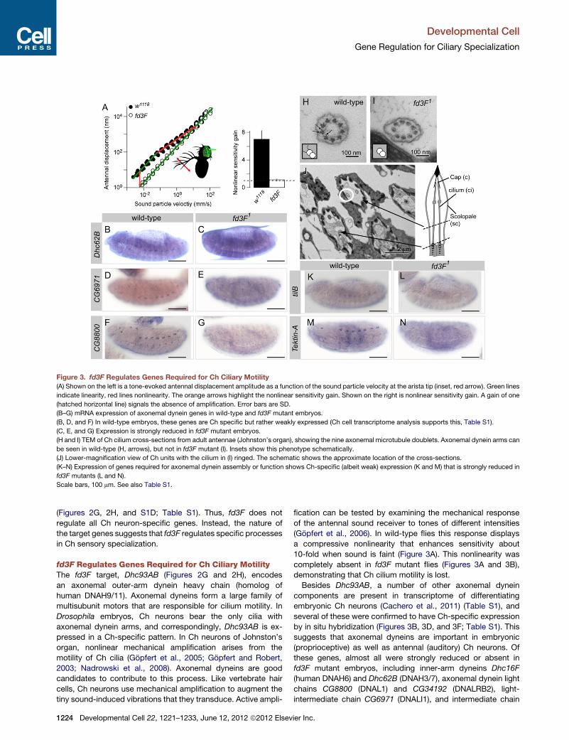
Figure 3. fd3F Regulates Genes Required for Ch Ciliary Motility
(A) Shown on the left is a tone-evoked antennal displacement amplitude as a function of the sound particle velocity at the arista tip (inset, red arrow). Green lines
indicate linearity, red lines nonlinearity. The orange arrows highlight the nonlinear sensitivity gain. Shown on the right is nonlinear sensitivity gain. A gain of one
(hatched horizontal line) signals the absence of amplification. Error bars are SD.
(B–G) mRNA expression of axonemal dynein genes in wild-type and fd3F mutant embryos.
(B, D, and F) In wild-type embryos, these genes are Ch specific but rather weakly expressed (Ch cell transcriptome analysis supports this, Table S1).
(C, E, and G) Expression is strongly reduced in fd3F mutant embryos.
(H and I) TEM of Ch cilium cross-sections from adult antennae (Johnston’s organ), showing the nine axonemal microtubule doublets. Axonemal dynein arms can
be seen in wild-type (H, arrows), but not in fd3F mutant (I). Insets show this phenotype schematically.
(J) Lower-magnification view of Ch units with the cilium in (I) ringed. The schematic shows the approximate location of the cross-sections.
(K–N) Expression of genes required for axonemal dynein assembly or function shows Ch-specific (albeit weak) expression (K and M) that is strongly reduced in
fd3F mutants (L and N).
Scale bars, 100 mm. See also Table S1.
Developmental Cell
Gene Regulation for Ciliary Specialization
(Figures 2G, 2H, and S1D; Table S1). Thus, fd3F does not
regulate all Ch neuron-specific genes. Instead, the nature of
the target genes suggests that fd3F regulates specific processes
in Ch sensory specialization.
fd3F Regulates Genes Required for Ch Ciliary MotilityThe fd3F target, Dhc93AB (Figures 2G and 2H), encodes
an axonemal outer-arm dynein heavy chain (homolog of
human DNAH9/11). Axonemal dyneins form a large family of
multisubunit motors that are responsible for cilium motility. In
Drosophila embryos, Ch neurons bear the only cilia with
axonemal dynein arms, and correspondingly, Dhc93AB is ex-
pressed in a Ch-specific pattern. In Ch neurons of Johnston’s
organ, nonlinear mechanical amplification arises from the
motility of Ch cilia (Gopfert et al., 2005; Gopfert and Robert,
2003; Nadrowski et al., 2008). Axonemal dyneins are good
candidates to contribute to this process. Like vertebrate hair
cells, Ch neurons use mechanical amplification to augment the
tiny sound-induced vibrations that they transduce. Active ampli-
1224 Developmental Cell 22, 1221–1233, June 12, 2012 ª2012 Elsev
fication can be tested by examining the mechanical response
of the antennal sound receiver to tones of different intensities
(Gopfert et al., 2006). In wild-type flies this response displays
a compressive nonlinearity that enhances sensitivity about
10-fold when sound is faint (Figure 3A). This nonlinearity was
completely absent in fd3F mutant flies (Figures 3A and 3B),
demonstrating that Ch cilium motility is lost.
Besides Dhc93AB, a number of other axonemal dynein
components are present in transcriptome of differentiating
embryonic Ch neurons (Cachero et al., 2011) (Table S1), and
several of these were confirmed to have Ch-specific expression
by in situ hybridization (Figures 3B, 3D, and 3F; Table S1). This
suggests that axonemal dyneins are important in embryonic
(proprioceptive) as well as antennal (auditory) Ch neurons. Of
these genes, almost all were strongly reduced or absent in
fd3F mutant embryos, including inner-arm dyneins Dhc16F
(human DNAH6) and Dhc62B (DNAH3/7), axonemal dynein light
chains CG8800 (DNAL1) and CG34192 (DNALRB2), light-
intermediate chain CG6971 (DNALI1), and intermediate chain
ier Inc.
Developmental Cell
Gene Regulation for Ciliary Specialization
CG13930 (IC138, WDR78) (Figures 3B–3G; Table S1). Together,
these observations suggest that fd3F regulates axonemal
dynein genes in general. To check that this also pertains to the
Johnston’s organ, Ch neuron ultrastructure in the antenna was
examined by transmission electron microscopy (TEM). In trans-
verse sections of mutant ciliary dendrites, normal axoneme
arrangement and ultrastructure were observed, but dynein outer
and inner arms were missing from the proximal cilium (Figures
3H–3J). Loss of dyneins with consequent loss of motility can
account for the loss of mechanical amplification by Johnston’s
organ neurons in fd3F mutants, although fd3F might also
regulate genes of the sensory transduction apparatus that are
required for the process (Nadrowski et al., 2008).
Several genes have been implicated in axonemal dynein
assembly. tilB is required for transport or assembly of axonemal
dyneins (Kavlie et al., 2010), and CG14905 is similar to
Chlamydomonas ODA-1, which is required for axonemal dynein
assembly (Kamiya, 1988). Ch neuron expression of both genes
was reduced in fd3F mutant embryos (Figures 3K and 3L; data
not shown). Tektins are required for correct motility of cilia, prob-
ably by participating in dynein inner-arm assembly or attachment
(Amos, 2008). We found that Tektin-A (TEKT4 ortholog) is
expressed exclusively in Ch neurons and that its expression is
strongly reduced in fd3F mutants (Figures 3M and 3N). Thus,
fd3F regulates genes required for several aspects of dynein
arm formation.
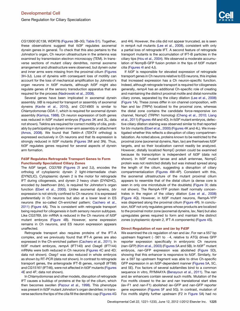
Fd3F Regulates Retrograde Transport Genes to FormFunctionally Specialized Ciliary ZonesThe fd3F target, CG3769 (Figures 2I and 2J), encodes the
ortholog of cytoplasmic dynein 2 light-intermediate chain
(DYN2LIC). Cytoplasmic dynein 2 is the motor for retrograde
IFT during ciliogenesis, and dynein 2 heavy chain (DYN2HC),
encoded by beethoven (btv), is required for Johnston’s organ
function (Eberl et al., 2000). Unlike axonemal dyneins, btv
expression is not strictly confined to Ch neurons: it is expressed
preferentially in Ch neurons but also at a lower level in ES
neurons (the so-called Ch-enriched pattern; Cachero et al.,
2011) (Figure 4A). This is consistent with retrograde transport
being required for ciliogenesis in both sensory neuron subtypes.
Like CG3769, btv mRNA is reduced in the Ch neurons of fd3F
mutant embryos (Figure 4B). However, some expression
remains in Ch neurons, and ES neuron expression appears
unaffected.
Retrograde transport also requires proteins of the IFT-A
complex, and we previously found that IFT-A genes are also
expressed in the Ch-enriched pattern (Cachero et al., 2011). In
fd3F mutant embryos, rempA (IFT140) and Oseg6 (IFT144)
mRNAs were both reduced in Ch neurons (Figures 4C and 4D;
data not shown). Oseg1 was also reduced in whole embryos
as shown by RT-PCR (data not shown). In contrast to retrograde
transport genes, the anterograde IFT-B genes, nompB (IFT88)
andCG15161 (IFT46), were not affected in fd3Fmutants (Figures
4E and 4F; data not shown).
InChlamydomonas and other models, disruption of retrograde
IFT causes a buildup of proteins at the tip of the cilium, which
then becomes swollen (Pazour et al., 1998). This phenotype
was present in fd3Fmutant Johnston’s organ dendrites: in trans-
verse sections the tips of the cilia fill the dendritic cap (Figures 4G
Developm
and 4H). However, the cilia did not appear truncated, as is seen
in rempA null mutants (Lee et al., 2008), consistent with only
a partial loss of retrograde IFT. A second feature of retrograde
transport mutants is the accumulation of IFT-B particles in the
ciliary tips (Hou et al., 2004). We observed a moderate accumu-
lation of NompB-GFP fusion protein in the tips of fd3F mutant
cilia (Figures 4I and 4J).
If fd3F is responsible for elevated expression of retrograde
transport genes in Ch neurons relative to ES neurons, this implies
that increased expression has a Ch neuron-specific function.
Indeed, although retrograde transport is required for ciliogenesis
generally, rempA has an additional Ch-specific role of creating
and maintaining the distinct proximal motile and distal nonmotile
ciliary zones, separated by the ciliary dilation (Lee et al., 2008)
(Figure 1A). These zones differ in ion channel composition, with
Nan and Iav (TRPV) localized to the proximal zone, whereas
the distal zone contains the candidate force-responsive ion
channel, NompC (TRPN1 homolog) (Cheng et al., 2010; Liang
et al., 2011) (Figures 4M and 4O). In fd3Fmutant embryos, defec-
tive ciliary dilation staining was observed similar to that reported
for btvmutants (Eberl et al., 2000) (Figures 4K and 4L). We inves-
tigated whether this reflects a disruption of ciliary compartmen-
talization. As noted above, proteins known to be restricted to the
proximal zone (axonemal dyneins, Nan, Iav) are themselves fd3F
targets, and so their localization cannot readily be analyzed.
However, distally localized NompC protein could be examined
because its transcription is independent of fd3F (data not
shown). In fd3F mutant larvae and adult antennae, NompC
protein was not restricted distally but was instead spread along
the length of the cilium, suggesting a disruption of ciliary
compartmentalization (Figures 4M–4P). Consistent with this,
the axonemal ultrastructure of the mutant proximal cilium
appears to resemble that of the distal cilium (a dense core is
seen in only one microtubule of the doublets) (Figure 3I; data
not shown). The RempA-YFP protein itself normally concen-
trates in the region of the ciliary dilation (Lee et al., 2008)
(Figure 4Q). However, in fd3F mutant neurons, RempA-YFP
was dispersed along the proximal cilium (Figure 4R). In conclu-
sion, fd3F not only regulates genes whose products are localized
to the proximal motor zone (axonemal dyneins, Iav, Nan) but also
upregulates genes required to form and maintain the distinct
zones (cytoplasmic dynein 2, IFT-A components) (Figure 4S).
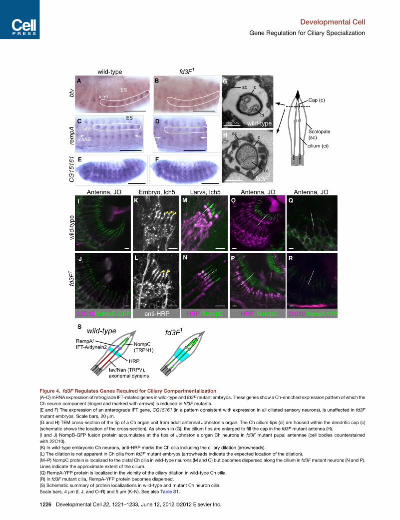
Direct Regulation of nan and iav by Fd3FWe examined the cis regulation of nan and iav. For nan a 557 bp
upstream fragment (�561 to �4, relative to ATG) drives GFP
reporter expression specifically in embryonic Ch neurons
(nan-GFP) (Kim et al., 2003) (Figures 5A and 5B). In fd3F1 mutant
embryos, nan-GFP expression was abolished (Figure 5D),
showing that this enhancer is responsive to fd3F. Similarly, for
iav a 597 bp upstream fragment was able to drive Ch-specific
GFP expression in an fd3F-dependent manner (Figures 5A, 5C,
and 5E). Fox factors of several subfamilies bind to a common
sequence in vitro, RYMAAYA (Benayoun et al., 2011). The nan
and iav enhancers contain several such motifs. Mutation of the
Fox motifs closest to the iav and nan translational start sites
(iav-F1 and nan-F1) abolished iav-GFP and nan-GFP reporter
gene expression (Figures 5F and 5G). In contrast, mutation of
Fox motifs slightly further upstream (F2 in Figure 5A) had no
ental Cell 22, 1221–1233, June 12, 2012 ª2012 Elsevier Inc. 1225
fd3F1
fd3F500 nm
500 nm
1
wild-type
wild-type
fd3F
1w
ild-ty
peA B
D
FE
C
G
H
I
J N
O Q
P R
S
MK
L
btv
rem
pAC
G15
161
NompB-GFP22C10 anti-HRP NompCHRP HRP NompC 22C10 RempA-YFP
Larva, lch5Antenna, JO Antenna, JO Antenna, JOEmbryo, lch5
lch5
lch5ES
ES
vchAB
RempA/IFT-A/dynein2
HRP
NompC(TRPN1)
Iav/Nan (TRPV),axonemal dyneins
wild-type
Scolopale(sc)
sc c
ci
Cap (c)
cilium (ci)
fd3F1
scc
ci
Figure 4. fd3F Regulates Genes Required for Ciliary Compartmentalization
(A–D)mRNA expression of retrograde IFT-related genes in wild-type and fd3Fmutant embryos. These genes show aCh-enriched expression pattern of which the
Ch neuron component (ringed and marked with arrows) is reduced in fd3F mutants.
(E and F) The expression of an anterograde IFT gene, CG15161 (in a pattern consistent with expression in all ciliated sensory neurons), is unaffected in fd3F
mutant embryos. Scale bars, 20 mm.
(G and H) TEM cross-section of the tip of a Ch organ unit from adult antennal Johnston’s organ. The Ch cilium tips (ci) are housed within the dendritic cap (c)
(schematic shows the location of the cross-section). As shown in (G), the cilium tips are enlarged to fill the cap in the fd3F mutant antenna (H).
(I and J) NompB-GFP fusion protein accumulates at the tips of Johnston’s organ Ch neurons in fd3F mutant pupal antennae (cell bodies counterstained
with 22C10).
(K) In wild-type embryonic Ch neurons, anti-HRP marks the Ch cilia including the ciliary dilation (arrowheads).
(L) The dilation is not apparent in Ch cilia from fd3F mutant embryos (arrowheads indicate the expected location of the dilation).
(M–P) NompC protein is localized to the distal Ch cilia in wild-type neurons (M and O) but becomes dispersed along the cilium in fd3Fmutant neurons (N and P).
Lines indicate the approximate extent of the cilium.
(Q) RempA-YFP protein is localized in the vicinity of the ciliary dilation in wild-type Ch cilia.
(R) In fd3F mutant cilia, RempA-YFP protein becomes dispersed.
(S) Schematic summary of protein localizations in wild-type and mutant Ch neuron cilia.
Scale bars, 4 mm (I, J, and O–R) and 5 mm (K–N). See also Table S1.
Developmental Cell
Gene Regulation for Ciliary Specialization
1226 Developmental Cell 22, 1221–1233, June 12, 2012 ª2012 Elsevier Inc.
fd3F1 fd3F1
Rfx–
XF
F F F
1kb FXF
F2 X boxCTGGTCTCATTGGTTTGGCTGGGTGGGTGGGGTTACCAGGACAACGAGCT ** *****C*C** * * *** ****G*******G*** * F1GGCTTCCTGCCTCTTGATATCATGGGTCATCGAACAAACAAGCCGAGAAG* * ** * * ** ** ************** ***
F2 X boxACAGCGTAAACAAAGCAAAGGAGCTCAAGTTCAGCTGTTGCATTGGCAAC ** ******* * ** ******************************* F1GGACGTGCACGGAAATGTTTTATCAATAGCCACCAATGGACAAGGACCTC** ** * * ********G**G*** *** ** *********** *
nanchungnuf
pod1 inactive
nan-GFP
wild-typeB C
E
K LK M N
D
G
A
F
HI J
wild-type
wild-typewild-type
lch5
v’ch1 v’ch1
vchAB vchAB
lch5 lch5
vchAB
vchAB
v’ch1
lch5
vchAB
v’ch1
v’ch1
lch5 lch5lch5
wild-type
iav RNA iav RNA
nan-F1*
Freeprobe
Complex
iav-F1*
FkhFkh
nan-GFP
iav-GFP
iav-GFP
nan-GFP-F1m iav-GFP-F1m
nan-GFPiav-GFP
nan-GFP-Xmiav-GFP-Xm
nan-F1m – – – + – – – – + – iav-F1mnan-F1 – – – – + – – – – + iav-F1Fd3F – + + + + – + + + + Fd3F
Figure 5. Fd3F Directly Regulates nan and iav with Rfx
(A) Schematics of the nan and iav genes showing locations of Foxmotifs (F) and X boxes (X) in their upstream regions. The sequences surrounding the X boxes are
shown, with asterisks representing identity in D. pseudoobscura.
(B and C) Stage 16 embryos showing expression of GFP in Ch neurons for nan-GFP (B) and iav-GFP (C) reporter genes.
(D and E) Expression of both reporter genes is lost in fd3F mutant embryos.
(F and G) Expression of both reporter genes is abolished upon mutation of a single Fox motif (F1) in either upstream region.
(H) Gel mobility shift assay with oligonucleotide probes (*) containing the nan-F1 or iav-F1 Fox motifs and purified Fd3F forkhead domain polypeptide (Fd3FFkh).
TheDNA-Fd3FFkh complexes are indicated. Addition of 100-fold excess of cold competitor oligonucleotide (nan-F1, iav-F1) reduces complex formation, but when
the Fox motif is mutated (nan-F1m, iav-F1m), the cold competitor has no effect on complex formation.
(I and J) Embryonic expression of iav in Ch neurons (I) is lost in Rfx49 homozygous mutant embryo (J) (embryos stained in parallel).
(K–N) Two abdominal segments from stage 16 embryos stained with anti-GFP (green) and 22C10 (magenta). Ch neuron expression of nan-GFP (K) and iav-GFP
(M) reporter genes is abolished when the upstream X box is mutated (L and N).
Scale bars, 100 mm (B–J) and 20 mm (K–M). See also Table S1.
Developmental Cell
Gene Regulation for Ciliary Specialization
effect on reporter gene expression (data not shown). To test
whether Fd3F can bind to these F1 motifs, the DNA binding
Forkhead domain of Fd3F (Fd3FFkd) was expressed as a GST
fusion protein and isolated from bacterial cells. When used in
an in vitro gel mobility shift assay, GST-Fd3FFkd was able to
bind specifically to the F1 sites of both nan and iav (Figure 5H).
Coregulation of Ciliary Specialization Genes by Fd3Fand Rfx Transcription FactorsTypically, Rfx factors regulate target genes via a single upstream
binding site (X box) (Efimenko et al., 2005; Laurencon et al.,
2007). The iav and nan enhancer sequences contain an X box
motif similar to that determined biochemically for human Rfx1
Developm
(GTNRCCN{0-3}RGYAAC; Emery et al., 1996), very close to
the functional Fd3F binding sites (Figure 5A). This raises the
possibility that although Rfx is a pan-ciliary transcription factor
required for both Ch and ES neurons (Dubruille et al., 2002), it
contributes to the regulation of Ch-specific Fd3F target genes.
Indeed, both nan and iav expression were strongly reduced
in Rfx mutant embryos (Figures 5I and 5J; data not shown). In
both cases mutation of the X box in the reporter constructs
largely abolished enhancer activity (Figures 5K–5N). We
therefore conclude that an X box/Fox motif combination is
required for nan and iav regulation in Ch neurons and that most
likely these sites are bound by Rfx and Fd3F. In support of the
conclusion that these transcription factors are coregulators, we
ental Cell 22, 1221–1233, June 12, 2012 ª2012 Elsevier Inc. 1227
Developmental Cell
Gene Regulation for Ciliary Specialization
found no evidence of a regulatory hierarchy between Rfx and
fd3F: in embryos we found that fd3F transcription does not
depend on Rfx function and vice versa (data not shown).
When we examined a selection of other fd3F target genes
(including Dhc93AB, rempA), we found that their expression
was also strongly reduced in Rfx mutant embryos (Table S1).
Moreover, we observed that almost all fd3F target genes contain
a closely spaced combination of conserved Fox and X box
motifs, usually within 50 bp of the transcriptional or translational
start site (Figure 6A; Table S2). Some of these genes were
suspected Rfx direct targets from other bioinformatic analyses
(Avidor-Reiss et al., 2004; Laurencon et al., 2007) (Table S2).
However, most of these genes were not previously predicted
to be Rfx targets, partly due to the fact that the X box motif often
does not conform completely to the palindromic consensus
sequence previously considered for Drosophila targets:
GTTGCCATGGCAAC (Avidor-Reiss et al., 2004); GYTRYYN(1-3)
RRHRAC (Laurencon et al., 2007) (Figure 6B). The juxtaposition
of Fox motifs and X boxes suggests that Fd3F and Rfx
are coregulators of a subset of Ch ciliary genes. Subsequent to
the bioinformatic analysis, we confirmed that the regulation of
Dhc93AB indeedmirrors that of nan and iav. An 820 bp promoter
fragment supports Ch neuron-specific reporter gene expression,
and mutation of the proximal-most X box and Fox motifs
reduced this expression. However, in this case expression
was not completely abolished, possibly due to redundancy
with other binding sites that are present in this longer enhancer
(Figures 6C–6F).
Target Genes Are Ectopically Activated upon Fd3FMisexpressionUsing flies transgenic for an inducible UAS-fd3F construct, we
ectopically expressed Fd3F in embryonic sensory precursor
cells using a scaGal4 neuroectodermal driver line (Figures 7A
and 7B). This induced the misexpression of several fd3F target
genes in ES neurons, including CG8800 (DNAL1), Tektin-A,
CG11253, and CG31320 (Figures 7C–7H; data not shown). In
addition the nan, iav, and Dhc93AB GFP reporter gene
constructs were all ectopically expressed upon fd3Fmisexpres-
sion (Figures 7I–7L; data not shown). Thus, fd3F is sufficient to
activate aspects of Ch-specific gene expression in other sensory
neurons. Interestingly, although scaGal4 drives expression
widely in neuroectodermal cells, misexpression of target genes
appears to be confined to ES neurons (Figures 7K and 7L).
This is consistent with the idea that Fd3F function is limited to
cells expressing its coregulator, Rfx.
DISCUSSION
Fd3F is a cell-type-specific transcriptional regulator of ciliary
sensory specialization. It is exclusively expressed in mechano-
sensory Ch neurons where it regulates aspects of Ch neuron
ciliogenesis and ciliary function. Fd3F is not a Ch neuron identity
factor or ‘‘master regulator’’ of Ch neuron differentiation: neural
differentiation and ciliogenesis occur largely normally in fd3F
mutant Ch neurons as attested by general morphology, and
many Ch-specific genes are fd3F independent. Instead, fd3F
regulates a program of gene expression for mechanosensory
specialization that is unique to Ch neuron cilia and is absolutely
1228 Developmental Cell 22, 1221–1233, June 12, 2012 ª2012 Elsev
required for their response to sensory stimulation. Specifically,
fd3F targets are concerned with the structurally and functionally
distinct ciliary zones of Ch neuron dendrites: fd3F regulates
genes for both the machinery for construction and delineation
of the zones (retrograde transport) and the proteins that populate
the specialized proximal motor zone (axonemal dyneins, tektin,
TRPV proteins). fd3F mutation has two consequences: ciliary
motility of JO neurons is lost (reflected by loss of mechanical
amplification), and sensory transduction is lost (reflected by
loss of electrical response to stimulus). Effects on sensory
transduction are both direct (loss of TRPV expression) and indi-
rect (disrupted localization of candidate force-gated channel,
TRPN1). Loss of axonemal dyneins might underlie the loss of
motility directly, or their role may be indirect through their poten-
tial function as adaptation motors in sensory transduction
(Nadrowski et al., 2008).
Mutation of fd3F does not lead to transformation of Ch cilium
morphology to that expected for ES neurons—only motility and
compartmentalization-related specializations are lost. Similarly,
fd3F misexpression does not convert ES cilia to a Ch cilium
morphology (unpublished data). We suggest that ES and
Ch neuron cilia are both specialized derivatives of a default
‘‘nonspecialized’’ cilium structure, such that loss of fd3F results
only in loss of specific Ch ciliary specializations. There may be
other aspects of specialization regulated by other factors in
both Ch neurons and other sensory neurons.
Consideration of fd3F target genes illuminates the outstanding
question of how ciliogenesis pathways are modulated to
produce specialized cilia. One might surmise that ciliary special-
ization requires subtype-restricted gene products. This is true to
some extent, as exemplified by the TRPV proteins and axonemal
dyneins. However, our findings suggest that quantitative differ-
ences in gene expression between sensory neuron subtypes
are also important for ciliary specialization. Compartmentaliza-
tion of Ch cilia into specializedmotile and sensory zones requires
retrograde transport proteins (Eberl et al., 2000; Lee et al., 2008).
Our results suggest that a ‘‘basal’’ level of retrograde transport
is sufficient for ciliogenesis in all sensory neurons (as is present
in ES neurons and fd3Fmutant Ch neurons), but compartmental-
ization of the Ch cilium requires a higher level of activity, as is
provided by fd3F regulation of the genes involved. Thus,
a quantitative difference in gene expression programs might
underlie a qualitative feature of Ch ciliary specialization. This
can be seen as a variation of the idea that differences in IFT
activity might contribute to ciliary specialization (Silverman and
Leroux, 2009).
One model for transcriptional regulation of ciliary specializa-
tion is that Rfx is required for pan-ciliary gene expression,
whereas other more restricted transcription factors regulate
subtype-specific gene expression (Silverman and Leroux,
2009). However, some targets of Rfx are expressed only in
subsets of ciliated cells (Efimenko et al., 2005), raising the ques-
tion of how a pan-ciliary factor can be responsible for subtype-
specific gene expression (Silverman and Leroux, 2009; Wang
et al., 2010). Rfx is required for all ciliated neurons in Drosophila,
but its target gene specificity is modulated in Ch neurons by fd3F
(Figure 7M). This suggests a general mechanism in which Rfx
cooperates with cell-type-specific transcription factors to regu-
late the genes required for cilia diversification. Most fd3F/Rfx
ier Inc.
Figure 6. Promoter Regions of Fd3F/Rfx Target Genes Contain a Paired Fox Motif/X Box Combination
(A) Schematics of example gene regions (first exons and upstream regions only) with locations of X box/Foxmotif pair. Below each is a screenshot from the UCSC
genome browser (http://genome.ucsc.edu/) representing the sequence alignment of 12 Drosophila species and showing degree of identity as a histogram.
X boxes and Fox motifs are outlined in blue and green, respectively.
(B) Sequence logos (http://weblogo.threeplusone.com) summarizing the alignment of X boxes (left) and Foxmotifs (right) from all identified fd3F target genes (see
Table S2). The representations of information content (bits) emphasize the fact that the X boxes often comprise one strong half-site and a more degenerate one.
(C–F) Embryos containing a Dhc93AB-GFP reporter gene.
(C) Stage 16 embryo showing specific expression of Dhc93AB-GFP in Ch neurons.
(D) Mutation of a proximal X box strongly reduces expression.
(E) Mutation of Fox motif F1 reduces expression.
(F) Mutation of Fox motif F2 has no discernable effect. Scale bars, 100 mm. See also Table S2.
Developmental Cell
Gene Regulation for Ciliary Specialization
targets have a conserved X box/Fox motif combination, demon-
strating that specificity factors (in this case Fd3F) may act in very
close molecular cooperation with Rfx, perhaps entailing cooper-
Developm
ative binding. Conversely, most if not all fd3F target genes are
also Rfx dependent, such that Rfx appears to be an obligate
cofactor of Fd3F. Interestingly, the X boxes associated with
ental Cell 22, 1221–1233, June 12, 2012 ª2012 Elsevier Inc. 1229

CG
3132
0C
G11
253
Tetk
in-A
na
n-G
FP
lch5lch5
vchAB
vesA,B
les
des
vchAB
lch5
vchAB
lch5
vchAB
lch5
vchAB
KA B
C D
E F
G H
I J
L
UAS-fd3F
Dhc93AB-GFP
Dhc93AB-GFP
Control
lch5
lch5
v’ch1
ves
v’es2
les
v’esA,B
vchA,B
vchA,B
des
scaGal4, UAS-fd3FControl
anti-
Fd3F
M
TRPV channels,axonemal dyneins, etc
IFT-A, cytoplasmic dynein 2
e.g. IFT-B, BBSome, TRPN1
Ch specific
Ch enriched
X
Fd3F Rfx Off Off
Off
wild-type Chfd3F Ch andwild-type ES Rfx Ch
F
X F
X F
X F
X F
X F
pan-ciliary OffX X X
--
Figure 7. Ectopic Activation of Target Genes by Fd3F Misexpression
(A and B) Expression of Fd3F in embryos from control (UAS-fd3F, no driver) and misexpressing (scaGal4, UAS-fd3F) lines.
(C–H) Expression of fd3F target genes. In (H), some of the ES neurons exhibiting ectopic expression of Tektin-A are indicated (cf. Figure 1A).
(I and J) Expression of nan-GFP reporter gene.
(K and L) Expression of Dhc93AB-GFP reporter gene. ES neurons exhibiting ectopic expression are labeled in (L).
(M) Summary of target gene regulation by fd3F and Rfx is shown. In wild-type Ch neurons, Fd3F and Rfx cooperate to regulate a subset of Ch-specific and
Ch-enriched genes required for ciliary specialization. In ES neurons, and in fd3F mutant Ch neurons, Rfx does not activate the Ch-specific genes but is able to
activate Ch-enriched genes at a low level for ‘‘basal’’ retrograde transport. In Rfx mutants, Fd3F is unable to activate these genes alone. pan-ciliary genes are
regulated by Rfx in Ch and ES cells. Note that in addition to these interactions, it is possible that ES neurons express their own ciliary specialization regulators, so
that fd3F mutant Ch cilia do not ‘‘revert’’ to an ES cilium state.
Scale bars, 100 mm (A–J) and 20 mm (K and L).
Developmental Cell
Gene Regulation for Ciliary Specialization
fd3F targets often do not conform to the classic palindromic Rfx
binding site (e.g., GTTGCCATGGCAAC; Avidor-Reiss et al.,
2004) but, instead, show a strong match in only one half-site
(RGYAAC). Half-sites and modified (nonpalindromic) X box sites
have been noted for a variety of other cilia genes and might be
especially associated with cell-type-restricted targets (Efimenko
et al., 2005; Piasecki et al., 2010).
1230 Developmental Cell 22, 1221–1233, June 12, 2012 ª2012 Elsev
Interestingly, the same X box/Fox site combination is shared
by both Ch-specific and Ch-enriched targets. Therefore, Fd3F
acts as an obligatory cofactor of Rfx for Ch-specific genes,
but for Ch-enriched genes it only enhances a basal level
of Rfx-dependent regulation that is already existent in Ch and
ES neurons (Figure 7M). To extend our model above, Rfx
regulates low-level retrograde transport activity sufficient for
ier Inc.
Developmental Cell
Gene Regulation for Ciliary Specialization
its ‘‘basal’’ ciliogenesis role, whereas fd3F/Rfx regulate the
higher activity in Ch neurons that is required for ciliary
compartmentalization.
In being required for Ch cilium motility, fd3F has a strikingly
similar role to vertebrate Foxj1 genes, albeit that Foxj1 muta-
tion has wider phenotypic consequences due to the many
roles performed by motile cilia in vertebrates. Is fd3F related
to Foxj1? The FoxJ subfamily is ancient, but bioinformatic
analyses previously detected no Drosophila or C. elegans or-
thologs, suggesting that FoxJ genes have been lost from
ecdysozoans (Mazet et al., 2003). Conversely, such analyses
also failed to assign fd3F to any Fox subfamily (Lee and
Frasch, 2004). However, a recent comprehensive analysis of
mosquito Fox genes tentatively placed fd3F and its mosquito
equivalent in the FoxJ subfamily (Hansen et al., 2007).
Although the sequence evidence is equivocal, we suggest
that fd3F is a highly diverged representative of the FoxJ
subfamily.
Support for this relationship comes from consideration of
target genes. Target gene analyses have indicated many
candidate direct or indirect targets for Foxj1 in mouse and
Xenopus (Jacquet et al., 2009; Stubbs et al., 2008). Several
fd3F target genes are homologs of Foxj1-dependent genes in
multiple vertebrate species (Thomas et al., 2010) (Table S1),
including Dhc93AB (DNAH9 in human), CG9313 (WDR66), tek-
tin-A (TEKT4), CG6971 (DNALI1), CG13930 (WDR78), Dhc62B
(DNAH3), Dhc16F (DNAH6), CG10064 (WDR16), and CG34192
(DNALRB2). The shared targets are all directly concerned with
motility, whereas retrograde transport genes are not Foxj1
targets in vertebrates. This suggests that regulation of axonemal
motor genes is an ancestral function of Foxj1/fd3F, whereas
regulation of retrograde transport was acquired later in the
Drosophila lineage, coinciding with the emergence of distinct
ciliary zones in the evolution of Ch neurons. Other fd3F target
genes have no known function: we suggest they provide a poten-
tial source of new ciliary motility genes.
Cilia are classically classified as either being sensory (primary)
with a 9+0microtubule axoneme or propulsive (motile) with a 9+2
axoneme. Ch cilia are 9+0 sensory cilia with a limited set of
motility-related features that are intimately linked to the Ch
sensory transduction mechanism. These cilia therefore differ
greatly from most propulsive cilia. Several Foxj1-dependent
cell types also bear 9+0 motile cilia somewhat reminiscent of
those on Ch neurons. These include the mouse embryonic
node, which is required for left-right asymmetry (Brody et al.,
2000), related cells in Xenopus and zebrafish (e.g., Kupffer’s
vesicle), and long 9+0 cilia of the neural tube floor plate in zebra-
fish (Stubbs et al., 2008; Yu et al., 2008).
In vertebrates, Rfx3 is required in many ciliated cells that
also require Foxj1, including the mouse embryonic node, CNS
ependymal cells (Baas et al., 2006), and chick neural tube floor
plate (Cruz et al., 2010). Moreover, in the embryonic node Rfx3
regulates genes involved not only with ciliogenesis but also
with cilium mobility (Bonnafe et al., 2004), and some Foxj1-
dependent genes are also affected in Rfx3 mutants, including
Dnahc9, the ortholog of Dhc93AB (El Zein et al., 2009). We
suggest that Rfx3/Foxj1 may work in combination to regulate
directly a subset of ciliary targets in the way we have identified
for Rfx/fd3F.
Developm
EXPERIMENTAL PROCEDURES
Fly Stocks
Flies were maintained on standard media at 25�C. We used w1118 as
wild-type control. The stocks elavGal4, UAS-mCD8-GFP, p{EP}fd3FEP1198,
Df(1)ED6716, ec1 were obtained from the Bloomington Stock Center
(Indiana University, Bloomington, IN, USA). Other stocks used were Rfx49
(Dubruille et al., 2002), rempA-YFP (Lee et al., 2008), and nompB-GFP
(Han et al., 2003).
Immunohistochemistry
Primary antibodies used were mAb-22C10 (1:200), RbAb-HRP (1:500), RbAb-
GFP (1:500) (Molecular Probes), and mAb-NompC (Liang et al., 2011). For
generating the Fd3F antibody, synthetic peptides matching the C-terminal
region of Fd3F were injected into two rabbits to produce anti-Fd3F antisera
(CovalAb/Eurogentec). The serum was purified using Melon Gel IgG Spin
Purification Kit (Thermo Scientific) and was used at 1:100. Secondary anti-
bodies were from Molecular Probes. mRNA in situ hybridizations to whole
embryos were by standard digoxigenin method. Gene fragments were ampli-
fied from genomic DNA by PCR (oligonucleotides shown in Supplemental
Experimental Procedures) and digoxigenin-labeled RNA probes prepared
using T7 RNA polymerase. For bright-field microscopy, embryos were
observed using an Olympus AX70 microscope and captured with a DP50
camera. For immunofluorescence microscopy, embryos were observed using
a Zeiss Pascal confocal microscope. Images were processed for contrast and
size in Adobe Photoshop.
Climbing Assay
A total of 20 mated female flies were placed in a measuring cylinder. After
a 1 min recovery period, the cylinder was banged firmly once on the bench,
and the percentage of flies passing a 10 cm threshold within 1 min of banging
was recorded. This was repeated five times each for four groups of flies from
each line.
Electron Microscopy
Whole-adult heads were removed and rinsed in 0.5% Triton X-100. The
proboscis was removed to facilitate infiltration of the fix, and the heads
were then fixed in 2.5% glutaraldehyde and 2% paraformaldehyde in 0.1 M
phosphate buffer (pH 7.4) overnight at 4�C. Heads were then washed in
0.1 M phosphate buffer (pH 7.4), postfixed with OsO4, dehydrated in an
ethanol series, and embedded in Polybed812. Ultrathin (75 nm) sections of
the antennae were then stained with aqueous uranyl-acetate and lead citrate
and examined with a Hitachi 7000 electron microscope (Electron Microscopy
Research Services, Newcastle University Medical School).
Promoter Fusions
Fragments were amplified from genomic DNA and cloned into pHStinger GFP
reporter vector. Site-directed mutagenesis of these constructs was carried out
using the QuikChange II Kit (Stratagene). Fox motifs were changed from
RYMAAYA to RYMACGA, and X boxes were changed from GYNRCCN{1-3}
RGYAAC to GYNRAAN{1-3}RGYAAC, which disrupt DNA binding in vitro by
Fox and Rfx proteins, respectively (Iwama et al., 1999; Kaufmann et al.,
1995). Transformants were made by microinjection into syncytial blastoderm
embryos. Two to four independent transformant lines were tested in each
case, and in cases of position effect-induced variability, the pattern given by
the majority of lines was recorded. Confocal imaging was performed using
identical nonsaturating settings for mutated and control fusion lines for direct
comparison of expression levels.
Expression and Purification of Fd3F Forkhead Domain
Total RNA was extracted from 5–21 hr embryos using RNeasy kit (QIAGEN).
This RNA was reversed transcribed to produce total embryonic cDNA using
ImProm-II Reverse Transcription System (Promega). The forkhead box of
the gene (fd3Ffkh) was amplified from the cDNA and cloned in pGEX-2T
(Supplemental Experimental Procedures). The pGEX- fd3Ffkh plasmid was
used to transform BL21-pLysS E. coli. The cells were grown to OD550 of
0.35, and expression of Fd3Ffkh was then induced with 0.5 M IPTG for 2 hr
at 20�C. GST-Fd3Ffkh was purified from the soluble cell lysate using
ental Cell 22, 1221–1233, June 12, 2012 ª2012 Elsevier Inc. 1231
Developmental Cell
Gene Regulation for Ciliary Specialization
glutathione Sepharose beads and eluted with 50 mM reduced glutathione and
0.4% deoxycholate in 250 mM Tris-HCl (pH 8).
Gel Mobility Shift Assay
Oligonucleotides (Supplemental Experimental Procedures) were labeled with
[g33P]ATP (Amersham) using T4 polynucleotide kinase (New England Biolabs).
Unincorporated [g33P]ATP was removed using ProbeQuant G-50 microcol-
umns (GE Healthcare). g33P-labeled duplexes were used at 0.1 nM in 20 ml
reaction mixtures in binding buffer (10 mM Tris-HCl, 1 mM dithiothreitol,
1 mM EDTA, and 100 mM NaCl). The duplexes were incubated with purified
Fd3Ffkh (12.5 mM in binding buffer) for 20 min on ice. The reaction mixture
was then electrophoresed on a native 6% polyacrylamide gel in 0.53 TBE at
40 mA for 1 hr before exposure to a phosphoimager screen. For competition
assays 10 nM unlabeled DNA duplex wasmixed with 0.1 nM labeled DNA prior
to adding Fd3Ffkh.
Misexpression of Fd3F
The fd3F ORF was amplified by RT-PCR on embryonic mRNA using a primer
from the 50 noncoding exon and the 30 end of the annotated fd3FORF (Supple-
mental Experimental Procedures). This yielded a fragment of 2.5 kb, which is
1 kb larger than expected from current genome annotation (http://www.
flybase.org). Sequencing revealed a large ORF consisting of the coding
regions of both fd3F and CG32779, which is currently annotated as a separate
transcription unit within the first intron of fd3F. Inspection of fd3F orthologs
from other Drosophila species and other insects suggests that CG32779 is
indeed part of fd3F (unpublished data). This ORF was therefore inserted into
pUAS-attB, and transgenic flies were obtained by microinjection of syncytial
blastoderm embryos.
Electrophysiological and Biophysical Analyses
For evaluating Johnston’s organ function, flies were exposed to pure tones of
different intensities at the individual best frequency of their antennal sound
receiver (Gopfert et al., 2005), and the resulting mechanical displacements
and antennal nerve potentials were simultaneously recorded. Receiver
displacements weremeasuredwith anOFV-400 Polytec laser Doppler vibrom-
eter at the tip of the antennal arista. Nerve responses were recorded by insert-
ing an electrolytically tapered tungsten wire between the first antennal
segment and the head capsule, and an indifferent electrode placed in the
thorax (Effertz et al., 2011). Sound particle velocities were measured with an
Emkay NR 3158 pressure gradient microphone at the position of the fly.
SUPPLEMENTAL INFORMATION
Supplemental Information includes one figure, two tables, and Supplemental
Experimental Procedures and can be found with this article online at
doi:10.1016/j.devcel.2012.05.010.
ACKNOWLEDGMENTS
For reagents, we are grateful to Maurice Kernan, Jonathan Howard, Benedicte
Durand, and Johannes Bischof. We thank Lynn Powell for technical advice and
for the initial nan-GFP and iav-GFP reporters, and Tracey Davies of Newcastle
University Medical School Electron Microscopy Research Services for tech-
nical services. This work was funded by The Wellcome Trust (077266) and
supported by BBSRC studentships to F.G.N. and D.J.M. S.K. was supported
by a stipend from the Neurosenses program and, together with M.C.G., by
a research grant of the German Science Foundation (DFG Go1092/1-1, to
M.C.G.).
Received: September 6, 2011
Revised: March 9, 2012
Accepted: May 14, 2012
Published online: June 11, 2012
REFERENCES
Amos, L.A. (2008). The tektin family of microtubule-stabilizing proteins.
Genome Biol. 9, 229.
1232 Developmental Cell 22, 1221–1233, June 12, 2012 ª2012 Elsev
Avidor-Reiss, T., Maer, A.M., Koundakjian, E., Polyanovsky, A., Keil, T.,
Subramaniam, S., and Zuker, C.S. (2004). Decoding cilia function: defining
specialized genes required for compartmentalized cilia biogenesis. Cell 117,
527–539.
Baas, D., Meiniel, A., Benadiba, C., Bonnafe, E., Meiniel, O., Reith, W., and
Durand, B. (2006). A deficiency in RFX3 causes hydrocephalus associated
with abnormal differentiation of ependymal cells. Eur. J. Neurosci. 24,
1020–1030.
Benayoun, B.A., Caburet, S., and Veitia, R.A. (2011). Forkhead transcription
factors: key players in health and disease. Trends Genet. 27, 224–232.
Bonnafe, E., Touka, M., AitLounis, A., Baas, D., Barras, E., Ucla, C., Moreau,
A., Flamant, F., Dubruille, R., Couble, P., et al. (2004). The transcription factor
RFX3 directs nodal cilium development and left-right asymmetry specification.
Mol. Cell. Biol. 24, 4417–4427.
Brody, S.L., Yan, X.H., Wuerffel, M.K., Song, S.K., and Shapiro, S.D. (2000).
Ciliogenesis and left-right axis defects in forkhead factor HFH-4-null mice.
Am. J. Respir. Cell Mol. Biol. 23, 45–51.
Cachero, S., Simpson, T.I., Zur Lage, P.I., Ma, L., Newton, F.G., Holohan, E.E.,
Armstrong, J.D., and Jarman, A.P. (2011). The gene regulatory cascade linking
proneural specification with differentiation in Drosophila sensory neurons.
PLoS Biol. 9, e1000568.
Cheng, L.E., Song, W., Looger, L.L., Jan, L.Y., and Jan, Y.N. (2010). The role of
the TRP channel NompC inDrosophila larval and adult locomotion. Neuron 67,
373–380.
Chu, J.S., Baillie, D.L., and Chen, N. (2010). Convergent evolution of RFX
transcription factors and ciliary genes predated the origin of metazoans.
BMC Evol. Biol. 10, 130.
Cruz, C., Ribes, V., Kutejova, E., Cayuso, J., Lawson, V., Norris, D., Stevens, J.,
Davey, M., Blight, K., Bangs, F., et al. (2010). Foxj1 regulates floor plate cilia
architecture and modifies the response of cells to sonic hedgehog signalling.
Development 137, 4271–4282.
Dubruille, R., Laurencon, A., Vandaele, C., Shishido, E., Coulon-Bublex, M.,
Swoboda, P., Couble, P., Kernan, M., and Durand, B. (2002). Drosophila
regulatory factor X is necessary for ciliated sensory neuron differentiation.
Development 129, 5487–5498.
Eberl, D.F., Hardy, R.W., and Kernan, M.J. (2000). Genetically similar
transduction mechanisms for touch and hearing in Drosophila. J. Neurosci.
20, 5981–5988.
Effertz, T.,Wiek, R., andGopfert, M.C. (2011). NompCTRP channel is essential
for Drosophila sound receptor function. Curr. Biol. 21, 592–597.
Efimenko, E., Bubb, K., Mak, H.Y., Holzman, T., Leroux, M.R., Ruvkun, G.,
Thomas, J.H., and Swoboda, P. (2005). Analysis of xbx genes in C. elegans.
Development 132, 1923–1934.
El Zein, L., Ait-Lounis, A., Morle, L., Thomas, J., Chhin, B., Spassky, N., Reith,
W., and Durand, B. (2009). RFX3 governs growth and beating efficiency of
motile cilia in mouse and controls the expression of genes involved in human
ciliopathies. J. Cell Sci. 122, 3180–3189.
Emery, P., Strubin, M., Hofmann, K., Bucher, P., Mach, B., and Reith, W.
(1996). A consensus motif in the RFX DNA binding domain and binding domain
mutants with altered specificity. Mol. Cell. Biol. 16, 4486–4494.
Gong, Z., Son, W., Chung, Y.D., Kim, J., Shin, D.W., McClung, C.A., Lee, Y.,
Lee, H.W., Chang, D.-J., Kaang, B.K., et al. (2004). Two interdependent
TRPV channel subunits, inactive and Nanchung, mediate hearing in
Drosophila. J. Neurosci. 24, 9059–9066.
Gopfert, M.C., and Robert, D. (2003). Motion generation byDrosophilamecha-
nosensory neurons. Proc. Natl. Acad. Sci. USA 100, 5514–5519.
Gopfert, M.C., Humphris, A.D., Albert, J.T., Robert, D., and Hendrich, O.
(2005). Power gain exhibited by motile mechanosensory neurons in
Drosophila ears. Proc. Natl. Acad. Sci. USA 102, 325–330.
Gopfert, M.C., Albert, J.T., Nadrowski, B., and Kamikouchi, A. (2006).
Specification of auditory sensitivity by Drosophila TRP channels. Nat.
Neurosci. 9, 999–1000.
ier Inc.
Developmental Cell
Gene Regulation for Ciliary Specialization
Han, Y.G., Kwok, B.H., and Kernan, M.J. (2003). Intraflagellar transport is
required in Drosophila to differentiate sensory cilia but not sperm. Curr. Biol.
13, 1679–1686.
Hansen, I.A., Sieglaff, D.H., Munro, J.B., Shiao, S.H., Cruz, J., Lee, I.W.,
Heraty, J.M., and Raikhel, A.S. (2007). Forkhead transcription factors regulate
mosquito reproduction. Insect Biochem. Mol. Biol. 37, 985–997.
Hou, Y., Pazour, G.J., and Witman, G.B. (2004). A dynein light intermediate
chain, D1bLIC, is required for retrograde intraflagellar transport. Mol. Biol.
Cell 15, 4382–4394.
Ishikawa, H., and Marshall, W.F. (2011). Ciliogenesis: building the cell’s
antenna. Nat. Rev. Mol. Cell Biol. 12, 222–234.
Iwama, A., Pan, J., Zhang, P., Reith, W., Mach, B., Tenen, D.G., and Sun, Z.
(1999). Dimeric RFX proteins contribute to the activity and lineage specificity
of the interleukin-5 receptor alpha promoter through activation and repression
domains. Mol. Cell. Biol. 19, 3940–3950.
Jacquet, B.V., Salinas-Mondragon, R., Liang, H., Therit, B., Buie, J.D.,
Dykstra, M., Campbell, K., Ostrowski, L.E., Brody, S.L., and Ghashghaei,
H.T. (2009). FoxJ1-dependent gene expression is required for differentiation
of radial glia into ependymal cells and a subset of astrocytes in the postnatal
brain. Development 136, 4021–4031.
Kamiya, R. (1988). Mutations at twelve independent loci result in absence
of outer dynein arms in Chylamydomonas reinhardtii. J. Cell Biol. 107,
2253–2258.
Kaufmann, E., Muller, D., and Knochel, W. (1995). DNA recognition site
analysis of Xenopus winged helix proteins. J. Mol. Biol. 248, 239–254.
Kavlie, R.G., Kernan, M.J., and Eberl, D.F. (2010). Hearing in Drosophila
requires TilB, a conserved protein associated with ciliary motility. Genetics
185, 177–188.
Kernan, M., Cowan, D., and Zuker, C. (1994). Genetic dissection of mechano-
sensory transduction: mechanoreception-defective mutations of Drosophila.
Neuron 12, 1195–1206.
Kim, J., Chung, Y.D., Park, D.Y., Choi, S.K., Shin, D.W., Soh, H., Lee, H.W.,
Son, W., Yim, J., Park, C.S., et al. (2003). A TRPV family ion channel required
for hearing in Drosophila. Nature 424, 81–84.
Larroux, C., Luke, G.N., Koopman, P., Rokhsar, D.S., Shimeld, S.M., and
Degnan, B.M. (2008). Genesis and expansion of metazoan transcription factor
gene classes. Mol. Biol. Evol. 25, 980–996.
Laurencon, A., Dubruille, R., Efimenko, E., Grenier, G., Bissett, R., Cortier, E.,
Rolland, V., Swoboda, P., and Durand, B. (2007). Identification of novel
Developm
regulatory factor X (RFX) target genes by comparative genomics in
Drosophila species. Genome Biol. 8, R195.
Lee, E., Sivan-Loukianova, E., Eberl, D.F., and Kernan, M.J. (2008). An IFT-A
protein is required to delimit functionally distinct zones in mechanosensory
cilia. Curr. Biol. 18, 1899–1906.
Lee, H.H., and Frasch, M. (2004). Survey of forkhead domain encoding genes
in the Drosophila genome: Classification and embryonic expression patterns.
Dev. Dyn. 229, 357–366.
Liang, X.,Madrid, J., Saleh, H.S., and Howard, J. (2011). NOMPC, amember of
the TRP channel family, localizes to the tubular body and distal cilium of
Drosophila campaniform and chordotonal receptor cells. Cytoskeleton
(Hoboken) 68, 1–7.
Mazet, F., Yu, J.K., Liberles, D.A., Holland, L.Z., and Shimeld, S.M. (2003).
Phylogenetic relationships of the Fox (Forkhead) gene family in the Bilateria.
Gene 316, 79–89.
Nadrowski, B., Albert, J.T., and Gopfert, M.C. (2008). Transducer-based
force generation explains active process in Drosophila hearing. Curr. Biol.
18, 1365–1372.
Pazour, G.J., Wilkerson, C.G., and Witman, G.B. (1998). A dynein light chain is
essential for the retrograde particle movement of intraflagellar transport (IFT).
J. Cell Biol. 141, 979–992.
Piasecki, B.P., Burghoorn, J., and Swoboda, P. (2010). Regulatory Factor X
(RFX)-mediated transcriptional rewiring of ciliary genes in animals. Proc.
Natl. Acad. Sci. USA 107, 12969–12974.
Silverman, M.A., and Leroux, M.R. (2009). Intraflagellar transport and the
generation of dynamic, structurally and functionally diverse cilia. Trends Cell
Biol. 19, 306–316.
Stubbs, J.L., Oishi, I., Izpisua Belmonte, J.C., and Kintner, C. (2008). The fork-
head protein Foxj1 specifies node-like cilia in Xenopus and zebrafish embryos.
Nat. Genet. 40, 1454–1460.
Thomas, J., Morle, L., Soulavie, F., Laurencon, A., Sagnol, S., and Durand, B.
(2010). Transcriptional control of genes involved in ciliogenesis: a first step in
making cilia. Biol. Cell 102, 499–513.
Wang, J., Schwartz, H.T., and Barr, M.M. (2010). Functional specialization of
sensory cilia by an RFX transcription factor isoform. Genetics 186, 1295–1307.
Yu, X., Ng, C.P., Habacher, H., and Roy, S. (2008). Foxj1 transcription
factors are master regulators of the motile ciliogenic program. Nat. Genet.
40, 1445–1453.
ental Cell 22, 1221–1233, June 12, 2012 ª2012 Elsevier Inc. 1233