Embed Size (px)
Citation preview
Edinburgh Research Explorer
Robust revascularisation in multiple models of limb ischemiausing a clinically translatable human stem cell-derivedendothelial cell productCitation for published version:Macaskill, MG, Saif, J, Condie, A, Jansen, MA, Macgillivray, TJ, Tavares, AS, Fleisinger, L, Spencer, HL,Besnier, M, Martin, E, Biglino, G, Newby, DE, Hadoke, PWF, Mountford, JC, Emanueli, C & Baker, AH2018, 'Robust revascularisation in multiple models of limb ischemia using a clinically translatable humanstem cell-derived endothelial cell product' Molecular Therapy, vol. 26, no. 7. DOI:10.1016/j.ymthe.2018.03.017
Digital Object Identifier (DOI):10.1016/j.ymthe.2018.03.017
Link:Link to publication record in Edinburgh Research Explorer
Document Version:Version created as part of publication process; publisher's layout; not normally made publicly available
Published In:Molecular Therapy
General rightsCopyright for the publications made accessible via the Edinburgh Research Explorer is retained by the author(s)and / or other copyright owners and it is a condition of accessing these publications that users recognise andabide by the legal requirements associated with these rights.
Take down policyThe University of Edinburgh has made every reasonable effort to ensure that Edinburgh Research Explorercontent complies with UK legislation. If you believe that the public display of this file breaches copyright pleasecontact [email protected] providing details, and we will remove access to the work immediately andinvestigate your claim.
Download date: 15. Feb. 2019
Accepted Manuscript
Robust revascularisation in multiple models of limb ischemia using a clinicallytranslatable human stem cell-derived endothelial cell product
Mark G. MacAskill, Jaimy Saif, Alison Condie, Maurits A. Jansen, Thomas J.MacGillivray, Adriana S. Tavares, Lucija Fleisinger, Helen L. Spencer, Marie Besnier,Ernesto Martin, Giovanni Biglino, David E. Newby, Patrick W.F. Hadoke, Joanne C.Mountford, Costanza Emanueli, Andrew H. Baker
PII: S1525-0016(18)30144-8
DOI: 10.1016/j.ymthe.2018.03.017
Reference: YMTHE 4612
To appear in: Molecular Therapy
Received Date: 19 December 2017
Accepted Date: 26 March 2018
Please cite this article as: MacAskill MG, Saif J, Condie A, Jansen MA, MacGillivray TJ, Tavares AS,Fleisinger L, Spencer HL, Besnier M, Martin E, Biglino G, Newby DE, Hadoke PWF, Mountford JC,Emanueli C, Baker AH, Robust revascularisation in multiple models of limb ischemia using a clinicallytranslatable human stem cell-derived endothelial cell product, Molecular Therapy (2018), doi: 10.1016/j.ymthe.2018.03.017.
This is a PDF file of an unedited manuscript that has been accepted for publication. As a service toour customers we are providing this early version of the manuscript. The manuscript will undergocopyediting, typesetting, and review of the resulting proof before it is published in its final form. Pleasenote that during the production process errors may be discovered which could affect the content, and alllegal disclaimers that apply to the journal pertain.

MANUSCRIP
T
ACCEPTED
ACCEPTED MANUSCRIPT
1
Title: 1
Robust revascularisation in multiple models of limb ischemia using a clinically translatable 2
human stem cell-derived endothelial cell product 3
4
Authors and Affiliations: 5
Mark G. MacAskill1,4
*, Jaimy Saif2*, Alison Condie
3, Maurits A. Jansen
1,4, Thomas J. 6
MacGillivray4, Adriana S. Tavares
1,4, Lucija Fleisinger
1, Helen L. Spencer
1, Marie Besnier
2, 7
Ernesto Martin2, Giovanni Biglino
2, David E. Newby
1, Patrick W.F. Hadoke
1, Joanne C. 8
Mountford3,5
, Costanza Emanueli2,6
and Andrew H. Baker1†
9
10
1. University/ BHF Centre for Cardiovascular Science, University of Edinburgh, Edinburgh, 11
UK. 12
2. Experimental Cardiovascular Medicine Division, Bristol Heart Institute, University of 13
Bristol, Bristol, UK. 14
3. Scottish National Blood Transfusion Service, Edinburgh, UK. 15
4. Edinburgh Imaging, University of Edinburgh, Edinburgh, UK. 16
5. Institute of Cardiovascular and Medical Sciences, University of Glasgow, Glasgow, UK. 17
6. National Heart and Lung Institute, Imperial College London, London, UK. 18
19
Correspondence: 20
Professor Andrew H Baker, BSc (Hons), PhD, FMedSci, FRSE 21
BHF Professor of Translational Cardiovascular Sciences 22
Gustav Born Chair of Vascular Biology 23
Centre for Cardiovascular Science 24
University of Edinburgh 25
The Queen’s Medical Research Institute 26
47 Little France Crescent 27
Edinburgh UK 28
EH16 4TJ 29
Tel: 0131 24 26728 30
Email: [email protected] 31
32
Running Title: Translation of hESC-ECP for Critical Limb Ischemia 33
34
Additional Footnotes: 35
*Co-first author, †Corresponding author. 36
MANUSCRIP
T
ACCEPTED
ACCEPTED MANUSCRIPT
2
Abstract 37
Pluripotent stem cell-derived differentiated endothelial cells offer high potential in 38
regenerative medicine in the cardiovascular system. With the aim of translating the use of a 39
human stem cell-derived endothelial cell product (hESC-ECP) for treatment of critical limb 40
ischemia (CLI) in man, we report a GMP-compatible protocol and detailed cell tracking and 41
efficacy data in multiple pre-clinical models. The clinical-grade cell line RC11 was used to 42
generate hESC-ECP which were identified as mostly endothelial (60% CD31+/CD144
+), with 43
the remainder of the subset expressing various pericyte/mesenchymal stem cell markers. 44
Cell tracking using MRI, PET and qPCR in a murine model of limb ischemia demonstrated 45
that hESC-ECP were detectable up to day 7 following injection. Efficacy in several murine 46
models of limb ischemia (immunocompromised/immunocompetent mice and mice with 47
either Type I/II diabetes mellitus) demonstrated significantly increased blood perfusion and 48
capillary density. Overall, we demonstrate a GMP-compatible hESC-ECP that improved 49
ischemic limb perfusion and increased local angiogenesis without engraftment, paving the 50
way for translation of this therapy. 51
52
53
54
MANUSCRIP
T
ACCEPTED
ACCEPTED MANUSCRIPT
3
Introduction 55
Peripheral arterial disease (PAD) is a common disorder and a major cause of morbidity and 56
mortality, with 202 million people living with PAD globally in 2010 1. The most severely 57
affected patients suffer from critical limb ischemia (CLI), characterized by rest pain, 58
ulcerations, and/or gangrene, and have a very poor prognosis, with high rates of limb 59
amputation and mortality 2. The situation is exacerbated in diabetes mellitus (DM), which is 60
one of the strongest risk factors for PAD. PAD is often asymptomatic in patients with DM 61
due to peripheral neuropathy; thus, they may present later with more severe disease and an 62
increased risk of amputation 3. Despite improvements in medical and surgical therapies, a 63
significant portion of patients with CLI are considered “no option” for revascularization, and 64
no medical therapy capable of reducing the need for amputation exists 4. Therefore, novel 65
therapies which promote tissue regeneration and stimulation of angiogenesis are urgently 66
needed for the treatment of CLI. Pro-angiogenic, cell-based therapies have significant 67
potential in the treatment of ischemic disease, but have not yet showed a clear clinical 68
success, with the majority of CLI clinical trials carried out to date utilizing autologous bone 69
marrow- or peripheral blood-derived cells in small, pilot trials 5,6
. A recent meta-analysis of 70
randomized controlled trials (RCT; 16 RCTs, involving 774 patients), demonstrated that cell 71
therapy in CLI is associated with reduced risk of major amputation 7. However, following 72
reanalysis using placebo-controlled RCTs these benefits were no longer significant. This calls 73
for the need to test for alternative sources of stem cells, expanding to allogenic approaches. 74
The efficacy of cells generated from pluripotent stem cells, such as human embryonic stem 75
cell (hESC)-derived endothelial cells (EC), has yet to be assessed in the clinic. Several 76
preclinical studies assessing hESC-EC in murine models of CLI have demonstrated significant 77
improvements in foot perfusion, accompanied, and hence possibly partially mediated, by 78
increases in capillary density within ischemic limbs 8–12
. Significantly, a breakthrough in the 79
cardiovascular cell therapy field was made in a study which demonstrated the ability of 80
hESC-cardiomyocytes to substantially engraft and regenerate infarcted non-human primate 81
hearts 13
, suggesting powerful regenerative effects of hESC-derived products. Therefore, a 82
major focus of this field is to translate the use of pluripotent cell therapy into the clinic to 83
assess its full potential. 84
85
A number of multi-step monolayer protocols have been shown to generate EC from human 86
pluripotent stem cells (hPSC). Whilst these are excellent tools for investigating 87
differentiation and development of EC function, the efficiency of conversion and reliance on 88
xenobiotic reagents limits their clinical application. Even the most well-defined culture 89
systems require use of animal products, such as Matrigel and/or foetal bovine serum (FBS), 90
at different stages 14–17
, which are not appropriate for clinical translation. The source and 91
grade of the starting PSC population is also important. Very few studies have used clinical 92
grade cells, with research grade hESC and human induced PSC (hiPSC) lines commonly 93
utilised 14–16
. To overcome these hurdles, we have adapted an appropriate protocol from the 94
Cowan lab 14
to provide compatibility for GMP and clinical grade-production, starting with 95
the use of a clinical grade cell line, RC11 18
. 96
97
We propose that administration of the hESC-ECP derived using this protocol would be 98
suitable for treatment of patients with CLI by stimulating angiogenesis in the affected limb. 99
The final cell product, rather than purified ECs, was used as it was considered that a further 100
MANUSCRIP
T
ACCEPTED
ACCEPTED MANUSCRIPT
4
cell sorting step would hinder GMP-compatible production and it is possible that multiple 101
cell populations contribute to the beneficial effect of cell administration. The aims of this 102
investigation were to translate the use of hESC-ECP therapy to the clinic for CLI by: 1) 103
developing a robust, clinical grade and good manufacturing practice (GMP)-compatible 104
protocol for the generation of hESC-ECP and characterisation of the final cell product, 2) 105
assessing the distribution of hESC-ECP post-transplantation using detailed imaging studies 106
and 3) evaluating the efficacy of hESC-ECP in a series of complex models using 107
immunosuppressed/ immunocompetent animals, mice with Type I or type II DM and 108
transplant of cells either immediately after ischemia induction (acute ischemia) or after 3 109
days (established ischemia). 110
111
112
MANUSCRIP
T
ACCEPTED
ACCEPTED MANUSCRIPT
5
Materials and Methods 113
114
Cell culture and endothelial differentiation 115
A clinical grade hESC line, RC11 was used throughout and was maintained in StemPro 116
complete medium (Life Technologies, UK) with 20ng/ml bFGF (R&D Systems, USA). Cultures 117
were passaged when confluent (every 6-7 days) using EZPassage disposable stem cell 118
passaging tools. Karyotype was regularly checked and by Giemsa (G)-band analysis was 119
found to be normal in all 30 cells examined, excluding more than 10% mosaicism at 95% 120
confidence (Cytogenetics Laboratory, UK). 121
122
Endothelial differentiation was based on modifications to the method by Patsch et al. 123
(2015). A detailed method and reagent list can be found in Supplemental Material. Briefly, 124
on day 0 one T25cm2 flask was pre-coated with recombinant human fibronectin (3μg/cm
2; 125
R&D Systems, USA). RC11 at 80-100% confluence were harvested using StemPro Accutase 126
(Life Technologies, UK) and plated onto the coated surface at between 2-4x10⁴ cells/cm
2 in 127
mTeSR (Stem Cell Technologies, UK) with 10μM Y27632 Rho-kinase inhibitor (Tocris, UK) 128
before overnight incubation. 129
130
On day 1 the medium was removed and replaced with N2B27 medium (comprising 250ml 131
DMEM/F12/GlutaMAX medium + 250ml CTS Neurobasal medium + 2.5mls GlutaMAX (100x) 132
+ 10ml CTS B27 + 5ml CTS N2 + 0.5ml β-Mercaptoethanol, and sterile filtered (all from Life 133
Technologies, UK) supplemented with 7μM CHIR-99021 (GSK3β inhibitor; Sigma Aldrich, UK) 134
and 25ng/ml rhBMP4 (R&D Systems, USA). Cultures were left until day 4 with no further 135
media change. On day 4, medium was removed and replaced with StemPro-34 SFM (Life 136
Technologies, UK) supplemented with 200ng/ml VEGF-165 (R&D Sytems, USA) and 2μM 137
forskolin (Sigma Aldrich, UK). Medium was changed daily to day 6. On day 6 medium was 138
removed and adherent cell layers were washed with DPBS and detached using 139
TrypleExpress (Life Technologies, UK). Washed cells were replated into fresh, T25 or T75 140
flasks with no fibronectin or other matrix, at 4x10⁴ cells/cm
2 in modified EGM-2 medium 141
(EBM-2 + EGM-2 SingleQuots (Lonza, Switzerland) omitting the FBS and VEGF) 142
supplemented with 1% vol:vol human AB serum (Sigma-Aldrich, UK or SNBTS) and 50ng/ml 143
VEGF-165. Medium was removed and replaced on day 7. On day 8, spent medium was 144
removed and cells were harvested with Tryple Express for in vitro analysis. If cells were to be 145
used for in vivo assays, they were not harvested but flasks were completely filled with 146
modified EGM-2 without the addition of VEGF and AB serum for expedited transport on day 147
7. Cells were transported in T75 flasks packaged in polystyrene boxes at ambient 148
temperature by dedicated courier to ensure minimal disruption. On arrival medium was 149
removed and replaced with complete modified EGM-2 including AB serum and VEGF, and 150
flasks incubated overnight for use on day 8. Cells were assessed for cell number and 151
viability before use. 152
153
Cell characterisation 154
Cells were analysed on d0 and d8 using flow cytometry, with the antibodies shown in 155
Supplemental Material, using the BD Canto II (Becton Dickinson, USA). Data were analysed 156
with FlowJo software (FlowJo LLC, USA). Cell counts and viability were performed using the 157
Cell Countess I system (Life Technologies, UK). Matrigel assays were performed on day 8 158
cells 19
with 1% AB serum and 50ng/ml VEGF-165 included in the substrate and assessed 159
MANUSCRIP
T
ACCEPTED
ACCEPTED MANUSCRIPT
6
after 3 and 6 hours. Expression of selected pluripotent and endothelial genes was assessed 160
by real time PCR on days 0, 6 and 8 of the differentiation protocol using the Taqman 7900 161
(Life Technologies, UK). 162
163
Mouse models of unilateral hindlimb ischemia. 164
All animal experiments were performed in accordance with the Animals (Scientific 165
Procedures) Act (UK) 1986 and under the auspices of UK Home Office project and personal 166
licences held within University of Bristol/ University of Edinburgh facilities. 167
Surgical procedures were performed under inhaled general anaesthesia (Isoflurane) and 168
with appropriate peri-operative analgesic cover (buprenorphine). Unilateral limb ischemia 169
(LI) was surgically induced by left femoral artery occlusion as previously reported 20
. This 170
procedure consists of ligation (6-0 silk suture) at two points and electrocoagulation of the 171
upper part of the left femoral artery, leaving the femoral vein and nerve untouched. Mice 172
were maintained for up to 21 days after surgery. 173
174
Cell administration 175
hESC-ECP was injected (1x10⁶
cells in three injection sites) into the ischemic adductor 176
muscle. For the majority of protocols, cells were delivered in the ischemic muscle 177
immediately after LI induction (acute treatment model). Engraftment studies were 178
performed in mice with/out LI (vide infra). To get closer to the clinical scenario, in which 179
patients are mainly suffering from chronic ischemia, part of the efficacy studies were 180
performed in subgroups of CD1 mice which were subjected to cell therapy at 3 days post-181
ischemia induction (established ischemia model, vide infra and Fig.7A). In all 182
aforementioned studies, control groups received the vehicle, EBM-2, rather than cells. 183
184
Post-Injection hESC-ECP Engraftment Analyses 185
To investigate the engraftment of hESC-ECP after intramuscular delivery, we combined in 186
vivo short-term dynamic and longitudinal MRI imaging with qPCR to detect human cells and 187
analyses of anti-human mitochondria immunofluorescence in the mouse muscles. 188
Immunocompromised Crl:CD1-Foxn1nu
mice (aged 7-8 weeks at the time of LI operation, 189
Charles River) were employed in these experiments. 190
191
• Longitudinal MRI tracking of hESC-ECP 192
MRI was used to assess the long-term (1-21 day) fate of SPIO-labelled hESC-ECP after 193
injection in murine limb muscles. The protocol was developed in mice that had either not 194
undergone surgery or that had undergone unilateral LI (aforementioned acute ischemia 195
model). Optimal cell labelling conditions were investigated based on previously reported 196
methods 21,22
. Briefly, 5µg/ml protamine sulfate (Sigma-Aldrich, UK) was added to 2.5 µg 197
Fe/ml of SPIO (FeraTrack™, Miltenyi Biotec GmbH, Germany) and left to form a complex (15 198
min, room temperature). The SPIO-protamine complex solution (10ml) was added to a T75 199
culture flask (Corning, USA) containing approximately 4x10⁶ d7 hESC-ECP and incubated (16 200
hours). The flask containing the SPIO-labelled hESC-ECP was then washed twice with DPBS 201
(Lonza, Switzerland), before the cells were harvested using TrypleExpress (Life Technologies, 202
MANUSCRIP
T
ACCEPTED
ACCEPTED MANUSCRIPT
7
UK). Cell density was then quantified by hemocytometer and adjusted to 1x10⁶ hESC-ECP/ 203
15µl. 204
205
SPIO-labelled hESC-ECP were injected into the adductor muscle at three sites (Total=1x10⁶ 206
cells in EBM-2). MRI scanning was performed 1, 7, 14 and 21 days after surgery. Mice were 207
anesthetized with 1.8 % isoflurane in oxygen/air (50/50, 1 L/min) and placed in a cradle 208
(Rapid Biomedical GmbH, Rimpar, Germany), with their temperature maintained at 37°C 209
throughout. All MRI experiments were performed using a 7 Tesla horizontal bore NMR 210
spectrometer (Agilent Technologies, Yarnton, UK), specific imaging parameters are detailed 211
in the supplementary methods. T2* maps were blinded before analysis using in-house 212
software (MAPPED V3.4, University of Edinburgh), also described in the supplementary 213
methods. 214
215
• Short-term dynamic PET cell tracking 216
Dynamic PET allows sensitive tracking of 18
F-FLT-labelled hESC-ECP over shorter timescales 217
(<4hours). 18
F-FLT was prepared using a standard FASTlab FLT Cassette (GE Healthcare, UK) 218
and was formulated in 9% ethanol in water. Radiochemical purity was >99% for 18
F-FLT. 18
F-219
FLT-hESC-ECP labelling was carried out as previously described 23
. Labelled hESC-ECP were 220
then harvested and suspended in EBM-2 at 1x10⁶ cells/ 15µl for immediate administration. 221
222 18
F-FLT-labelled hESC-ECP (1x10⁶ cells) were injected into the adductor muscle at three sites 223
(Total= 100-200 kBq/1x106 cells), after which the surgical wound was closed using 224
interrupted sutures (4/0 silk) and the animal was placed into the nanoPET/CT scanner. 225
Anaesthesia was maintained throughout the imaging session using isoflurane (1.5%, 0.5:0.5 226
Oxygen/Nitrous Oxide, 1L/min). All PET data were acquired using a nanoPET/CT scanner 227
(Mediso, Hungary). Post administration of radiolabelled cells, a 240 min whole-body 228
emission scan was obtained followed by a CT scan. Acquisition, reconstruction and analysis 229
were performed as previously described to calculate percentage cell retention 23
. 230
231
• qPCR detection of hESC-ECP in mouse LI 232
Mice received unlabelled hESC-ECP (1x10⁶) at 3 injection sites in the adductor muscle 233
immediately after LI and were culled 0hr, 4hr, 24hr, 7d, 14d, or 21d following surgery. 234
Whole animal carcasses were snap frozen on dry ice prior to isolation of the entire hindlimb 235
musculature to limit loss of transplanted hESC-ECP at the early time-points. Entire hindlimb 236
muscle tissue was homogenised and the DNA extracted by a standard phenol-chloroform 237
protocol. Experimental samples and standard mixtures of mouse (skeletal muscle) and 238
human DNA (hESC-ECP) were then run in a paired-qPCR assay (lightCycler, Roche, 239
Switzerland) by an independent researcher with human/mouse-specific primers 240
(Sup.Table.1) to determine the percentage human cells within each experimental sample. As 241
previously described 24
, known ratios of mouse and hESC-ECP DNA were quantified by qPCR 242
using species specific primers (Sup.Table 1) and amplification ratios of human: mouse were 243
generated to produce a standard curve (Sup.Fig.6). Positive (human and mouse only DNA) 244
and negative (assay buffer) controls were also run. 245
246
MANUSCRIP
T
ACCEPTED
ACCEPTED MANUSCRIPT
8
• Anti-human mitochondria immunofluorescence 247
To assess the distribution of human cells at early and late time points, mice were culled 4hr 248
and 21d following LI surgery and immediately after injection of unlabelled hESC-ECP. Whole 249
hindlimb muscle samples were fixed (10% formalin; 24-48 hours) before being dehydrated 250
and wax embedded. Muscle sections from 9 levels within each sample, along with human 251
liver (positive control) and mouse skeletal muscle (negative control), were selected for the 252
detection of human cells using an anti-human mitochondria antibody. Briefly, rehydrated 253
slides were blocked with 10% goat serum and incubated overnight (4°C) with anti-human 254
mitochondria (1/100, MAB1273, Merck Millipore, USA). Primary antibody was detected with 255
an anti-mouse-HRP conjugated antibody (1/500, PO447, Agilent Technologies, USA), and by 256
TSA-Cy3 (1/50, NEL744, Perkin Elmer, USA). Slides were analysed on an LSM710 confocal 257
microscope (Zeiss, Germany). 258
259
Investigation of the Therapeutic Potential of hESC-ECP in the LI setting. 260
The therapeutic potential of hESC-ECP was primarily assessed by determining the capacity of 261
the cells to induce post-LI blood flow recovery in the several mouse models mentioned 262
above. 263
Eight to nine week old CD1 or nude (Crl:CD1-Foxn1nu
) female mice (Charles River, UK) were 264
randomly allocated into treatment or control groups. Type 1 diabetes mellitus (T1DM) was 265
induced in 6-7 week old CD1 female mice using streptozotocin (STZ; Sigma Aldrich, UK), as 266
described previously 25
. STZ was delivered intraperitoneally (i.p.) for five consecutive days 267
(40 mg kg−1
in citrate buffer per day). Fourteen days after the first STZ injection, glycosuria 268
was measured and only mice with overt glycosuria entered the protocol. Three months after 269
the onset of hyperglycaemia, mice underwent the LI procedure. Eleven-12-week old male 270
db/db mice (BKS Cg-Dock7m +/+ Leprdb/J) (Envigo, UK) were used as a type 2 diabetes 271
model. Each of the above mouse types was used for experiments where cell administration 272
was performed immediately after LI induction. In addition, subgroups of CD1 mice (left 273
untouched until surgery time) received either hESC-ECP or fresh EBM-2 medium at 3 days 274
following arterial ligation. 275
In addition to the longitudinal blood flow recovery analyses, the capillary density of the 276
ischemic muscles of the different treatment and control groups was determined at 277
necroscopy (21 days post-surgery). 278
279
• Influence of hESC-ECP administration on blood flow in murine hindlimb ischemia 280
models 281
Blood flow in the ischemic and contralateral feet was sequentially analysed (30 min, and 7, 282
14 and 21 days after surgery) by colour laser Doppler (Moor, UK) on anesthetised mice (1% 283
isoflurane, 1.5L/min) placed on a heating plate at 37°C to minimise temperature variations. 284
The ratio of blood flow between the ischemic and contralateral foot was calculated to use as 285
an index of percentage blood flow recovery. Colour laser Doppler scans were quantified by 286
two or three blinded, independent investigators. 287
288
Capillary Density 289
MANUSCRIP
T
ACCEPTED
ACCEPTED MANUSCRIPT
9
At 21 days post-ischemia, after the last colour laser Doppler analysis, mice were terminally 290
anesthetized and the abdominal aorta was cannulated to perfuse the hindlimbs with 5mM 291
EDTA in PBS followed by 10% buffered formalin. The ischemic adductor muscle was 292
harvested, stored overnight at RT in 4% PFA, washed in PBS and incubated in 30% sucrose 293
for 24h before being embedded in OCT. 294
For analysis of capillary densities, adductor muscle sections were stained with isolectin B4 295
(Molecular Probes, I21411; 1:100) to identify the ECs and Wheat germ agglutinin (Thermo 296
Fischer Scientific, W32466) to stain muscle fibre. Slides were observed under a fluorescence 297
microscope (Zeiss Z1 fluorescent microscope). 10 high-power fields were captured (×200) 298
and the number of capillaries and muscle fibres per field were counted. Capillary density 299
was expressed in two ways: 1) capillaries per mm2 of transverse muscle section and 2) 300
capillary number to myofibre number ratio. Capillary density quantification was conducted 301
by two investigators blinded to the treatment groups. 302
303
Statistical Analysis 304
Data are presented as mean±SEM and n indicates the number of animals or experimental 305
repeats which were performed, as indicated in the figure legends. Statistical analyses, 306
power calculations and graphical representations were performed with appropriate 307
software (GraphPad Prism, GraphPad Software Inc, La Jolla, CA, USA; Stata v. 13, StataCorp 308
LLC, College Station, TX, USA). For statistical comparison, the following tests were used as 309
indicated in the figure legends; one-way ANOVA paired, one-way ANOVA with Tukey’s post-310
hoc test, one-way ANOVA unpaired/paired with post-hoc Dunnett’s test, Mann-Whitney U 311
test and Student’s t-test. The interaction of the effect of treatment group and time in 312
different tested scenarios was assessed with analysis of response profiles. Significance level 313
was set at p<0.05. 314
MANUSCRIP
T
ACCEPTED
ACCEPTED MANUSCRIPT
10
Results 315
Endothelial differentiation of clinical grade hESC using GMP compatible reagents. 316
We first set a product profile requiring that greater than 25% of differentiated cells co-317
express the mature endothelial markers CD31/CD144, and less than 0.5% of the final 318
product are double positive for pluripotent markers SSEA4/TRA1-81 or SSEA3/TRA1-60. As 319
shown in Fig.1.A and summarised in Fig.1.B, assessment of 21 independent hESC-ECP 320
preparations, generated using the protocol, reproducibly and robustly exceeded our 321
requirements with 60.6±3.0% CD31+/CD144
+ cells whilst residual pluripotent cells were only 322
detected in 3/21 samples, giving a mean of <0.01% for either combination of markers 323
(SSEA4/TRA1-81 or SSEA3/TRA1-60). Of those 3 samples the highest proportion of double 324
positive cells was 0.097% (SSEA3/TRA1-60), in all other cases (18/21) the percentage was 325
lower than the isotype control. The hESC-ECP were able to form tubules on Matrigel 326
(Sup.Fig.1.B), and this ability, in addition to CD31/CD144 expression, was not affected by 7hr 327
(during transportation) at room temperature on d7 of the protocol (Sup.Fig.2.). hESC-ECP 328
were subsequently used for in vitro and in vivo characterisation. 329
330
To determine the identity of the remaining 40% of cells that were not double positive for 331
the characteristic endothelial combination of CD31/CD144, we assessed expression of a 332
wider panel of surface markers by FACs, with a focus on mesenchyme, pericyte and 333
haematopoietic cell markers. On day 8 of differentiation all cells positive for CD144 were 334
also positive for CD31; therefore we assessed combinations of CD144 and additional 335
markers. All of the additional markers were expressed on either ≥95% or on ≤5% of cells, no 336
bi-modal populations were observed and, therefore, markers were scored as positive or 337
negative (Fig.2.A). The pattern of staining fell into 3 groups (Fig.2.B); markers typically 338
observed on less mature endothelial cells and co-expressed on only CD144 positive cells 339
(e.g. CD34, CD105, CD309), MSC and pericyte markers on all cells (e.g. CD73, CD44, CD90, 340
CD146), and haematopoietic /earlier progenitors which were negative on all cells (e.g. CD14, 341
CD45, CD56, CD133). Analysis of mRNA from the day 8 population also demonstrated down-342
regulation of pluripotent associated genes, to similar levels to those of human umbilical vein 343
endothelial cells (HUVECs). HUVECs were chosen as a control as they are foetal endothelial 344
cells and therefore closer in terms of developmental age to hESC-ECPs than adult ECs. 345
Expression profiles of endothelial genes also reflected the immature stage of the hESC-ECP; 346
in hESC-ECP, CD31 and CD144 increased over time (8 days) to levels similar to those in 347
HUVECs whereas expression levels of KDR and CD34 increased to levels that were 348
significantly higher than in HUVECs (Fig.1.C and Sup.Fig.1.A). As the unmanipulated/non-349
purified cell product produced by this protocol is the one intended for clinical use, the total 350
heterogeneous cell population was used throughout this study and referred to as hESC-ECP 351
due to the majority endothelial phenotype. Both the endothelial and non-endothelial (based 352
on CD144 sorting) components of this heterogenous population expressed genes associated 353
with angiogenesis (Sup.Fig.10). 354
355
Longitudinal biodistribution of transplanted hESC-ECP 356
It was hypothesised that transplanted hESC-ECP would be present and engraft for the 357
duration of the study (d21). Therefore, long-term distribution of hESC-ECP was assessed in 358
MANUSCRIP
T
ACCEPTED
ACCEPTED MANUSCRIPT
11
the first instance using superparamagnetic iron oxide nanoparticle (SPIO) labelling of hESC-359
ECP in combination with magnetic resonance imaging (MRI). hESC-ECP uptake of SPIO was 360
optimised to ensure sufficient contrast without affecting hESC-ECP viability or function 361
(Sup.Fig.3). Following injection, MRI imaging of labelled cells in mice without LI 362
demonstrated a moderate signal suppression due to the presence of SPIO at the injection 363
site on day 1, which returned to baseline values by day 7 (Fig.3.A & B). This did not reach 364
statistical significance by one-way ANOVA. The liver and spleen were also imaged as these 365
organs are suitable for T2* mapping and are associated with uptake of cells from the 366
circulation/ SPIO clearance (Fig.3.C). There was no signal suppression within the liver and a 367
minor time-dependent suppression of signal within the spleen. hESC-ECP tracking in nude 368
mice with LI (Fig.3.D & E) demonstrated greater signal suppression (~30%, vs ~20%) which 369
was maintained for longer, when compared with unoperated mice (Fig.3.B). Significant 370
signal suppression was evident at 1 and 7 days post-injection vs. baseline, with no 371
statistically significant differences at days 14 and 21 (Fig.3.E). In these mice there was no 372
signal suppression within the liver or spleen (Fig.3.F). 373
374
Short-term biodistribution of transplanted hESC-ECP 375
Since long-term tracking suggested that the hESC-ECP was lost from the hindlimb within the 376
first week following injection, detailed, dynamic analysis of cell distribution immediately 377
following injection was necessary. In order to track the initial distribution of hESC-ECP within 378
ischemic hindlimbs of nude mice, cells were labelled with 18
F-FLT and imaged by dynamic 379
PET. We previously determined optimal hESC-ECP labelling with 18
F-FLT 23
and showed that 380
this did not affect hESC-ECP viability, proliferation or function (Sup.Fig.4). Dynamic PET 381
imaging post-injection of labelled hESC-ECP detected signal in the mouse hindlimb that 382
dropped rapidly over the 4 hour scanning period (10, 20, 30, 60, 90, 120, 180 and 240 mins; 383
Fig.4.A & B). This indicated a significant loss of transplanted cells from the injection site, 384
with 23.5±2.2% cell retention at 4 hr. These data were corrected for radioactive decay, as 385
well as leakage of free radiotracer from the cells (which accumulated within the elimination 386
organs; Sup.Fig.5.A). The maximum accumulation of signal within the other measured sites 387
was 0.28 %ID, 99-fold lower than in the elimination organs at 4 hr (Sup.Fig.5.B). 388
389
qPCR and histological detection of human cells 390
To validate the cell distribution profiles demonstrated using MRI and PET, nude mice with LI 391
received an intramuscular injection of hESC-ECP and were sacrificed at time points 392
corresponding to the imaging studies. The percentage of human cells within each ischemic 393
hindlimb was calculated at each time point (Fig.4.C), using a standard curve with decreasing 394
human: mouse DNA ratios (Sup.Fig.6). There was no decrease in the presence of human 395
cells within the first 4 hours post-injection, and at 24 hr and 7 d hESC-ECP was still present 396
within the ischemic hindlimb, even if at a reduced amount (0.7±0.2% (24 hr) and 0.4±0.1% 397
(7 d) vs 1.8±0.6% at 0 hr, P<0.05). At days 14 and 21, human cells were no longer present 398
within the ischemic hindlimb. Qualitative analysis of human-specific mitochondrial staining 399
within ischemic muscle harvested at 21 days post-injection demonstrated no significant 400
signal, compared to positively stained, cell dense patches of hESC-ECP in tissue harvested 4 401
hr post-injection and human control tissue (Fig.4.D). 402
MANUSCRIP
T
ACCEPTED
ACCEPTED MANUSCRIPT
12
403
Intramuscular injection of hESC-ECP improves post-ischemic blood flow recovery in 404
immunocompromised and immunocompetent mice 405
The therapeutic efficacy of hESC-ECP was tested in different mouse models of acute 406
hindlimb ischemia. We first investigated the efficacy of hESC-ECP in immunocompromised 407
Crl:CD1-Foxn1nu
mice, which are particularly indicated for xenotransplantation. Laser 408
Doppler measurement showed reduction of blood flow in ischemic limb shortly after 409
surgical occlusion of femoral artery, confirming successful induction of acute LI. Follow-up 410
showed progressive recovery of blood flow with significantly higher post-ischemic 411
hemodynamic recovery in hESC-ECP treated mice (Fig.5.A, Sup.Fig.7.A). Furthermore, cell-412
treated mice showed increased capillary density within ischemic limb muscles 21 days post-413
surgery, as demonstrated by immunofluorescent staining (Fig.5.B & C, Sup. Fig.7.B). 414
415
To determine whether hESC-ECP therapy requires an immunocompromised host, the same 416
protocol was repeated in immunocompetent CD1 mice with hindlimb ischemia. hESC-ECP 417
administration induced a significant improvement in hindlimb blood flow when compared to 418
control CD1 mice (Fig.5.D, Sup.Fig.7.C). There was also a significant increase in capillary 419
density within adductor muscles of hESC-ECP treated mice compared to controls (Fig.5.E & 420
F, Sup. Fig.7.D). 421
422
hESC-ECP improves post-ischemic blood flow recovery in diabetic mouse models of 423
hindlimb ischemia. 424
As the majority of patients with CLI have diabetes mellitus, which further impairs 425
angiogenesis 26,27
, we tested the efficacy of hESC-ECP in type 1 and type 2 diabetic mouse 426
models. We used streptozotocin-induced (STZ) type 1 diabetic CD1 mice and genetically 427
modified db/db mice (BKS.Cg-Dock7m+/+LeprdbJ) with type 2 diabetes. Laser Doppler 428
assessment of blood flow showed progressive recovery of blood flow in ischemic hindlimbs 429
injected with hESC-ECP both in STZ-type 1 diabetic mice (Fig.6.A and Sup.Fig.8.A) and in 430
db/db type 2 diabetic mice (Fig.6.D and Sup.Fig.8.C) compared with EBM-2-treated controls. 431
Moreover, hESC-ECP increased capillary to myofibre number ratio both in STZ- mice (Fig.6.B 432
& C) and in db/db mice (Fig.6.E & F) compared with controls. hESC-ECP increased capillary 433
density per mm2 in STZ-mice, only (Sup.Fig.8.B & D). 434
435
Comparison of hESC-ECP efficacy administered in CD1 mice 3 days after establishment of 436
ischemia 437
To get closer to the clinical scenario, in which cell therapies for CLI involve cell transplant 438
into established ischemic conditions, we investigated whether hESC-ECP were still effective 439
when delivered at 3 days post-ischemia induction (Fig.7.A). CD1 mice which received hESC-440
ECP on day 3 post-ischemia induction showed increased blood flow recovery (Fig.7.B, 441
Sup.Fig.9). However, hESC-ECP failed to improve capillary density at day 21 from surgery 442
(Fig.7.C, D & E). 443
444
445
446
MANUSCRIP
T
ACCEPTED
ACCEPTED MANUSCRIPT
13
447
448
Discussion 449
For the first time, we have developed a GMP-compatible protocol using clinical grade cells 450
and have shown that this cell product consistently stimulates increased recovery of blood 451
perfusion to the ischemic limb in a number of complementary animal models of limb 452
ischemia, including in animals with diabetes. Cell tracking experiments demonstrated 453
clearance of transplanted hESC-ECP within 2 weeks post-injection, suggesting a paracrine 454
mechanism of action. Combined, this study paves the way for translation of this method 455
into the clinic. 456
457
Through the adaptation of previously-published methodology 14
, we have developed a GMP-458
compatible protocol using a quality management system which is free from xenobiotic 459
reagents, using methods that can be simply completed in a clean room environment and 460
importantly utilising a clinical grade hESC line (RC11). Generally, hESC-EC differentiation 461
protocols have a low efficiency, most usually between 10-30% 28–31
, resulting in the need for 462
the final cell product to be sorted using immunomagnetic beads which introduces additional 463
regulatory and cost considerations. The highly efficient system reported by Patsch et 464
al.(2015) relies on a sorting step which generates 80% PSC-ECs. Our newly-adapted 465
protocol presented in this study results in 60% CD31+/CD144
+ cells with <0.05% pluripotent 466
markers; thus this improvement in endothelial marker positive cells removes the need for 467
cell sorting, improving the translatability of this cell product. In addition, during this 468
multicentre study, hESC-ECP were successfully transported by couriers for ~7 hours and 469
~370 miles (from Edinburgh to Bristol,) without impairing their function, which is an 470
important consideration for the translation of this cell product. Our differentiation method 471
produced a preparation of endothelial progenitors, in addition to a smaller population of 472
cells which are not overtly endothelial, expressing markers characteristic of MSC and 473
pericyte lineages. Pericytes perform a supporting role during angiogenesis, and have been 474
shown to improve reperfusion in murine hindlimb ischemia previously 32
. Therefore, it is 475
possible that both the EC progenitor population, and those expressing MSC and pericyte 476
characteristics, may contribute to the efficacy of this product, although this study has not 477
assessed this aspect. Overall, the important point to note is that this cell product has shown 478
robust efficacy and it is intended to be used as a heterogeneous population for clinical 479
translation. In addition, this is the first study to describe a fully GMP-compatible 480
methodology, using clinical grade cells, which has a high endothelial differentiation 481
efficiency and the ability to withstand the logistics required as part of a multi-centre project. 482
483
The next component of this study was to assess the retention and distribution of 484
transplanted hESC-ECP using clinically-relevant imaging techniques; the first of which was 485
the use of SPIO-labelling and MRI for longitudinal tracking. For consistency with previous 486
intervention studies, the cell product was administered using the standard intra-muscular 487
cell-delivery method (i.e. along/ near the projection of the adductor muscle11,12,17,32–36
) 488
previously shown to increase blood flow in the pre-clinical model of limb 489
ischemia11,12,17,32,33,35,36. There is molecular evidence of ischemia in the adductor in this 490
model, although it is likely that studies in patients will require cell injections at numerous 491
sites16. Firstly, we demonstrated that retention of cells within ischemic muscles, in which the 492
SPIO signal was present up to 7 days post-injection, was enhanced in comparison with 493
MANUSCRIP
T
ACCEPTED
ACCEPTED MANUSCRIPT
14
healthy skeletal muscle, which is encouraging for use of cells in the clinical setting. 494
However, at 14 days, labelled cells were no longer detectable despite their ability to 495
improve reperfusion and capillary density. Therefore, with MRI suggesting a fast clearance 496
of hESC-ECP, it was evident that the early tracking of transplanted hESC-ECP was vital. Short-497
term cell tracking was developed using dynamic PET imaging of 18
F-FLT-labelled hESC-ECP. In 498
agreement with our MRI findings, PET demonstrated a rapid clearance of signal over the 499
first few hours. In addition, qPCR and histology directly confirmed this early clearance 500
profile, with the cells no longer detectable by the 14 day time point. These results suggest 501
the prevalence of paracrine mechanisms behind hESC-ECP induced improvement in foot 502
reperfusion. Previous and independent preclinical cell therapy studies 35,37
already 503
demonstrate improved perfusion of blood to the ischemic hindlimb in the absence of direct 504
engraftment of transplanted cells. However, it should be noted that in other instances 505
within the heart extensive engraftment has been reported after hESC-CM transplantation 506 13,38
. A strategy to improve the post-transplant survival and retention of our GMP-507
compatible cells in the future could utilize biological matrix or scaffolds, such as the recently 508
published extracellular matrix–mimicking peptide amphiphile nanomatrix gel 17
. 509
510
To translate hESC-ECP into the clinic, it is imperative to robustly demonstrate their efficacy 511
in a number of models. To our knowledge, this is the first preclinical study to demonstrate 512
the efficacy of hESC-ECP in several clinically-relevant models (including type 1 and type 2 513
diabetes and cell transplant in muscles with established ischemia) in addition to undertaking 514
detailed cell biodistribution studies. Moreover, another important question this study 515
approached was whether transplanted hESC-ECP could improve ischemic limb reperfusion in 516
the presence and absence of an intact immune response. In terms of translation, it is 517
envisaged that allogenic hESC-ECP therapy will take place alongside transient 518
immunosuppression of the patient 39
. However, in this study, we demonstrate that hESC-519
ECP can improve reperfusion to the ischemic foot and increase the capillary density within 520
the skeletal muscle in both immunocompromised (nude) and immunocompetent (CD1) 521
mice. Demonstration of efficacy in the presence of an intact immune response is 522
encouraging, and suggests that it may be possible to deliver these cells in the absence of 523
immunosuppression in the future. Notably, other clinical trials, such as the PISCES Trial, 524
deliver neural stem cells into the brain in the absence of immunosuppression and have 525
reported no immunological or cell-related adverse events 40
. The next question to be 526
approached by this study was whether hESC-ECP could elicit their beneficial effects when 527
administered during established ischemia, as opposed to administration in the period 528
immediately after the LI procedure. This experimental design is currently lacking in many 529
preclinical studies 8–12
, in spite of the fact that, in clinical trials and practice, hESC-ECP will be 530
transplanted into muscles which are chronically ischemic. hESC-ECP administered on day 3 531
significantly increased reperfusion to the foot by day 21. However, unlike the other LI 532
experiments no significant improvement was seen in capillary density. This is perhaps not 533
surprising, as arteriogenesis and angiogenesis are distinct processes in this model 41
, both of 534
which are important to vascular regeneration 41
. In addition, we have recently published 535
findings using this model to compare the recovery response in various genotypes, which 536
demonstrated that reperfusion of blood to the foot does not directly correlate with changes 537
in capillary density in this model 42
. Therefore, evaluation of both capillary density and foot 538
perfusion are important in the preclinical assessment of cell therapies, although perhaps 539
greater emphasis should be placed on the latter. 540
MANUSCRIP
T
ACCEPTED
ACCEPTED MANUSCRIPT
15
541
hESC-ECP therapy is also likely to be undertaken in the presence of DM. Diabetes induced 542
impairment of angiogenesis has long been known 26,27
, and, therefore, we assessed the 543
impact of this condition on the efficacy of hESC-ECP. In both Type I and II DM, hESC-ECP 544
significantly improved foot reperfusion and skeletal muscle capillary density. Therefore, this 545
study demonstrates that our GMP-compatible hESC-ECP can rescue blood flow within the 546
ischemic limb and improve capillary density in a number of clinically-relevant models, 547
suggesting that these cells may prove useful in humans, warranting further study in the 548
clinical setting. 549
550
While there are some cell therapy products, such as PLX PAD (ClinicalTrials.gov Identifier: 551
NCT03006770), under investigation in phase III clinical trials, there remains no FDA 552
approved gene or cell therapy for the treatment of CLI. This may be, in part, due to the fact 553
that these therapies must demonstrate an increase in amputation-free survival, rather than 554
other benefits such as reduction in pain and ulcer healing, in order to be approved 43
. In two 555
recently published retrospective meta-analyses 7,44
, cell therapy studies produced no overall 556
reduction in limb amputations. The only significant improvement demonstrated was a 557
decrease in pain score following cell treatment vs. placebo 44
. The failure of these studies 558
could be attributed to a number of factors, including: an overambitious and imprecise 559
primary endpoint, undefined populations, differing production methods and/or differing 560
administration regimens (route, dose, timing). Increasingly, clinical trials are moving away 561
from intra-arterial/ intravenous administration, towards intramuscular injections across as 562
many as 30 sites45
. These refinements are crucial to the progression of the field. Also, we 563
believe the use of a robust, well-defined pluripotent cell-derived product may result in more 564
favourable results, and clearly our study warrants such translation using a well-defined 565
population of cells. To date, trials of hESC-derived products are underway in several 566
conditions including: macular degeneration, type I diabetes mellitus (for re-establishing 567
insulin-secreting beta-cells), spinal cord injury, and heart failure 46
, with the limited results 568
published so far demonstrating their safety 47
. Clinical assessment of hESC-ECP in CLI will be 569
an important step in the field, and may offer renewed hope for therapeutic angiogenesis. 570
This approach may also prove applicable to the myocardium post-myocardial infarction 48
. 571
572
While the utility of hESC-ECP and pluripotent cell-derived products offers obvious 573
advantages over multipotent or somatic cells, they are faced with disadvantages, such as 574
the possible requirement for immunosuppression and risk of teratoma formation 5. One 575
limitation of our study is that we are yet to undertake assessment of potential teratoma 576
formation. While we have demonstrated that cells expressing pluripotent markers are rare 577
in our final hESC-ECP, teratoma or tumour formation assessment is planned before clinical 578
translation. In addition, all preclinical studies are limited by their relation to the clinical 579
scenario. However, where possible, we have included the most relevant models in our 580
study. Future studies using larger animal models was allow for more clinically relevant 581
outcome measures to be assessed, such as Ankle Brachial Index and Transcutaneous Oxygen 582
Pressure49,50
, as well as allowing for further optimisation of administration route and dose. It 583
is unlikely that future clinical trials will inject cells solely into ischemic or non-ischemic areas. 584
Areas with intermediate levels of ischemia (between healthy and necrotic) are likely to 585
present better options for cell delivery. Given the difficulties in targeting such areas in the 586
MANUSCRIP
T
ACCEPTED
ACCEPTED MANUSCRIPT
16
ischemic mouse hindlimb, we have limited ourselves to essential proof-of-concept studies. It 587
is anticipated that future clinical safety/ feasibility studies will address optimum route of 588
administration, possibly in patients with no other treatment options available. Finally, our 589
differentiation protocol has been validated with other lines including H9 hESC and an iPSC 590
line NAS2 51
(Sup.Fig.11 & 12) with similar results. This differentiation is robust and 591
reproducible and we are now using it as a model in which to study the role of non-coding 592
RNAs during endothelial commitment and differentiation. Ultimately though, hESC-ECP 593
derived from RC11 cells will be the product which we intend to assess in a first-in-man trial; 594
therefore, we have concentrated our efficacy and cell tracking efforts on that clinical grade 595
hESC line. We are now considering scale-up in a closed culture system such as CellStack or 596
Hyperflask (Corning, USA), and cryopreservation of the product to ease manufacturing and 597
distribution schedules. 598
599
Conclusion: 600
In this study, we present a GMP-compatible hESC-ECP production protocol, with precise 601
characterisation of the cellular subsets, which we aim to use as a heterogeneous endothelial 602
cell product. Positive efficacy studies in clinically-relevant models of limb ischemia 603
demonstrate the robust efficacy our product; with the addition of detailed cell tracking 604
studies pointing towards a paracrine mechanism of action. Efforts should now be focussed 605
towards a first-in-man clinical trial utilising the results from this study. 606
MANUSCRIP
T
ACCEPTED
ACCEPTED MANUSCRIPT
17
Acknowledgments: 607
This work was funded by the Medical Research Council (MR/K00719X/1), British Heart 608
Foundation UK Cardiovascular Regenerative Medicine Centre (RM/13/2/30158) and British 609
Heart Foundation Research Excellence Award (RE/13/3/30183). DEN is funded by the British 610
Heart Foundation (CH/09/002) and is the recipient of a Wellcome Trust Senior Investigator 611
Award (WT103782AIA). AHB is funded by the British Heart Foundation Chair of Translational 612
Cardiovascular Sciences. CE would like to acknowledge the support of the British Heart 613
Foundation (RG/15/5/31446 and CH/15/1/31199). Authors at UoE would also like to 614
acknowledge the support of the BHF CoRE. 615
616
We are grateful to Dr. Parul Dixit (Bristol Heart Institute) for help in tissue sectioning and 617
staining. The authors would also like to thank Katy Jepson (Wolfson Bioimaging Facility, 618
University of Bristol) for help in confocal microscopy. Finally, we would like to thank Carlos 619
Alcaide Corral, Ross Lennen within Edinburgh Imaging Facility (PRECLINICAL), and Christophe 620
Lucatelli, Tashfeen Walton and the radiochemistry team within Edinburgh Imaging Facility 621
(QMRI) for their crucial work in supporting the MRI and PET studies. 622
623
Conflict of Interest: 624
The authors declare that they have no competing financial interests. 625
626
627
Author Contribution Statement: 628
M.G.M, J.S., D.E.N., P.W.F.H, J.C.M., C.E. and A.H.B. contributed to the study concept and 629
design. M.G.M, M.A.J, T.J.M, A.S.T., L.F and H.L.S. conducted and/or analysed the 630
experiments conducted at the University of Edinburgh. J.S., M.B., E.M. and G.B. conducted 631
and/or analysed the experiments conducted at the University of Bristol. J.C.M and A.C. 632
produced all batches of hESC-ECP used in this study, and conducted/ analysed the 633
experiments characterising the hESC-ECP. All authors participated in the drafting of the 634
article and have given full approval of this version to be submitted. 635
636
637
638
639
640
641
642
643
644
645
646
MANUSCRIP
T
ACCEPTED
ACCEPTED MANUSCRIPT
18
Figure Legends: 647
648
Figure 1. Endothelial differentiation of the clinical grade hESC line RC11. Differentiated 649
cells analysed on day 8 of the protocol predominantly co-expressed the endothelial markers 650
CD31 and CD144 with few, if any, detectable residual pluripotent hESC. A) Representative 651
flow cytometric analysis for the endothelial (left panels) and pluripotent markers (middle 652
and right panels) with the appropriate isotype controls. Cells were pre-gated for viable cells 653
(FSC/SSC; 10,000 events) and doublet exclusion (FSC-A/FSC-H). B) Day 8 hESC-ECP 654
characteristics assessed against a target profile determined at the start of the study, n=21 655
replicates. C) qPCR detected expression of selected pluripotent (NANOG, OCT4, SOX2) and 656
endothelial (CD31, KDR, CD34) genes in differentiated RC11 cells shows the down-regulation 657
of pluripotency and acquisition of endothelial phenotype, in comparison to mRNA from 658
human umbilical vein endothelial cells (HUVEC) cells as a positive control. Data are shown as 659
2ΔCt x1000 compared to the housekeeping gene β-actin. hESC data are n=4 biological 660
replicates assayed in triplicate, HUVEC n=3 in triplicate, *p<0.05, **p≤0.01, ***p≤0.001 661
denotes significance compared to d0, †p<0.05, ††p≤0.01, †††p≤0.001 denotes level of 662
significance compared to HUVEC using one-way ANOVA with Tukey’s post-hoc test. 663
664
Figure 2. Extended surface marker analysis of differentiated RC11 hESC-ECP. An extended 665
panel of surface markers known to be expressed in mesodermal, mesenchymal, 666
haematopoietic or pericyte differentiation of hESC was also assessed on the CD144/CD31 667
population (CD144+) and on those cells not expressing CD144 (CD144
-). A) Summaries of the 668
data from 3 biological replicates. B) Examples of additional markers demonstrating 3 669
different patterns of expression: left panel– Only CD144+ cells are positive for another 670
marker (CD105); middle panel– CD144+ and CD144
- cells are both positive for other marker 671
(CD73); right panel– CD144+ and CD144
-cells are both negative for other marker (CD133). 672
EC = endothelial cell, HSC= haematopoietic stem cell, MSC = mesenchymal stromal cell, SC = 673
stem cell. 674
675
Figure 3. Longitudinal tracking of hESC-ECP in control and ischemic limbs. A) 676
Representative images of control (without limb ischemia), nude (Crl:CD1-Foxn1nu
) mouse 677
limbs post-injection of SPIO-labelled hESC-ECP with dark regions indicating SPIO-mediated 678
signal suppression (circled), taken from the 2nd
echo of a T2* mapping sequence. B) 679
Quantification of mean T2* relaxation time within the injection site relative to the 680
contralateral control limb, n=7, and C) Quantification of mean T2* relaxation time within the 681
liver and spleen, n=7, ‡= p<0.05 by one-way ANOVA paired for signal suppression over time. 682
D) Representative images of ischemic mouse limbs post-injection of SPIO-labelled hESC-ECP 683
with dark regions indicating SPIO-mediated signal suppression (circled), taken from the 2nd
684
echo of a T2* mapping sequence. E) Quantification of mean T2* relaxation time within the 685
injection site relative to the contralateral control limb, n=7, *= p<0.05 vs. Baseline, by one-686
way ANOVA paired with post-hoc Dunnett’s test. F) Quantification of mean T2* relaxation 687
time within the liver and spleen, n=7. 688
689
MANUSCRIP
T
ACCEPTED
ACCEPTED MANUSCRIPT
19
Figure 4. PET, qPCR and histological detection of hESC-ECP within ischemic hindlimbs at 690
early and late time points. A) Representative average time-frames of nude (Crl:CD1-691
Foxn1nu
) mice with hindlimb ischemia up to 4 hr post-injecdon. *Injecdon Site, ‡Urinary 692
Bladder. B) Estimation of cell retention within the injection site, n=5, **p≤0.01, ***p≤0.001 693
and ****p≤0.0001 vs. Baseline, by one-way ANOVA paired with post-hoc Dunnett’s test. C) 694
qPCR quantification of human DNA in ischemic hindlimbs harvested at time-points 695
corresponding to imaging studies. n=6, *p<0.05, **p≤0.01, ***p≤0.001 vs. Baseline, by one-696
way ANOVA with post-hoc Dunnett’s test. D) Immunofluorescence for human mitochondria 697
(Green) within i) ischemic muscle 21 days post-injection (PI), ii) ischemic muscle 4 hr post 698
injection and iii) human positive control (liver). Nuclei counterstained with DAPI (blue), 699
images representative of n=3. All data represent mean ± S.E.M. 700
701
Figure 5. hESC-ECP significantly increase post ischemic blood flow recovery and capillary 702
density in nude Crl:CD1-Foxn1nu
and immunocompetent CD1 mouse model of hindlimb 703
ischemia. Foot perfusion was assessed following hindlimb ischemia in immunodeficient 704
(Crl:CD1-Foxn1nu
) and immunocompetent mice treated with vehicle (EBM-2, Control) or 705
hESC-ECP. The rate of blood flow recovery was expressed as the ratio of ischaemic to 706
contralateral limb blood flow at 0, 7, 14 and 21 days post treatment for A) immunodeficient, 707
p value<0.01, n=12, correlated outcome analysis and D) immunocompetent mice, p 708
value<0.01, n=21, correlated outcome analysis. Representative images demonstrating 709
endothelial cell (green), muscle (wheat germ agglutinin; red), and nuclear (DAPI; (blue) 710
markers in control (top) and hESC-ECP treated (bottom) adductor muscles from B) 711
immunodeficient and E) immunocompetent mice. Scale bar indicates 100 μm. Capillary 712
density expressed as the ratio of capillaries/ muscle fibre in C) immunodeficient, n=7, 713
p<0.05, Mann-Whitney U test and F) immunocompetent mice, n=12, p<0.05, Mann-Whitney 714
U test. 715
716
Figure 6. hESC-ECP significantly increase post ischemic blood flow recovery in type 1 and 717
type 2 diabetic mice. Foot perfusion was assessed following hindlimb ischemia in 718
streptozotocin (STZ)-induced type 1 diabetic CD1 mice and in BKS.Cg-Dock7m+/+LeprdbJ 719
(db/db) type 2 diabetic mice treated with either vehicle (EBM-2, Control) or hESC-ECP. Blood 720
flow recovery was expressed as the ratio of ischaemic to contralateral blood flow at 0, 7, 14 721
and 21 days post treatment for A) STZ-induced type 1 diabetic mice and D) db/db type 2 722
diabetic mice, n=12, p value <0.001, correlated outcome analysis. Representative images 723
demonstrating endothelial cell (green), muscle (wheat germ agglutinin; red) and nuclear 724
(DAPI; blue) markers in control (top) and hESC-ECP treated (bottom) adductor muscles from 725
B) STZ-induced type 1 diabetic mice and E) db/db type 2 diabetic mice are shown. Scale bar 726
indicates 100 μm. C) Capillary density expressed as the ratio of capillaries/ muscle fibre in 727
STZ induced type 1 diabetic mice, n=7, p value <0.001 and F) db/db type 2 diabetic mice, 728
n=8, p value <0.001, Mann Whitney U test. 729
730
Figure 7. hESC-ECP efficacy in CD1 mice with established hindlimb ischemia. A) Time scale 764
of induction of hindlimb ischemia and cell therapy in acute and established ischemia 765
models. B) Blood flow recovery expressed as the ratio of ischemic to contralateral limb 766
blood flow at 0, 7, 14 and 21 days after surgery in an established ischemia model, n=9, p 767
value<0.01, correlated outcome analysis. C) Representative images demonstrating 768
MANUSCRIP
T
ACCEPTED
ACCEPTED MANUSCRIPT
20
endothelial cell (green), muscle (wheat germ agglutinin; red) and nuclear (DAPI; blue) 769
markers in adductor muscles. Scale bar indicates 100 μm. D) Capillary density expressed as 770
the ratio of capillaries/ mm2 and E) capillaries/muscle fiber in established ischemia, n=5, p 771
value=NS for any comparison, Mann Whitney U test. 772
773
774
775
776
777
778
779
780
781
782
783
784
785
786
787
788
789
790
791
792
793
794
795
796
797
798
799
800
801
802
803
804
805
806
807
808
809
810
811
812
813
814
815
MANUSCRIP
T
ACCEPTED
ACCEPTED MANUSCRIPT
21
816
817
References: 818
1. Fowkes, FGR, Rudan, D, Rudan, I, Aboyans, V, Denenberg, JO, McDermott, MM, et al. 819
(2013). Comparison of global estimates of prevalence and risk factors for peripheral 820
artery disease in 2000 and 2010: A systematic review and analysis. Lancet 382: 1329–821
1340. 822
2. Faglia, E, Clerici, G, Clerissi, J, Gabrielli, L, Losa, S, Mantero, M, et al. (2006). Early and 823
Five-year Amputation and Survival Rate of Diabetic Patients with Critical Limb 824
Ischemia: Data of a Cohort Study of 564 Patients. Eur. J. Vasc. Endovasc. Surg. 32: 825
484–490. 826
3. American Diabetes Association (2003). Peripheral Arterial Disease in People With 827
Diabetes. Diabetes Care 26: 3333–3341. 828
4. Gupta, R and Losordo, DW (2011). Cell therapy for critical limb ischemia moving 829
forward one step at a time. Circ. Cardiovasc. Interv. 4: 2–5. 830
5. Cooke, JP and Losordo, DW (2015). Modulating the Vascular Response to Limb 831
Ischemia: Angiogenic and Cell Therapies. Circ. Res. 116: 1561–1578. 832
6. Compagna, R, Amato, B, Massa, S, Amato, M, Grande, R, Butrico, L, et al. (2015). Cell 833
therapy in patients with critical limb ischemia. Stem Cells Int. 2015. 834
7. Liew, A, Bhattacharya, V, Shaw, J and Stansby, G (2016). Cell Therapy for Critical Limb 835
Ischemia. Angiology 67: 444–455. 836
8. Kane, NM, Meloni, M, Spencer, HL, Craig, M a, Strehl, R, Milligan, G, et al. (2010). 837
Derivation of endothelial cells from human embryonic stem cells by directed 838
differentiation: analysis of microRNA and angiogenesis in vitro and in vivo. 839
Arterioscler. Thromb. Vasc. Biol. 30: 1389–97. 840
9. Huang, NF, Niiyama, H, Peter, C, De, A, Natkunam, Y, Fleissner, F, et al. (2010). 841
Embryonic stem cell-derived endothelial cells engraft into the ischemic hindlimb and 842
restore perfusion. Arterioscler. Thromb. Vasc. Biol. 30: 984–91. 843
10. Staudacher, DL, Sela, Y, Itskovitz-Eldor, J and Flugelman, MY (2011). Intra-arterial 844
injection of human embryonic stem cells in athymic rat hind limb ischemia model 845
leads to arteriogenesis. Cardiovasc. Revascularization Med. 12: 228–234. 846
11. Cho, S-W, Moon, S-H, Lee, S-H, Kang, S-W, Kim, J, Lim, JM, et al. (2007). Improvement 847
of postnatal neovascularization by human embryonic stem cell derived endothelial-848
like cell transplantation in a mouse model of hindlimb ischemia. Circulation 116: 849
2409–19. 850
12. Lai, W-H, Ho, JCY, Chan, Y-C, Ng, JHL, Au, K-W, Wong, L-Y, et al. (2013). Attenuation of 851
hind-limb ischemia in mice with endothelial-like cells derived from different sources 852
of human stem cells. PLoS One 8: e57876. 853
13. Chong, JJH, Yang, X, Don, CW, Minami, E, Liu, Y-W, Weyers, JJ, et al. (2014). Human 854
embryonic-stem-cell-derived cardiomyocytes regenerate non-human primate hearts. 855
Nature 510: 273–7. 856
14. Patsch, C, Challet-Meylan, L, Thoma, EC, Urich, E, Heckel, T, O’Sullivan, JF, et al. 857
(2015). Generation of vascular endothelial and smooth muscle cells from human 858
pluripotent stem cells. Nat. Cell Biol. 17: 994–1003. 859
15. Palpant, NJ, Pabon, L, Friedman, CE, Roberts, M, Hadland, B, Zaunbrecher, RJ, et al. 860
(2016). Generating high-purity cardiac and endothelial derivatives from patterned 861
mesoderm using human pluripotent stem cells. Nat. Protoc. 12: 15–31. 862
MANUSCRIP
T
ACCEPTED
ACCEPTED MANUSCRIPT
22
16. Liu, X, Qi, J, Xu, X, Zeisberg, M, Guan, K and Zeisberg, EM (2016). Differentiation of 863
functional endothelial cells from human induced pluripotent stem cells: A novel, 864
highly efficient and cost effective method. Differentiation 92: 225–236. 865
17. Lee, S-J, Sohn, Y-D, Andukuri, A, Kim, S, Byun, J, Han, JW, et al. (2017). Enhanced 866
Therapeutic and Long-Term Dynamic Vascularization Effects of Human Pluripotent 867
Stem Cell–Derived Endothelial Cells Encapsulated in a Nanomatrix Gel. Circulation 868
136: 1939–1954. 869
18. De Sousa, PA, Tye, BJ, Bruce, K, Dand, P, Russell, G, Collins, DM, et al. (2016). 870
Derivation of the clinical grade human embryonic stem cell line RCe021-A (RC-17). 871
Stem Cell Res. 17: 1–5. 872
19. Logie, JJ, Ali, S, Marshall, KM, Heck, MMS, Walker, BR and Hadoke, PWF (2010). 873
Glucocorticoid-mediated inhibition of angiogenic changes in human endothelial cells 874
is not caused by reductions in cell proliferation or migration. PLoS One 5. 875
20. Emanueli, C, Minasi, a, Zacheo, a, Chao, J, Chao, L, Salis, MB, et al. (2001). Local 876
delivery of human tissue kallikrein gene accelerates spontaneous angiogenesis in 877
mouse model of hindlimb ischemia. Circulation 103: 125–132. 878
21. Arbab, AS, Yocum, GT, Kalish, H, Jordan, EK, Anderson, S a, Khakoo, AY, et al. (2004). 879
Efficient magnetic cell labeling with protamine sulfate complexed to ferumoxides for 880
cellular MRI. Blood 104: 1217–23. 881
22. Richards, JMJ, Shaw, C a, Lang, NN, Williams, MC, Semple, SIK, MacGillivray, TJ, et al. 882
(2012). In vivo mononuclear cell tracking using superparamagnetic particles of iron 883
oxide: feasibility and safety in humans. Circ. Cardiovasc. Imaging 5: 509–17. 884
23. MacAskill, MG, Tavares, AS, Wu, J, Lucatelli, C, Mountford, JC, Baker, AH, et al. (2017). 885
PET Cell Tracking Using 18F-FLT is Not Limited by Local Reuptake of Free Radiotracer. 886
Sci. Rep. 7: 44233. 887
24. Malek, A, Catapano, C V, Czubayko, F and Aigner, A (2010). A sensitive polymerase 888
chain reaction-based method for detection and quantification of metastasis in human 889
xenograft mouse models. Clin. Exp. Metastasis 27: 261–271. 890
25. Emanueli, C, Salis, MB, Pinna, A, Stacca, T, Milia, AF, Spano, A, et al. (2002). 891
Prevention of diabetes-induced microangiopathy by human tissue kallikrein gene 892
transfer. Circulation 106: 993–999. 893
26. Rivard, A, Silver, M, Chen, D, Kearney, M, Magner, M, Annex, B, et al. (1999). Rescue 894
of diabetes-related impairment of angiogenesis by intramuscular gene therapy with 895
adeno-VEGF. Am J Pathol 154: 355–363. 896
27. Hazarika, S, Dokun, AO, Li, Y, Popel, AS, Kontos, CD and Annex, BH (2007). Impaired 897
angiogenesis after hindlimb ischemia in type 2 diabetes mellitus: Differential 898
regulation of vascular endothelial growth factor receptor 1 and soluble vascular 899
endothelial growth factor receptor 1. Circ. Res. 101: 948–956. 900
28. Sahara, M, Hansson, EM, Wernet, O, Lui, KO, Später, D and Chien, KR (2014). 901
Manipulation of a VEGF-Notch signaling circuit drives formation of functional vascular 902
endothelial progenitors from human pluripotent stem cells. Cell Res. 24: 820–841. 903
29. Yang, L, Soonpaa, MH, Adler, ED, Roepke, TK, Kattman, SJ, Kennedy, M, et al. (2008). 904
Human cardiovascular progenitor cells develop from a KDR+ embryonic-stem-cell-905
derived population. Nature 453: 524–528. 906
30. Orlova, V V, van den Hil, FE, Petrus-Reurer, S, Drabsch, Y, Ten Dijke, P and Mummery, 907
CL (2014). Generation, expansion and functional analysis of endothelial cells and 908
pericytes derived from human pluripotent stem cells. Nat. Protoc. 9: 1514–31. 909
MANUSCRIP
T
ACCEPTED
ACCEPTED MANUSCRIPT
23
31. Lian, X, Bao, X, Al-Ahmad, A, Liu, J, Wu, Y, Dong, W, et al. (2014). Efficient 910
differentiation of human pluripotent stem cells to endothelial progenitors via small-911
molecule activation of WNT signaling. Stem Cell Reports 3: 804–816. 912
32. Dar, A, Domev, H, Ben-Yosef, O, Tzukerman, M, Zeevi-Levin, N, Novak, A, et al. (2012). 913
Multipotent vasculogenic pericytes from human pluripotent stem cells promote 914
recovery of murine ischemic limb. Circulation 125: 87–99. 915
33. Kane, NM, Meloni, M, Spencer, HL, Craig, M a., Strehl, R, Milligan, G, et al. (2010). 916
Derivation of endothelial cells from human embryonic stem cells by directed 917
differentiation: Analysis of microrna and angiogenesis in vitro and in vivo. 918
Arterioscler. Thromb. Vasc. Biol. 30: 1389–1397. 919
34. Emanueli, C, Graiani, G, Salis, MB, Gadau, S, Desortes, E and Madeddu, P (2004). 920
Prophylactic gene therapy with human tissue kallikrein ameliorates limb ischemia 921
recovery in type 1 diabetic mice. Diabetes 53: 1096–1103. 922
35. Lian, Q, Zhang, Y, Zhang, J, Zhang, HK, Wu, X, Zhang, Y, et al. (2010). Functional 923
mesenchymal stem cells derived from human induced pluripotent stem cells 924
attenuate limb ischemia in mice. Circulation 121: 1113–1123. 925
36. Kinnaird, T, Stabile, E, Burnett, MS, Lee, CW, Barr, S, Fuchs, S, et al. (2004). Marrow-926
Derived Stromal Cells Express Genes Encoding a Broad Spectrum of Arteriogenic 927
Cytokines and Promote In Vitro and In Vivo Arteriogenesis Through Paracrine 928
Mechanisms. Circ. Res. 94: 678–685. 929
37. Rufaihah, AJ, Huang, NF, Jamé, S, Lee, JC, Nguyen, HN, Byers, B, et al. (2011). 930
Endothelial cells derived from human iPSCS increase capillary density and improve 931
perfusion in a mouse model of peripheral arterial disease. Arterioscler. Thromb. Vasc. 932
Biol. 31: e72-9. 933
38. Shiba, Y, Gomibuchi, T, Seto, T, Wada, Y, Ichimura, H, Tanaka, Y, et al. (2016). 934
Allogeneic transplantation of iPS cell-derived cardiomyocytes regenerates primate 935
hearts. Nature 538: 388–391. 936
39. Ishii, T (2014). Fetal stem cell transplantation: Past, present, and future. World J. Stem 937
Cells 6: 404. 938
40. Kalladka, D, Sinden, J, Pollock, K, Haig, C, McLean, J, Smith, W, et al. (2016). Human 939
neural stem cells in patients with chronic ischaemic stroke (PISCES): a phase 1, first-940
in-man study. Lancet 388: 787–796. 941
41. Limbourg, A, Korff, T, Napp, LC, Schaper, W, Drexler, H and Limbourg, FP (2009). 942
Evaluation of postnatal arteriogenesis and angiogenesis in a mouse model of hind-943
limb ischemia. Nat. Protoc. 4: 1737–1746. 944
42. Wu, J, Hadoke, PWF, Takov, K, Korczak, A, Denvir, MA and Smith, LB (2016). Influence 945
of androgen receptor in vascular cells on reperfusion following hindlimb ischaemia. 946
PLoS One 11: 1–13. 947
43. Benoit, E, O’Donnell, TF, Kitsios, GD and Iafrati, MD (2012). Improved amputation-948
free survival in unreconstructable critical limb ischemia and its implications for clinical 949
trial design and quality measurement. J. Vasc. Surg. 55: 781–789. 950
44. Peeters Weem, SMOMO, Teraa, M, de Borst, GJ, Verhaar, MCC, Moll, FL, Borst, GJ De, 951
et al. (2015). Bone Marrow derived Cell Therapy in Critical Limb Ischemia : A Meta-952
analysis of Randomized Placebo Controlled Trials. Eur. J. Vasc. Endovasc. Surg. 50: 953
772–80. 954
45. Wijnand, JGJ, Teraa, M, Gremmels, H, van Rhijn-Brouwer, FCC, de Borst, GJ and 955
Verhaar, MC (2017). Rationale and design of the SAIL trial for intramuscular injection 956
MANUSCRIP
T
ACCEPTED
ACCEPTED MANUSCRIPT
24
of allogeneic mesenchymal stromal cells in no-option critical limb ischemia. J. Vasc. 957
Surg. 67: 656–661. 958
46. Trounson, A and McDonald, C (2015). Stem Cell Therapies in Clinical Trials: Progress 959
and Challenges. Cell Stem Cell 17: 11–22. 960
47. Schwartz, SD, Regillo, CD, Lam, BL, Eliott, D, Rosenfeld, PJ, Gregori, NZ, et al. (2015). 961
Human embryonic stem cell-derived retinal pigment epithelium in patients with age-962
related macular degeneration and Stargardt’s macular dystrophy: Follow-up of two 963
open-label phase 1/2 studies. Lancet 385: 509–516. 964
48. Zhang, H, van Olden, C, Sweeney, D and Martin-Rendon, E (2014). Blood vessel repair 965
and regeneration in the ischaemic heart. Open Hear. 1: e000016–e000016. 966
49. Yin, C, Liang, Y, Zhang, J, Ruan, G, Li, Z, Pang, R, et al. (2016). Umbilical Cord-Derived 967
Mesenchymal Stem Cells Relieve Hindlimb Ischemia through Enhancing Angiogenesis 968
in Tree Shrews. Stem Cells Int. 2016. 969
50. Long, CA, Sweet, M, Chadid, T, Koutakis, P, Goodchild, T, Lefer, D, et al. (2016). A 970
Novel Large-Animal Model of Peripheral Arterial Disease. J. Vasc. Surg. 63: 293–294. 971
51. Devine, MJ, Ryten, M, Vodicka, P, Thomson, AJ, Burdon, T, Houlden, H, et al. (2011). 972
Parkinson’s disease induced pluripotent stem cells with triplication of the alpha-973
synuclein locus. Nat Commun 2: 440. 974
975
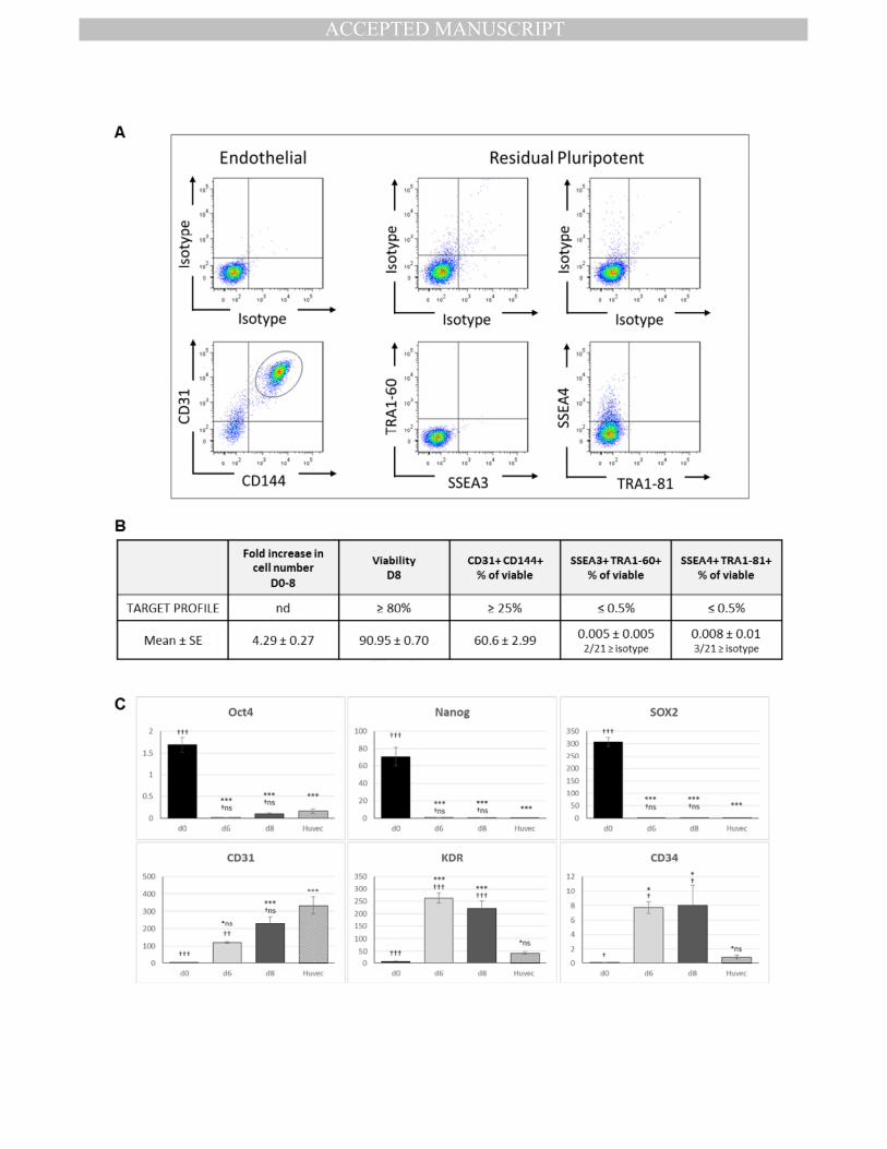
MANUSCRIP
T
ACCEPTED
ACCEPTED MANUSCRIPT
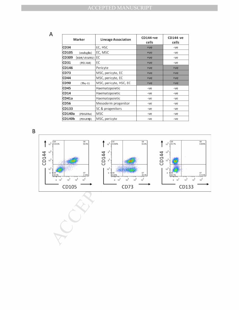
MANUSCRIP
T
ACCEPTED
ACCEPTED MANUSCRIPT
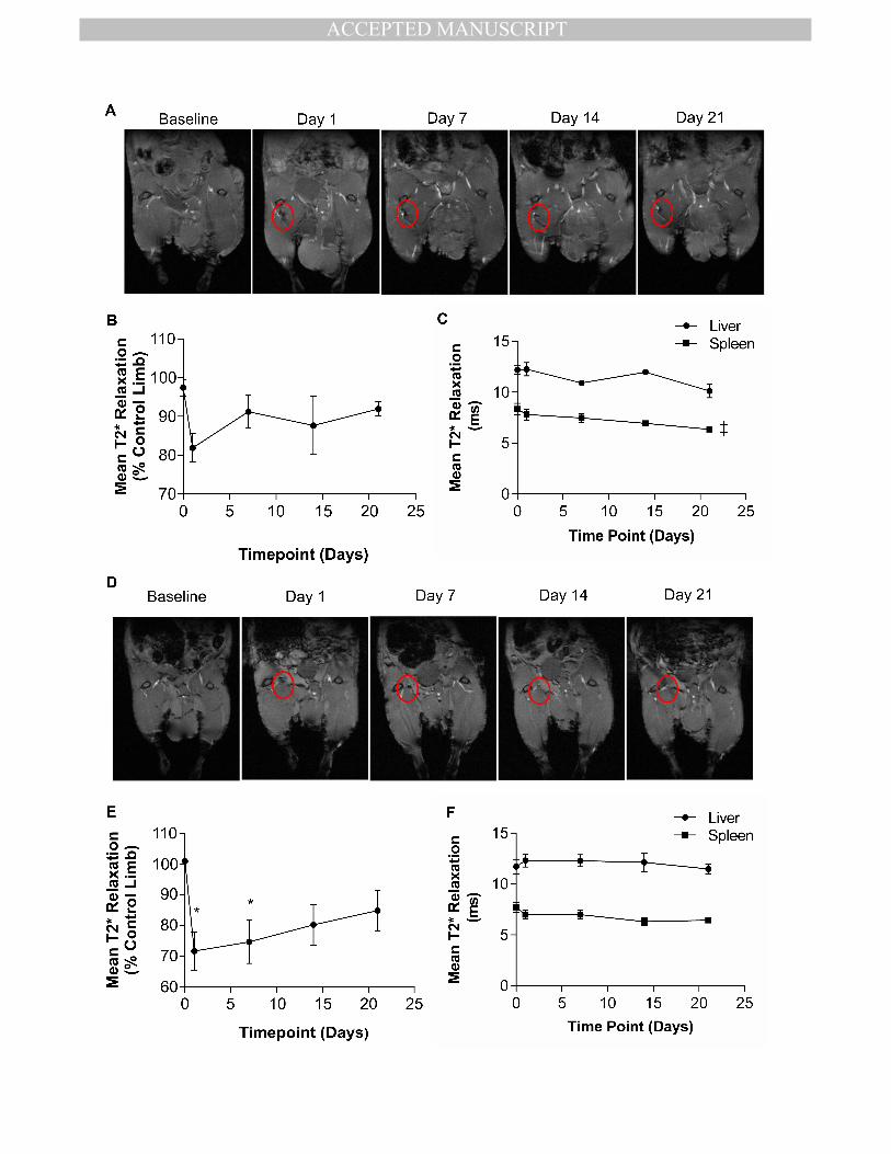
MANUSCRIP
T
ACCEPTED
ACCEPTED MANUSCRIPT
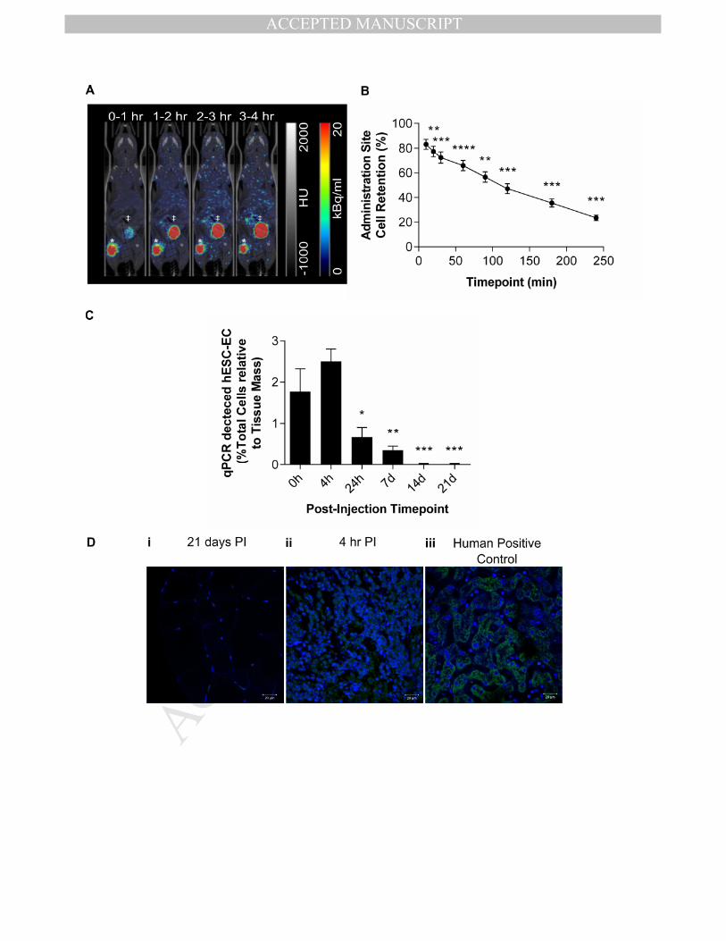
MANUSCRIP
T
ACCEPTED
ACCEPTED MANUSCRIPT
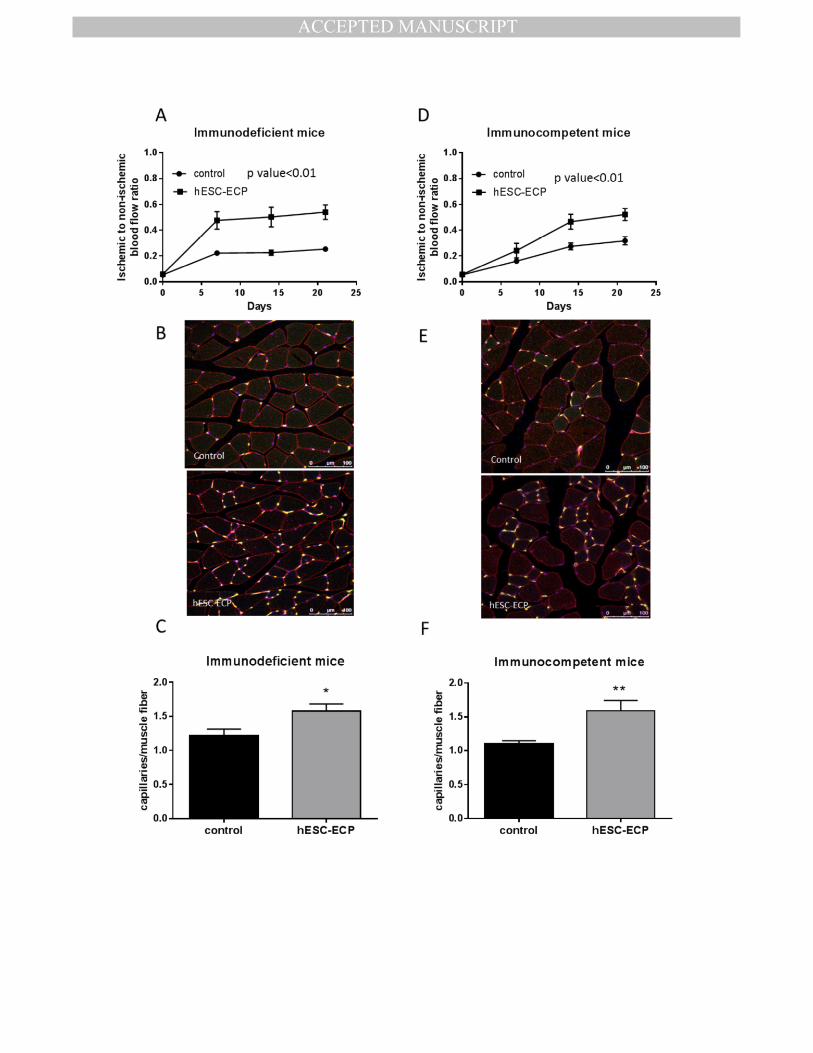
MANUSCRIP
T
ACCEPTED
ACCEPTED MANUSCRIPT
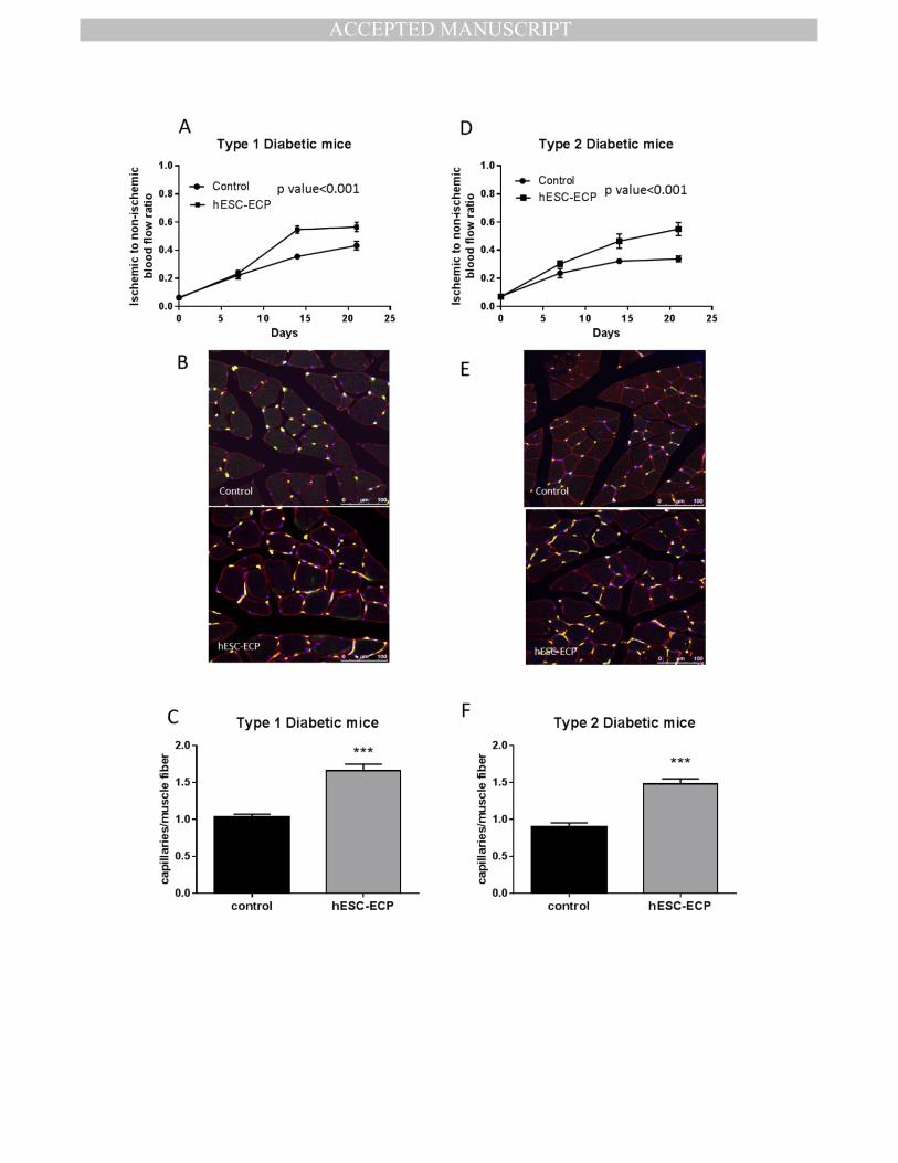
MANUSCRIP
T
ACCEPTED
ACCEPTED MANUSCRIPT
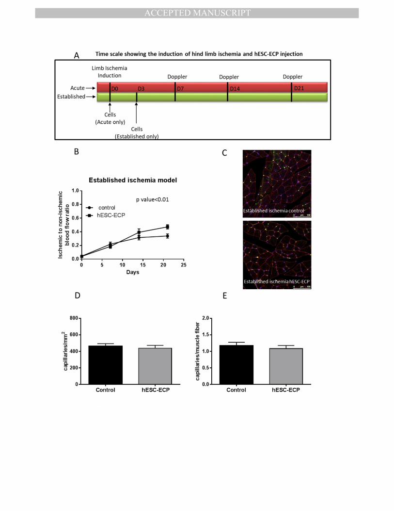
MANUSCRIP
T
ACCEPTED
ACCEPTED MANUSCRIPT

MANUSCRIP
T
ACCEPTED
ACCEPTED MANUSCRIPT
eTOC Synopsis:
To translate hESC-ECP into the clinic for CLI treatment, MacAskill et al. developed a robust, GMP-
compatible method for hESC-ECP generation. hESC-ECP remained within ischemic limbs for ~7 days,
and significantly improved foot perfusion and capillary density in murine models of limb ischemia
with/without an intact immune system or diabetes mellitus.


![· Web viewText, 9(1), 69-92. [[[Literacy] [Indo Pacific Languages] [Discourse Analysis] [Sociolinguistics] [Text Analysis] [Spoken vs Written Communication] [Emotion]]] Besnier,](https://img.pdfslide.us/doc/110x75/5b29122f7f8b9aae368b4626/-web-viewtext-91-69-92-literacy-indo-pacific-languages-discourse.jpg)