Embed Size (px)
Citation preview

Edinburgh Research Explorer
An Improved Method of Renal Tissue Engineering, by CombiningRenal Dissociation and Reaggregation with a Low-VolumeCulture Technique, Results in Development of EngineeredKidneys Complete with Loops of Henle
Citation for published version:Davies, J & Chang, C-H 2012, 'An Improved Method of Renal Tissue Engineering, by Combining RenalDissociation and Reaggregation with a Low-Volume Culture Technique, Results in Development ofEngineered Kidneys Complete with Loops of Henle' Nephron. Experimental nephrology. DOI:10.1159/000345514
Digital Object Identifier (DOI):10.1159/000345514
Link:Link to publication record in Edinburgh Research Explorer
Document Version:Peer reviewed version
Published In:Nephron. Experimental nephrology
Publisher Rights Statement:This article may not be used for commercial purposes.
© 2013 S. Karger AG, Basel
General rightsCopyright for the publications made accessible via the Edinburgh Research Explorer is retained by the author(s)and / or other copyright owners and it is a condition of accessing these publications that users recognise andabide by the legal requirements associated with these rights.
Take down policyThe University of Edinburgh has made every reasonable effort to ensure that Edinburgh Research Explorercontent complies with UK legislation. If you believe that the public display of this file breaches copyright pleasecontact [email protected] providing details, and we will remove access to the work immediately andinvestigate your claim.
Download date: 28. Apr. 2017

1
An improved method of renal tissue engineering, by combining renal dissociation and re-aggregation with a low-volume culture technique, results in development of engineered
kidneys complete with loops of Henle
C-Hong Chang and Jamie A. Davies
University of Edinburgh Centre for Integrative Physiology; Hugh Robson Building; Edinburgh, Scotland UK EH8 9XB
Cite as: Chang C-H, Davies J, A, An Improved Method of Renal Tissue Engineering, by Combining Renal Dissociation and Reaggregation with a Low-Volume Culture Technique, Results in Development of Engineered Kidneys Complete with Loops of Henle. Nephron Exp Nephrol 2012;121:e79-e85DOI: 10.1159/000345514Available online: http://www.karger.com/Article/FullText/345514

2
Abstract
Background: Tissue engineering of functional kidney tissue is an important goal for clinical
restoration of renal function in patients damaged by infectious, toxicological, or genetic
disease. One promising approach is the use of the self-organizing abilities of embryonic
kidney cells to arrange themselves, from a simply reaggregated cell suspension, into
engineered organs similar to foetal kidneys. The previous state-of-the-art method for this
results in the formation of a branched collecting duct tree, immature nephrons (S-shaped
bodies) beside and connected to it, and supportive stroma. It does not, though, result in the
significant formation of morphologically detectable loops of Henle - anatomical features of
the nephron that are critical to physiological function.
Methods: We have combined the best existing technique for renal tissue engineering from
cell suspensions, with a low-volume culture technique that allows intact kidney rudiments to
make loops of Henle, to test whether engineered kidneys can produce these loops.
Results: The result is the formation of loops of Henle in engineered cultured ‘foetal kidneys’,
very similar in both morphology and in number to those formed by intact organ rudiments.
Conclusion: This brings the engineering technique one important step closer to production of
a fully realistic organ.
Keywords: embryonic kidney, re-aggregates, tissue engineering, low-volume culture, loop of Henle

3
Introduction
The primary goal of renal tissue engineering is to construct an organ that resembles the
natural kidney as closely as possible [1-4]. One strategy for achieving this is to exploit the
capacity of renal cells for self-organization.
Two years ago, Unbekandt and Davies invented a method to recapitulate the early stages of
organotypic renal structure from simple suspensions of isolated embryonic renal cells [5].
Because the cell suspension was obtained by dissociation of early (E11.5, ‘T-bud stage’)
embryonic mouse kidneys, the method was called the ‘dissociation-reaggregation technique’.
In its basic form, it produced immature nephrons arranged around ureteric bud tissue, but the
ureteric bud cells were arranged as a multitude of small ureteric buds/ collecting duct trees
rather than as one single coherent collecting duct tree system, and therefore failed to
reproduce a key feature of normal renal anatomy tree [5,6]. The ‘nephrosphere’ technique
developed by Lusis and colleagues in the same year [7] did not include collecting ducts at all,
so suffered from an even more severe version of this problem.
To resolve this limitation, Ganeva et al. developed a serial dissociation and re-aggregation
system [6]. They first used the original dissociation and re-aggregation system to make re-
aggregates with multiple, small ureteric buds. They then manually isolated one of these small
re-aggregated ureteric buds and combined it with fresh disaggregated and reaggregated
mesenchyme: the result was development of immature nephrons that were arranged around
one, highly-branched ureteric bud / collecting duct system. This was a good reflection of the
structure of mouse embryonic kidneys at about 13 days gestation.

4
As normal kidneys mature, from about 14 days of mouse development [8], they develop
distinct cortical and medullary zones. Bowman’s capsules, proximal tubules and distal
tubules are restricted to the cortex, while the medulla consists of collecting ducts and loops of
Henle, elements of the nephron that extend radially inwards from the cortex. This
arrangement is vital for normal physiology, particularly the recovery of water (which depends
on loops of Henle making the medullary interstitium very hypertonic compared to normal
body fluids). Any useful system for renal tissue engineering must therefore be able to
reproduce this feature. The standard, Trowell-screen culture methods used for development
of existing dissociation-reaggregation methods do not support efficient development of loops
of Henle even when they are used to culture normal, intact kidney rudiments. There is
therefore neither positive nor reliable negative evidence about the potential for reaggregated
kidneys to organize themselves to produce realistic cortico-medullary zonation or loops of
Henle.
A recently published novel culture method, based on growing rudiments on silicone-bounded
glass slides with extremely low volumes of medium (just tens of microlitres), allows an intact
kidney isolated directly from an E11.5 embryo to develop organotypic cortico-medullary
zonation with loops of Henle over the course of 7-10 days [9]. In this short report, we
combine the idea of tissue engineering, from cell suspensions by the serial dissociation-
reaggregation method, with the low-volume culture method for cortico-medullary zonation.
The result is the production, from cell suspensions, of kidneys with distinct cortical and
medullary zones and with loops of Henle extending radially inwards. This marks a further
step towards engineering a realistic foetal kidney from simple suspensions of cells, and
provides a potential path by which renal stem cells could be used to make kidney rudiments
for clinical applications.

5
Materials and Methods
Organ culture
The main method is depicted in diagrammatically in Fig 1. Kidney rudiments were obtained
from E11.5 CD1 mouse embryos (morning of plug check considered to be E0.5) by manual
dissection in Eagle’s Minimum Essential Medium with Earle’s Salts (Sigma cat # M5650).
For whole kidney conventional (Trowell screen) culture, they were placed on a 5 µm Isopore
membrane filter (Millipore cat # TMTP02500) supported by a stainless steel grid in a 3.5 cm
culture dish at the gas-medium interface. The medium was Kidney Culture Medium (KCM):
Eagle’s Mimimum Essential Medium (Sigma cat # M5650) with 10% foetal bovine serum
(FBS: Invitrogen cat # 10108165) and 1% penicillin/ streptomycin (Sigma cat # P4333), as
described by Unbekandt et al. [5]. For the low-volume culture system, we followed the
method described in Sebinger et al. [9]. The Cone shape ‘A’ silicon ring (SARSTEDT cat #
94.6077.434) was attached to a 22×22 mm glass cover slip (VWR International cat # 631-
0125) and the re-aggregates were placed in the centre of it in 85 µl of KCM.
Tissue engineering by re-aggregation
E11.5 embryonic kidneys from CD1 mice were dissected in MEM (Sigma cat # M5650). The
kidney rudiments were then dissociated enzymatically and reaggregated exactly as described
in Unbekandt and Davies, 2010 [5]. They were cultured in KCM on Isopore filters supported
by metal grids as described above. For the first 24 h, the ROCK inhibitor, 1.25 µM glycyl-
H1152-dihydrochloride (TOCRIS batch # 1A/93503), was added to KCM. This medium was
then replaced with drug-free KCM for the remaining 3-4 days of culture, as described [5].
After this first incubation, single ureteric bud cysts were isolated from the whole-kidney re-

6
aggregates by manual dissection. Dissociated fresh metanephric mesenchyme was isolated as
described by Ganeva et al. [6]; briefly, 10 to 15 kidneys were incubated in 2× trypsin/EDTA
(Sigma cat # T4174) in MEM for 2 min at 37°C and quenched in KCM. Following this, the
mesenchymes were peeled away from the ureteric bud. The mesenchymes were collected in a
500µl tube, dissociated by gentle pipetting and re-aggregated by centrifugation at 3000 rpm
for 2 min in a micro-centrifuge. The reaggregated ureteric buds from the dissociation-
reaggregation experiment were combined, singly, with reaggregated fresh mesenchymes on a
membrane filter in the conventional culture system. They were then cultured for 1-2 days in
KCM; during this time, they became solid enough to manipulate. They were removed from
their filters and transferred to the low-volume culture system. They were incubated for a
further 5-7 days (so to a total of 6-9 days from application of mesenchyme to bud cyst),
medium being changed every 2 days.
Immunohistochemistry
Tissues were fixed using cold methanol initially at -20°C and allowed to warm up towards
room temperature during the 15 min fixation. They were rinsed in PBS for 30 min at room
temperature. Primary antibodies applied to the tissues were diluted 1:100 in PBS and applied
overnight at 4°C; primary antibodies were mouse anti-Calbindin (Abcam cat # ab9481),
mouse anti-pan cytokeratin (Sigma cat # C2562), chicken anti-laminin (Abcam cat #
ab14055), rabbit anti-laminin (Sigma cat # L9393), and rabbit anti-Human Tamm-Horsfall
glycoprotein (Bioquote cat # bt-590). The next day, tissues were washed for a few hours in
PBS and secondary antibodies were applied overnight at 4°C. Secondary antibodies were
goat FITC anti-chicken (Abcam cat # ab97134), goat FITC anti-mouse (Sigma cat # F2012)
and goat TRITC anti-rabbit (Sigma cat # T6778), and were applied at 1/100 in PBS. Finally,

7
tissues were washed in PBS for few hours. Those grown on filters in Trowell culture were
mounted, still on their pieces of filter, between two 22x64mm coverslips that had 22x22mm
coverslips sandwiched between them at their ends as spacers, to keep the longer converslips
apart and prevent the samples being crushed: the whole assembly was sealed with nail
varnish (Portobello Pink, Rimmel) and mounted loosely on a microscope slide so that the
coverslip assembly could be inverted if the filter-and-kidney combination happened to be
upside-down. These samples were viewed on a Zeiss Axioscope epifluorescence microscope.
Organs and reaggregates grown in the low volume system were viewed using a Zeiss
Axiovert epifluorescence microscope.
Identification and counting of loops of Henle
Loops of Henle were identified and counted primarily by morphological criteria because the
existence of an actual loop (rather than mere expression of marker genes, some of which
appear in the S-shaped body before real loops form) has greater physiological relevance: see
‘Results and Discussion’. Anti-laminin staining (see above) was used to trace the shapes of
all tubules. The criterion used to define the presence of an LoH, and to count them, was the
existence of a tube that was bent sharply back on itself like a hair grip (US: ‘bobby pin’),
extending from the mid-portion of a nephron. The straight part of the tube had to be more
than a tubule diameter in length before it was considered to be a bona fide loop of Henle (in
practice, they were much longer). As an additional test, immunostaining for Tamm-Horsfall
glycoprotein was used in some experiments to confirm that the morphologically identified
loops do indeed express this loop of Henle marker in the expected manner (they do: see
Results and Discussion). Counting was done using a low-power image of the whole kidney
and using high-power views to confirm loop morphology as shown in Figs 2-4.

8
We also recorded whether loops of Henle extended towards the middle of the kidney. To do
this, two lines were drawn on an image: one ran along the long axis of the loop itself and the
other, radius line, ran from the centre of the kidney to and beyond the tip of the loop (‘centre
of the kidney’ defined as the first branching point of the collecting duct system). The angle
between the axis of the loop and the radius line beyond the tip was then measured, to assess
how accurately the loop of Henle was orientated radially towards the centre of the kidney:
perfect radial alignment would yield an angle of zero. Where the angle was less than 45
degrees, the loop was counted as extending towards the centre (‘centripetal’).
9
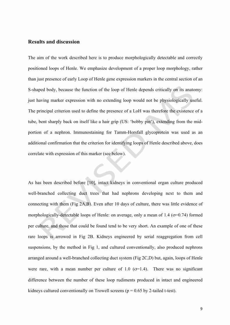
Results and discussion
The aim of the work described here is to produce morphologically detectable and correctly
positioned loops of Henle. We emphasize development of a proper loop morphology, rather
than just presence of early Loop of Henle gene expression markers in the central section of an
S-shaped body, because the function of the loop of Henle depends critically on its anatomy:
just having marker expression with no extending loop would not be physiologically useful.
The principal criterion used to define the presence of a LoH was therefore the existence of a
tube, bent sharply back on itself like a hair grip (US: ‘bobby pin’), extending from the mid-
portion of a nephron. Immunostaining for Tamm-Horsfall glycoprotein was used as an
additional confirmation that the criterion for identifying loops of Henle described above, does
correlate with expression of this marker (see below).
As has been described before [10], intact kidneys in conventional organ culture produced
well-branched collecting duct trees that had nephrons developing next to them and
connecting with them (Fig 2A,B). Even after 10 days of culture, there was little evidence of
morphologically-detectable loops of Henle: on average, only a mean of 1.4 (σ=0.74) formed
per culture, and those that could be found tend to be very short. An example of one of these
rare loops is arrowed in Fig 2B. Kidneys engineered by serial reaggregation from cell
suspensions, by the method in Fig 1, and cultured conventionally, also produced nephrons
arranged around a well-branched collecting duct system (Fig 2C,D) but, again, loops of Henle
were rare, with a mean number per culture of 1.0 (σ=1.4). There was no significant
difference between the number of these loop rudiments produced in intact and engineered
kidneys cultured conventionally on Trowell screens (p = 0.65 by 2-tailed t-test).

10
To overcome this restriction, we used the low-volume culture method of Sebinger et al. [9].
For intact kidneys, this technique was used exactly as published. For serial re-aggregates, the
combination of a ureteric bud cyst from the first reaggregation (Fig1, step 1) with fresh
mesenchyme (from Fig1, step 2) was cultured conventionally for 1-2 days before being
transferred to the low-volume culture system (Fig1, step 3). This period of conventional
culture before low volume culture had to be used because the low-volume culture method
requires an organ rudiment to be placed in a specific place (the centre, where the medium is
at its shallowest so that surface tension presses down on the tissue [9]). This accurate
placement was not possible until reaggregation had proceeded far enough to make a solid
‘tissue’ that could be manipulated by pipette.
Intact kidneys behaved in the low volume culture system exactly as has been described before
[9]. The organ rudiments spread over a large area and formed a well-branched collecting duct
tree (Fig 3A,B). Under these culture conditions, loops of Henle could be seen; a mean of 16.3
(σ=3.0) per culture, some long and extended and some shorter but still identifiable: examples
of both can be seen, arrowed, in the higher magnification view of the sample of Fig 3A that is
presented in Fig 3B. This increase in loop production was highly significant (p = 0.00004 by
a 2-tailed t-test). Most (95 of 98: 97%) loops extended correctly towards the middle of the
kidney (‘centripetally’, defined for measurement purposes as heading towards the first
ureteric branch with an error of less than ± 45 degrees).
Kidneys engineered by serial reaggregation, pre-incubated in conventional culture for 2 days
and then transferred to low-volume culture, also spread over a large area and formed a well-
branched collecting duct system (Fig 3C, D). Except for the fact that the intact kidneys had

11
an overall polarity arising from the ureteric bud entry point while the engineered ones had no
unique entry point and therefore showed radial symmetry, these engineered organs were
difficult to distinguish from their intact counterparts (compare Fig 3C with Fig 3A).
Importantly, under these culture conditions the engineered kidneys produced morphologically
identifiable loops of Henle, which can be seen in Fig 3C and more easily in the successively
higher magnification views, Fig 3D and 3E. Quantitatively, the engineered kidneys produced
a mean of 14.0 (σ=1.73) morphologically identifiable loops per culture. This was highly
significantly different from loop production in conventional culture (p= 0.0005 by a 2-tailed
t-test). Encouragingly, it was not significantly different from the performance of intact
kidneys in low-volume culture (a 2-tailed t-test yields p = 0.19; no significant difference).
Once again, most (40 of 42: 95%) loops were orientated towards the centre of the kidney. The
quantitative behaviour of kidneys and engineered kidneys in these culture systems, with
respect to Loop of Henle formation, is shown in Fig 3F.
To confirm morphological identification of loops of Henle, we examined the expression of
Tamm-Horsfall protein (THP), which is expressed strongly in the ascending limb of the
mature loop of Henle [11,12]. In intact kidneys and in serial reaggregates, THP expression
could be seen in the growing loops of Henle (Fig 4A-C). It is striking to note that, in these
early kidneys (both intact and engineered), THP expression is particularly strong near the
bend of the growing loop. THP expression begins a little after loop emergence, which means
that some shorter morphologically defined loops did not express THP. Nevertheless, counting
only the THP-positive loops shows the same pattern (Fig 4D): very few in conventional
culture and significantly more (p=0.02 by 2-tailed t-test) in low-volume cultures of intact and

12
engineered kidneys. Again, there was no significant difference (p=0.90 by 2-tailed t-test)
between the numbers in intact and engineered.
The development of loops of Henle brings engineered kidneys an important step closer to
being properly representative of kidneys that have developed normally in vivo. Essentially, it
makes the anatomical development of the epithelial tubules very similar to that found in a
normal late-gestation foetal murine kidney. Important remaining steps include the
introduction of properly patterned and integrated vascular and nervous systems.
Acknowlegements
We thank David Sebinger, Mathieu Unbekandt, Veronika Ganeva and Peter Hohenstein for
helpful advice. This work was funded in part by the NC3Rs.

13
Figures
Figure 1. Schematic description of the method to incubate the re-aggregated kidney cells in
the conventional and low-volume culture system.

14
Figure 2. In 10-days of conventional Trowell-screen culture, both intact kidneys (A, B) and
kidneys engineered (C, D) through serial reaggregation produce an organotypic arrangement
of nephrons around a single collecting duct tree, but there is little sign of development of
loops of Henle (one rare example is arrowed in B). Green shows the ureteric bud marker,
Calbindin-D-28k, and red the basement membrane marker, laminin.

15
Figure 3. In low volume culture, kidneys engineered by series reaggregation form loops of
Henle like those formed by intact kidneys in low volume culture. (A) shows a low-power
view of an intact kidney in low-volume culture. The arrows point to positions of developing
loops of Henle, identified by examination of higher magnification images; (B) shows an
example of a higher magnification image, with loops of Henle marked with arrows (the
arrows point at the loops and have nothing to do with the orientation of the loops). (C) shows
a low-power view of a kidney engineered by serial reaggregation in low-volume culture.
Again, the arrows point to developing loops of Henle, identified by examination of higher
magnification images; (D), (E) show examples of successively higher magnification images,

16
with loops of Henle marked with arrows. (F) Shows the average total number of loops of
Henle per kidney formed in each method, and the average number of loops that extend
radially inwards (heading towards the first ureteric branch with less than a 45 degree error):
these are called ‘centripetal’, ie centre-seeking, on the graph). Intact kidneys were cultured
for 10 days and serial reaggregates for 2 days of conventional culture (from final aggregation
of UB cyst with fresh MM) followed by 6 days of low-volume culture. Error bars show
standard deviation; p values are given in the main text; the groups contained 8, 6, 4 and 3
cultures respectively. Green shows the ureteric bud marker, Calbindin-D-28k, and red the
basement membrane marker, laminin.

17
Figure 4. THP expressed loops were shown in the intact and engineered kidneys in low
volume system. (A) shows an intact kidney cultured in the low-volume system, with a loop of
Henle (arrow) marked with U-shaped adluminal expression of THP (green), the basement
membrane again being stained for laminin (red). (B) shows a similarly stained image of a
serial reaggregate kidney, with several loops visible (arrowed: the arrow with the larger head
marks a loop, the bend of which can clearly be seen as such in this plane of focus). (C) shows
a low-power view to demonstrate the specificity of anti-THP for loops of Henle (ie absence
of stain in other parts of the kidney). Graph (D) shows the average number of THP-
expressing loops of Henle per kidney formed in each method (error bars = standard error of
the mean). Intact kidneys were cultured for 9 days and serial reaggregates for 2 days of
conventional culture followed by 6 days of low-volume culture.

18
References
1. Rosines E, Sampogna RV, Johkura K, Vaughn DA, Choi Y, Sakurai H, Shah MM, Nigam SK: Staged in vitro reconstitution and implantation of engineered rat kidney tissue. Proceedings of the National Academy of Sciences 2007;104:20938-20943. 2. Perin L, Da Sacco S, De Filippo RE: Regenerative medicine of the kidney. Advanced Drug Delivery Reviews 2011;63:379-387. 3. Davies JA: Self-organization as a tool in mammalian tissue engineering; in Winslet-Gendebien S (ed.): Regenerative Medicine, Intech, 2011, pp261-274. 4. Steer DL, Nigam SK: Developmental approaches to kidney tissue engineering. Am. J. Physiol. Renal Physiol. 2004;286:F1-F7. 5. Unbekandt M, Davies JA: Dissociation of embryonic kidneys followed by reaggregation allows the formation of renal tissues. Kidney International 2010;77:407-16. 6. Ganeva V, Unbekandt M, Davies JA: An improved kidney dissociation and reaggregation culture system results in nephrons arranged organotypically around a single collecting duct system. Organogenesis 2011;7:83-7. 7. Lusis M, Li J, Ineson J, Christensen ME, Rice A, Little MH: Isolation of clonogenic, long-term self renewing embryonic renal stem cells. Stem Cell Research 2010;5:23-39. 8. Little MH, Brennan J, Georgas K, Davies JA, Davidson DR, Baldock RA, Beverdam A, Bertram JF, Capel B, Chiu HS, Clements D, Cullen-McEwen L, Fleming J, Gilbert T, Herzlinger D, Houghton D, Kaufman MH, Kleymenova E, Koopman PA, Lewis AG, McMahon AP, Mendelsohn CL, Mitchell EK, Rumballe BA, Sweeney DE, Valerius MT, Yamada G, Yang Y, Yu J: A high-resolution anatomical ontology of the developing murine genitourinary tract. Gene Expression Patterns 2007; 7:680-699. 9. Sebinger DD, Unbekandt M, Ganeva VV, Ofenbauer A, Werner C, Davies JA: A Novel, Low-Volume Method for Organ Culture of Embryonic Kidneys That Allows Development of Cortico-Medullary Anatomical Organization. PLoS ONE 2010;5:e10550. 10. Saxen L: Organogenesis of the kidney. Cambridge University Press, 1985. 11. Tamm I, Horsfall FL: Characterization and separation of an inhibitor of viral hemagglutination present in urine. Proc Soc Exp Biol Med. 1950;74:106-8. 12. Vyletal P, Bleyer AJ, Kmoch S: Uromodulin biology and pathophysiology--an update. Kidney Blood Press Res. 2010;33:456-75.