Embed Size (px)
Citation preview
Introduction & Background
Ediacaran Reef Construction and the Earliest Evidence for Animal BiomineralizationAshely Fernandes | GEOL394 | Spring 2020 | Advisor: Dr. Alan Jay Kaufman
Hypotheses
Results Discussion & Conclusions
Reef Construction: Cloudina preserved in microbial mats reflect in-situ growth because they appear to be intact, in life-position, and interlayered with the mats.
Shell Composition: Cloudina shells from the Arasab locality contain primary phosphate minerals because the Shuram Excursion brought about high concentrations of phosphorus in the ocean (Cui et al., 2016).
Vital Effects: Early Cloudina did not fractionate the seawater isotopic composition because the vital effect has not previously been observed in Cloudina (Grant, 1992).
Feeding Mechanism: Cloudina had chemosymbionts, in the form of sulfide-oxidizing bacteria, living within them that helped them to manage the sulfide in their surroundings because a similar process is observed in the modern-day deep-sea tube worm, Riftia pachyptila (Felbeck, 1981).
The earliest evidence of animals capable of biomineralizing shells, Cloudina, wasrecently discovered in Ediacaran-aged strata of the lower Nama Group in Arasabfarmstead, Namibia. The specimens are preserved in close association withmicrobial mats. The appearance of the Cloudina coincides with the end of thegreatest carbon isotope anomaly in Earth history, the Shuram Excursion, at 575 Ma(Rooney, 2019). This study analyzes (i) whether the Cloudina contributed to theconstruction of microbial reefs, (ii) the original composition of the Cloudina shells,(iii) if the Cloudina fractionated seawater isotopic compositions (δ13C and δ18O ) toform their shells, and (iv) if Cloudina harbored sulfide-oxidizing bacteria within theirbodies in a symbiotic relationship that provided the Cloudina with organic food.
Reef Construction
Shell Composition
Special thanks go out to Dr. Jay Kaufman and Dr. Philip Piccoli for their advice throughout my senior thesis project. I would like to thank Julia Poggi and Orion Jenkins-Houk for their assistance withusing Adobe Photoshop to outline and measure the orientation of Cloudina. I thank Dr. Piccoli for his assistance with the EPMA. I thank Dr. Penniston-Dorland for instructing me on how to use thepetrographic microscope and rock saw. I am grateful to Dr. Steven Goderis and Dr. Huan Cui for providing data from the micro-XRF spectrometer. I thank Dr. Mike Evans, Dr. Jay Kaufman, Dr. ShuiwangDuan, and Cristy Ho for their assistance with the use of stable isotope mass spectrometers. I am grateful to Dr. Thomas Holtz for directing me to new published research on Cloudina. A final thanksgoes out to the faculty that have given me constructive feedback on my presentations and proposal.
Becker-Kerber, B., Pacheco, M. L. A. F., Rudnitzki, I. D., Galante, D., Rodrigues, F., & Leme, J. M. (2017). Ecological interactions in Cloudina from the Ediacaran of Brazil: implications for the rise of animal biomineralization. Sci. Rep., 7(1), 1-11.
Cui, H., Xiao, S., Zhou, C., Peng, Y., Kaufman, A. J., & Plummer, R. E. (2016). Phosphogenesis associated with the Shuram Excursion: Petrographic and geochemical observations from the Ediacaran Doushantuo Formation of South China. Sediment. Geol., 341(2016), 134-146.
Felbeck, H. (1981). Chemoautotrophic Potential of the Hydrothermal Vent Tube Worm, Riftia pachyptila Jones (Vestimentifera). Science, 213(4505), 336-338.Fisher, C. R., Childress, J. J., & Minnich, E. (1989). Autotrophic Carbon Fixation by the Chemoautotrophic Symbionts of Riftia pachyptila. Biol. Bull., 177(3), 372-385.Grant, S. W. F. (1992). Carbon isotopic vital effect and organic diagenesis, Lower Cambrian Forteau Formation, northwest Newfoundland: Implications for δ13C chemostratigraphy. Geology, 20(3), 243-
246.Kaplan, I. R. & Rittenberg, S. C. (1964). Microbiological Fractionation of Sulfur Isotopes. MicroSoc., 34(2), 195-212.Rooney, A. (2019). New Re-Os ages and Os geochemistry from the Neoproterozoic: Fresh insights into links between climate, animal evolution and biogeochemical cycles. Geol. Soc. Am. Abstr., 51(5).Schiffbauer, J. D., Selly, T., Jacquet, S. M., Merz, R. A., Nelson, L. L., Strange, M. A., Cai, Y., & Smith, E. F. (2020). Discovery of bilaterian-type through-guts in cloudinomorphs from the terminal
Ediacaran Period. Nat. Commun., 11(2020), 1-12.Wood, R. (2011). Paleoecology of the earliest skeletal metazoan communities: Implications for early biomineralization. Earth-Sci. Rev., 106(1-2), 184-190.Wood, R., Ivantsov, A. Y., & Zhuravlev, A. Y., Zhuravlev, A. Y. (2017). First macrobiota biomineralization was environmentally triggered. Proc. Royal Soc. B, 284(1851), 1-7.
Figure 1: Samples. 1A: Boulders were analyzed at the micro- and macroscopic levels. 1B: Cross sections of boulders (slabs) were analyzed with a micro-XRF spectrometer at the cm-scale. Slabs were also micro-drilled for δ13C and δ18O analyses. 1C: 30-µm thin sections were analyzed with a petrographic microscope and an EPMA at the mm-scale.
Figure 2: 2A: GigaPan image of hundreds of Cloudina exposed on the surface of lithified microbial mats. The inset shows 288 Cloudina. 2B: Adobe Photoshop measurements of the growth direction of several Cloudina. 2C: Rose diagram compiling 193 individual orientations into 15° intervals. Black Resultant Vector: Magnitude = 0.25, Orientation = 295°, and Circular Variance = 0.75. Orange Resultant Vector (accounting for bimodal variance): Magnitude = 0.27, Orientation = 33° & 213°, and Circular Variance = 0.73.
Figure 3: EPMA Data (Three Examples). Left: WDS phosphorus (P) maps. Right: Petrographic images of the areas analyzed (PPL, 1.5x). P is elevated in areas associated with Cloudina shells but lacking in the microbial mat and Cloudina interior. EDS spectra indicate the presence of apatite (Ca5(PO4)3(OH,F,Cl)).
Figure 4: Micro-XRF Data. 4A: Slab consisting of alternating Cloudina-dominated and mat-dominated intervals. 4B: Strontium (Sr) map showing that Cloudina-intervals are significantly lacking in Sr, a proxy for aragonite (Siegel, 1960). 4C: Manganese (Mn) map showing that mat-intervals are significantly lacking in Mn clusters, signifying a change in redox conditions.
Comparison of the Cloudinaand black microbial mats (a proxy for seawater conditions) reveal similar values for δ13C (-4.25‰ +/- 0.46‰ and -4.36‰ +/- 0.42‰, respectively).
AB
C
A
C
Figure 5: The Cloudina layer on the surface of lithified microbial mats is 13.14‰ to 22.76‰ more enriched in 32S than the underlying mats.
Reef Construction: Hypothesis is supported Based on the high circular variance and low resultant
magnitude, the Cloudina are randomly orientedDetrital Cloudina shells would have likely become aligned and
broken up by the waves that deposited them (Becker-Kerber et al. 2017), so the Cloudina in this study are in situ
Shell Composition: Hypothesis is supported An original aragonitic shell composition can be ruled out Cloudina likely took advantage of the high availability of Ca
and P brought about by the Shuram Excursion (Cui et al., 2016), to form calcium phosphate shells
Cloudina transitioned to forming calcium carbonate shells later in the Ediacaran Period, due to the high availability of carbonate ions (Wood 2011; Wood et al., 2017) and decreased availability of P
Vital Effects: Hypothesis might be supported It is possible that the recrystallized calcitic interior of the
Cloudina were also drilled and included in the samples, so an alternative interpretation is that the Cloudina were cemented rapidly after death Feeding Mechanism: More information is needed
Microbes that reduce sulfate tend to fractionate the S such that the resulting hydrogen sulfide (H2S) is more enriched in 32S (Kaplan & Rittenberg, 1964)
H2S may have been concentrated in Cloudina within a trophosome, contrary to the interpretation by Schiffbauer et al. (2020) that recently discovered internal cylindrical structures were a one-way through-gut
Alternatively, H2S may have been concentrated in the surficial microbial mat surrounding the Cloudina
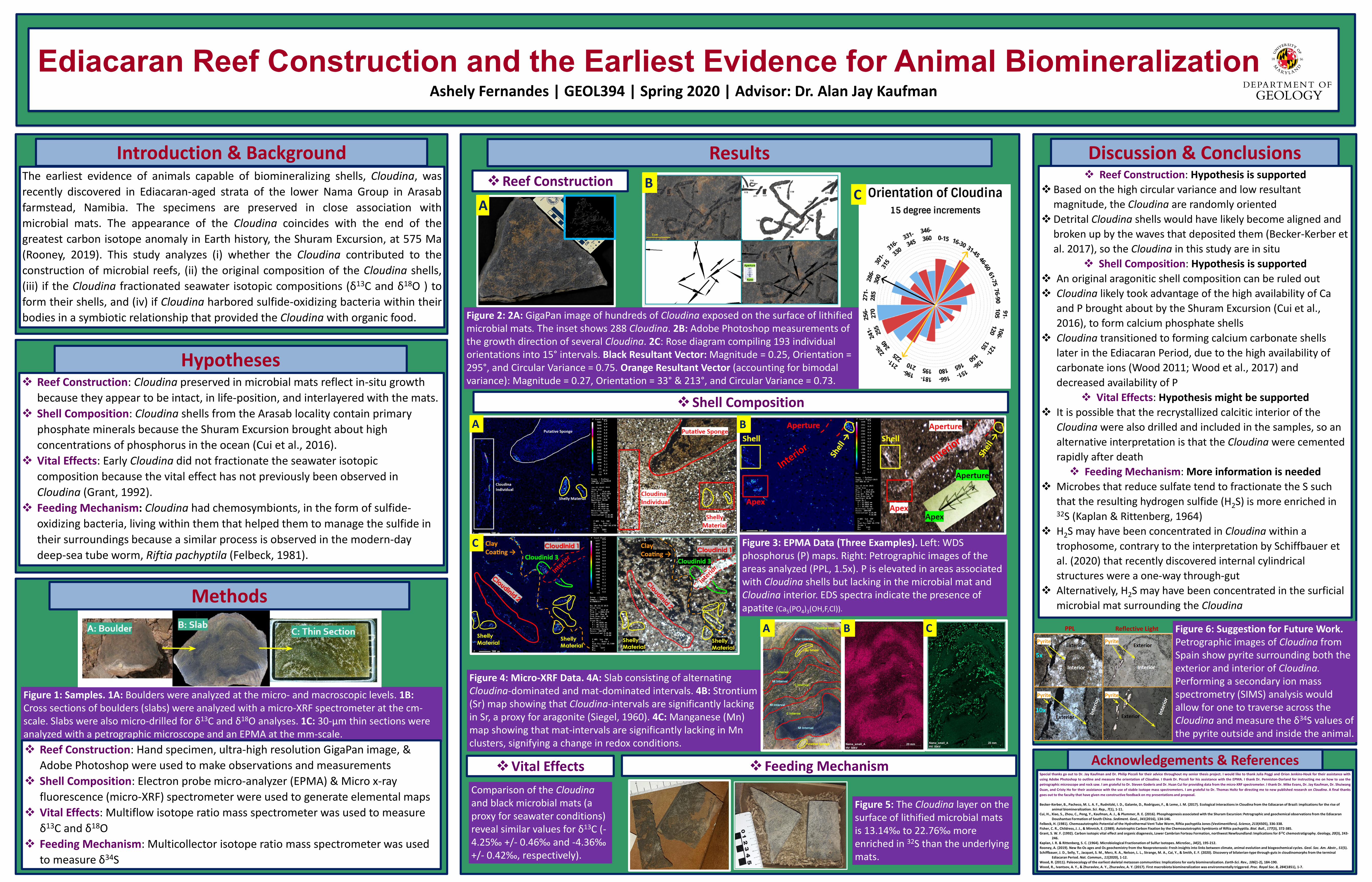
Figure 6: Suggestion for Future Work. Petrographic images of Cloudina from Spain show pyrite surrounding both the exterior and interior of Cloudina. Performing a secondary ion mass spectrometry (SIMS) analysis would allow for one to traverse across the Cloudina and measure the δ34S values of the pyrite outside and inside the animal.
B
Acknowledgements & ReferencesFeeding MechanismVital Effects Reef Construction: Hand specimen, ultra-high resolution GigaPan image, &
Adobe Photoshop were used to make observations and measurements Shell Composition: Electron probe micro-analyzer (EPMA) & Micro x-ray
fluorescence (micro-XRF) spectrometer were used to generate elemental maps Vital Effects: Multiflow isotope ratio mass spectrometer was used to measure
δ13C and δ18O Feeding Mechanism: Multicollector isotope ratio mass spectrometer was used
to measure δ34S
Methods
C
A B PPL Reflective Light
5x
10x