Embed Size (px)
Citation preview
THIEME
38 Case Report
Ectodermal Dysplasia—A Case ReportDamanpreet Kaur1 Ashima Behl1 Parminder Pal Singh Isher2 Dipti Bhatnagar3
1Department of Oral Medicine & Radiology, BJS Dental College, Hospital and Research Institute, Ludhiana, Punjab, India
2Conservative Dentistry & Endodontics, SKSS Dental College & Hospital, Sarabha, Ludhiana, Punjab, India
3Department of Oral Medicine & Radiology, Bhatnagar Orthodontic and Dental Centre, Chandigarh, Chandigarh, India
received September 26, 2018accepted after revision February 11, 2019published onlineApril 4, 2019
Address for correspondence Damanpreet Kaur, MDS, Department of Oral Medicine & Radiology, BJS Dental College, Hospital and Research Institute, Chandigarh Road, Ludhiana 141010, Punjab, India (e-mail: [email protected]).
Ectodermal dysplasias (EDs) include large group of syndromes that are clinically and genetically heterogeneous and are identified by anomalies in structures of ectodermal origin. Hereditary ectodermal dysplasia is a condition associated with defect of structures originating from ectoderm and is typically inherited as cross-linked recessive trait, more pronounced in males than in females. The frequency of different ectodermal dysplasias in a given population is highly variable. It is estimated to be 7 in 1,000 births. This case report is presented with an objective to show two patients, siblings, with ED. Both presented with classic symptoms of anodontia, hypohidrosis, and hypotrichosis.
Abstract
Keywords ► ectodermal dysplasia ► anodontia ► hypohidrosis ► hypotrichosis
Dent J Adv Stud 2019;7:38–41
DOI https://doi.org/ 10.1055/s-0039-1684943 ISSN 2321-1482.
©2019 Bhojia Dental College and Hospital affiliated to Himachal Pradesh University
IntroductionEctodermal dysplasias (EDs) include a large group of syndromes that are clinically and genetically heterogenous and are identified by anomalies in structures of ectodermal origin.1 EDs are a group of inherited disorders that share common developmental defects involving at least two of the major structures classically held to derive from the embryonic ectoderm—hair, teeth, nails, sweat glands.2 It was first described by Thurnam in 1848 and was coined by Weech in 1929. In 1875, Charles Darwin documented it among the Hindu family of Scinde in which men in the course of four generations were affected and remarkably no daughter was affected.3
It is typically inherited as a cross-linked recessive trait so that the frequency and severity of condition are much more pronounced in males than in females.4 Its incidence rate is estimated at 7 cases per 1,000 births, and more than 192 disorders have been identified in this disease.5 Hypodontia or anodontia is a major problem in ED patients.1 If any teeth are present, these are conical or peg shaped. Patients’ hair is thin and sparse, and skin is dry due to lack of perspiration. These patients are intolerant to heat. Also, in many cases finger and toe nails are deformed.6 The different types of EDs are caused by gene mutation or deletion of certain gene located on certain chromosome.7
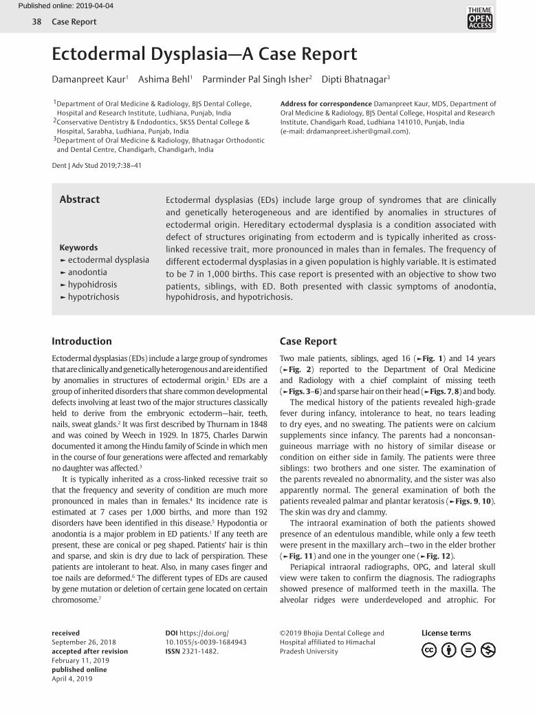
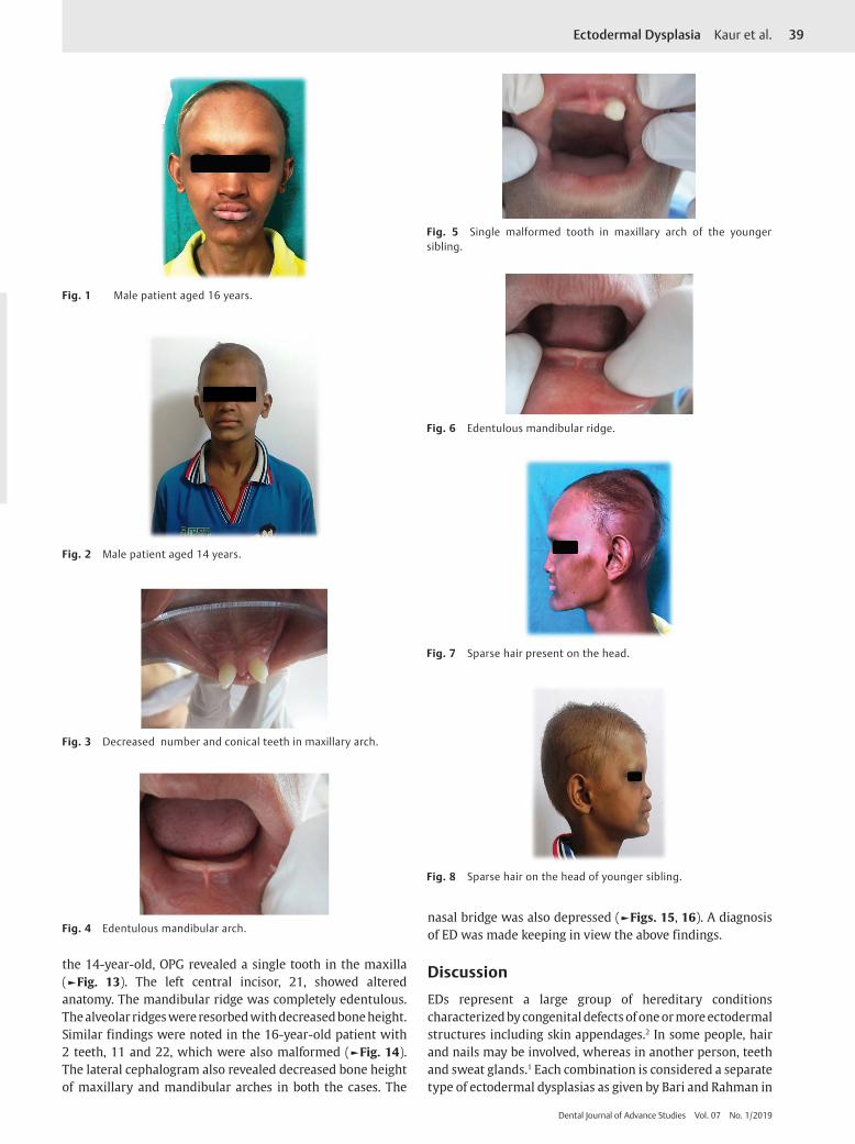
Case ReportTwo male patients, siblings, aged 16 (►Fig. 1) and 14 years (►Fig. 2) reported to the Department of Oral Medicine and Radiology with a chief complaint of missing teeth (►Figs. 3–6) and sparse hair on their head (►Figs. 7, 8) and body.
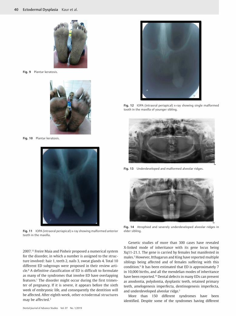
The medical history of the patients revealed high-grade fever during infancy, intolerance to heat, no tears leading to dry eyes, and no sweating. The patients were on calcium supplements since infancy. The parents had a nonconsan-guineous marriage with no history of similar disease or condition on either side in family. The patients were three siblings: two brothers and one sister. The examination of the parents revealed no abnormality, and the sister was also apparently normal. The general examination of both the patients revealed palmar and plantar keratosis (►Figs. 9, 10). The skin was dry and clammy.
The intraoral examination of both the patients showed presence of an edentulous mandible, while only a few teeth were present in the maxillary arch—two in the elder brother (►Fig. 11) and one in the younger one (►Fig. 12).
Periapical intraoral radiographs, OPG, and lateral skull view were taken to confirm the diagnosis. The radiographs showed presence of malformed teeth in the maxilla. The alveolar ridges were underdeveloped and atrophic. For
Published online: 2019-04-04
39Ectodermal Dysplasia Kaur et al.
Dental Journal of Advance Studies Vol. 07 No. 1/2019
Fig. 1 Male patient aged 16 years.
Fig. 2 Male patient aged 14 years.
Fig. 3 Decreased number and conical teeth in maxillary arch.
Fig. 4 Edentulous mandibular arch.
Fig. 5 Single malformed tooth in maxillary arch of the younger sibling.
Fig. 6 Edentulous mandibular ridge.
Fig. 7 Sparse hair present on the head.
Fig. 8 Sparse hair on the head of younger sibling.
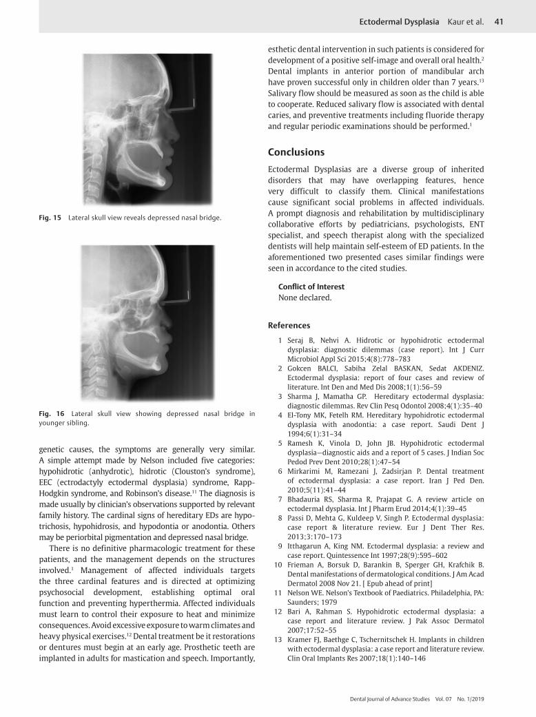
the 14-year-old, OPG revealed a single tooth in the maxilla (►Fig. 13). The left central incisor, 21, showed altered anatomy. The mandibular ridge was completely edentulous. The alveolar ridges were resorbed with decreased bone height. Similar findings were noted in the 16-year-old patient with 2 teeth, 11 and 22, which were also malformed (►Fig. 14). The lateral cephalogram also revealed decreased bone height of maxillary and mandibular arches in both the cases. The
nasal bridge was also depressed (►Figs. 15, 16). A diagnosis of ED was made keeping in view the above findings.
DiscussionEDs represent a large group of hereditary conditions characterized by congenital defects of one or more ectodermal structures including skin appendages.2 In some people, hair and nails may be involved, whereas in another person, teeth and sweat glands.1 Each combination is considered a separate type of ectodermal dysplasias as given by Bari and Rahman in
40 Ectodermal Dysplasia Kaur et al.
Dental Journal of Advance Studies Vol. 07 No. 1/2019
Fig. 9 Plantar keratosis.
Fig. 10 Plantar keratosis.
Fig. 11 IOPA (intraoral periapical) x-ray showing malformed anterior teeth in the maxilla.
Fig. 12 IOPA (intraoral periapical) x-ray showing single malformed tooth in the maxilla of younger sibling.
Fig. 13 Underdeveloped and malformed alveolar ridges.
Fig. 14 Atrophied and severely underdeveloped alveolar ridges in elder sibling.
2007.12 Freire Maia and Pinheir proposed a numerical system for the disorder, in which a number is assigned to the struc-ture involved: hair 1, teeth 2, nails 3, sweat glands 4. Total 10 different ED subgroups were proposed in their review arti-cle.8 A definitive classification of ED is difficult to formulate as many of the syndromes that involve ED have overlapping features.1 The disorder might occur during the first trimes-ter of pregnancy. If it is severe, it appears before the sixth week of embryonic life, and consequently the dentition will be affected. After eighth week, other ectodermal structures may be affected.3
Genetic studies of more than 300 cases have revealed X-linked mode of inheritance with its gene locus being Xq11-21.1. The gene is carried by females but manifested in males.3 However, Itthagarun and King have reported multiple siblings being affected and of females suffering with this condition.9 It has been estimated that ED is approximately 7 in 10,000 births, and all the mendelian modes of inheritance have been reported.10 Dental defects in many EDs can present as anodontia, polydontia, dysplastic teeth, retained primary teeth, amelogenesis imperfecta, dentinogenesis imperfecta, and underdeveloped alveolar ridge.2
More than 150 different syndromes have been identified. Despite some of the syndromes having different
41Ectodermal Dysplasia Kaur et al.
Dental Journal of Advance Studies Vol. 07 No. 1/2019
genetic causes, the symptoms are generally very similar. A simple attempt made by Nelson included five categories: hypohidrotic (anhydrotic), hidrotic (Clouston’s syndrome), EEC (ectrodactyly ectodermal dysplasia) syndrome, Rapp- Hodgkin syndrome, and Robinson’s disease.11 The diagnosis is made usually by clinician’s observations supported by relevant family history. The cardinal signs of hereditary EDs are hypo-trichosis, hypohidrosis, and hypodontia or anodontia. Others may be periorbital pigmentation and depressed nasal bridge.
There is no definitive pharmacologic treatment for these patients, and the management depends on the structures involved.1 Management of affected individuals targets the three cardinal features and is directed at optimizing psychosocial development, establishing optimal oral function and preventing hyperthermia. Affected individuals must learn to control their exposure to heat and minimize consequences. Avoid excessive exposure to warm climates and heavy physical exercises.12 Dental treatment be it restorations or dentures must begin at an early age. Prosthetic teeth are implanted in adults for mastication and speech. Importantly,
Fig. 15 Lateral skull view reveals depressed nasal bridge.
Fig. 16 Lateral skull view showing depressed nasal bridge in younger sibling.
esthetic dental intervention in such patients is considered for development of a positive self-image and overall oral health.2 Dental implants in anterior portion of mandibular arch have proven successful only in children older than 7 years.13 Salivary flow should be measured as soon as the child is able to cooperate. Reduced salivary flow is associated with dental caries, and preventive treatments including fluoride therapy and regular periodic examinations should be performed.1
ConclusionsEctodermal Dysplasias are a diverse group of inherited disorders that may have overlapping features, hence very difficult to classify them. Clinical manifestations cause significant social problems in affected individuals. A prompt diagnosis and rehabilitation by multidisciplinary collaborative efforts by pediatricians, psychologists, ENT specialist, and speech therapist along with the specialized dentists will help maintain self-esteem of ED patients. In the aforementioned two presented cases similar findings were seen in accordance to the cited studies.
Conflict of InterestNone declared.
References
1 Seraj B, Nehvi A. Hidrotic or hypohidrotic ectodermal dysplasia: diagnostic dilemmas (case report). Int J Curr Microbiol Appl Sci 2015;4(8):778–783
2 Gokcen BALCI, Sabiha Zelal BASKAN, Sedat AKDENIZ. Ectodermal dysplasia: report of four cases and review of literature. Int Den and Med Dis 2008;1(1):56–59
3 Sharma J, Mamatha GP. Hereditary ectodermal dysplasia: diagnostic dilemmas. Rev Clin Pesq Odontol 2008;4(1):35–40
4 El-Tony MK, Fetelh RM. Hereditary hypohidrotic ectodermal dysplasia with anodontia: a case report. Saudi Dent J 1994;6(1):31–34
5 Ramesh K, Vinola D, John JB. Hypohidrotic ectodermal dysplasia—diagnostic aids and a report of 5 cases. J Indian Soc Pedod Prev Dent 2010;28(1):47–54
6 Mirkarimi M, Ramezani J, Zadsirjan P. Dental treatment of ectodermal dysplasia: a case report. Iran J Ped Den. 2010;5(11):41–44
7 Bhadauria RS, Sharma R, Prajapat G. A review article on ectodermal dysplasia. Int J Pharm Erud 2014;4(1):39–45
8 Passi D, Mehta G, Kuldeep V, Singh P. Ectodermal dysplasia: case report & literature review. Eur J Dent Ther Res. 2013;3:170–173
9 Itthagarun A, King NM. Ectodermal dysplasia: a review and case report. Quintessence Int 1997;28(9):595–602
10 Frieman A, Borsuk D, Barankin B, Sperger GH, Krafchik B. Dental manifestations of dermatological conditions. J Am Acad Dermatol 2008 Nov 21. [ Epub ahead of print]
11 Nelson WE. Nelson’s Textbook of Paediatrics. Philadelphia, PA: Saunders; 1979
12 Bari A, Rahman S. Hypohidrotic ectodermal dysplasia: a case report and literature review. J Pak Assoc Dermatol 2007;17:52–55
13 Kramer FJ, Baethge C, Tschernitschek H. Implants in children with ectodermal dysplasia: a case report and literature review. Clin Oral Implants Res 2007;18(1):140–146