Embed Size (px)
Citation preview

Echocardiographic Anatomy in the Fetus
CHAPTER 2 • The Four-Chamber View I
ENRICO M. CHIAPPA • ANDREW C. COOK
GIANNI BOTTA • NORMAN H. SILVERMAN
Echocardiographic
Anatomy in the Fetus
Indice IIICHAPTER 2 • The Four-Chamber View III
ENRICO M. CHIAPPAMaternal and Perinatal Cardiology UnitDivision of Pediatricf Cardiology“Meyer” Children’s HospitalFlorence, Italy
ANDREW C. COOKBritish Heart FoundationtCardiac Morphology UnitInstitute of Childf HealthUniversity College LondonLondon, United Kingdom
GIANNI BOTTAFetal and Maternal PathologyDepartment oft Pathologyf“O.I.R.M. – S. Anna”Children & Women’s HospitalTurin, Italy
NORMAN H. SILVERMAN
Pediatric and Fetal Echocardiography LaboratoriesDivision of Pediatricf CardiologyLucile Packard Children’s HospitalStanford University Medical CenterPalo Alto, California, USA
IV Echocardiographic Anatomy in the Fetus
With a contribution byRABIH CHAOUI (Chapter 20)rCenter forr Prenatalr Diagnosisand Human GeneticsBerlin, Germany
Library of Congressf Control Number: 2008931599
ISBN 978-88-470-0572-3 Springer Milanr Berlin Heidelberg New Yorke-ISBN 978-88-470-0573-0
This work is subject to copyright. All rights are reserved, whether ther whole or partr oft thef materialis concerned, specifically the rights of translation,f reprinting, reuse of illustrations,f recitation, broad-casting, reproduction on microfilm or inr any other way,r and storage in data banks. Duplication ofthis publication or partsr thereof isf permitted only under ther provisions of thef Italian Copyright Lawin its current version,t and permission for user must alwayst be obtained from Springer. Violations areliable to prosecution under ther Italian Copyright Law.t
Springer isr a part oft Springerf Science+Businessr Mediaspringer.com© Springer-Verlag Italia 2008, 2nd printing 2009
The use of generalf descriptive names, registered names, trademarks, etc. in this publication does notimply, even in the absence of af specific statement, that such names are exempt from the relevant pro-ttective laws and regulations and therefore free for generalr use.Product liability:t The publishers cannot guarantee the accuracy of anyf information about dosaget andapplication contained in this book. In every individual case the user mustr checkt such information byconsulting the relevant literature.t
The copyright fort ther images printed in this work is retained by the authors, according to the listingat thet end of thisf book.
Cover image:r Simona Colombo, Milan, ItalyTypesetting: C & G di Cerri e Galassi, Cremona, ItalyPrinting: Printer Trentor s.r.l., Trento, Italy
Printed ind ItalySpringer-Verlag Italia S.r.l., Via Decembrio 28, I-20137 Milan, Italy

To our parents, who paved the way of our lives
Prefaceby Norman H. Silverman
Over the last two decades, the value of examining the fetal heart has moved from anexperimental procedure of diagnostic curiosity to a front-line form of evaluating fetalcardiac health and disease. There have been numerous advances in the associated tech-nology, including high-resolution imaging, the introduction of reliable color flow andpulse Doppler, and M-Mode and continuous-wave Doppler recordings in some instru-ments. Such advances continue, with the potential for 3D imaging using spatiotempo-ral image correlation (STIC) and full-volume fetal technology.
The techniques used by obstetric sonographers in all fields, including physiciansfrom the fields of radiology, obstetrics, and pediatric cardiology, together with tech-nologists who support and do most of the scanning, require a fundamental understandingof ultrasound as well as anatomy, physiology, and the various cardiac pathologies thatoccur in the fetus. This book addresses these fundamentals, providing correlations bymeans of diagrams and images of fetal cardiac morphology and pathology. The scansare quite unique, having been collected over several years by the principal author,Dr. Enrico M. Chiappa, from his laboratories in Italy, and provide exquisite echocar-diography of normal and congenitally malformed hearts. These are complemented bythe excellent pathological images of Dr. Andrew C. Cook and Dr. Gianni Botta, whoprovided high-quality images of normal and pathological fetal heart conditions, whichare displayed as support for the echocardiographic images.
The organization of this book is oriented toward practitioners. The first section pro-vides general guidelines for imaging the fetal body and heart, for segmental analysis, andfor diagnosis. The second section takes a view-oriented approach, describing first thetransverse views and then the longitudinal views of the fetal body and how each echocar-diographic projection best displays a particular pathological entity. The third section con-tains essential information pertaining to the new technique of 3D/4D echocardiographyand the role of the pathologist in heart disease, which expands further the value of thistext for providing references and comparisons with standard imaging techniques.
The authors obviously gave a great deal of thought to this project – from the choiceof images in the text, which include the clearest descriptions and labels, to the ac-companying DVD, which contains complex moving echocardiographic images. Theobjective is to provide the reader with something greater than a static representationof the fetal cardiac morphology while retaining the ability to refer directly to mor-phological comparisons and consult with the text for greater detail. This work will havegreat appeal to physicians and technologists involved in obtaining and interpreting suchimages and will provide the obstetric, cardiological, and radiological communities withan excellent reference for comparing cases seen in their daily practice.
Indice VIICHAPTER 2 • The Four-Chamber View VII
Prefaceby Enrico M. Chiappa
The number ofr congenitalf heart diseasest detected in utero is still low, even in countrieswith well-advanced screening programs. Only 20% of newf cases of congenitalf heartdisease come from traditionalm high-risk pregnancies.k Ultrasound screening of allf preg-nancies is therefore necessary to improve the detection rate of congenitalf heart diseasestand to manage them mostm effectively.t However, prenatal screening studies have shownwidely divergent results,t with low detection rates in most cases.t Several reasons havebeen advocated to explain why these programs seem tom fail, and different solutionst havebeen proposed to improve the success rate. There is a broada consensus that examinertskill plays a cruciala role in this setting and that appropriatet training is mandatory.
It ist our beliefr thatf peoplet responsible for fetalr cardiology units in centers of excel-flence should invest resourcest to fully educate the personnel involved in prenatal ultra-sound screening. To do this, a fundamentala step is to improve, simply and comprehen-sibly, their, knowledger about thet anatomy of thef fetal cardiovascular system.r As Prof.Robert Andersont beautifully stated in a recenta reviewt [Anderson RH, Razavi R, Tay-lor AMr (2004) Cardiac anatomy revisited. J Anat 205:159-177],t one convention of thefhuman anatomy is that allt structures should be described in the setting of theirf anatom-rical position within the body and of thef relationship of organsf to each other. This con-vention has not alwayst been strictly applied when describing the heart. In the past, theso-called Valentine approach has prevailed, that is,t the convention of representingf theheart isolatedt from them surrounding structures and balanced on its apex, with the atriaabove the ventricles. This approach generated confusion, particularly, in the field of con-fgenital heart disease,t where, the position of thef heart andt the location of thef cardiac seg-ments is variable. Therefore, as Prof. Anderson wrote: “students should be introducedto the anatomy of thef heart ast it liest within the body, as revealed with clinical tomo-graphic images”. To this purpose, the use of tomographicf sections of isolatedf hearts,frequently used in textbooks of echocardiography,f is only partially effective in prena-tal ultrasound examination,d where, the views of thef surrounding structures are much widerand the approaches to the fetal thorax more variable than in the postnatal setting.
We decided,e therefore,, to, perform imagem sectionse of thef wholee fetale bodyl to obtain tomon -graphic views of thef heart, thus, showing the relationship between cardiacn and extracardiacdstructures. As indicated ind Chaptern 21,r tomographic, sections of thef fetal body were obtain-ned ind an limiteda numberd ofr fetusesf under ther gestational age of 20f weeks following informedconsent oft parentsf and ind strictn adherencet with theh Italian legislationn onn thisn subject. Theidea toa obtain thesen types of sectionsf stemmed fromd almostm 20t years of fundamentalf workby Alf Staudachf [Staudachh Ah (1989)A Sectional fetal anatomy in ultrasound.n Springer], with,the advantages of computerf technologyr in photographicn and ultrasoundd images.d
Rather thanr systematically describing congenital heart diseasest in the fetus, the goalof thisf work is to provide a basic tool for understandingr the normal and abnormalechocardiographic anatomy of thef fetal heart. In this way, this, textbook isk complementaryand not alternative to the more extensive publications on this subject.
The first sectiont ofn thisf book describesk the basic principles of diagnosis,f illustrating,assessment oft thef laterality of thef fetal body, the, visceroatrial arrangement, and, thed car-diac position. This order reproducesr the logical sequence the examiner shouldr followd whenstudying the fetal heart. In then second section,d all, echocardiographic projections in then fe-
Indice IXCHAPTER 2 • The Four-Chamber View IX

X Preface
tus are described, from those most familiar to obstetric sonographers, to those usually ob-tained only by pediatric cardiologists. Particular emphasis is given to imaging the short-axis sections of the fetal body, which has recently proven to be a powerful method forcomplete examination of the fetal heart. The chapters pertaining to echocardiographic pro-jection are in logical sequence in two series: the transverse views of the fetal thorax, pre-sented from the bottom to the top, and the sagittal and parasagittal views, presented fromthe right side to the left side. The sequence of these planes in some ways imitates thechanges of the scanning plane that can be obtained by the examiner tilting or translatingthe probe manually with traditional probes or electronically with modern 3D ultrasound.
The third section consists of two chapters. The first describes the essential use of mod-ern 3D/4D ultrasound techniques to image the fetal heart, including new volume manip-ulation, such as spatial and temporal image correlation (STIC), and rendering, such as glass-body, minimum-transparent, and inversion modes. The second chapter emphasizes the im-portance of autopsy in providing information regarding anomalies of the fetal heart anddescribes optimal techniques for dissection and photography of the autopsic specimens.This section emphasizes the crucial role of the echocardiographic projections describedelsewhere in this book, which maintain their fundamental role in the comprehension ofnew advances in ultrasound and the future application of MRI/CT in postmortem studiesof the fetal heart. These two different chapters are incorporated in this section because theapproach to volume data sets of 3D/4D sonography and to blocks of pathological speci-mens shows many similarities. The information displayed by either technique depends onthe level at which the examiner cuts the cardiac volume, no matter if a digital tool or apathologist’s blade is used. Moreover, some image-rendering techniques are comparablewith the fine-art photographic technique available to the pathologist to display with out-standing clarity the subtle details of congenital heart disease in fetal heart specimens.
The problem of image orientation was thoroughly discussed with cardiac mor-phologists Andrew C. Cook, Gianni Botta, and Robert H. Anderson from the very be-ginning of the editorial phase of this book. The reader will notice that some anatomi-cal images of the transverse sections of the thorax do not “at first sight” match theechocardiographic images. We decided to maintain the echocardiographic images intheir original caudocranial orientation, a standard that is accepted in MR and CT imag-ing, rather than flip them horizontally, which would reduce the resolution of the digi-tal clips on the accompanying DVD. We decided, instead, to display a compass withevery image, thus illustrating its orientation.
As to the abnormal heart, rather than fully describe a single congenital heart dis-ease in different projections, the book and DVD describe how a specific projection ap-pears in the normal heart and in some abnormal conditions. Although we understandthat this is not sufficient for a comprehensive assessment of a specific disease, we be-lieve this type of presentation reproduces the usual approach of the examiner, who isinitially unaware whether the case being examining is normal or abnormal. The morethe examiner understands the normal anatomy, the easier it is to recognize what is wrong.
Ultrasound assessment of the heart, whether in fetal life or postnatally, is based onmoving images, and books therefore have limited value in teaching echocardiography.With recent advances in ultrasound systems, storing multiple digital frames and clipswith superb image quality has become a reality. These advances have brought innova-tive applications to the clinical field and can be utilized as powerful multimedia pre-sentations for teaching purposes. The accompanying DVD is such a tool. This is a com-plex DVD that includes more than 300 clips of normal and abnormal views of the fe-tal heart frames of each topic, with 11–13, representing a single cardiac cycle, each ofwhich is displayed in a loop. The DVD in many respects is not simply a copy of thebook. The text window contains essential information of the case being presented, and– in the four-chamber section in particular – a short unit describes the essential featuresof each specific congenital heart disease. We believe this presentation will be a highlyuseful tool for all those interested in the echocardiographic study of the fetal heart.
Acknowledgements
First of all, a special tribute to Prof. Robert H. Anderson, a giant in the field of mor-phology in congenital heart disease, whose teaching through the countless numbers ofpapers, books, lectures, and courses deeply impacts the practice of modern pediatriccardiology and cardiac surgery and, inevitably and unavoidably, the pages of this book.
I am particularly grateful to Norman H. Silverman, who supported this editorialproject. During an intense period at the University of California San Francisco, he taughtme new insights into fetal and pediatric echocardiography, a powerful mixture of mor-phology and physiology, along with the never-ending enthusiasm for teaching and col-lecting outstanding images.
A special note of thanks to Andrew C. Cook, who gave Gianni Botta and me thevaluable opportunity to learn from his passionate work as scientist coupled with hisrefined technique of dissection and photography of fetal heart specimens. Thanks al-so to Gianni Botta, a colleague and friend whose day-to-day collaboration providedthe reciprocal opportunity for growth and understanding in the field of perinatal con-genital heart diseases. I am indebted to Rabih Chaoui for agreeing to write the chap-ter on 3D/4D fetal echocardiography. We all recognize his substantial contribution tothe knowledge of this technique through his fundamental papers published in the in-ternational literature.
We owe a special thanks to our esteemed colleagues who directly and indirectlycontributed to this textbook: Tullia Todros, Mario Campogrande, Elsa Viora, PieroGaglioti, Vlasta Fesslovà, Roberto Tumbarello, Roberto Conturso, Patrizia D’Ajello,Vincenzo Trengia, Marco Pagliano, Simona Sdei, Giuseppe Errante, Simona Bastonero,Maria Grazia Alemanno, Maria Brizzolara, Rosalba Giachello, Ruggiero Crocco, Al-fio Oddo, Maria Renzetti, Giuseppe De Sanso, Vittorio Guaragna, Antonio Guelfi,Bruno Inaudi, Pierluigi Mazzucco, Alessandra Lesca, and Elena Gullino. Thanks tothem and many others not mentioned, we had the opportunity to share clinical caseexperiences. Last but not least, I gratefully acknow ledge Andrea Sciarrone for his valu-able collaboration in the clinical field and his help in proofreading the references ofthis work.
Our thanks to fellows and students in the many courses on fetal echocardiographywhose comments and criticism provided much of the stimulus that prompted this work.
We are deeply grateful to the families of affected fetuses, families whose cooper-ation allowed us to discover the natural history of these diseases and the result of treat-ment and whose strength taught us courage and fortitude.
A special note of thanks to the personnel of the divisions of Obstetrics, Neonatology,Pediatric and Women’s Intensive Care Units, Pediatric Cardiology, and Pediatric CardiacSurgery of the O.I.R.M. – S. Anna, Children & Women’s Hospital, Turin, where manycases presented in this text were treated, with whom I was honored to work for manyyears. Because of their professional abilities matched with dedication, positive results werepossible on many occasions. It is our wish that such results will be possible in our newlocation at the Meyer Children’ Hospital in Florence.
Our thanks to Sofia Redaelli (CNR Massa) and Eugenio Picano (CNR Pisa) Ital-ian National Research Council (CNR) who helped make this book possible.
CHAPTER 2 • The Four-Chamber View XI
We are particularly indebted to Giuliano Kraft – efficiently supported in the finalphase of this work by Tiziano Carducci – for the patience and strength he showed astime went on and it became apparent that the work was evolving beyond our expec-tations. Thanks to his creativity and professional ability in the digital world, he con-tributed brilliantly to the shape and construction of the complex structure of the ac-companying DVD.
We also thank the staff at Springer-Verlag Italia, and in particular Antonella Cerri,executive editor, for first approaching us with, and then believing in, this project; MarieThompson, for her outstanding work in copyediting the manuscript; and AlessandraBorn and Barbara Ferrario, assistant editors, for their efficiency in helping to keep every-thing straight and on track.
Finally, my deep appreciation to Gabriella, my lovely companion in life, who be-lieved in me and tolerated many lonely evenings and weekends over the long periodit took to complete this project. The authors’ wish only that through this work, manyfamilies will gain more than our families have lost.
Florence, August 2008 E.M. Chiappa
XII Aknowledgements
PART I Basic Principles of Diagnosis
CHAPTER 1 General Guidelines . . . . . . . . . . . . . . . . . . . . . . . . . . . . . . . . . . . . . . . . . . . . . . . . . . . . . . 3Introduction . . . . . . . . . . . . . . . . . . . . . . . . . . . . . . . . . . . . . . . . . . . . . . . . . . . . . . . . . . . . . . . 3Equipment . . . . . . . . . . . . . . . . . . . . . . . . . . . . . . . . . . . . . . . . . . . . . . . . . . . . . . . . . . . . . . . . 4Major Planes of the Body and Heart . . . . . . . . . . . . . . . . . . . . . . . . . . . . . . . . . . . . . . . . . . . . . 6Basic Probe Manipulations . . . . . . . . . . . . . . . . . . . . . . . . . . . . . . . . . . . . . . . . . . . . . . . . . . . . 7Axial and Lateral Resolution . . . . . . . . . . . . . . . . . . . . . . . . . . . . . . . . . . . . . . . . . . . . . . . . . . . 9Method for Comprehensive Cardiac Evaluation . . . . . . . . . . . . . . . . . . . . . . . . . . . . . . . . . . . . 10References . . . . . . . . . . . . . . . . . . . . . . . . . . . . . . . . . . . . . . . . . . . . . . . . . . . . . . . . . . . . . . . . 12
CHAPTER 2 Determining the Laterality of the Fetal Body and Image Orientation . . . . . 15Introduction . . . . . . . . . . . . . . . . . . . . . . . . . . . . . . . . . . . . . . . . . . . . . . . . . . . . . . . . . . . . . . . 15Methods . . . . . . . . . . . . . . . . . . . . . . . . . . . . . . . . . . . . . . . . . . . . . . . . . . . . . . . . . . . . . . . . . . 16– Procedure . . . . . . . . . . . . . . . . . . . . . . . . . . . . . . . . . . . . . . . . . . . . . . . . . . . . . . . . . . . . . . . 17Image Orientation . . . . . . . . . . . . . . . . . . . . . . . . . . . . . . . . . . . . . . . . . . . . . . . . . . . . . . . . . . 19Reference . . . . . . . . . . . . . . . . . . . . . . . . . . . . . . . . . . . . . . . . . . . . . . . . . . . . . . . . . . . . . . . . . 20
CHAPTER 3 The Visceroatrial Arrangement (Situs) . . . . . . . . . . . . . . . . . . . . . . . . . . . . . . . . . . . . 21Introduction . . . . . . . . . . . . . . . . . . . . . . . . . . . . . . . . . . . . . . . . . . . . . . . . . . . . . . . . . . . . . . . 21Usual Arrangement (Situs Solitus) . . . . . . . . . . . . . . . . . . . . . . . . . . . . . . . . . . . . . . . . . . . . . . 21Mirror-Image Arrangement (Situs Inversus) . . . . . . . . . . . . . . . . . . . . . . . . . . . . . . . . . . . . . . . 23Left Isomerism (Polysplenia) . . . . . . . . . . . . . . . . . . . . . . . . . . . . . . . . . . . . . . . . . . . . . . . . . . . 24Right Isomerism (Asplenia) . . . . . . . . . . . . . . . . . . . . . . . . . . . . . . . . . . . . . . . . . . . . . . . . . . . 26References . . . . . . . . . . . . . . . . . . . . . . . . . . . . . . . . . . . . . . . . . . . . . . . . . . . . . . . . . . . . . . . . 28
CHAPTER 4 The Cardiac Position and Axis Orientation . . . . . . . . . . . . . . . . . . . . . . . . . . . . . . . . 29Introduction . . . . . . . . . . . . . . . . . . . . . . . . . . . . . . . . . . . . . . . . . . . . . . . . . . . . . . . . . . . . . . . 29Heart in the Left Side of the Chest (Levocardia) . . . . . . . . . . . . . . . . . . . . . . . . . . . . . . . . . . . . 30Heart in the Middle of the Chest (Mesocardia) . . . . . . . . . . . . . . . . . . . . . . . . . . . . . . . . . . . . 31Heart in the Right Side of the Chest (Dextrocardia) . . . . . . . . . . . . . . . . . . . . . . . . . . . . . . . . . 31Isomerism of the Atrial Appendages and Cardiac Position . . . . . . . . . . . . . . . . . . . . . . . . . . . . 35References . . . . . . . . . . . . . . . . . . . . . . . . . . . . . . . . . . . . . . . . . . . . . . . . . . . . . . . . . . . . . . . . 37
CHAPTER 5 Principles of Segmental Analysis . . . . . . . . . . . . . . . . . . . . . . . . . . . . . . . . . . . . . . . . . 39Introduction . . . . . . . . . . . . . . . . . . . . . . . . . . . . . . . . . . . . . . . . . . . . . . . . . . . . . . . . . . . . . . . 39The Atria . . . . . . . . . . . . . . . . . . . . . . . . . . . . . . . . . . . . . . . . . . . . . . . . . . . . . . . . . . . . . . . . . . 39The Ventricles . . . . . . . . . . . . . . . . . . . . . . . . . . . . . . . . . . . . . . . . . . . . . . . . . . . . . . . . . . . . . . 40The Arterial Trunks . . . . . . . . . . . . . . . . . . . . . . . . . . . . . . . . . . . . . . . . . . . . . . . . . . . . . . . . . . 42The Atrioventricular Connection . . . . . . . . . . . . . . . . . . . . . . . . . . . . . . . . . . . . . . . . . . . . . . . . 42The Ventriculoarterial Connection . . . . . . . . . . . . . . . . . . . . . . . . . . . . . . . . . . . . . . . . . . . . . . 44References . . . . . . . . . . . . . . . . . . . . . . . . . . . . . . . . . . . . . . . . . . . . . . . . . . . . . . . . . . . . . . . . 48
Elenco autori XIII
Contents
CHAPTER 2 • The Four-Chamber View XIII
XIV Contents
PART II Echocardiographic Projections
A Transverse Views of the Fetal Body
CHAPTER 6 The Transverse Views of the Upper Abdomen . . . . . . . . . . . . . . . . . . . . . . . . . . . . . 51Introduction . . . . . . . . . . . . . . . . . . . . . . . . . . . . . . . . . . . . . . . . . . . . . . . . . . . . . . . . . . . . . . . 51The Umbilical Cord Insertion . . . . . . . . . . . . . . . . . . . . . . . . . . . . . . . . . . . . . . . . . . . . . . . . . . . 51The Lower Liver . . . . . . . . . . . . . . . . . . . . . . . . . . . . . . . . . . . . . . . . . . . . . . . . . . . . . . . . . . . . 52The Mid Liver . . . . . . . . . . . . . . . . . . . . . . . . . . . . . . . . . . . . . . . . . . . . . . . . . . . . . . . . . . . . . . 52The Portal Sinus . . . . . . . . . . . . . . . . . . . . . . . . . . . . . . . . . . . . . . . . . . . . . . . . . . . . . . . . . . . . 52The Infracardiac Vena Cava . . . . . . . . . . . . . . . . . . . . . . . . . . . . . . . . . . . . . . . . . . . . . . . . . . . . 55The Suprahepatic Veins . . . . . . . . . . . . . . . . . . . . . . . . . . . . . . . . . . . . . . . . . . . . . . . . . . . . . . . 55References . . . . . . . . . . . . . . . . . . . . . . . . . . . . . . . . . . . . . . . . . . . . . . . . . . . . . . . . . . . . . . . . 57
CHAPTER 7 The Four-Chamber View . . . . . . . . . . . . . . . . . . . . . . . . . . . . . . . . . . . . . . . . . . . . . . . . . 59The Section Plane . . . . . . . . . . . . . . . . . . . . . . . . . . . . . . . . . . . . . . . . . . . . . . . . . . . . . . . . . . . 59The Normal Morphology . . . . . . . . . . . . . . . . . . . . . . . . . . . . . . . . . . . . . . . . . . . . . . . . . . . . . . 59The Normal Echocardiogram – 2D . . . . . . . . . . . . . . . . . . . . . . . . . . . . . . . . . . . . . . . . . . . . . . 60Dimensions of the Heart . . . . . . . . . . . . . . . . . . . . . . . . . . . . . . . . . . . . . . . . . . . . . . . . . . . . . . 63Measurement . . . . . . . . . . . . . . . . . . . . . . . . . . . . . . . . . . . . . . . . . . . . . . . . . . . . . . . . . . . . . . 63Ventricular Function . . . . . . . . . . . . . . . . . . . . . . . . . . . . . . . . . . . . . . . . . . . . . . . . . . . . . . . . . 65The Normal Echocardiogram – Color Flow Mapping and Pulsed Doppler . . . . . . . . . . . . . . . . 65– The Atrioventricular Valves . . . . . . . . . . . . . . . . . . . . . . . . . . . . . . . . . . . . . . . . . . . . . . . . . . 65– The Pulmonary Veins . . . . . . . . . . . . . . . . . . . . . . . . . . . . . . . . . . . . . . . . . . . . . . . . . . . . . . 69Cardiac Rhythm . . . . . . . . . . . . . . . . . . . . . . . . . . . . . . . . . . . . . . . . . . . . . . . . . . . . . . . . . . . . 71Ventricular Output . . . . . . . . . . . . . . . . . . . . . . . . . . . . . . . . . . . . . . . . . . . . . . . . . . . . . . . . . . 71Four-Chamber-View Checklist . . . . . . . . . . . . . . . . . . . . . . . . . . . . . . . . . . . . . . . . . . . . . . . . . 72References . . . . . . . . . . . . . . . . . . . . . . . . . . . . . . . . . . . . . . . . . . . . . . . . . . . . . . . . . . . . . . . . 74
CHAPTER 8 The Five-Chamber View . . . . . . . . . . . . . . . . . . . . . . . . . . . . . . . . . . . . . . . . . . . . . . . . . . 77The Section Plane . . . . . . . . . . . . . . . . . . . . . . . . . . . . . . . . . . . . . . . . . . . . . . . . . . . . . . . . . . . 77The Normal Morphology . . . . . . . . . . . . . . . . . . . . . . . . . . . . . . . . . . . . . . . . . . . . . . . . . . . . . . 77The Normal Echocardiogram – 2D . . . . . . . . . . . . . . . . . . . . . . . . . . . . . . . . . . . . . . . . . . . . . . 77The Normal Echocardiogram – Color Flow Mapping and Pulsed Doppler . . . . . . . . . . . . . . . . 79References . . . . . . . . . . . . . . . . . . . . . . . . . . . . . . . . . . . . . . . . . . . . . . . . . . . . . . . . . . . . . . . . 86
CHAPTER 9 The Three-Vessel View . . . . . . . . . . . . . . . . . . . . . . . . . . . . . . . . . . . . . . . . . . . . . . . . . . . 87Introduction . . . . . . . . . . . . . . . . . . . . . . . . . . . . . . . . . . . . . . . . . . . . . . . . . . . . . . . . . . . . . . . 87The Normal Morphology . . . . . . . . . . . . . . . . . . . . . . . . . . . . . . . . . . . . . . . . . . . . . . . . . . . . . . 87The Normal Echocardiogram – 2D . . . . . . . . . . . . . . . . . . . . . . . . . . . . . . . . . . . . . . . . . . . . . . 88The Normal Echocardiogram – Color Flow Mapping and Pulsed Doppler . . . . . . . . . . . . . . . . 88Three-Vessel-View Abnormalities . . . . . . . . . . . . . . . . . . . . . . . . . . . . . . . . . . . . . . . . . . . . . . . 89Ascending Aorta and Pulmonary Artery Disproportion . . . . . . . . . . . . . . . . . . . . . . . . . . . . . . 96Aortic Arch Sidedness . . . . . . . . . . . . . . . . . . . . . . . . . . . . . . . . . . . . . . . . . . . . . . . . . . . . . . . . 96Relationship of the Great Arteries and Cardiac Connections . . . . . . . . . . . . . . . . . . . . . . . . . . . 96References . . . . . . . . . . . . . . . . . . . . . . . . . . . . . . . . . . . . . . . . . . . . . . . . . . . . . . . . . . . . . . . . 99
CHAPTER 10 The Arterial Duct Transverse View . . . . . . . . . . . . . . . . . . . . . . . . . . . . . . . . . . . . . . . . 101The Section Plane . . . . . . . . . . . . . . . . . . . . . . . . . . . . . . . . . . . . . . . . . . . . . . . . . . . . . . . . . . . 101The Normal Morphology . . . . . . . . . . . . . . . . . . . . . . . . . . . . . . . . . . . . . . . . . . . . . . . . . . . . . . 101The Normal Echocardiogram – 2D . . . . . . . . . . . . . . . . . . . . . . . . . . . . . . . . . . . . . . . . . . . . . . 102The Normal Echocardiogram – Color Flow Mapping and Pulsed Doppler . . . . . . . . . . . . . . . . 102
The Arterial Duct – Reversed Flow . . . . . . . . . . . . . . . . . . . . . . . . . . . . . . . . . . . . . . . . . . . . . . 103The Arterial Duct – Short Length . . . . . . . . . . . . . . . . . . . . . . . . . . . . . . . . . . . . . . . . . . . . . . . 105The Arterial Duct – Abnormal Shape . . . . . . . . . . . . . . . . . . . . . . . . . . . . . . . . . . . . . . . . . . . . 105The Arterial Duct – Premature Constriction or Closure . . . . . . . . . . . . . . . . . . . . . . . . . . . . . . . 108The Arterial Duct – Abnormal Position . . . . . . . . . . . . . . . . . . . . . . . . . . . . . . . . . . . . . . . . . . . 110References . . . . . . . . . . . . . . . . . . . . . . . . . . . . . . . . . . . . . . . . . . . . . . . . . . . . . . . . . . . . . . . . 111
CHAPTER 11 The Aortic Arch Transverse View . . . . . . . . . . . . . . . . . . . . . . . . . . . . . . . . . . . . . . . . . . 113The Section Plane . . . . . . . . . . . . . . . . . . . . . . . . . . . . . . . . . . . . . . . . . . . . . . . . . . . . . . . . . . . 113The Normal Morphology . . . . . . . . . . . . . . . . . . . . . . . . . . . . . . . . . . . . . . . . . . . . . . . . . . . . . . 113The Normal Echocardiogram – 2D . . . . . . . . . . . . . . . . . . . . . . . . . . . . . . . . . . . . . . . . . . . . . . 113– The Thymus . . . . . . . . . . . . . . . . . . . . . . . . . . . . . . . . . . . . . . . . . . . . . . . . . . . . . . . . . . . . . 114The Normal Echocardiogram – Color Flow Mapping and Pulsed Doppler . . . . . . . . . . . . . . . . 115Aortic Arch Abnormal Size . . . . . . . . . . . . . . . . . . . . . . . . . . . . . . . . . . . . . . . . . . . . . . . . . . . . 116Aortic Arch Interruption . . . . . . . . . . . . . . . . . . . . . . . . . . . . . . . . . . . . . . . . . . . . . . . . . . . . . . 117Aortic Arch Sidedness . . . . . . . . . . . . . . . . . . . . . . . . . . . . . . . . . . . . . . . . . . . . . . . . . . . . . . . . 117References . . . . . . . . . . . . . . . . . . . . . . . . . . . . . . . . . . . . . . . . . . . . . . . . . . . . . . . . . . . . . . . . 121
CHAPTER 12 The Arterial Duct and Aortic Arch Transverse View . . . . . . . . . . . . . . . . . . . . . . . . 123The Section Plane . . . . . . . . . . . . . . . . . . . . . . . . . . . . . . . . . . . . . . . . . . . . . . . . . . . . . . . . . . . 123The Normal Morphology . . . . . . . . . . . . . . . . . . . . . . . . . . . . . . . . . . . . . . . . . . . . . . . . . . . . . . 123The Normal Echocardiogram – 2D . . . . . . . . . . . . . . . . . . . . . . . . . . . . . . . . . . . . . . . . . . . . . . 123The Normal Echocardiogram – Color Flow Mapping . . . . . . . . . . . . . . . . . . . . . . . . . . . . . . . . 124The Aortic Arch Abnormalities . . . . . . . . . . . . . . . . . . . . . . . . . . . . . . . . . . . . . . . . . . . . . . . . . 126– Severe Aortic Outflow Obstruction . . . . . . . . . . . . . . . . . . . . . . . . . . . . . . . . . . . . . . . . . . . . 126– Severe Pulmonary Outflow Obstruction . . . . . . . . . . . . . . . . . . . . . . . . . . . . . . . . . . . . . . . . 126– Pseudoatresia of the Pulmonary Valve . . . . . . . . . . . . . . . . . . . . . . . . . . . . . . . . . . . . . . . . . 128– Vascular Ring . . . . . . . . . . . . . . . . . . . . . . . . . . . . . . . . . . . . . . . . . . . . . . . . . . . . . . . . . . . . 128Azygos Vein Dilatation . . . . . . . . . . . . . . . . . . . . . . . . . . . . . . . . . . . . . . . . . . . . . . . . . . . . . . . 128The Transverse Views of the Fetal Body: an Overview . . . . . . . . . . . . . . . . . . . . . . . . . . . . . . . 129References . . . . . . . . . . . . . . . . . . . . . . . . . . . . . . . . . . . . . . . . . . . . . . . . . . . . . . . . . . . . . . . . 131
B Longitudinal Views of the Fetal Body
CHAPTER 13 The Inferior and Superior Vena Cava Long-Axis View (The Bicaval View) . . . 135The Section Plane . . . . . . . . . . . . . . . . . . . . . . . . . . . . . . . . . . . . . . . . . . . . . . . . . . . . . . . . . . . 135The Normal Morphology . . . . . . . . . . . . . . . . . . . . . . . . . . . . . . . . . . . . . . . . . . . . . . . . . . . . . . 135The Normal Echocardiogram – 2D . . . . . . . . . . . . . . . . . . . . . . . . . . . . . . . . . . . . . . . . . . . . . . 137The Normal Echocardiogram – Color Flow Mapping and Pulsed Doppler . . . . . . . . . . . . . . . . 137The Azygos Vein . . . . . . . . . . . . . . . . . . . . . . . . . . . . . . . . . . . . . . . . . . . . . . . . . . . . . . . . . . . . 139Vena Cava Disproportion . . . . . . . . . . . . . . . . . . . . . . . . . . . . . . . . . . . . . . . . . . . . . . . . . . . . . 139Azygos Vein Dilatation . . . . . . . . . . . . . . . . . . . . . . . . . . . . . . . . . . . . . . . . . . . . . . . . . . . . . . . 139References . . . . . . . . . . . . . . . . . . . . . . . . . . . . . . . . . . . . . . . . . . . . . . . . . . . . . . . . . . . . . . . . 142
CHAPTER 14 The Aortic Arch Long-Axis View . . . . . . . . . . . . . . . . . . . . . . . . . . . . . . . . . . . . . . . . . . 143The Section Plane . . . . . . . . . . . . . . . . . . . . . . . . . . . . . . . . . . . . . . . . . . . . . . . . . . . . . . . . . . . 143The Normal Morphology . . . . . . . . . . . . . . . . . . . . . . . . . . . . . . . . . . . . . . . . . . . . . . . . . . . . . . 143The Normal Echocardiogram – 2D . . . . . . . . . . . . . . . . . . . . . . . . . . . . . . . . . . . . . . . . . . . . . . 145The Normal Echocardiogram – Color Flow Mapping and Pulsed Doppler . . . . . . . . . . . . . . . . 146The Aortic Arch Abnormalities . . . . . . . . . . . . . . . . . . . . . . . . . . . . . . . . . . . . . . . . . . . . . . . . . 148– Severe Aortic Outflow Obstructions . . . . . . . . . . . . . . . . . . . . . . . . . . . . . . . . . . . . . . . . . . . 148– Severe Pulmonary Outflow Obstructions . . . . . . . . . . . . . . . . . . . . . . . . . . . . . . . . . . . . . . . 149
Contents XV

– Variability of the Plane of the Aortic Arch Long-Axis View . . . . . . . . . . . . . . . . . . . . . . . . . 151– The Aortic Arch in Mitral Valve Atresia with Ventricular Septal Defect . . . . . . . . . . . . . . . . 151References . . . . . . . . . . . . . . . . . . . . . . . . . . . . . . . . . . . . . . . . . . . . . . . . . . . . . . . . . . . . . . . . 153
CHAPTER 15 The Arterial Duct Long-Axis View . . . . . . . . . . . . . . . . . . . . . . . . . . . . . . . . . . . . . . . . . 155The Section Plane . . . . . . . . . . . . . . . . . . . . . . . . . . . . . . . . . . . . . . . . . . . . . . . . . . . . . . . . . . . 155The Normal Morphology . . . . . . . . . . . . . . . . . . . . . . . . . . . . . . . . . . . . . . . . . . . . . . . . . . . . . . 155The Normal Echocardiogram – 2D . . . . . . . . . . . . . . . . . . . . . . . . . . . . . . . . . . . . . . . . . . . . . . 156The Normal Echocardiogram – Color Flow Mapping and Pulsed Doppler . . . . . . . . . . . . . . . . 157Duct-Dependent Systemic Circulation . . . . . . . . . . . . . . . . . . . . . . . . . . . . . . . . . . . . . . . . . . . 159Duct-Dependent Pulmonary Circulation . . . . . . . . . . . . . . . . . . . . . . . . . . . . . . . . . . . . . . . . . . 159The Arterial Duct – Premature Constriction or Closure . . . . . . . . . . . . . . . . . . . . . . . . . . . . . . . 162References . . . . . . . . . . . . . . . . . . . . . . . . . . . . . . . . . . . . . . . . . . . . . . . . . . . . . . . . . . . . . . . . 164
CHAPTER 16 Special Considerations on the Arterial Duct and Aortic Arch Views . . . . . . . . 165Technique . . . . . . . . . . . . . . . . . . . . . . . . . . . . . . . . . . . . . . . . . . . . . . . . . . . . . . . . . . . . . . . . . 165
CHAPTER 17 The Right Ventricle Outflow View . . . . . . . . . . . . . . . . . . . . . . . . . . . . . . . . . . . . . . . . 169The Section Plane . . . . . . . . . . . . . . . . . . . . . . . . . . . . . . . . . . . . . . . . . . . . . . . . . . . . . . . . . . . 169The Normal Morphology . . . . . . . . . . . . . . . . . . . . . . . . . . . . . . . . . . . . . . . . . . . . . . . . . . . . . . 169The Normal Echocardiogram – 2D . . . . . . . . . . . . . . . . . . . . . . . . . . . . . . . . . . . . . . . . . . . . . . 171The Normal Echocardiogram – Color Flow Mapping and Pulsed Doppler . . . . . . . . . . . . . . . . 171
CHAPTER 18 The Left Ventricle Short-Axis View . . . . . . . . . . . . . . . . . . . . . . . . . . . . . . . . . . . . . . . 177The Section Plane . . . . . . . . . . . . . . . . . . . . . . . . . . . . . . . . . . . . . . . . . . . . . . . . . . . . . . . . . . . 177The Normal Morphology . . . . . . . . . . . . . . . . . . . . . . . . . . . . . . . . . . . . . . . . . . . . . . . . . . . . . . 177– The Level of the Mitral and Tricuspid Valves . . . . . . . . . . . . . . . . . . . . . . . . . . . . . . . . . . . . . 177– The Level of the Papillary Muscles . . . . . . . . . . . . . . . . . . . . . . . . . . . . . . . . . . . . . . . . . . . . 178
C Oblique Views of the Fetal Body
CHAPTER 19 The Left Ventricle Long-Axis View . . . . . . . . . . . . . . . . . . . . . . . . . . . . . . . . . . . . . . . . 187The Section Plane . . . . . . . . . . . . . . . . . . . . . . . . . . . . . . . . . . . . . . . . . . . . . . . . . . . . . . . . . . . 187The Normal Morphology . . . . . . . . . . . . . . . . . . . . . . . . . . . . . . . . . . . . . . . . . . . . . . . . . . . . . . 187The Normal Echocardiogram – 2D . . . . . . . . . . . . . . . . . . . . . . . . . . . . . . . . . . . . . . . . . . . . . . 189The Normal Echocardiogram – Color Flow Mapping and Pulsed Doppler . . . . . . . . . . . . . . . . 189
PART III Echocardiographic and Morphologic Overview
CHAPTER 20 Three-Dimensional Ultrasound Assessment of the Fetal Heart . . . . . . . . . . . . 199Rabih ChaouiIntroduction . . . . . . . . . . . . . . . . . . . . . . . . . . . . . . . . . . . . . . . . . . . . . . . . . . . . . . . . . . . . . . . 199Technical Principles of 3D/4D Fetal Echocardiography . . . . . . . . . . . . . . . . . . . . . . . . . . . . . . . 199– Volume Acquisition . . . . . . . . . . . . . . . . . . . . . . . . . . . . . . . . . . . . . . . . . . . . . . . . . . . . . . . 199
• Static 3D . . . . . . . . . . . . . . . . . . . . . . . . . . . . . . . . . . . . . . . . . . . . . . . . . . . . . . . . . . . . . . 200• Real-Time 3D (Direct Volume Scan Real Time: Online 4D) . . . . . . . . . . . . . . . . . . . . . . . . 200• STIC (Indirect Volume Scan, Motion Gated: Off-Line 4D) . . . . . . . . . . . . . . . . . . . . . . . . . 200
– Volume Display . . . . . . . . . . . . . . . . . . . . . . . . . . . . . . . . . . . . . . . . . . . . . . . . . . . . . . . . . . 200• Planes: One Plane, Multiplanar Orthogonal, and Multiplanar Tomography . . . . . . . . . . . 200
XVI Contents
– Rendering . . . . . . . . . . . . . . . . . . . . . . . . . . . . . . . . . . . . . . . . . . . . . . . . . . . . . . . . . . . . . . 201• Surface Mode . . . . . . . . . . . . . . . . . . . . . . . . . . . . . . . . . . . . . . . . . . . . . . . . . . . . . . . . . . 201• Glass-Body Mode with Color Doppler . . . . . . . . . . . . . . . . . . . . . . . . . . . . . . . . . . . . . . . . 203• Minimum Transparent and Inversion Mode . . . . . . . . . . . . . . . . . . . . . . . . . . . . . . . . . . . 208
Conclusion . . . . . . . . . . . . . . . . . . . . . . . . . . . . . . . . . . . . . . . . . . . . . . . . . . . . . . . . . . . . . . . . 208References . . . . . . . . . . . . . . . . . . . . . . . . . . . . . . . . . . . . . . . . . . . . . . . . . . . . . . . . . . . . . . . . 210
CHAPTER 21 The Role of the Pathologist in the Diagnosis of Fetal Heart Disease . . . . . . . . . 211Introduction . . . . . . . . . . . . . . . . . . . . . . . . . . . . . . . . . . . . . . . . . . . . . . . . . . . . . . . . . . . . . . . 211The Morphologic Study . . . . . . . . . . . . . . . . . . . . . . . . . . . . . . . . . . . . . . . . . . . . . . . . . . . . . . . 211– Instrumentation . . . . . . . . . . . . . . . . . . . . . . . . . . . . . . . . . . . . . . . . . . . . . . . . . . . . . . . . . . 211– Exposure of the Heart and Great Vessels . . . . . . . . . . . . . . . . . . . . . . . . . . . . . . . . . . . . . . . 212– Heart Examination In Situ . . . . . . . . . . . . . . . . . . . . . . . . . . . . . . . . . . . . . . . . . . . . . . . . . . 212– Removal of the Heart and Lung Block . . . . . . . . . . . . . . . . . . . . . . . . . . . . . . . . . . . . . . . . . 213– Methods of Heart Dissection . . . . . . . . . . . . . . . . . . . . . . . . . . . . . . . . . . . . . . . . . . . . . . . . 213
• Dissection Following Blood Flow . . . . . . . . . . . . . . . . . . . . . . . . . . . . . . . . . . . . . . . . . . . 213• Tomographic Method . . . . . . . . . . . . . . . . . . . . . . . . . . . . . . . . . . . . . . . . . . . . . . . . . . . . 213• Windowing the Heart . . . . . . . . . . . . . . . . . . . . . . . . . . . . . . . . . . . . . . . . . . . . . . . . . . . . 216
New Technique for the Autopsy . . . . . . . . . . . . . . . . . . . . . . . . . . . . . . . . . . . . . . . . . . . . . . . . 216Description of Cardiovascular Defects . . . . . . . . . . . . . . . . . . . . . . . . . . . . . . . . . . . . . . . . . . . . 217Iconographic Documentation . . . . . . . . . . . . . . . . . . . . . . . . . . . . . . . . . . . . . . . . . . . . . . . . . . 217– Choice of Equipment . . . . . . . . . . . . . . . . . . . . . . . . . . . . . . . . . . . . . . . . . . . . . . . . . . . . . . 218– Lighting . . . . . . . . . . . . . . . . . . . . . . . . . . . . . . . . . . . . . . . . . . . . . . . . . . . . . . . . . . . . . . . . 218– Background . . . . . . . . . . . . . . . . . . . . . . . . . . . . . . . . . . . . . . . . . . . . . . . . . . . . . . . . . . . . . 218– Image Quality . . . . . . . . . . . . . . . . . . . . . . . . . . . . . . . . . . . . . . . . . . . . . . . . . . . . . . . . . . . 218– Exposure Time and Depth of Field . . . . . . . . . . . . . . . . . . . . . . . . . . . . . . . . . . . . . . . . . . . . 219Conclusion . . . . . . . . . . . . . . . . . . . . . . . . . . . . . . . . . . . . . . . . . . . . . . . . . . . . . . . . . . . . . . . . 220References . . . . . . . . . . . . . . . . . . . . . . . . . . . . . . . . . . . . . . . . . . . . . . . . . . . . . . . . . . . . . . . . 221
Credits for Figures . . . . . . . . . . . . . . . . . . . . . . . . . . . . . . . . . . . . . . . . . . . . . . . . . . . . . . . . . . . . . . . . . . . . . . 223
CHAPTER 2 • The Four-Chamber View XVII
PART I
Basic Principles of Diagnosis
Introduction
Congenital heart diseases are the most common con-genital malformations, affecting six to eight per1,000 live births [1-3], and their prevalence in abor-tuses has been shown to be even higher [4, 5]. More-over, heart diseases are the leading cause of deathamong infants with congenital anomalies, causingnearly 20% of neonatal deaths and up to 50% of in-fant deaths due to congenital anomalies. Recent stud-ies have demonstrated a positive impact of prenataldiagnosis in morbility and mortality rates in specif-ic groups of congenital heart disease [6-8]. Only 20%of congenital heart disease in the fetus occurs inhigh-risk pregnancies; therefore, routine screeningof all pregnancies is necessary. Prenatal screeningcannot be conferred solely to pediatric cardiologistsbecause of the limited number of these specialists.The alternative, chosen in many developed countries,is to assess the heart in a simplified form – the four-chamber view – during routine “anomaly scan” at18-20 weeks’ gestation and to provide extra train-ing for all obstetric sonographers. The anticipated po-tential of the four-chamber view to detect most ofthe severe cardiac anomalies has been disproved bydiscouraging false-negative rates of congenital heartdisease in many screening programs utilizing thisprojection only [9-11]. Most forms of conotruncalanomalies, such as transposition of the great arter-ies, tetralogy of Fallot, truncus arteriosus, and dou-ble-outlet right ventricle, may appear completely nor-mal in the four-chamber view. Some screening pro-grams have demonstrated that the detection rate forcongenital heart disease is significantly improved byan extended cardiac examination [12-16]. Therefore,there is a growing consensus that assessment of theventriculoarterial connection should be included inthe routine fetal anomaly scan [17, 18]. Detailedfetal echocardiography may identify most signifi-cant congenital heart diseases, but it is time con-suming and requires special knowledge of the nor-mal and abnormal cardiovascular system, which is
not a requirement for every examiner. Most authorsagree that to improve results in fetal echocardiog-raphy, efforts are needed to expand the skills of theoperator involved with a clear and effective edu-cational method. Positive results of training sono-graphers to recognize cardiac abnormality in the fe-tus have been proven [19, 20]. We believe the in-formation provided in this work will be most usefulto that end.
The background of this project comes from twomain considerations. First, whether in fetal life orpostnatally, echocardiographic diagnosis is based onmoving images. Consequently, books have limitedvalue in teaching echocardiography, and the use ofvideotapes is time consuming and relies on incon-sistent image quality. With recent advances in ultra-sound systems, storing multiple digital frames andclips with superb image quality has become a real-ity. These advances have brought innovative appli-cations into the clinical field and can be utilized inpowerful multimedia presentations for teaching pur-poses. Second, imaged sections of cardiac specimensare usually compared with their correspondingechocardiographic views in textbooks of echocar-diography. These sections are mainly obtained fromisolated hearts because they are technically easier andless time consuming to obtain. Nevertheless, imagedsections of the whole body are better tools by whichto understand the relationship between cardiac andextracardiac structures. This understanding is par-ticularly necessary in fetal echocardiography, wherethe number of visible structures around the heart ismuch greater and the approaches to the fetal thoraxare more variable.
For this project, we created a digital presentation,included in the DVD, in which images from tomo-graphic sections of the whole fetal body are com-bined with dynamic echocardiographic images ofnormal fetuses and of some of the most common con-genital heart defects. All projections are accompa-nied by schemes and full text for better understand-ing. We believe this collection will be a major toolfor all those interested in studying the fetal heart.
General GuidelinesCHAPTER
1
Equipment
A thorough discussion of the principles of ultrasoundin prenatal diagnosis is beyond the scope of this pro-ject, and the reader must refer to papers and booksthat extensively cover this subject [21-24].
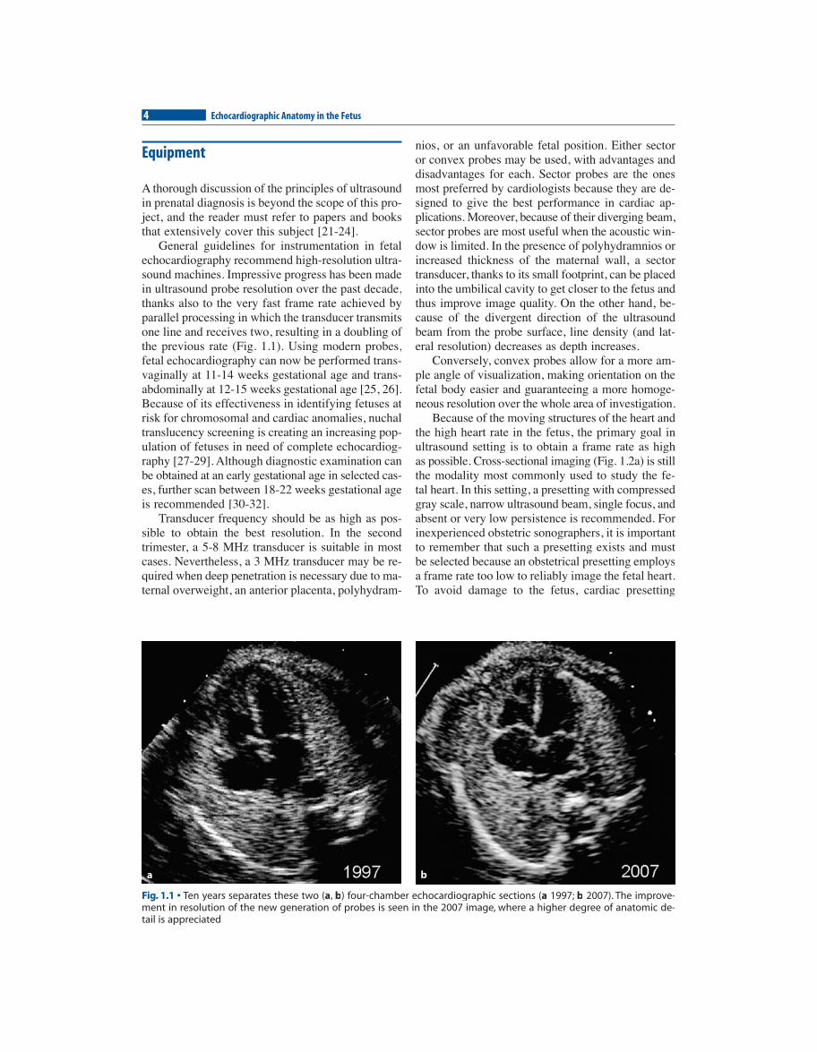
General guidelines for instrumentation in fetalechocardiography recommend high-resolution ultra-sound machines. Impressive progress has been madein ultrasound probe resolution over the past decade,thanks also to the very fast frame rate achieved byparallel processing in which the transducer transmitsone line and receives two, resulting in a doubling ofthe previous rate (Fig. 1.1). Using modern probes,fetal echocardiography can now be performed trans-vaginally at 11-14 weeks gestational age and trans-abdominally at 12-15 weeks gestational age [25, 26].Because of its effectiveness in identifying fetuses atrisk for chromosomal and cardiac anomalies, nuchaltranslucency screening is creating an increasing pop-ulation of fetuses in need of complete echocardiog-raphy [27-29]. Although diagnostic examination canbe obtained at an early gestational age in selected cas-es, further scan between 18-22 weeks gestational ageis recommended [30-32].
Transducer frequency should be as high as pos-sible to obtain the best resolution. In the secondtrimester, a 5-8 MHz transducer is suitable in mostcases. Nevertheless, a 3 MHz transducer may be re-quired when deep penetration is necessary due to ma-ternal overweight, an anterior placenta, polyhydram-
nios, or an unfavorable fetal position. Either sectoror convex probes may be used, with advantages anddisadvantages for each. Sector probes are the onesmost preferred by cardiologists because they are de-signed to give the best performance in cardiac ap-plications. Moreover, because of their diverging beam,sector probes are most useful when the acoustic win-dow is limited. In the presence of polyhydramnios orincreased thickness of the maternal wall, a sectortransducer, thanks to its small footprint, can be placedinto the umbilical cavity to get closer to the fetus andthus improve image quality. On the other hand, be-cause of the divergent direction of the ultrasoundbeam from the probe surface, line density (and lat-eral resolution) decreases as depth increases.
Conversely, convex probes allow for a more am-ple angle of visualization, making orientation on thefetal body easier and guaranteeing a more homoge-neous resolution over the whole area of investigation.
Because of the moving structures of the heart andthe high heart rate in the fetus, the primary goal inultrasound setting is to obtain a frame rate as highas possible. Cross-sectional imaging (Fig. 1.2a) is stillthe modality most commonly used to study the fe-tal heart. In this setting, a presetting with compressedgray scale, narrow ultrasound beam, single focus, andabsent or very low persistence is recommended. Forinexperienced obstetric sonographers, it is importantto remember that such a presetting exists and mustbe selected because an obstetrical presetting employsa frame rate too low to reliably image the fetal heart.To avoid damage to the fetus, cardiac presetting
4 Echocardiographic Anatomy in the Fetus
Fig. 1.1 • Ten years separates these two (a, b) four-chamber echocardiographic sections (a 1997; b 2007). The improve-ment in resolution of the new generation of probes is seen in the 2007 image, where a higher degree of anatomic de-tail is appreciated
a b
1 • General Guidelines 5
should also be used to ensure that spatial temporalaverage power output for color and pulsed Doppleris less than 100 mW/cm2.
The possibility of image magnification using thezoom allows for better definition of the size andstructure of the fetal heart, whereas the cine loop nowmakes it possible to record a long series of consec-utive digital images that can be immediately reex-amined as single frames. This function is particularlyuseful for identifying structural or flow anomaliesthat may be overlooked should the images be eval-uated in real time only. With recent instrumentation,storing digital clips on the hard disk of the built-incomputer is possible. Instruments equipped withcine-loop and digital-clip storing capabilities providea user-friendly system in which moving clips andstill-frame digital images are readily accessible forreview – a powerful means for off-line analysis andcase discussion that is largely superior to professionalvideotape recorders.
When investigating the fetal heart, the use ofDoppler is complementary to the morphologicalstudy with the cross-sectional technique. To allowan accurate flow evaluation, the insonation angle
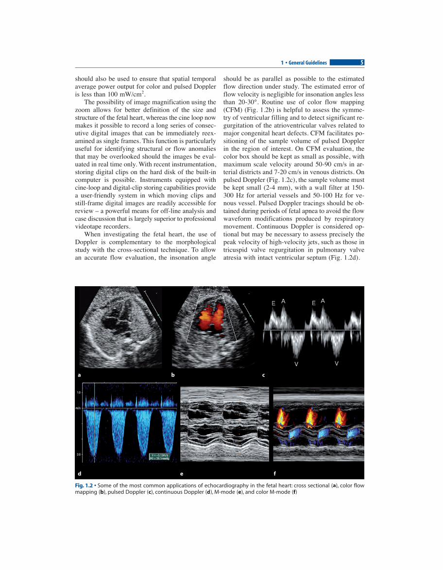
should be as parallel as possible to the estimatedflow direction under study. The estimated error offlow velocity is negligible for insonation angles lessthan 20-30°. Routine use of color flow mapping(CFM) (Fig. 1.2b) is helpful to assess the symme-try of ventricular filling and to detect significant re-gurgitation of the atrioventricular valves related tomajor congenital heart defects. CFM facilitates po-sitioning of the sample volume of pulsed Dopplerin the region of interest. On CFM evaluation, thecolor box should be kept as small as possible, withmaximum scale velocity around 50-90 cm/s in ar-terial districts and 7-20 cm/s in venous districts. Onpulsed Doppler (Fig. 1.2c), the sample volume mustbe kept small (2-4 mm), with a wall filter at 150-300 Hz for arterial vessels and 50-100 Hz for ve-nous vessel. Pulsed Doppler tracings should be ob-tained during periods of fetal apnea to avoid the flowwaveform modifications produced by respiratorymovement. Continuous Doppler is considered op-tional but may be necessary to assess precisely thepeak velocity of high-velocity jets, such as those intricuspid valve regurgitation in pulmonary valveatresia with intact ventricular septum (Fig. 1.2d).
Fig. 1.2 • Some of the most common applications of echo cardiography in the fetal heart: cross sectional (a), color flowmapping (b), pulsed Doppler (c), continuous Doppler (d), M-mode (e), and color M-mode (f)
a b c
d e f
Due to the high frame rate and time resolution,M-mode tracing (Fig. 1.2e) is the most accuratetmethod tod provide information onn walln thickness, ven-,tricular diameter,r and shortening fraction. However,the application of M-modef in the fetus is limited be-cause M-mode measurements require an ultrasoundbeam orientationm that ist difficult andt sometimes im-possible to obtain. Color M-moder color (Fig.r 1.2f)combined withd pulsedh Dopplerd hasr been demonstratednto be an invaluable tool in assessing fetal arrhythmias.
Major Planes of the Body and Heart
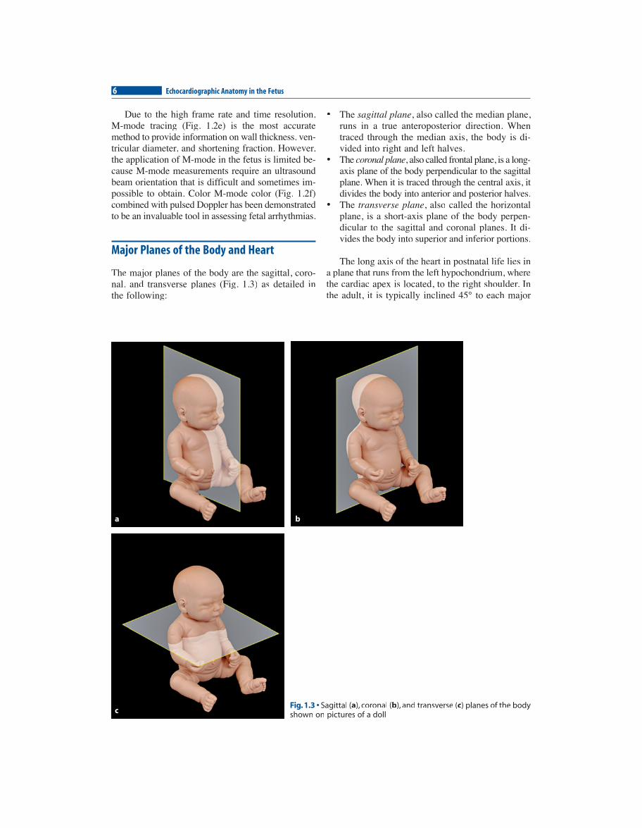
The major planesr of thef body are the sagittal, coro-nal, and transverse planes (Fig. 1.3) as detailed inthe following:
• The sagittal planel , also called the median plane,runs in a true anteroposterior direction.r Whentraced through the median axis, the body is di-vided into right andt left halves.t
• The coronal planel , also, calledo frontald plane,l is, a long-aaxis plane of thef body perpendicular tor the sagittalplane. When itn ist traced throughd theh central axis, it,divides the body into anterior andr posteriord halves.r
• The transverse plane, also called the horizontalplane, is a short-axis plane of thef body perpen-dicular tor the sagittal and coronal planes. It di-tvides the body into superior andr inferior portions.r
The long axis of thef heart in postnatal life lies ina planea that runst from them left hypochondrium,t where,the cardiac apex is located, to the right shoulder.t Inthe adult, it ist typically inclined 45° to each major
6 Echocardiographic Anatomy in the Fetus
Fig. 1.3 • Sagittal (a), coronal, (b), and, transverse (c) planes of thef bodyshown on pictures of af doll
a
c
b
1 • General Guidelines 7
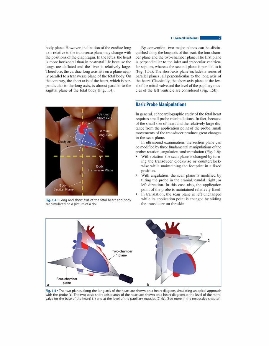
body plane. However, inclination of the cardiac longaxis relative to the transverse plane may change withthe positions of the diaphragm. In the fetus, the heartis more horizontal than in postnatal life because thelungs are deflated and the liver is relatively large.Therefore, the cardiac long axis sits on a plane near-ly parallel to a transverse plane of the fetal body. Onthe contrary, the short axis of the heart, which is per-pendicular to the long axis, is almost parallel to thesagittal plane of the fetal body (Fig. 1.4).
By convention, two major planes can be distin-guished along the long axis of the heart: the four-cham-ber plane and the two-chamber plane. The first planeis perpendicular to the inlet and trabecular ventricu-lar septum, whereas the second plane is parallel to it(Fig. 1.5a). The short-axis plane includes a series ofparallel planes, all perpendicular to the long axis ofthe heart. Classically, the short-axis plane at the lev-el of the mitral valve and the level of the papillary mus-cles of the left ventricle are considered (Fig. 1.5b).
Basic Probe Manipulations
In general, echocardiographic study of the fetal heartrequires small probe manipulations. In fact, becauseof the small size of heart and the relatively large dis-tance from the application point of the probe, smallmovements of the transducer produce great changesin the scan plane.
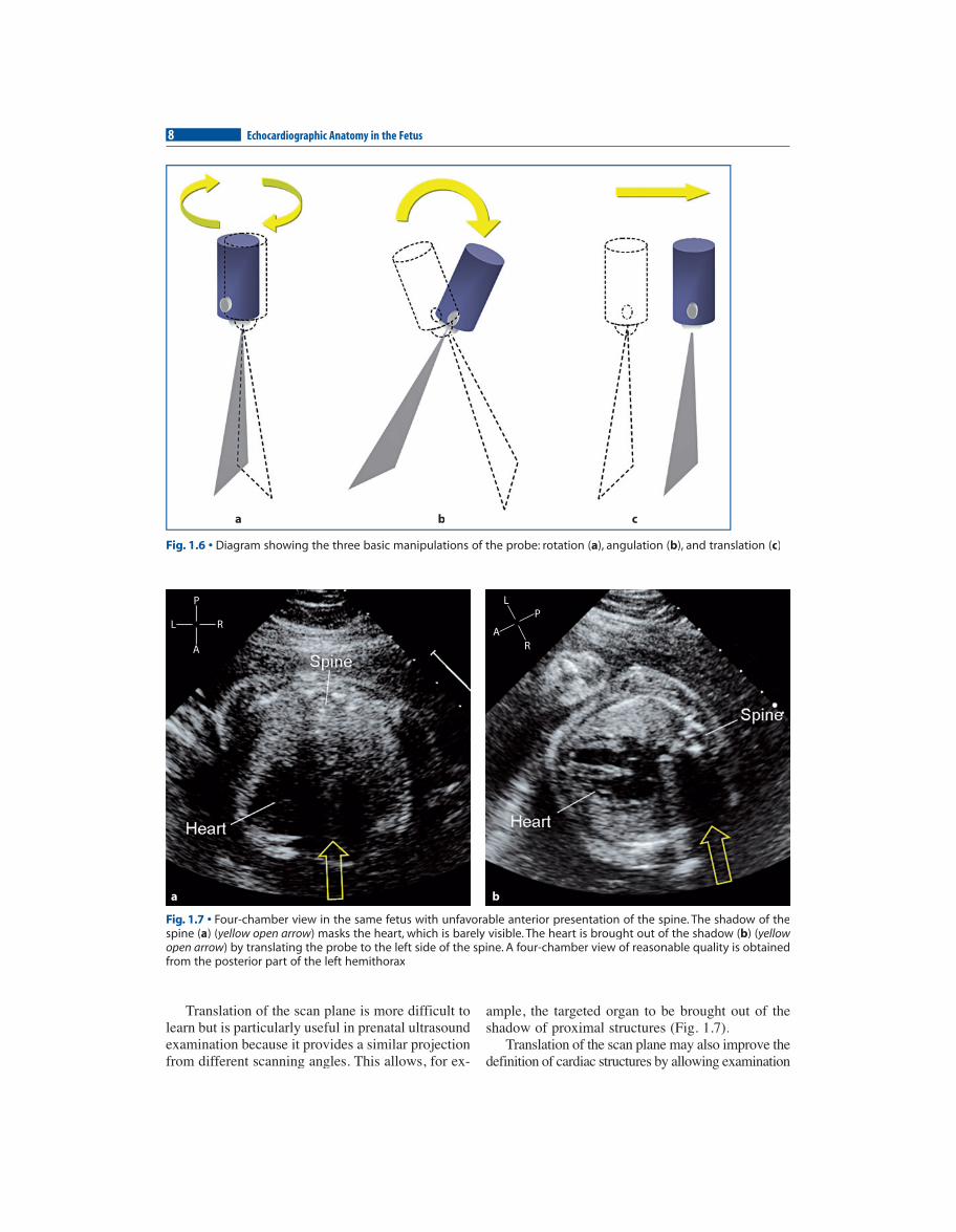
In ultrasound examination, the section plane canbe modified by three fundamental manipulations of theprobe: rotation, angulation, and translation (Fig. 1.6):• With rotation, the scan plane is changed by turn-
ing the transducer clockwise or counterclock-wise while maintaining the footprint in a fixedposition.
• With angulation, the scan plane is modified bytilting the probe in the cranial, caudal, right, orleft direction. In this case also, the applicationpoint of the probe is maintained relatively fixed.
• In translation, the scan plane is left unchangedwhile its application point is changed by slidingthe transducer on the skin.
Fig. 1.5 • The two planes along the long axis of the heart are shown on a heart diagram, simulating an apical approachwith the probe (a). The two basic short-axis planes of the heart are shown on a heart diagram: at the level of the mitralvalve (or the base of the heart) (1) and at the level of the papillary muscles (2) (b). (See more in the respective chapter)
a b
Fig. 1.4 • Long and short axis of the fetal heart and bodyare simulated on a picture of a doll
Translation of the scan plane is more difficult tolearn but is particularly useful in prenatal ultrasoundexamination because it provides a similar projectionfrom different scanning angles. This allows, for ex-
ample, the targeted organ to be brought out of theshadow of proximal structures (Fig. 1.7).
Translation of the scan plane may also improve thedefinition of cardiac structures by allowing examination
8 Echocardiographic Anatomy in the Fetus
Fig. 1.6 • Diagram showing the three basic manipulations of the probe: rotation (a), angulation (b), and translation (c)
aa bb cc
Fig. 1.7 • Four-chamber view in the same fetus with unfavorable anterior presentation of the spine. The shadow of thespine (a) (yellow open arrow( ) masks the heart, which is barely visible. The heart is brought out of the shadow (ww b) (yellow(open arrow) by translating the probe to the left side of the spine. A four-chamber view of reasonable quality is obtainedwfrom the posterior part of the left hemithorax
a b
L R
P
A
L
RA
P

1 • General Guidelines 9
with axial instead of lateral resolution (Fig. 1.8). Infetal echocardiography, similar projections of the heartcan be obtained from different positions of the trans-ducer because the lungs do not represent an obstacleto imaging, as they are filled with fluid and not air.This allows projections through the rib cage, even fromthe back, which are unobtainable postnatally (Fig. 1.7).Nevertheless, projections from the back produce at-tenuation of the ultrasonic energy, particularly in thethird trimester when bony structures of the thoraciccage absorb most of the ultrasound energy.
In general, the best definition of cardiac structuresis achieved through the fetal abdomen. If initially un-favorable, fetal presentation may change for the bet-ter by translating the scan plane or by turning themother on her side, having her empty a full bladder,or having her take a little walk. Sometimes, the ex-amination must be postponed for hours or a few days.
Axial and Lateral Resolution
The axial resolution is the shortest distance betweentwo points lying on the same insonation axis, which
the ultrasound system is able to represent as sepa-rate. The lateral resolution refers to the shortest dis-tance between two adjacent points that an ultra-sound system is able to represent as distinct. In vir-tually all cross-sectional ultrasound systems, theaxial resolution is better than the lateral resolution.This implies that structures that are orthogonal tothe ultrasound beam will be better represented thanstructures that are parallel. The four-chamber viewprovides one of the best examples to explain thisconcept. This view can be obtained in apical ortransverse orientation. Despite showing sections ofthe fetal thorax across identical structures, the api-cal and transverse four-chamber views provide dif-ferent information. In fact, in the apical approach,the moderator band, the atrioventricular valves, andthe venoatrial connection are better represented; inthe transverse approach, ventricular-wall thickness,ventricular transverse diameter, and foramen ovalemorphology and function are more precisely as-sessed. Conversely, in the apical approach, it is dif-ficult to identify the endocardial borders of the heartchambers and assess precisely ventricular-wallthickness and transverse diameter of cardiac cham-
Fig. 1.8 • Diagram simulating a transverse section of the fetal thorax at the level of the four-chamber view within the am-niotic cavity (black area). The apical four-chamber view (a) can be changed to a transverse four-chamber view (b) by trans-lating the probe (yellow arrow( ) on the mother’s abdomen while maintaining the section plane unchangedww
a b
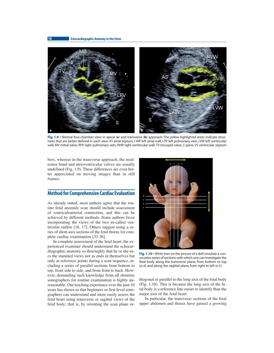
bers, whereas in the transverse approach, the mod-erator band and atrioventricular valves are usuallyundefined (Fig. 1.9). These differences are even bet-ter appreciated on moving images than in stillframes.
Method for Comprehensive Cardiac Evaluation
As already stated, most authors agree that the rou-tine fetal anomaly scan should include assessmentof ventriculoarterial connection, and this can beachieved by different methods. Some authors favorincorporating the views of the two so-called ven-tricular outlets [16, 17]. Others suggest using a se-ries of short-axis sections of the fetal thorax for com-plete cardiac examination [33-36].
In complete assessment of the fetal heart, the ex-perienced examiner should understand the echocar-diographic anatomy so thoroughly that he or she us-es the standard views not as ends in themselves butonly as reference points during a scan sequence, in-cluding a series of parallel sections from bottom totop, from side to side, and from front to back. How-ever, demanding such knowledge from all obstetricsonographers for routine examination is highly un-reasonable. Our teaching experience over the past 10years has shown us that beginners or first-level sono-graphers can understand and more easily assess thefetal heart using transverse or sagittal views of thefetal body; that is, by orienting the scan plane or-
thogonal or parallel to the long axis of the fetal body(Fig. 1.10). This is because the long axis of the fe-tal body is a reference line easier to identify than themajor axis of the fetal heart.
In particular, the transverse sections of the fetalupper abdomen and thorax have gained a growing
10 Echocardiographic Anatomy in the Fetus
Fig. 1.10 • White lines on the picture of a doll simulate a con-secutive series of sections with which one can investigate thefetal body along the transverse plane, from bottom to top (a-d), and along the sagittal plane, from right to left (dd e-h)
Fig. 1.9 • Normal four-chamber view in apical (a) and transverse (b) approach. The yellow highlighted areas indicate struc-tures that are better defined in each view. AS atrial septum, LAW left atrial wall, LPV left pulmonary vein, LVW left ventricularwall, MV mitral valve, RPV right pulmonary vein, RVW right ventricular wall, TV tricuspid valve, S spine, VS ventricular septum
a b