Embed Size (px)
Citation preview
K. RAMAKRISHNAN COLLEGE OF ENGINEERINGK. RAMAKRISHNAN COLLEGE OF ENGINEERINGK. RAMAKRISHNAN COLLEGE OF ENGINEERINGK. RAMAKRISHNAN COLLEGE OF ENGINEERING Samayapuram, Tiruchirappalli – 621 112.
DEPARTMENT OF ELECTRONICS AND COMMUNICATION ENGINEERING
Department of ECE Medical Electronics
QUESTION BANKQUESTION BANKQUESTION BANKQUESTION BANK
Subject Code/Subject: : : : EC1001 / MEDICAL ELECTRONICSEC1001 / MEDICAL ELECTRONICSEC1001 / MEDICAL ELECTRONICSEC1001 / MEDICAL ELECTRONICS
Name : V.SRINIVASANV.SRINIVASANV.SRINIVASANV.SRINIVASAN Designation : LECTURERLECTURERLECTURERLECTURER
Dept : ECEECEECEECE Semester : VIIVIIVIIVII
UNIT-I
ELECTROELECTROELECTROELECTRO----PHYSIOLOGY AND BIOPHYSIOLOGY AND BIOPHYSIOLOGY AND BIOPHYSIOLOGY AND BIO----POTENTIAL RECORDINGPOTENTIAL RECORDINGPOTENTIAL RECORDINGPOTENTIAL RECORDING
The origin of Bio-potentials; biopotential electrodes, biological amplifiers, ECG, EEG,
EMG, PCG, EOG, lead systems and recording methods, typical waveforms and signal
characteristics.
Part – A (2 Marks)
1. Explain the Cell Structure. The basic living unit of the body is a cell. Each organ in our body is an aggregate of many
different cells held together by intercellular supporting structures. Each type of cell is meant for performing one particular function. Each cell consists of a centrally located nucleus, also called cell core, surrounded by cytoplasm. The nucleus is separated from the cytoplasm is separated from the surrounding fluids by a cell membrane. The different substances that make up the cell one collectively called protoplasm, which is mainly composed of water, electrolytes, proteins, carbohydrates and lipids. 2. What are the applications of piezo electric sensors? 1) In cardiology 2) In phonocardiography 3) In blood pressure measurement 4) In measuring physiological accelerations 3. Define Resting Potential.
Certain type of cells within the body such as nerve and muscle cell are encased in a semi permeable membrane that permits same substances to pass through the membrane, while others are kept out, surrounding the cells of the body are the body fluids which are Conductive solutions of charge ions. The principal ions are sodium (Na+), Potassium (K+) and chloride (cl-) the membrane of the excitable cells readily permits entry of potassium and chloride ions but blocks the entry of sodium ions .since the various ions seek balance between inside the cell and outside. Equilibrium is reached with the potential difference across the membrane – ve on the inside and + ve on the outside of the cell. This membrane potential is called the resting of the cell.

K. RAMAKRISHNAN COLLEGE OF ENGINEERINGK. RAMAKRISHNAN COLLEGE OF ENGINEERINGK. RAMAKRISHNAN COLLEGE OF ENGINEERINGK. RAMAKRISHNAN COLLEGE OF ENGINEERING Samayapuram, Tiruchirappalli – 621 112.
DEPARTMENT OF ELECTRONICS AND COMMUNICATION ENGINEERING
Department of ECE Medical Electronics
4. Define Action Potential.
When a section of cell membrane is excited by a flow of ionic current or same form of externally applied voltage the membrane changes its permaliablity and begins to allow to some of the sodium ions to enter. This movement of sodium ions into the cell results in an ionic current flow that further reduces the barrier of membrane to sodium ion rush into the Cell to try to reach to balance with the ions outside. At the same time potassium ions which were higher in the concentration inside the cell during the resting stage try to leave the cell, but are unable to move as rapidly as sodium ions. As a result the cell has a slightly positive potential on the due to imbalance of potassium ions. This +ve potential is called action Potential and this approximately +20mv .A cell in the action potential stage is said to be Depolarized. The process of changing from resting stage to action potential stage is called Depolarization. 5. Explain Bioelectric Potential.
Bioelectric potential are generated at a cellular level that is each cell is a Minute voltage generator, because positive and negative ions tend to concentrate unequally inside and outside the cell wall, a potential difference is established and the cell becomes a tiny biological battery. In the normal resting state of the cell it interior is negative with respect to the outside when the cells “fires” however, the outside of the cell becomes momentarily negative with respect to the interior. A short time later, the cell regains the normal state in which the inside again negative with respect to outside. This “discharging” and “recharging” of the cell known as depolarisation and repolarisation respectively. 6. Name the factors that are considered in the design of biomedical instrument system.
1. Range 2. Sensitivity 3. Linearity 4. Frequency Response 5. Accuracy 6. Stability 7. Isolation 8. Simplicity 9. Signal to noise ratio.
7. Name the physiological systems of the body.
1. Bio chemical System 2. Cardio vascular System 3. Regulated System 4. Nervous System
8. State the principal of the sodium pump.
Once the rush of sodium ions through the cell membrane has stopped that is a new stage of equilibrium is reached, the ionic currents that lowered the barrier to sodium ions are no longer present and the membrane comes back into its original selectively permeable condition, where in the passage of sodium ions from the outside to inside of the cell is again blocked. This take a long time for the resting potential to develop again .But by the active process called sodium pump, the sodium ions are quickly transported outside of the cell and the cell again Becomes polarized and assumes its restrict potential. This process is called repolarisation.
K. RAMAKRISHNAN COLLEGE OF ENGINEERINGK. RAMAKRISHNAN COLLEGE OF ENGINEERINGK. RAMAKRISHNAN COLLEGE OF ENGINEERINGK. RAMAKRISHNAN COLLEGE OF ENGINEERING Samayapuram, Tiruchirappalli – 621 112.
DEPARTMENT OF ELECTRONICS AND COMMUNICATION ENGINEERING
Department of ECE Medical Electronics
9. Name the different types of electrodes.
1. Micro Electrode a) Metallic b) Non –Metallic
2. Depth and needle Electrode 3. Surface Electrode
10. What are the requirements of physiological signal amplifier or biomedical pre amplifier?
a) The voltage gain should be more than 100 db. b) It should have low frequency response. c) There is no drift in the amplifier. d) The output impedance of the amplifier should be very small.
11. What are the different modes of operation of differential amplifier?
a) Single ended mode b) Differential mode c) Common mode
12. What is single ended mode? When either v1 or v2 is equal to zero, the operation of the differential amplifier is known
as single ended mode of operation. 13. What is differential mode?
The two input signals are equal but have opposite polarity at every instant of time. Vo=Rf/Ri(V2-V1)
In this case, the input signals are called differential mode signals.
14. What is common mode signal? The input voltages appearing at the input terminals 1 and 2 are identical both in amplitude
and phase at every instant of time and the circuit is said to be in common mode. V1=V2=Vcm Vo=0.
15. What is CMRR in a differential amplifier?
It is the ratio of the amplification of the differential voltage ti the amplification of the common mode voltage.
CMRR=Ad/Ac. CMRR in db=20 log10 CMRR.
16. What is noise figure?
It is defined as the ratio of the signal to noise ratio at the input to the signal to noise ratio at the output. 17. What are the advantages of the pre amplifier or instrumentation amplifier?
a) High stability b) Higher fidelity c) High CMRR d) High input impedance with the required gain.
K. RAMAKRISHNAN COLLEGE OF ENGINEERINGK. RAMAKRISHNAN COLLEGE OF ENGINEERINGK. RAMAKRISHNAN COLLEGE OF ENGINEERINGK. RAMAKRISHNAN COLLEGE OF ENGINEERING Samayapuram, Tiruchirappalli – 621 112.
DEPARTMENT OF ELECTRONICS AND COMMUNICATION ENGINEERING
Department of ECE Medical Electronics
18. What is chopper amplifier?
The chopper amplifier is used convert the dc or low frequency signal into a high frequency signal. Then this modulated high frequency signal is amplified by conventional ac amplifier. Then this is demodulated and filtered to get low frequency or dc signal. 19. What are the types of chopper amplifier?
a) Mechanical chopper amplifier. b) Non mechanical chopper amplifier.
20. What is Electrocardiography?
It deals with the study of the electrical activity of the heart muscles. The potentials Originated in the individual fibres of heart muscle are added to produce the ECG waveform. 21. What are the various parts of generalized instrumentation system?
1. Measured 2. Primary sensing element 3. Variable conversion element 4. Signal processing unit 5. Output display 6. Control & feedback element
22. Give the classifications of biomedical instruments. i) According to the quantity that is sensed, pressure, flow or temperature sensing devices. ii) According to the principle of transduction used, resistive, inductive, capacitive, ultrasonic or electrochemical devices. iii) According to the measurement techniques, cardio vascular, pulmonary, nervous & endocrine systems. iv) According to the clinical medical specialities, paediatrics, obstetrics, cardiology or radiology.
23. What are the different types of ECG lead configurations?
Ø Bipolar limb leads Ø Augmented Unipolar limb leads Ø Chest leads Systems.
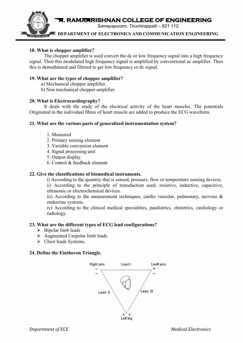
24. Define the Einthoven Triangle.
K. RAMAKRISHNAN COLLEGE OF ENGINEERINGK. RAMAKRISHNAN COLLEGE OF ENGINEERINGK. RAMAKRISHNAN COLLEGE OF ENGINEERINGK. RAMAKRISHNAN COLLEGE OF ENGINEERING Samayapuram, Tiruchirappalli – 621 112.
DEPARTMENT OF ELECTRONICS AND COMMUNICATION ENGINEERING
Department of ECE Medical Electronics
The closed path RA to LA to LL and back to RA is called Einthoven triangle. According
to Einthoven, in a frontal plane of the body, the cardiac electric field vector is a two dimensional one. 25. What are the important parts of ECG recorder?
• Patient cable and defibrillator protection circuit. • Lead selector switch • Calibrator • Bio- amplifier • Auxiliary amplifier • Isolated power supply • Output unit • Power switch
26. What is Electroencephalography?
It deals with the recording and study of electrical activity of the brain. By means of electrodes attached to the skull of a patient, brain waves can be picked up and recorded. 27. What is Electromyography?
It is the science of interpreting and recording the electrical activity of the muscles action potentials. Meanwhile, the recording of the peripheral nerve’s action potential is called electroneurography. 28. What is Electrooculography?
It deals with the recording of the corneal- retinal potentials associated with eye movements. 29. What is Electroretinography?
It deals with the recording and interpreting of the electrical activity of the eye. If the illumination of the retina is changed, the potential changes slightly in a complex manner. The recording of these changes is called Electroretinography. 30. List the brain waves and their frequency.
Alpha- 8 to 13Hz, Beta-13 to 30 Hz , Theta- 4 to 8 Hz, Delta- 0.5 to 4 Hz. 31. Define latency.
It is defined as the elapsed time between the simulating impulse and the muscle’s action potential. 32. What are the different sounds made by the heart?
Valve closure sounds, Ventricular filling sounds, Valve opening sounds, Extra cardiac sounds. 33. Name the parts of the heart conduction system.
Sino atrial node, Atrio ventricular node, Bundle of His , Purkinje fibres. 34. What is the colour coding of the different leads?
White –RA, Black- LA, Green- RL, Red- LL, Brown- Chest
K. RAMAKRISHNAN COLLEGE OF ENGINEERINGK. RAMAKRISHNAN COLLEGE OF ENGINEERINGK. RAMAKRISHNAN COLLEGE OF ENGINEERINGK. RAMAKRISHNAN COLLEGE OF ENGINEERING Samayapuram, Tiruchirappalli – 621 112.
DEPARTMENT OF ELECTRONICS AND COMMUNICATION ENGINEERING
Department of ECE Medical Electronics
35. Mention any four specifications of the ordinary ECG recorder.
Maximum sensitivity – 20 mm/mV, Input impedance –5 mega ohms, Output impedance -<100 ohms, CMRR- 10000:1. 36. What is Electrode Potential?
The voltage developed at an electrode-electrolyte interface is known as Electrode Potential. 37. What is the purpose of electrode paste?
The electrode paste decreases the impedance of the contact the artifacts resulting from the movement of the electrode or patient. 38. Give the different types of Surface electrodes?
• Metal Plate electrodes • Suction cup electrodes • Adhesive tape electrodes • Multi point electrodes • Floating electrodes.
39. What is PH electrode?
The chemical balance of human body is identified by measurement of Ph content of blood and other body fluids. PH is defined as logarithm of reciprocal of hydrogen ion concentration.
40. Define polarized and non polarized electrode. Electrodes in which no net transfer of charge occurs across the metal electrolyte interface is called as perfectly polarized electrodes. An electrode in which un hindered exchange of charge occurs across the metal electrode interface is called perfectly non polaraisable electrodes. 41. What is plethysmograph? The instrument used for measuring blood volume is called plethysmograph. 42. Write Goldman’s equation? Vr=-kt/q{Pk(k+)i +(Pna+)I +Pcl(cl-)o/ Pk(k+)o+ Pna(Na+)o+Pcl(cl-)i 43. Define All or nothing law. All or nothing law states that regardless of the method of excitation of cells or by the intensity of the stimulus, the action potential is same for any given cell. 44. What is absolute refractory period? It is the time duration in which cell cannot respond to any new stimulus. Generally it is about 1ms in nerve cell. 45. What is Relative refractory Period? It is one during which another action potential can be triggered but a higher stimulus is required to reinitiate the action potential and the subsequent contraction of muscles . Generally the relative refractory period is several milliseconds.
K. RAMAKRISHNAN COLLEGE OF ENGINEERINGK. RAMAKRISHNAN COLLEGE OF ENGINEERINGK. RAMAKRISHNAN COLLEGE OF ENGINEERINGK. RAMAKRISHNAN COLLEGE OF ENGINEERING Samayapuram, Tiruchirappalli – 621 112.
DEPARTMENT OF ELECTRONICS AND COMMUNICATION ENGINEERING
Department of ECE Medical Electronics
46. Define Conduction Velocity. The rate at which an action potential moves down a fibre or propagated from cell to cell is termed as propagation rate. 47. What are the characteristics of resting potential?
• The value of potential is maintained as constant. • It depends on temperature. • Permeability varies
Part – B
1. Define resting potential & Action potential. Explain how these potentials are generated in human body.
v Resting & Action Potential generation v Polarization & Depolarization of cell v Sodium pump –description
2. Explain in detail the different types of Electrodes used for biomedical Applications.
§ Microelectrodes § Depth & Needle electrodes § Surface electrodes § Chemical electrodes
3. Explain the factors that influence the design and application of a medical instruction
system / discuss the different characteristics of a medical instrument system. (8)
4. Explain the man-instrument system with a neat block diagram /Explain with a block
diagram the components of the bio-medical instrument system. (8)
5. Discuss the problems encountered in measuring a living system /discuss the major
differences encountered between measurements in a physiological system as distinct from
a physical system. (8)
4. Draw the structure of a living cell of our body and explain its constituents. (8)
5. Discuss the different ways of transport of ions through the cell membrane (4)
6. Give an account on the different chemical compositions in the intra and extra cellular
fluids and their effects in the case of blood serum. (4)
7. Discuss the development of action potential and muscular contraction. (8)
8. Draw the electrical equivalent circuit of microelectrode and explain its electrical nature.
(8)
9. What are Biopotential electrodes? Distinguish between metallic microelectrode and non
metallic microelectrode. (4)
10. Draw the micropipette non-metallic electrode and explain (8)
K. RAMAKRISHNAN COLLEGE OF ENGINEERINGK. RAMAKRISHNAN COLLEGE OF ENGINEERINGK. RAMAKRISHNAN COLLEGE OF ENGINEERINGK. RAMAKRISHNAN COLLEGE OF ENGINEERING Samayapuram, Tiruchirappalli – 621 112.
DEPARTMENT OF ELECTRONICS AND COMMUNICATION ENGINEERING
Department of ECE Medical Electronics
11. With a neat block diagram, explain the working of ECG recorder (8)
12. Discuss the different lead configuration used in ECG. (8)
13. Explain with a neat diagram the resting potential (8)
14. Explain polarization, depolarization the depolarization (8)
15. Draw the circuit diagram of an ECG isolation amplifier and explain its action. (8)
16. What are chopper amplifiers and explain. (8)
17. Explain with a diagram medical preamplifier and explain its action (8)
18. Explain a bridge voltage amplifier and explain (8)
19. Explain buffer amplifier and explain (8)
20. Explain a current amplifier circuit and explain its working. (8)
21. Draw the curves of ECG and diagnose any form of disturbance in heart rhythm (8)
22. Draw the block diagram of an EEG unit and explain the different parts in it. (8)
23. Give the origin of brain waves and describe the 10-20 electrode system used in EEG. (8)
24. Describe the recording setup used in EMG (8)
25. Write a note on ERG and EOG (8)
26. Explain the origin of different heart sounds (8)
27. Explain with diagram the salient features of Phonocardiography (PCG) (8)
28. Draw the frequency response of
a. An electromyogram. (2)
b. Blood flow measurements. (2)
c. Phonocardiogram. (2)
d. Plethysmogram (2)
29. (a) Write down the ‘Nernst Equation’ and ‘Goldman Equation’ and explain about the
constants used.(8)
(b) Explain ‘Bio Electric Potentials from the brain’ and ‘Resting Rhythms of the Brain’.
(8)
******************************
K. RAMAKRISHNAN COLLEGE OF ENGINEERINGK. RAMAKRISHNAN COLLEGE OF ENGINEERINGK. RAMAKRISHNAN COLLEGE OF ENGINEERINGK. RAMAKRISHNAN COLLEGE OF ENGINEERING Samayapuram, Tiruchirappalli – 621 112.
DEPARTMENT OF ELECTRONICS AND COMMUNICATION ENGINEERING
Department of ECE Medical Electronics
UNIT - II
BIOBIOBIOBIO----CHEMICAL AND NON ELECTRICAL PARAMETER CHEMICAL AND NON ELECTRICAL PARAMETER CHEMICAL AND NON ELECTRICAL PARAMETER CHEMICAL AND NON ELECTRICAL PARAMETER
MEASUREMENT MEASUREMENT MEASUREMENT MEASUREMENT
PH, PO2, PCO2, PHCO3, Electrophoresis, colorimeter, photometer, Auto analyzer,
Blood flow meter, cardiac output, respiratory measurement, Blood pressure,
temperature, pulse, Blood cell counters.
Part – A (2 Marks)
1. What are the types of measurements of blood pressure? 1. Indirect or non-invasive method. 2. Direct or invasive method.
2. How is the blood pressure measured in the indirect method?
The indirect method of measuring blood pressure involves the use of a Sphygmomanometer and a stethoscope. The sphygmomanometer consists of an inflatable pressure cuff and a mercury or aneroid manometer to measure the pressure in the cuff. The cuff is normally manually inflated, with a rubber bulb and deflated slowly through a needle valve.
3. Explain the principle of sphygmomanometer.
The sphygmomanometer works on the principle that when the cuff is placed on the upper arm and inflated, the arterial blood can flow past the cuff only when the arterial pressure exceeds the pressure in the cuff. Furthermore, when the cuff is inflated to a pressure that only occludes the brachial artery, turbulence is generated in the blood as it spurts through the tiny arterial opening during each systole. The sounds generated by this turbulence, Korotkoff sounds, can be heard through the stethoscope placed over the artery downstream from the cuff.
4. What are the methods involved in direct blood pressure measurement?
1. Auscultatory method 2. Palpatory method
Auscultatory method locates the systolic and diastolic pressure valves by listening to the Korotkoff. Diastolic pressure can be easily measured.
Palpatory method is a alternative method that the physician identifies the flow of blood in the artery by feeling the pulse of the patient downstream from the cuff instead of listening for the korotkoff sounds. In this method, systolic pressure can be easily measured. 5. What is meant by mean arterial pressure (MAP)?
Mean Arterial pressure is the weighted average of the systolic and diastolic pressure MAP falls about one- third of the way between the diastolic low and Systolic peak. Formula for calculating MAP is,
MAP = 1/3 (systolic –diastolic) + diastolic
K. RAMAKRISHNAN COLLEGE OF ENGINEERINGK. RAMAKRISHNAN COLLEGE OF ENGINEERINGK. RAMAKRISHNAN COLLEGE OF ENGINEERINGK. RAMAKRISHNAN COLLEGE OF ENGINEERING Samayapuram, Tiruchirappalli – 621 112.
DEPARTMENT OF ELECTRONICS AND COMMUNICATION ENGINEERING
Department of ECE Medical Electronics
6. What are the methods involved in direct blood pressure measurement?
1. Percutaneous insertion 2. Catheterization (Vessel Cut down) 3. Implantation of a transducer in a vessel or in the heart. 4. Other methods such as clamping a transducer on the intact artery have also been used. But they are not common.
7. Explain the two ways involved in measurement of blood pressure with a catheter?
Measurement of blood pressure with a catheter can be achieved in two ways. 1. The first is to introduce a sterile saline solution into catheter so the fluid pressure is
transmitted to a transducer outside the body a complete fluid pressure system is set up with provisions for checking against atmospheric pressure and for establishing a reference point. The frequency response of this system is a combination of the frequency response of the transducer and the fluid column in the catheter.
2. In the second method, pressure measurements are obtained at the source. Here, the transducer is introduced into the catheter and pushed to the point at which the pressure is to be measured, or the transducer is mounted at the tip of the catheter. This device is called a catheter-tip blood pressure transducer. 8. Discuss the technique involved in direct measurement?
1) A catheterization method involving the sensing of the blood pressure through a liquid column. In this method the transducer is external to the body and the blood pressure is transmitted through a saline solution column in a catheter to this transducer.
2) A catheterization method involving the placement of the transducer through the catheter at the actual size of measurement. In the bloodstream or by mounting the transducer on the tip of the catheter.
3) Percutaneous methods in which the blood pressure is sensed in the vessel just under the skin by the use of a needle or catheter.
4) Implantation techniques in which the transducer is more permanently placed in the blood vessels or the heart by surgical methods. 9) What are the different types of blood flow meters?
1) Magnetic blood flow meter –Based on the principle of Magnetic induction. 2) Ultrasonic blood flow meter-Based on the principle if Doppler. 3) Thermal convection-The rate of cooling is proportional to the rate of the flow of the
medium. This principle is also used to measure the gas flow. 4) Determination by Radiographic method-By the injection of a contrast medium into a
blood vessel, the circulation pattern can be made visible. Record of the X-ray image, obstruction can be detected and the blood flow in the blood vessels can be estimated. This technique is known as ‘angiography’. 10) What is cardiac output?
The blood flow at any point in the circulatory system is the volume of blood that passes that point during a unit of time. It is measured normally in milli meter per min or litres per min. Blood flow are highest in the pulmonary artery and the aorta, where the blood vessels leave the heart. The flow at these points is called ‘cardiac output’.
K. RAMAKRISHNAN COLLEGE OF ENGINEERINGK. RAMAKRISHNAN COLLEGE OF ENGINEERINGK. RAMAKRISHNAN COLLEGE OF ENGINEERINGK. RAMAKRISHNAN COLLEGE OF ENGINEERING Samayapuram, Tiruchirappalli – 621 112.
DEPARTMENT OF ELECTRONICS AND COMMUNICATION ENGINEERING
Department of ECE Medical Electronics
11) What is meant by pH?
pH can be defined as the logarithm of the reciprocal of the H+ ion concentration. It is a measure of the acid-base balance of a fluid.
pH= - log10 [H+] = log10( 1/[H+]) 12) What is the pH value for blood?
The pH value of normal arterial blood ranges between 7.38 and 7.42.The pH of venous blood is 7.35, because of the extra CO2. 13) Define GSR.
GSR is used for measuring variations in perspiration. In response to an external stimulus, such as touching a sharp point, the resistance of the skin shows a characteristic decrease and this is known as Galvanic Skin Response. The GSR is believed to be caused by the activity of the sweat glands. 14) Give the name of the instrument used for respiratory volume measurements and what are its types? The most widely used instrument for respiratory volume measurements in there cording spirometer. The different types of spirometer are
• Standard spirometer • Waterless spirometer • Wedge spirometer • Electronic spirometer • Broncho spirometer
15) Give the name of the instrument used for measuring airflow and explain its principle. Pneumotachometer can be used for measuring airflow. This device utilizes the principle that air flowing through an orifice produces a pressure difference across the orifice that is a function of the velocity of the air. 16) Define MVV.
Maximal voluntary ventilation is a measure of the maximum amount of air that can be breathed in and blown out over a sustained interval, such as 15 or 20seconds.
17) What is FVC?
Forced Vital Capacity (FVC) is the total amount of air that can forcibly be expired as quickly as possible after taking the deepest possible breath. 18) What is FRC?
The functional residual capacity (FRC) is the volume of gas remaining in the lungs at the end expiratory level. It the sum of the residual volume and the expiratory reserve volume.
19) Differentiate between tidal volume and residual volume.
The tidal volume (TV) or normal depth of breathing is the volume of gas inspired or expired during each normal, quiet, respiration cycle. The residual volume (RV), is the volume of gas remaining in the lungs at the end of a maximal expiration.
K. RAMAKRISHNAN COLLEGE OF ENGINEERINGK. RAMAKRISHNAN COLLEGE OF ENGINEERINGK. RAMAKRISHNAN COLLEGE OF ENGINEERINGK. RAMAKRISHNAN COLLEGE OF ENGINEERING Samayapuram, Tiruchirappalli – 621 112.
DEPARTMENT OF ELECTRONICS AND COMMUNICATION ENGINEERING
Department of ECE Medical Electronics
20) Define total lung capacity.
Total Lung Capacity is the amount of gas contained in the lungs at the end of a maximal inspiration .It is also the sum of residual volume and vital capacity. 21. Mention various types of chemical electrodes.
Hydrogen electrode, ph electrode, po2 electrode, pco2 electrode. 22. Define circulation and respiration?
We can define from the engineering point of view, the circulation is a high resistance circuit with a large pressure gradient between the arteries and veins. The exchange of any gases in any biological process is termed as respiration 23. What is mean by transducer?
It is a device which detects or senses the bio signal and converts it in to an electrical signal for bio signal processing
24. What is electrophoresis?
Electrophoresis is a technique used to separate biological molecules, such as nucleic acids, carbohydrates, and amino acids, based on their movement due to the influence of a direct electric current in a buffered solution. Positively charged molecules move toward the negative electrode, while negatively charged molecules move toward the positive electrode.
25. What are the different methods to measure the blood pressure?
1. Indirect method using sphygmomanometer. 2. Direct method.
26. What is the use of blood flowmeter in bio medical instrumentation?
Blood flow meters are used to monitor the blood flow in various blood vessels and it also helps to measure cardiac output.
27. Give some applications of electromagnetic blood flow meters.
Blood flow measurements during cardiac surgery, blood flow measurements during shunt operations, blood flow measurements during carotid artery, blood flow measurements in rural arteries, blood flow measurements during organ transplantation.
28. What happens when there is a fall in cardiac output?
A fall in cardiac output may result in low blood pressure, reduces tissues oxygenation, acidosis, poor renal function and shock. 29. What are the different types of dilution methods?
Indicator dilution method, Dye dilution method, Thermal dilution method.
30. How Cardiac output is measured in thermal dilution method? A thermal indicator of known volume introduced into either the right or left atrium will
produce a resultant temperature change in the pulmonary artery or in the aorta respectively, the integral of which is inversely proportional to the cardiac output.
Cardiac output=a constant X(blood temp-injectate temp)/area under dilution curve.
K. RAMAKRISHNAN COLLEGE OF ENGINEERINGK. RAMAKRISHNAN COLLEGE OF ENGINEERINGK. RAMAKRISHNAN COLLEGE OF ENGINEERINGK. RAMAKRISHNAN COLLEGE OF ENGINEERING Samayapuram, Tiruchirappalli – 621 112.
DEPARTMENT OF ELECTRONICS AND COMMUNICATION ENGINEERING
Department of ECE Medical Electronics
31. What is the use of blood flow meter in bio medical instrumentation?
Blood flow meters are used to monitor the blood flow in various blood vessels and it also helps to measure cardiac output.
32. What are the two different principles used in ultrasonic blood flow measurement?
• Transit Time method: In this method, a piezo electric crystal emits a brief pulse of ultrasound which propagates diagonally across the blood vessel.
• Doppler effect based method: In this method, as per Doppler effect, there is a change in frequency of reflected ultrasonic wave, due to motion of blood , when it crosses blood.
33. Define transit time principle of ultrasonic blood flow meter.
• In Transit time method a piezo electric crystal emits a brief pulse of ultrasound which propagates diagonally across the blood vessel.
• The pulse reaches a receiving crystal situated on the opposite side wall of the blood vessel.
• Electronic circuitry attached externally interprets transit time to velocity. 34. What is Sphygmomanometer?
• Sphygmomanometer is a device used by the physician to measure blood pressure. • It is used for indirect BP measurement and it consists of inflatable rubber bladder called
the cuff, a rubber squeeze ball pump and value assembly and a manometer. 35. What is korotkoff sound?
In the BP measurement, when the systolic pressure exceeds the cuff pressure, then the doctor can hear some crashing, snapping sound through the stethoscope. This is known as korotkoff sound. 36. What is cardiac output?
Cardiac output is the amount of blood delivered by heart to the aorota per minute. 37. What are the various methods to measure cardiac output?
Ficks method, Indicator dilution method, Measurement of impedance change.
38. Differentiate systolic and diastolic pressure. • The maximum pressure reached during cardiac output is called systolic pressure. • The maximum pressure occurring at the end of ventricular relaxation is termed as diastolic
pressure. 39. What are the two types to measure pulse rate?
Transmittance method, Reflectance method. 40. What is a colorimeter?
Colorimeter is otherwise called flame photometer, which is used for measuring transmittance and absorbance in the given solutions.
K. RAMAKRISHNAN COLLEGE OF ENGINEERINGK. RAMAKRISHNAN COLLEGE OF ENGINEERINGK. RAMAKRISHNAN COLLEGE OF ENGINEERINGK. RAMAKRISHNAN COLLEGE OF ENGINEERING Samayapuram, Tiruchirappalli – 621 112.
DEPARTMENT OF ELECTRONICS AND COMMUNICATION ENGINEERING
Department of ECE Medical Electronics
41. What is the use of infrared thermometer? It is a device to measure skin surface temperature. It is used to locate breast cancer. It is
also used to identify the spots in which blood circulation is poor. 42. What are the two methods for counting the blood? 1. Conductivity method, 2. Laser based cell counting method.
Part – B
1. a. Explain how the PCO2of blood is measured. (8) b. Explain how the PHCO3 of blood is measured. (8)
2. a. Write down the application of Electrophoresis and explain the basic principles involved.
(8) b. Explain the working principle of an Electromagnetic blood flow meter. (8)
3. a. Define the term residual volume, tital volume, vital capacity and total lung capacity (8)
b. Discuss the Fick’s method for determining cardiac output. (8) 4. a. Explain about ultrasonic blood flow meter (Doppler type). (8)
b. Explain about Respiratory measurement Technique. (8) 5. Explain the following. (16)
(i) Temperature (ii) Pulse
6. Explain the following electrodes with neat diagram
(i) Hydrogen (8)
(ii) pH (8)
7. Explain the following electrodes with neat diagram
(i) Pco2 (8)
(ii) Po2 (8)
8. a. Explain how the PH of blood is measured. (8) b. Explain how the PO2 of blood is measured. (8)
9. What are biomedical electrodes? Explain the electrode PHCO3 with neat diagram. (8)
10. Draw the block diagram of an automatic blood cell counter and explain its functioning. (8)
11. Describe the principle of laser based blood cell counting using a schematic diagram. (8)
12. Explain the following photometers with suitable diagrams.
a. Filter photometer (8)
b. Flame photometer (8)
13. Explain the working principle of spectrophotometer. (8)
K. RAMAKRISHNAN COLLEGE OF ENGINEERINGK. RAMAKRISHNAN COLLEGE OF ENGINEERINGK. RAMAKRISHNAN COLLEGE OF ENGINEERINGK. RAMAKRISHNAN COLLEGE OF ENGINEERING Samayapuram, Tiruchirappalli – 621 112.
DEPARTMENT OF ELECTRONICS AND COMMUNICATION ENGINEERING
Department of ECE Medical Electronics
14. Explain the principle of chromatography and its applications in medicine. (8)
15. Discuss the principle and working of electromagnetic blood flow meters. (8)
16. Describe an ultrasonic blood flow meter used in the measurement of velocity of blood
flowing in the blood vessels. (8)
17. Describe ultrasonic Doppler blood flow meters. (16)
18. Explain with a block diagram the laser based blood flow meter. (8)
19. Explain the Fick’s method for the determination of cardiac output. (8)
20. Explain the Indicator dilution method of cardiac output measurement. (8)
21. Explain the thermo dilution method of cardiac output measurement. (8)
22. Describe a method for the measurement of total lung capacity./ Describe the
plethysmograph method of measuring the total lung capacity. (8)
23. Describe a spirometer with a suitable schematic diagram. (8)
24. Explain in detail any one of the methods used for measuring blood pressure (8)
25. Explain in detail any one of the methods used for measuring temperature (8)
26. What is pneumotachograph? Give its importance in the pulmonary function analysis. (8)
27. Write down the application of Electrophoresis and explain the basic principle involved (8)
******************************
K. RAMAKRISHNAN COLLEGE OF ENGINEERINGK. RAMAKRISHNAN COLLEGE OF ENGINEERINGK. RAMAKRISHNAN COLLEGE OF ENGINEERINGK. RAMAKRISHNAN COLLEGE OF ENGINEERING Samayapuram, Tiruchirappalli – 621 112.
DEPARTMENT OF ELECTRONICS AND COMMUNICATION ENGINEERING
Department of ECE Medical Electronics
UNIT – III
ASSIST DEVICES AND BIOASSIST DEVICES AND BIOASSIST DEVICES AND BIOASSIST DEVICES AND BIO----TELEMETRYTELEMETRYTELEMETRYTELEMETRY
Cardiac pacemakers, DC Defibrillator, Telemetry principles, frequency selection,
Bio-telemetry, radio-pill and tele-stimulation.
Part – A (2 Marks)
1. Define circulatory system It is a type of transport system. It helps in supplying the oxygen and digested food to
different parts of our body and removing CO2 from the blood. The heart is the center of the circulatory system.
2. Define heart, lung?
Heart is a pumping organ which eats regularly and continuously for years. It beats seventy times a minute at rest. Contraction is systole and relaxation is diastole. 3. Define circulation and respiration?
We can define from the engineering point of view; the circulation is a high Resistance circuit with a large pressure.
4. Classify the pacemakers
• Fixed rate pacemakers. • Ventricular Synchronous pacemakers • Demand pacemakers • Atrial Synchronous pacemakers •
5. Different methods of stimulation External stimulation, internal stimulation
6. What is a Defibrillator? A defibrillator is an electronic device that creates a sustained myocardial depolarization of
a patient s heart in order to stop ventricular fibrillation or artial fibrillation. 7. Which are the elements of bio-telemetry system?
The essential elements are biological signal, transducer, conditioner, transmission link. 8. What are the types of radio telemetry systems?
o Single channel telemetry system o Radio telemetry with a sub-carrier o Multiple channel telemetry system
9. What are the types of multiple channel telemetry systems?
Ø Frequency system multiplex Ø Time division multiplex
K. RAMAKRISHNAN COLLEGE OF ENGINEERINGK. RAMAKRISHNAN COLLEGE OF ENGINEERINGK. RAMAKRISHNAN COLLEGE OF ENGINEERINGK. RAMAKRISHNAN COLLEGE OF ENGINEERING Samayapuram, Tiruchirappalli – 621 112.
DEPARTMENT OF ELECTRONICS AND COMMUNICATION ENGINEERING
Department of ECE Medical Electronics
10. What are the measurements in single channel telemetry system?
ü Active measurements ü Passive measurements
11. What is a pacemaker?
Pacemaker is an electrical pulse generator that starts or maintains the normal heart rhythm (i.e) application of electrical pulses to the heart is pacing action. 12. Explain the classification of pacemaker?
Pacemaker is broadly classified into internal & external pacemaker. Total AV block requires internal pacemaker. It has a mini energy of 10µJ-100µJ (5V,10mA,2ms).At a level of 400µJ, it causes Ventricular Fibrillation. Cardiac Standstill is obtained by external pacemaker. 13. What are the types of pacemaker?
i. Ventricular synchronous(fixed rate pulse) ii. Ventricular asynchronous(stand by pacemaker) iii. Ventricular inhibited(demand pacemaker) iv. Atrial synchronous pacemaker. v. Atrial sequential ventricular inhibited pacemaker.
14. Explain the application of ventricular asynchronous or stand by pacemaker?
Ventricular asynchronous orstand by pacemaker is basically a simple astable multivibrator that produces a stimulus at a fixed rate irrespective of the heart rhythm. 15. What is the application of ventricular inhibited pacemaker?
i. The R wave inhibited pacemaker allows the heart to pace at its normal rhythm when it is able to. If the R wave is missing for a preset period of time, the pacer will supply a stimulus.
ii. When the sensor ( shielded inside the pacemaker) is slightly stressed or bent by the patient’s body activity, the pacemaker can automatically increase or decrease its rate. Thus it can match with the greater physical effort. 16. What is the application of Atrial synchronous pacemaker?
i. This type of pacing is used for young patients with a mostly stable block. ii. It is used in stress testing & coronary artery diseases, in the evaluation of severity of
mitral stenosis & in the evaluation of various conduction mechanisms. iii. It has been used to terminate Atrial flutter & paroxysmal Atrial tachycardia. iv. It can act as a temporary pacemaker for the Atrial fibrillation.
17. What is an atrial sequential ventricular inhibited pacemaker and mention its advantage?
Atrial sequential ventricular inhibited pacemaker has the capability of stimulating both the atria & ventricles and adopts its method of stimulation to the patient’s need .If Atrial function fails, this pacemaker will stimulate the atrium & then sense the subsequent ventricular beat. 18. What is a defibrillator?
A defibrillator is an electronic device that creates a sustained myocardial polarisation of a patient’s heart in order to stop ventricular fibrillation or atrial fibrillation.
K. RAMAKRISHNAN COLLEGE OF ENGINEERINGK. RAMAKRISHNAN COLLEGE OF ENGINEERINGK. RAMAKRISHNAN COLLEGE OF ENGINEERINGK. RAMAKRISHNAN COLLEGE OF ENGINEERING Samayapuram, Tiruchirappalli – 621 112.
DEPARTMENT OF ELECTRONICS AND COMMUNICATION ENGINEERING
Department of ECE Medical Electronics
19 Explain ventricular fibrillation and how can it be eliminated?
Ventricular fibrillation is a serious cardiac energy resulting from asynchronous contraction of the heart muscle. This uncoordinated movement of ventricle walls of the heart may result from coronary occlusion, electric shock or abnormalities of body chemistry. 20. What are the different types of defibrillators?
i. Internal Defibrillator ii. External Defibrillator
a. AC. Defibrillator b. DC. Defibrillator c. Synchronous DC. Defibrillator d. Square Pulse Defibrillator e. Double Square Pulse Defibrillator f. Biphasic DC Defibrillator
Part – B
1. Describe the cardiac pacemaker waveforms and explain their importance. Compare
external and implanted pacemakers. (8)
2. Explain with a diagram the ventricular asynchronous pacemaker (fixed rate pacemaker).
(8)
3. Explain the ventricular synchronous pacemaker. (8)
4. Explain working principle of demand pacemaker with a diagram. (8)
5. Explain the atrial synchronous pacemaker. (8)
6. Explain with a neat diagram, the working principle of D.C. defibrillator. (8)
7. Explain with a neat diagram, the working principle of synchronized D.C. defibrillator. (8)
8. Explain the square pulse defibrillator. (8)
9. Explain the block diagram of a bio-telemetry system. Discuss its design. (8)
10. Explain the single channel telemetry system. (8)
11. Draw and explain the telemetry circuit for the transmission of EMG, ECG, EEG and
respiration rate. (16)
12. Explain the subcarrier biotelemetry system. (8)
13. Explain the multiple channel telemetry systems with neat diagrams. (16)
14. What are the problems associated with the implant telemetry circuits? Explain the uses of
biotelemetry. (8)
K. RAMAKRISHNAN COLLEGE OF ENGINEERINGK. RAMAKRISHNAN COLLEGE OF ENGINEERINGK. RAMAKRISHNAN COLLEGE OF ENGINEERINGK. RAMAKRISHNAN COLLEGE OF ENGINEERING Samayapuram, Tiruchirappalli – 621 112.
DEPARTMENT OF ELECTRONICS AND COMMUNICATION ENGINEERING
Department of ECE Medical Electronics
15. Explain the various modulation techniques used for transmitting a biosignal in a telemetry
system (8)
16. Write short notes on telestimulation (8)
17. What are the precautions to be followed in hospitals while using defibrillators (4)
18. Write briefly about the power sources used for implantable type of pacemaker (8)
19. What is radiopill? Explain. (8)
20. Write technical properties of electrodes used in Defibrillator (4)
21. Write short notes on ‘Frequency Selection’ w.r.t. Biotelemetry (8)
22. Explain the basic concepts (including the modulation types) of radio transmission used in
biotelemetry (16)
******************************
K. RAMAKRISHNAN COLLEGE OF ENGINEERINGK. RAMAKRISHNAN COLLEGE OF ENGINEERINGK. RAMAKRISHNAN COLLEGE OF ENGINEERINGK. RAMAKRISHNAN COLLEGE OF ENGINEERING Samayapuram, Tiruchirappalli – 621 112.
DEPARTMENT OF ELECTRONICS AND COMMUNICATION ENGINEERING
Department of ECE Medical Electronics
UNIT-IV
RADIOLOGICAL EQUIPMENTSRADIOLOGICAL EQUIPMENTSRADIOLOGICAL EQUIPMENTSRADIOLOGICAL EQUIPMENTS
Ionosing radiation, Diagnostic x-ray equipments, use of Radio Isotope in diagnosis,
Radiation Therapy.
Part – A (2 Marks)
1. How the X-rays are produced?
When the fast moving electron enters into the orbit of the anode material atom, its velocity is continuously decreased due to scattering by the orbiting elevtrons, thus, the loss of energy of that incident electron appears in the form of continuous X-rays. This type of radiation is referred as Bremstrahlung radiation. 2. What is the need for aluminium filters in X-ray machine? It absorbs lower frequencies and hence the intensity of lower frequency X-rays incident on the patient is reduced. 3. What are the applications of X-rays?
Ø To visualize skeletal structures. Ø To take chest radiograph. Ø Bronchography. Ø Gastrointestinal tract and Urinary tract can be examined.
4. Write short notes on Angiography? Angiography is a special X-ray imaging technique through which high contrast can be obtained. The outlines of the blood vessel are visible in angiogram. The various angiograms are angiocardiography, cerebral angiography, nephroangiography, etc. 5. What is the principle of computer tomography (CT)? A new method of forming images from X-rays. Measurements are taken from the transmitted X-rays through the body and contain information on all the constituents of the body in the path of the X-ray beam. By using multi direction al scanning of the object, multiple data are collected. 6. What are the applications of CT?
ü Scanning of Brain. ü CT stereotaxy. ü CT scan will detect small bone fracture. ü It detects the cancer and tumour. ü Radiotherapy planning.
7. What are the types of transducer used in ultrasonography?
v Linear v Sector v Convex array
K. RAMAKRISHNAN COLLEGE OF ENGINEERINGK. RAMAKRISHNAN COLLEGE OF ENGINEERINGK. RAMAKRISHNAN COLLEGE OF ENGINEERINGK. RAMAKRISHNAN COLLEGE OF ENGINEERING Samayapuram, Tiruchirappalli – 621 112.
DEPARTMENT OF ELECTRONICS AND COMMUNICATION ENGINEERING
Department of ECE Medical Electronics
8. What are the types of display modes used in ultrasonography?
⊗ A-mode ⊗ B-mode ⊗ M-mode
9. What are the recording devices used in ultrasonography?
• Strip chart recorder • Video printer • Video recording • Polaroid camera
10. What are the artifacts in ultrasonography?
§ Related to instrument problems § Improper operator technique § Due to interaction of sound.
11. Give the characteristics of X- Ray radiation.
When the fast moving electrons enters into the orbit of the anode material atom, its velocity is continuously decreased due to the scattering of the orbiting electrons. Thus the loss of energy of that incident electron appears in the form of continuous X-Rays or White X-Rays which are called Bremsstrahlung Radiation. 12. Define Efficiency.
Efficiency is defined as the ratio of X-Ray beam energy to the electron beam energy which is normally 1.4*10-9ZVA. Where Z is the atomic number of anode material ,VA anode voltage normally in diagnosing radiology ,tungsten is used as the anode material which has high melting point of about 33700C its atomic number Z=74.
The minimum wavelength emitted by the X-Ray is given by λmin
= hc/eva = 12408/VA Ao
13. What is meant by soft and hard X-Ray?
The anode voltage increases the λ min decreases and hence X-Rays are called as hard X-Ray .These are mainly used for therapeutic purpose .If the anode voltage VA decreases then λmin increases and these are called soft X-Ray . 14. List the basic components of X-Ray Machine
1. Power supply arrangement 2. Collimator 3. Diaphragm 4. Film 5. Lead shield
15. Define contrast.
It is a measure of darkness of a desired image compare to its surroundings. The contrast between two tissues is given by
C 12=10log I1/I2 Db
K. RAMAKRISHNAN COLLEGE OF ENGINEERINGK. RAMAKRISHNAN COLLEGE OF ENGINEERINGK. RAMAKRISHNAN COLLEGE OF ENGINEERINGK. RAMAKRISHNAN COLLEGE OF ENGINEERING Samayapuram, Tiruchirappalli – 621 112.
DEPARTMENT OF ELECTRONICS AND COMMUNICATION ENGINEERING
Department of ECE Medical Electronics
16. State the use of Bucky Diaphragm.
It is introduced between the patient and the film to improve the sharpness of the image .It consists of thin lead veins separated by spaces of a low attenuation material. The lead veins are usually angled so that the primary radiation which carries the information can pass between them while these scattered radiations from the object are observed. 17. Why aluminium filter is used in X-Ray tube.
The emitted rays of unwanted frequencies increase the patient those and the decrease the image contrast. An Aliminuim filter observes the lowest X-Ray frequency and hence the intensity of low X-Ray frequencies incident on the patient is ready in use. Hence the negative effects produce by low frequency X-Rays are reduced. 18. Explain the function of collimator.
Between the patient and the X-Ray tube the collimator is placed .I t is an aperture Diaphragm which restricts the beam falling on the patient. the necessary shaping of the X-Ray beam is done by it. 19. State the classification of Artifact. It can be classified into 4 types
1. Noise Artifact 2. Motion Artifact 3. Artifact due to high differential absorption in the adjacent tissue 4. Technical errors and computer Artifacts.
20. Define NMR
In the presence of large magnetic field the spinning of nucleus in the atom and its axis of rotation will process about the magnetic field. Each spin state has different energy. At equilibrium, the lower state has more nuclei than the higher state. Using RF radiation with an energy exactly equal to the energy difference between two nuclear energy states. One state can achieve population inversion by raising the nuclei from the lower energy states to the higher energy state .The excited nuclear spins will slowly return to its equilibrium. Emitting the RF called Nuclear Magnetic Resonance.
Part – B
1. Explain the principle involved in the production of x-rays. (16) 2. a. Draw and explain the block diagram of an Image Intensifier unit. (8)
b. Write short notes on Scintillation Detectors. (8) 3. a. Explain principle of angiography. (8)
b. Compare Radiography and Fluoroscopy. (8) 4. a. Write short notes on ionization chamber. (10)
b. (i) Properties of x-rays. (6)
K. RAMAKRISHNAN COLLEGE OF ENGINEERINGK. RAMAKRISHNAN COLLEGE OF ENGINEERINGK. RAMAKRISHNAN COLLEGE OF ENGINEERINGK. RAMAKRISHNAN COLLEGE OF ENGINEERING Samayapuram, Tiruchirappalli – 621 112.
DEPARTMENT OF ELECTRONICS AND COMMUNICATION ENGINEERING
Department of ECE Medical Electronics
5. Discuss in detail the radiation therapy techniques. (8)
6. Explain with suitable diagram the diagnostic X –Ray machine. What are the applications
of X-Ray examination? (16)
7. Explain with suitable diagrams the working principle of the two types of Scintillation
detectors for gamma radiation. (8)
8. With a block diagram, explain the instrumentation system for radioisotope procedures. (8)
9. Write short notes on the following detectors for beta radiation:
(a) Gas flow counter (4)
(b) Liquid scintillation counter (4)
10. Describe the principle of visualizing body organs by radioisotope methods. (8)
11. List out the properties of X-Rays (4)
******************************
K. RAMAKRISHNAN COLLEGE OF ENGINEERINGK. RAMAKRISHNAN COLLEGE OF ENGINEERINGK. RAMAKRISHNAN COLLEGE OF ENGINEERINGK. RAMAKRISHNAN COLLEGE OF ENGINEERING Samayapuram, Tiruchirappalli – 621 112.
DEPARTMENT OF ELECTRONICS AND COMMUNICATION ENGINEERING
Department of ECE Medical Electronics
UNIT-V
RECENT TRENDS IN MEDICAL INSTRUMENTATIONRECENT TRENDS IN MEDICAL INSTRUMENTATIONRECENT TRENDS IN MEDICAL INSTRUMENTATIONRECENT TRENDS IN MEDICAL INSTRUMENTATION
Thermograph, endoscopy unit, Laser in medicine, Diathermy units, Electrical
safety in medical equipment.
Part – A (2 Marks)
1. Define thermography. It is the process of recording true thermal images of the surface of object under study. It is a diagnosis aid for breast cancer, pneumatic disease or joint disease. 2. What are the types of thermography.
ü Infrared thermography. ü Liquid crystal thermography. ü Microwave thermography.
3. What are the medical applications of thermography?
1. Tumours 2. Inflammation 3. Brain diseases. 4. Burns 5. Orthopaedic diseases.
4. Define endoscopes and mention some of its types. It is a tubular instrument to inspect or view the body cavities which are not visible to the naked eye normally. Types of endoscopes are cardioscope, bronchoscope, laparoscope, otoscope, gastroscope, etc. 5. Name the types of lasers used in medicine.
Ø Pulsed Nd-YaG laser. Ø Continuous wave Co2 laser. Ø Continuous wave organ ion laser.
6. What are the advantages of laser surgery?
• Highly sterile. • Highly localised and precise. • Non-contact and bloodless surgery. • Short period of surgical time and painless surgery.
7. What are the applications of laser in medicine?
v Laser is used in ophthalmology. v Gynaecology, plastic surgery, skin cancer, etc.
K. RAMAKRISHNAN COLLEGE OF ENGINEERINGK. RAMAKRISHNAN COLLEGE OF ENGINEERINGK. RAMAKRISHNAN COLLEGE OF ENGINEERINGK. RAMAKRISHNAN COLLEGE OF ENGINEERING Samayapuram, Tiruchirappalli – 621 112.
DEPARTMENT OF ELECTRONICS AND COMMUNICATION ENGINEERING
Department of ECE Medical Electronics
8. Define Let-go current. Let-go current is the minimum current to produce muscular contraction. For men à about 16 mA. For women à about 10.5 mA. 9. Define Macro shock. A physiological response to a current applied to the surface of the body that produces unwanted stimulation like tissue injury or muscle contraction is called as macro shock. 10. Define Micro shock. A physiological response to a current applied to the surface of the heart that results in unnecessary stimulation like muscle contractions or tissue injury is called as micro shock. 11. What are the different types of current? Threshold current, pain current, let-go current, paralysis current, fibrillation and defibrillation current. 12. Define leakage current. Leakage current is an extraneous current flowing along a path other than those intended. It is due to –
I. Ungrounded equipment. II. Broken ground wire. III. Unequal ground potentials.
13. What are the devices used to protect against electrical hazards?
Ø Ground fault interrupter. Ø Isolation transformer.
14. Define Resuscitation unit. It plays a major role in intensive care unit (ICU). In modern hospitals, the resuscitation units are in the form of a mobile trolley. Various equipments mounted on the trolley are:
o Dual trace oscilloscope. o Heart rate meter. o Graphic recorder. o Dc defibrillator. o Pacemaker.
15. What are the advantages of diathermy?
• The treatment can be controlled easily. • Use of appropriate electrodes permits the heat to be localised only in the region to
be treated. 16. What are the different types of diathermy? The various types are
ü Short wave diathermy. ü Microwave diathermy. ü Ultrasonic diathermy. ü Surgical diathermy.
K. RAMAKRISHNAN COLLEGE OF ENGINEERINGK. RAMAKRISHNAN COLLEGE OF ENGINEERINGK. RAMAKRISHNAN COLLEGE OF ENGINEERINGK. RAMAKRISHNAN COLLEGE OF ENGINEERING Samayapuram, Tiruchirappalli – 621 112.
DEPARTMENT OF ELECTRONICS AND COMMUNICATION ENGINEERING
Department of ECE Medical Electronics
17. What are two methods of short wave diathermy? Two methods are Capacitive method and Inductive method.
Part – B 1. Explain with block diagram the infrared thermograph technique and its merits
and demerits. (8)
2. What are the medical applications of thermography (8)
3. Mention the details of laser instrumentation for biomedical applications. (8)
4. Discuss the laser principle and mention the different laser interactions on our body. (8)
5. Write short notes on HE-NE laser and the general applications of laser in medicine (8)
6. What are the uses of endoscopes in medicine? Describe any one of the therapeutic
Instrument using an endoscope. (8)
7. What are the different types of commonly available endoscopes and their diagnostic
Applications? (4)
8. Explain the liquid crystal thermograph in brief. (4)
9. What are the techniques involved in electro surgery techniques using diathermy units? (8)
10. Draw the block diagram of short wave diathermy unit and explain. (8)
11. Draw the block diagram of ultrasonic diathermy. (8)
12. Explain in brief the salient features of microwave diathermy. (4)
13. Discuss the range and area of irritation of different heating techniques in diathermy. (4)
14. Explain the physiological effects of current at commercial frequencies on human body (8)
15. Describe the possibilities of occurrence of micro shock hazards in a hospital. (16)
16. a. Explain the basic principle of operation of an ultrasonic Diathermy unit. (8) b. Explain the Working principle of an Infrared Thermography unit with a neat Block
diagram. (8) 17. a. List out the Physiological effect of electric current on Humans. (8)
b. Explain about Thermography. (8) 18. a. What is an Endoscope? Discuss the Working of an Endoscopic unit. (8)
b. Write Short Notes on Ground Fault Circuit Interrupter. (8) 19. Explain about micro shock and macro shock. (16)
******************************