Embed Size (px)
Citation preview
1
EbolaEbola
Christina Liscynesky, MDAssistant Professor of Internal Medicine
Associate Medical Director of EpidemiologyDivision of Infectious Diseases
The Ohio State University Wexner Medical Center
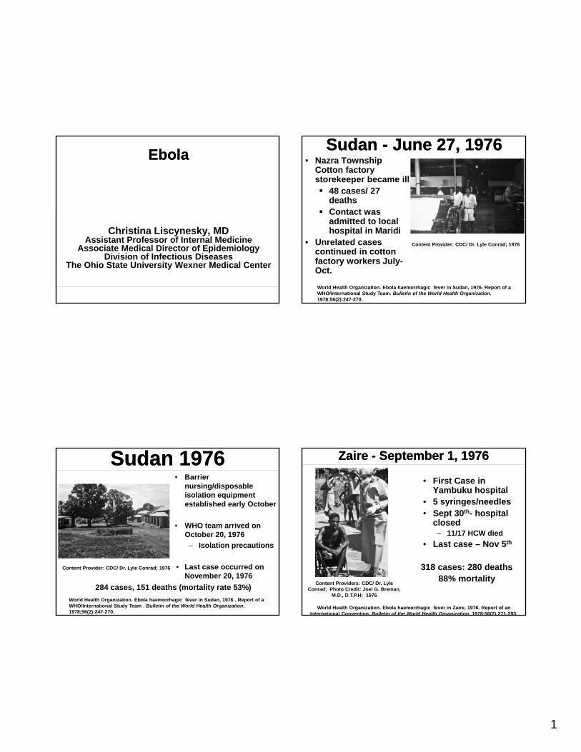
Sudan - June 27, 1976Sudan - June 27, 1976• Nazra Township
Cotton factory storekeeper became ill 48 cases/ 27
deaths Contact was
admitted to localadmitted to local hospital in Maridi
• Unrelated cases continued in cotton factory workers July-Oct.
World Health Organization. Ebola haemorrhagic fever in Sudan, 1976. Report of a WHO/International Study Team. Bulletin of the World Health Organization. 1978;56(2):247-270.
Content Provider: CDC/ Dr. Lyle Conrad; 1976
Sudan 1976Sudan 1976• Barrier
nursing/disposable isolation equipment established early October
• WHO team arrived on O t b 20 1976
284 cases, 151 deaths (mortality rate 53%)
October 20, 1976
– Isolation precautions
• Last case occurred on November 20, 1976
World Health Organization. Ebola haemorrhagic fever in Sudan, 1976 . Report of a WHO/International Study Team . Bulletin of the World Health Organization. 1978;56(2):247-270.
Content Provider: CDC/ Dr. Lyle Conrad; 1976
Zaire - September 1, 1976Zaire - September 1, 1976
• First Case in Yambuku hospital
• 5 syringes/needles• Sept 30th- hospital
closed 11/17 HCW died– 11/17 HCW died
• Last case – Nov 5th
318 cases: 280 deaths88% mortality
Content Providers: CDC/ Dr. Lyle Conrad; Photo Credit: Joel G. Breman,
M.D., D.T.P.H; 1976
World Health Organization. Ebola haemorrhagic fever in Zaire, 1976. Report of an International Convention. Bulletin of the World Health Organization. 1978;56(2):271-293.

2
EbolavirusEbolavirus• Sudan ebolavirus (SUDV)
• Zaïre ebolavirus (EBOV)
• Taï Forest (Ivory Coast) ebolavirus (TAFV)(TAFV)
• Bundibugyo ebolavirus (BDBV)
• Reston ebolavirus (RESTV)
Mandell, Douglas, and Bennett’s Principals and Practice of Infectious Diseases, Seventh Edition. Gerald L. Mandell, John E. Bennett, and Raphael Dolin. 164, 2259-2263
Guinea- December 2013 Guinea- December 2013 • Guéckédou, Guinea
– 2yr old Child– Fever, black stool, vomiting– Onset Dec. 2, 2013; died Dec. 6, 2013
• Transmitted from HCW (patient #14) to neighboring towns• HCW died Feb. 10, 2014
• March 10, 2014 – Guinea Ministry of Health Notified
• March 14, 2014- Outbreak team in place
111 suspect cases: 79 deaths (71% mortality)
Baize S. et al. Emergence of Zaire Ebola Virus Disease in Guinea – Preliminary Report. N Engl J Med. 2014 Apr 16.
August 21, 2014 – Ebola CasesAugust 21, 2014 – Ebola Cases
• Suspected and Confirmed Case Count: 2473
• Suspected Case D th 1350Deaths: 1350
• Laboratory Confirmed Cases: 1460
TransmissionTransmission• Zoonotic -introduced to humans through
close contact with infected animal’s bodily fluids
– Fruit bats
– Chimpanzeesp
– Gorillas
– Monkeys
– Forest Antelope
– Porcupines Author: Mnolf
GFDL & CC ShareAlike 2.0

3
Human to Human TransmissionHuman to Human Transmission• Direct contact with infected bodily secretions
• Indirect contact with contaminated environments
– In lab study: Ebola can remain active for up to 6 days– Environmental cxs: 2/33 samples positive for Ebola
• Blood stained physical glove• Bloody IV insertion site• Bloody IV insertion site
• Direct contact with infected corpses
• Men who survive can transmit virus via semen for up to 7 weeks
http://www.who.int/mediacentre/factsheets/fs103/en/Sagripanti JL, Rom AM, Holland LE. Persistence in darkness of virulent alphaviruses, Ebola virus, and Lassa virus deposited on solid surfaces. Arch Virol 2010; 155:2035-2039Bausch DG et al. Assessment of the Risk of Ebola Virus Transmission from Bodily Fluids and FomitesThe J of Infect Dis 2007; 196:S142–7
High Risk ExposuresHigh Risk Exposures• Percutaneous
– needle stick or mucous membrane exposure to body fluids
• Direct care or exposure to body fluids without appropriate personal protective equipment (PPE)
• Participation in funeral rites
Low Risk ExposuresLow Risk Exposures
• Household member or other casual contact with an EVD patient
• Providing patient care or casual contact• Providing patient care or casual contact without high-risk exposure with EVD patients
Ineffective TransmissionIneffective Transmission
Previous epidemics have calculated that 1 primary human case of Ebola generates only 1 to 3 secondary cases on average.
1 case of Measles in West Africa generates14-17 cases.
Chowell G, Hengartner NW, Castillo-Chavez C,Fenimore PW, Hyman JM. The basic reproductive number of Ebola and the effects of public health measures: the cases of Congo and Uganda. J Theor Biol 2004;229:119-126Legrand J, Grais RF, Boelle PY, Valleron AJ,Flahault A. Understanding the dynamics of Ebola epidemics. Epidemiol Infect 2007;135:610-621

4
Clinical ManifestationsClinical Manifestations• Incubation period of 8-10 days (range 2-21)
• Abrupt onset of fever, with HA and myalgia
– Nausea, vomiting, abdominal pain, and diarrhea
Maculopapular rash by day 5 7– Maculopapular rash by day 5-7
– Chest pain, shortness of breath
– Hemorrhage
– Confusion, seizures
Kortepeter MG, Bausch DG, Bray M. Basic clinical and laboratory features of filoviral hemorrhagic fever. J Infect Dis. 2011 Nov;204 Suppl 3:S810–6
Differential DiagnosisDifferential Diagnosis• Malaria
• Typhoid Fever
• Dengue
• Lassa Fever
• Marburg Virus
• Yellow fever
• Viral hepatitis
• Anthrax
• Shigellosis
• Meningococcal septicemia
• Plague
• Relapsing fever
• Chikungunya fever
• Leptospirosis
• Typhus
PathogenesisPathogenesis• Monocytes, macrophages, and
dendritic cells are infected early• Virus suppresses type 1 interferon
responses and induces cytokine and chemokine releasechemokine release
• Virus replicates, released and migrates to local lymph nodes, travels through the lymphatic system to blood
• Virus disseminated throughout the body
Feldmann H, Geisbert TW. Ebola haemorrhagic fever. Lancet. 2011 Mar 5;377(9768):849-62.
• Lymphocytes undergo apoptosis, which undermines adaptive immunity
• Hepatocellular necrosis DIC
• Adrenal necrosis Hypotension Impaired Steroid Synthesisp y
• Extensive tissue necrosis and shock

5
Diagnostic labs testsDiagnostic labs tests
• Ebola virus is detectable in blood only after onset of symptoms
• Detectable by real-time RT-PCR between 3 to 10 days post-onset of symptoms
Lab AbnormalitiesLab Abnormalities
• Leukopenia
• Thrombocytopenia – 50 to 100K range
• Transminitis: AST>ALT
• Proteinuria may be present
• PT and PTT prolonged
• Fibrin elevated
Fatal IllnessFatal Illness
• LFTS and D-dimer higher in fatal illness.
• Calcium <6mg/dL associated with death
• Median survival of 9 days
• Most patients die during the second week
• Alive on day 14 portends >75% survival
• Fatally infected patients do not develop an AB response
Kortepeter MG, Bausch DG, Bray M. Basic clinical and laboratory features of filoviral hemorrhagic fever. J Infect Dis. 2011 Nov;204 Suppl 3:S810–6
TreatmentTreatment• Supportive care
• Antibiotics for secondary infections
• September 4-5, WHO scheduled pconference on potential Ebola therapies and vaccines in Geneva

6
Additional ReferencesAdditional References
CDC Ebola Hemorrhagic Fever site: www.cdc.gov/ebola
WHO:http://www.who.int/csr/disease/ebol
/ /
DDrr. Margaret Isaacson as she was . Margaret Isaacson as she was tending to the needs of an Ebola tending to the needs of an Ebola patient in a patient in a YambukuYambuku, Zaire hospital , Zaire hospital theatre block that was used theatre block that was used as as a a temporary ICU for Ebola patients temporary ICU for Ebola patients during the country’s 1976 outbreak.during the country’s 1976 outbreak.
Content Providers: CDC/ Dr. Lyle Conrad; Photo Credit: Joel G. Breman, M.D., D.T.P.H; 1976
a/en/
http://www.nejm.org/page/ebola-outbreak
EbolaEbola
Naeem Ali, MDMedical Director,
The Ohio State University Wexner Medical Center
Hospital preparednessHospital preparedness
EVD
Are you prepared?Are you prepared?
Why prepare?Why prepare?
Author: PA2 Dan Tremper, USCG cropped by USer:Ultra7

7
Why prepare?Why prepare? Why prepare?Why prepare?
Why prepare?Why prepare? Why prepare?Why prepare?

8
Why prepare?Why prepare? Why prepare?Why prepare?
Why prepare?Why prepare? Why prepare?Why prepare?

9
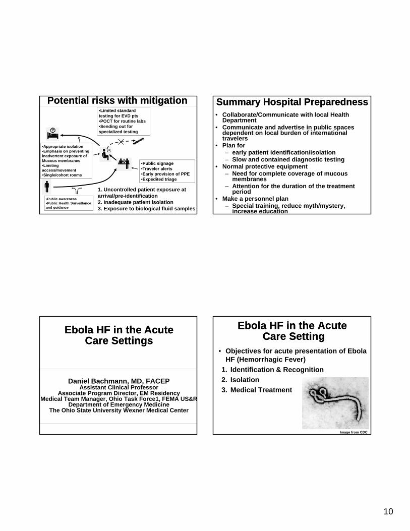
Potential risksPotential risksPotential risksPotential risks
1. Uncontrolled patient exposure at arrival/pre-identification2. Inadequate patient isolation3. Exposure to biological fluid samples
Preparedness: more than patient isolationPreparedness: more than patient isolation
Within minutes, he dispatched one member of his staff to make sure that the sick man remained isolated and that doctors and nurses were taking precautions to protect themselves against contracting the virus. Then he turned to a second staff member.
“Stop what you’re doing right now,” Dr. Phillips told him, and sent him to the hospital’s laboratory.
The lab?
NYT, 8/10/2014, Rachel Swarnshttp://www.nytimes.com/2014/08/11/nyregion/fighting-deadly-diseases-without-breaking-a-sweat.html?module=Search&mabReward=relbias%3Aw
Case study: CJDCase study: CJDRisk of infection Tissue Special procedures
High Brain, spinal cord • Notification• PPE disposal• Laundry disposal• Terminal room cleaning• Disposable equipment use• Special equipment decon.
procedures
Low CSF, liver, lymph node • Notification• Dedicated instrument• Usual cleaning after
sample assay
None Whole blood, nasal mucous, saliva, sputum,feces
• Notification• Usual cleaning after
sample assay
EVD comparisonEVD comparison• Blood and secretions are infectious
– PCR positive in blood only
• Implications
Common laboratory testing samples for– Common laboratory testing samples for the febrile patient ARE infectious
• Mitigation?
10
Potential risks with mitigationPotential risks with mitigation•Limited standard testing for EVD pts•POCT for routine labs•Sending out for specialized testing
•Appropriate isolation•Emphasis on preventing i d t t f
1. Uncontrolled patient exposure at arrival/pre-identification2. Inadequate patient isolation3. Exposure to biological fluid samples
•Public awareness•Public Health Surveillance and guidance
•Public signage•Traveler alerts•Early provision of PPE•Expedited triage
inadvertent exposure of Mucous membranes•Limiting access/movement•Single/cohort rooms
Summary Hospital PreparednessSummary Hospital Preparedness• Collaborate/Communicate with local Health
Department• Communicate and advertise in public spaces
dependent on local burden of international travelers
• Plan for – early patient identification/isolationy p– Slow and contained diagnostic testing
• Normal protective equipment– Need for complete coverage of mucous
membranes– Attention for the duration of the treatment
period • Make a personnel plan
– Special training, reduce myth/mystery, increase education
Ebola HF in the Acute Ebola HF in the Acute Care Settings Care Settings
Daniel Bachmann, MD, FACEPAssistant Clinical Professor
Associate Program Director, EM ResidencyMedical Team Manager, Ohio Task Force1, FEMA US&R
Department of Emergency MedicineThe Ohio State University Wexner Medical Center
Ebola HF in the AcuteCare Setting
Ebola HF in the AcuteCare Setting
• Objectives for acute presentation of Ebola HF (Hemorrhagic Fever)
1. Identification & Recognition
2. Isolation
3. Medical Treatment
Image from CDC

11
Identification of Ebola HF Patients
Identification of Ebola HF Patients
• Education of staff
• Increased awareness levels at portals of entry
– Clinics
– Urgent care centers
– Emergency departments
• Public health awareness
– Patient self-selection
– Signage & outreachImage from CDC
Identification of Ebola HF Patients
Identification of Ebola HF Patients
• Screening criteria for Ebola HF in the ED:
– Any of the following symptoms:
• Fever >101.5F
• Headache
• Joint / muscle achesJoint / muscle aches
• Weakness
• Vomiting
• Diarrhea
Author: Mikael Häggström. – Public Domain
Identification of Ebola HF PatientsIdentification of Ebola HF Patients
• Ask about travel to high-risk areas <21days:– Guinea
– Liberia
– Nigeria
– Sierra Leone
• Concerning symptoms PLUS high risk travel requires immediate isolation!
Image from CDC
Isolation of Ebola HF PatientsIsolation of Ebola HF Patients• Place a mask on the patient
– Initiate contact, droplet, & airborne precautions
• Staff personal protective equipment includes:includes:
– Gown
– Gloves
– Mask
– Eye protection
Image from CDC

12
Isolation of Ebola HF PatientsIsolation of Ebola HF Patients
• Outpatient setting
– Arrange transfer to ED (via ambulance with PPE)
• Emergency Department
– Place patient in private room with bathroomPlace patient in private room with bathroom
– Negative airflow not required
– Contact, Droplet & Airborne isolation precautions
– Dedicated medical equipment should be disposable when possible
Isolation of Ebola HF PatientsIsolation of Ebola HF Patients• Notifications:
– ED Attending
– Critical Events Officer
– Infectious Disease Team
– Public Health Department (via the CE Officer)p ( )
• This notification chain is institution dependent
• Initiation of laboratory testing
CC-BY-SA-3.0-MIGRATED
Author: Tristanb
Acute Medical Treatment of Ebola HFAcute Medical Treatment of Ebola HF
• Supportive therapy:– Fluids and electrolytes– Blood pressure and oxygen
status – Treatment of complicating
i f tiinfections• Initial management of
complications• Admission to the inpatient setting
– Level of care depends on acuity of illness
Author: Harmid Public Domain