Embed Size (px)
Citation preview
Early Versus Late Onset IUGR Usefulness of fetal Doppler evaluation
DANIEL MURESAN
UNIVERSITY OF MEDICINE “IULIU HATIEGANU”
CLUJ-NAPOCA, ROMANIA
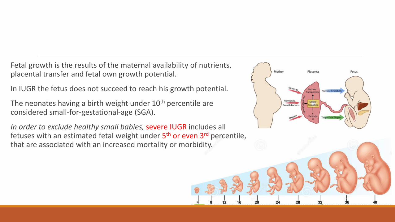
Fetal growth is the results of the maternal availability of nutrients, placental transfer and fetal own growth potential.
In IUGR the fetus does not succeed to reach his growth potential.
The neonates having a birth weight under 10th percentile are considered small-for-gestational-age (SGA).
In order to exclude healthy small babies, severe IUGR includes all fetuses with an estimated fetal weight under 5th or even 3rd percentile, that are associated with an increased mortality or morbidity.
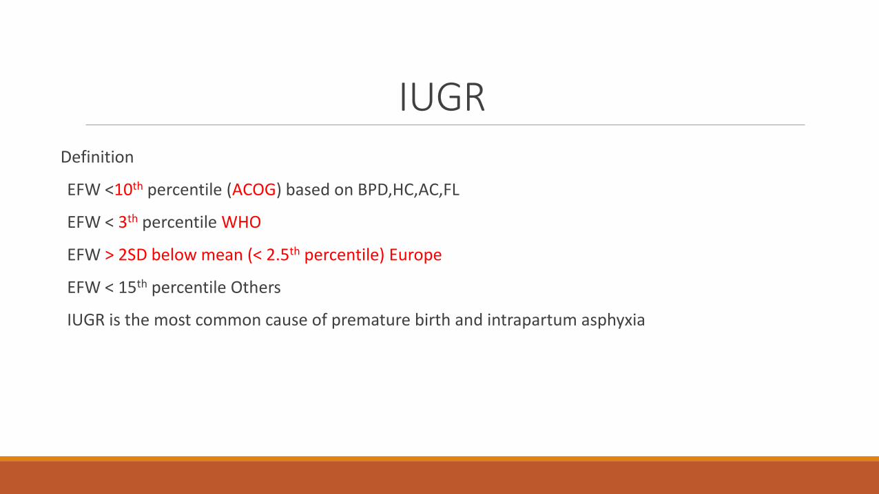
IUGR Definition
EFW <10th percentile (ACOG) based on BPD,HC,AC,FL
EFW < 3th percentile WHO
EFW > 2SD below mean (< 2.5th percentile) Europe
EFW < 15th percentile Others
IUGR is the most common cause of premature birth and intrapartum asphyxia
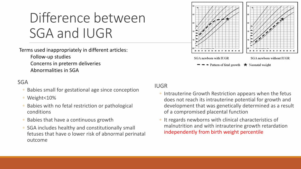
Difference between SGA and IUGR
SGA ◦ Babies small for gestational age since conception
◦ Weight<10%
◦ Babies with no fetal restriction or pathological conditions
◦ Babies that have a continuous growth
◦ SGA includes healthy and constitutionally small fetuses that have o lower risk of abnormal perinatal outcome
IUGR ◦ Intrauterine Growth Restriction appears when the fetus
does not reach its intrauterine potential for growth and development that was genetically determined as a result of a compromised placental function
◦ It regards newborns with clinical characteristics of malnutrition and with intrauterine growth retardation independently from birth weight percentile
Terms used inappropriately in different articles: Follow-up studies Concerns in preterm deliveries Abnormalities in SGA
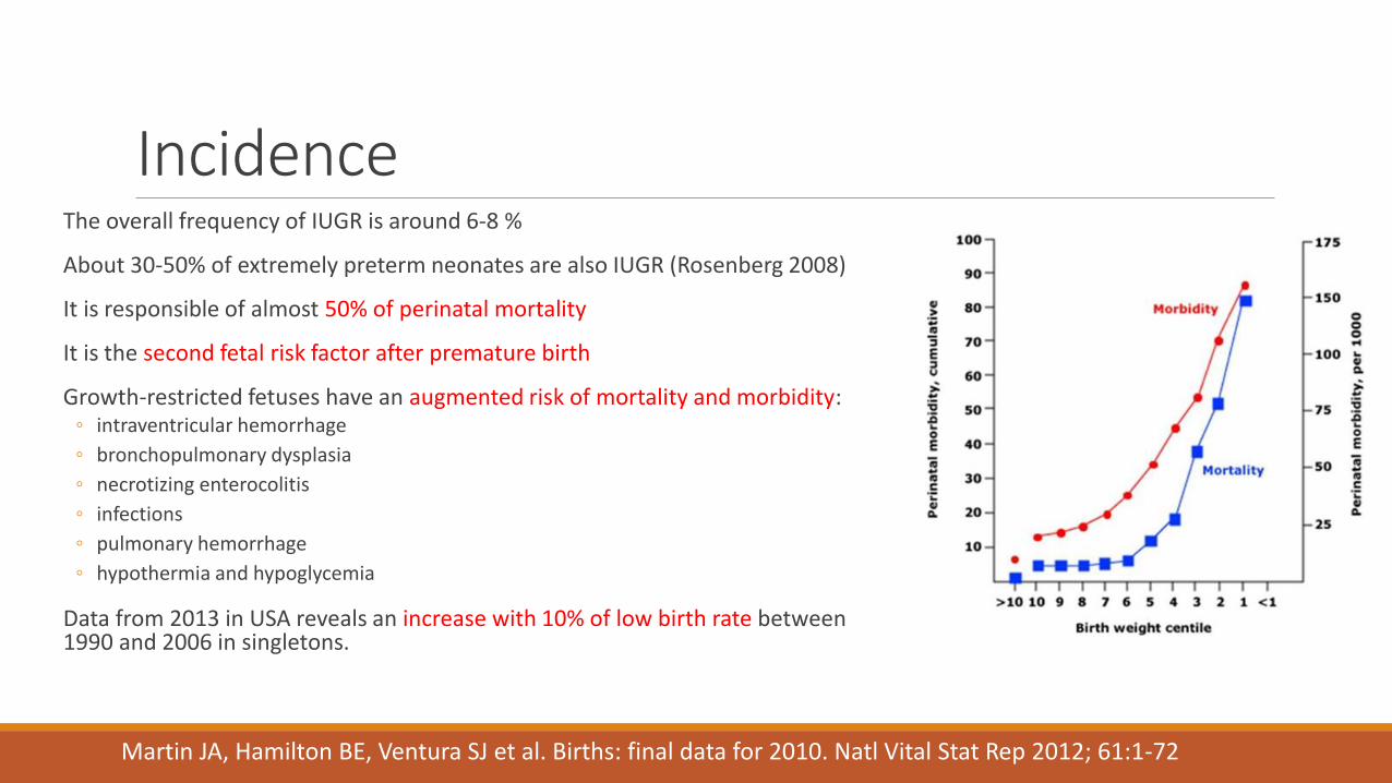
Incidence The overall frequency of IUGR is around 6-8 %
About 30-50% of extremely preterm neonates are also IUGR (Rosenberg 2008)
It is responsible of almost 50% of perinatal mortality
It is the second fetal risk factor after premature birth
Growth-restricted fetuses have an augmented risk of mortality and morbidity: ◦ intraventricular hemorrhage
◦ bronchopulmonary dysplasia
◦ necrotizing enterocolitis
◦ infections
◦ pulmonary hemorrhage
◦ hypothermia and hypoglycemia
Data from 2013 in USA reveals an increase with 10% of low birth rate between 1990 and 2006 in singletons.
Martin JA, Hamilton BE, Ventura SJ et al. Births: final data for 2010. Natl Vital Stat Rep 2012; 61:1-72
IUGR diagnostic
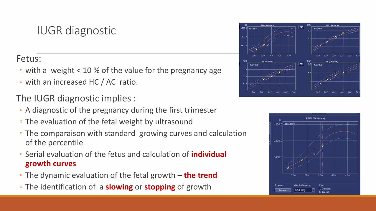
Fetus: ◦ with a weight < 10 % of the value for the pregnancy age
◦ with an increased HC / AC ratio.
The IUGR diagnostic implies : ◦ A diagnostic of the pregnancy during the first trimester
◦ The evaluation of the fetal weight by ultrasound
◦ The comparaison with standard growing curves and calculation of the percentile
◦ Serial evaluation of the fetus and calculation of individual growth curves
◦ The dynamic evaluation of the fetal growth – the trend
◦ The identification of a slowing or stopping of growth
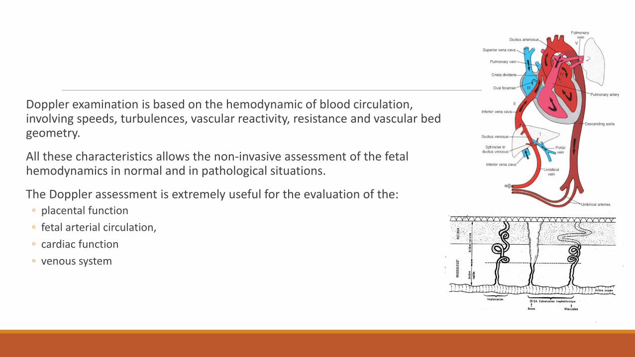
Doppler examination is based on the hemodynamic of blood circulation, involving speeds, turbulences, vascular reactivity, resistance and vascular bed geometry.
All these characteristics allows the non-invasive assessment of the fetal hemodynamics in normal and in pathological situations.
The Doppler assessment is extremely useful for the evaluation of the: ◦ placental function
◦ fetal arterial circulation,
◦ cardiac function
◦ venous system

There are two phenotypes of IUGR: E-IUGR and L-IUGR that are distinct by: ◦ the moment of onset,
◦ evolution
◦ Doppler parameters modifications and
◦ postnatal outcome.
The best cut-off between the two IUGR forms is 32 weeks in terms of perinatal outcome.
The antenatal diagnostic, treatment and timely delivery could diminish the risks significantly.
Savchev S, Figueras F, Sanz-Cortes M et al. Evaluation of an optimal gestational age cut-off for the definition of early- and late-onset fetal growth restriction. Fetal Diagn Ther 2014; 36:99-105.
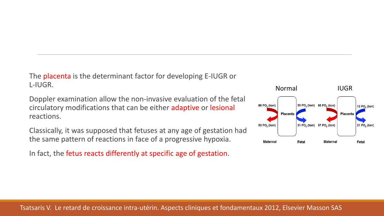
The placenta is the determinant factor for developing E-IUGR or L-IUGR.
Doppler examination allow the non-invasive evaluation of the fetal circulatory modifications that can be either adaptive or lesional reactions.
Classically, it was supposed that fetuses at any age of gestation had the same pattern of reactions in face of a progressive hypoxia.
In fact, the fetus reacts differently at specific age of gestation.
Tsatsaris V. Le retard de croissance intra-utérin. Aspects cliniques et fondamentaux 2012, Elsevier Masson SAS
Normal IUGR
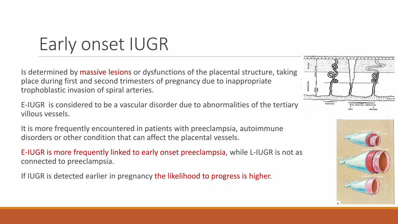
Early onset IUGR Is determined by massive lesions or dysfunctions of the placental structure, taking place during first and second trimesters of pregnancy due to inappropriate trophoblastic invasion of spiral arteries.
E-IUGR is considered to be a vascular disorder due to abnormalities of the tertiary villous vessels.
It is more frequently encountered in patients with preeclampsia, autoimmune disorders or other condition that can affect the placental vessels.
E-IUGR is more frequently linked to early onset preeclampsia, while L-IUGR is not as connected to preeclampsia.
If IUGR is detected earlier in pregnancy the likelihood to progress is higher.
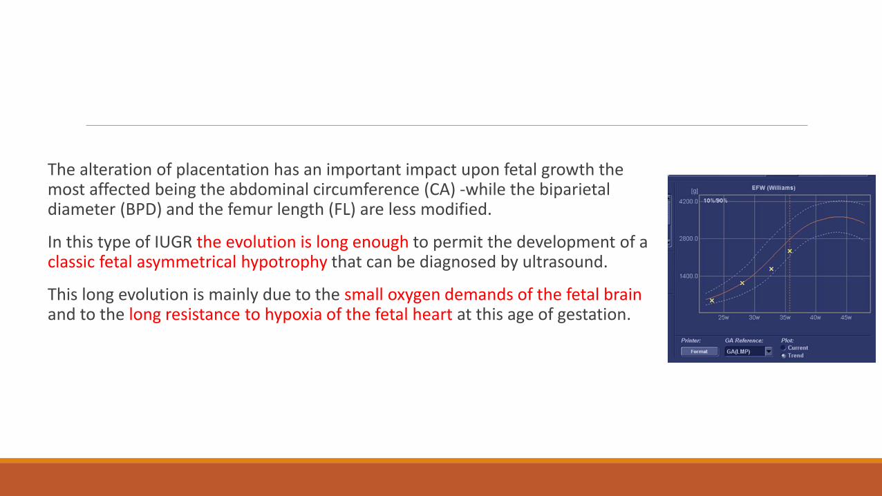
The alteration of placentation has an important impact upon fetal growth the most affected being the abdominal circumference (CA) -while the biparietal diameter (BPD) and the femur length (FL) are less modified.
In this type of IUGR the evolution is long enough to permit the development of a classic fetal asymmetrical hypotrophy that can be diagnosed by ultrasound.
This long evolution is mainly due to the small oxygen demands of the fetal brain and to the long resistance to hypoxia of the fetal heart at this age of gestation.
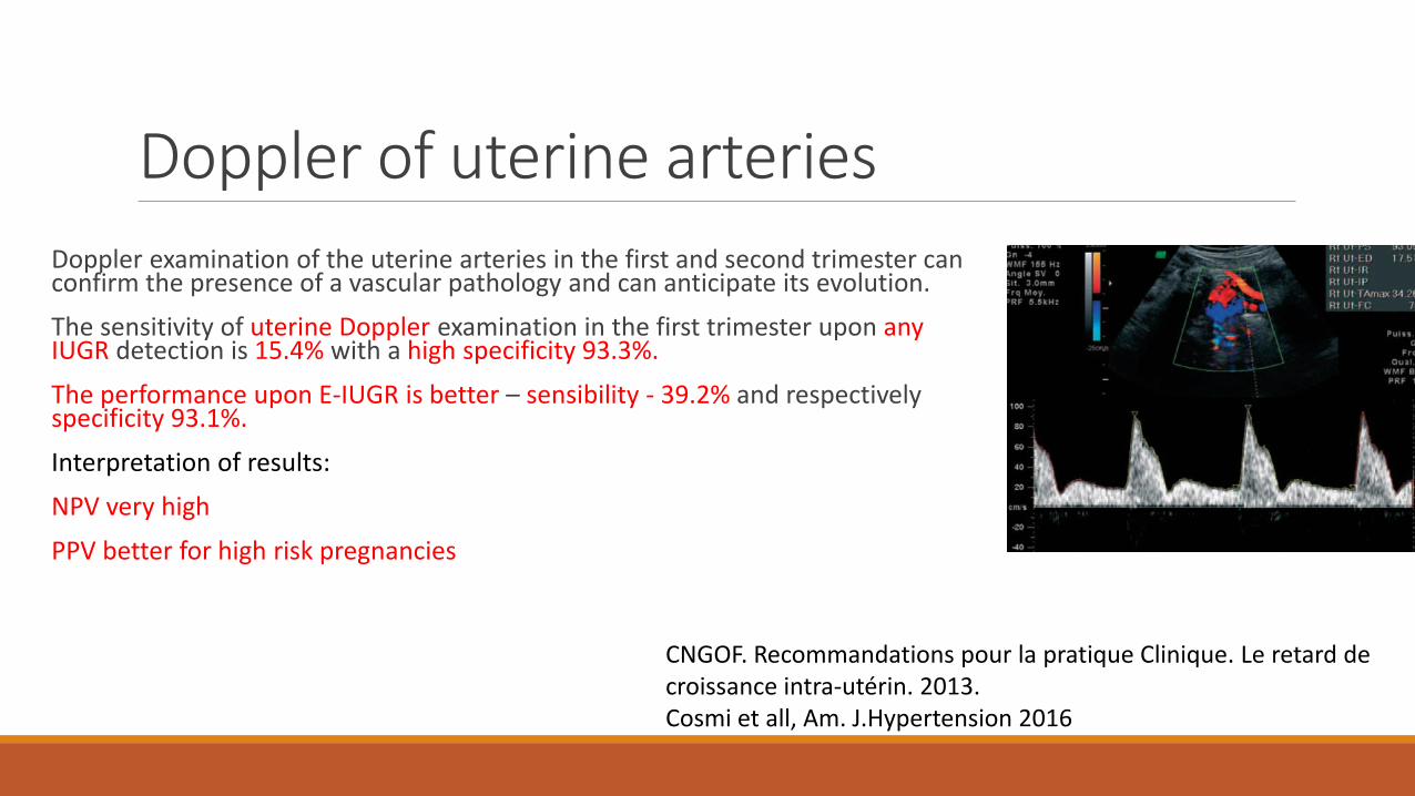
Doppler of uterine arteries
Doppler examination of the uterine arteries in the first and second trimester can confirm the presence of a vascular pathology and can anticipate its evolution.
The sensitivity of uterine Doppler examination in the first trimester upon any IUGR detection is 15.4% with a high specificity 93.3%.
The performance upon E-IUGR is better – sensibility - 39.2% and respectively specificity 93.1%.
Interpretation of results:
NPV very high
PPV better for high risk pregnancies
CNGOF. Recommandations pour la pratique Clinique. Le retard de croissance intra-utérin. 2013. Cosmi et all, Am. J.Hypertension 2016
Physiopathology of IUGR
Reduced transfer of oxygen and nutrients toward the fetus
Reduced perfusion of the liver with impaired liver protein synthesis
Blood flow redirected towards vital organs: brain, heart, surrenal glands
Reduced blood flow to muscles, bowel and kidneys
The sequence of a classical, progressive
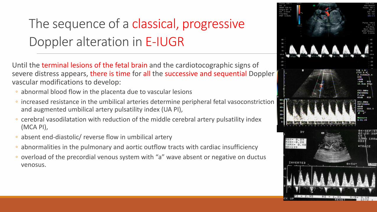
Doppler alteration in E-IUGR Until the terminal lesions of the fetal brain and the cardiotocographic signs of severe distress appears, there is time for all the successive and sequential Doppler vascular modifications to develop:
◦ abnormal blood flow in the placenta due to vascular lesions
◦ increased resistance in the umbilical arteries determine peripheral fetal vasoconstriction and augmented umbilical artery pulsatility index (UA PI),
◦ cerebral vasodilatation with reduction of the middle cerebral artery pulsatility index (MCA PI),
◦ absent end-diastolic/ reverse flow in umbilical artery
◦ abnormalities in the pulmonary and aortic outflow tracts with cardiac insufficiency
◦ overload of the precordial venous system with “a” wave absent or negative on ductus venosus.
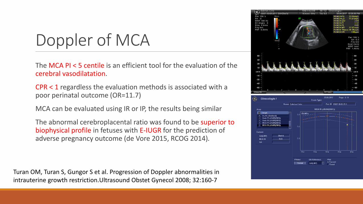
Doppler of MCA The MCA PI < 5 centile is an efficient tool for the evaluation of the cerebral vasodilatation.
CPR < 1 regardless the evaluation methods is associated with a poor perinatal outcome (OR=11.7)
MCA can be evaluated using IR or IP, the results being similar
The abnormal cerebroplacental ratio was found to be superior to biophysical profile in fetuses with E-IUGR for the prediction of adverse pregnancy outcome (de Vore 2015, RCOG 2014).
Turan OM, Turan S, Gungor S et al. Progression of Doppler abnormalities in intrauterine growth restriction.Ultrasound Obstet Gynecol 2008; 32:160-7
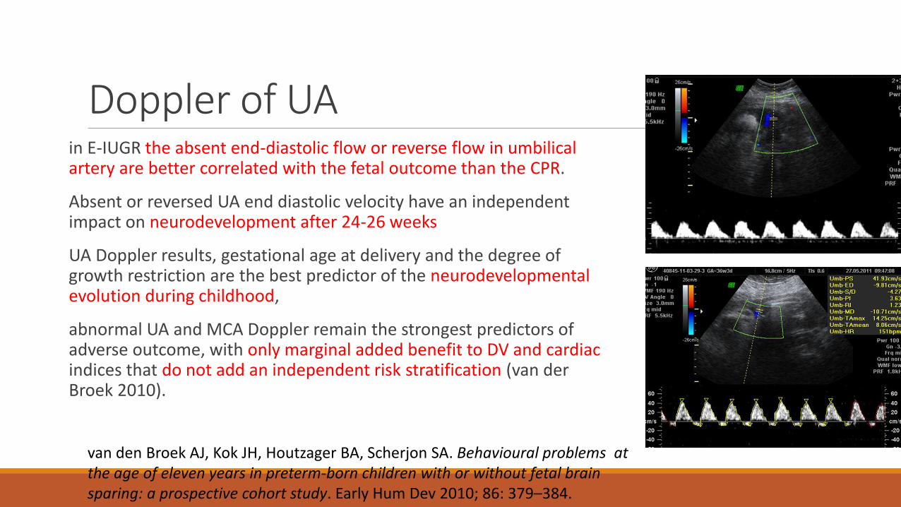
Doppler of UA in E-IUGR the absent end-diastolic flow or reverse flow in umbilical artery are better correlated with the fetal outcome than the CPR.
Absent or reversed UA end diastolic velocity have an independent impact on neurodevelopment after 24-26 weeks
UA Doppler results, gestational age at delivery and the degree of growth restriction are the best predictor of the neurodevelopmental evolution during childhood,
abnormal UA and MCA Doppler remain the strongest predictors of adverse outcome, with only marginal added benefit to DV and cardiac indices that do not add an independent risk stratification (van der Broek 2010).
van den Broek AJ, Kok JH, Houtzager BA, Scherjon SA. Behavioural problems at the age of eleven years in preterm-born children with or without fetal brain sparing: a prospective cohort study. Early Hum Dev 2010; 86: 379–384.
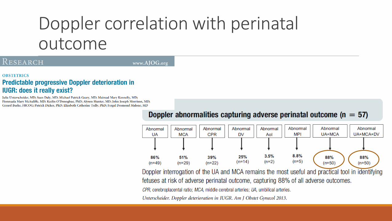
Doppler correlation with perinatal outcome
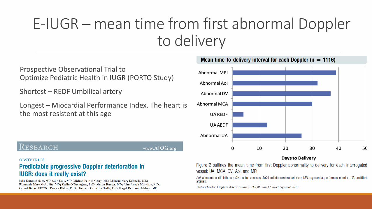
E-IUGR – mean time from first abnormal Doppler to delivery
Prospective Observational Trial to Optimize Pediatric Health in IUGR (PORTO Study)
Shortest – REDF Umbilical artery
Longest – Miocardial Performance Index. The heart is the most resistent at this age
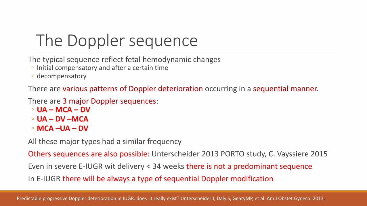
The Doppler sequence The typical sequence reflect fetal hemodynamic changes
◦ Initial compensatory and after a certain time ◦ decompensatory
There are various patterns of Doppler deterioration occurring in a sequential manner.
There are 3 major Doppler sequences: ◦ UA – MCA – DV ◦ UA – DV –MCA ◦ MCA –UA – DV
All these major types had a similar frequency
Others sequences are also possible: Unterscheider 2013 PORTO study, C. Vayssiere 2015
Even in severe E-IUGR wit delivery < 34 weeks there is not a predominant sequence
In E-IUGR there will be always a type of sequential Doppler modification
Predictable progressive Doppler deterioration in IUGR: does it really exist? Unterscheider J, Daly S, GearyMP, et al. Am J Obstet Gynecol 2013;
L- IUGR Represents the failure of the fetus to reach its growth potential at term,
Fetal hypoxemia/hypoxia secondary of placental insufficiency represents the main cause of L-IUGR.
In most cases the placental lesions have a late onset and/or do not have a significant extent in order to increase the resistivity of the placenta and translated into augmented UA IP.
In terms of frequency L-IUGR is far more prevalent than E-IUGR.
It has placental anomalies such as villous immaturity with less impact upon placental resistance, therefore the umbilical Doppler indices can be unaffected.
Oros D, Figueras F, Cruz-Martinez R, Meler E, Munmany M, Gratacos E. Longitudinal changes in uterine, umbilical and fetal cerebral Doppler indices in late-onset small-for-gestational age fetuses. Ultrasound Obstet Gynecol. 2011.
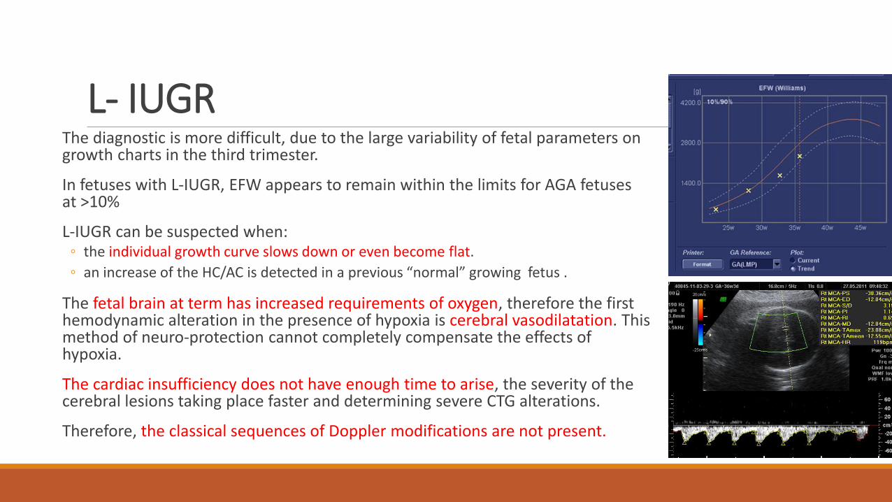
L- IUGR The diagnostic is more difficult, due to the large variability of fetal parameters on growth charts in the third trimester.
In fetuses with L-IUGR, EFW appears to remain within the limits for AGA fetuses at >10%
L-IUGR can be suspected when: ◦ the individual growth curve slows down or even become flat.
◦ an increase of the HC/AC is detected in a previous “normal” growing fetus .
The fetal brain at term has increased requirements of oxygen, therefore the first hemodynamic alteration in the presence of hypoxia is cerebral vasodilatation. This method of neuro-protection cannot completely compensate the effects of hypoxia.
The cardiac insufficiency does not have enough time to arise, the severity of the cerebral lesions taking place faster and determining severe CTG alterations.
Therefore, the classical sequences of Doppler modifications are not present.
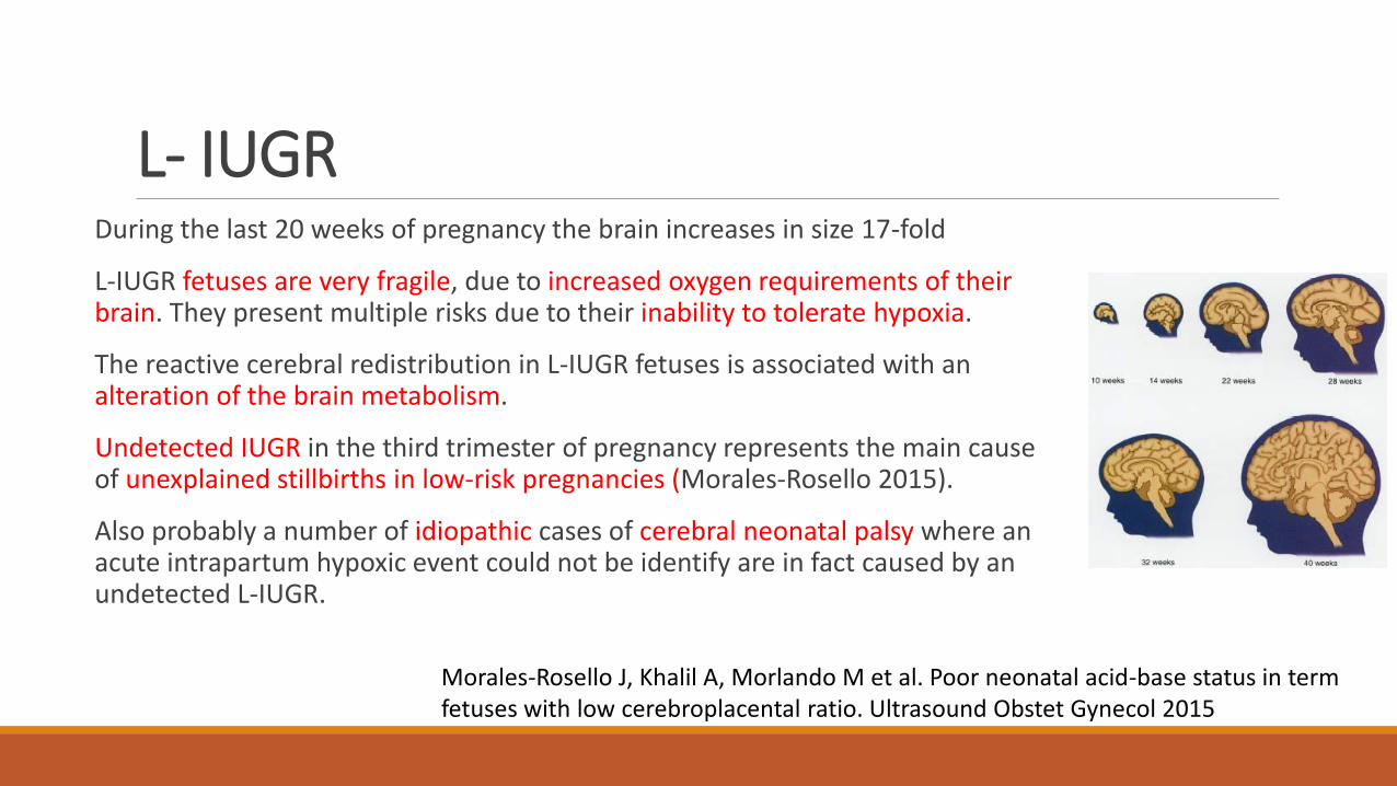
L- IUGR During the last 20 weeks of pregnancy the brain increases in size 17-fold
L-IUGR fetuses are very fragile, due to increased oxygen requirements of their brain. They present multiple risks due to their inability to tolerate hypoxia.
The reactive cerebral redistribution in L-IUGR fetuses is associated with an alteration of the brain metabolism.
Undetected IUGR in the third trimester of pregnancy represents the main cause of unexplained stillbirths in low-risk pregnancies (Morales-Rosello 2015).
Also probably a number of idiopathic cases of cerebral neonatal palsy where an acute intrapartum hypoxic event could not be identify are in fact caused by an undetected L-IUGR.
Morales-Rosello J, Khalil A, Morlando M et al. Poor neonatal acid-base status in term fetuses with low cerebroplacental ratio. Ultrasound Obstet Gynecol 2015
L- IUGR L-IUGR is a challenging diagnosis associated with:
◦ increased fetal morbidity and mortality,
◦ impaired postnatal outcome
◦ suboptimal neuro-development.
In fetuses with L- IUGR the placental insufficiency seems not to be not reflected in UA Doppler.
Umbilical artery Doppler and the sequence of Doppler modifications in multiple fetal vessels are not reliable for the fetal surveillance.
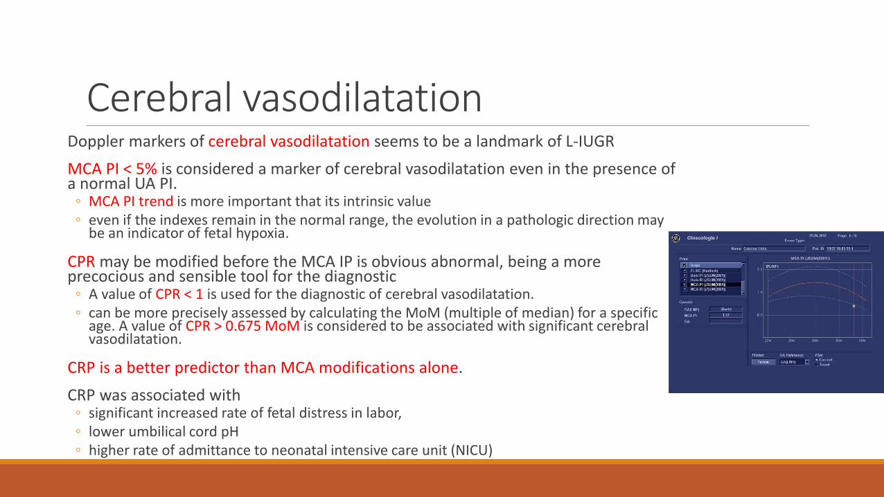
Cerebral vasodilatation Doppler markers of cerebral vasodilatation seems to be a landmark of L-IUGR
MCA PI < 5% is considered a marker of cerebral vasodilatation even in the presence of a normal UA PI.
◦ MCA PI trend is more important that its intrinsic value ◦ even if the indexes remain in the normal range, the evolution in a pathologic direction may
be an indicator of fetal hypoxia.
CPR may be modified before the MCA IP is obvious abnormal, being a more precocious and sensible tool for the diagnostic
◦ A value of CPR < 1 is used for the diagnostic of cerebral vasodilatation. ◦ can be more precisely assessed by calculating the MoM (multiple of median) for a specific
age. A value of CPR > 0.675 MoM is considered to be associated with significant cerebral vasodilatation.
CRP is a better predictor than MCA modifications alone.
CRP was associated with ◦ significant increased rate of fetal distress in labor, ◦ lower umbilical cord pH ◦ higher rate of admittance to neonatal intensive care unit (NICU)
Cerebral vasodilatation “If in the third trimester the obstetrician will analyses only the umbilical artery PI, these fetuses will be miss, and the potential intrapartum and postnatal complications cannot be anticipated” [Cruz-Martinez R, Figueras F 2011].
An abnormal CPR and low birthweight centile were identified as significantly and independently associated with emergency caesarean section in both appropriate for gestational age (AGA) and L-IUGR groups.
CPR perform better in identifying fetuses with adverse outcome than did the biophysical profile, and was a better predictor than birthweight centile for the necessity of NICU admission .
This data encourage the recommendation of usage of CPR for risk stratification in L-IUGR fetuses.
DeVore GR. The importance of the cerebroplacental ratio in the evaluation of fetal well-being in SGA and AGA fetuses.Am J Obstet Gynecol 2015
Postnatal outcome The short and medium neonatal outcome are influenced by the cerebral vasodilatation.
Term L-IUGR fetuses have poorer neurodevelopmental outcomes and an increased risk of neonatal motor activity, lower score of communication and problem solving scores at 2 years of age. [Meher 2015].
This may suggest the development of a neurological injury from early stages of fetal hemodynamic adaptation to hypoxia.
L-IUGR is also associated with a risk of neurocognitive dysfunction. It is likely that stillbirth represents the outcome of a pathological process, and that for every fetal demise many more fetuses might theoretically be affected by neurological impairment as a consequence of mild hypoxia and suboptimal growth at term.
L-IUGR fetuses are at higher risk of cardiovascular diseases, hypertension, glucose intolerance, diabetes, dyslipidemia and obesity in childhood and in adulthood
The rates of acute fetal distress during delivery and the number of emergency caesarean sections in L- IUGR with cerebral redistribution are significantly increased (DeVore 2015, Meher S 2015, Khalil AA 2015).
Meher S, Hernandez-Andrade E, Basheer SN, Lees C. Impact of cerebral redistribution on neurodevelopmental outcome in small-for-gestational-age or growth-restricted babies: a systematic review. Ultrasound Obstet Gynecol. 2015;
Implications for clinical practice The use of Doppler evaluation of the uterine arteries during the first and second trimester of pregnancy represents a helpful tool for the prediction of IUGR (RCOG 2014).
Once an early/late IUGR fetus has been identified based on the ultrasound measurements, the evaluation must include the Doppler analysis of umbilical and cerebral circulation (CPR,MCA) (DeVore 2015, RCOG 2014).
This evaluation allows the risk stratification of IUGR fetuses pointing out the fetuses with an increased risk for perinatal complications.
E-IUGR Umbilical Artery Doppler is capital for surveillance and obstetrical management
MCA, Ductus Venosus and cardiac Doppler bring little benefit and are not included in standard protocols (RCOG,ACOG, French College of Gynaecologists and Obstetricians )
The sequence of Doppler modifications is usually the classic one, but not always.
At this period the fetal cerebral resistance to hypoxia is superior to the cardiac one, so the evolution may arrive at cardiac insufficiency before major fetal accidents arrive.
The timing of delivery should be chosen taking into account the gestational age, umbilical Doppler, Ductus Venosus Doppler and cardiotocography
The biophysical profile is not usefull in premature pregnancies
L-IUGR
In fetuses with L-IUGR frequently the UA Doppler is within normal range therefore it cannot constitute a useful diagnostic.
The cerebral vasodilatation being the first reaction of the fetus to hypoxia in L-IUGR, the MCA Doppler is the most valuable examination tool.
Generaly there is no sequence of Doppler alteration.
At this period the fetal cerebral resistance to hypoxia is inferior to the cardiac one, so there is possible to have major fetal accidents before cardiac and ductus venosus Doppler modification appears.
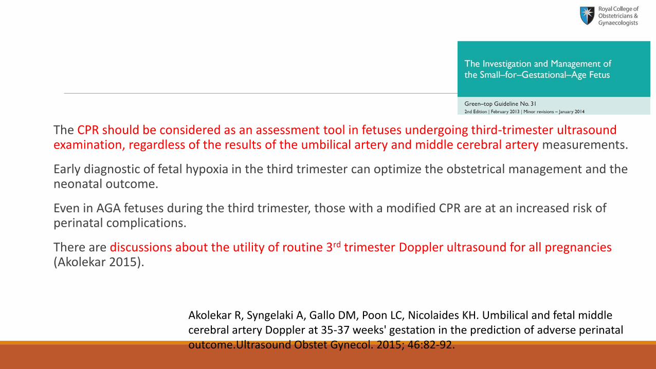
The CPR should be considered as an assessment tool in fetuses undergoing third-trimester ultrasound examination, regardless of the results of the umbilical artery and middle cerebral artery measurements.
Early diagnostic of fetal hypoxia in the third trimester can optimize the obstetrical management and the neonatal outcome.
Even in AGA fetuses during the third trimester, those with a modified CPR are at an increased risk of perinatal complications.
There are discussions about the utility of routine 3rd trimester Doppler ultrasound for all pregnancies (Akolekar 2015).
Akolekar R, Syngelaki A, Gallo DM, Poon LC, Nicolaides KH. Umbilical and fetal middle cerebral artery Doppler at 35-37 weeks' gestation in the prediction of adverse perinatal outcome.Ultrasound Obstet Gynecol. 2015; 46:82-92.
The MCA Doppler evaluation is important for the decision of the delivery timing especially when the UA Doppler is normal.
Recent guidelines recommend delivery of L-IUGR fetuses with brain sparring effect, because it is predictive of acidosis at birth
Cerebral redistribution may be use in association with other clinical elements in the decision of delivery of L-IUGR fetuses, even in the presence of a normal UA Doppler.
After 34 weeks, the risk of serious morbidity and of mortality is low and the delivery of the fetuses with L-IUGR will minimize the long term sequelae.
The paradigm that a normal UA Doppler pattern in the third trimester confirms a normal pregnancy and does not need any further Doppler evaluation of other fetal vessels definitely needs to be modified, with important impact upon the clinical follow-up.

Management of IUGR There is no effective intrauterine treatment
The objectives of a correct managements are: ◦ Early diagnostic
◦ Prevention and reduction of risk factors where possible
◦ Surveillance of the fetus with IUGR
Delivery at the best moment ◦ If delivery before 32 WG consider magnesium sulfat for fetal neuroprotection
◦ If delivery before 34 WG corticosteroids are indicated
Doppler ultrasound is the chief tool
The optimal gestation age to deliver the E-IUGR fetus
the timing of delivery (immediately or after some days) has not a direct impact on the neurological development.
◦ In E-IUGR detected before 32 weeks, with AREDF on UA delivery is decided when DV become abnormal.
◦ Even if DV is normal, because the fetal mortality and morbidity decrease much after 30 weeks, in AREDF, delivery is recommended at 32 weeks and must be proposed at 30-32 weeks.
◦ The timing of delivery is NOT based on MCA Doppler
RCOG 2014
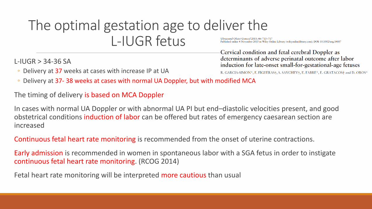
The optimal gestation age to deliver the L-IUGR fetus
L-IUGR > 34-36 SA ◦ Delivery at 37 weeks at cases with increase IP at UA
◦ Delivery at 37- 38 weeks at cases with normal UA Doppler, but with modified MCA
The timing of delivery is based on MCA Doppler
In cases with normal UA Doppler or with abnormal UA PI but end–diastolic velocities present, and good obstetrical conditions induction of labor can be offered but rates of emergency caesarean section are increased
Continuous fetal heart rate monitoring is recommended from the onset of uterine contractions.
Early admission is recommended in women in spontaneous labor with a SGA fetus in order to instigate continuous fetal heart rate monitoring. (RCOG 2014)
Fetal heart rate monitoring will be interpreted more cautious than usual

Thank You! Teşekkürler!