Embed Size (px)
Citation preview
Early Prenatal Critical Period for ChordaTympani Nerve Terminal Field
Development
ROBIN F. KRIMM AND DAVID L. HILL*
Department of Psychology, University of Virginia, Charlottesville, Virginia 22903-2477
ABSTRACTIn order to determine whether the developing central gustatory system responds to
dietary manipulation during restricted developmental periods, terminal fields of the chordatympani nerve within the nucleus of the solitary tract were investigated via anterogradetransport of horseradish peroxidase in control rats and in rats in which a low sodium diet wassystematically fed during specific periods of development. Rats fed a low sodium diet (0.03%NaCl) from embryonic day 3 (E3) to day E12 and then fed a sodium replete diet to at least 60days postnatal exhibited enlarged and irregularly shaped chorda tympani terminal fields.Specifically, the dorsal zone of the field was the smallest in controls, whereas it was the largestin restricted rats, occupying more territory within the nucleus. This alteration in the terminalfield was apparent in all groups of rats fed the low-NaCl diet beginning at E3, and continuingbeyond E12. In contrast, no effects of the dietary manipulation on the developing chordatympani field was evident when it occurred from E3 to day E9, from E0 to day E9 or when itoccurred at adulthood only. Therefore, only 9 days of maternal exposure to a sodium-restricteddiet is required for a permanent expansion of the chorda tympani terminal field in theoffspring. Moreover, a brief period from E9 to E12 must be included within the 9-day dietaryrestriction to yield the expanded field. Since this period is before taste receptors appear onthe tongue, it is likely that nonactivity-dependent factors determine the formation of thechorda tympani terminal field during later development. J. Comp. Neurol. 378:254–264,1997. r 1997 Wiley-Liss, Inc.
Indexing terms: taste; development; horseradish peroxidase; sensory afferents; nucleus of the
solitary tract
For many sensory systems, it is apparent that normalfunctional and morphological sensory maturation dependsupon proper stimulation during well-defined periods ofdevelopment. During these developmental periods of sus-ceptibility, the neural apparatus can be modified easily(Aslin, 1981; Mistretta and Bradley, 1978). An extensiveliterature exists which examines the consequences ofsensory restriction during development and the reversal ofsuch effects in the olfactory, auditory, visual, and somato-sensory systems (e.g., Brunjes and Frazier, 1986; Deitchand Rubel, 1984; Hubel and Weisel, 1970; Renehan et al.,1989). These studies have been important not only inelucidating the capacity of the respective sensory systemto respond to abnormal environmental conditions, but alsoto understand the processes necessary for normal develop-ment. That is, the goal of much of this work is to learnabout normal development by perturbing the system.In comparison to other sensory systems, little emphasis
has been placed on clarifying the role of sensory experiencein the developing gustatory system. One strategy to under-
stand such a role has been to alter dietary componentsduring early periods of development. This experimentalprocedure has yielded many important insights about theeffects of dietary manipulation, as well as providing cluesabout normal development. For example, restriction ofmaternal dietary sodium, beginning on or before embry-onic (E) day 8 and continuing throughout development,results in dramatically reduced neurophysiological re-sponses selectively to sodium salts in the chorda tympaninerve (Hill et al., 1986; Hill, 1987; Hill and Przekop, 1988;Ye et al., 1993b). Since sodium salts are the ‘‘best stimuli’’for the adult rat chorda tympani nerve, early sodiumrestriction results in a dramatic loss of stimulus-elicited
Contract grant sponsor: National Insitutes of Mental Health; Contractgrant numbers: DC00407, HD07323.*Correspondence to: Dr. David L. Hill, Department of Psychology, Gilmer
Hall, University of Virginia, Charlottesville, VA 22903-2477.Received 8 September 1996; Revised: 23 July 1996; Accepted 24 Septem-
ber 1996
THE JOURNAL OF COMPARATIVE NEUROLOGY 378:254–264 (1997)
r 1997 WILEY-LISS, INC.
afferent activity transmitted to the brain. This resultsuggests that normal taste response development may bedependent on proper amounts of gustatory stimulationwith sodium salts.Interestingly, sodium taste transduction is only affected
by sodium restriction when the dietary manipulationbegins on or before E8 (Hill and Przekop, 1988). Thisdietary-induced effect is especially intriguing when placedwithin the time course of normal gustatory development.Taste buds first appear in rat fungiform papillae at birth(E21; Farbman, 1965; Mistretta, 1972), and amiloride-sensitive sodium taste responses begin at postnatal day 11(Hill and Bour, 1985). Thus, the end of the sensitive periodis 13 days before the appearance of the first taste buds and24 days before expression of functional amiloride-sensitivesodium channels, the transduction element primarily in-volved in rodent sodium taste (Avenet and Lindemann,1988; Brand et al., 1985; DeSimone and Ferrell, 1985;Formaker and Hill, 1988; Ye et al., 1993a) and the elementaffected most by early dietary sodium restriction (Hill,1987; Ye et al., 1993b). Even though the onset of this periodof susceptibility must occur very early in fetal develop-ment, there is no clear offset of the period. Namely, normalperipheral gustatory system function in early sodium-restricted rats can be restored at any time during develop-ment and even in adulthood (Hill, 1987; Stewart and Hill,1995). These findings suggest that events very early in de-velopment can alter the normal development of peripheralsensory function that emerges as much as 3 weeks later.Given the striking effects of dietary sodium restriction
on the functional development of the peripheral gustatorysystem, the altered afferent input may have profoundeffects on central gustatory development. Indeed, dietarysodium restriction during pre- and postnatal developmentproduces both abnormally distributed and irregularlyshaped chorda tympani terminal fields in the nucleus ofthe solitary tract (NST; King andHill, 1991). It is critical tonote that the central anatomical effects are limited to thechorda tympani field; the size and topography of theprojections from another gustatory nerve, the lingual-tonsilar branch of the glossopharyngeal, are unaffected bydietary manipulations (King and Hill, 1991). Further-more, the central morphological consequences are notreversible. Institution of a sodium-replete diet in develop-mentally sodium-restricted rats exaggerates the effectinstead of eliminating it (King and Hill, 1991).While it is clear that sodium restriction begun early in
development and continued throughout pre- and postnatalperiods has widespread, permanent effects on centralgustatory development, it is not clear what defines theperiod of susceptibility for the dietary-induced effects.Identification of this period may be useful in understand-ing the events that occur normally in directing the develop-ment of the chorda tympani terminal field. For example,similar to peripheral functional development, events veryearly in development may have a major influence inshaping later morphological development of central gusta-tory structures. Alternatively, later events that coincidewith onset of taste function may be especially important,suggesting that primary afferent activity shapes centralmorphological features. Therefore, to begin elucidating theperiod(s) that determines central gustatory development,terminal fields of the chorda tympani nerve in the NSTwere studied after systematically manipulating the periodand timing of dietary sodium restriction.
MATERIALS AND METHODS
Animals
Terminal fields of the chorda tympani nerve in ratswith different developmental histories of dietary sodiumrestriction were studied with anterograde transport ofhorseradish peroxidase (HRP). Sodium restriction wasaccomplished by feeding pregnant Sprague-Dawley rats asodium-deficient diet (0.03%NaCl) from embryonic (E) day3 through E9, E12, E15, the day of birth, or postnatal (PN)days 5 or 28 of the offspring. Upon completion of therestricted dietary schedule, animals were fed the stan-dard, sodium-replete diet (1.0% NaCl), and were main-tained on that diet until they were at least 60 days of age.Control groups consisted of ratsmaintained on the sodium-replete diet throughout development, and of rats fed thesodium-restricted diet for at least 50 days, but only duringadulthood. The period of sodium restriction in adult-restricted controls exceeded that of all the development-ally restricted rats.
Surgical preparations
Rats were anesthetized with sodium Brevital (60 mg/kg,i.p.). Access to the left chorda tympani nerve was accom-plished by exposing the nerve through the neck at thepoint of bifurcation with the lingual branch of the trigemi-nal nerve. The chorda tympani nerve was transected, thecentral end placed on a small piece of parafilm, and treatedbriefly with dimethyl sulfoxide. HRP crystals (Type IV;Boeringer Manheim) were then placed on the nerve for20–30 minutes before closing wounds with surgical silk.The optimal time for transport of HRP to the NST was
determined in pilot work to be 24 hours (also see Kingand Hill, 1991). Thus, approximately 24 hours after label-ing the nerve with HRP, animals were sacrificed with anoverdose of urethane, perfused through the heart withKrebs solution, and fixed with 2% glutaraldehyde followedby a mixture of 2% paraformaldehyde/2.5% glutaralde-hyde. All fixatives were dissolved in 0.1 M phosphatebuffer (pH 5 7.4). Brainstems were sectioned in the hori-zontal plane at 50 µm with a Vibratome to allow visualiza-tion of the entire extent of the terminal fields in both therostral-caudal andmedial-lateral planes. Sectioning in thehorizontal plane was chosen because of the horizontalorientation of incoming chorda tympani fibers and cellularprocesses within the NST (Davis, 1988; Lasiter et al.,1989; Whitehead, 1986), and because the largest dietary-induced effects seen earlier in chorda terminal field devel-opment occurred in these planes (King and Hill, 1991).Sectioning only in this plane precluded a precise determi-nation of the subnuclei in the NSTmost affected. Addition-ally, precise localization of the field within specific subnu-clei was prevented because the tissue could not becounterstained with the appropriate array of stains whileretaining the fidelity of HRP label. Tissue was processedaccording to amodified tetramethylbenzidine (TMB) histo-chemical technique (Mesulam, 1978). In order to maximizevisualization of the chorda tympani terminal field, theTMB reaction proceeded for up to 45 minutes. Thesereactions led to an easily visualized HRP reaction product(see Fig. 1).
Quantification
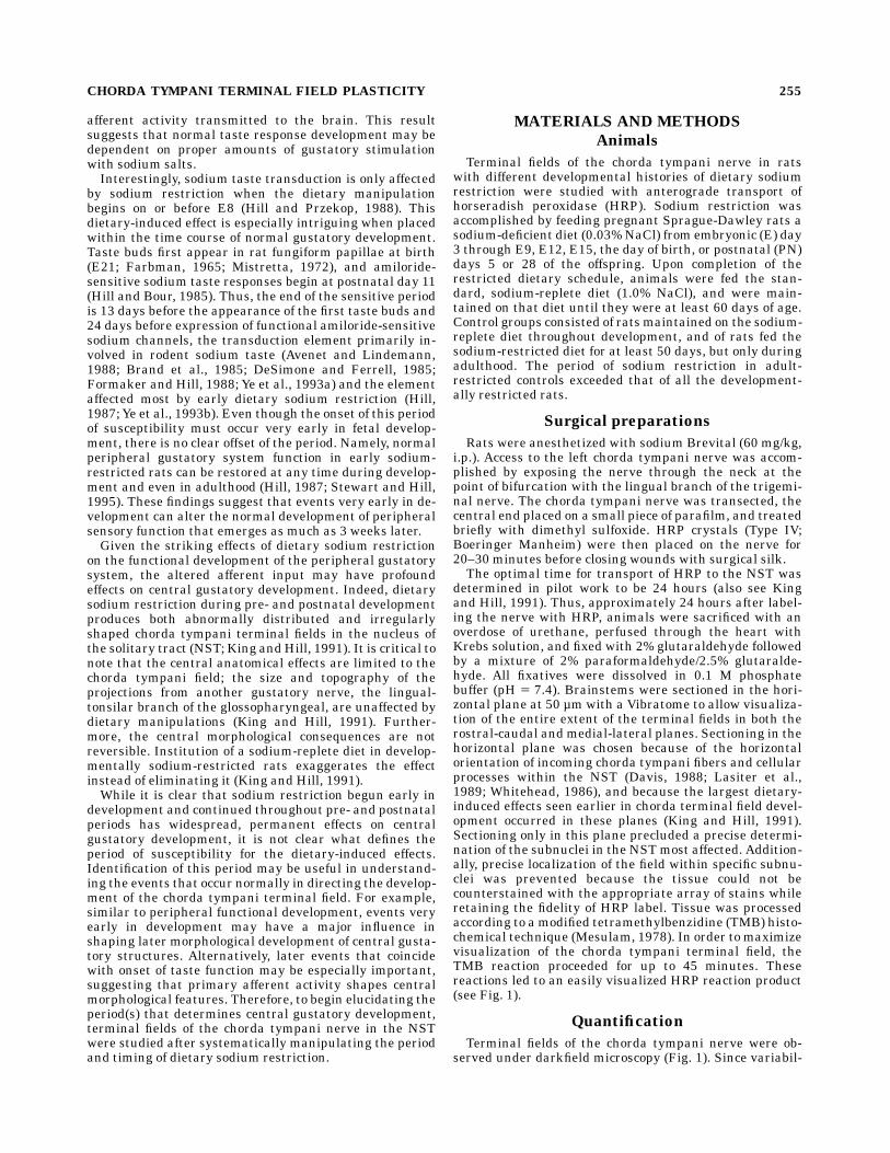
Terminal fields of the chorda tympani nerve were ob-served under darkfield microscopy (Fig. 1). Since variabil-
CHORDA TYMPANI TERMINAL FIELD PLASTICITY 255
Figure 1
256 R.F. KRIMM AND D.L. HILL
ity in the ‘‘intensity’’ of the HRP labeling was apparentwithin and between groups, the volume of the label withinthe NST, rather than the density of the label, was mea-sured. In order to obtain volumes, the perimeter encompass-ing the anterograde label in each 50-µm section wasoutlined and the corresponding area was computed by anOlympus Cue-2 image analysis system. Only areas inwhich fine puncta labeling occurred were measured; thiscorresponds to terminations of afferents and not labeledfibers (e.g., Lasiter, 1992). The areas from all sections foreach animal were summed and multiplied by 50 µm toderive an estimate of the total volume of the chordatympani terminal fields. These techniques are similar tothose used by us and others (Davis and Jang, 1986; Kingand Hill, 1991; Lasiter et al., 1989). To allow for compari-sons with our earlier work (King and Hill, 1991), theterminal field in each rat was divided into three contiguousdorsal-ventral zones, termed here as dorsal, intermediate,and ventral. Each terminal field zone comprised the labelfound in two consecutive 50-µm sections. Typically, thedorsal zone was distinguished by the presence of thesolitary tract at the lateral edge of the nucleus and by thedorsalmost portion of the hypoglossal nucleus. The interme-diate zone, which is the next 100 µm of tissue, was justbelow the level of the solitary tract. The ventral zone of thefield was designated as the label in the next 100 µm oftissue. Sections containing the ventral zone of the terminalfield typically contained cells of the superior salivatorynucleus.Additionally, the rostral to caudal and medial to lateral
distances were measured for the dorsal, intermediate, andventral regions of the terminal field. This was accom-plished by measuring the longest rostral to caudal dis-tance of the terminal field parallel to themedial edge of theNST, and then measuring the longest distance of the fieldperpendicular to the rostral-caudal measurement to ob-tain the largest medial-lateral terminal field distance.
Statistical analysis
The total volume occupied by the chorda tympani termi-nal field in the NST was analyzed for 53 rats (see Resultsfor n/group). Analyses of variance (ANOVA) were used todetermine the effect of the dietary manipulation on thetotal volume, volumes within each of the three dorsal toventral regions, and the rostral-caudal and medial-lateraldistances within each of the three regions. Following asignificant ANOVA, posttests were accomplished with a
one-tailed Dunnett’s test for comparisons involving acontrol mean.
RESULTS
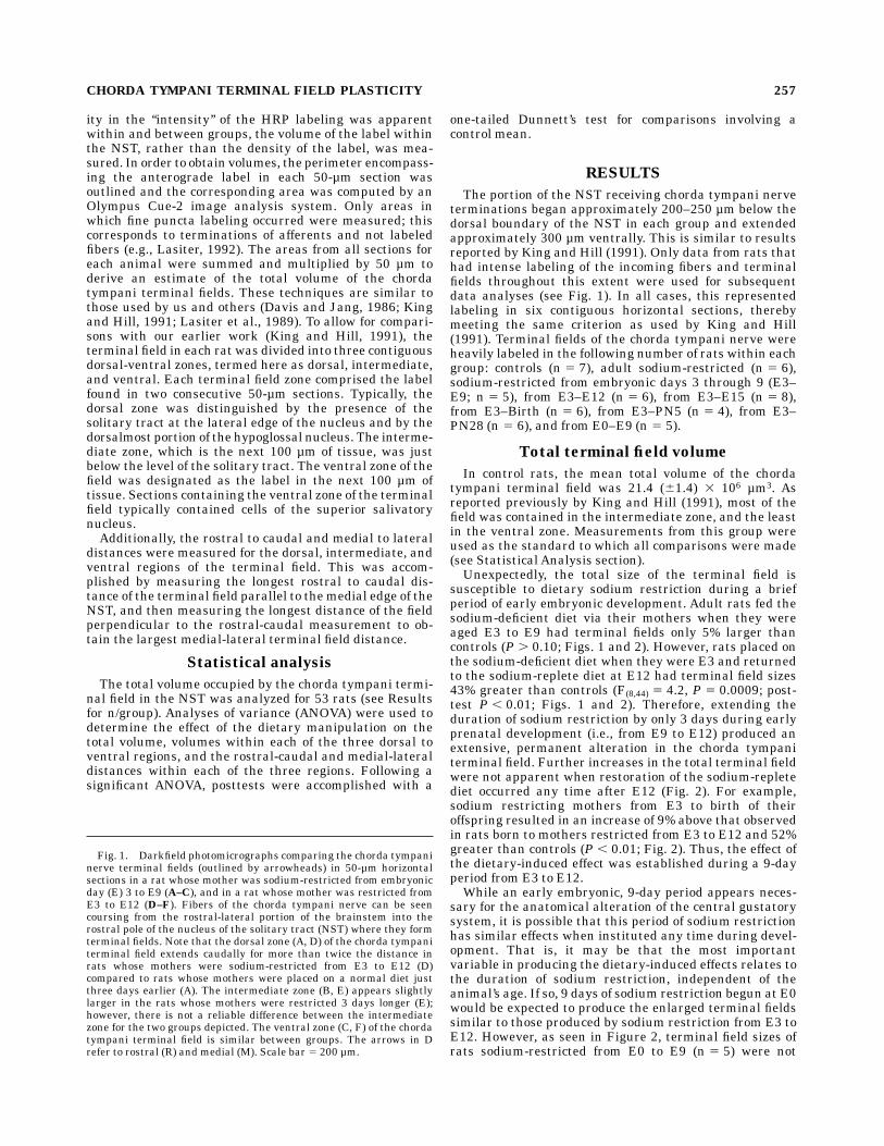
The portion of the NST receiving chorda tympani nerveterminations began approximately 200–250 µm below thedorsal boundary of the NST in each group and extendedapproximately 300 µm ventrally. This is similar to resultsreported by King and Hill (1991). Only data from rats thathad intense labeling of the incoming fibers and terminalfields throughout this extent were used for subsequentdata analyses (see Fig. 1). In all cases, this representedlabeling in six contiguous horizontal sections, therebymeeting the same criterion as used by King and Hill(1991). Terminal fields of the chorda tympani nerve wereheavily labeled in the following number of rats within eachgroup: controls (n 5 7), adult sodium-restricted (n 5 6),sodium-restricted from embryonic days 3 through 9 (E3–E9; n 5 5), from E3–E12 (n 5 6), from E3–E15 (n 5 8),from E3–Birth (n 5 6), from E3–PN5 (n 5 4), from E3–PN28 (n 5 6), and from E0–E9 (n 5 5).
Total terminal field volume
In control rats, the mean total volume of the chordatympani terminal field was 21.4 (61.4) 3 106 µm3. Asreported previously by King and Hill (1991), most of thefield was contained in the intermediate zone, and the leastin the ventral zone. Measurements from this group wereused as the standard to which all comparisons were made(see Statistical Analysis section).Unexpectedly, the total size of the terminal field is
susceptible to dietary sodium restriction during a briefperiod of early embryonic development. Adult rats fed thesodium-deficient diet via their mothers when they wereaged E3 to E9 had terminal fields only 5% larger thancontrols (P . 0.10; Figs. 1 and 2). However, rats placed onthe sodium-deficient diet when they were E3 and returnedto the sodium-replete diet at E12 had terminal field sizes43% greater than controls (F(8,44) 5 4.2, P 5 0.0009; post-test P , 0.01; Figs. 1 and 2). Therefore, extending theduration of sodium restriction by only 3 days during earlyprenatal development (i.e., from E9 to E12) produced anextensive, permanent alteration in the chorda tympaniterminal field. Further increases in the total terminal fieldwere not apparent when restoration of the sodium-repletediet occurred any time after E12 (Fig. 2). For example,sodium restricting mothers from E3 to birth of theiroffspring resulted in an increase of 9% above that observedin rats born to mothers restricted from E3 to E12 and 52%greater than controls (P , 0.01; Fig. 2). Thus, the effect ofthe dietary-induced effect was established during a 9-dayperiod from E3 to E12.While an early embryonic, 9-day period appears neces-
sary for the anatomical alteration of the central gustatorysystem, it is possible that this period of sodium restrictionhas similar effects when instituted any time during devel-opment. That is, it may be that the most importantvariable in producing the dietary-induced effects relates tothe duration of sodium restriction, independent of theanimal’s age. If so, 9 days of sodium restriction begun at E0would be expected to produce the enlarged terminal fieldssimilar to those produced by sodium restriction from E3 toE12. However, as seen in Figure 2, terminal field sizes ofrats sodium-restricted from E0 to E9 (n 5 5) were not
Fig. 1. Darkfield photomicrographs comparing the chorda tympaninerve terminal fields (outlined by arrowheads) in 50-µm horizontalsections in a rat whose mother was sodium-restricted from embryonicday (E) 3 to E9 (A–C), and in a rat whose mother was restricted fromE3 to E12 (D–F). Fibers of the chorda tympani nerve can be seencoursing from the rostral-lateral portion of the brainstem into therostral pole of the nucleus of the solitary tract (NST) where they formterminal fields. Note that the dorsal zone (A, D) of the chorda tympaniterminal field extends caudally for more than twice the distance inrats whose mothers were sodium-restricted from E3 to E12 (D)compared to rats whose mothers were placed on a normal diet justthree days earlier (A). The intermediate zone (B, E) appears slightlylarger in the rats whose mothers were restricted 3 days longer (E);however, there is not a reliable difference between the intermediatezone for the two groups depicted. The ventral zone (C, F) of the chordatympani terminal field is similar between groups. The arrows in Drefer to rostral (R) and medial (M). Scale bar 5 200 µm.
CHORDA TYMPANI TERMINAL FIELD PLASTICITY 257
Figure 2
258 R.F. KRIMM AND D.L. HILL
significantly different from controls (P . 0.10). Likewise,dietary sodium restriction begun during adulthood hadlittle effect on terminal fields in the NST. Rats placed on asodium-restricted diet during adulthood and maintainedon the diet for a minimum of 50 days had terminal fieldsizes similar to controls (P . 0.10). Thus, from theseresults, it is clear that the period during gestation coinci-dent with sodium restriction is a more important variablethan the duration of dietary restriction.
Terminal field volume within dorsalto ventral zones
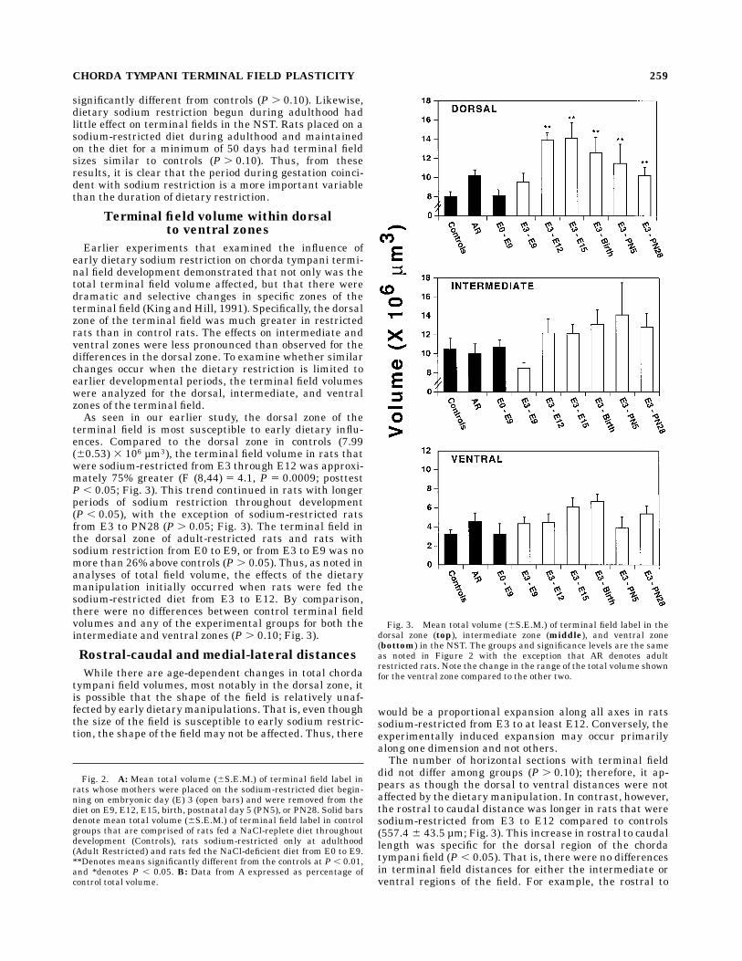
Earlier experiments that examined the influence ofearly dietary sodium restriction on chorda tympani termi-nal field development demonstrated that not only was thetotal terminal field volume affected, but that there weredramatic and selective changes in specific zones of theterminal field (King andHill, 1991). Specifically, the dorsalzone of the terminal field was much greater in restrictedrats than in control rats. The effects on intermediate andventral zones were less pronounced than observed for thedifferences in the dorsal zone. To examine whether similarchanges occur when the dietary restriction is limited toearlier developmental periods, the terminal field volumeswere analyzed for the dorsal, intermediate, and ventralzones of the terminal field.As seen in our earlier study, the dorsal zone of the
terminal field is most susceptible to early dietary influ-ences. Compared to the dorsal zone in controls (7.99(60.53) 3 106 µm3), the terminal field volume in rats thatwere sodium-restricted from E3 through E12 was approxi-mately 75% greater (F (8,44) 5 4.1, P 5 0.0009; posttestP , 0.05; Fig. 3). This trend continued in rats with longerperiods of sodium restriction throughout development(P , 0.05), with the exception of sodium-restricted ratsfrom E3 to PN28 (P . 0.05; Fig. 3). The terminal field inthe dorsal zone of adult-restricted rats and rats withsodium restriction from E0 to E9, or from E3 to E9 was nomore than 26% above controls (P . 0.05). Thus, as noted inanalyses of total field volume, the effects of the dietarymanipulation initially occurred when rats were fed thesodium-restricted diet from E3 to E12. By comparison,there were no differences between control terminal fieldvolumes and any of the experimental groups for both theintermediate and ventral zones (P . 0.10; Fig. 3).
Rostral-caudal and medial-lateral distances
While there are age-dependent changes in total chordatympani field volumes, most notably in the dorsal zone, itis possible that the shape of the field is relatively unaf-fected by early dietarymanipulations. That is, even thoughthe size of the field is susceptible to early sodium restric-tion, the shape of the field may not be affected. Thus, there
would be a proportional expansion along all axes in ratssodium-restricted from E3 to at least E12. Conversely, theexperimentally induced expansion may occur primarilyalong one dimension and not others.The number of horizontal sections with terminal field
did not differ among groups (P . 0.10); therefore, it ap-pears as though the dorsal to ventral distances were notaffected by the dietary manipulation. In contrast, however,the rostral to caudal distance was longer in rats that weresodium-restricted from E3 to E12 compared to controls(557.4 6 43.5 µm; Fig. 3). This increase in rostral to caudallength was specific for the dorsal region of the chordatympani field (P , 0.05). That is, there were no differencesin terminal field distances for either the intermediate orventral regions of the field. For example, the rostral to
Fig. 2. A: Mean total volume (6S.E.M.) of terminal field label inrats whose mothers were placed on the sodium-restricted diet begin-ning on embryonic day (E) 3 (open bars) and were removed from thediet on E9, E12, E15, birth, postnatal day 5 (PN5), or PN28. Solid barsdenote mean total volume (6S.E.M.) of terminal field label in controlgroups that are comprised of rats fed a NaCl-replete diet throughoutdevelopment (Controls), rats sodium-restricted only at adulthood(Adult Restricted) and rats fed the NaCl-deficient diet from E0 to E9.**Denotes means significantly different from the controls at P , 0.01,and *denotes P , 0.05. B: Data from A expressed as percentage ofcontrol total volume.
Fig. 3. Mean total volume (6S.E.M.) of terminal field label in thedorsal zone (top), intermediate zone (middle), and ventral zone(bottom) in the NST. The groups and significance levels are the sameas noted in Figure 2 with the exception that AR denotes adultrestricted rats. Note the change in the range of the total volume shownfor the ventral zone compared to the other two.
CHORDA TYMPANI TERMINAL FIELD PLASTICITY 259
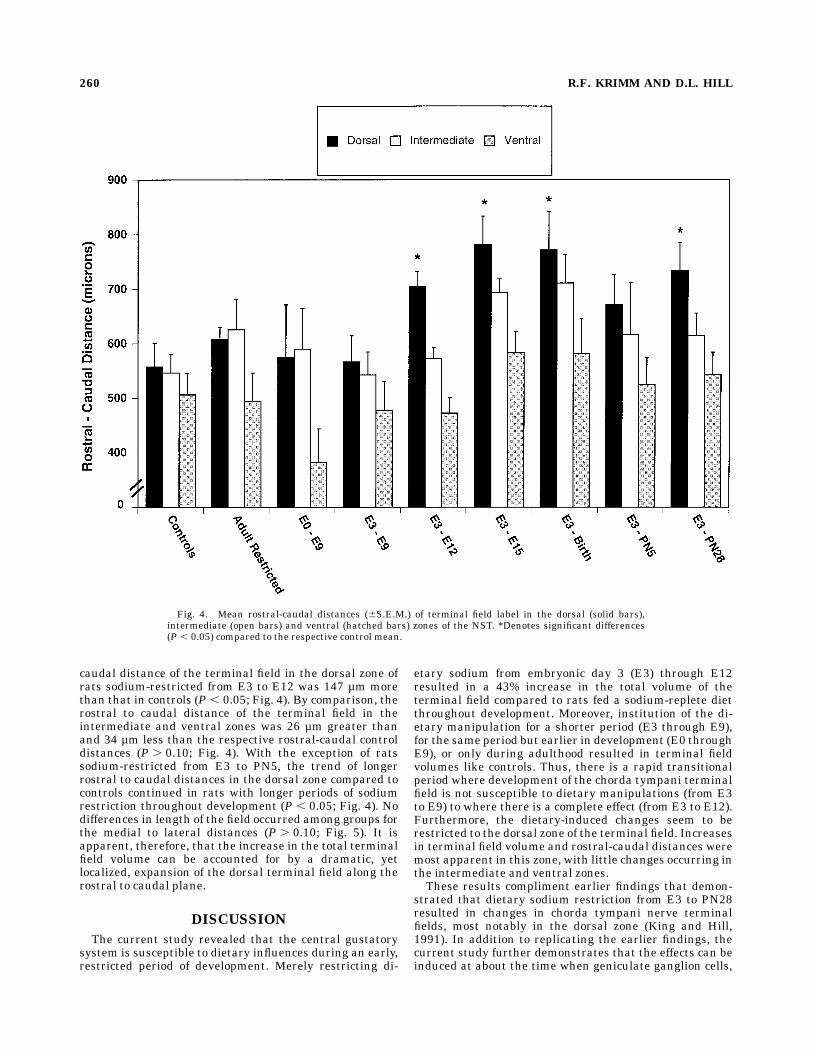
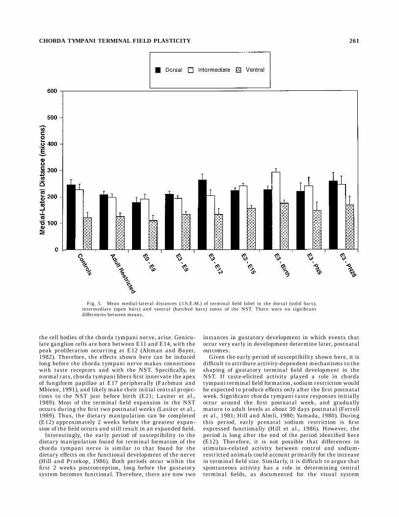
caudal distance of the terminal field in the dorsal zone ofrats sodium-restricted from E3 to E12 was 147 µm morethan that in controls (P , 0.05; Fig. 4). By comparison, therostral to caudal distance of the terminal field in theintermediate and ventral zones was 26 µm greater thanand 34 µm less than the respective rostral-caudal controldistances (P . 0.10; Fig. 4). With the exception of ratssodium-restricted from E3 to PN5, the trend of longerrostral to caudal distances in the dorsal zone compared tocontrols continued in rats with longer periods of sodiumrestriction throughout development (P , 0.05; Fig. 4). Nodifferences in length of the field occurred among groups forthe medial to lateral distances (P . 0.10; Fig. 5). It isapparent, therefore, that the increase in the total terminalfield volume can be accounted for by a dramatic, yetlocalized, expansion of the dorsal terminal field along therostral to caudal plane.
DISCUSSION
The current study revealed that the central gustatorysystem is susceptible to dietary influences during an early,restricted period of development. Merely restricting di-
etary sodium from embryonic day 3 (E3) through E12resulted in a 43% increase in the total volume of theterminal field compared to rats fed a sodium-replete dietthroughout development. Moreover, institution of the di-etary manipulation for a shorter period (E3 through E9),for the same period but earlier in development (E0 throughE9), or only during adulthood resulted in terminal fieldvolumes like controls. Thus, there is a rapid transitionalperiod where development of the chorda tympani terminalfield is not susceptible to dietary manipulations (from E3to E9) to where there is a complete effect (from E3 to E12).Furthermore, the dietary-induced changes seem to berestricted to the dorsal zone of the terminal field. Increasesin terminal field volume and rostral-caudal distances weremost apparent in this zone, with little changes occurring inthe intermediate and ventral zones.These results compliment earlier findings that demon-
strated that dietary sodium restriction from E3 to PN28resulted in changes in chorda tympani nerve terminalfields, most notably in the dorsal zone (King and Hill,1991). In addition to replicating the earlier findings, thecurrent study further demonstrates that the effects can beinduced at about the time when geniculate ganglion cells,
Fig. 4. Mean rostral-caudal distances (6S.E.M.) of terminal field label in the dorsal (solid bars),intermediate (open bars) and ventral (hatched bars) zones of the NST. *Denotes significant differences(P , 0.05) compared to the respective control mean.
260 R.F. KRIMM AND D.L. HILL
the cell bodies of the chorda tympani nerve, arise. Genicu-late ganglion cells are born between E11 and E14, with thepeak proliferation occurring at E12 (Altman and Bayer,1982). Therefore, the effects shown here can be inducedlong before the chorda tympani nerve makes connectionswith taste receptors and with the NST. Specifically, innormal rats, chorda tympani fibers first innervate the apexof fungiform papillae at E17 peripherally (Farbman andMbiene, 1991), and likely make their initial central projec-tions to the NST just before birth (E21; Lasiter et al.,1989). Most of the terminal field expansion in the NSToccurs during the first two postnatal weeks (Lasiter et al.,1989). Thus, the dietary manipulation can be completed(E12) approximately 2 weeks before the greatest expan-sion of the field occurs and still result in an expanded field.Interestingly, the early period of susceptibility to the
dietary manipulation found for terminal formation of thechorda tympani nerve is similar to that found for thedietary effects on the functional development of the nerve(Hill and Przekop, 1986). Both periods occur within thefirst 2 weeks postconception, long before the gustatorysystem becomes functional. Therefore, there are now two
instances in gustatory development in which events thatoccur very early in development determine later, postnataloutcomes.Given the early period of susceptibility shown here, it is
difficult to attribute activity-dependent mechanisms to theshaping of gustatory terminal field development in theNST. If taste-elicited activity played a role in chordatympani terminal field formation, sodium restrictionwouldbe expected to produce effects only after the first postnatalweek. Significant chorda tympani taste responses initiallyoccur around the first postnatal week, and graduallymature to adult levels at about 30 days postnatal (Ferrellet al., 1981; Hill and Almli, 1980; Yamada, 1980). Duringthis period, early prenatal sodium restriction is firstexpressed functionally (Hill et al., 1986). However, theperiod is long after the end of the period identified here(E12). Therefore, it is not possible that differences instimulus-related activity between control and sodium-restricted animals could account primarily for the increasein terminal field size. Similarly, it is difficult to argue thatspontaneous activity has a role in determining centralterminal fields, as documented for the visual system
Fig. 5. Mean medial-lateral distances (6S.E.M.) of terminal field label in the dorsal (solid bars),intermediate (open bars) and ventral (hatched bars) zones of the NST. There were no significantdifferences between means.
CHORDA TYMPANI TERMINAL FIELD PLASTICITY 261
(Sretavan et al., 1988), because geniculate ganglion cellsdo not arise until the end of the period of susceptibility.Thus, functional peripheral and central connections of thechorda tympani nerve would not be present until after thedietary manipulation was terminated. While it is clearthat activity plays a major role in developing sensorysystems (e.g., Antonini and Stryker, 1993; Casagrande andCondo, 1988; Guthrie et al., 1990; Shatz, 1996; Shatz andStryker, 1988), there are instances in other sensory sys-tems (e.g., somatosensory; Chiaia et al., 1992; Hendersonet al., 1992) in addition to the gustatory system wherenonactivity dependent factors are involved.The apparent lack of neuronal activity in determining
terminal field size here is in contrast to that advanced byLasiter and colleagues (Lasiter, 1995; Lasiter and Diaz,1992; Lasiter and Kachele, 1990). Taste receptor destruc-tion in the anterior tongue of postnatal rats aged 2 daysresults in a decreased chorda tympani field in the NST,whereas similar destruction after this age fails to influencethe size of the field (Lasiter and Kachele, 1990). Since tasteresponses initially occur at about this age (Hill and Almli,1980), this result suggests that the onset of function isimportant in determining terminal field characteristics.Similarly, lack of lingual stimulation with salts in intactrats during early postnatal development results in de-creased field size (Lasiter, 1995). Such observations are inconcordance with those of other sensory systems in whichactivity plays a major role in forming central structures(e.g., Antonini and Stryker, 1993; Casagrande and Condo,1988; Guthrie et al., 1990; Shatz, 1996; Shatz and Stryker,1988). The results from Lasiter (1995) are different fromthose reported here. With receptor destruction and limitedpostnatal gustatory experience, terminal field size is de-creased. With early dietary sodium restriction, the termi-nal field size is increased.It is likely that the different outcomes reflect different
mechanisms. Neural activity may be crucial in shaping thechorda tympani terminal field once the proper numbers ofgeniculate ganglion neurons are present and once theinitial central contacts are made. In fact, early receptorcell destruction does not alter geniculate ganglion cellnumbers (Lasiter andKachele, 1990). However, determina-tion of proper ganglion cell numbers and initial centralcontacts may be under the control of much earlier events.Specifically, early dietary manipulations employed heremay have consequences on the survival of geniculateganglion cells, perhaps by way of altered neurotrophiclevels. Indeed, nutritional manipulations during earlydevelopmental periods can result in altered amounts ofneurotrophic factors (Diamond et al., 1991). It is possiblethat such an alteration in the gustatory system due toearly sodium restriction may result in increased numbersof geniculate ganglion cells surviving throughout earlydevelopment. That is, there may not be the normal amountof ganglion cell death which is characteristic of developingperipheral sensory neurons (e.g., Lam et al., 1982). Accord-ingly, the increased numbers of central processes fromincreased numbers of geniculate ganglion cells may spreadbeyond the normal terminal field borders, resulting in anenlarged terminal field. Such an effect of increased sensoryganglion cell survival and the resultant centralmorphologi-cal alteration has been shown in the trigeminal system.Increased numbers of trigeminal ganglion cells resulting
from increased levels of exogenous NGF during prenataldevelopment disrupted whisker-related patterns in thetrigeminal brainstem complex postnatally (Henderson etal., 1994). Therefore, similar to what may have occurred inthe present study, increased survival of projecting neuronsinto the brainstem resulted in expanded terminal fields.Experiments that examine geniculate ganglion cell num-bers and pattern of terminal field development in normaland in dietary-restricted rats will be important in reconcil-ing these experimental differences.Although activity may contribute to chorda tympani
field development, there is converging evidence that sys-temic factorsmay also contribute significantly to gustatorydevelopment, namely: (1) sodium taste response develop-ment in taste receptor cells is coincident with the expres-sion of many hormonal or growth factors; (2) dietarysodium restriction has to be implemented long before theappearance of functional responses but during times whenmany hormonal and growth factor systems are forming; (3)recovery of sodium sensitivity requires postingestionalabsorption of sodium and not direct taste stimulation withthe ion; and (4) results from studies of sodium channelimmunoreactivity indicate a systemic-factor dependence(Bondy, 1991; Millan et al., 1989; Nielsen et al., 1991; Ponset al., 1991; Stewart and Hill, 1995; Tribollet et al., 1991).It is reasonable to hypothesize that circulating or localfactors may also be important in defining the structuraldevelopment of the chorda tympani nerve. Indeed, thedevelopmental timing of the period identified here isconsistent with such a hypothesis.Regardless of the actual mechanisms (i.e., neuronal
activity or systemic factors), there are several potentialanatomical bases that account for the increased size of theterminal field. These would include arbor expansion ofindividual chorda tympani neurons into regions not nor-mally occupied and/or displacement of normal-shapedarbors of neurons outside the typical terminal field bound-ary. Such an expansion or displacement would occur alongthe rostral to caudal axis, which is the same axis by whichnormal development of the field proceeds. Accordingly, theaberrant projection of the chorda tympani nerve wouldlikely invade territory normally occupied only by theglossopharyngeal nerve (Hamilton and Norgren, 1984).Therefore, processes that determine the caudal bound-aries of the field may be altered in sodium-restricted rats(e.g., extracellular matrix molecules). As such, the func-tional and structural implications of chorda tympani neu-rons invading the ‘‘new’’ territory are numerous. Forexample, chorda tympani fibers may instruct the aberrantdendritic expansion of postsynaptic cells during develop-ment that occurs in developmentally sodium-restrictedrats (King andHill, 1993). In this example, themorphologi-cal changes, perhaps directed by the abnormal chordatympani nerve development, would result in functionalchanges such as differential convergence of taste informa-tion from the anterior and posterior tongue, and theformation of new gustatory coding strategies. Indeed,persistent functional changes are apparent in NST neu-rons of developmentally sodium restricted rats (Vogt andHill, 1993).In summary, these results show that the first synaptic
relay of the gustatory system is susceptible to dietarymanipulations during a remarkably brief period of very
262 R.F. KRIMM AND D.L. HILL
early embryonic development. The effects are profoundand permanent. Correspondingly, these findings have ma-jor implications concerning the role of maternal diet oncentral neural development and behavior.
ACKNOWLEDGMENTS
This work was supported by NIH grants DC00407 andHD07323. We extend our appreciation to Drs. A. KurtThaw, Benjamin Walker, Lynnette Phillips, and Ms.Dianna Cummings for their reviews of an earlier version ofthe manuscript.
LITERATURE CITED
Altman, J., and S. Bayer (1982) Development of the cranial nerve gangliaand related nuclei in the rat. Adv. Anat. Embryol. Cell Biol. 74:1–90.
Antonini, A., and M.P. Stryker (1993) Rapid remodeling of axonal arbors inthe visual cortex. Science 260:1819–1820.
Aslin, R.N. (1981) Experimental influences and sensitive periods in per-ceptual development: A unified model. In R.N. Aslin, J.R. Alberts,M.R. Peterson (eds): The Development of Perception: PsychobiologicalPerspectives, The Visual System. NewYork:Academic Press, pp. 45–93.
Avenet, P., and B. Lindemann (1988) Amiloride-blockable sodium currentsin isolated taste receptor cells. J. Memb. Biol. 105:245–255.
Bondy, C.A. (1991) Transient IGF-1 gene expression during the maturationof functionally related central projection neurons. J. Neurosci. 11:3442–3455.
Brand, J.B., J.H. Teeter, and W.L. Silver (1985) Inhibition by amiloride ofchorda tympani responses evoked by monovalent salts. Brain Res.334:207–214.
Brunjes, P.C., and L.L. Frazier (1986) Maturation and plasticity in theolfactory system of vertebrates. Brain Res. Rev. 11:1–45.
Casagrande, V.A., and G.J. Condo (1988) The effect of altered neuronalactivity on the development of layers in the lateral geniculate nucleus.J. Neurosci. 8:395–416.
Chiaia, N.L., S.E. Fish, W.R. Bauer, C.A. Bennett-Clarke, andR.W. Rhoades (1992) Postnatal blockade of cortical activity by tetrodo-toxin does not disrupt the formation of vibrissa-related patterns in therat’s somatosensory cortex. Dev. Brain Res. 66:244–250.
Davis, B.J. (1988) Computer generated rotational analyses reveal a keythree-dimensional feature of the nucleus of the solitary tract. BrainRes. Bull. 20:545–548.
Davis, B.J., and T. Jang (1986) The gustatory zone of the nucleus of thesolitary tract in the hamster: Light microscopic morphometric studies.Chem. Senses 11:213–228.
Deitch, J.S., and E.W. Rubel (1984) Afferent influences on brain stemauditory nuclei of the chicken: Time course and specificity of dendriticatrophy following deafferentation. J. Comp. Neurol. 229:66–79.
DeSimone, J.A., and F. Ferrell (1985) Analysis of amiloride inhibition ofchorda tympani taste response of rat to NaCl. Am. J. Physiol. 249:R52–R61.
Diamond, J., M. Cameron, and B. Cassells (1991) Functional impairmentsthat develop in NGF-sensitive neuronal projection in the PNS and CNSof aged rats are ameliorated by dietary interventions. In Y. Christen(ed): Growth Factors in Alzheimer’s Disease. New York: Springer-Verlag, pp. 131–148.
Farbman, A.I. (1965) Electron microscope study of the developing taste budin the rat fungiform papilla. Dev. Biol. 11:110–135.
Farbman, A.I., and J.-P. Mbiene (1991) Early development and innervationof taste bud-bearing papillae on the rat tongue. J. Comp. Neurol.304:172–186.
Ferrell, M.F., C.M. Mistretta, and R.M. Bradley (1981) Development ofchorda tympani taste responses in rat. J. Comp. Neurol. 198:37–44.
Formaker, B.K., and D.L. Hill (1988) An analysis of residual NaCl tasteresponse after amiloride. Am. J. Physiol. 255:R1002–R1007.
Guthrie, K.M., D.A. Wilson, and M. Leon (1990) Early unilateral depriva-tion modifies olfactory bulb function. J. Neurosci. 10:3402–3412.
Hamilton, R.B., and R. Norgren (1994) Central projections of gustatorynerves in the rat. J. Comp. Neurol. 222:560–577.
Henderson, T.A., T.A. Woolsey, and M.F. Jacquin (1992) Infraorbital nerveblockade from birth does not disrupt central trigeminal pattern forma-tion in the rat. Dev. Brain Res. 66:146–152.
Henderson, T.A., E.M. Johnson, P.A. Osborne, and M.F. Jacquin (1994)Fetal NGF augmentation preserves excess trigeminal ganglion cellsand interrupts whisker-related pattern formation. J. Neurosci. 14:3389–3403.
Hill, D.L. (1987) Susceptibility of the developing rat gustatory system to thephysiological effects of dietary sodium deprivation. J. Physiol. Lond.393:423–434.
Hill, D.L., and C.R. Almli (1980) Ontogeny of chorda tympani nerveresponses to gustatory stimuli in the rat. Brain Res. 20:310–313.
Hill, D.L., and T.C. Bour (1985) Addition of functional amiloride-sensitivecomponents to the receptor membrane: A possible mechanism foraltered taste responses during development. Dev. Brain Res. 20:310–313.
Hill, D.L., and P.R. Przekop (1988) Influences of dietary sodium onfunctional taste receptor development: A sensitive period. Science241:1826–1828.
Hill, D.L., C.M. Mistretta, and R.M. Bradley (1986) Effects of dietary NaCldeprivation during early development on behavioral and neurophysi-ological taste responses. Behav. Neurosci. 100:390–398.
Hubel, D.H., and T.N. Weisel (1970) The period of susceptibility to thephysiological effects of unilateral eye closure in kittens. J. Physiol.Lond. 206:419–436.
King, C.T., and D.L. Hill (1991) Dietary sodium chloride deprivationthroughout development selectively influences the terminal field organi-zation of gustatory afferent fibers projecting to the rat nucleus of thesolitary tract. J. Comp. Neurol. 303:159–169.
King, C.T., and D.L. Hill (1993) Neuroanatomical alterations in the ratnucleus of the solitary tract following early maternal NaCl deprivationand subsequent NaCl repletion. J. Comp. Neurol. 333:531–542.
Lam, K., A.J. Sefton, and M.R. Bennett (1982) Loss of axons from the opticnerve of the rat during early postnatal development. Dev. Brain Res.3:487–491.
Lasiter, P.S. (1995) Effects of orochemical stimulation on postnatal develop-ment of gustatory recipient zones within the nucleus of the solitarytract. Brain Res. Bull. 38:1–9.
Lasiter, P.S., and J. Diaz (1992) Artificial rearing alters development of thenucleus of the solitary tract. Brain Res. Bull. 29:407–410.
Lasiter, P.S., and D.L. Kachele (1990) Effects of early postnatal receptordamage on development of gustatory recipient zones within the nucleusof the solitary tract. Dev. Brain Res. 55:57–71.
Lasiter, P.S., D.M.Wong, and D.L. Kachele (1989) Postnatal development ofthe rostral solitary nucleus in rat: Dendritic morphology and mitochon-drial enzyme activity. Brain Res. Bull. 22:313–321.
Mesulam, M.-M. (1978) Tetramethyl benzidine for horseradish peroxidaseneurohistochemistry: A non-carcinogenic blue reaction product withsuperior sensitivity for visualizing neural afferents and efferents. J.Histochem. Cytochem. 26:166–177.
Millan, M.A., P. Carvallo, S.-I. Izumi, S. Zemel, K. Catt, and G. Aguilera(1989) Novel sites of expression of functional angiotensin II receptors inthe late gestation fetus. Science 244:1340–1342.
Mistretta, C.M. (1972) Topographical and histological study of the develop-ing rat tongue, palate and taste buds. In: J.F. Bosma (ed): ThirdSymposium onOral Sensation and Perception. Springfield: Thomas, pp.163–187.
Mistretta, C.M., and R.M. Bradley (1978) Effects of early sensory experi-ence on brain and behavioral development. In G. Gottlieb (ed): Studieson the Development of Behavior and the Nervous System, Vol. 4. NewYork: Academic Press, pp. 215–247.
Nielsen, F.C., E. Wang, and S. Gammeltoft (1991) Receptor binding,endocytosis, and mitogenesis of insulin-like growth factors I and II infetal rat brain neurons. J. Neurochem. 56:12–21.
Pons, S., M.T. Rejas, and I. Torres-Aleman (1991) Ontogeny of insulin-likegrowth factor I, its receptor, and its binding proteins in the rathypothalamus. Dev. Brain. Res. 62:169–175.
Renehan, W.E., R.W. Rhoades, and M.F. Jacquin (1989) Structure-functionrelationships in rat brainstem subnucleus interpolaris: VII: Primaryafferent central terminal arbors in adults subjected to infraorbitalnerve section at birth. J. Comp. Neurol. 289:493–508.
Shatz, C.J. (1996) Emergence of order in visual system development. Proc.Natl. Acad. Sci. USA 93:602–608.
Shatz, C.J., and M.P. Stryker (1988) Prenatal tetrodotoxin infusion blockssegregation of retinogeniculate afferents. Science 242:87–89.
CHORDA TYMPANI TERMINAL FIELD PLASTICITY 263

Sretavan, D.W., C.J. Shatz, and M.P. Stryker (1988) Modification of retinalganglion cell axon morphology by prenatal infusion of tetrodotoxin.Nature 336:468–471.
Stewart, R.E., and D.L. Hill (1995) Time course of saline-induced recoveryof the gustatory system in sodium-restricted rats. Am. J. Physiol.270:R704–R712.
Tribollet, E., M. Goumaz, M. Raggenbass, M. Dubois-Dauphin, and J.J.Dreifuss (1991) Early appearance and transient expression of vasopres-sin receptors in the brain of rat fetus and infant. An autoradiographicaland electrophysiological study. Dev. Brain Res. 58:13–24.
Vogt, M.B., and D.L. Hill (1993) Enduring alterations in neurophysiologicaltaste responses after early dietary sodium deprivation. J. Neurophysiol.69:832–841.
Whitehead, M.C. (1986) Anatomy of the gustatory system in the hamster:Synaptology of facial afferent terminals in the solitary nucleus. J.Comp. Neurol. 244:72–85.
Yamada, T. (1980) Chorda tympani responses to gustatory stimuli indeveloping rats. Japan. J. Physiol. 30:631–643.
Ye, Q., G.L. Heck, and J.A. DeSimone (1993a) Voltage dependence of the ratchorda tympani response to Na1 salts: Implications for the functionalorganization of taste receptor cells. J. Neurophysiol. 70:167–178.
Ye, Q., R.E. Stewart, G.L. Heck, D.L. Hill, and J.A. DeSimone (1993b)Na1-restricted rats lack functional Na1 channels in taste cell apicalmembranes: Proof by membrane voltage perturbation. J. Neurophysiol.70:1713–1716.
264 R.F. KRIMM AND D.L. HILL