Embed Size (px)
Citation preview
www.jcomjournal.com Vol. 22, No. 6 June 2015 JCOM 257
AbstrAct• Objective:To provide an overview of the importance
and challenges of accurate diagnosis of early idio-pathicParkinson’sdiseaseandpracticalguidelinesforclinicians.
• Methods:Reviewoftherelevantliterature.• Results:IdiopathicParkinson’sdiseaseisacommon
neurodegenerativedisordercausingawidespectrumofmotorandnonmotorsymptoms.Thecardinalmotorfeaturesincluderestingtremors,bradykinesia,rigidity,andpostural instability.Thediagnosisisclinical,andancillary laboratory or radiology tests are unneces-saryintypicalcases.Despitetheuseofvalidateddi-agnosticcriteria,misdiagnosisiscommon,especiallyearlyinthediseaseprocess.ThisislargelyduetothephenotypicheterogeneityintheidiopathicParkinson’sdisease population as well phenotypic overlappingwith other diseases. The diseases most commonlyconfused with idiopathic Parkinson’s disease aretheParkinson-plussyndromes (dementiawithLewybodies, multiple system atrophy, progressive supra-nuclearpalsy,andcorticobasaldegeneration),vascu-lar parkinsonism, drug-induced parkinsonism, doparesponsivedystonia,normalpressurehydrocephalus,and essential tremor. Since the diagnosis of theseother diseases is also clinical, familiarity with theirtypicalpresentationsandmostcurrentdiagnosticcri-teriaishelpful.BrainMRIcanbehelpfulindiagnosingsomeofthediseases,thoughbrainimagingismostcommonlyunremarkableinidiopathicParkinson’sdis-ease.DaTscanhasanFDAindicationtoassistintheevaluationofadultswithparkinsoniansyndromes. Itshouldnotbeusedintypicalcasesbutcanbeause-fuladjuncttootherdiagnosticevaluationsinatypicalcases.
• Conclusion: Despite the challenges involved, ac-curateandearly diagnosisof idiopathicParkinson’s
disease is essential for optimal patient education,counseling,andtreatment.
Idiopathic Parkinson’s disease (IPD) is a common neurodenerative disease, affecting 1% of the popula-tion over the age of 65 [1]. A definitive diagnosis
requires the postmortem findings of degeneration of the substantia nigra pars compacta and the presence of Lewy bodies (insoluble cytoplasmic inclusions composed of aggregated alpha-synuclein). In the later stages of the disease, a correct clinical diagnosis is made in more than 90% of patients [2]. Early on, however, clinical diagnosis is less reliable. For clinicians, distinguishing early IPD from other parkinsonian syndromes can be extraordi-narily challenging because these conditions, especially in the earliest stages, present with highly variable yet overlapping phenotypes [3]. Furthermore, most of the diseases in the differential diagnosis, including IPD itself, are clinical diagnoses made on the basis of history and ex-amination without the benefit of laboratory or radiology data. A high level of clinical acumen is therefore required for early and accurate diagnosis. Recent clinical trials in which subspecialists performed stringent diagnostic as-sessments to identify subjects with clinically diagnosed IPD later found that some subjects had normal function-al dopamine imaging, suggesting that they probably did not have IPD [4,5]. These trials served to highlight the possibility of misdiagnosis, even in the hands of highly trained subspecialists. Early and accurate diagnosis is of paramount importance for many reasons. First, treat-ment approaches differ significantly across many of these diseases. Second, as neuroprotective interventions that
Early Parkinsonism: Distinguishing Idiopathic Parkinson’s Disease from Other SyndromesJessica B. Lehosit, DO, and Leslie J. Cloud, MD, MSc
CliniCal Review
From the VA Medical Center (Dr. Lehosit) and the Parkinson’s and Movement Disorders Center, Virginia Commonwealth University (Dr. Cloud), Richmond, VA.
258 JCOM June 2015 Vol. 22, No. 6 www.jcomjournal.com
Early Parkinsonism
are currently under investigation become available, long-term outcomes may significantly improve with earlier
diagnosis and intervention. Third, some of these diseases are prognostically very different from one another, so accurate diagnosis enables better counseling and setting realistic expectations for progression.
This review will discuss the most common presenting signs and symptoms of early IPD, present the most wide-ly used diagnostic criteria, and introduce the ancillary laboratory and imaging tests that may be helpful in dis-tinguishing it from its mimics. The diseases most com-monly confused with early IPD will also be discussed with an emphasis on the ways they most commonly differ from IPD. We will begin our discussion with the present-ing signs and symptoms of IPD.
Idiopathic Parkinson’s Disease
IPD typically has a subtle and insidious onset with char-acteristic features developing over months to years. IPD most often presents in patients after age 60, and age is the most consistent risk factor for developing IPD; however, approximately 5% of IPD cases begin before age 40 years. These young-onset cases are likely to be caused by genetic mutations [6]. The widely recognized cardinal motor fea-tures of IPD include asymmetric resting tremor, rigidity, bradykinesia and postural instability [7]. Asymmetry is a key feature, as symptoms typically start on one side and remain more prominent on that side as the disease pro-gresses. In fact, lack of asymmetry suggests an alternative diagnosis. Of the cardinal motor features, tremor is most often reported by patients as the first symptom [8]. How-ever, IPD can alternately present with various other motor or even nonmotor complaints that will be discussed later.
As stated previously, only the clinician can make the diagnosis. Ancillary tests are more often utilized to ex-clude other disease etiologies rather than to make the ac-tual diagnosis of IPD itself. Brain imaging with MRI or CT is generally unremarkable but can sometimes be use-ful in ruling out other conditions in atypical cases. While genetic tests for the known causative genetic mutations are commercially available, they are never required for diagnosis and do not significantly alter the management approach. They are, therefore, most commonly reserved for the purposes of genetic counseling in individuals with a strong family history of PD. The UK Parkinson’s Disease Society Brain Bank clinical criteria are the most widely used diagnostic criteria for IPD and are featured in Table 1. Despite the use of clinical criteria, the diag-nostic certainty is still only between 75% and 90% when compared to autopsy results [9,10].
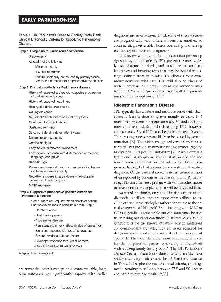
Table 1. UKParkinson’sDiseaseSocietyBrainBankClinicalDiagnosticCriteriaforIdiopathicParkinson’sDisease
Step 1. Diagnosis of Parkinsonian syndrome
Bradykinesia
Atleast1ofthefollowing:
-Muscularrigidity
-4-6Hzresttremor
-Posturalinstabilitynotcausedbyprimaryvisual,vestibular,cerebellarorproprioceptivedysfunction
Step 2. Exclusion criteria for Parkinson’s disease
Historyofrepeatedstrokeswithstepwiseprogressionofparkinsonianfeatures
Historyofrepeatedheadinjury
Historyofdefiniteencephalitis
Oculogyriccrises
Neuroleptictreatmentatonsetofsymptoms
Morethan1affectedrelative
Sustainedremission
Strictlyunilateralfeaturesafter3years
Supranucleargazepalsy
Cerebellarsigns
Earlysevereautonomicinvolvement
Earlyseveredementiawithdisturbancesofmemory,language,andpraxis
Babinskisign
Presenceofcerebraltumororcommunicationhydro-cephalusonimagingstudy
Negativeresponsetolargedosesoflevodopainabsenceofmalabsorption
MPTPexposure
Step 3. Supportive prospective positive criteria for Parkinson’s disease
ThreeormorearerequiredfordiagnosisofdefiniteParkinson’sdiseaseincombinationwithStep1
-Unilateralonset
-Resttremorpresent
-Progressivedisorder
-Persistentasymmetryaffectingsideofonsetmost
-Excellentresponse(70-100%)tolevodopa
-Severelevodopa-inducedchorea
-Levodoparesponsefor5yearsormore
-Clinicalcourseof10yearsormore
Adaptedfromreference9.
www.jcomjournal.com Vol. 22, No. 6 June 2015 JCOM 259
Motor FeaturesResting tremor is the most common presenting sign/symptom of early IPD, found in approximately 70% of patients [8]. The tremor typically is asymmetric and in-termittent at onset, often starting in one hand. It is some-times, though not necessarily, described as a “pill-rolling” rhythmic movement of the thumb and first finger while the hand is at rest. Patients will usually report a worsening of tremor with stress, anxiety, and increased fatigue. The tremor does not persist during sleep and diminishes with voluntary activity of the affected limb(s). By having the patient perform mentally challenging tasks (such as count-ing backwards) or motor movements of other body parts (such as finger tapping with the other hand or walking), the examiner may notice an increase in tremor amplitude [11]. There may also be a resting tremor of the lip or lower jaw, but true head tremor suggests an alternate diagnosis such as essential tremor [12]. Postural tremor can co-exist with resting tremor in IPD, which often leads to diagnos-tic confusion, especially when the postural tremor is more prominent than the resting tremor. In this scenario, the distinction between IPD and essential tremor (discussed later) can become more difficult.
Rigidity is characterized as the presence of increased resistance to passive stretch throughout the range of motion [13]. “Lead pipe” rigidity remains sustained throughout the motion of the joint, while “cogwheel” rigidity is intermittent through the movement. The examiner must take care to distinguish between true rigidity and other forms of increased tone such as spastic-ity (a velocity dependent increase in tone) and paratonia (a resistance to passive motion created by the patient). Subtle rigidity can be enhanced in a limb by having the patient perform a voluntary movement of the contralat-eral limb [14]. Rigidity in early IPD is also asymmetric and most commonly found in the upper extremities, but it can be seen in the neck and lower extremities as well. Patients may initially complain of shoulder pain and stiff-ness that is diagnosed as rotator cuff disease or arthritis, when this pain is actually due to rigidity from Parkinson’s disease [15]. Severe axial rigidity out of proportion to appendicular rigidity, however, should suggest an alter-nate diagnosis in the early stages of the disease (such as progressive supranuclear palsy which is further discussed below).
Bradykinesia refers to decreased amplitude and speed of voluntary motor movements. This sign can be found throughout the body in the form of hypometric saccades,
decreased blink rate, decreased facial expressions (“masked facies”) and softening of speech (hypophonia) [16]. Patients may initially report a general slowing down of movements as well as difficulty with handwriting due to their writing becoming smaller (micrographia) [17]. Bradykinesia is evaluated by testing the speed, ampli-tude, and rhythmicity of voluntary movements such as repetitive tapping of the thumb and first finger together, alternation of supination and pronation of the forearm and hand, opening and closing the hand and tapping the foot rhythmically on the floor. The examiner should also evaluate for generalized bradykinesia by viewing the patient rise from a seated to standing position as well as observing the patient’s normal speed of ambulation and speed and symmetry of arm swing.
Gait disturbance and postural instability can some-times be found in early IPD; however, significant im-pairment of postural reflexes, gait impairment and early falls may point to a diagnosis other than IPD. Early IPD postural changes include mild flexion of the neck or trunk that may be accompanied by a slight leaning to one side. On examination of natural gait, the patient may exhibit asymmetrically reduced arm swing, slowing of gait and turning, shortened stride length and intermittent shuffling of the feet. With disease progression, all of these become more severe and there may be festination of gait (“hur-ried” gate with increased cadence and difficulty stopping). This can lead to instability and falls as the patient’s center of balance is displaced forward. Freezing of gait can also develop, but is rarely found in early IPD [18]. Postural stability is evaluated by the “pull test” where the patient is asked to stand in a comfortable stance with eyes open and feet apart and instructed to resist falling backwards when pulled by the examiner. The patient is allowed to take one step backwards with either foot if necessary to prevent fall-ing. This test is usually normal in early IPD, but it often becomes abnormal with disease progression.
Because of dramatic heterogeneity in the expression of these cardinal motor features in IPD, patients are often subcategorized based upon the most prominent features of their motor exam. Well-recognized motor subtypes include tremor-predominant, akinetic-rigid, postural insta-bility gait disorder PD (PIGD), and mixed [19]. Tremor- predominant patients are those with significant tremors that overshadow the other motor features of the disease, while akinetic-rigid patients have prominent bradykinesia and rigidity with little to no tremor. PIGD patients have prominent postural and gait abnormalities, while mixed
CliniCal Review

260 JCOM June 2015 Vol. 22, No. 6 www.jcomjournal.com
Early Parkinsonism
patients have roughly equal amounts of all of the cardi-nal motor features. Recent research has suggested that these motor subtypes differ with regard to the frequency of comorbid nonmotor features, disease prognosis, and response to certain treatments [20–22]. For example, tremor-predominant patients generally have a good prog-nosis with slow disease progression while PIGD patients have a poor prognosis with rapid progression, dementia, and depression [19].
Nonmotor SymptomsAlong with the classic motor features of IPD, patients often suffer from a variety of nonmotor symptoms that can sometimes precede the onset of motor symptoms by several years [23]. When nonmotor symptoms are the presenting symptoms, diagnosis is often delayed at 1.6 years versus 1.0 year for individuals with motor presenta-tions [2]. Recognition of a nonmotor prodrome of PD has instigated a debate about whether new diagnostic cri-teria for early-stage and prodromal PD should be created [24]; for now, however, a diagnosis of PD still requires the motor syndrome. The spectrum of nonmotor symp-toms in IPD can include olfactory dysfunction, urinary dysfunction, constipation, depression, anxiety, apathy, cognitive decline, sleep disorders such as REM (rapid eye movement) sleep behavior disorder and restless legs syn-drome, fatigue and orthostatic hypotension. While many of these nonmotor symptoms are common in the general population and are certainly not specific to IPD, their presence in conjunction with early parkinsonism can help further support an IPD diagnosis.
Patients with IPD should exhibit a robust and sus-tained response to levodopa therapy. Over time, as the degenerative disease progresses, doses need to be in-creased and complications of therapy are likely to emerge, most commonly levodopa-induced dyskinesia, motor and nonmotor fluctuations [25]. The various forms of par-kinsonism (discussed later) may have an initial response to levodopa therapy; however, this response is generally transient and wanes quickly despite increases in dose. Many will have no response at all.
Differential DiagnosisThe differential diagnosis for IPD most commonly includes the Parkinson-plus syndromes (dementia with Lewy bodies, multiple system atrophy, progressive supra-nuclear palsy, and corticobasal degeneration), vascular parkinsonism, drug-induced parkinsonism, dopa respon-
sive dystonia, normal pressure hydrocephalus, and essen-tial tremor. Each of these conditions will be discussed in further detail below.
Parkinson-Plus syndromes
Dementia with Lewy bodies (DLB) may initially resemble IPD as it can present with parkinsonian motor signs, but the distinguishing feature of this disease is the presence of a progressive dementia with deficits in atten-tion and executive function that occurs before or within 1 year of the development of parkinsonian motor signs [26]. This is in contrast to the dementia that can develop in IPD, which usually occurs many years into the disease course. Patients with DLB often have well-formed, visual hallucinations with this disorder. Motor parkinsonian symptoms do not improve with dopaminergic therapy and caution should be used with these patients as psy-chiatric symptoms may be exacerbated by even small doses of these medications [27]. Diagnostic criteria for probable DLB require the presence of dementia plus at least 2 of the following 3 core features: fluctuating attention and concentration, recurrent well-formed visual hallucinations, and spontaneous parkinsonian motor signs. Suggestive clinical features include REM behavior disorder, severe neuroleptic sensitivity, and low dopamine transporter uptake in the basal ganglia on SPECT or PET imaging. In the absence of 2 core features, the diag-nosis of probable DLB can also be made if dementia plus at least 1 suggestive feature is present with just 1 core fea-ture. Possible DLB can be diagnosed with the presence of dementia plus 1 core or suggestive feature. These criteria are 83% sensitive and 95% specific for the presence of neocortical Lewy bodies at autopsy [27]. Other support-ive clinical features include repeated falls, syncope, tran-sient loss of consciousness, severe autonomic dysfunction, depression, and systematized delusions or hallucinations in other sensory and perceptual modalities [27]. Defini-tive diagnosis requires pathological confirmation.
Multiple system atrophy (MSA), which presents with autonomic failure in combination with motor symptoms, often poses a diagnostic challenge due to dramatic pheno-typic variability. Two clinical phenotypes are recognized: MSA-C exhibits predominantly cerebellar exam features and MSA-P exhibits predominantly parkinsonian exam fea-tures and is therefore more likely to be confused with early IPD [28]. MSA-P patients can have a mild early response to dopaminergic therapy and commonly have a symmetric onset of parkinsonian features (in contrast to the asymme-
www.jcomjournal.com Vol. 22, No. 6 June 2015 JCOM 261
try that is a hallmark of IPD). A diagnosis of probable MSA requires urinary incontinence or an orthostatic decrease in blood pressure within 3 minutes of standing by at least 30 mm Hg systolic or 15 mm Hg diastolic in addition to the motor symptoms [29]. If the autonomic dysfunction does not meet this requirement, a diagnosis of possible MSA can be made if there is at least 1 of the additional clinical or neuroimaging features (Table 2). Additional supporting clinical features include orofacial dystonia, dis-proportionate antecollis (forward flexion of neck), camp-tocormia (forward flexion of the spine) or Pisa syndrome (flexion of the body and head to one side), contractures of the hands or feet, inspiratory sighs, severe dysphonia, severe dysarthria, new or increased snoring, cold hands and feet, pathologic laughter or crying, and a jerky myoclonic postural/action tremor [29]. Aside from atrophy in the brain regions listed in Table 2, typical MSA brain MRI findings include T2 hyperintensities and degeneration in the pontocerebellar tracts creating a “hot cross bun sign” in the pons. MSA-P patients have also been reported to have a finding of a hyperintense putaminal rim on T2 weighted images [30]. The reader should note that dementia is not a characteristic feature of MSA.
Progressive supranuclear palsy (PSP) is Parkinson-plus syndrome that often presents with parkinsonian motor signs. Some patients report an early response to dopami-nergic medications, though this is typically not sustained. Other significant signs such as supranuclear vertical gaze palsy (especially in downward gaze), postural instability with repeated falls as well as frontal dementia develop early on in this condition and help to distinguish it from IPD. Gait disturbance and falls have been reported to be the presenting symptom in 90% and 62% (respectively) of PSP patients, versus IPD with gait disturbance as the pre-senting symptom in only 11% of patients [31,32]. Swal-lowing and speech difficulties are more common and more severe in PSP as well. PSP patients also typically have a symmetric onset of parkinsonian features versus the asymmetry found in most early IPD patients. Clinical criteria for the diagnosis of PSP are featured in Table 3. Characteristic MRI findings in PSP include midbrain atrophy (reduction of antero-posterior midline midbrain diameter in axial images as well as thinning of cerebral peduncles, giving a “mickey mouse” appearance) as well as flattening or concave outline to the superior aspect of the midbrain on sagittal imaging, giving a “hummingbird sign” (normally would have an upward convex outline) [33].
Corticobasal degeneration (CBD) is more rare than the previously described Parkinson-plus syndromes. CBD typically presents with a markedly unilateral/asymmetric motor features and can mimic early IPD, but other defin-ing features include cortical signs of progressive unilateral apraxia, limb dystonia and visual-tactile neglect (“alien limb” sign) that can lead to loss of voluntary control of the extremity. This sign has been reported in approxi-mately half of all patients with CBD [34]. As the disease progresses, cognitive decline, dementia, dysarthria, pos-tural instability and gait dysfunction can all occur [35]. Patients with CBD typically do not show any response to dopaminergic therapy. CBD brain MRI findings include asymmetric cortical atrophy (most commonly in the superior parietal region), bilateral basal ganglia atrophy, corpus callosum atrophy and T2 hyperintensi-ties of the subcortical white matter and posterolateral putamen [36]. In recently published consensus criteria, Armstrong et al broadened the clinical phenotype as-sociated with CBD to acknowledge the spectrum and overlapping phenotypes of tau-related neurodegenera-tive diseases [37]. The criteria for probable corticobasal syndrome require asymmetric presentation of 2 of: (a) limb rigidity or akinesia, (b) limb dystonia, (c) limb myoclonus plus 2 of: (d) orobuccal or limb apraxia, (e) cortical sensory deficit, (f) alien limb phenomena (more than simple levitation). Possible corticobasal syndrome may be symmetric and requires 1 of: (a) limb rigidity or akinesia, (b) limb dystonia, (c) limb myoclonus plus 1 of: (d) orobuccal or limb apraxia, (e) cortical sensory deficit, (f) alien limb phenomena (more than simple levitation). Unfortunately, these new criteria have not improved the specificity of diagnosis compared to previous criteria as
CliniCal Review
Table 2. AdditionalFeaturesofPossibleMSA-P*
Babinskisignwithhyperreflexia
Stridor
Rapidlyprogressiveparkinsonism
Poorlevodoparesponse
Posturalinstabilitywithin3yearsofmotoronset
Gaitataxia,cerebellardysarthria,limbataxia,orcerebellaroculomotordysfunction
Dysphagiawithin5yearsofmotoronset
AtrophyonMRIofputamen,middlecerebellarpeduncle,pons,orcerebellum
*Atleast1mustbepresentifautonomicdysfunctiondoesnotfulfillcriteriafordiagnosisofprobableMSA.
262 JCOM June 2015 Vol. 22, No. 6 www.jcomjournal.com
Early Parkinsonism
shown by a recent longitudinal clinical and neuropatho-logical study that found that all of their patients with a cortiocobasal syndrome but without corticobasal pathol-ogy had all met the new diagnostic criteria for possible or probable CBD [38]. The reader should be aware that Armstrong et al acknowledged that memory dysfunction is common in CBD, although this was not incorporated into the diagnostic criteria.
Other causes of Parkinsonism
Vascular parkinsonism results from the accumulation of multiple infarcts in the basal ganglia and/or subcorti-cal white matter [39]. It may account for up to 12% of all cases of parkinsonism [40]. There are not any specific clinical diagnostic criteria for vascular parkin-sonism; however, the clinical presentation is somewhat distinctive. Vascular parkinsonism initially presents with gait problems, and the upper extremities are less affected than the lower extremities. Vascular parkinsonism has been referred to as “lower body parkinsonism” due to this distribution of symptoms. Patients often present with a characteristic shuffling gait, but may also exhibit significant freezing of gait, even early in the course of the disease (in contrast to IPD). Tremor is reported less consistently and other pyramidal tract signs, urinary symptoms, dementia and pseudobulbar affect resulting from various ischemic lesions often co-exist [41]. Patients
tend to have a history of cerebrovascular risk factors. Re-sponse to dopaminergic therapy is present in one-third to one-half of patients and is typically short-lived [42]. Brain MRI findings in vascular parkinsonism include dif-fuse subcortical white or gray matter lesions, particularly involving the globus pallidus, thalamus, substantia nigra and frontal lobes. One study reported a “cutoff” point to help differentiate between vascular parkinsonism and the normal vascular changes associated with aging at 0.6% lesioned volume of brain tissue [43]. It is important to re-member that microvascular lesions are commonly seen on MRI scans of older patients and therefore the presence of these lesions on imaging does not necessarily convey a diagnosis of vascular parkinsonism.
Evaluation of any parkinsonian patient should involve careful scrutiny of the medication list (current and past) to exclude the possibility of drug-induced parkinsonism (DIP). DIP is typically, though not always, symmetric in onset. Drugs causing DIP include all of the typical and atypical antipsychotics, dopamine depleters such as reser-pine and tetrabenazine, gastrointestinal drugs with dopa-mine receptor blocking activity such as antiemetics and metoclopramide, calcium channel blockers, valproic acid, selective serotonin reuptake inhibiters and lithium [44]. Traditionally this syndrome was thought to be reversible with discontinuation of the offending drug; however, resolution can require many months and at least 10%
Table 3. NINDS-SPSPClinicalCriteriafortheDiagnosisofProgressiveSupranuclearPalsy(PSP)
Possible PSP(83%sensitivity,93%specificity,83%PPV)
Probable PSP(50%sensitivity,100%specificity,100%PPV)
Definite PSP
Mandatory inclusion criteria Graduallyprogressivedisorder,age>40,eitherverticalsu-pranucleargazepalsyorbothslowingofverticalsaccadesandprominentposturalinsta-bilitywithfallsinthefirstyearofdiseaseonset
Graduallyprogressivedisorder,age>40,verticalsupranuclearpalsyandprominentposturalin-stabilitywithfallsinthefirstyearofdiseaseonset
Clinicallyprobableorpossi-blePSPandhistopathologi-calevidenceoftypicalPSP
Mandatory exclusion criteria Recentencephalitis,alienlimbsyndrome,corticalsensorydeficits,focalfrontalortemporoparietalatrophy,hallucinationsordelusionsunrelatedtodopaminergictherapy,corticaldementiaofAlzheimer’stype,prominentearlycerebellarsymptoms,prominentearlydysautonomia,severeasymmetricparkinsoniansigns,neuroradiologicevidenceofrelevantstructuralabnormality,Whipple’sdisease
Supportive criteria Symmetricakinesiaorrigidity(proximalmorethandistal),abnormalneckposture(especiallyretrocollis),poororabsentlevodoparesponse,earlydysphagia/dysarthria,earlycognitiveimpairmentincludingatleast2ofthefollowing:apathy,impairedabstraction,decreasedverbalfluency,utilizationorimitationbehavior,frontalreleasesigns
PPV=positivepredictivevalue.(Adaptedfromreference60.)
www.jcomjournal.com Vol. 22, No. 6 June 2015 JCOM 263
of patients with DIP develop persistent and progressive parkinsonism despite discontinuation of the drug [45].
Dopa-responsive dystonia (DRD) most typically pres-ents in childhood with initial onset of lower limb dysto-nia with parkinsonism developing over time. Symptoms respond robustly to low doses of levodopa, hence the name DRD. Occasionally, however, DRD can present in adulthood. In adult-onset cases of DRD, parkinsonism usually develops before dystonia. Because it presents with parkinsonism and is levodopa responsive, adult-onset DRD can easily be confused with young-onset IPD [46]. Clues to the presence of DRD include diurnal fluc-tuation, stability of symptoms over time, and a normal DaTscan (discussed later) [46].
Other rare causes of parkinsonism include expo-sure to toxins (MPTP, manganese, carbon monoxide, methanol), metabolic disorders (hypoparathyroidism, hypothyroidism, acquired hepatocerebral degeneration), early-onset and genetic disorders (Wilson’s disease, juve-nile Huntington’s disease, spinocerebellar ataxia types 2 and 3, and neurodegeneration with brain iron accumula-tion), infectious diseases, trauma, space-occupying brain lesions, autoimmune diseases (Sjogren’s syndrome) and paraneoplastic disorders [47–51]. Further discussion of these more rare causes parkinsonism is beyond the scope of this review; however, clinicians should always care-fully consider the past medical, family, and social history, along with the review of systems, as these aspects of the patient history may point to one of these causes of par-kinsonism.
Normal pressure hydrocephalus (NPH) refers to chronic communicating hydrocephalus with adult onset. The classic clinical triad of NPH includes cognitive im-pairment, urinary incontinence, and gait disturbance in the absence of signs of increased intracranial pressure such as papilledema. NPH can present with motor signs similar to those found in vascular parkinsonism, possibly due to the close proximity of basal ganglia structures to the ventricular system [52]. The gait of NPH typically shows a decrease in step height and foot clearance as well as a decrease in walking speed. This is often referred to as a “magnetic gait.” In contrast to Parkinson’s disease patients, the gait disturbance in NPH does not improve with visual cues or dopaminergic therapy [53]. Dementia also occurs early on in the course of NPH and is mostly characterized by apathy, forgetfulness, and impaired recall. Urinary incontinence and urgency is a later find-ing of the disease in contrast to IPD in which urinary
dysfunction is often an early nonmotor symptom. MRI and CT scans of the brain reveal enlarged ventricles (out of proportion to surrounding cerebral atrophy if present) and should be followed by a diagnostic high volume lum-bar puncture. Clinical improvement following lumbar puncture is supportive of the diagnosis of NPH and helps to identify patients who may benefit from ventriculoperi-toneal shunting [54].
Essential tremor (ET) is characterized by postural and action tremors, rather than resting tremors, though some ET patients can have co-existing resting tremors. Though it is usually bilateral, it is often asymmetric, adding to the potential for diagnostic confusion with IPD. It typically has a higher frequency than the tremor of IPD. The absence of rigidity, bradykinesia, postural and gait disturbances and no response to dopaminergic therapy help distinguish it further from IPD [55]. There is phenotypic overlap between these two conditions and some patients with IPD have more postural tremor than rest tremor (or even postural tremor with no rest tremor), while some with long-standing essential tremor may go on to develop parkinsonism [56].
the role of Datscan in Diagnosing Early Parkinsonism
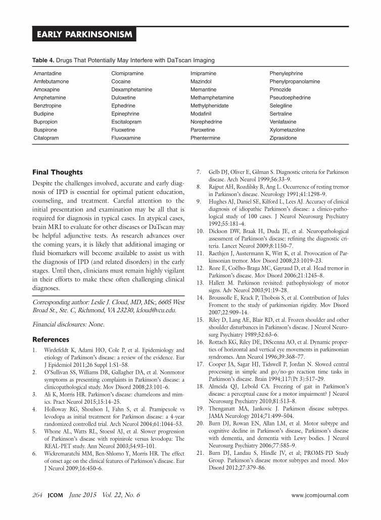
DaTscan is an imaging modality that uses (123I)Ioflupane injection with single photon emission computed tomog-raphy (SPECT) for detecting dopamine transporters. By binding to the dopamine transporters, a quantitative measure of the transporters in the striatal region of the brain can be obtained [57]. Dopaminergic deficit can be quantified in this manner. While this technology can be extremely useful in certain clinical situations, clinicians should be aware of its limitations. DaTscan cannot dif-ferentiate conditions in which there is loss of striatonigral dopaminergic neurons (IPD, PSP, MSA, CBD, LBD), nor can it distinguish between conditions where there is no loss of dopamine neurons (ET, DIP, psychogenic condi-tions) [58]. In clinical use, these scans are mostly used for differentiation of IPD from essential tremor, and are more often used if tremor is the most prominent symptom (which can make distinguishing between IPD and essen-tial tremor difficult). It is important to mention that drugs that bind to the dopamine transporter with high affinity may interfere with the image, and the impact of dopamine agonists and antagonists on the imaging results has not been established. Drugs that may potentially interfere with DaTscan imaging are listed in Table 4 [59].
CliniCal Review
264 JCOM June 2015 Vol. 22, No. 6 www.jcomjournal.com
Early Parkinsonism
Final thoughts
Despite the challenges involved, accurate and early diag-nosis of IPD is essential for optimal patient education, counseling, and treatment. Careful attention to the initial presentation and examination may be all that is required for diagnosis in typical cases. In atypical cases, brain MRI to evaluate for other diseases or DaTscan may be helpful adjunctive tests. As research advances over the coming years, it is likely that additional imaging or fluid biomarkers will become available to assist us with the diagnosis of IPD (and related disorders) in the early stages. Until then, clinicians must remain highly vigilant in their efforts to make these often challenging clinical diagnoses.
Corresponding author: Leslie J. Cloud, MD, MSc, 6605 West Broad St., Ste. C, Richmond, VA 23230, [email protected].
Financial disclosures: None.
references
1. Wirdefeldt K, Adami HO, Cole P, et al. Epidemiology and etiology of Parkinson’s disease: a review of the evidence. Eur J Epidemiol 2011;26 Suppl 1:S1–58.
2. O’Sullivan SS, Williams DR, Gallagher DA, et al. Nonmotor symptoms as presenting complaints in Parkinson’s disease: a clinicopathological study. Mov Disord 2008;23:101–6.
3. Ali K, Morris HR. Parkinson’s disease: chameleons and mim-ics. Pract Neurol 2015;15:14–25.
4. Holloway RG, Shoulson I, Fahn S, et al. Pramipexole vs levodopa as initial treatment for Parkinson disease: a 4-year randomized controlled trial. Arch Neurol 2004;61:1044–53.
5. Whone AL, Watts RL, Stoessl AJ, et al. Slower progression of Parkinson’s disease with ropinirole versus levodopa: The REAL-PET study. Ann Neurol 2003;54:93–101.
6. Wickremaratchi MM, Ben-Shlomo Y, Morris HR. The effect of onset age on the clinical features of Parkinson’s disease. Eur J Neurol 2009;16:450–6.
7. Gelb DJ, Oliver E, Gilman S. Diagnostic criteria for Parkinson disease. Arch Neurol 1999;56:33–9.
8. Rajput AH, Rozdilsky B, Ang L. Occurrence of resting tremor in Parkinson’s disease. Neurology 1991;41:1298–9.
9. Hughes AJ, Daniel SE, Kilford L, Lees AJ. Accuracy of clinical diagnosis of idiopathic Parkinson’s disease: a clinico-patho-logical study of 100 cases. J Neurol Neurosurg Psychiatry 1992;55:181–4.
10. Dickson DW, Braak H, Duda JE, et al. Neuropathological assessment of Parkinson’s disease: refining the diagnostic cri-teria. Lancet Neurol 2009;8:1150–7.
11. Raethjen J, Austermann K, Witt K, et al. Provocation of Par-kinsonian tremor. Mov Disord 2008;23:1019–23.
12. Roze E, Coêlho-Braga MC, Gayraud D, et al. Head tremor in Parkinson’s disease. Mov Disord 2006;21:1245–8.
13. Hallett M. Parkinson revisited: pathophysiology of motor signs. Adv Neurol 2003;91:19–28.
14. Broussolle E, Krack P, Thobois S, et al. Contribution of Jules Froment to the study of parkinsonian rigidity. Mov Disord 2007;22:909–14.
15. Riley D, Lang AE, Blair RD, et al. Frozen shoulder and other shoulder disturbances in Parkinson’s disease. J Neurol Neuro-surg Psychiatry 1989;52:63–6.
16. Rottach KG, Riley DE, DiScenna AO, et al. Dynamic proper-ties of horizontal and vertical eye movements in parkinsonian syndromes. Ann Neurol 1996;39:368–77.
17. Cooper JA, Sagar HJ, Tidswell P, Jordan N. Slowed central processing in simple and go/no-go reaction time tasks in Parkinson’s disease. Brain 1994;117(Pt 3):517–29.
18. Almeida QJ, Lebold CA. Freezing of gait in Parkinson’s disease: a perceptual cause for a motor impairment? J Neurol Neurosurg Psychiatry 2010;81:513–8.
19. Thenganatt MA, Jankovic J. Parkinson disease subtypes. JAMA Neurology 2014;71:499–504.
20. Burn DJ, Rowan EN, Allan LM, et al. Motor subtype and cognitive decline in Parkinson’s disease, Parkinson’s disease with dementia, and dementia with Lewy bodies. J Neurol Neurosurg Psychiatry 2006;77:585–9.
21. Burn DJ, Landau S, Hindle JV, et al; PROMS-PD Study Group. Parkinson’s disease motor subtypes and mood. Mov Disord 2012;27:379–86.
Table 4. DrugsThatPotentiallyMayInterferewithDaTscanImaging
Amantadine
Amfebutamone
Amoxapine
Amphetamine
Benztropine
Budipine
Bupropion
Buspirone
Citalopram
Clomipramine
Cocaine
Dexamphetamine
Duloxetine
Ephedrine
Epinephrine
Escitalopram
Fluoxetine
Fluvoxamine
Imipramine
Mazindol
Memantine
Methamphetamine
Methylphenidate
Modafinil
Norephedrine
Paroxetine
Phentermine
Phenylephrine
Phenylpropanolamine
Pimozide
Pseudoephedrine
Selegiline
Sertraline
Venlafaxine
Xylometazoline
Ziprasidone
www.jcomjournal.com Vol. 22, No. 6 June 2015 JCOM 265
22. Katz M, Luciano MS, Carlson K, et al; CSP 468 study group. Differential effects of deep brain stimulation target on motor subtypes in Parkinson’s disease. Ann Neurol 2015;77:710–9.
23. Savica R, Rocca WA, Ahlskog JE. When does Parkinson dis-ease start? Arch Neurol 2010;67:798–801.
24. Berg D, Postuma RB, Bloem B, et al. Time to redefine PD? Introductory statement of the MDS Task Force on the defini-tion of Parkinson’s disease. Mov Disord 2014;29:454–62.
25. Aquino CC, Fox SH. Clinical spectrum of levodopa-induced complications. Mov Disord 2015;30:80–9.
26. Geser F, Wenning GK, Poewe W, McKeith I. How to diag-nose dementia with Lewy bodies: state of the art. Mov Disord 2005;20 Suppl 12:S11–20.
27. Karantzoulis S, Galvin JE. Update on dementia with Lewy bod-ies. Curr Transl Geriatr Exp Gerontol Rep 2013;2:196–204.
28. Gilman S, Low PA, Quinn N, et al. Consensus statement on the diagnosis of multiple system atrophy. J Neurol Sci 1999;163:94–8.
29. Kim HJ, Jeon BS, Jellinger KA. Diagnosis and differential diagnosis of MSA: boundary issues. J Neurol 2015 Feb 7. [Epub ahead of print]
30. Lee EA, Cho HI, Kim SS, Lee WY. Comparison of magnetic resonance imaging in subtypes of multiple system atrophy. Parkinsonism Relat Disord 2004;10:363–8.
31. Golbe LI, Davis PH, Schoenberg BS, Duvoisin RC. Preva-lence and natural history of progressive supranuclear palsy. Neurology 1988;38:1031–4.
32. Maher ER, Lees AJ. The clinical features and natural history of the Steele-Richardson-Olszewski syndrome (progressive supranuclear palsy). Neurology 1986;36:1005–8.
33. Gröschel K, Kastrup A, Litvan I, Schulz JB. Penguins and hummingbirds: midbrain atrophy in progressive supranuclear palsy. Neurology 2006;66:949–50.
34. Rinne JO, Lee MS, Thompson PD, Marsden CD. Cor-ticobasal degeneration. A clinical study of 36 cases. Brain 1994117(Pt 5):1183–96.
35. Grimes DA, Lang AE, Bergeron CB. Dementia as the most common presentation of cortical-basal ganglionic degenera-tion. Neurology 1999;53:1969–74.
36. Tokumaru AM, O’uchi T, Kuru Y, et al. Corticobasal degen-eration: MR with histopathologic comparison. AJNR Am J Neuroradiol 1996;17:1849–52.
37. Armstrong MJ, Litvan I, Lang AE, et al. Criteria for the diagno-sis of corticobasal degeneration. Neurology 2013;80:496–503.
38. Alexander SK, Rittman T, Xuereb JH, et al. Validation of the new consensus criteria for the diagnosis of corticobasal degen-eration. J Neurol Neurosurg Psychiatry 2014;85:925–9.
39. Sibon I, Fenelon G, Quinn NP, Tison F. Vascular parkinson-ism. J Neurol 2004;251:513–24.
40. Thanvi B, Lo N, Robinson T. Vascular parkinsonism--an important cause of parkinsonism in older people. Age Ageing 2005;34:114–9.
41. Kalra S, Grosset DG, Benamer HT. Differentiating vascular parkinsonism from idiopathic Parkinson’s disease: a systematic review. Mov Disord 2010;25:149–56.
42. Mehanna R, Jankovic J. Movement disorders in cerebrovascu-lar disease. Lancet Neurol 2013; 12:597–608.
43. Josephs KA. Frontotemporal lobar degeneration. Neurol Clin 2007;25:683–96, vi.
44. Lopez-Sendon J, Mena MA, de Yebenes JG. Drug-induced parkinsonism. Expert Opin Drug Saf 2013;12:487–96.
45. Mena MA, de Yebenes JG. Drug-induced parkinsonism. Ex-pert Opin Drug Saf 2006;5:759–71.
46. Brajkovic LD, Svetel MV, Kostic VS, et al. Dopamine trans-porter imaging (123)I-FP-CIT (DaTSCAN) SPET in differ-ential diagnosis of dopa-responsive dystonia and young-onset Parkinson’s disease. Hell J Nucl Med 2012;15:134–8.
47. Krusz JC, Koller WC, Ziegler DK. Historical review: abnor-mal movements associated with epidemic encephalitis lethar-gica. Mov Disord 1987;2:137–41.
48. Langston JW, Ballard P, Tetrud JW, Irwin I. Chronic Par-kinsonism in humans due to a product of meperidine-analog synthesis. Science 1983;219:979–80.
49. Jankovic J. Searching for a relationship between man-ganese and welding and Parkinson’s disease. Neurology 2005;64:2021–8.
50. Jankovic J, Kirkpatrick JB, Blomquist KA, et al. Late-onset Hallervorden-Spatz disease presenting as familial parkinson-ism. Neurology 1985;35:227–34.
51. Cloud L, Jankovic J. Systemic disease and movement disor-ders. In: Burn DJ, editor. Oxford textbook of clinical neurol-ogy on movement disorders. Oxford University Press; 2013.
52. Bugalho P, Guimaraes J. Gait disturbance in normal pressure hydrocephalus: a clinical study. Parkinsonism Relat Disord 2007;13:434–7.
53. Jankovic J, Newmark M, Peter P. Parkinsonism and acquired hydrocephalus. Mov Disord 1986;1:59–64.
54. Bergsneider M, Black PM, Klinge P, et al. Surgical manage-ment of idiopathic normal-pressure hydrocephalus. Neurosur-gery 2005;57(3 Suppl): S29-39; discussion ii-v.
55. Bain P, Brin M, Deuschl G, et al. Criteria for the diagnosis of essential tremor. Neurology 2000; 54(11 Suppl 4): S7.
56. Jankovic J. Essential tremor and Parkinson’s disease. Ann Neurol 1989;25:211–2.
57. Catafau AM, Tolosa E; DaTSCAN Clinically Uncertain Parkinsonian Syndromes Study Group. Impact of dopamine transporter SPECT using 123I-Ioflupane on diagnosis and management of patients with clinically uncertain Parkinsonian syndromes. Mov Disord 2004;19:1175–82.
58. Bajaj N, Hauser RA, Grachev ID. Clinical utility of dopamine transporter single photon emission CT (DaT-SPECT) with (123I) ioflupane in diagnosis of parkinsonian syndromes. J Neurol Neurosurg Psychiatry 2013;84:1288–95.
59. Kagi G, Bhatia KP, Tolosa E. The role of DAT-SPECT in movement disorders. J Neurol Neurosurg Psychiatry 2010;81:5–12.
60. Litvan I, Agid Y, Calne D, et al. Clinical research criteria for the diagnosis of progressive supranuclear palsy (Steele-Rich-ardson-Olszewski syndrome): report of the NINDS-SPSP international workshop. Neurology 1996;47:1–9.
Copyright 2015 by Turner White Communications Inc., Wayne, PA. All rights reserved.
CliniCal Review


![Vascular parkinsonism · Vascular parkinsonism – REVIEW future science groupfuture science group 239 20%) suffered from parkinsonism with strong evidence of CVD [23]](https://img.pdfslide.us/doc/110x75/5c12e69c09d3f208438bb500/vascular-parkinsonism-vascular-parkinsonism-review-future-science-groupfuture.jpg)