Embed Size (px)
Citation preview

Available online at www.sciencedirect.com
www.elsevier.com/locate/brainres
b r a i n r e s e a r c h 1 5 1 5 ( 2 0 1 3 ) 1 9 – 2 8
0006-8993/$ - see frohttp://dx.doi.org/10
nCorrespondence toE-mail address: r1These authors
Research Report
Early life permethrin exposure induces long-termbrain changes in Nurr1, NF-kB and Nrf-2
Manuel Carlonia,1, Cinzia Nasutia,1, Donatella Fedelia,1, Maura Montanib,M.S Dhivya Vadhanac, Augusto Amicib, Rosita Gabbianellia,n
aSchool of Pharmacy, University of Camerino, ItalybSchool of Biosciences and Biotechnology, University of Camerino, ItalycSchool of Advanced Studies, University of Camerino, Italy
a r t i c l e i n f o
Article history:
Accepted 29 March 2013
Pesticide exposure during brain development represents an important risk factor for the
onset of brain-aging processes. Here, the impact of permethrin administered to rats from
Available online 6 April 2013
Keywords:
Rat
Early-life-permethrin-treatment
Brain
Nurr1
NF-kB
Nrf-2
nt matter & 2013 Elsevie.1016/j.brainres.2013.03.04
: School of Pharmacy, Viosita.gabbianelli@unicamcontributed equally to th
a b s t r a c t
6th to 21st day of life, at a dose near to “no observed adverse effect level” (NOAEL), was
studied when animals reached 500 day-old. The permethrin treatment induced a decrease
in Nurr1 gene expression in striatum, an increase in hippocampus and cerebellum, while
the protein level changed only in striatum where it was increased. NF-kB p65 gene
expression was increased in cerebellum, while its protein level augmented in cerebellum
and in prefrontal cortex and decreased in hippocampus of treated rats compared to control
ones. Nrf-2 gene expression resulted significantly higher only in cerebellum of treated
animals. The results suggest that early life permethrin treatment induces long-lasting
effects leading to dopaminergic neuronal disorders, monitored by Nurr1 alteration. More-
over the impairment of NF-kB and Nrf-2, important for the balance between pro- and anti-
inflammatory systems, confirms that the neonatal permethrin treatment can influence
genes involved with the onset of brain-ageing processes.
& 2013 Elsevier B.V. All rights reserved.
r B.V. All rights reserved.8
a Gentile III da Varano, UNICAM, 62032 Camerino, MC, Italy. Fax: +39 0737 403290..it (R. Gabbianelli).e study
1. Introduction
Ageing is a physiological change characterized by a decline inprotein homeostasis and accumulation of macromoleculardamage in different organs as well as in brain. Genetic andenvironmental factors, life style, metabolic processes, as wellas reactive oxygen species have an impact on the neurode-generative process (Parrón et al., 2011; Zhang et al., 2006).Moreover on the development of different types of neurode-generation (i.e. Alzheimer disease (AD), Parkinson’s diseases
(PD) etc.) that characterize ageing processes, the stochasticevent is the most determinant, because it influences bothgene expression and epigenetic pathways, and could partiallyexplain the reported variability and random fluctuations ofindividual gene expression in genetically identical organisms(Raj and Van Oudenaarden, 2008).
Studies on neurodegenerative diseases are dominated byresearch performed in model systems, such as yeast, worms,flies and genetically modified mice because animal modelspermit the assessment of markers of neurodegeneration in
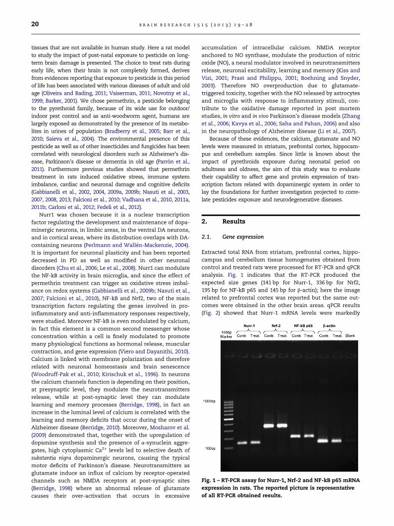
Fig. 1 – RT-PCR assay for Nurr-1, Nrf-2 and NF-kB p65 mRNA
expression in rats. The reported picture is representative
of all RT-PCR obtained results.
b r a i n r e s e a r c h 1 5 1 5 ( 2 0 1 3 ) 1 9 – 2 820
tissues that are not available in human study. Here a rat modelto study the impact of post-natal exposure to pesticide on long-term brain damage is presented. The choice to treat rats duringearly life, when their brain is not completely formed, derivesfrom evidences reporting that exposure to pesticide in this periodof life has been associated with various diseases of adult and oldage (Oliveira and Bading, 2011; Vaiserman, 2011; Novotny et al.,1999; Barker, 2001). We chose permethrin, a pesticide belongingto the pyrethroid family, because of its wide use for outdoor/indoor pest control and as anti-woodworm agent, humans arelargely exposed as demonstrated by the presence of its metabo-lites in urines of population (Bradberry et al., 2005; Barr et al.,2010; Saieva et al., 2004). The environmental presence of thispesticide as well as of other insecticides and fungicides has beencorrelated with neurological disorders such as Alzheimer’s dis-ease, Parkinson’s disease or dementia in old age (Parrón et al.,2011). Furthermore previous studies showed that permethrintreatment in rats induced oxidative stress, immune systemimbalance, cardiac and neuronal damage and cognitive deficits(Gabbianelli et al., 2002, 2004, 2009a, 2009b; Nasuti et al., 2003,2007, 2008, 2013; Falcioni et al., 2010; Vadhana et al., 2010, 2011a,2011b; Carloni et al., 2012; Fedeli et al., 2012).
Nurr1 was chosen because it is a nuclear transcriptionfactor regulating the development and maintenance of dopa-minergic neurons, in limbic areas, in the ventral DA neurons,and in cortical areas, where its distribution overlaps with DA-containing neurons (Perlmann and Wallén-Mackenzie, 2004).It is important for neuronal plasticity and has been reporteddecreased in PD as well as modified in other neuronaldisorders (Chu et al., 2006; Le et al., 2008). Nurr1 can modulatethe NF-kB activity in brain microglia, and since the effect ofpermethrin treatment can trigger an oxidative stress imbal-ance on redox systems (Gabbianelli et al., 2009b; Nasuti et al.,2007; Falcioni et al., 2010), NF-kB and Nrf2, two of the maintranscription factors regulating the genes involved in pro-inflammatory and anti-inflammatory responses respectively,were studied. Moreover NF-kB is even modulated by calcium,in fact this element is a common second messenger whoseconcentration within a cell is finely modulated to promotemany physiological functions as hormonal release, muscularcontraction, and gene expression (Viero and Dayanithi, 2010).Calcium is linked with membrane polarization and thereforerelated with neuronal homeostasis and brain senescence(Woodruff-Pak et al., 2010; Kirischuk et al., 1996). In neuronsthe calcium channels function is depending on their position,at presynaptic level, they modulate the neurotransmittersrelease, while at post-synaptic level they can modulatelearning and memory processes (Berridge, 1998), in fact anincrease in the luminal level of calcium is correlated with thelearning and memory deficits that occur during the onset ofAlzheimer disease (Berridge, 2010). Moreover, Mosharov et al.(2009) demonstrated that, together with the upregulation ofdopamine synthesis and the presence of α-synuclein aggre-gates, high cytoplasmic Ca2+ levels led to selective death ofsubstantia nigra dopaminergic neurons, causing the typicalmotor deficits of Parkinson’s disease. Neurotransmitters asglutamate induce an influx of calcium by receptor-operatedchannels such as NMDA receptors at post-synaptic sites(Berridge, 1998) where an abnormal release of glutamatecauses their over-activation that occurs in excessive
accumulation of intracellular calcium. NMDA receptoranchored to NO synthase, modulate the production of nitricoxide (NO), a neural modulator involved in neurotransmittersrelease, neuronal excitability, learning and memory (Kiss andVizi, 2001; Prast and Philippu, 2001; Boehning and Snyder,2003). Therefore NO overproduction due to glutamate-triggered toxicity, together with the NO released by astrocytesand microglia with response to inflammatory stimuli, con-tribute to the oxidative damage reported in post mortemstudies, in vitro and in vivo Parkinson’s disease models (Zhanget al., 2006; Kavya et al., 2006; Saha and Pahan, 2006) and alsoin the neuropathology of Alzheimer disease (Li et al., 2007).
Because of these evidences, the calcium, glutamate and NOlevels were measured in striatum, prefrontal cortex, hippocam-pus and cerebellum samples. Since little is known about theimpact of pyrethroids exposure during neonatal period onadultness and oldness, the aim of this study was to evaluatetheir capability to affect gene and protein expression of tran-scription factors related with dopaminergic system in order tolay the foundations for further investigation projected to corre-late pesticides exposure and neurodegenerative diseases.
2. Results
2.1. Gene expression
Extracted total RNA from striatum, prefrontal cortex, hippo-campus and cerebellum tissue homogenates obtained fromcontrol and treated rats were processed for RT-PCR and qPCRanalysis. Fig. 1 indicates that the RT-PCR produced theexpected size genes (141 bp for Nurr-1, 336 bp for Nrf2,195 bp for NF-kB p65 and 145 bp for β-actin); here the imagerelated to prefrontal cortex was reported but the same out-comes were obtained in the other brain areas. qPCR results(Fig. 2) showed that Nurr-1 mRNA levels were markedly

b r a i n r e s e a r c h 1 5 1 5 ( 2 0 1 3 ) 1 9 – 2 8 21
decreased in the treated rats striatum, while no changes wereobserved in Nrf-2 and NF-kB p65 mRNA levels. No Nurr-1,Nrf-2 and NF-kB p65mRNA levels changes were observed inprefrontal cortex. In hippocampus, a significant Nurr-1 mRNAlevel increase was observed with respect to the control, whileno changes were observed in Nrf-2 and NF-kB p65 mRNAlevels. A significant increase was observed in Nurr-1, Nrf-2and NF-kB p65 mRNA levels in cerebellum of treated rats withrespect to the control.
2.2. Western blot
Fig. 3 shows the Western blot analysis in striatum, prefrontalcortex, hippocampus and cerebellum tissues obtained fromcontrol and treated rats with anti-Nurr-1, anti-Nrf2 and anti-NF-kB p65 antibodies. The densitometry analysis shows thatNurr-1 protein level increased (15879.54%) in striatum oftreated group with respect to the control one (100%) (Po0.05),while no changes in NF-kB p65 and Nrf2 protein levels wereobserved. Nurr-1 and Nrf2 protein levels unchanged in pre-frontal cortex, while NF-kB p65 increased (167720.94%) sig-nificantly (Po0.05) compared to control (100%).
A significant decrease in NF-kB p65 levels (63711.61%) inhippocampus from treated group compared to control one(100%) was measured, while no changes in Nurr-1 and Nrf2were measured. NF-kB p65 protein level increased (228734%)in cerebellum from treated group while Nurr1 and Nrf2 wereunchanged.
2.3. Glutamate levels
Fig. 4 shows the glutamate levels in striatum, prefrontal cortex,hippocampus and cerebellum tissue homogenates obtainedfrom control and treated rats. A significant glutamate decreasewas detected in cerebellum from treated rats (41.04 70.00 nmol/mg protein) compared to the control group (49.5472.14 nmol/mg
Fig. 2 – qPCR for quantifying changes in gene expression of Nu
hippocampus and cerebellum of control and treated rats. All exp
used as an internal control. Po0.05 § vs control.
protein) (Po0.05). No changes were detected in the otherbrain areas.
2.4. Calcium levels
Fig. 5 shows the Ca++ levels in striatum, prefrontal cortex,hippocampus and cerebellum tissue homogenates obtainedfrom control and treated rats. A significant Ca++ levelincrease (197.44725.61 nmol/mg protein) was observed instriatum of treated rats with respect to the control(124.72720.10 nmol/mg protein) (Po0.05). The same behaviorwas observed in prefrontal cortex (65.1375.67 nmol/mg pro-tein for control and 104.2778.29 nmol/mg protein for treated)(Po0.05) and cerebellum (365.2273.89 nmol/mg protein forcontrol and 410.3877.98 nmol/mg protein for treated)(Po0.05), while the opposite behavior was observed in hippo-campus (295.6373.28 nmol/mg protein for control and246.6473.91 nmol/mg protein for treated) (Po0.05).
2.5. NO levels
Fig. 6 shows the NO levels in striatum, prefrontal cortex,hippocampus and cerebellum tissue homogenates obtainedfrom control and treated rats. Cerebellum from treated ratsshows a significant decrease (1.13070.18 nmol/mg protein) inNO production with respect to the control (3.0770.30 nmol/mg protein) (Po0.05), while no changes are measured in theothers brain regions.
3. Discussion
Pesticides accumulated through the food chain and environ-mental exposures are identified as one of the main riskfactors leading to psychiatric disorders and neurodegenera-tive diseases (Parrón et al., 2011; Zhang et al., 2006; Hatcher
rr-1, Nrf-2, and NF-kB p65 in striatum, prefrontal cortex,
ression values were normalized to the value of β-actin gene

Fig. 4 – Glutamate levels in striatum, prefrontal cortex, hippocampus and cerebellum obtained from control and treated rat
groups. Po0.05 § vs control.
Fig. 3 – Western blotting detection and densitometry analysis of Nurr-1, NF-kB p65, Nrf-2 protein expression in striatum,
prefrontal cortex, hippocampus and cerebellum of control and treated rats. Density ratio percentage of each treated area
sample was referred to its respective control fixed as 100%. Po0.05 § vs control. The immunoblot exhibited in the figure is
representative of three experiments.
b r a i n r e s e a r c h 1 5 1 5 ( 2 0 1 3 ) 1 9 – 2 822
et al., 2008). They can modify gene expression changingprotein synthesis in different tissues, leading to the initiationand progression of age-related diseases (Parrón et al., 2011;Zhang et al., 2006; Hatcher et al., 2008; Giasson and Lee 2000;Relton and Smith, 2010).
Although pyrethroids are excreted within 24 h, reducing theimpact linked to body accumulation, they are characterized bypronounced lipophilicity compared to other classes of pesticides,which makes them able to easily cross the blood–brain barrierand exert their toxic effect directly on the central nervous system(Barr et al., 2010; Bradberry et al., 2005).Besides, it should be
underlined that early life is a developing phase, which isparticularly vulnerable to the effects of adverse environmentalexposures because pathways of many systems, as well asstructure and function of several macromolecules are pro-grammed during this period and, for this reason, neonatal periodcan influence the program of gene expression in adulthood(Oliveira et al., 2011; Vaiserman, 2011). According to this evi-dence, rats exposed during early life to endocrine disruptorshave lifelong effects on neuroendocrine gene expression andDNAmethylation, together with progression of senescence (Goreet al., 2011).

Fig. 6 – NO levels in striatum striatum, prefrontal cortex,
hippocampus and cerebellum obtained from control and
treated rat groups. Po0.05 § vs control.
Fig. 5 – Ca++ levels in striatum, prefrontal cortex,
hippocampus and cerebellum obtained from control and
treated rat groups. Po0.05 § vs control.
b r a i n r e s e a r c h 1 5 1 5 ( 2 0 1 3 ) 1 9 – 2 8 23
In our experiment, 500 day-old rats treated in early lifewith permethrin showed an increase of Ca+2 level in stria-tum, prefrontal cortex and cerebellum with respect to thecontrols. In addition, no glutamate level variation wasobserved between the groups, with the exception of thecerebellum where the glutamate levels were decreased intreated group. These results led us to hypothesize that theincreased Ca2+ concentration could be due to an increasedcell membrane depolarization, that is a direct consequence ofpermethrin’s mode of action through its binding with thevoltage-gated sodium channel α-subunit, occurred duringearly life permethrin treatment that was able to lead perma-nent alterations of gene expression profile (Imamura et al.,2006; Narahashi, 1996). Moreover, as reported by Wu et al.(2009), synthetic pyrethroids can interact with voltage-gatedsodium channels to modify the kinetics of sodium currentsand there after neuronal transmission.
It should be underlined that the lower Ca2+ level inhippocampus from 500 day-old treated rats compared to thecontrols, can be properly related to the decreased NF-kBprotein level, because Ca2+ is one of the factors that is ableto modulate this transcription factor, which regulates thepro-inflammatory responses, and that can be inhibited alsoby the increased Nurr1 gene expression detected in this brainarea. This outcome fit with our previous findings where aselective effect of permethrin treatment on cognitive testsdemonstrated an involvement of frontal cortico-striatal cir-cuitry rather than to a role for the hippocampus (Nasuti et al.,
2013), in fact an increase of calcium level has been linkedwith learning, memory and motor deficits occurring duringon neurodegenerative diseases (Mosharov et al., 2009;Berridge, 2010). Nurr1, a transcription factor belonging tothe orphan nuclear receptor family, regulates genes involvedin dopamine neurotransmission such as tyrosine hydroxy-lase and vesicular monoamine transporter 2 (Bensinger andTontonoz, 2009). Reduction in Nurr1 gene expression wasreported in Parkinson’s diseases, schizophrenia and manicdepression (Chu et al., 2006; Le et al., 2008) and in rheumatoidarthritis (Davies et al., 2005). Besides, Nurr1 has a neuropro-tective effect because of its capability to inhibit the produc-tion of inflammatory mediators in microglia by interactionwith CoREST repressor and by the promotion of the boundformation with NF-kB subunit p65 (Bensinger and Tontonoz,2009; Saijo et al., 2009).
In striatum from 500 day-old treated rats, the increasedNurr1 protein level could be consequent to an adaptiveresponse to balance the decreased Nurr1 mRNA gene expres-sion measured in this brain area. To explain this outcome itcould be hypothesized a compensative post-transcriptionalregulation that increases the Nurr1 mRNA stability allowingto increase the RNA translation with the consequent highNurr1 protein level (Scheme 1). This regulation is largelyused to maintain early response genes in a active state sothat protein level can be increased according to cellrequirements (Stellato, 2004). Moreover it could be alsohypothesized an increase in protein stability by abolishingthe Nurr1 site phosphorylation by Akt, that controls theubiquitinylation of Nurr1 and then its stability (Scheme 1)(Jo et al., 2009). However at this stage it is unknown if thispathway is due to a direct interaction of permethrin on Akt orto an adaptive mechanism for control neuronal survival. Onthe other hand the decreased Nurr 1 gene expression could bedue to neuronal death caused by oxidative stress induced bypermethrin via accelerated dopamine turnover. Furthermore,this hypothesis is supported by the low dopamine levelalready measured in striatum from this animal model inadolescent age (Nasuti et al., 2007) and in 500 day-old rats(Nasuti et al., 2013). In addition, the change in Nurr1 instriatum may be correlated with previous outcomes onadolescent rats, that received the same early life permethrintreatment, because those rats displayed significant signs ofprotein, DNA and lipid oxidation together with decreasedGSH levels due to increased oxidative stress following higherdopamine degradation (Nasuti et al., 2007; Spina and Cohen,1989).
In prefrontal cortex from 500 day-old rats treated withpermethrin in early life, we observed an higher neuronal andglial calcium concentration with respect to control rats, thatcould be associated with the increased NF-kB protein levelsmeasured in this brain area. This transcription factor has acrucial role in the activation of pro-inflammatory genes andits increase is associated with ageing biomarkers (i.e. cellularsenescence, inflammation, tissue atrophy) and age relateddiseases (i.e. atherosclerosis, neuronal degeneration)(Salminen et al., 2008; Salminen and Kaarniranta, 2010).However, in order to maintain homeostasis, organisms havean efficient repressor system (Salminen and Kaarniranta,2010) working to inhibit NF-kB together with transcriptor
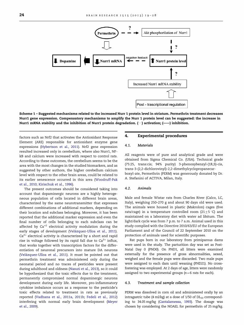
Scheme 1 – Suggested mechanisms related to the increased Nurr 1 protein level in striatum. Permethrin treatment decreases
Nurr1 gene expression. Compensatory mechanisms to amplify the Nurr 1 protein level can be suggested: the increase in
Nurr1 mRNA stability and the inhibition of Nurr1 protein degradation. (___) activation; (——) inhibition.
b r a i n r e s e a r c h 1 5 1 5 ( 2 0 1 3 ) 1 9 – 2 824
factors such as Nrf2 that activates the Antioxidant ResponseElement (ARE) responsible for antioxidant enzyme geneexpressions (Hybertson et al., 2011). Nrf2 gene expressionresulted increased only in cerebellum, where also Nurr1, NF-kB and calcium were increased with respect to control rats.According to these outcomes, the cerebellum seems to be thearea with the most changes in the studied biomarkers, and assuggested by other authors, the higher cerebellum calciumlevel with respect to the other brain areas, could be related toits earlier senescence occurred in this area (Woodruff-Paket al., 2010; Kirischuk et al., 1996).
The present outcomes should be considered taking intoaccount that dopaminergic neurons are a highly heteroge-neous population of cells located in different brain areas,characterized by the same neurotransmitter that expressesdifferent combinations of additional markers, depending ontheir location and subclass belonging. Moreover, it has beenreported that the additional marker expression and even thefinal number of cells belonging to each subclass can beaffected by Ca+2 electrical activity modulation during theearly stages of development (Velázquez-Ulloa et al., 2011).Ca2+ electrical activity is characterized by a short and rapidrise in voltage followed by its rapid fall due to Ca2+ influx,that works together with transcription factors for the differ-entiation of neuronal precursors into mature DA neurons(Velázquez-Ulloa et al., 2011). It must be pointed out thatpermethrin treatment was administered only during theneonatal period and no traces of pesticides were presentduring adulthood and oldness (Nasuti et al., 2013), so it couldbe hypothesized that the toxic effects due to the treatment,permanently compromised normal dopaminergic neuronsdevelopment during early life. Moreover, pro-inflammatorycytokine imbalance occurs as a response to the pesticide’stoxic effects related to treatment in rats as previouslyreported (Vadhana et al., 2011a, 2011b; Fedeli et al., 2012)interfering with normal early brain development (Meyeret al., 2009).
4. Experimental procedures
4.1. Materials
All reagents were of pure and analytical grade and wereobtained from Sigma Chemical Co. (USA). Technical grade(75:25, trans:cis; 94% purity) 3-phenoxybenzyl-(1R,S)-cis,trans-3-(2,2-dichlorovinyl)-2,2-dimethylcyclopropanecar-boxyl-ate, Permethrin (PERM) was generously donated by Dr.A. Stefanini of ACTIVA, Milan, Italy.
4.2. Animals
Male and female Wistar rats from Charles River (Calco, LC,Italy), weighing 250–270 g and about 90 days old were used.The animals were housed in plastic (Makrolon) cages (fiverats/cage) in a temperature controlled room (2175 1C) andmaintained on a laboratory diet with water ad libitum. Thelight/dark cycle was from 7 p.m. to 7 a.m. Animal used in thisstudy complied with the Directive 2010/63/EU of the EuropeanParliament and of the Council of 22 September 2010 on theprotection of animals used for scientific purposes.
Rat pups born in our laboratory from primiparous damswere used in the study. The parturition day was set as Post-Natal Day 0 (PND0). On PND1, all litters were examinedexternally for the presence of gross abnormalities, sexed,weighed and the female pups were discarded. Two male pupswere assigned to each dam until weaning (PND21). No cross-fostering was employed. At 2 days of age, litters were randomlyassigned to two experimental groups (n¼6 rats for each).
4.3. Treatment and sample collection
PERM was dissolved in corn oil and administered orally by anintragastric tube (4 ml/kg) at a dose of 1/50 of DL50 correspond-ing to 34.05 mg/kg (Cantalamessa, 1993). The dosage waschosen by considering the NOAEL for permethrin of 25mg/kg.

b r a i n r e s e a r c h 1 5 1 5 ( 2 0 1 3 ) 1 9 – 2 8 25
The compounds were administered for 15 days, once a day inthe morning from PND6 to PND21. Control rats were treatedwith vehicle (corn oil 4 ml/kg) on a similar schedule. Thevolume of the compound administered was adjusted dailybased on body weight measured during the dosing period. OnPND21, the offspring were weaned and the littermates werehoused together. At old age (PND 500), six rats from each group(PERM treated and control groups) were sacrificed by exposureto CO2, and their brain areas (striatum, prefrontal cortex,hippocampus and cerebellum) were collected and immediatelyfrozen in liquid nitrogen and stored at −80 1C until analysis.This age was chosen because it corresponds to about 50 year-old humans (Andreollo et al., 2012) and it could be representa-tive of the beginning of the first brain alterations that candevelop to brain ageing diseases such as AD and PD (Levy, 2007;Lindsay et al., 1997).
All data were analyzed considering the litter as thesmallest unit.
4.4. RNA extraction and cDNA preparation
Total RNA was extracted from the pool of striatum, prefrontalcortex, hippocampus and cerebellum obtained from controland treated rats by using a RNA isolation kit (NucleoSpinRNA II Purification Kit, Clontech Laboratories, Inc., USA).RNA quality was ensured by gel visualization and spectro-photometric analysis (OD260/280), while its quantity was mea-sured using the OD260 on a Nano-Drop spectrophotometer.The RNA was in vitro transcribed to cDNA by using MaximaFirst Strand cDNA Synthesis Kit for RT-qPCR (Fermentas,Thermo Fisher Scientific Inc., USA) according to the manu-facturer’s instructions.
4.5. Gene expression analysis
RT-PCR and qPCR were employed to evaluate mRNA expres-sion of genes of interest. The following specific sense andantisense primers were designed on the basis of gene andmRNA sequences available online (http://www.ncbi.nlm.nih.gov/gene) and purchased from Sigma Chemical Co. (USA):
β-Actin: TAAAGACCTCTATGCCAACACAGTGC (forward),AGAGTACTTGCGCTCAGGAGGAG (reverse);Nurr-1: GGTTTCTTTAAGCGCACGGTG (forward),TTCTTTAACCATCCCAACAGCCAG (reverse);Nrf-2: CAGCACATCCAGACAGACACCA (forward),CGTATTAAGACACTGTAACTCGGGAATGG (reverse);NF-kB p65: CTGTTTCCCCTCATCTTTCCCTC (forward),TCCCGTGTAGCCATTGATCTTG (reverse).
These primer sets specifically recognized only the genes ofinterest, as indicated by the PCR amplification products
(145 bp for β-actin, 141 bp for Nurr-1, 336 bp for Nrf2 and195 bp for NF-kB p65). Amplification of β-actin, a relativelyinvariant internal reference RNA, was performed in parallel.First strand cDNA was amplified by using Phire Hot Start IIDNA Polymerase (Finnzymes Oy, Finland) in a total volume of20 ml containing 100 ng of cDNA, 0.5 mM of sense and anti-sense gene-specific primers and 200 μM of dNTP Mix (Fer-mentas, Thermo Fisher Scientific Inc., USA). The temperatureprofile was as follows: 30 s at 98 1C; 35 cycles of 5 s at 98 1C, 5 sat 63 1C, 15 s at 72 1C and hold 1 min at 72 1C. The amplifica-tion products were analyzed by electrophoresis on a 1.7%agarose gel in 1� TAE buffer (40 mM Tris-acetate and 1 mMEDTA) containing 0.5 mg/ml ethidium bromide. GeneRuler100 bp DNA Ladder (Fermentas, Thermo Fisher ScientificInc., USA) was used to size DNA fragments. Gel images werecaptured using the KODAK Image Station 2000r Systems.qPCR analysis was performed in a total volume of 20 μlcontaining 25 ng of template cDNA, 0.25 μM sense and anti-sense primers, 10 μl of iQ SYBR Green Supermix (Bio-Rad Inc.,USA) by using a Stratagene MX3000P. The same RT-PCRspecific sense and antisense upon cited primers were used.The real-time PCR program was: initial denaturation at 95 1Cfor 3 min and 40 cycles at 95 1C for 30 s, 63 1C for 30 s and72 1C for 1 min. The program was terminated by a finalextension at 95 1C for 1 min, 60 1C for 30 s and 95 1C for 30 s.Relative mRNA expression on each tissue sample was quan-tified according to the ΔΔCt method. A ΔCt value wascalculated, first by subtracting the Ct value for the house-keeping gene β-actin from the Ct value for each sample.A ΔΔCt value was then calculated by subtracting the ΔCtvalue for the control from the ΔCt value for treated. Foldchanges compared to the controls were then determined by2−ΔΔCt. Each PCR was run in triplicate on three separateoccasions for each independent experiment.
4.6. Tissues preparation
Pools of striatum, prefrontal cortex, hippocampus and cere-bellum obtained from control and treated rats were lysatedusing RIPA buffer [1% NP40, 0.5% Na-deoxycolic acid and 0.1%SDS in phosphate buffered saline (PBS)] with fresh addedprotease inhibitors. The lysates were passed several timesthrough a 22-gauge needle in order to shatter the DNAmolecules.
4.7. Western blot
Proteins in tissue lysates were quantified using the Lowrymethod (Lowry et al., 1951) and equal amounts (30 μg) ofprotein from each cell lysate were separated using SDS-PAGE(10%) and electrophoretically blotted on a nitrocellulose sup-port (Hybond C, Amersham Bioscience, Little Chalfont, UK).
For each experiment, reactive sites were then blocked withPBS containing 0.05% Tween 20 (PBS-T) and bovine serumalbumin (10%) for 1 h at 42 1C, then washed three times in PBS-T and incubated, respectively, with polyclonal Nurr-1 (diluted1:100), polyclonal Nrf-2 (diluted 1:200) and polyclonal NF-kB p65(diluted 1:200) anti-rabbit antibodies (Santacruz Inc, USA) andthen with secondary anti-rabbit antibody diluted 1:5000, (KPL,USA). β-actin was utilized as a control for equal protein loading:membranes were stripped and reprobed for β-actin with an anti-rabbit monoclonal antibody (Abcam plc, UK) diluted 1:3000.Antibodies were diluted in PBS containing 0.05% Tween 20 andbovine serum albumin (2%). Every gel was loaded with molecularweight markers including proteins with MW from 250 to 4 kDa(Invitrogen, USA). HeLa cells lysate was used as positive control(data not shown). Images capturing and densitometry analysiswere performed using the KODAK Image Station 2000r Systems.

b r a i n r e s e a r c h 1 5 1 5 ( 2 0 1 3 ) 1 9 – 2 826
4.8. Glutamate levels
Glutamate quantification on pools of striatum, prefrontal cortex,hippocampus and cerebellum tissue homogenates obtainedfrom control and treated rats was assessed using a commerciallyavailable enzymatic assay kit (BioVision Inc., USA) according tothe manufacturer’s instructions. The amount of glutamate isquantified by colorimetric (spectrophotometry at λ¼450 nm)method and expressed in nmol glutamate/mg protein.
4.9. Calcium levels
Total Ca++ level derived from both glial and neuronal cellswas measured on pools of striatum, prefrontal cortex hippo-campus and cerebellum tissue obtained from control andtreated rats. Calcium Green-1 AM (Invitrogen, USA), a fluor-escent indicator that exhibits an increase in fluorescenceupon binding Ca2+, was reconstituted in high-quality anhy-drous DMSO and used at final concentration of 5 μM in areaction mixture containing equal amounts (0.3 mg/ml) ofprotein from each brain area tissue obtained from control andtreated rats. The fluorescence, was monitored for 10 s using aHitachi 4500 spectrofluorometer, emission wavelength531 nm, excitation wavelength 506 nm, excitation slit 5 andemission slit 5. A standard curve was done using CaCl2 in PBSat different concentrations in the presence of 5 μM CalciumGreen-1 AM to report the results as nmol Ca++/mg protein.
4.10. NO levels
NO quantification on pools of striatum, prefrontal cortexhippocampus and cerebellum tissue homogenates obtainedfrom control and treated rats was assessed using a commer-cially available non enzymatic assay kit (Neogen Corporation,USA) according to the manufacturer’s instructions. NO levelsare expressed in nmol NO/mg protein.
4.11. Statistical analysis
Data are expressed as mean values7SEM. Each experimentwas performed in triplicate and repeated three times. Statis-tical analysis was carried out using Student’s t test (StatsoftStatistica Software, 9.0). A P valueo0.05 was consideredstatistically significant.
5. Conclusion
In conclusion, trough modifications that occur during early life,whose mechanisms (i.e. epigenetic modulation) will be object offuture investigations, the post-natal treatment with permethrinis thought to be capable of affecting gene and protein expres-sion of transcriptor factors related with dopaminergic systemdevelopment together with impairment of pro and anti-inflammatory status in brain areas of 500 day-old rats. Sincethe changes observed in the brain could develop into a well-defined neurodegeneration, future studies will be performed tocheck the impact of early life permethrin pesticide treatment onlongevity and brain status in elderly rats (more than 500 day-old rats).
Disclosure statement
The authors declare that there are no conflicts of interest.
Contributors
M. C. performed gene expression, critical revision of data andmanuscript preparation; C. N. performed animal treatmentand manuscript preparation; D. F. performed glutamate andNO analysis, western blotting and critical revision of data; M.M. and A. A. contributed in the western blotting analysis; D.V. M.S performed calcium measurement; R. G. providedexperimental design, critical revision of data and manuscriptpreparation.
Acknowledgments
This work was supported by MIUR, National Grant No2008ZW3FJ3 to R.G.
r e f e r e n c e s
Andreollo, N.A., Santos, E.F., Araújo, M.R., Lopes, L.R., et al., 2012.Rat’s age versus human’s age: what is the relationship?. Arq.Bras. Cir. Dig. 25 (1), 49–51.
Barker, D.J.P., et al., 2001. Fetal and infant origins of adult disease.Monatsschr. Kinderheilkd. 49, 2–6.
Barr, D.B., Wong, O.A., Unubka, S., Baker, S.E., Whitehead, R.D.,Magsumbol, M.S., Williams, B.L., Needham, L.L., et al., 2010.Urinary concentration of metabolites of pyrethroidinsecticides in the general US population: National health andnutrition examination survey 1999–2002. Environ. HealthPerspect. 118, 742–748.
Bensinger, S.J., Tontonoz, P., et al., 2009. A nurr1 Pathway forneuroprotection. Cell 137, 26–28.
Berridge, M., et al., 1998. Neuronal calcium signaling. Neuron 21,13–26.
Berridge, M., et al., 2010. Calcium hypothesis of Alzheimer’sdisease. Pflugers Arch. 459, 441–449.
Boehning, D., Snyder, S.H., et al., 2003. Novel neural modulators.Annu. Rev. Neurosci. 26, 105–131.
Bradberry, S.M., Cage, S.A., Proudfoot, A.T., Vale, J.A., et al., 2005.Poisoning due to pyrethroids. Toxicol. Rev. 24, 93–106.
Cantalamessa, F., et al., 1993. Acute toxicity of two pirethyroids,permethrin and cypermethrin in neonatal and adult rats.Arch. Toxicol. 67, 510–513.
Carloni, M., Nasuti, C., Fedeli, D., Montani, M., Amici, A., Vadhana,M.S.D., Gabbianelli, R., et al., 2012. The impact of early lifepermethrin exposure on development of neurodegenerationin adulthood. Exp. Gerontol. 47 (1), 60–66.
Chu, Y., Le, W., Kompoliti, K., Jankovic, J., Mufson, E.J., Kordower,J.H., et al., 2006. Nurr1 in Parkinson’s disease and relateddisorders. J. Comp. Neurol. 494 (3), 495–514.
Davies, M.R., Harding, C.J., Raines, S., Tolley, K., Parker, A.E.,Downey-Jones, M., Needham, M.R.C., et al., 2005. Nurr1dependent regulation of pro-inflammatory mediators inimmortalised synovial fibroblasts. J. Inflamm. 2, 15–23.
Falcioni, M.L., Nasuti, C, Bergamini, C., Fato, R, Lenaz, G.,Gabbianelli, R., et al., 2010. The primary role of GSH againstnuclear DNA damage of striatum induced by permethrin in rats.Neuroscience 168, 2–10.

b r a i n r e s e a r c h 1 5 1 5 ( 2 0 1 3 ) 1 9 – 2 8 27
Fedeli, D., Montani, M., Carloni, M., Nasuti, C., Amici, A.,Gabbianelli, R., et al., 2012. Leukocyte Nurr1 as peripheralbiomarker of early life environmental exposure to permethrininsecticide. Biomarkers 17 (7), 604–609.
Gabbianelli, R., Falcioni, G., Nasuti, C., Cantalamessa, F., et al.,2002. Cypermethrin-induced plasma membrane perturbationon erythrocytes from rats: reduction of fluidity in thehydrophobic core and in glutathione peroxidase activity.Toxicology 175, 91–101.
Gabbianelli, R., Falcioni, M.L., Cantalamessa, F., Nasuti, C., et al.,2009a. Permethrin induces Endo III and Fpg lymphocyte DNAdamage and change in monocyte respiratory burst in rats.J. Appl. Toxicol. 29, 317–322.
Gabbianelli, R., Falcioni, M.L., Nasuti, C., Cantalamessa, F., Imada,I., Inoue, M., et al., 2009b. Effect of permethrin insecticide onrat polymorphonuclear neutrophils. Chem. Biol. Interact. 182,245–252.
Gabbianelli, R., Nasuti, C., Falcioni, G., Cantalamessa, F., et al.,2004. Lymphocyte DNA damage in rats exposed topyrethroids: effect of supplementation with vitamins E and C.Toxicology 203, 17–26.
Giasson, B.I., Lee, V.M.-Y., et al., 2000. A new link betweenpesticides and Parkinson’s disease nature. Neuroscience 3,1227–1228.
Gore, A.C., Walker, D.M., Zama, A.M., Armenti, A.E., Uzumcu, M.,et al., 2011. Early life exposure to endocrine-disruptingchemicals causes lifelong molecular reprogramming of thehypothalamus and premature reproductive aging. Mol.Endocrinol. 25, 2157–2168.
Hatcher, J.M., Pennell, K.D., Miller, G.W., et al., 2008. Parkinson.sdisease and pesticides: a toxicological perspective. TrendsPharmacol. Sci. 9, 322–329.
Hybertson, B.M., Gao, G., Bose, S.K., McCord, J.M., et al., 2011.Oxidative stress in health and disease: the therapeuticpotential of Nrf2 activation. Mol. Aspects Med. 32, 234–246.
Imamura, L., Yasuda, M., Kuramitsu, K., Hara, D., Tabuchi, A.,Tsuda, M., et al., 2006. Deltamethrin, a pyrethroidinsecticide, is a potent inducer for the activity-dependentgene expression of brain-derived neurotrophic factor inneurons. J. Pharmacol. Exp. Ther. 316, 136–143.
Jo, A.Y., Kim, M.Y., Lee, H.S., Rhee, Y.H., Lee, J.E., Baek, K.H., Park,C.H., Koh, H.C., Shin, I., Lee, Y.S., Lee, S.H., et al., 2009.Generation of dopamine neurons with improved cell survivaland phenotype maintenance using a degradation-resistantnurr1 mutant. Stem Cells 27, 2238–2246.
Kavya, R., Saluja, R., Singh, S., Dikshit, M., et al., 2006. Nitric oxidesynthase regulation and diversity: implications in Parkinson’sdisease. Nitric Oxide 15, 280–294.
Kiss, J.P., Vizi, E.S., et al., 2001. Nitric oxide: a novel link betweensynaptic and nonsynaptic transmission. Trends Neurosci. 24,211–215.
Kirischuk, S., Voitenko, N., Kostyuk, P., Verkhratsky, A., et al.,1996. Age-associated changes of cytoplasmic calciumhomeostasis in cerebellar granule neurons in situ:investigation on thin cerebellar slices. Exp. Gerontol. 31,475–487.
Le, W., Pan, T., Huang, M., Xu, P., Xie, W., Zhu, W., Zhang, X.,Deng, H., Jankovic, J., et al., 2008. Decreased NURR1 geneexpression in patients with Parkinson’s disease. J. Neurol. Sci.273, 29–33.
Levy, G., et al., 2007. The relationship of Parkinson disease withaging. Arch. Neurol. 64 (9), 1242–1246.
Li, W., Xue, J., Niu, C., Fu, H., Lam, C.S., Luo, J., Chan, H.H., Xue, H.,Kan, K.K., Lee, N.T., Li, C., Pang, Y., Li, M., Tsim, K.W., Jiang, H.,Chen, K., Li, X., Han, Y., et al., 2007. Synergisticneuroprotection by bis(7)-tacrine via concurrent blockade ofN-methyl-d-aspartate receptors and neuronal nitric-oxidesynthase. Mol. Pharmacol. 71, 1258–1267.
Lindsay, A.F., Cupples, L.A., Haines, J.L., Hyman, B., Kukull, W.A.,Mayeux, R., Myers, R.H., Pericak-Vance, M.A., Risch, N., VanDuijn, C.M., et al., 1997. Effects of age, sex, and ethnicity onthe association between apolipoprotein E genotype andAlzheimer disease. J. Am. Med. Assoc. 278 (16), 1349–1356.
Lowry, O.H., Rosebrough, N.J., Farr, A.L., Randall, R.L., et al., 1951.Protein measurement with the Folin phenol reagent. J. Biol.Chem. 193, 265–275.
Meyer, U., Feldon, J., Yee, B.K., et al., 2009. A review of the fetalbrain cytokine imbalance hypothesis of Schizophrenia.Schizophr. Bull. 35, 959–972.
Mosharov, E.V., Larsen, K.E., Kanter, E., Phillips, K.A., Wilson, K.,Schmitz, Y., Krantz, D.E., Kobayashi, K., Edwards, R.H., Sulzer,D., et al., 2009. Interplay between cytosolic dopamine,calcium, and alpha-synuclein causes selective death ofsubstantia nigra neurons. Neuron 62, 218–229.
Narahashi, T., et al., 1996. Neuronal ion channels as the targetsites of insecticides. Pharmacol. Toxicol. 79, 1–14.
Nasuti, C., Cantalamessa, F., Falcioni, G., Gabbianelli, R., et al.,2003. Different effects of type I and type II pyrethroids onerythrocyte plasma membrane properties and enzymaticactivity in rats. Toxicology 191, 233–244.
Nasuti, C., Falcioni, M.L., Nwankwo, I.E., Cantalamessa, F.,Gabbianelli, R., et al., 2008. Effect of permethrin plusantioxidants on locomotor activity and striatum in adolescentrats. Toxicology 251, 45–50.
Nasuti, C., Gabbianelli, R., Falcioni, M.L., Di Stefano, A., Sozio, P.,Cantalamessa, F., et al., 2007. Dopaminergic systemmodulation, behavioral changes, and oxidative stress afterneonatal administration of pyrethroids. Toxicology 229,194–205.
Nasuti, C., Carloni, M., Fedeli, D., Gabbianelli, R., Di Stefano, A.,Sozio, P., Silva, I., Domingues, V, Ciccocioppo, R., F., et al., 2013.Effects of early life permethrin exposure on spatial workingmemory and on monoamine levels in different brain areas ofpre-senescent rats. Toxicology 303, 162–168.
Novotny, J., Bourova, L., Malkova, O., Svoboda, P., Kolar, F., et al.,1999. G proteins, beta-adrenoreceptors and beta-adrenergicresponsiveness in immature and adult rat ventricularmyocardium: influence of neonatal hypo- andhyperthyroidism. J. Mol. Cell. Cardiol. 31, 761–772.
Oliveira, A.M., Bading, H., et al., 2011. Calcium signaling incognition and aging-dependent cognitive decline. Biofactors37, 168–174.
Oliveira, L.S., Da Silva, L.P., Da Silva, A.I., Magalhães, C.P., DeSouza, S.L., De Castro, R.M., et al., 2011. Effects of earlyweaning on the circadian rhythm and behavioral satietysequence in rats. Behav. Process. 86, 119–124.
Parrón, T., Requena, M., Hernández, A.F., Alarcón, R., et al., 2011.Association between environmental exposure to pesticidesand neurodegenerative diseases. Toxicol. Appl. Pharmacol.256, 379–385.
Perlmann, T., Wallén-Mackenzie, A., et al., 2004. Nurr1, anorphan nuclear receptor with essential functions indeveloping dopamine cells. Cell Tissue Res. 318, 45–52.
Prast, H., Philippu, A., et al., 2001. Nitric oxide as modulator ofneuronal function. Prog. Neurobiol. 64, 51–58.
Raj, A., Van Oudenaarden, A., et al., 2008. Nature, nurture, orchance: stochastic gene expression and its consequences.Cell 135, 216–226.
Relton, C.L., Smith, G.D., et al., 2010. Epigenetic epidemiology ofcommon complex disease: prospects for prediction,prevention, and treatment. PLoS Med. 7, 1–10.
Saieva, C., Aprea, C., Tumino, R., Masala, G., Salvini, S., Frasca, G.,Giurdanella, M.C., Zanna, I., Decarli, A., Sciarra, G., Pallia, D, etal., 2004. Twenty-four-hour urinary excretion of tenpesticide metabolites in healthy adults in two different areasof Italy (Florence and Ragusa). Sci. Total Environ. 33, 271–280.

b r a i n r e s e a r c h 1 5 1 5 ( 2 0 1 3 ) 1 9 – 2 828
Saha, R.N., Pahan, K., et al., 2006. Regulation of inducible nitricoxide synthase gene in glial cells. Antioxid. Redox Signal 8,929–947.
Saijo, K., Winner, B., Carson, C.T., Collier, J.G., Boyer, L., Rosenfeld,M.G., Gage, F.H., Glass, C.K, et al., 2009. A Nurr1/CoRESTtransrepression pathway attenuates neurotoxic inflammationin activated microglia and astrocytes. Cell 137, 47–59.
Salminen, A., Kaarniranta, K., et al., 2010. Genetics vs. entropy:longevity factors suppress the NF-kB-driven entropic agingprocess. Ageing Res. Rev. 9, 298–314.
Salminen, A., Ojala, J., Huuskonen, J., Kauppinen, A., Suuronen,T., Kaarniranta, K., et al., 2008. Interaction of aging-associatedsignaling cascades: Inhibition of NF-kB signaling by longevityfactors FoxOs and SIRT1. Cell. Mol. Life Sci. 65, 1049–1058.
Spina, M.B., Cohen, G., et al., 1989. Dopamine turnover andglutathione oxidation: implications for Parkinson disease.Proc. Natl. Acad. Sci. U. S. A. 86, 1398–1400.
Stellato, C., et al., 2004. Post-transcriptional and nongenomiceffects of glucocorticoids. Proc. Am. Thorac. Soc. 1, 255–263.
Vadhana, M.S.D., Carloni, M., Nasuti, C, Fedeli, D., Gabbianelli, R.,et al., 2011a. Early life permethrin insecticide treatmentleads to heart damage in adult rats. Exp. Gerontol. 46,731–738.
Vadhana, M.S.D., Carloni, M., Nasuti, C, Gabbianelli, R., et al.,2011b. Perturbation of rat heart plasma membrane fluidity dueto permethrin insecticide metabolites. Cardiovas. Toxicol. 11,226–234.
Vadhana, M.S.D., Nasuti, C, Gabbianelli, R., et al., 2010. OxidativeDNA damage and repair following permethrin insecticidetreatment in rat atrial cells. Cardiovas. Toxicol. 10, 199–207.
Vaiserman, A., et al., 2011. Early-life origin of adult disease: evidencefrom natural experiments. Exp. Gerontol. 46, 189–192.
Velázquez-Ulloa, N.A., Spitzer, N.C., Dulcis, D., et al., 2011.Contexts for dopamine specification by calcium spike activityin the central nervous system. J Neurosci. 31, 78–88.
Viero, C., Dayanithi, G., et al., 2010. Decoding calcium signals inliving cells. IIOAB J. 1, 11–16.
Woodruff-Pak, D.S., Foy, M.R., Akopian, G.G., Lee, K.H., Zach, J.,Nguyen, K.P., Comalli, D.M., Kennard, J.A., Agelan, A.,Thompson, R.F., et al., 2010. Differential effects and rates ofnormal aging in cerebellum and hippocampus. Proc. Natl. Acad.Sci. U. S. A. 107, 1624–1629.
Wu, S.N., Wu, Y.H., Chen, B.S., Lo., Y.C., Liu, Y.C., et al., 2009.Underlying mechanism of actions of tefluthrin, a pyrethroidinsecticide, on voltage-gated ion currents and on action currentsin pituitary tumor (GH3) cells and GnRH-secreting (GT1-7)neurons. Toxicology 258, 70–77.
Zhang, L., Dawson, V.L., Dawson, T.M., et al., 2006. Role of nitricoxide in Parkinson’s disease. Pharmacol. Ther. 109, 33–41.
Zhang, X., Jones, D., Gonzalez-Lima, F., et al., 2006.Neurodegeneration produced by rotenone in themouse retina: apotential model to investigate environmental pesticidecontributions to neurodegenerative diseases. J. Toxicol.Environ. Health A 69, 1681–1697.