Embed Size (px)
Citation preview

Early Development of
Vertebrates
Biology 4361
July 13, 2009
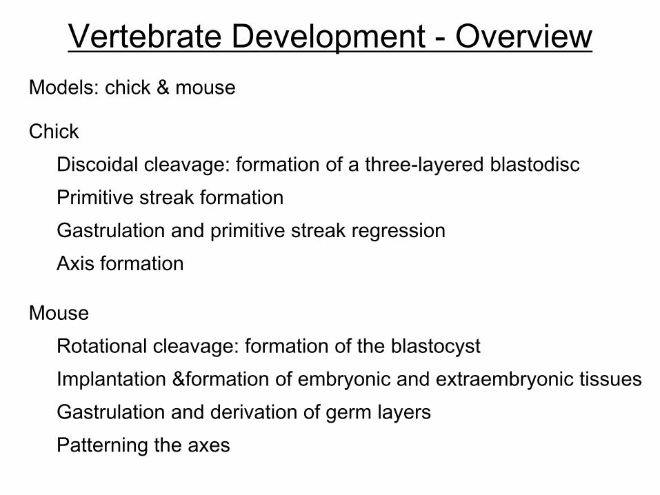
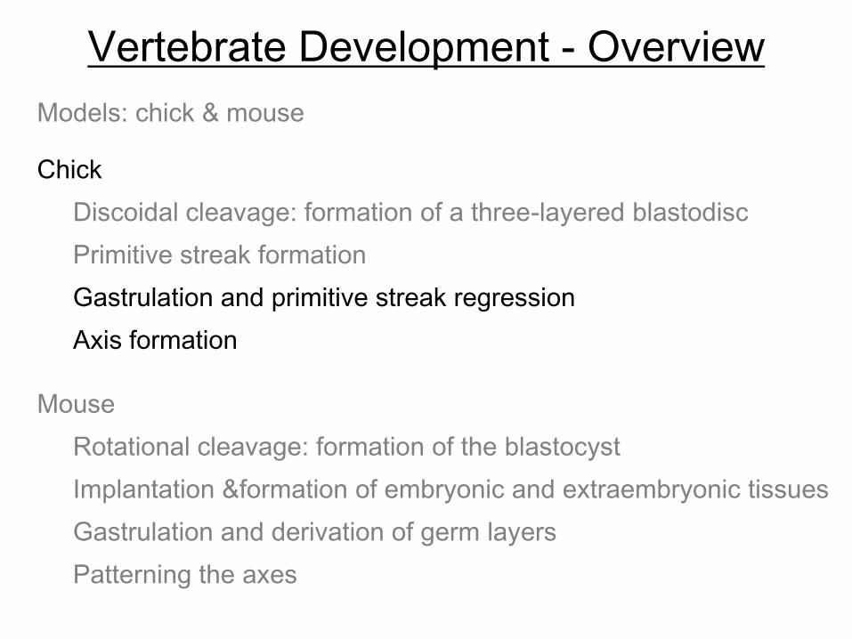
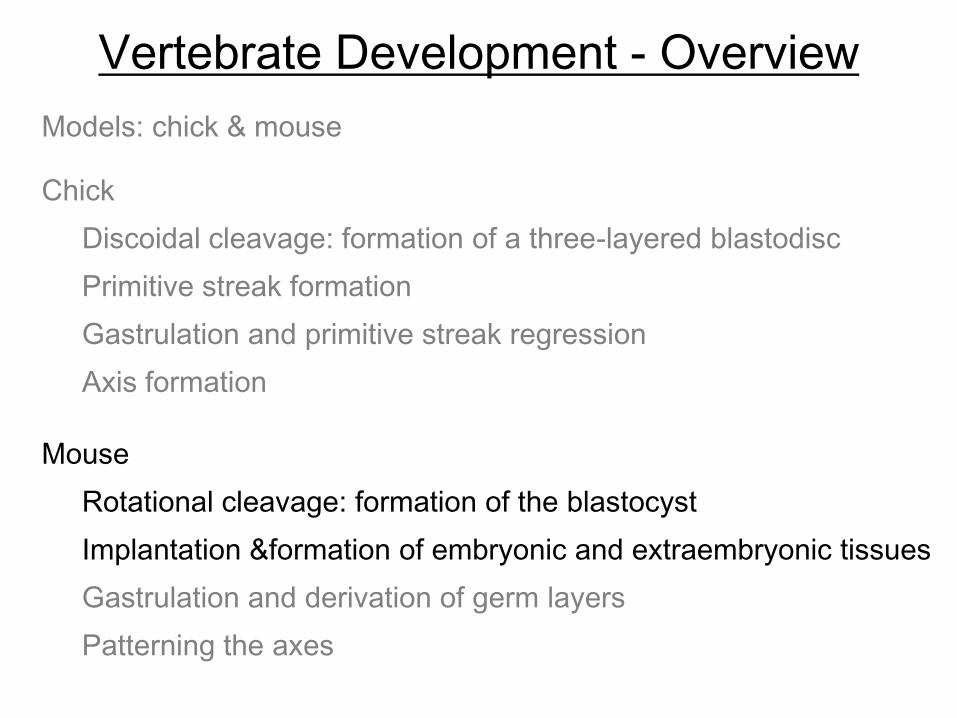
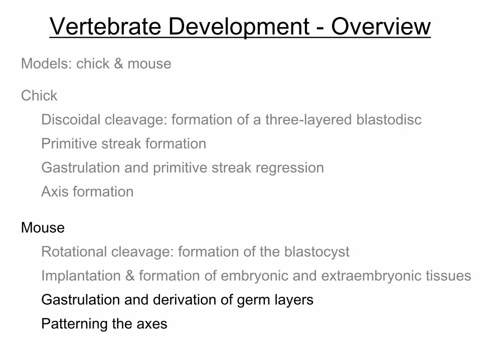
Vertebrate Development - Overview
Models: chick & mouse
Mouse
Rotational cleavage: formation of the blastocyst
Implantation &formation of embryonic and extraembryonic tissues
Gastrulation and derivation of germ layers
Patterning the axes
Discoidal cleavage: formation of a three-layered blastodisc
Gastrulation and primitive streak regression
Chick
Axis formation
Primitive streak formation
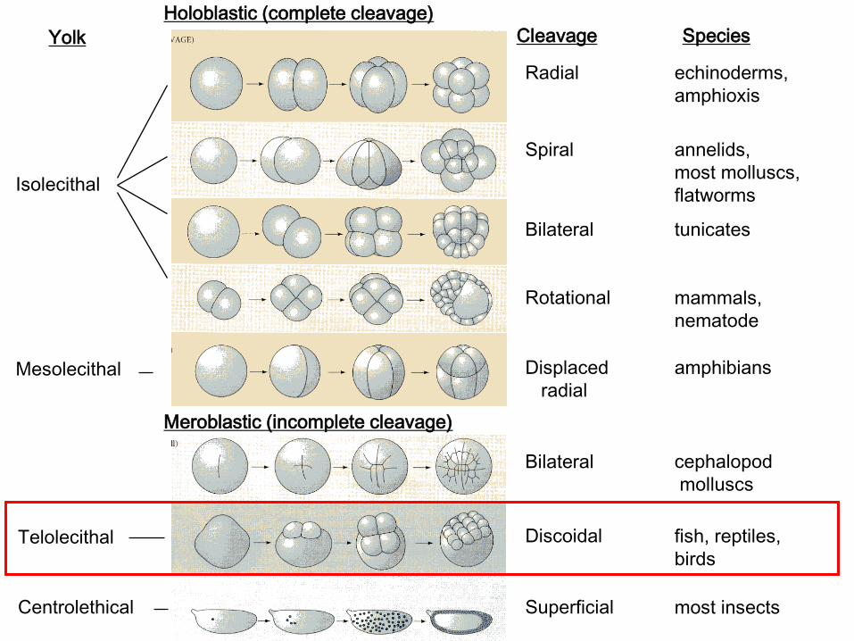
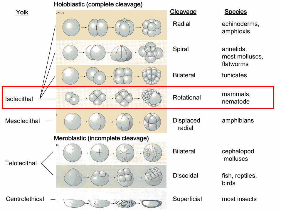
Cleavage Patterns
Meroblastic (incomplete cleavage)
Holoblastic (complete cleavage)
cephalopod
molluscs
fish, reptiles,
birds
most insects
echinoderms,
amphioxis
annelids,
most molluscs,
flatworms
tunicates
mammals,
nematode
amphibians
Species
Spiral
Bilateral
Radial
Rotational
Displaced
radial
Bilateral
Discoidal
Superficial
CleavageYolk
Isolecithal
Centrolethical
Mesolecithal
Telolecithal
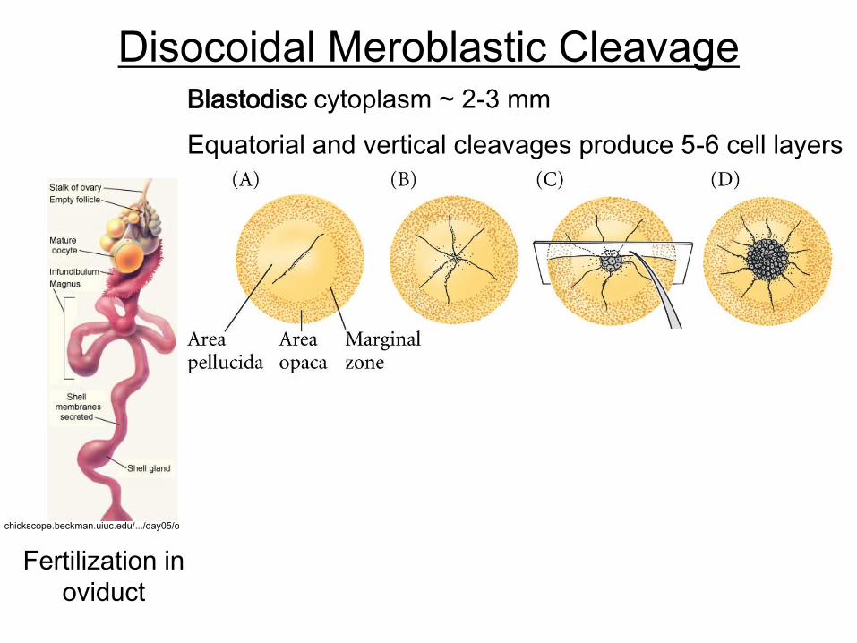
Disocoidal Meroblastic Cleavage
Fertilization in
oviduct
chickscope.beckman.uiuc.edu/…/day05/ovary.html
Blastodisc cytoplasm ~ 2-3 mm
Equatorial and vertical cleavages produce 5-6 cell layers
- vertical layers eventually reduced
to single layer - epiblast

Three-Layered Blastodisc
- 5 – 6 cell layer blastodisc sheds cells in the center- single layer remains = area pellucida (epiblast)
- edges – area opaca
- marginal zone – important for determining cell fate
- some area pellucida cells detach; form poly-invagination islands
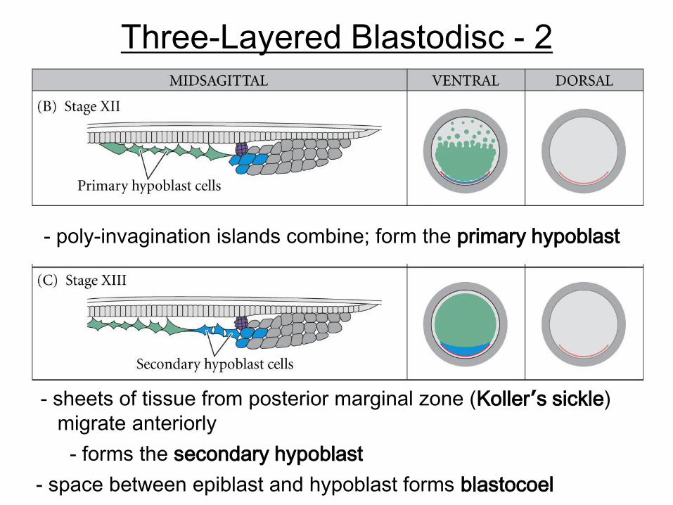
Three-Layered Blastodisc - 2
- sheets of tissue from posterior marginal zone (Koller’s sickle)
migrate anteriorly
- poly-invagination islands combine; form the primary hypoblast
- forms the secondary hypoblast
- space between epiblast and hypoblast forms blastocoel
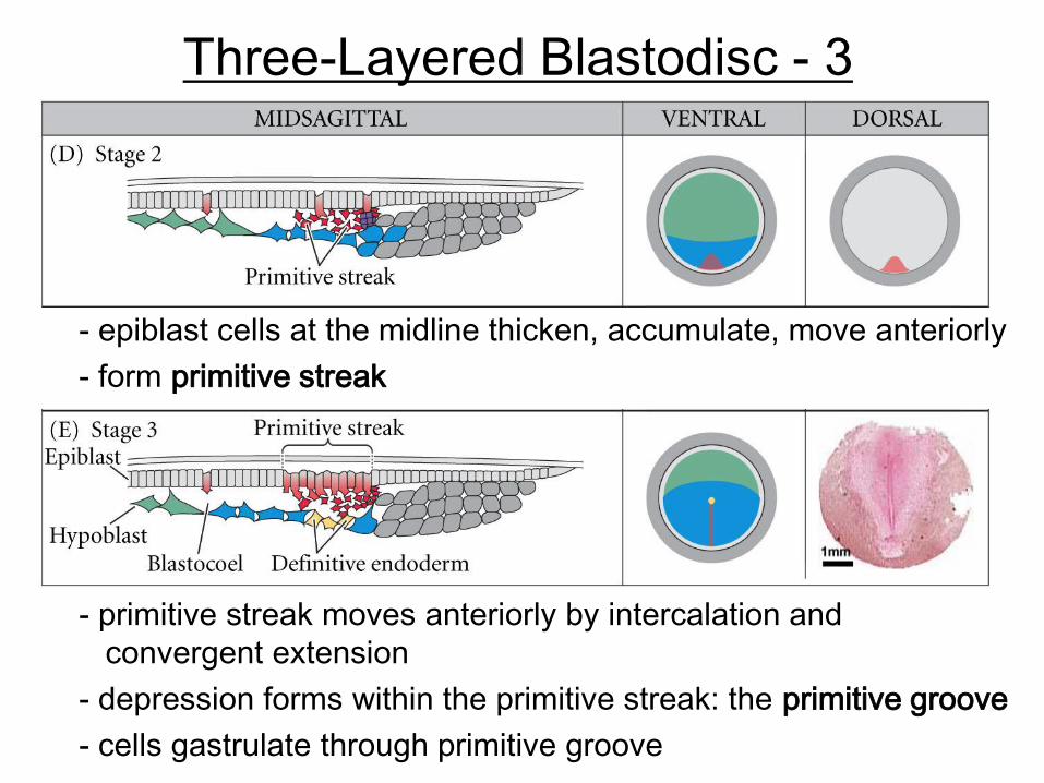
Three-Layered Blastodisc - 3
- epiblast cells at the midline thicken, accumulate, move anteriorly
- primitive streak moves anteriorly by intercalation and
convergent extension
- depression forms within the primitive streak: the primitive groove
- cells gastrulate through primitive groove
- form primitive streak
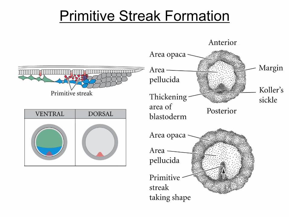
Primitive Streak Formation
Primitive
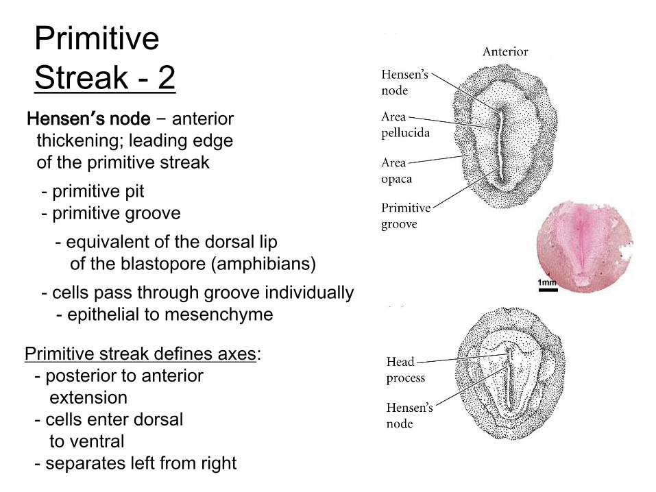
Streak - 2Hensen’s node – anterior
thickening; leading edge
of the primitive streak
- primitive pit
- primitive groove
- equivalent of the dorsal lip
of the blastopore (amphibians)
- cells pass through groove individually
- epithelial to mesenchyme
Primitive streak defines axes:
- posterior to anterior
extension
- cells enter dorsal
to ventral
- separates left from right

Primitive
Streak - 2
somites

Primitive Streak - 3
Hensen’s node extends
60 – 75% length of
area pellucida;
then regresses
- creates posterior
dorsal axis
Posterior forms
anal region
NOTE – avian embryos
exhibit distinct
anterior-to-posterior
gradient of
developmental maturity
Vertebrate Development - Overview
Models: chick & mouse
Mouse
Rotational cleavage: formation of the blastocyst
Implantation &formation of embryonic and extraembryonic tissues
Gastrulation and derivation of germ layers
Patterning the axes
Discoidal cleavage: formation of a three-layered blastodisc
Gastrulation and primitive streak regression
Chick
Axis formation
Primitive streak formation
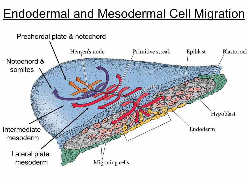
Endodermal and Mesodermal Cell Migration
Prechordal plate & notochord
Notochord &
somites
Intermediate
mesoderm
Lateral plate
mesoderm
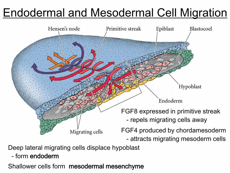
Endodermal and Mesodermal Cell Migration
FGF8 expressed in primitive streak
- repels migrating cells away
FGF4 produced by chordamesoderm
- attracts migrating mesoderm cells
Deep lateral migrating cells displace hypoblast
- form endoderm
Shallower cells form mesodermal mesenchyme
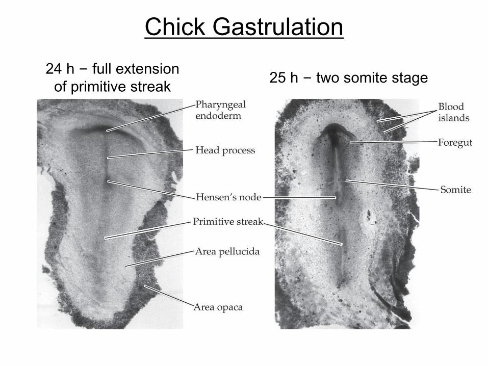
Chick Gastrulation
24 h – full extension
of primitive streak25 h – two somite stage
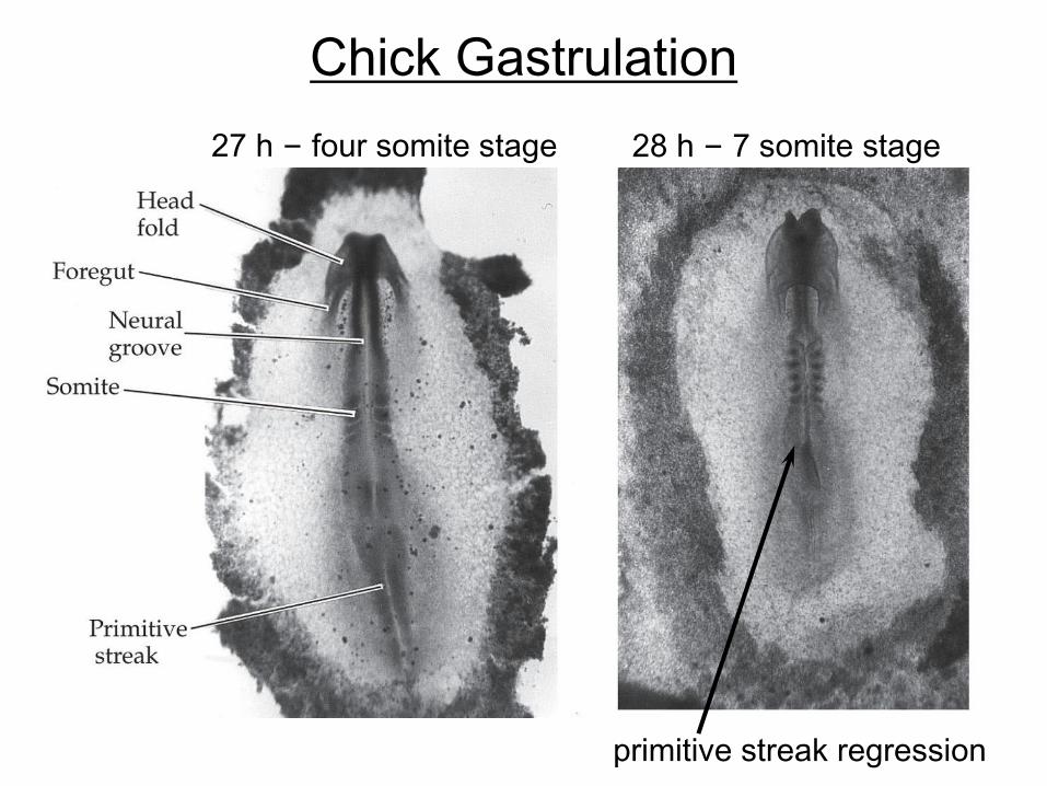
Chick Gastrulation
27 h – four somite stage 28 h – 7 somite stage
primitive streak regression
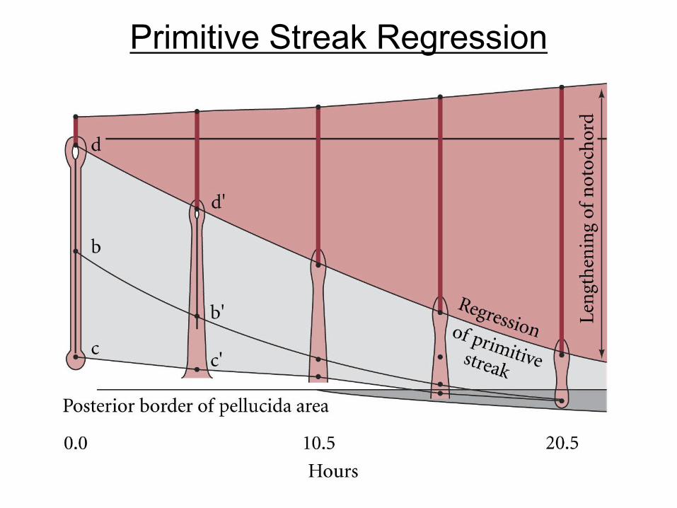
Primitive Streak Regression
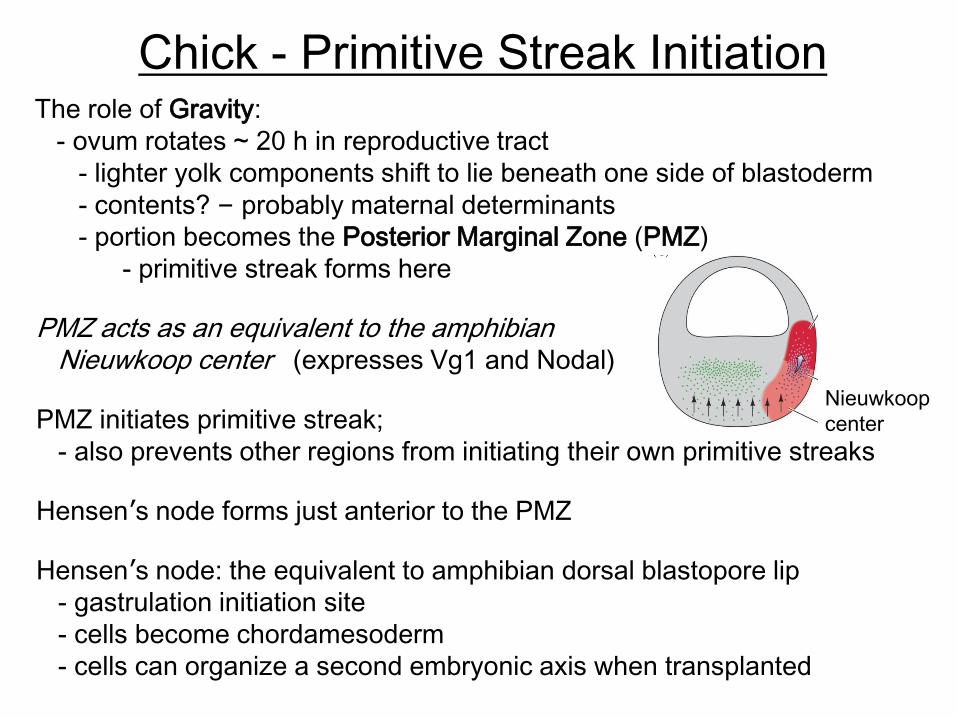
Chick - Primitive Streak InitiationThe role of Gravity:
- ovum rotates ~ 20 h in reproductive tract
- lighter yolk components shift to lie beneath one side of blastoderm
- contents? – probably maternal determinants
- portion becomes the Posterior Marginal Zone (PMZ)
- primitive streak forms here
PMZ acts as an equivalent to the amphibian Nieuwkoop center (expresses Vg1 and Nodal)
PMZ initiates primitive streak;
- also prevents other regions from initiating their own primitive streaks
Hensen’s node forms just anterior to the PMZ
Nieuwkoop
center
Hensen’s node: the equivalent to amphibian dorsal blastopore lip
- gastrulation initiation site
- cells become chordamesoderm
- cells can organize a second embryonic axis when transplanted
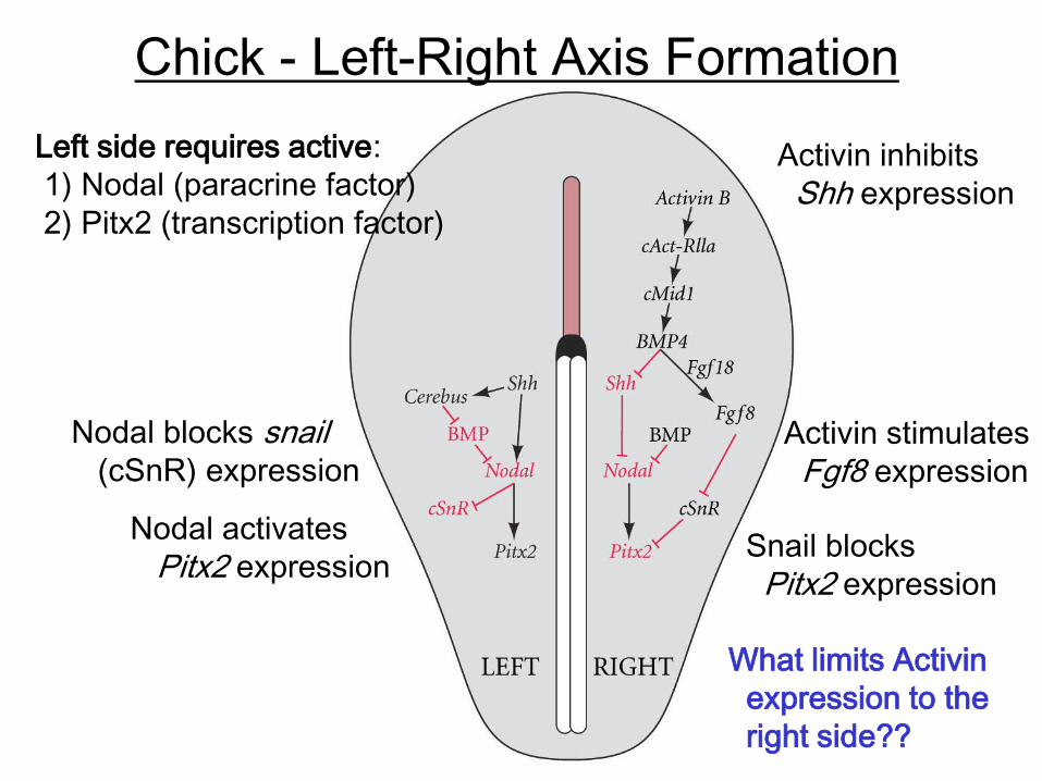
Chick - Left-Right Axis Formation
Left side requires active:
1) Nodal (paracrine factor)
2) Pitx2 (transcription factor)
Snail blocks
Pitx2 expression
Nodal blocks snail(cSnR) expression
Nodal activates
Pitx2 expression
Activin inhibits
Shh expression
Activin stimulates
Fgf8 expression
What limits Activin
expression to the
right side??
Vertebrate Development - Overview
Models: chick & mouse
Mouse
Rotational cleavage: formation of the blastocyst
Implantation &formation of embryonic and extraembryonic tissues
Gastrulation and derivation of germ layers
Patterning the axes
Discoidal cleavage: formation of a three-layered blastodisc
Gastrulation and primitive streak regression
Chick
Axis formation
Primitive streak formation
Early Development in Mammals
Mammalian development is difficult to study.
- zygotes are very small; ~ 100 μm diameter
- produced in relatively low numbers
- development inside another body makes observation
very difficult
Although mammalian eggs are isolecithal and contain
very little yolk, their embryos act as if they are sitting
on top a large imaginary ball of yolk
- i.e. gastrulate like fish, reptiles, and birds
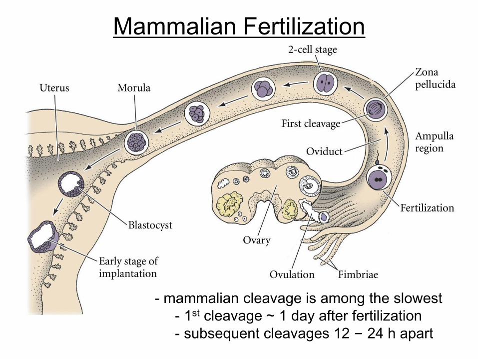
Mammalian Fertilization
- mammalian cleavage is among the slowest
- 1st cleavage ~ 1 day after fertilization
- subsequent cleavages 12 – 24 h apart
Cleavage Patterns
Meroblastic (incomplete cleavage)
Holoblastic (complete cleavage)
cephalopod
molluscs
fish, reptiles,
birds
most insects
echinoderms,
amphioxis
annelids,
most molluscs,
flatworms
tunicates
mammals,
nematode
amphibians
Species
Spiral
Bilateral
Radial
Rotational
Displaced
radial
Bilateral
Discoidal
Superficial
CleavageYolk
Isolecithal
Telolecithal
Centrolethical
Mesolecithal
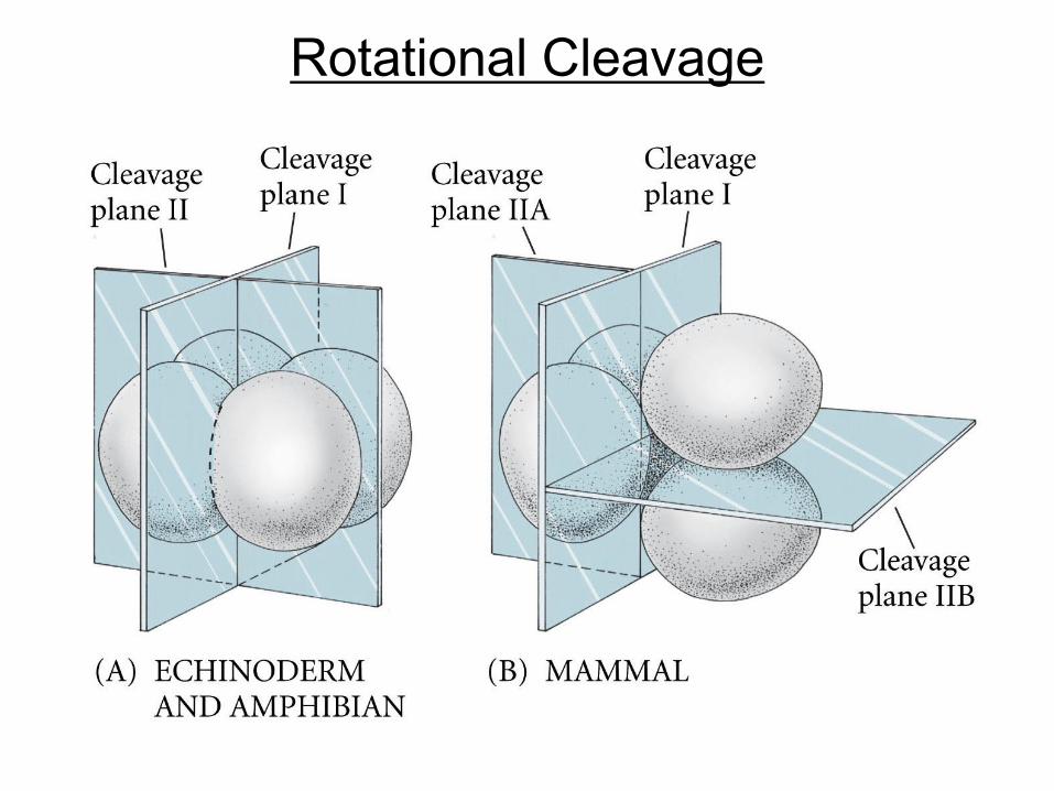
Rotational Cleavage
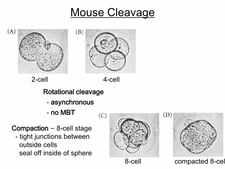
Mouse Cleavage
Rotational cleavage
- asynchronous
- no MBT
2-cell 4-cell
Compaction – 8-cell stage
- tight junctions between
outside cells
seal off inside of sphere
8-cell compacted 8-cell
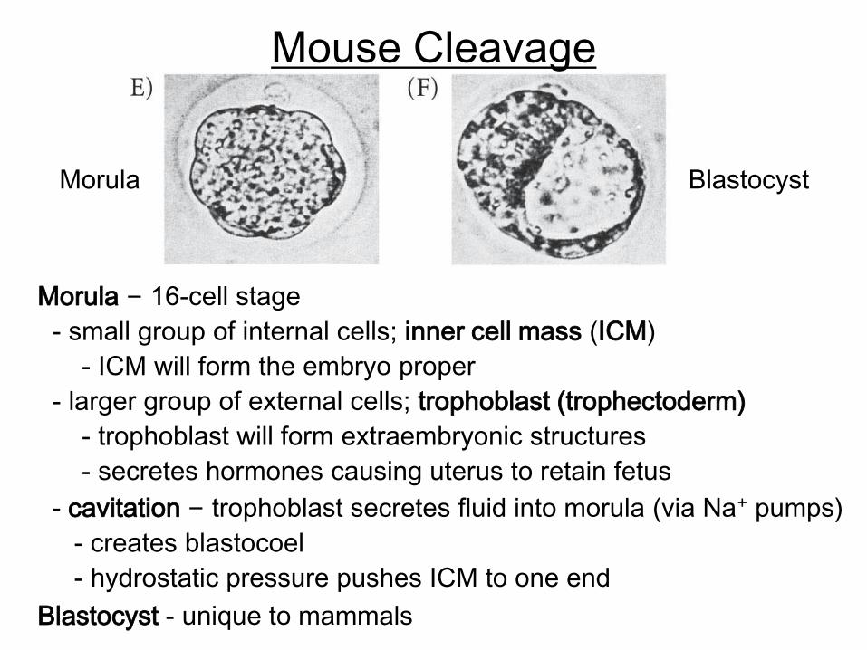
Mouse Cleavage
Blastocyst - unique to mammals
Morula – 16-cell stage
- small group of internal cells; inner cell mass (ICM)
- ICM will form the embryo proper
- larger group of external cells; trophoblast (trophectoderm)
- trophoblast will form extraembryonic structures
- secretes hormones causing uterus to retain fetus
- cavitation – trophoblast secretes fluid into morula (via Na+ pumps)
- creates blastocoel
- hydrostatic pressure pushes ICM to one end
Morula Blastocyst

Blastocyst Hatching & Implantation
Zona pellucida prevents adhesion
to uterine wall
(premature adhesion =
ectopic pregnancy)
Note – ICM cells are pluripotent
(source of embryonic stem cells)
ICM – forms the embryo proper
- also, the yolk sac, allantois,
and amnion
Trophoblast secretes proteases
- digests uterine ECM
- blastocyst implants
Trophoblast attaches to uterine wall
- forms the chorion – embryonic
portion of the placenta
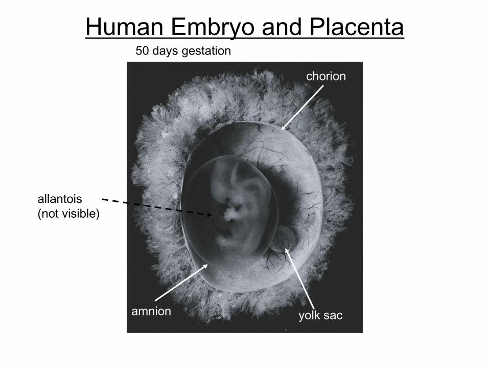
Human Embryo and Placenta
chorion
yolk sacamnion
50 days gestation
allantois
(not visible)
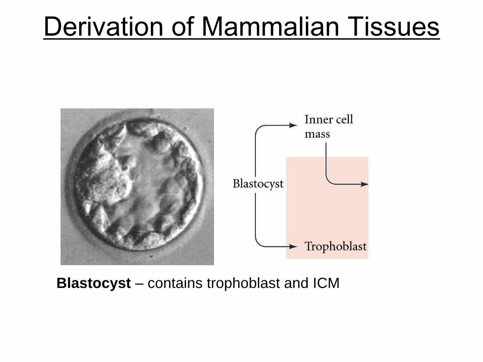
Derivation of Mammalian Tissues
Blastocyst – contains trophoblast and ICM
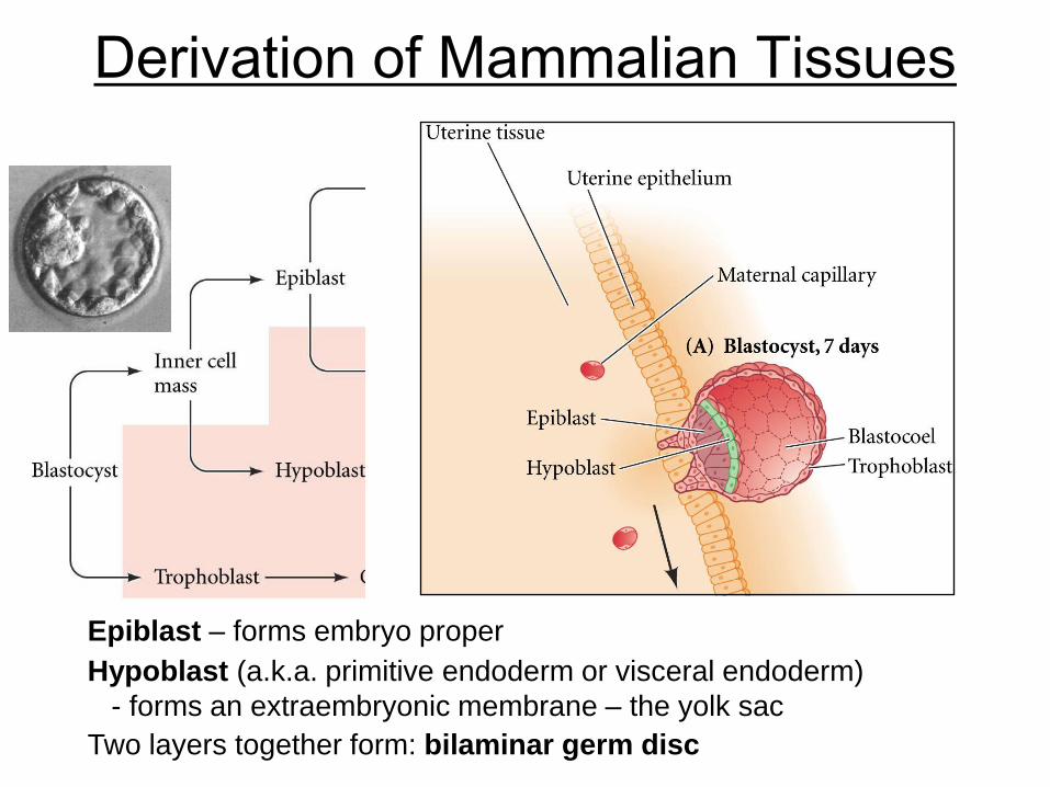
Derivation of Mammalian Tissues
Hypoblast (a.k.a. primitive endoderm or visceral endoderm)
- forms an extraembryonic membrane – the yolk sac
Epiblast – forms embryo proper
Two layers together form: bilaminar germ disc
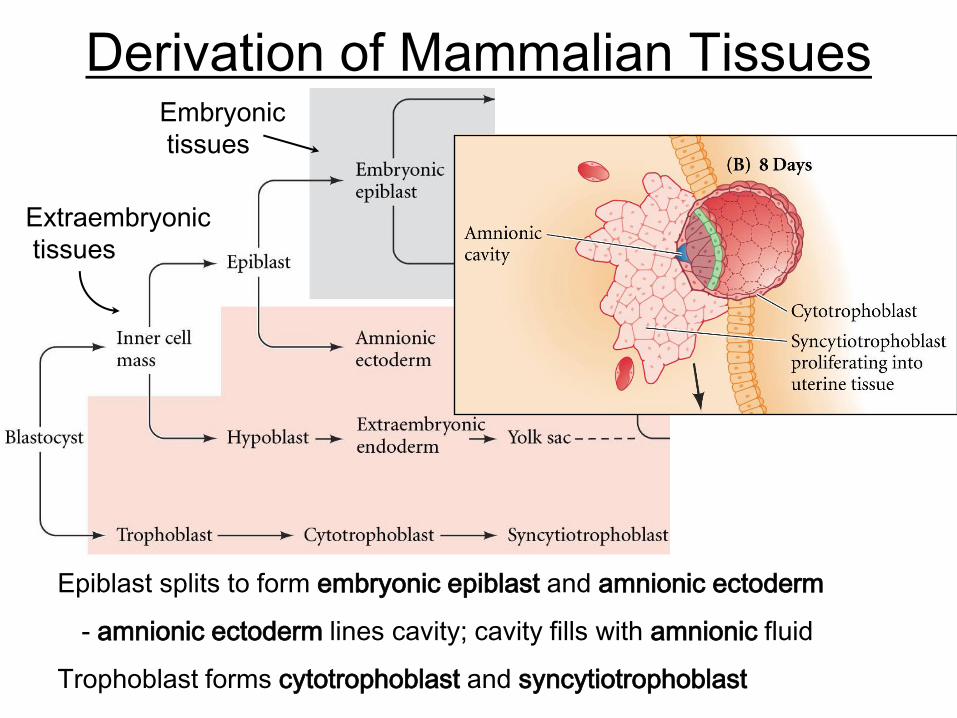
Derivation of Mammalian Tissues
Epiblast splits to form embryonic epiblast and amnionic ectoderm
- amnionic ectoderm lines cavity; cavity fills with amnionic fluid
Trophoblast forms cytotrophoblast and syncytiotrophoblast
Embryonic
tissues
Extraembryonic
tissues
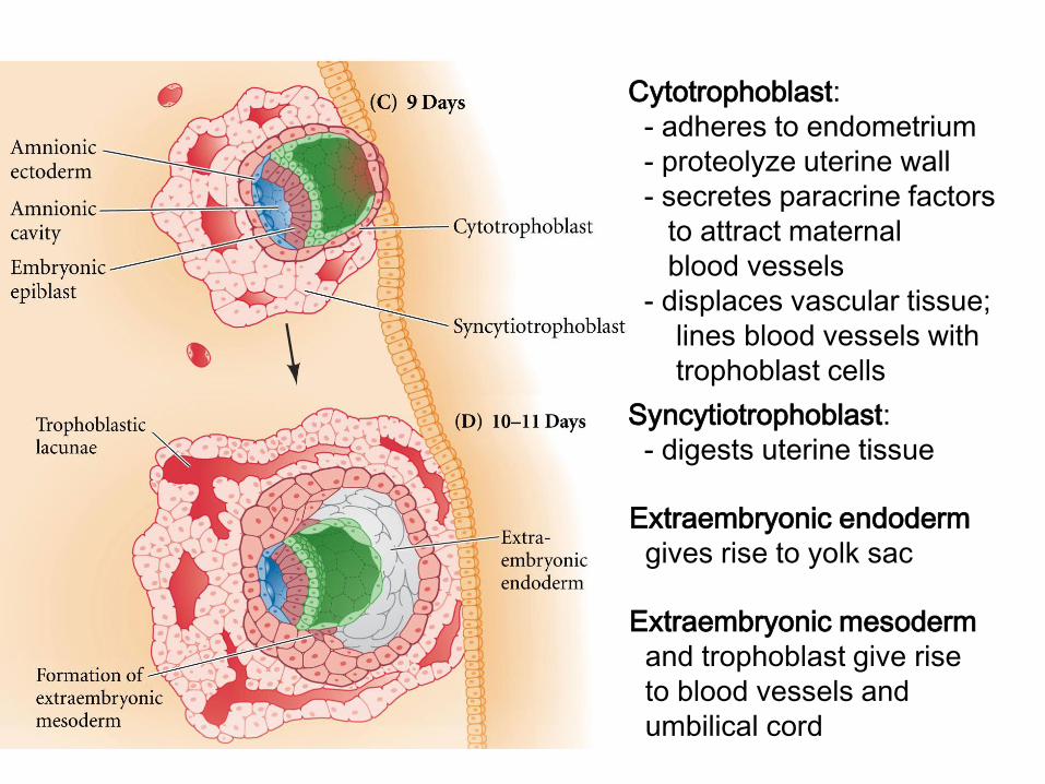
Cytotrophoblast:
- adheres to endometrium
- proteolyze uterine wall
- secretes paracrine factors
to attract maternal
blood vessels
- displaces vascular tissue;
lines blood vessels with
trophoblast cells
Syncytiotrophoblast:
- digests uterine tissue
Extraembryonic mesoderm
and trophoblast give rise
to blood vessels and
umbilical cord
Extraembryonic endoderm
gives rise to yolk sac
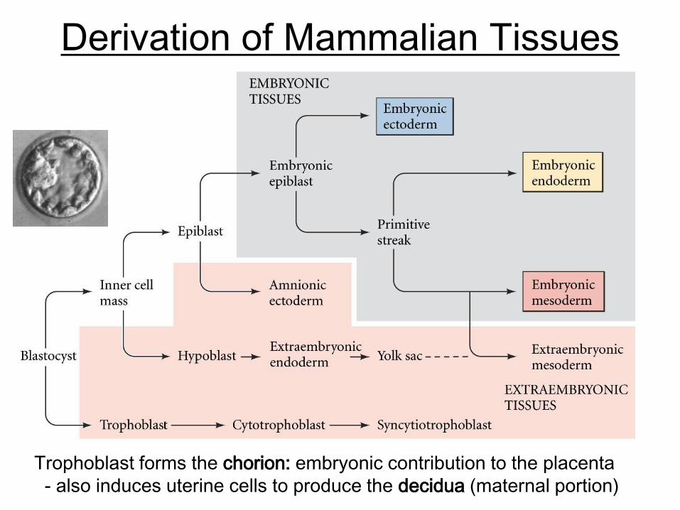
Derivation of Mammalian Tissues
Trophoblast forms the chorion: embryonic contribution to the placenta
- also induces uterine cells to produce the decidua (maternal portion)
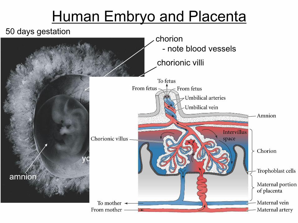
Human Embryo and Placenta
chorion
- note blood vessels
yolk sac
amnion
50 days gestation
chorionic villi
Vertebrate Development - Overview
Models: chick & mouse
Mouse
Rotational cleavage: formation of the blastocyst
Implantation & formation of embryonic and extraembryonic tissues
Gastrulation and derivation of germ layers
Patterning the axes
Discoidal cleavage: formation of a three-layered blastodisc
Gastrulation and primitive streak regression
Chick
Axis formation
Primitive streak formation
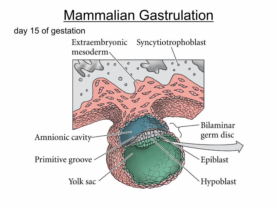
Mammalian Gastrulationday 15 of gestation
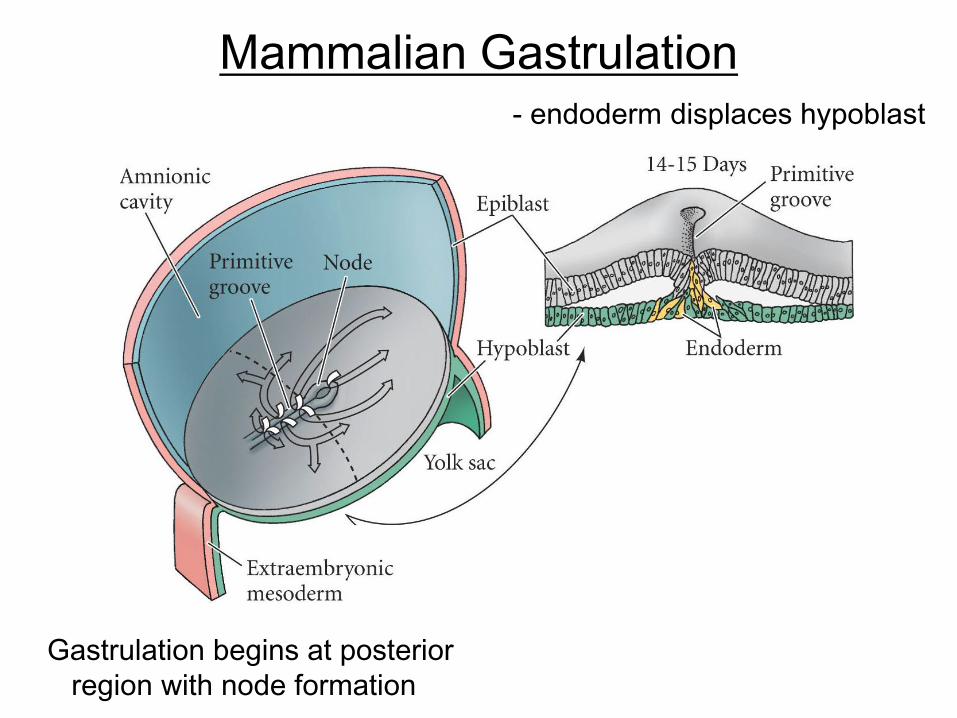
Mammalian Gastrulation
Gastrulation begins at posterior
region with node formation
- endoderm displaces hypoblast
- mesoderm follows endoderm

Anterior-Posterior Patterning2) BMP and Wnt
antagonists
(e.g. Chordin)
expressed by
the node,
notochord,
and head
mesoderm
Retinoic acid gradient: Low anterior – high posterior
1) Gradients of Wnts,
BMPs, FGFs form
from posterior
to anterior
- BMPs, Wnts,
FGFs are all
posterior/
mesoderm
determinants
A-P patterning determined by Hox genes
Summary: Posterior regions determined by RA, BMPs, Wnts, & FGFs
Anterior determined (in part) by blocking posterior signals
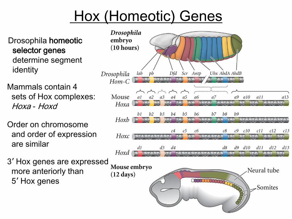
Hox (Homeotic) Genes
Drosophila homeotic
selector genes
determine segment
identity
Mammals contain 4
sets of Hox complexes:
Hoxa - Hoxd
Order on chromosome
and order of expression
are similar
3’ Hox genes are expressed
more anteriorly than
5’ Hox genes
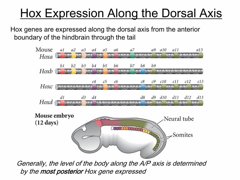
Hox Expression Along the Dorsal Axis
Hox genes are expressed along the dorsal axis from the anterior
boundary of the hindbrain through the tail
Generally, the level of the body along the A/P axis is determined by the most posterior Hox gene expressed

Hox Expression Along the Dorsal Axis
e.g. expression of different Hox genes
specify different vertebrae type; in mice:
7 cervical (neck)
14 thoracic (rib)
6 lumbar (abdominal)
4 sacral (hip)
variable number of tail
Hox knockouts (all paralogous copies) =
shifts in vertebra identity
Hox 10 -/-
Hox 11 -/-
Many Hox genes are sensitive to retinoic acid
- RA gradient (high posterior)
- controlled by differential synthesis
and degradation
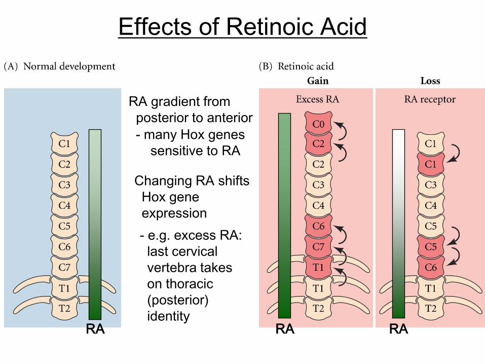
Effects of Retinoic Acid
RA gradient from
posterior to anterior
- many Hox genes
sensitive to RA
RA
Changing RA shifts
Hox gene
expression
- e.g. excess RA:
last cervical
vertebra takes
on thoracic
(posterior)
identityRA RA
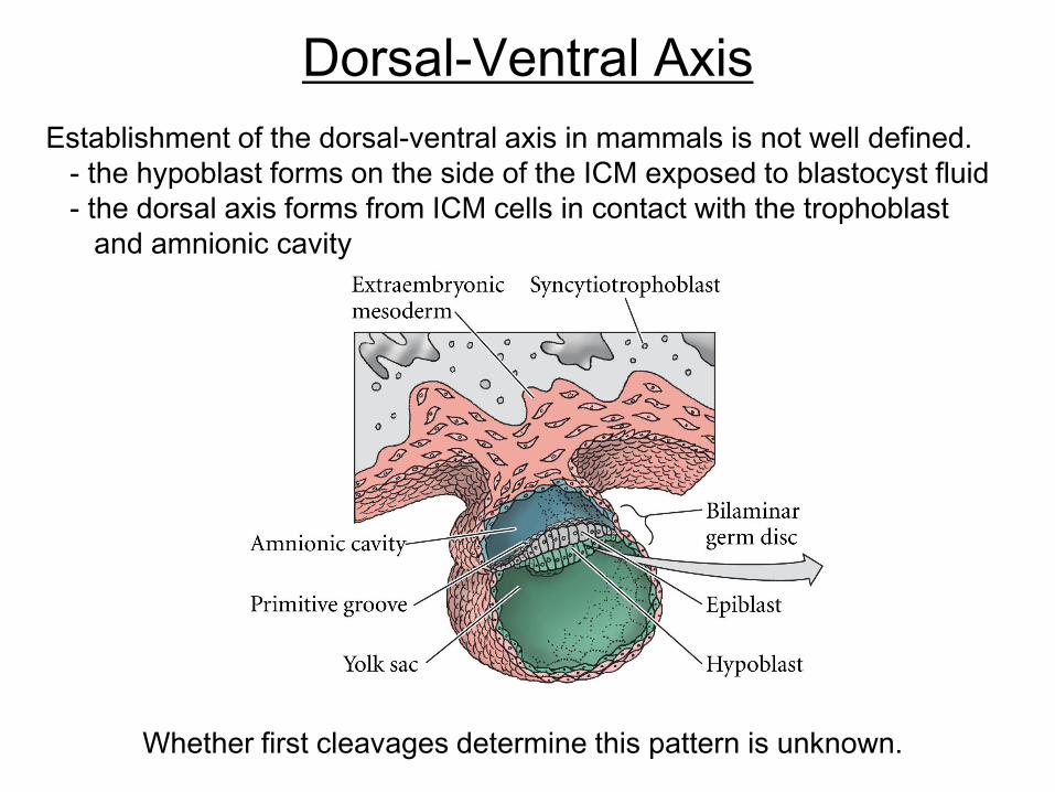
Dorsal-Ventral Axis
Establishment of the dorsal-ventral axis in mammals is not well defined.
- the hypoblast forms on the side of the ICM exposed to blastocyst fluid
- the dorsal axis forms from ICM cells in contact with the trophoblast
and amnionic cavity
Whether first cleavages determine this pattern is unknown.
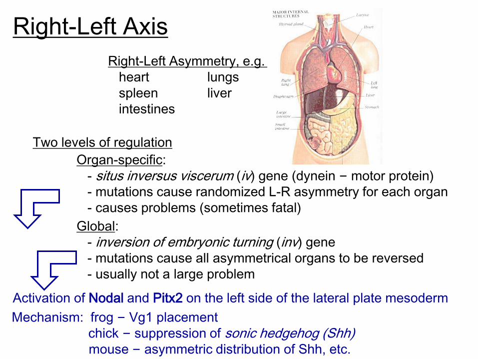
Right-Left Axis
Two levels of regulation
Organ-specific:
- situs inversus viscerum (iv) gene (dynein – motor protein)
- mutations cause randomized L-R asymmetry for each organ
- causes problems (sometimes fatal)
Global:
- inversion of embryonic turning (inv) gene
- mutations cause all asymmetrical organs to be reversed
- usually not a large problem
Right-Left Asymmetry, e.g.
heart lungs
spleen liver
intestines
Activation of Nodal and Pitx2 on the left side of the lateral plate mesoderm
Mechanism: frog – Vg1 placement
chick – suppression of sonic hedgehog (Shh)mouse – asymmetric distribution of Shh, etc.
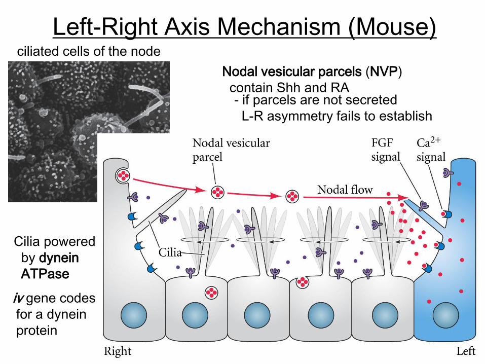
Left-Right Axis Mechanism (Mouse)ciliated cells of the node
Cilia powered
by dynein
ATPase
iv gene codes
for a dynein
protein
Nodal vesicular parcels (NVP)
contain Shh and RA- if parcels are not secreted
L-R asymmetry fails to establish