Embed Size (px)
Citation preview

s521
Acta Scientiae Veterinariae. 38(Supl 2): s521-s533, 2010.
ISSN 1678-0345 (Print)ISSN 1679-9216 (Online)
Early development of ruminant embryos, autonomous process or theresult of a positive dialog with surrounding maternal tissues?
Pascal Mermillod1,3, Barbara Schmaltz1, Christine Perreau1, Guillaume Tsikis1, EmmanuelleMartinot1, Amanda Cordova1 & Yann Locatelli1,2
ABSTRACT
Background: After artificial insemination and multiple ovulation and embryo transfer (MOET), in vitro production ofembryos (IVP) represents the third generation of techniques aimed at a better control of animal reproduction. Thistechnique involves four major steps: oocyte collection, oocyte in vitro maturation (IVM), in vitro fertilization (IVF) andin vitro development of the resulting embryos (IVD). These different steps are now well established in domesticruminant species (cattle, sheep and goat) although the variability of the number and quality of the oocytes collectedand the low viability of frozen – thawed in vitro produced embryos still limit the large-scale use of this promisingtechnology. Beyond the potential use of IVP in breeding schemes, this technique is also required for the establishmentof new biotechnologies such as cloning and transgenesis. Additionally, the knowledge of oocyte and embryo physiologyacquired through IVP techniques may stimulate the further development of other techniques such as marker assistedand genomic selection of preimplantation embryos and also benefit to assisted procreation in human being. Thispaper will discuss the possible function of maternal environment in the regulation of early development and theconsequences of these functions for IVP, in view to improve IVP embryos viability.
Review: Comparisons between in vivo and in vitro produced embryos pointed out several differences in morphology,metabolism and gene expression. IVP embryos have a modified lipid metabolism, resulting in increased triglyceridsaccumulation, translating into different density. This altered lipid metabolism may account for differences in membranestructure and increased sensitivity to oxidative stress, resulting in lower cryoresistance of these IVP embryos. Theidentification of modified metabolic pathways leading to these lipidic disorders will provide clues for modification ofculture conditions in view to restore normal lipid metabolism through appropriate precursors supplementation of themedia. The natural embryo environment from fertilization to blastocyst stage is the oviduct. In vivo, oviduct epithelialcells provide ideal development support by regulating physico-chemical embryo microenvironment. Under in vitroconditions, the lack oviduct support may result in embryo exposure to toxic metabolites and oxidative stress. Inaddition, in vitro developing embryos may lack oviduct originated embryotrophic factors that regulate and stimulateearly development in vivo. The use of co culture systems to mimic natural embryo environment in vitro may allow toimprove embryo development, restore normal metabolic parameters, and increase embryo viability and cryoresistance.In addition, such co culture systems involving oviduct epithelial cells will help to identify critical development parametersand to point out potential embryotrophic factors.
Conclusion: In vitro embryo production is a promising technique for improvement of selection schemes and diffusionof genetic gain through safe exchanges of embryos. To allow a lager use of this technology, improvements should beobtained in management of oocyte collection and in vitro treatment to improve its quality and in embryo in vitrodevelopment systems. A better knowledge of interactions between the developing embryo and maternal environmentwill allow to improve in vitro systems to produce high viability embryos.
Keywords: Ruminants, embryo, oviduct, development, freezing
1Institut National de la Recherche Agronomique (INRA), UMR6175, Physiologie de la Reproduction et des Comportements, Nouzilly,France.2 Muséum National d’Histoire Naturelle (MNHN), Parc de la Haute Touche, Obterre, France.3 Corresponding author: INRA, Physiologie de la Reproduction et des Comportements, 37380 Nouzilly, France.([email protected]), + 33 2 47 42 79 20 ; +33 2 47 42 77 43.
11_SBTE_MERMILLOD.P65 4/8/2010, 17:33521

s522
Mermillod P, Schmaltz B, Perreau C, Tsikis G, Martinot E, Cordova A, Locatelli Y. 2010. Early development of ruminantembryos, autonomous process or the result of a positive dialog... Acta Scientiae Veterinariae. 38 (Supl 2): s521-s533
I INTRODUCTION
II OVIDUCT EFFECT ON EARLY DEVELOPMENT
III GAMETES - EMBRYO REGULATED OCIDUCT ACTIVITIES
IV CONCLUSIONS
I INTRODUCTION
The first attempts of early development of ruminant embryos in vitro resulted in a systematic block at the8-16 Cell stage [72]. This stage of development corresponds to the maternal to zygotic transition (MZT) of geneexpression in ruminant embryos [64]. The first successes of ruminant embryo development up to the blastocyststage in vitro have been obtained by using co culture systems involving oviduct epithelial cells [10,16]. Furtherevolution of this technique involved use of new media especially designed to support early embryo developmentbased on the composition of oviduct fluid, such as SOF medium [14] and bovine oviduct medium for embryo culture(BOMEC) [39] or sequential media fitted to changing embryo requirements [32]. However, it is generally accepted thatin vitro produced blastocysts are less resistant to cryopreservation procedures than in vivo derived embryos [45,58].In addition, a better knowledge of embryo – maternal interactions would provide interesting clues for deciphering earlyreproductive physiology and design improved methods for embryo in vitro production (IVP) in mammals [52]. Recentstudies comparing bovine oocyte maturation, fertilization and embryo culture in vivo vs. in vitro have demonstratedthat the origin of the oocyte is the main factor affecting blastocyst yield while the post-fertilization culture environmentis critical in determining blastocyst quality, measured in terms of cryotolerance and relative transcript abundancewhatever the origin of the oocyte [57,59]. Early development is often considered as an autonomous process, regulatedby the embryo itself (maternal transcripts and embryo transcription activity). However, it is clear that embryo environmentcould act through specific sensitive windows to modulate embryo metabolism, gene expression pattern and morphologythrough epigenetically regulated mechanisms [68]. These data underline the key effect of maternal microenvironmenton the success of early embryo development in terms of viability of these embryos. In addition, early embryonicmortality may explain up to 40% of reproductive failure [24], highlighting the need for a better knowledge of earlyembryo development regulation. This paper will review some aspects of the molecular and functional dialog betweencleaving embryo and maternal surrounding, leading to efficient development and full embryo viability.
II OVIDUCT EFFECT ON EARLY DEVELOPMENT
The oviduct appears to play a crucial role in different aspects of early reproduction control like gametesfinal preparation and transport, fertilization, early embryo development [25]. The oviduct may regulate early developmentthrough several mechanisms: regulating metabolites in oviduct fluid to fit embryo requirements and metabolismcapacities, protect embryos against oxidative stress by removing toxic compounds from the medium (negativeaction), regulate embryo cell proliferation through the secretion (positive action) of growth factors [4]. In vivo studiesof these activities are difficult due to low accessibility of the oviduct (surgery requirement) and to the low quantity ofbiological material available. The use of in vitro models is required to allow the study of oviduct cell activities. Thesemodels rely on co culture of early embryos with oviduct cells. To avoid any bias, it is important to make sure that invitro systems reflect more or less the physiological situation. Therefore, the origin of oviducts as well as the culturemethods should be carefully designed to develop pertinent co culture systems [73].
Oviduct cell culture
Anatomically, the oviduct is a tube with a virtual lumen bordered by an epithelial cell layer, composed of ciliatedand secreting cells. This epithelium is surrounded by conjunctive tissue and smooth muscle cells. Bovine oviduct epithelialcells (Boec) could be collected after enzymatic digestion using trypsin or collagenase. Alternatively, Boec could be collectedmechanically, by scrapping the inner layer after longitudinal dissection of the oviduct or by gentle squeezing of the wholeorgan. Mechanical methods are less detrimental to sensitive structures like cilia and allow to collect larger numbers of cells[66]. Whereas enzymatic Boec collection provides mainly individual cell suspensions, mechanical methods allow to collectlarge sheets of epithelium. Once in culture, these sheets rapidly form vesicles bordered by a single layer of epithelial cells.The apical pole of the cells being outside of the vesicles, the cilia movements make the vesicles turning and moving in the
11_SBTE_MERMILLOD.P65 4/8/2010, 17:33522

Mermillod P, Schmaltz B, Perreau C, Tsikis G, Martinot E, Cordova A, Locatelli Y. 2010. Early development of ruminantembryos, autonomous process or the result of a positive dialog... Acta Scientiae Veterinariae. 38 (Supl 2): s521-s533
s523
medium. These vesicles could be used for embryo co culture [63] but the use of monolayers allow a better standardizationof the co culture conditions and easier access to functional study of OEC activities. Within few days, in the presence ofserum, these vesicles attach to the culture support and cells proliferate to reach confluence within 5 to 7 days [48,67].Numerous traditional media could be used to culture Boec (TCM-199, DMEM/F12, RPMI1640,…). Although TCM-199 isfrequently used, media more fitted to embryo needs, like SOF, could be used alternatively. In our laboratory, we usually useTCM-199 medium supplemented with 10% FCS to initiate Boec cultures and drive them to confluence and then replace itwith SOF supplemented with 5% FCS 24h before using them for embryo development support [60].
The origin of the oviduct (stage of the female cycle) could influence the cell differentiation status and molecularactivities [3]. Even the side of the oividuct (ipsi- vs. Contralateral of previous ovulation) as well as the oviduct anatomicalregion (isthmus vs. Ampulla) could influence protein secretion pattern [2]. For example, oviduct specific glycoprotein(OGP), one of the major oviduct secreted proteins (OSP) is expressed by oviduct cells from ovulation and decreasesafter 3 to 4 days in vivo and it is more expressed in the ampulla, as compared to isthmus [6]. However, once in culture,cell dedifferentiation may at least partially equalize these differences in molecular activities [62]. The use of specificculture systems like coated culture inserts, favouring cell attachment and polarization, could help to maintain oviductcell morphological and functional differentiation in culture [8]. As shown in Figure 1, Boec secreted proteins are more orless stable during the time period at which cells are generally used for supporting embryo development in co culture (day5 to 13), although some slight variations between secreted protein patterns could be observed. It is interesting to notethat proteins found in conditioned medium (lanes S5 and S13) do not correspond to major cellular proteins (lane Cell),indicating that they are not the result of passive release after cellular death.
Figure 1. SDS-PAGE analysis of bOEC conditioned media collected after 5 (SD5) or 13 (SD13) days of culture, compared to cell lysateobtained on Day 13. Conditioned media were concentrated 80 fold by centrifugation on Vivaspin 5 kD cut off membranes concentrators(Sartorius, Paris, France) before analysis. The gel was stained with Commassie brillant blue. Arrows indicate differential protein bandsbetween Day 5 and Day 13. Molecular weight markers are indicated on the left (kDa).
11_SBTE_MERMILLOD.P65 4/8/2010, 17:33523
s524
Mermillod P, Schmaltz B, Perreau C, Tsikis G, Martinot E, Cordova A, Locatelli Y. 2010. Early development of ruminantembryos, autonomous process or the result of a positive dialog... Acta Scientiae Veterinariae. 38 (Supl 2): s521-s533
Oviduct – embryos co culture
It has been well established that the environment encountered by the embryo during its early developmentinfluences its gene expression at the blastocyst stage [43,51]. These differences translate into modification of severalembryo parameters and may result in long-term alterations of embryo and offspring physiology [13,69]. Maternalenvironment induces higher cell number and allocation between trophectoderm and inner cell mass, modified embryogene expression pattern and embryo metabolism. All together, these modifications result in higher viability andimproved cryoresistance of the IVP embryos [57,59].
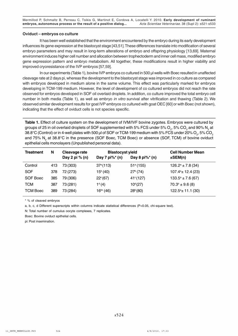
In our experiments (Table 1), bovine IVP embryos co cultured in 500 µl wells with Boec resulted in unaffectedcleavage rate at 2 days pi, whereas the development to the blastocyst stage was improved in co culture as comparedwith embryos developed in medium alone in the same volume. This effect was particularly marked for embryosdeveloping in TCM-199 medium. However, the level of development of co cultured embryos did not reach the rateobserved for embryos developed in SOF oil overlaid droplets. In addition, co culture improved the total embryo cellnumber in both media (Table 1), as well as embryo in vitro survival after vitrification and thawing (Table 2). Weobserved similar development results for goat IVP embryos co cultured with goat OEC [60] or with Boec (not shown),indicating that the effect of oviduct cells is not species specific.
Table 1. Effect of culture system on the development of IVM/IVF bovine zygotes. Embryos were cultured bygroups of 25 in oil overlaid droplets of SOF supplemented with 5% FCS under 5% O
2, 5% CO
2 and 90% N
2 at
38.8°C (Control) or in 4-well plates with 500 µl of SOF or TCM-199 medium with 5% FCS under 20% O2, 5% CO
2
and 75% N2 at 38.8°C in the presence (SOF Boec, TCM Boec) or absence (SOF, TCM) of bovine oviduct
epithelial cells monolayers (Unpublished personal data).
Treatment N Cleavage rate Blastocyst yield Cell Number MeanDay 2 pi % (n) Day 7 pi%* (n) Day 8 pi%* (n) ±SEM(n)
Control 413 73 (303) 37a (113) 51a (155) 126.2a ± 7.8 (34)
SOF 378 72 (273) 15b (40) 27b (74) 107.4a± 12.4 (23)
SOF Boec 385 79 (306) 22c (67) 41c (127) 133.5a ± 7.6 (67)
TCM 387 73 (281) 1d (4) 10d (27) 70.3b ± 9.6 (8)
TCM Boec 389 73 (284) 16bc (46) 28b (80) 122.5a± 11.1 (30)
* % of cleaved embryos
a, b, c, d Different superscripts within columns indicate statistical differences (P<0.05, chi-square test).
N: Total number of cumulus oocyte complexes, 7 replicates.
Boec: Bovine oviduct epithelial cells.
pi: Post insemination.
11_SBTE_MERMILLOD.P65 4/8/2010, 17:33524

Mermillod P, Schmaltz B, Perreau C, Tsikis G, Martinot E, Cordova A, Locatelli Y. 2010. Early development of ruminantembryos, autonomous process or the result of a positive dialog... Acta Scientiae Veterinariae. 38 (Supl 2): s521-s533
s525
Table 2. Effect of culture system on the in vitro survival of vitrified bovine IVP embryos. IVP embryos weredeveloped by groups of 25 in oil overlaid droplets of SOF supplemented with 5% FCS under 5% O
2, 5% CO
2
and 90% N2 at 38.8°C (Control) or in 4-well plates with 500 µl of SOF or TCM-199 medium with 5% FCS under
20% O2, 5% CO
2 and 75% N
2 at 38.8°C in the presence (SOF Boec, TCM Boec) or absence (SOF, TCM) of
bovine oviduct epithelial cells monolayers. At 7 days post insemination, blastocysts were vitrified – thawedand placed in culture in 4-well plates in 500 µl of SOF supplemented with 5% FCS for 48h. Survival (reexpansionand/or hatching) was evaluated at different times of post thawing culture (5, 24 and 48h) (Unpublished personaldata).
Treatment Thawed Survival rate post-thawingembryos (n) 5h% (n) 24h% (n) 48h% (n) 48h*% (n)
Control 67 43ax (29) 18bx (12) 9bx (6) 10bx (3)
SOF 32 22ay (7) 22ax (7) 0by 0by
SOF Boec 51 59az (30) 59az (30) 39az (20) 30az (9)
TCM 10 17ay (2) 0by 0by 0by
TCM Boec 33 67az (22) 67az (22) 54az (18) 41az (9)
* % of hatched embryos/number of blastocysts 5h after thawing.
Within lines, values with different letters (a–c) differ significantly (P < 0.05, Chi-square).
Within columns, values with different letters (x–z) differ significantly (P < 0.05, Chi-square).
One of the possible effect of somatic cells in co culture is the regulation of gas partial pressure in themedium and particularly the diminution of oxygen [14,42]. In our co culture system (Table 3), we observed that therate of embryo development to the blastocyst stage was significantly reduced under 20% O
2 and that this effect was
suppressed in co culture. In addition, the rate of development in co culture under 20% O2 in SOF or TCM-199 was
higher than the development in the same media under 5% O2 without co culture, indicating that Boec exert othereffects in addition to the control of oxygen level.
11_SBTE_MERMILLOD.P65 4/8/2010, 17:33525

s526
Mermillod P, Schmaltz B, Perreau C, Tsikis G, Martinot E, Cordova A, Locatelli Y. 2010. Early development of ruminantembryos, autonomous process or the result of a positive dialog... Acta Scientiae Veterinariae. 38 (Supl 2): s521-s533
Table 3. Effect of culture system on the development of IVM/IVF bovine zygotes. Embryos were cultured bygroups of 25 in oil overlaid droplets of SOF supplemented with 5% FCS (Control) or in 4-well plates with 500 µlof SOF or TCM-199 medium with 5% FCS in the presence of 5% O
2, 5% CO
2 and 90% N
2 (group 5% O
2). In
group 20% O2, embryos were cultured in the same media in the presence or absence of bovine oviduct
epithelial cells monolayers under 20% O2, 5% CO
2, 75% N
2 atmosphere (Unpublished personal data).
Treatment n No. of cleaved Blastocyst yield No. of Hatchedoocytes blastocystDay 2 pi % (n) Day 7 pi%* (n) Day 8 pi%* (n) Day 8 pi % (n)
5% O2
Control 171 59 (101) 22a (22) 30ac (30) 30ac (34)
SOF 161 65 (104) 19a (20) 19a (20) 40ac (23)
TCM 158 73 (116) 5b (6) 10b (11) 0b
20% O2
SOF 137 65 (89) 6b (5) 6b (5) 0b
TCM 170 63 (107) 4b (4) 4b (4) 0b
SOF Boec 187 67 (126) 24a (30) 35c (44) 43a (19)
TCM Boec 185 66 (122) 17a (21) 27ac(33) 23c (7)
* % of cleaved/embryos
a, b, c, d Different superscripts within columns indicate statistical differences (P<0.05, Chi-square).
n: Total number of cumulus oocytes complexes, 3 replicates.
Boec: Bovine oviduct epithelial cells.
pi: Post insemination.
We used the goat model to confirm increased viability of vitrified – thawed IVP embryos after OEC coculture [60]. Results are represented in Table 4. Vitrified – thawed IVP embryos displayed a very low survival (9% ofkids born) as compared with fresh ones (62% of kids born). Survival of IVP embryos was increased after Goec coculture (33%% of kids born) but remained significantly lower compared to fresh ones, indicating that co culture tendto restore in vivo-like development conditions but that the method remains to be improved. Any further improvementof IVP technique in terms of quality, viability and cryoresistance of the embryos produced will require a betterknowledge of the embryo maternal communication regulating early development [52].
Table 4. Effect of culture system on the viability of goat IVP embryos. Embryos were developed in SOFsupplemented with 5% FCS with our without goat oviduct epithelial cells support. They were then vitrified at theblastocyst stage (Day 7 pi), two embryos per straw [20]. Straws were thawed and their content was transferredto synchronized recipients. Pregnancy rate was evaluated at Days 34 and 90 by echography and kiddings wererecorded. Modified from [60].
Treatment Recip. Emb. Pregnant Day PregnantDay Kidding Kids bornN n 34n (%) 90n (%) n (%) n (%)
SOF Fresh 13 26 12a (92) 12a (92) 12a (92) 16a (62)
SOF vit. 29 58 6b (21) 4b (14) 4b (14) 5b (9)
gOEC vit. 18 36 13a (72) 10c (56) 10c (56) 12c (33)
a,b,c Values with different superscripts in the same column are significantly different (P<0.05, Chi-square).
gOEC: goat oviduct epithelial cells.
pi: Post insemination.
11_SBTE_MERMILLOD.P65 4/8/2010, 17:33526

Mermillod P, Schmaltz B, Perreau C, Tsikis G, Martinot E, Cordova A, Locatelli Y. 2010. Early development of ruminantembryos, autonomous process or the result of a positive dialog... Acta Scientiae Veterinariae. 38 (Supl 2): s521-s533
s527
Oviduct secreted proteins
The knowledge of embryo – maternal communication in the oviduct relies on the identification of oviductsecreted proteins (OSP). Many OSP have been identified already [18,28]. In our co culture system, we investigatedthe level of expression in oviduct cells at different culture times of transcripts encoding for some of these proteinsinvolved in different mechanism that can explain early development support. The expression of these genes werecompared in freshly collected cells (F0), at Day 5 of culture, representing the beginning of confluence and the momentof co culture initiation and at Day 13, representing the end of co culture, for cells cultured in SOF or TCM-199 with orwithout developing embryos.
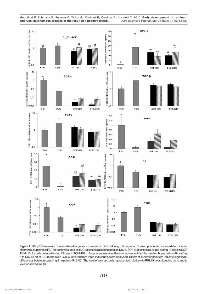
We quantified the mRNA two antioxidant enzymes: Copper, Zinc Superoxide Dismutase (Cu,Zn-SOD) andPhospholipid Hydroperoxide Glutathione Peroxidase (GPx-4). Indeed, Cu,Zn-SOD scavenges superoxide radicalsand GPx-4 reduces lipid hydroperoxides and H
2O
2, the by-product of Cu,Zn-SOD action [34,44,53]. As shown in Figure
2, Boec expressed continuously Cu,Zn-SOD during all the culture period. This result is in accordance with thoseobtained in vivo showing constant Cu,Zn-SOD Mrna level all along the oviduct and a constant enzymatic activitythroughout the oestrus cycle [33]. GPx-4 transcript has been detected throughout the culture with a significantincrease at confluence (Day 5) followed by a steady level up to the end of the culture (Day 13). GPx-4 is probablystimulated by oxidative stress due to high oxygen concentration (20%) during initial Boec culture. Indeed, oxygentension in the oviduct is approximately 5-6 % [44]. GPX-4 is the only GPx family member with the ability to reducelipid hydroperoxides bound to cell membrane. GPx-4 mRNA expression in bovine oviduct is up-regulated at theperiovulatory period [34] which is characterized by increased lipid synthesis [22]. Then, in our experiments, thebeneficial effect of Boec on blastocyst rate and quality may be at least partly explained by detoxification of highlydiffusible ROS by Boec secreted SOD and GPx-4 antioxidant enzymes. However, blastocyst rates were alwayshigher in Boec co-culture system even under a reduced oxygen concentration, suggesting that Boec may have otherways to support blastocyst formation. This additional effect could be mediated by growth factors.
In our experiments, control embryos cultured in reduced volume of SOF (1 µl per embryo) showed higherblastocyst and hatching rates as well as cell number per embryo as compared with those produced in a larger volumeof SOF (500 µl). These results are consistent with those obtained in mice by [53] showing that embryos culturedsingly had a lower development rate as compared with those cultured in groups in the same volume of medium (25µl).They also reported that the development of embryos cultured singly was markedly improved by the addition of growthfactors (EGF, TGF-α and TGF-β), indicating that these growth factors may mediate the cooperation between embryos.A similar collaboration between developing embryos was also reported in cattle [12]. Growth factors secretion may atleast partly account for the positive effect of Boec observed in our study. In view to test this hypothesis, we quantifiedthe Mrna of some growth factors known to have a role in embryo development such as insulin-like growth factors IGF-I and –II, transforming growth factor-α and –β1 (TGF-α and TGF-α1) , and basic fibroblast growth factor (FGF2).
TGF-α mRNA significantly decreased throughout the culture and particularly after 13 days of culture whateverthe medium. This decrease was partly abolished in the presence of embryos. TGF-α expression was found in theinner cell mass cells and antisense oligonucleotides against EGF receptor attenuated embryo cell proliferation [5].Therefore, TGF-α produced by inner cell mass may stimulate trophectoderm proliferation through EGF receptors in aparacrine manner. Oviduct produced TGF-α may enhance this paracrine mechanism.
FGF2 and TGF-α1 expression in Boec cultured during 13 days with or without embryos did not differ. FGF2is an activator of protein synthesis and a potent mitogen [35]. The exposure of bovine embryos to media containingFGF2 increased in vitro development from the morulae and early blastocyst stages but not at earlier stages [36].Furthermore, basic fibroblast growth factor (bFGF), a related growth factor is a maternal transcript in the bovineoocyte. Maternal bFGF transcripts are detectable in cleaved embryos up to the eight-cell stage [70]. Therefore,embryos may lack autocrine FGF family factors from the morula stage and oviduct secretion may then replace thisautocrine production. Transforming Growth Factor β stimulates cell proliferation and induces a variety of other cellulareffects [27]. TGF-β1 has been reported to improve early development in the mouse, in conjunction with EGF [53] andin the bovine [41]. In bovine embryos, Mrna for both TGF beta type I and II receptors were detected throughoutpreimplantation development [61]. All together, these results suggest both autocrine and paracrine activities for thisgrowth factor.
The insulin-like family of growth factors is made up of insulin-like growth factors (IGF)-I and –II, IGF bindingproteins (IGFBP) and IGF receptors [27]. In our study, IGF-I and IGF-II transcripts have been detected throughout the
11_SBTE_MERMILLOD.P65 4/8/2010, 17:33527
s528
Mermillod P, Schmaltz B, Perreau C, Tsikis G, Martinot E, Cordova A, Locatelli Y. 2010. Early development of ruminantembryos, autonomous process or the result of a positive dialog... Acta Scientiae Veterinariae. 38 (Supl 2): s521-s533
Figure 2. RT-qPCR measure of several marker genes expression in bOEC during culture period. Transcript abundance was determined atdifferent culture times: F(0) for freshly isolated cells, C(5) for cells at confluence on Day 5, SOF (13) for cells cultured during 13 days in SOF,TCM (13) for cells cultured during 13 days in TCM-199 in the presence (striped bars) or absence (black bars) of embryos cultured from Day5 to Day 13 on bOEC monolayer. BOEC isolated from three individuals were analysed. Different superscript letters indicate significantdifferences between sampling time points (P<0.05). The level of expression is represented relatively to RPL19 housekeeping gene and tolevel observed in F(0).
11_SBTE_MERMILLOD.P65 4/8/2010, 17:33528

Mermillod P, Schmaltz B, Perreau C, Tsikis G, Martinot E, Cordova A, Locatelli Y. 2010. Early development of ruminantembryos, autonomous process or the result of a positive dialog... Acta Scientiae Veterinariae. 38 (Supl 2): s521-s533
s529
culture with a significant decrease at Day 5, indicating that when cells were in proliferation, transcriptional activity forthese two genes was low. In contrast, when cells reached confluence, transcriptional levels were increased again upto the end of culture. Although it was not significant, it seems that in TCM-199, the presence of embryos stimulatedIGF-I mRNA expression. These results are in agreement with previous data showing that IGF-I was stimulating the invitro development of bovine embryos [46], as well as blastocyst formation and cell proliferation in the inner cell massof human embryos [40]. IGF-I stimulated uptake of both amino acids and glucose and antibodies directed againstIGF-I receptor completely inhibited these effects in human [40] and in bovine [47], suggesting that preimplantationdevelopment in vitro is stimulated by IGF-I. Furthermore, [54] and [75], failed to detect IGF-I transcript in mouse andbovine embryos, indicating that this growth factor is probably produced by maternal environment in vivo. In a previousstudy, [56] has shown a significantly higher IGF-II transcript level in in vivo derived blastocyst as compared with invitro produced ones. Then, it is possible to hypothesize that in our co-culture system, IGF-II has been implicated inthe improvement of blastocyst rates, in SOF and in TCM.
Complement C3 and OGP mRNA levels decreased significantly between culture initiation and confluenceand were maintained at a steady level thereafter, with a tendency of stimulation of transcription in the presence ofembryos for OGP. C3 protein is expressed in the porcine [7] and human [38] oviduct. Previous studies have demonstratedthat C3 enhanced trophectoderm development, blastocyst size and hatching rate in mouse [74]. OGP has beenshown to increase sperm capacitation and its ability to fertilize bovine oocytes [29], the cleavage and blastocystrates in ovine [31] but no effect was found when OGP was added after IVF [23].
Finally, C3-deficient mice are fertile and can produce offspring with normal appearance [71] and null mutationof OGP gene did not affect mice fertility [1] signifying that the role of these genes on embryo development is notcrucial in rodent reproduction.
Other authors [55] found the same down regulation for OGP transcript suggesting a clear dedifferentiationof primary bovine oviduct cells in vitro in a static culture system. The addition of hΧΓ increased bovine embryonicdevelopment in co-culture with bovine oviduct epithelial cells but not in medium alone. This effect was abolished whenOGP or LH receptor synthesis were inhibited [49]. Recently, OGP has been shown to take part in sperm-oocyteinteraction prior to fertilization by modulating zona pellucid hardening [9]. C3 expression is estradiol regulated in pigoviduct [7]. In our co-culture system, Boec were not treated by exogenous hormones. Then, the decrease of C3 andOGP expression between fresh cells and confluence may be explained by the lack of hormonal stimulation. OGPexpression has been reported to be stimulated also by estradiol in pig oviduct [6] and in Boec [65]. Steroid receptorsare expressed throughout culture of oviduct cells in our system, indicating that these cells could be responsive tosuch stimulus from exogenous origin or from the embryos.
In our study, osteopontin mRNA is expressed throughout culture and increased significantly after confluence.The incubation of in vitro matured porcine occytes with osteopontin during fertilization and development increasedblastocyst rate and decreased apoptosis and fragmentation [21]. Although osteopontin have obvious effects ongametes [19], it may also directly affect zygote and blastocyst formation [50] possibly by reducing apoptosis toenhances embryo quality which translates into higher cell count and improved cryotolerance. Further examinationswill be carried out to establish direct impact of osteopontin on embryo development and cryoresistance.
These data indicate that Boec cultures display a gene expression profile compatible with the regulation ofembryo early development through different mechanisms of growth factors stimulation. This observation is in agreementwith the ability of Boec conditioned media to support embryo development in the absence of cells and with the factthat this activity could be suppressed by decomplementation treatment or by ultrafiltration through a 10 kDa ultrafiltrationmembrane [48].
III GAMETES – EMBRYO REGULATED OVIDUCT ACTIVITIES
Despite numerous studies of the effect of oviduct cells on early embryo development, only few data areavailable on the ability of embryos to modulate oviduct activities. In vivo studies are difficult given the scarcebiological material available. In addition, the embryo may exert a local action, specifically on immediately surroundingcells, increasing the difficulty of any possible approaches.
The oviduct is a highly regulated milieu. At ovulation, the released of follicular fluid with high steroidscontents together with the changing hormonal status of the female are influencing oviduct physiology. In addition,
11_SBTE_MERMILLOD.P65 4/8/2010, 17:33529

s530
Mermillod P, Schmaltz B, Perreau C, Tsikis G, Martinot E, Cordova A, Locatelli Y. 2010. Early development of ruminantembryos, autonomous process or the result of a positive dialog... Acta Scientiae Veterinariae. 38 (Supl 2): s521-s533
spermatozoa have been shown to influence oviduct cell transcription activity in mice [11], prostaglandin secretion incattle [30] and protein secretion in pig [17]. Cumulus oocyte complexes may also modulate oviduct cells activities[15] or modulate the effect of developing embryo on oviduct cells gene expression activity [26].
The observation of gene expression differences between ipsi- vs. contralateral oviduct seems to indicatethat oviduct is sensitive to the presence of gametes and/or embryos. However, E2 sensitive genes (OGP, GPX4)specifically stimulated by steroids release at ovulation may explain partly the differences observed between oviductscontaining oocyte – embryo or not. However, specific action of the presence of embryos on oviduct gene expressionpatterns have been observed in mice [37].
Suitable in vitro models will allow the development of high throughput transcriptomic and proteomicapproaches that will help to decipher oviduct – early embryo interactions and to progressively build an integratedmodel of this dialog that will help to develop more suited methods of embryo development in vitro.
IV CONCLUSIONS
Increasing lines of evidence suggest a molecular communication between early developing embryo andmaternal tissues in the oviduct. The functional significance of this dialog remains to be fully established. In vitrosystems may mimic these interactions and allow the use of up-to-date potent molecular tools to identify oviduct –embryo communication actors and functions. This new knowledge of early development regulation will open the wayfor increased comprehension of basic mechanisms of cleavage and differentiation regulation in early embryo andprovide new tools for improvement of reproductive technologies such as IVP or cloning.
Acknowledgements. This work received a financial support from Région Centre (contract PIVER: Production in vitrod’embryons de Ruminants). Amanda Cordova PhD studies are supported by cofunding from French Ministry ofResearch and UNCEIA (Union Nationale des Coopératives d’Elevage et d’Insémination Artificielle).
REFERENCES
1 Araki Y., Nohara M., Yoshida-Komiya H., Kuramochi T., Ito M., Hoshi H., Shinkai Y. & Sendai Y. 2003. Effect of a null mutation of theoviduct-specific glycoprotein gene on mouse fertilization. Biochemical Journal. 374: 551-557.
2 Bauersachs S., Blum H., Mallok S., Wenigerkind H., Rief S., Prelle K. & Wolf E. 2003. Regulation of ipsilateral and contralateral bovineoviduct epithelial cell function in the postovulation period: a transcriptomics approach. Biology of Reproduction. 68: 1170-1177.
3 Bauersachs S., Rehfeld S., Ulbrich S.E., Mallok S., Prelle K., Wenigerkind H., Einspanier R., Blum H. & Wolf E. 2004. Monitoringgene expression changes in bovine oviduct epithelial cells during the oestrous cycle. Journal of Molecular Endocrinology. 32: 449-466.
4 Bongso A. & Fong C.Y. 1993. The effect of coculture on human zygote development. Current Opinion in Obstetrics and Gynecology. 5:585-593.
5 Brice E.C., Wu J.X., Muraro R., Adamson E.D. & Wiley L.M. 1993. Modulation of mouse preimplantation development by epidermalgrowth factor receptor antibodies, antisense RNA, and deoxyoligonucleotides. Developmental Genetics. 14: 174-184.
6 Buhi W.C. 2002. Characterization and biological roles of oviduct-specific, oestrogen-dependent glycoprotein. Reproduction. 123: 355-362.
7 Buhi W.C. & Alvarez I.M. 2003. Identification, characterization and localization of three proteins expressed by the porcine oviduct.Theriogenology. 60: 225-238.
8 Cox C.I. & Leese H.J. 1997. Retention of functional characteristics by bovine oviduct and uterine epithelia in vitro. Animal ReproductionScience. 46: 169-178.
9 Coy P., Canovas S., Mondejar I., Saavedra M.D., Romar R., Grullon L., Matas C. & Aviles M. 2008. Oviduct-specific glycoprotein andheparin modulate sperm-zona pellucida interaction during fertilization and contribute to the control of polyspermy. Proceedings of theNational Academy of Sciences of the United States of America. 105: 15809-15814.
10 Eyestone W.H. & First N.L. 1989. Co-culture of early cattle embryos to the blastocyst stage with oviducal tissue or in conditionedmedium. Journal of Reproduction and Fertility. 85: 715-720.
11_SBTE_MERMILLOD.P65 4/8/2010, 17:33530

Mermillod P, Schmaltz B, Perreau C, Tsikis G, Martinot E, Cordova A, Locatelli Y. 2010. Early development of ruminantembryos, autonomous process or the result of a positive dialog... Acta Scientiae Veterinariae. 38 (Supl 2): s521-s533
s531
11 Fazeli A., Affara N.A., Hubank M. & Holt W.V. 2004. Sperm-induced modification of the oviductal gene expression profile after naturalinsemination in mice. Biology of Reproduction. 71: 60-65.
12 Ferry L., Mermillod P., Massip A. & Dessy F. 1994. Bovine embryos cultured in serum-poor oviduct-conditioned medium needcooperation to reach the blastocyst stage. Theriogenology. 42: 445-453.
13 Fleming T.P., Kwong W.Y., Porter R., Ursell E., Fesenko I., Wilkins A., Miller D.J., Watkins A.J. & Eckert J.J. 2004. The embryo andits future. Biology of Reproduction. 71: 1046-1054.
14 Fukui Y., McGowan L.T., James R.W., Pugh P.A. & Tervit H.R. 1991. Factors affecting the in vitro development to blastocysts of bovineoocytes matured and fertilized in vitro. Journal of Reproduction and Fertility. 92: 125-131.
15 Gabler C., Odau S., Muller K., Schon J., Bondzio A. & Einspanier R. 2008. Exploring cumulus-oocyte-complex-oviductal cellinteractions: gene profiling in the bovine oviduct. Journal of Physiology and Pharmacology. 59: (Suppl 9): 29-42.
16 Gandolfi F. & Moor R.M. 1987. Stimulation of early embryonic development in the sheep by co-culture with oviduct epithelial cells.Journal of Reproduction and Fertility. 81: 23-28.
17 Georgiou A.S., Sostaric E., Wong C.H., Snijders A.P., Wright P.C., Moore H.D. & Fazeli A. 2005. Gametes alter the oviductalsecretory proteome. Molecular & Cellular Proteomics. 4: 1785-1796.
18 Goncalves R.F., Staros A.L. & Killian G.J. 2008. Oviductal fluid proteins associated with the bovine zona pellucida and the effect onin vitro sperm-egg binding, fertilization and embryo development. Reproduction in Domestic Animals. 43: 720-729.
19 Goncalves R.F., Wolinetz C.D. & Killian G.J. 2007. Influence of arginine-glycine-aspartic acid (RGD), integrins (alphaV and alpha5)and osteopontin on bovine sperm-egg binding, and fertilization in vitro. Theriogenology. 67: 468-474.
20 Guignot F., Bouttier A., Baril G., Salvetti P., Pignon P., Beckers J.F., Touze J.L., Cognie J., Traldi A.S., Cognie Y. & Mermillod P.2006. Improved vitrification method allowing direct transfer of goat embryos. Theriogenology. 66: 1004-1011.
21 Hao Y., Murphy C.N., Spate L., Wax D., Zhong Z., Samuel M., Mathialagan N., Schatten H. & Prather R.S. 2008. Osteopontinimproves in vitro development of porcine embryos and decreases apoptosis. Molecular Reproduction & Development. 75: 291-298.
22 Henault M.A. & Killian G.J. 1993. Synthesis and secretion of lipids by bovine oviduct mucosal explants. Journal of Reproduction andFertility. 98: 431-438.
23 Hill J.L., Wade M.G., Nancarrow C.D., Kelleher D.L. & Boland M.P. 1997. Influence of ovine oviducal amino acid concentrations andan ovine oestrus-associated glycoprotein on development and viability of bovine embryos. Molecular Reproduction & Development. 47:164-169.
24 Humblot P. 2001. Use of pregnancy specific proteins and progesterone assays to monitor pregnancy and determine the timing,frequencies and sources of embryonic mortality in ruminants. Theriogenology. 56: 1417-1433.
25 Hunter R.H. 2005. Fallopian tube physiology: preliminaries to monospermic fertilization and cellular events post-fertilization. ErnstSchering Research Foundation Workshop. 245-261.
26 Hunter R.H., Einer-Jensen N. & Greve T. 2005. Somatic cell amplification of early pregnancy factors in the fallopian tube. Italian Journalof Anatomy and Embryology. 110: 195-203.
27 Kane M.T., Morgan P.M. & Coonan C. 1997. Peptide growth factors and preimplantation development. Human Reproduction Update. 3:137-157.
28 Killian G.J. 2004. Evidence for the role of oviduct secretions in sperm function, fertilization and embryo development. Animal ReproductionScience. 82-83: 141-153.
29 King R.S., Anderson S.H. & Killian G.J. 1994. Effect of bovine oviductal estrus-associated protein on the ability of sperm to capacitateand fertilize oocytes. Journal of Andrology. 15: 468-478.
30 Kodithuwakku S.P., Miyamoto A. & Wijayagunawardane M.P. 2007. Spermatozoa stimulate prostaglandin synthesis and secretionin bovine oviductal epithelial cells. Reproduction. 133: 1087-1094.
31 Kouba A.J., Abeydeera L.R., Alvarez I.M., Day B.N. & Buhi W.C. 2000. Effects of the porcine oviduct-specific glycoprotein onfertilization, polyspermy, and embryonic development in vitro. Biology of Reproduction. 63: 242-250.
32 Lane M., Gardner D.K., Hasler M.J. & Hasler J.F. 2003. Use of G1.2/G2.2 media for commercial bovine embryo culture: equivalentdevelopment and pregnancy rates compared to co-culture. Theriogenology. 60: 407-419.
33 Lapointe J. & Bilodeau J.F. 2003. Antioxidant defenses are modulated in the cow oviduct during the estrous cycle. Biology ofReproduction. 68: 1157-1164.
34 Lapointe J., Kimmins S., Maclaren L.A. & Bilodeau J.F. 2005. Estrogen selectively up-regulates the phospholipid hydroperoxideglutathione peroxidase in the oviducts. Endocrinology. 146: 2583-2592.
35 Larson R.C., Ignotz G.G. & Currie W.B. 1992. Transforming growth factor beta and basic fibroblast growth factor synergisticallypromote early bovine embryo development during the fourth cell cycle. Molecular Reproduction & Development. 33: 432-435.
36 Lee E.S. & Fukui Y. 1995. Effect of various growth factors in a defined culture medium on in vitro development of bovine embryosmatured and fertilized in vitro. Theriogenology. 44: 71-83.
37 Lee K.F., Yao Y.Q., Kwok K.L., Xu J.S. & Yeung W.S. 2002. Early developing embryos affect the gene expression patterns in the mouseoviduct. Biochemical and Biophysical Research Communications. 292: 564-570.
38 Lee Y.L., Lee K.F., Xu J.S., He Q.Y., Chiu J.F., Lee W.M., Luk J.M. & Yeung W.S. 2004. The embryotrophic activity of oviductal cell-derived complement C3b and iC3b, a novel function of complement protein in reproduction. The Journal of Biological Chemistry. 279:12763-12768.
11_SBTE_MERMILLOD.P65 4/8/2010, 17:33531

s532
Mermillod P, Schmaltz B, Perreau C, Tsikis G, Martinot E, Cordova A, Locatelli Y. 2010. Early development of ruminantembryos, autonomous process or the result of a positive dialog... Acta Scientiae Veterinariae. 38 (Supl 2): s521-s533
39 Leese H.J., Hugentobler S.A., Gray S.M., Morris D.G., Sturmey R.G., Whitear S.L. & Sreenan J.M. 2008. Female reproductive tractfluids: composition, mechanism of formation and potential role in the developmental origins of health and disease. Reproduction, Fertilityand Development. 20: 1-8.
40 Lighten A.D., Moore G.E., Winston R.M. & Hardy K. 1998. Routine addition of human insulin-like growth factor-I ligand could benefitclinical in-vitro fertilization culture. Human Reproduction. 13: 3144-3150.
41 Lim J.M. & Hansel W. 1996. Roles of growth factors in the development of bovine embryos fertilized in vitro and cultured singly in adefined medium. Reproduction, Fertility and Development, 8, 1199-1205.
42 Lonergan P., O’Kearney-Flynn M. & Boland M.P. 1999. Effect of protein supplementation and presence of an antioxidant on thedevelopment of bovine zygotes in synthetic oviduct fluid medium under high or low oxygen tension. Theriogenology. 51: 1565-1576.
43 Lonergan P., Rizos D., Gutierrez-Adan A., Fair T. & Boland M.P. 2003. Effect of culture environment on embryo quality and geneexpression - experience from animal studies. Reprod Biomed Online. 7: 657-663.
44 Maas D.H., Storey B.T. & Mastroianni L., Jr. 1976. Oxygen tension in the oviduct of the rhesus monkey (Macaca mulatta). Fertility andSterility. 27: 1312-1317.
45 Massip A., Mermillod P. & Dinnyes A. 1995. Morphology and biochemistry of in vitro produced bovine embryos: Implications for theircryopreservation. Human Reproduction. 10: 3004-3011.
46 Matsui M., Takahashi Y., Hishinuma M. & Kanagawa H. 1995. Insulin and insulin-like growth factor-I (IGF-I) stimulate the developmentof bovine embryos fertilized in vitro. The Journal of Veterinary Medical Science. 57: 1109-1111.
47 Matsui M., Takahashi Y., Hishinuma M. & Kanagawa H. 1997. Stimulation of the development of bovine embryos by insulin and insulin-like growth factor-I (IGF-I) is mediated through the IGF-I receptor. Theriogenology. 48: 605-616.
48 Mermillod P., Vansteenbrugge A., Wils C., Mourmeaux J.L., Massip A. & Dessy F. 1993. Characterization of the embryotrophicactivity of exogenous protein-free oviduct-conditioned medium used in culture of cattle embryos. Biology of Reproduction. 49: 582-587.
49 Mishra S., Lei Z.M. & Rao Ch V. 2003. A novel role of luteinizing hormone in the embryo development in cocultures. Biology ofReproduction. 68: 1455-1462.
50 Monaco E., Gasparrini B., Boccia L., De Rosa A., Attanasio L., Zicarelli L. & Killian G. 2009. Effect of osteopontin (OPN) on in vitroembryo development in cattle. Theriogenology. 71: 450-457.
51 Natale D.R., De Sousa P.A., Westhusin M.E. & Watson A.J. 2001. Sensitivity of bovine blastocyst gene expression patterns to cultureenvironments assessed by differential display RT-PCR. Reproduction. 122: 687-693.
52 Orsi N.M. & Reischl J.B. 2007. Mammalian embryo co-culture: trials and tribulations of a misunderstood method. Theriogenology. 67:441-458.
53 Paria B.C. & Dey S.K. 1990. Preimplantation embryo development in vitro: cooperative interactions among embryos and role of growthfactors. Proceedings of the National Academy of Sciences of the United States of America. 87: 4756-4760.
54 Rappolee D.A., Sturm K.S., Behrendtsen O., Schultz G.A., Pedersen R.A. & Werb Z. 1992. Insulin-like growth factor II acts throughan endogenous growth pathway regulated by imprinting in early mouse embryos. Genes & Development. 6: 939-952.
55 Reischl J., Prelle K., Schol H., Neumuller C., Einspanier R., Sinowatz F. & Wolf E. 1999. Factors affecting proliferation anddedifferentiation of primary bovine oviduct epithelial cells in vitro. Cell and Tissue Research. 296: 371-383.
56 Rizos D., Gutierrez-Adan A., Perez-Garnelo S., De La Fuente J., Boland M.P. & Lonergan P. 2003. Bovine embryo culture in thepresence or absence of serum: implications for blastocyst development, cryotolerance, and messenger RNA expression. Biology ofReproduction. 68: 236-243.
57 Rizos D., Lonergan P., Boland M.P., Arroyo-Garcia R., Pintado B., de la Fuente J. & Gutierrez-Adan A. 2002. Analysis ofdifferential messenger RNA expression between bovine blastocysts produced in different culture systems: implications for blastocystquality. Biology of Reproduction. 66: 589-595.
58 Rizos D., Ward F., Boland M.P. & Lonergan P. 2001. Effect of culture system on the yield and quality of bovine blastocysts as assessedby survival after vitrification. Theriogenology. 56: 1-16.
59 Rizos D., Ward F., Duffy P., Boland M.P. & Lonergan P. 2002. Consequences of bovine oocyte maturation, fertilization or early embryodevelopment in vitro versus in vivo: implications for blastocyst yield and blastocyst quality. Molecular Reproduction & Development. 61:234-248.
60 Rodriguez-Dorta N., Cognie Y., Gonzalez F., Poulin N., Guignot F., Touze J.L., Baril G., Cabrera F., Alamo D., Batista M., GraciaA. & Mermillod P. 2007. Effect of coculture with oviduct epithelial cells on viability after transfer of vitrified in vitro produced goatembryos. Theriogenology. 68: 908-913.
61 Roelen B.A., Van Eijk M.J., Van Rooijen M.A., Bevers M.M., Larson J.H., Lewin H.A. & Mummery C.L. 1998. Molecular cloning,genetic mapping, and developmental expression of a bovine transforming growth factor beta (TGF-beta) type I receptor. MolecularReproduction & Development. 49: 1-9.
62 Rottmayer R., Ulbrich S.E., Kolle S., Prelle K., Neumueller C., Sinowatz F., Meyer H.H., Wolf E. & Hiendleder S. 2006. A bovineoviduct epithelial cell suspension culture system suitable for studying embryo-maternal interactions: morphological and functionalcharacterization. Reproduction. 132: 637-648.
63 Sirard M.A., Roy F., Patrick B., Mermillod P. & Guilbault L.A. 1995. Origin of the follicular-fluid added to the media during bovine-ivminfluences embryonic-development. Theriogenology. 44: 85-94.
11_SBTE_MERMILLOD.P65 4/8/2010, 17:33532

Mermillod P, Schmaltz B, Perreau C, Tsikis G, Martinot E, Cordova A, Locatelli Y. 2010. Early development of ruminantembryos, autonomous process or the result of a positive dialog... Acta Scientiae Veterinariae. 38 (Supl 2): s521-s533
s533
64 Telford N.A., Watson A.J. & Schultz G.A. 1990. Transition from maternal to embryonic control in early mammalian development: acomparison of several species. Molecular Reproduction & Development. 26: 90-100.
65 Ulbrich S.E., Kettler A. & Einspanier R. 2003. Expression and localization of estrogen receptor alpha, estrogen receptor beta andprogesterone receptor in the bovine oviduct in vivo and in vitro. The Journal of Steroid Biochemistry and Molecular Biology. 84: 279-289.
66 Ulbrich S.E., Zitta K., Hiendleder S. & Wolf E. 2010. In vitro systems for intercepting early embryo-maternal cross-talk in the bovineoviduct. Theriogenology. 73: 802-816.
67 Van Langendonckt A., Vansteenbrugge A., Dessy-Doize C., Flechon J.E., Charpigny G., Mermillod P., Massip A. & Dessy F.1995. Characterization of bovine oviduct epithelial cell monolayers cultured under serum-free conditions. In Vitro Cellular & DevelopmentalBiology Animal. 31: 664-670.
68 Watkins A.J., Papenbrock T. & Fleming T.P. 2008. The preimplantation embryo: handle with care. Seminars in Reproductive Medicine.26: 175-185.
69 Watkins A.J., Platt D., Papenbrock T., Wilkins A., Eckert J.J., Kwong W.Y., Osmond C., Hanson M. & Fleming T.P. 2007. Mouseembryo culture induces changes in postnatal phenotype including raised systolic blood pressure. Proceedings of the National Academyof Sciences of the United States of America. 104: 5449-5454.
70 Watson A.J., Hogan A., Hahnel A., Wiemer K.E. & Schultz G.A. 1992. Expression of growth factor ligand and receptor genes in thepreimplantation bovine embryo. Molecular Reproduction & Development. 31: 87-95.
71 Wessels M.R., Butko P., Ma M., Warren H.B., Lage A.L. & Carroll M.C. 1995. Studies of group B streptococcal infection in mice deficientin complement component C3 or C4 demonstrate an essential role for complement in both innate and acquired immunity. Proceedingsof the National Academy of Sciences of the United States of America. 92: 11490-11494.
72 Wintenberger S., Dauzier L. & Thibault C. 1953. Development in vitro of the ovum of the sheep and of the goat. Comptes Rendus desSeances de la Societe de Biologie et de ses Filiales. 147: 1971-1974.
73 Wolf E., Arnold G.J., Bauersachs S., Beier H.M., Blum H., Einspanier R., Frohlich T., Herrler A., Hiendleder S., Kolle S., Prelle K.,Reichenbach H.D., Stojkovic M., Wenigerkind H. & Sinowatz F. 2003. Embryo-maternal communication in bovine - strategies fordeciphering a complex cross-talk. Reproduction in Domestic Animals. 38: 276-289.
74 Xu J.S., Cheung T.M., Chan S.T., Ho P.C. & Yeung W.S. 2001. Temporal effect of human oviductal cell and its derived embryotrophicfactors on mouse embryo development. Biology of Reproduction. 65: 1481-1488.
75 Yaseen M.A., Wrenzycki C., Herrmann D., Carnwath J.W. & Niemann H. 2001. Changes in the relative abundance of mRNAtranscripts for insulin-like growth factor (IGF-I and IGF-II) ligands and their receptors (IGF-IR/IGF-IIR) in preimplantation bovineembryos derived from different in vitro systems. Reproduction. 122: 601-610.
www.ufrgs.br/favet/revista
Supl 1
11_SBTE_MERMILLOD.P65 4/8/2010, 17:33533

11_SBTE_MERMILLOD.P65 4/8/2010, 17:33534