Embed Size (px)
Citation preview
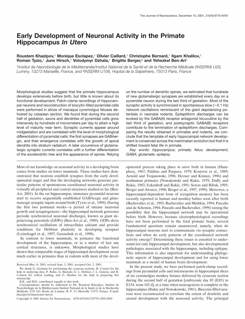
Early Development of Neuronal Activity in the PrimateHippocampus In Utero
Roustem Khazipov,1 Monique Esclapez,1 Olivier Caillard,1 Christophe Bernard,1 Ilgam Khalilov,1Roman Tyzio,1 June Hirsch,1 Volodymyr Dzhala,1 Brigitte Berger,2 and Yehezkel Ben-Ari1
1Institut de Neurobiologie de la Mediterranee/Institut National de la Sante et de la Recherche Medicale (INSERM) U29,Luminy, 13273 Marseille, France, and 2INSERM U106, Hopital de la Salpetriere, 75013 Paris, France
Morphological studies suggest that the primate hippocampusdevelops extensively before birth, but little is known about itsfunctional development. Patch-clamp recordings of hippocam-pal neurons and reconstruction of biocytin-filled pyramidal cellswere performed in slices of macaque cynomolgus fetuses de-livered by cesarean section. We found that during the secondhalf of gestation, axons and dendrites of pyramidal cells growintensively by hundreds of micrometers per day to attain a highlevel of maturity near term. Synaptic currents appear aroundmidgestation and are correlated with the level of morphologicaldifferentiation of pyramidal cells: the first synapses are GABAer-gic, and their emergence correlates with the growth of apicaldendrite into stratum radiatum. A later occurrence of glutama-tergic synaptic currents correlates with a further differentiationof the axodendritic tree and the appearance of spines. Relying
on the number of dendritic spines, we estimated that hundredsof new glutamatergic synapses are established every day on apyramidal neuron during the last third of gestation. Most of thesynaptic activity is synchronized in spontaneous slow (�0.1 Hz)network oscillations reminiscent of the giant depolarizing po-tentials in neonatal rodents. Epileptiform discharges can beevoked by the GABA(A) receptor antagonist bicuculline by thelast third of gestation, and postsynaptic GABA(B) receptorscontribute to the termination of epileptiform discharges. Com-paring the results obtained in primates and rodents, we con-clude that the template of early hippocampal network develop-ment is conserved across the mammalian evolution but that it isshifted toward fetal life in primate.
Key words: hippocampus; primate; fetus; development;GABA; glutamate; epilepsy
Most of our knowledge on neuronal activity in a developing braincomes from studies on lower mammals. These studies have dem-onstrated that neurons establish synapses from the early devel-opmental stages and that the developing networks generate par-ticular patterns of spontaneous coordinated neuronal activity invirtually all peripheral and central structures studied so far (Ben-Ari, 2001). In the rat hippocampus, immature pyramidal neuronsstart to receive sequentially established GABAergic and gluta-matergic synaptic inputs around birth (Tyzio et al., 1999). Duringthe first two postnatal weeks—a period of intense neuronalgrowth and synaptogenesis—the hippocampal network generatesperiodic synchronized neuronal discharges, known as giant de-polarizing potentials (GDPs) (Ben-Ari et al., 1989). GDPs drivesynchronized oscillations of intracellular calcium and provideconditions for Hebbian plasticity in developing synapses(Leinekugel et al., 1997; Garaschuk et al., 1998).
In contrast to lower mammals, in primates the functionaldevelopment of the hippocampus, or as a matter of fact anycortical structures, is unknown. Morphological studies haveshown that comparable stages of hippocampal development occurmuch earlier in primates than in rodents with most of the devel-
opmental process taking place in utero both in human (Hum-phrey, 1967; Paldino and Purpura, 1979; Kostovic et al., 1989;Arnold and Trojanowski, 1996; Hevner and Kinney, 1996) andnonhuman primates (Nowakowski and Rakic, 1979; Duffy andRakic, 1983; Eckenhoff and Rakic, 1991; Seress and Ribak, 1995;Berger and Alvarez, 1996; Berger et al., 1997, 1999). Moreover, ahippocampal-dependent form of recognition memory has beenrecently reported in human and monkey babies soon after birth(Bachevalier et al., 1993; Bachevalier and Mishkin, 1994; Pascalisand de Schonen, 1994; Pascalis and Bachevalier, 1999) raising thepossibility that the hippocampal network may be operationalbefore birth. However, because electrophysiological recordingshave not been performed in the fetal primate brain, severalfundamental questions remain unanswered, namely, when dohippocampal neurons start to communicate via synaptic connec-tions and when do early patterns of the coordinated networkactivity emerge? Determining these issues is essential to under-stand not only hippocampal development, but also developmentalpathologies associated with the hippocampus, including epilepsy.This information is also important for understanding phyloge-netic aspects of hippocampal development and for using lowermammals as a model of human brain development.
In the present study, we have performed patch-clamp record-ings from pyramidal cells and interneurons in hippocampal slicesof six cynomolgus monkey fetuses delivered by cesarean sectionduring the second half of gestation [embryonic day 85 (E85) toE154, term 165 d], at a time when neurogenesis is complete in thehippocampus (Rakic and Nowakowski, 1981). Biocytin-filled neu-rons were reconstructed to correlate the extent of dendritic andaxonal development with the neuronal activity. The principal
Received May 14, 2001; revised Sept. 5, 2001; accepted Oct. 2, 2001.We thank G. Germain for performing the cesarean sections, R. Cossart for the
help in analyzing data, P. Rakic, G. Buzsaki, G. L. Holmes, J. L. Gaiarsa, and R.Cannon for critical reading, and G. Medina for the help in preparing thismanuscript.
R.K. and M.E. contributed equally to this work.Correspondence should be addressed to Dr. Roustem Khazipov, Institut de
Neurobiologie de la Mediterranee/Institut National de la Sante et de la RechercheMedicale, U29 163, Route de Luminy, B. P. 13, Marseille 13273, France. E-mail:[email protected] © 2001 Society for Neuroscience 0270-6474/01/219770-12$15.00/0
The Journal of Neuroscience, December 15, 2001, 21(24):9770–9781

conclusion of this study is that in primates, neuronal activitiesemerge early in utero and that there is a rapid pace of maturationthat enables the neurons, essentially silent at the beginning ofmidgestation, to have all the morphological and physiologicalproperties required to generate network-driven endogenous andepileptiform activities before birth.
MATERIALS AND METHODSPreparation of animals and tissue. The study was conducted under theapproval and guidelines of the Ethical Committee of the Institut Nationalde La Recherche Agronomique Primate Center at Jouy-en-Josas,France. Six cynomolgus monkey (Macaca fascicularis) fetuses of agesE85, E105, E109, E119, E134, and E154 (term 165 d) were delivered bycesarean section under anesthesia with 10 mg/kg ketamine and 1 mg/kgdiazepam. The fetuses were intracardially perfused with cold (2–4°C)oxygenated (95% O2 and 5% CO2) modified artificial CSF (ACSF)(Hirsch et al., 1996). The brains were quickly removed and immersed fordissection into ice-cold oxygenated standard ACSF that contained (inmM): 126 NaCl, 3.5 KCl, 2.0 CaCl2, 1.3 MgCl2, 25 NaHCO3, 1.2 NaH2PO4,and 11 glucose. Transverse hippocampal slices (400-�m-thick) were cutwith a Leica VT 1000E (Nussloch, Germany) tissue vibroslicer, transferredto a beaker containing oxygenated ACSF and kept at room temperature(20–22°C) for at least 1–2 hr before use. Slices were recorded in a sub-merged chamber perfused with oxygenated ACSF (30–32°C).
Electrophysiology. Patch-clamp recordings were performed using Axo-patch 200 (Axon Instruments, Union City, CA) and EPC-9 (HEKAElektronik, GmbH, Lambrecht /Pfalz, Germany) amplifiers. Cells wereeither recorded blindly or under visual control using infrared microscopy,with microelectrodes of 7–10 M� resistance when filled with a solutioncontaining (in mM): 135 K gluconate, 2 MgCl2, 0.1 CaCl2, 1 EGTA, 2Na2 ATP, and 10 HEPES, pH 7.25, osmolarity 270–280 mol/kg. Thecells were identified as neurons based on the following criteria: actionpotential firing in response to a depolarizing step, presence of a synapticactivity (except for “silent” neurons), and morphology. Usually the cellswere held at �70 or 0 mV to record glutamatergic and GABAergicsynaptic currents, respectively. Electrical stimulation (0–80 V; 10–30�sec; delivered at 0.02– 0.05 Hz) was provided by a bipolar electrodeplaced in the stratum radiatum. All the neurons were filled with bio-cytin (0.4%) for post hoc morphological analysis. Electrophysiologi-cal data were analyzed using Acquis (G. Sadoc, Paris, France),Clampex (Axon Instruments), and Origin (Microcal Software,Northampton, MA).
Morphology. All the recorded slices were processed for biocytin-filledneuron detection. In addition, some hippocampal slices were collectedimmediately after slice preparation, for immunohistochemistry. Theslices were fixed overnight at 4°C in a solution containing 4% parafor-maldehyde in 0.1 M phosphate buffer (PB), pH 7.4. After fixation, sliceswere rinsed in PB, cryoprotected in sucrose for 16 hr, and quickly frozenon dry ice. The detection of biocytin-filled neurons was performed onunsectioned slices. To neutralize an endogenous peroxidase, slices werepretreated for 30 min in 1% H2O2. After several rinses in 0.1 M PBS, pH7.4, slices were incubated for 24 hr at 4°C in 1:100 avidin-biotinylatedperoxidase complex diluted in PBS containing 0.3% Triton X-100. After30 min rinses in PBS, slices were processed with 0.06% 3,3�-diaminobenzidine tetrahydrochloride (DAB; Sigma, St. Louis, MO) and0.006% H2O2 diluted in PBS, rinsed, mounted on gelatin-coated slides,and coverslipped in an aqueous medium (Crystal /Mount; Biomeda,Foster City, CA).
Among 147 recorded neurons, 100 neurons were morphologicallyidentified including 28 CA1 pyramidal cells, 27 CA3 pyramidal cells, 31hippocampal interneurons, 10 pyramids and interneurons in subiculumand neocortex, and 6 granular cells. 25 CA3 and 20 CA1 pyramidal cellswith a complete labeling of dendritic and axonal arbors were recon-structed for morphometric analysis using the Neurolucida system(MicroBrightField Inc., Colchester, VT). All the spines observed alongthe dendritic arborization of pyramidal cells were marked for furthernumerical analysis. Statistical analysis of the morphometric data wasperformed using Origin software (Microcal, Northampton, MA).Morphometric parameters from pooled CA1–CA3 pyramidal cells werefitted with a Boltzmann function: y � Amin � (Amax � Amin)/(1 �exp((x � x0)/dx)), with Amin fixed to 0. The rate of development of axonaland dendritic lengths, together with the increase in the number of spines,were deduced from the derivative of the fits.
Immunohistochemistry. The slices to be processed for histochemistry
were resectioned (40 �m) on a cryostat. Sections were rinsed in 0.01 MPBS, pH 7.4, (1� PBS), collected sequentially in tubes containing anethylene glycol-based cryoprotective solution and stored at �20°C untilprocessing. For each animal, adjacent sections were processed for im-munohistochemistry with unlabeled antibodies that are specific toGAD-65 (mouse monoclonal GAD-6) and GAD-67 (rabbit polyclonalK2) and standard avidin-biotin-peroxidase immunolabeling methods(Vectastain Elite rabbit IgG kit; Vector Laboratories, Burlingame, CA)as described previously (Esclapez et al., 1994).
RESULTS
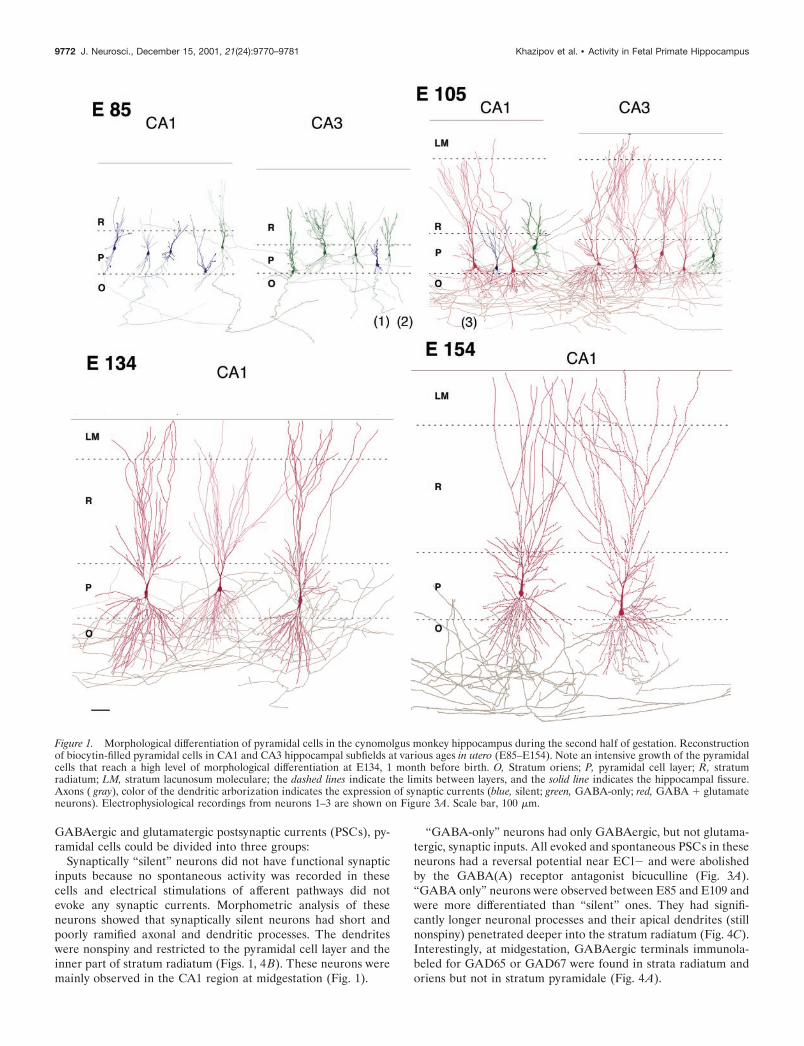
The second half of gestation: a period of intensivegrowth of pyramidal cellsThe second half of gestation was characterized by an intensivemorphological differentiation of pyramidal cells, including thegrowth and ramification of axonal and dendritic arbors and theformation of spines. As a result, pyramidal cells that were veryimmature with little neuritic extensions at midgestation wereendowed with a high level of differentiation 1 month before birth(Fig. 1, Table 1).
At midgestation (E85), immature pyramidal neurons displayeda relatively long axonal process running in stratum oriens but onlyshort apical dendrites with few branches and little or no basaldendrites. Apical dendrites were mainly restricted to the cell bodylayer in CA1 but entered the proximal part of stratum radiatum inCA3. Many dendritic and axonal processes ended with growthcones.
At E105–E109, the apical dendrites extended through stratumradiatum to lacunosum moleculare; the basal dendrites pene-trated and ramified in stratum oriens. At this stage, the firstdendritic spines were observed. Axons of CA3 pyramidal cellsgave rise to numerous recurrent and Schaffer collaterals; CA1pyramidal cells emitted many local axonal branches running instrata oriens and radiatum as well as projections to the subiculum.
At E134 and E154, pyramidal cells had well differentiateddendritic and axonal arbors that extended through all theirproper layers. The densely ramified apical dendrites crossed thestratum lacunosum moleculare up to the hippocampal fissure.Apical and basal dendrites were covered with spines.
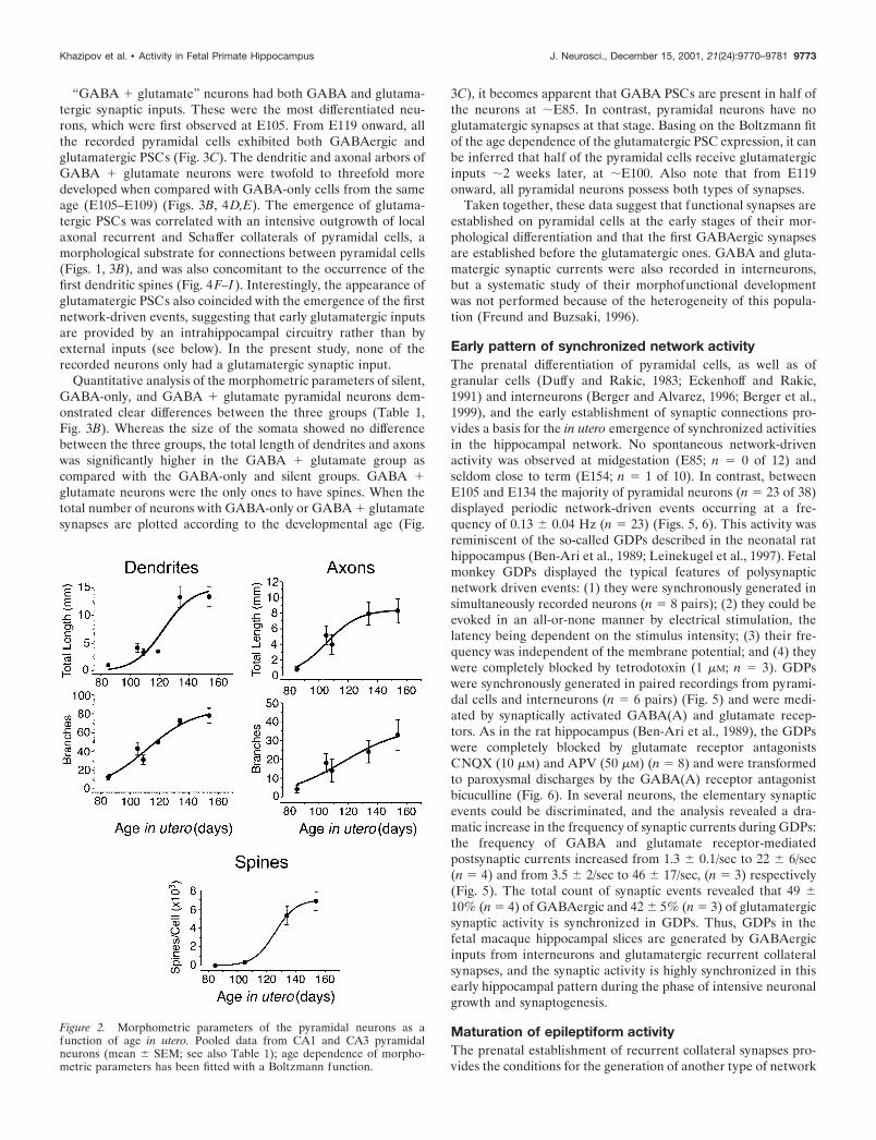
A morphometric analysis of the total length and the number ofbranches for dendrites and axons of pyramidal cells from CA3and CA1 revealed a progressive increase of all these parametersfrom E85 to E134. From E134 to E154, the length and number ofdendritic and axonal branches did not change significantly (Fig. 2,Table 1). The number of spines increased continuously from E105onward to attain some 7000 spines per pyramidal cell before birth(E154) (Fig. 2, Table 1). The density of spines at E154 was 55spines/100 �m and matched the one observed in the neonaterhesus monkey (47/100 �m; Seress and Ribak, 1995). Thornyexcrescences were not observed on CA3 pyramidal cells, confirm-ing that the complex mossy fiber synapses mature mainly afterbirth (Vijayan, 1986; Seress and Ribak, 1995). Thus, the morpho-logical differentiation of the pyramidal cells in the primate hip-pocampus occurs essentially during the second half of gestation,and the pyramidal cells already attain a high level of maturitybefore birth.
The sequential expression of GABA and glutamatergicsynaptic currents correlates with the morphologicalmaturation of the pyramidal cellsSynaptic activity was observed in hippocampal pyramidal neuronsstarting from midgestation, and the expression of synaptic cur-rents was highly correlated with their degree of morphologicaldifferentiation (Fig. 3, Table 1). Based on the expression of
Khazipov et al. • Activity in Fetal Primate Hippocampus J. Neurosci., December 15, 2001, 21(24):9770–9781 9771
GABAergic and glutamatergic postsynaptic currents (PSCs), py-ramidal cells could be divided into three groups:
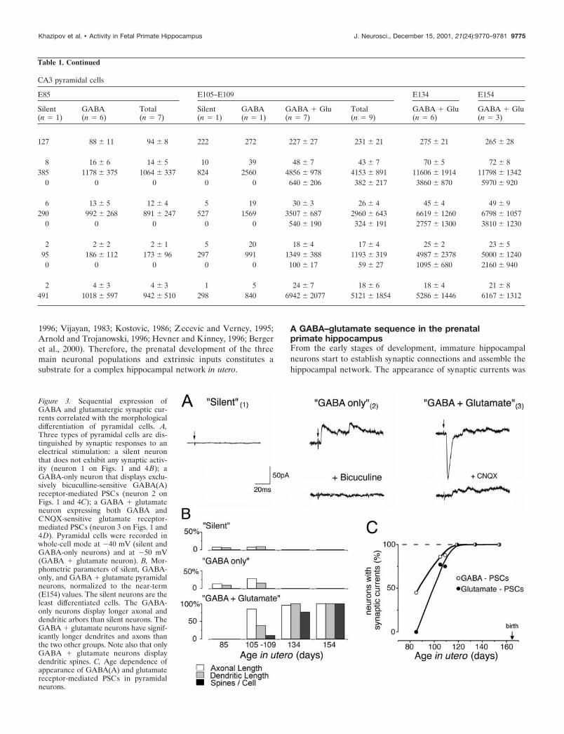
Synaptically “silent” neurons did not have functional synapticinputs because no spontaneous activity was recorded in thesecells and electrical stimulations of afferent pathways did notevoke any synaptic currents. Morphometric analysis of theseneurons showed that synaptically silent neurons had short andpoorly ramified axonal and dendritic processes. The dendriteswere nonspiny and restricted to the pyramidal cell layer and theinner part of stratum radiatum (Figs. 1, 4B). These neurons weremainly observed in the CA1 region at midgestation (Fig. 1).
“GABA-only” neurons had only GABAergic, but not glutama-tergic, synaptic inputs. All evoked and spontaneous PSCs in theseneurons had a reversal potential near ECl� and were abolishedby the GABA(A) receptor antagonist bicuculline (Fig. 3A).“GABA only” neurons were observed between E85 and E109 andwere more differentiated than “silent” ones. They had signifi-cantly longer neuronal processes and their apical dendrites (stillnonspiny) penetrated deeper into the stratum radiatum (Fig. 4C).Interestingly, at midgestation, GABAergic terminals immunola-beled for GAD65 or GAD67 were found in strata radiatum andoriens but not in stratum pyramidale (Fig. 4A).
Figure 1. Morphological differentiation of pyramidal cells in the cynomolgus monkey hippocampus during the second half of gestation. Reconstructionof biocytin-filled pyramidal cells in CA1 and CA3 hippocampal subfields at various ages in utero (E85–E154). Note an intensive growth of the pyramidalcells that reach a high level of morphological differentiation at E134, 1 month before birth. O, Stratum oriens; P, pyramidal cell layer; R, stratumradiatum; LM, stratum lacunosum moleculare; the dashed lines indicate the limits between layers, and the solid line indicates the hippocampal fissure.Axons ( gray), color of the dendritic arborization indicates the expression of synaptic currents (blue, silent; green, GABA-only; red, GABA � glutamateneurons). Electrophysiological recordings from neurons 1–3 are shown on Figure 3A. Scale bar, 100 �m.
9772 J. Neurosci., December 15, 2001, 21(24):9770–9781 Khazipov et al. • Activity in Fetal Primate Hippocampus
“GABA � glutamate” neurons had both GABA and glutama-tergic synaptic inputs. These were the most differentiated neu-rons, which were first observed at E105. From E119 onward, allthe recorded pyramidal cells exhibited both GABAergic andglutamatergic PSCs (Fig. 3C). The dendritic and axonal arbors ofGABA � glutamate neurons were twofold to threefold moredeveloped when compared with GABA-only cells from the sameage (E105–E109) (Figs. 3B, 4D,E). The emergence of glutama-tergic PSCs was correlated with an intensive outgrowth of localaxonal recurrent and Schaffer collaterals of pyramidal cells, amorphological substrate for connections between pyramidal cells(Figs. 1, 3B), and was also concomitant to the occurrence of thefirst dendritic spines (Fig. 4F–I). Interestingly, the appearance ofglutamatergic PSCs also coincided with the emergence of the firstnetwork-driven events, suggesting that early glutamatergic inputsare provided by an intrahippocampal circuitry rather than byexternal inputs (see below). In the present study, none of therecorded neurons only had a glutamatergic synaptic input.
Quantitative analysis of the morphometric parameters of silent,GABA-only, and GABA � glutamate pyramidal neurons dem-onstrated clear differences between the three groups (Table 1,Fig. 3B). Whereas the size of the somata showed no differencebetween the three groups, the total length of dendrites and axonswas significantly higher in the GABA � glutamate group ascompared with the GABA-only and silent groups. GABA �glutamate neurons were the only ones to have spines. When thetotal number of neurons with GABA-only or GABA � glutamatesynapses are plotted according to the developmental age (Fig.
3C), it becomes apparent that GABA PSCs are present in half ofthe neurons at �E85. In contrast, pyramidal neurons have noglutamatergic synapses at that stage. Basing on the Boltzmann fitof the age dependence of the glutamatergic PSC expression, it canbe inferred that half of the pyramidal cells receive glutamatergicinputs �2 weeks later, at �E100. Also note that from E119onward, all pyramidal neurons possess both types of synapses.
Taken together, these data suggest that functional synapses areestablished on pyramidal cells at the early stages of their mor-phological differentiation and that the first GABAergic synapsesare established before the glutamatergic ones. GABA and gluta-matergic synaptic currents were also recorded in interneurons,but a systematic study of their morphofunctional developmentwas not performed because of the heterogeneity of this popula-tion (Freund and Buzsaki, 1996).
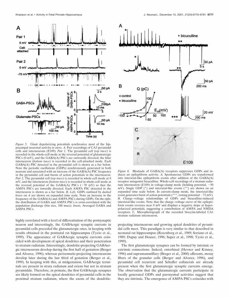
Early pattern of synchronized network activityThe prenatal differentiation of pyramidal cells, as well as ofgranular cells (Duffy and Rakic, 1983; Eckenhoff and Rakic,1991) and interneurons (Berger and Alvarez, 1996; Berger et al.,1999), and the early establishment of synaptic connections pro-vides a basis for the in utero emergence of synchronized activitiesin the hippocampal network. No spontaneous network-drivenactivity was observed at midgestation (E85; n � 0 of 12) andseldom close to term (E154; n � 1 of 10). In contrast, betweenE105 and E134 the majority of pyramidal neurons (n � 23 of 38)displayed periodic network-driven events occurring at a fre-quency of 0.13 0.04 Hz (n � 23) (Figs. 5, 6). This activity wasreminiscent of the so-called GDPs described in the neonatal rathippocampus (Ben-Ari et al., 1989; Leinekugel et al., 1997). Fetalmonkey GDPs displayed the typical features of polysynapticnetwork driven events: (1) they were synchronously generated insimultaneously recorded neurons (n � 8 pairs); (2) they could beevoked in an all-or-none manner by electrical stimulation, thelatency being dependent on the stimulus intensity; (3) their fre-quency was independent of the membrane potential; and (4) theywere completely blocked by tetrodotoxin (1 �M; n � 3). GDPswere synchronously generated in paired recordings from pyrami-dal cells and interneurons (n � 6 pairs) (Fig. 5) and were medi-ated by synaptically activated GABA(A) and glutamate recep-tors. As in the rat hippocampus (Ben-Ari et al., 1989), the GDPswere completely blocked by glutamate receptor antagonistsCNQX (10 �M) and APV (50 �M) (n � 8) and were transformedto paroxysmal discharges by the GABA(A) receptor antagonistbicuculline (Fig. 6). In several neurons, the elementary synapticevents could be discriminated, and the analysis revealed a dra-matic increase in the frequency of synaptic currents during GDPs:the frequency of GABA and glutamate receptor-mediatedpostsynaptic currents increased from 1.3 0.1/sec to 22 6/sec(n � 4) and from 3.5 2/sec to 46 17/sec, (n � 3) respectively(Fig. 5). The total count of synaptic events revealed that 49 10% (n � 4) of GABAergic and 42 5% (n � 3) of glutamatergicsynaptic activity is synchronized in GDPs. Thus, GDPs in thefetal macaque hippocampal slices are generated by GABAergicinputs from interneurons and glutamatergic recurrent collateralsynapses, and the synaptic activity is highly synchronized in thisearly hippocampal pattern during the phase of intensive neuronalgrowth and synaptogenesis.
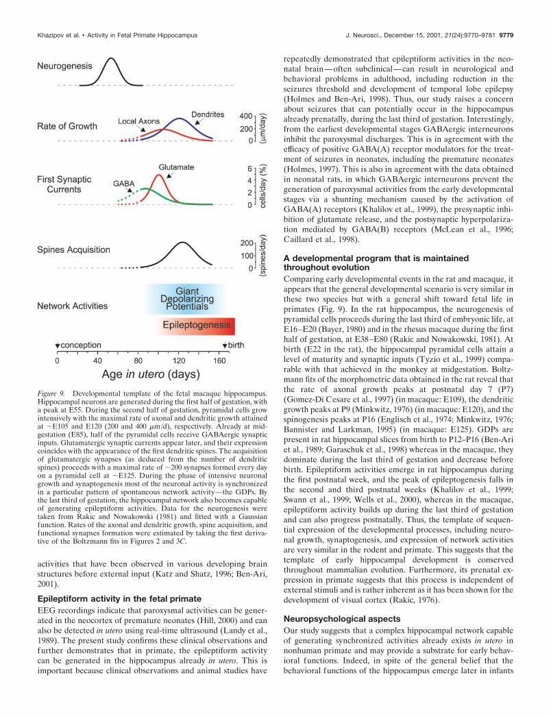
Maturation of epileptiform activityThe prenatal establishment of recurrent collateral synapses pro-vides the conditions for the generation of another type of network
Figure 2. Morphometric parameters of the pyramidal neurons as afunction of age in utero. Pooled data from CA1 and CA3 pyramidalneurons (mean SEM; see also Table 1); age dependence of morpho-metric parameters has been fitted with a Boltzmann function.
Khazipov et al. • Activity in Fetal Primate Hippocampus J. Neurosci., December 15, 2001, 21(24):9770–9781 9773
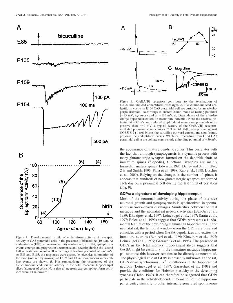
activity—epileptiform discharges. Therefore, in the next series ofexperiments we studied the development of the epileptiformactivity induced by the GABA(A) receptor antagonist bicuculline(10 �M) (Figs. 6, 7). At midgestation, no epileptiform activity wasobserved, in keeping with the large percentage of either silent orGABA-only neurons at this stage. The first epileptiform events inthe presence of bicuculline were observed at E105–E109, whichcoincides with the outgrowth of axonal collaterals of pyramidalcells and the appearance of spines and glutamatergic synapticcurrents. The occurrence and severity of epileptiform eventsincreased through gestation in parallel with the development ofpyramidal cells and the increase in the number of spines. Onemonth before birth (E134), powerful epileptiform events wereobserved in all recorded neurons (Fig. 7). Interictal-like eventswere synchronized by glutamatergic connections because: (1) theyreversed near 0 mV and the charge transfer during these eventsdisplayed a slope of negative conductance at negative membranepotentials suggesting the contribution of both AMPA andNMDA types of glutamate receptors (Fig. 6B) (n � 4) and (2)they were completely blocked by the AMPA and NMDA receptorantagonists CNQX (10 �M) and APV (50 �M; n � 4).
During epileptiform discharges, interneurons received a pow-erful glutamatergic input and fired bursts of action potentials(n � 8) (Fig. 6A), providing the conditions for the activation ofGABA(B) receptors. Indeed, at E134 and E154, the epileptiformevents were curtailed by an afterhyperpolarization that reversedat �93 3 mV (n � 5) and displayed an inward rectification thatis characteristic of the GABA(B) receptor-activated potassiumconductance (Fig. 8). The GABA(B) receptor antagonist CGP55812 (1 �M) blocked the outward currents curtailing the epilep-tiform events and significantly prolonged the discharges (n � 3),further suggesting that the activation of the GABA(B) receptorscontributes to the termination of epileptiform discharges. How-ever, at earlier stages, the GABA(B) receptor-mediated currentswere not observed (Fig. 6), which is in agreement with a delayed
expression of the postsynaptic GABA(B) receptor-mediated in-hibition in the rat (Fukuda et al., 1993; Gaiarsa et al., 1995).
Thus, in primate, the hippocampal network becomes capable ofgenerating paroxysmal discharges already in utero, and the pro-gression of the paroxysmal activity likely reflects the developmentof a recurrent connectivity between pyramidal cells. The obser-vation that epileptiform activities can be generated in the hip-pocampus already in utero provides additional evidence that thehippocampal network reaches a high level of maturity beforebirth.
DISCUSSIONThis study provides the first evidence that in primate, a complexhippocampal network capable of generating spontaneous andparoxysmal synchronized activities is already established in utero.It also provides some of the key steps involved in the formation ofthe network elements, including morphological differentiation ofpyramidal neurons, sequential establishment of GABA and glu-tamatergic synaptic connections, and emergence of synchronizednetwork activity (Fig. 9).
Prenatal maturation of hippocampal primate neuronsWe found that the second half of gestation is a principal period ofpyramidal cell differentiation, when dendrites and axons inten-sively grow and ramify. Whereas pyramidal cells already reach ahigh level of differentiation 1 month before birth, further growthand remodeling of the axonal and dendritic arbors can probablycontinue after birth; determining a complete picture of the de-velopment of pyramidal cells will require a postnatal study. Themajority of dentate gyrus granular cells (Duffy and Rakic, 1983;Eckenhoff and Rakic, 1991) and GABAergic interneurons(Berger and Alvarez, 1996; Berger et al., 1999; Esclapez et al.,1999) also differentiate prenatally. In addition, major extrinsicafferences invade the hippocampus from midgestation and shouldprovide inputs from other brain regions (Berger and Alvarez,
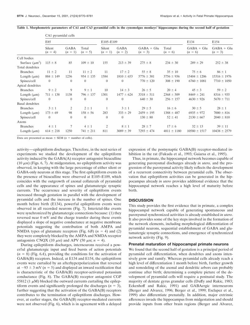
Table 1. Morphometric parameters of CA1 and CA3 pyramidal cells in the cynomolgus monkeys’ hippocampus during the second half of gestation
CA1 pyramidal cells
E85 E105–E109 E134 E154
Silent(n � 4)
GABA(n � 1)
Total(n � 5)
Silent(n � 1)
GABA(n � 2)
GABA � Glu(n � 3)
Total(n � 6)
GABA � Glu(n � 6)
GABA � Glu(n � 3)
Cell bodiesSurface (�m2) 115 8 85 109 10 155 213 39 275 8 234 30 289 29 252 38
Total dendritesBranches 11 2 11 11 2 11 17 2 55 8 35 10 75 6 86 1Length (�m) 884 149 1236 954 135 1584 1810 653 5776 381 3756 936 15404 1206 15316 1976Spines/cell 0 0 0 0 0 770 120 308 190 6760 1081 7710 1050
Apical dendritesBranches 9 2 9 9 1 10 14 3 26 5 20 4 45 3 59 2Length (�m) 711 138 1138 796 137 1301 1477 624 3318 511 2368 509 8469 241 8316 935Spines/cell 0 0 0 0 0 640 30 256 157 4630 926 5670 711
Basal dendritesBranches 3 1 2 2 1 1 3 1 29 5 16 6 30 5 28 1Length (�m) 173 69 98 158 56 283 333 29 2459 195 1388 487 6935 972 7000 816Spines/cell 0 0 0 0 0 130 80 52 41 2130 667 2040 810
AxonsBranches 4 1 7 4 1 2 8 1 28 7 17 6 32 13 39 11Length (�m) 614 218 1250 741 211 811 3089 39 7293 474 4811 1180 10580 1517 10438 2579
Data are presented as mean SEM (n � number of cells).
9774 J. Neurosci., December 15, 2001, 21(24):9770–9781 Khazipov et al. • Activity in Fetal Primate Hippocampus
1996; Vijayan, 1983; Kostovic, 1986; Zecevic and Verney, 1995;Arnold and Trojanowski, 1996; Hevner and Kinney, 1996; Bergeret al., 2000). Therefore, the prenatal development of the threemain neuronal populations and extrinsic inputs constitutes asubstrate for a complex hippocampal network in utero.
A GABA–glutamate sequence in the prenatalprimate hippocampusFrom the early stages of development, immature hippocampalneurons start to establish synaptic connections and assemble thehippocampal network. The appearance of synaptic currents was
Figure 3. Sequential expression ofGABA and glutamatergic synaptic cur-rents correlated with the morphologicaldifferentiation of pyramidal cells. A,Three types of pyramidal cells are dis-tinguished by synaptic responses to anelectrical stimulation: a silent neuronthat does not exhibit any synaptic activ-ity (neuron 1 on Figs. 1 and 4 B); aGABA-only neuron that displays exclu-sively bicuculline-sensitive GABA(A)receptor-mediated PSCs (neuron 2 onFigs. 1 and 4C); a GABA � glutamateneuron expressing both GABA andCNQX-sensitive glutamate receptor-mediated PSCs (neuron 3 on Figs. 1 and4D). Pyramidal cells were recorded inwhole-cell mode at �40 mV (silent andGABA-only neurons) and at �50 mV(GABA � glutamate neuron). B, Mor-phometric parameters of silent, GABA-only, and GABA � glutamate pyramidalneurons, normalized to the near-term(E154) values. The silent neurons are theleast differentiated cells. The GABA-only neurons display longer axonal anddendritic arbors than silent neurons. TheGABA � glutamate neurons have signif-icantly longer dendrites and axons thanthe two other groups. Note also that onlyGABA � glutamate neurons displaydendritic spines. C, Age dependence ofappearance of GABA(A) and glutamatereceptor-mediated PSCs in pyramidalneurons.
Table 1. Continued
CA3 pyramidal cells
E85 E105–E109 E134 E154
Silent(n � 1)
GABA(n � 6)
Total(n � 7)
Silent(n � 1)
GABA(n � 1)
GABA � Glu(n � 7)
Total(n � 9)
GABA � Glu(n � 6)
GABA � Glu(n � 3)
127 88 11 94 8 222 272 227 27 231 21 275 21 265 28
8 16 6 14 5 10 39 48 7 43 7 70 5 72 8385 1178 375 1064 337 824 2560 4856 978 4153 891 11606 1914 11798 1342
0 0 0 0 0 640 206 382 217 3860 870 5970 920
6 13 5 12 4 5 19 30 3 26 4 45 4 49 9290 992 268 891 247 527 1569 3507 687 2960 643 6619 1260 6798 1057
0 0 0 0 0 540 190 324 191 2757 1300 3810 1230
2 2 2 2 1 5 20 18 4 17 4 25 2 23 595 186 112 173 96 297 991 1349 388 1193 319 4987 2378 5000 12400 0 0 0 0 100 17 59 27 1095 680 2160 940
2 4 3 4 3 1 5 24 7 18 6 18 4 21 8491 1018 597 942 510 298 840 6942 2077 5121 1854 5286 1446 6167 1312
Khazipov et al. • Activity in Fetal Primate Hippocampus J. Neurosci., December 15, 2001, 21(24):9770–9781 9775
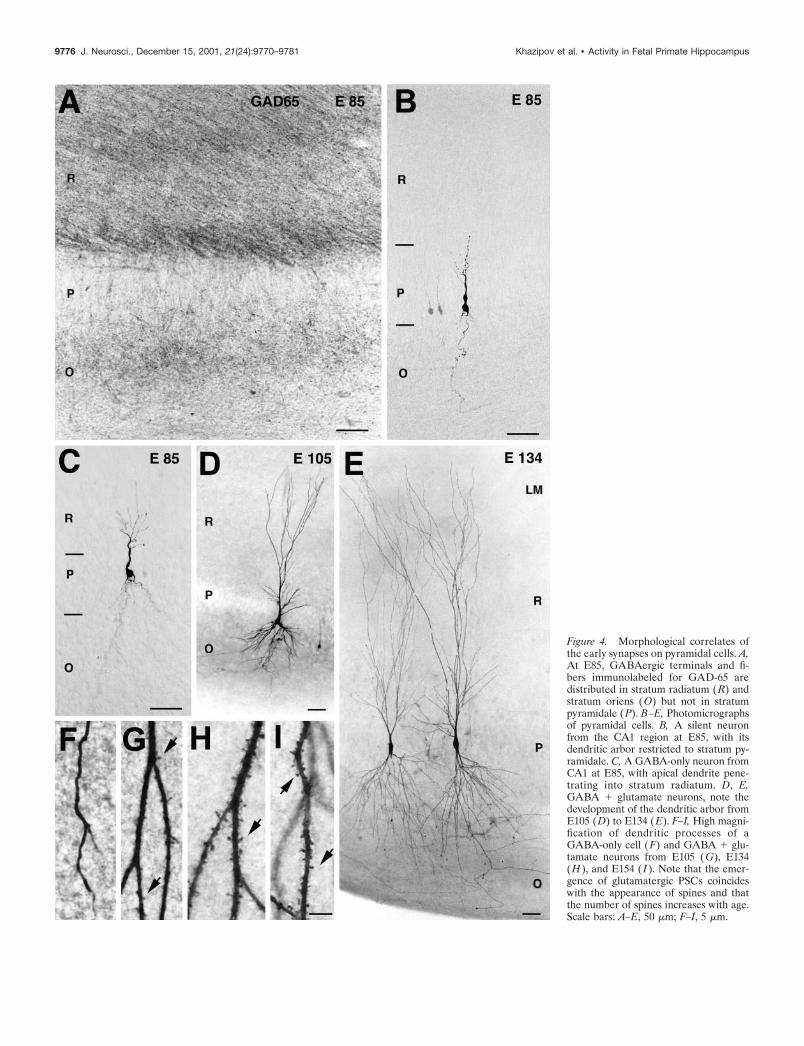
Figure 4. Morphological correlates ofthe early synapses on pyramidal cells. A,At E85, GABAergic terminals and fi-bers immunolabeled for GAD-65 aredistributed in stratum radiatum (R) andstratum oriens (O) but not in stratumpyramidale (P). B–E, Photomicrographsof pyramidal cells. B, A silent neuronfrom the CA1 region at E85, with itsdendritic arbor restricted to stratum py-ramidale. C, A GABA-only neuron fromCA1 at E85, with apical dendrite pene-trating into stratum radiatum. D, E,GABA � glutamate neurons, note thedevelopment of the dendritic arbor fromE105 (D) to E134 (E). F–I, High magni-fication of dendritic processes of aGABA-only cell (F) and GABA � glu-tamate neurons from E105 (G), E134(H ), and E154 ( I ). Note that the emer-gence of glutamatergic PSCs coincideswith the appearance of spines and thatthe number of spines increases with age.Scale bars: A–E, 50 �m; F–I, 5 �m.
9776 J. Neurosci., December 15, 2001, 21(24):9770–9781 Khazipov et al. • Activity in Fetal Primate Hippocampus
highly correlated with a level of differentiation of the postsynapticneuron and interestingly, the GABAergic synaptic currents inpyramidal cells preceded the glutamatergic ones, in keeping withresults obtained in the postnatal rat hippocampus (Tyzio et al.,1999). The appearance of GABAergic synaptic currents coin-cided with development of apical dendrites and their penetrationto stratum radiatum. Interestingly, dendritic-projecting GABAer-gic interneurons develop during the first half of gestation (Bergerand Alvarez, 1996), whereas perisomatic-projecting interneuronsdevelop later during the last third of gestation (Berger et al.,1999). In keeping with this, at midgestation, GABAergic termi-nals are present in strata radiatum and oriens but not in stratumpyramidale. Therefore, in primate, the first GABAergic synapsesare likely formed on the apical dendrites of pyramidal cells in theproximal stratum radiatum, where the axons of the dendritic-
projecting interneurons and growing apical dendrites of pyrami-dal cells meet. This paradigm is very similar to that described inneonatal rat hippocampus (Rozenberg et al., 1989; Soriano et al.,1989; Dupuy and Houser, 1996; Super et al., 1998; Tyzio et al.,1999).
The first glutamatergic synapses can be formed by intrinsic orextrinsic connections. Indeed, entorhinal (Hevner and Kinney,1996) and supramammillary (Berger et al., 2000) afferents, mossyfibers of the granular cells (Berger and Alvarez, 1996), andpyramidal cell recurrent and Schaffer collaterals are alreadypresent when the first glutamatergic synaptic currents emerge.The observation that the glutamatergic currents participate inlocally generated GDPs and paroxysmal activities suggest thatthey are intrinsic. The emergence of AMPA PSCs coincides with
Figure 5. Giant depolarizing potentials synchronize most of the hip-pocampal neuronal activity in utero. A, Pair recordings of CA3 pyramidalcells and interneurons (E109). Pair 1, The pyramidal cell (top trace) isrecorded in the whole-cell mode at the reversal potential of glutamatergicPSCs (0 mV), and the GABA(A) PSCs are outwardly directed; the hilarinterneuron (bottom trace) is recorded in the cell-attached mode. EachGABA(A) PSC detected in the pyramidal cell is shown as a bar below.Note the periodic oscillations (GDPs) synchronously generated in bothneurons and associated with an increase of the GABA(A) PSC frequencyin the pyramidal cell and bursts of action potentials in the interneuron.Pair 2, The pyramidal cell (top trace) is recorded in whole-cell mode at 0mV, and the interneuron (bottom trace) is recorded in whole-cell mode atthe reversal potential of the GABA(A) PSCs (�70 mV) so that theAMPA PSCs are inwardly directed. Each AMPA PSC detected in theinterneuron is shown as a bar below. B, Left, GDPs outlined by dashedboxes on A are shown on expanded time scale. Note an increase in thefrequency of the GABA(A) and AMPA PSCs during GDPs. On the right,the distribution of GABA and AMPA PSCs is cross-correlated with thepopulation discharge (bin size, 100 msec). Insets, Averaged GABA andAMPA PSCs.
Figure 6. Blockade of GABA(A) receptors suppresses GDPs and in-duces an epileptiform activity. A, Spontaneous GDPs are transformedinto interictal-like epileptiform events after addition of the GABA(A)receptor antagonist bicuculline. Whole-cell recordings of a stratum radia-tum interneuron (E109) in voltage-clamp mode (holding potential, �50mV). Single GDP (*) and interictal-like events (**) are shown on anexpanded time scale below. In current-clamp mode, the interictal-likeevent generates burst of action potentials (***, resting potential �72 mV).B, Charge–voltage relationships of GDPs and bicuculline-inducedinterictal-like events. Note that the charge–voltage curve of the epilepti-form events reverses near 0 mV and displays a negative slope at hyper-polarized potentials, suggesting a contribution of AMPA and NMDAreceptors. C, Microphotograph of the recorded biocytin-labeled CA1stratum radiatum interneuron.
Khazipov et al. • Activity in Fetal Primate Hippocampus J. Neurosci., December 15, 2001, 21(24):9770–9781 9777
the appearance of mature dendritic spines. This correlates withthe fact that although synaptogenesis is a dynamic process withmany glutamatergic synapses formed on the dendritic shaft orimmature spines (filopodia), functional synapses are mainlyformed on mature spines (Edwards, 1995; Dailey and Smith, 1996;Ziv and Smith, 1996; Fiala et al., 1998; Rao et al., 1998; Luscheret al., 2000). Relying on the changes in the number of spines, itappears that hundreds of new glutamatergic synapses are formedeach day on a pyramidal cell during the last third of gestation(Fig. 9).
GDPs: a signature of developing hippocampusMost of the neuronal activity during the phase of intensiveneuronal growth and synaptogenesis is synchronized in sponta-neous network-driven discharges. Similarities between the fetalmacaque and the neonatal rat network activities (Ben-Ari et al.,1989; Khazipov et al., 1997; Leinekugel et al., 1997; Strata et al.,1997; Bolea et al., 1999) suggest that GDPs represents a funda-mental feature of the developing mammalian hippocampus. In theneonatal rat, the temporal window when the GDPs are observedcoincides with a period when GABA depolarizes and excites theimmature neurons (Ben-Ari et al., 1989; Khazipov et al., 1997;Leinekugel et al., 1997; Garaschuk et al., 1998). The presence ofGDPs in the fetal monkey hippocampal slices suggests thatGABA might be excitatory in the immature macaque hippocam-pal neurons; this however remains to be directly demonstrated.The physiological role of GDPs is presently unknown. In the rat,GDPs drive synchronous Ca2� oscillations in the hippocampalneurons (Leinekugel et al., 1997; Garaschuk et al., 1998) andprovide the conditions for Hebbian plasticity in the developingsynapses (Hebb, 1949). It can therefore be suggested that GDPsparticipate in the activity-dependent formation of the hippocam-pal circuitry similarly to other internally generated spontaneous
Figure 7. Developmental profile of epileptiform activity. A, Synapticactivity in CA3 pyramidal cells in the presence of bicuculline (10 �M). Atmidgestation (E85), no seizure activity is observed; at E105, epileptiformevents emerge and progress in occurrence and severity during the secondhalf of gestation. Whole-cell recordings at holding potential of �60 mV.At E85 and E105, the responses were evoked by electrical stimulation ofthe slice (marked by arrows); at E109 and E154, spontaneous interictal-like events are shown. B, Plot summarizing the occurrence of thebicuculline-induced seizure activity in the fetal macaque hippocampalslices (number of cells). Note that all neurons express epileptiform activ-ities from E134 onward.
Figure 8. GABA(B) receptors contribute to the termination ofbicuculline-induced epileptiform discharges. A, Bicuculline-induced epi-leptiform events in E134 CA3 pyramidal cell are curtailed by an afterhy-perpolarization. Recordings in current-clamp mode at resting potential(�75 mV; top trace) and at �110 mV. B, Dependence of the afterdis-charge hyperpolarization on membrane potential. Note the reversal po-tential at �92 mV and reduced amplitude at membrane potentials morepositive than �40 mV, a typical feature of the GABA(B) receptor-mediated potassium conductances. C, The GABA(B) receptor antagonistCGP55812 (1 �M) blocks the curtailing outward current and significantlyprolongs the epileptiform events. Whole-cell recording from E134 CA3pyramidal cell in the voltage-clamp mode at holding potential of �50 mV.
9778 J. Neurosci., December 15, 2001, 21(24):9770–9781 Khazipov et al. • Activity in Fetal Primate Hippocampus
activities that have been observed in various developing brainstructures before external input (Katz and Shatz, 1996; Ben-Ari,2001).
Epileptiform activity in the fetal primateEEG recordings indicate that paroxysmal activities can be gener-ated in the neocortex of premature neonates (Hill, 2000) and canalso be detected in utero using real-time ultrasound (Landy et al.,1989). The present study confirms these clinical observations andfurther demonstrates that in primate, the epileptiform activitycan be generated in the hippocampus already in utero. This isimportant because clinical observations and animal studies have
repeatedly demonstrated that epileptiform activities in the neo-natal brain—often subclinical—can result in neurological andbehavioral problems in adulthood, including reduction in theseizures threshold and development of temporal lobe epilepsy(Holmes and Ben-Ari, 1998). Thus, our study raises a concernabout seizures that can potentially occur in the hippocampusalready prenatally, during the last third of gestation. Interestingly,from the earliest developmental stages GABAergic interneuronsinhibit the paroxysmal discharges. This is in agreement with theefficacy of positive GABA(A) receptor modulators for the treat-ment of seizures in neonates, including the premature neonates(Holmes, 1997). This is also in agreement with the data obtainedin neonatal rats, in which GABAergic interneurons prevent thegeneration of paroxysmal activities from the early developmentalstages via a shunting mechanism caused by the activation ofGABA(A) receptors (Khalilov et al., 1999), the presynaptic inhi-bition of glutamate release, and the postsynaptic hyperpolariza-tion mediated by GABA(B) receptors (McLean et al., 1996;Caillard et al., 1998).
A developmental program that is maintainedthroughout evolutionComparing early developmental events in the rat and macaque, itappears that the general developmental scenario is very similar inthese two species but with a general shift toward fetal life inprimates (Fig. 9). In the rat hippocampus, the neurogenesis ofpyramidal cells proceeds during the last third of embryonic life, atE16–E20 (Bayer, 1980) and in the rhesus macaque during the firsthalf of gestation, at E38–E80 (Rakic and Nowakowski, 1981). Atbirth (E22 in the rat), the hippocampal pyramidal cells attain alevel of maturity and synaptic inputs (Tyzio et al., 1999) compa-rable with that achieved in the monkey at midgestation. Boltz-mann fits of the morphometric data obtained in the rat reveal thatthe rate of axonal growth peaks at postnatal day 7 (P7)(Gomez-Di Cesare et al., 1997) (in macaque: E109), the dendriticgrowth peaks at P9 (Minkwitz, 1976) (in macaque: E120), and thespinogenesis peaks at P16 (Englisch et al., 1974; Minkwitz, 1976;Bannister and Larkman, 1995) (in macaque: E125). GDPs arepresent in rat hippocampal slices from birth to P12–P16 (Ben-Ariet al., 1989; Garaschuk et al., 1998) whereas in the macaque, theydominate during the last third of gestation and decrease beforebirth. Epileptiform activities emerge in rat hippocampus duringthe first postnatal week, and the peak of epileptogenesis falls inthe second and third postnatal weeks (Khalilov et al., 1999;Swann et al., 1999; Wells et al., 2000), whereas in the macaque,epileptiform activity builds up during the last third of gestationand can also progress postnatally. Thus, the template of sequen-tial expression of the developmental processes, including neuro-nal growth, synaptogenesis, and expression of network activitiesare very similar in the rodent and primate. This suggests that thetemplate of early hippocampal development is conservedthroughout mammalian evolution. Furthermore, its prenatal ex-pression in primate suggests that this process is independent ofexternal stimuli and is rather inherent as it has been shown for thedevelopment of visual cortex (Rakic, 1976).
Neuropsychological aspectsOur study suggests that a complex hippocampal network capableof generating synchronized activities already exists in utero innonhuman primate and may provide a substrate for early behav-ioral functions. Indeed, in spite of the general belief that thebehavioral functions of the hippocampus emerge later in infants
Figure 9. Developmental template of the fetal macaque hippocampus.Hippocampal neurons are generated during the first half of gestation, witha peak at E55. During the second half of gestation, pyramidal cells growintensively with the maximal rate of axonal and dendritic growth attainedat �E105 and E120 (200 and 400 �m/d), respectively. Already at mid-gestation (E85), half of the pyramidal cells receive GABAergic synapticinputs. Glutamatergic synaptic currents appear later, and their expressioncoincides with the appearance of the first dendritic spines. The acquisitionof glutamatergic synapses (as deduced from the number of dendriticspines) proceeds with a maximal rate of �200 synapses formed every dayon a pyramidal cell at �E125. During the phase of intensive neuronalgrowth and synaptogenesis most of the neuronal activity is synchronizedin a particular pattern of spontaneous network activity—the GDPs. Bythe last third of gestation, the hippocampal network also becomes capableof generating epileptiform activities. Data for the neurogenesis weretaken from Rakic and Nowakowski (1981) and fitted with a Gaussianfunction. Rates of the axonal and dendritic growth, spine acquisition, andfunctional synapses formation were estimated by taking the first deriva-tive of the Boltzmann fits in Figures 2 and 3C.
Khazipov et al. • Activity in Fetal Primate Hippocampus J. Neurosci., December 15, 2001, 21(24):9770–9781 9779
because of the amnesia of the first years of human life (Nadel andZola-Morgan, 1984), recent neuropsychological studies have pro-vided evidence of the emergence of a hippocampal-dependentform of recognition memory early after birth (Bachevalier et al.,1993; Bachevalier and Mishkin, 1994; Pascalis and de Schonen,1994; Pascalis and Bachevalier, 1999). The results of the presentstudy add morphofunctional support to these observations.
REFERENCESArnold SE, Trojanowski JQ (1996) Human fetal hippocampal develop-
ment: I. Cytoarchitecture, myeloarchitecture, and neuronal morpho-logic features. J Comp Neurol 367:274–292.
Bachevalier J, Mishkin M (1994) Effects of selective neonatal temporallobe lesions on visual recognition memory in rhesus monkeys. J Neu-rosci 14:2128–2139.
Bachevalier J, Brickson M, Hagger C (1993) Limbic-dependent recogni-tion memory in monkeys develops early in infancy. NeuroReport4:77–80.
Bannister NJ, Larkman AU (1995) Dendritic morphology of CA1 pyra-midal neurones from the rat hippocampus: II. Spine distributions.J Comp Neurol 360:161–171.
Bayer SA (1980) Development of the hippocampal region in the rat. II.Morphogenesis during embryonic and early postnatal life. J CompNeurol 190:115–134.
Ben-Ari Y (2001) Developing networks play a similar melody. TrendsNeurosci 24:353–360.
Ben-Ari Y, Cherubini E, Corradetti R, Gaiarsa JL (1989) Giant synapticpotentials in immature rat CA3 hippocampal neurones. J Physiol(Lond) 416:303–325.
Berger B, Alvarez C (1996) Neurochemical development of the hip-pocampal region in the fetal rhesus monkey. III: Calbindin-D28K,calretinin and parvalbumin with special mention of Cajal-Retzius cellsand the retrosplenial cortex. J Comp Neurol 366:674–699.
Berger B, Alvarez C, Pelaprat D (1997) Retrosplenial /presubicular con-tinuum in primates: a developmental approach in fetal macaques usingneurotensin and parvalbumin as markers. Brain Res Dev Brain Res101:207–224.
Berger B, De Grissac N, Alvarez C (1999) Precocious development ofparvalbumin-like immunoreactive interneurons in the hippocampal for-mation and entorhinal cortex of the fetal cynomolgus monkey. J CompNeurol 403:309–331.
Berger B, Esclapez M, Alvarez C, Meyer G, Catala M (2000) Humanand monkey fetal brain development of the supramammillary-hippocampal projections: A system involved in the regulation of thetarhythm activity. J Comp Neurol 429:515–529.
Bolea S, Avignone E, Berretta N, Sanchez-Andres JV, Cherubini E(1999) Glutamate controls the induction of GABA-mediated giantdepolarizing potentials through AMPA receptors in neonatal rat hip-pocampal slices. J Neurophysiol 81:2095–2102.
Caillard O, McLean HA, Ben-Ari Y, Gaiarsa JL (1998) Ontogenesis ofpresynaptic GABAB receptor-mediated inhibition in the CA3 region ofthe rat hippocampus. J Neurophysiol 79:1341–1348.
Dailey ME, Smith SJ (1996) The dynamics of dendritic structure indeveloping hippocampal slices. J Neurosci 16:2983–2994.
Duffy CJ, Rakic P (1983) Differentiation of granule cell dendrites in thedentate gyrus of the rhesus monkey: a quantitative Golgi study. J CompNeurol 214:224–237.
Dupuy ST, Houser CR (1996) Prominent expression of two forms ofglutamate decarboxylase in the embryonic and early postnatal rathippocampal formation. J Neurosci 16:6919–6932.
Eckenhoff MF, Rakic P (1991) A quantitative analysis of synaptogenesisin the molecular layer of the dentate gyrus in the rhesus monkey. BrainRes Dev Brain Res 64:129–135.
Edwards FA (1995) Anatomy and electrophysiology of fast central syn-apses lead to a structural model for long-term potentiation. Physiol Rev75:759–787.
Englisch HJ, Kunz G, Wenzel J (1974) Distribution of spines on thepyramidal neurons in the CA-1 region of the hippocampus in the rat. ZMikrosk Anat Forsch 88:85–102.
Esclapez M, Tillakaratne NJ, Kaufman DL, Tobin AJ, Houser CR (1994)Comparative localization of two forms of glutamic acid decarboxylaseand their mRNAs in rat brain supports the concept of functionaldifferences between the forms. J Neurosci 14:1834–1855.
Esclapez M, Dinocourt C, Ben-Ari Y, Berger B (1999) Developmentalchanges of GABA interneurons in the fetal hippocampal formation inthe cynomolgus monkey. Soc Neurosci Abstr 29:2266.
Fiala JC, Feinberg M, Popov V, Harris KM (1998) Synaptogenesis viadendritic filopodia in developing hippocampal area CA1. J Neurosci18:8900–8911.
Freund TF, Buzsaki G (1996) Interneurons of the hippocampus. Hip-pocampus 6:347–470.
Fukuda A, Mody I, Prince DA (1993) Differential ontogenesis of pre-synaptic and postsynaptic GABAB inhibition in rat somatosensorycortex. J Neurophysiol 70:448–452.
Gaiarsa JL, McLean H, Congar P, Leinekugel X, Khazipov R, Tseeb V,Ben-Ari Y (1995) Postnatal maturation of gamma-aminobutyricacidA and B-mediated inhibition in the CA3 hippocampal region of therat. J Neurobiol 26:339–349.
Garaschuk O, Hanse E, Konnerth A (1998) Developmental profile andsynaptic origin of early network oscillations in the CA1 region of ratneonatal hippocampus. J Physiol (Lond) 507:219–236.
Gomez-Di Cesare CM, Smith KL, Rice FL, Swann JW (1997) Axonalremodeling during postnatal maturation of CA3 hippocampal pyrami-dal neurons. J Comp Neurol 384:165–180.
Hebb DO (1949) The organization of behaviour. New York: Wiley.Hevner RF, Kinney HC (1996) Reciprocal entorhinal-hippocampal con-
nections established by human fetal midgestation. J Comp Neurol372:384–394.
Hill A (2000) Neonatal seizures. Pediatr Rev 21:117–121.Hirsch JC, Quesada O, Esclapez M, Gozlan H, Ben-Ari Y, Bernard CL
(1996) Enhanced NMDAR-dependent epileptiform activity is con-trolled by oxidizing agents in a chronic model of temporal lobe epilepsy.J Neurophysiol 76:4185–4189.
Holmes GL (1997) Epilepsy in the developing brain: lessons from thelaboratory and clinic. Epilepsia 38:12–30.
Holmes GL, Ben-Ari Y (1998) Seizures in the developing brain: perhapsnot so benign after all. Neuron 21:1231–1234.
Humphrey T (1967) The development of the human hippocampal fis-sure. J Anat 101:655–676.
Katz LC, Shatz CJ (1996) Synaptic activity and the construction ofcortical circuits. Science 274:1133–1138.
Khalilov I, Dzhala V, Ben-Ari Y, Khazipov R (1999) Dual role ofGABA in the neonatal rat hippocampus. Dev Neurosci 21:310–319.
Khazipov R, Leinekugel X, Khalilov I, Gaiarsa JL, Ben-Ari Y (1997)Synchronization of GABAergic interneuronal network in CA3 subfieldof neonatal rat hippocampal slices. J Physiol (Lond) 498:763–772.
Kostovic I (1986) Prenatal development of nucleus basalis complex andrelated fiber systems in man: a histochemical study. Neuroscience17:1047–1077.
Kostovic I, Seress L, Mrzljak L, Judas M (1989) Early onset of synapseformation in the human hippocampus: a correlation with Nissl-Golgiarchitectonics in 15- and 16.5-week-old fetuses. Neuroscience30:105–116.
Landy HJ, Khoury AN, Heyl PS (1989) Antenatal ultrasonographic di-agnosis of fetal seizure activity. Am J Obstet Gynecol 161:308.
Leinekugel X, Medina I, Khalilov I, Ben-Ari Y, Khazipov R (1997)Ca 2� oscillations mediated by the synergistic excitatory actions ofGABA(A) and NMDA receptors in the neonatal hippocampus. Neu-ron 18:243–255.
Luscher C, Nicoll RA, Malenka RC, Muller D (2000) Synaptic plasticityand dynamic modulation of the postsynaptic membrane. Nat Neurosci3:545–550.
McLean HA, Caillard O, Khazipov R, Ben-Ari Y, Gaiarsa JL (1996)Spontaneous release of GABA activates GABAB receptors and con-trols network activity in the neonatal rat hippocampus. J Neurophysiol76:1036–1046.
Minkwitz HG (1976) Development of neuronal structure in the hip-pocampus during pre- and post-natal ontogenesis in the albino rat. III.Morphometric determination of ontogenetic changes in dendrite struc-ture and spine distribution on pyramidal neurons (CA1) of the hip-pocampus. J Hirnforsch 17:255–275.
Nadel L, Zola-Morgan S (1984) Infantile amnesia: a neurobiologicalperspective. In: Infant memory (Moscovitch M, ed), pp145–172. NewYork: Plenum.
Nowakowski RS, Rakic P (1979) The mode of migration of neurons tothe hippocampus: a Golgi and electron microscopic analysis in foetalrhesus monkey. J Neurocytol 8:697–718.
Paldino AM, Purpura DP (1979) Branching patterns of hippocampalneurons of human fetus during dendritic differentiation. Exp Neurol64:620–631.
Pascalis O, Bachevalier J (1999) Neonatal aspiration lesions of the hip-pocampal formation impair visual recognition memory when assessedby paired-comparison task but not by delayed nonmatching-to-sampletask. Hippocampus 9:609–616.
Pascalis O, de Schonen S (1994) Recognition memory in 3- to 4-day-oldhuman neonates. NeuroReport 5:1721–1724.
Rakic P (1976) Prenatal genesis of connections subserving ocular dom-inance in the rhesus monkey. Nature 261:467–471.
Rakic P, Nowakowski RS (1981) The time of origin of neurons in thehippocampal region of the rhesus monkey. J Comp Neurol 196:99–128.
Rao A, Kim E, Sheng M, Craig AM (1998) Heterogeneity in the mo-lecular composition of excitatory postsynaptic sites during developmentof hippocampal neurons in culture. J Neurosci 18:1217–1229.
Rozenberg F, Robain O, Jardin L, Ben-Ari Y (1989) Distribution ofGABAergic neurons in late and early postnatal rat hippocampus. DevBrain Res 50:177–187.
9780 J. Neurosci., December 15, 2001, 21(24):9770–9781 Khazipov et al. • Activity in Fetal Primate Hippocampus
Seress L, Ribak CE (1995) Postnatal development of CA3 pyramidalneurons and their afferents in the Ammon’s horn of rhesus monkeys.Hippocampus 5:217–231.
Soriano E, Cobas A, Fairen A (1989) Neurogenesis of glutamic aciddecarboxylase immunoreactive cells in the hippocampus of the mouse.II. Area dentata. J Comp Neurol 281:603–611.
Strata F, Atzori M, Molnar M, Ugolini G, Tempia F, Cherubini E (1997)A pacemaker current in dye-coupled hilar interneurons contributes tothe generation of giant GABAergic potentials in developing hippocam-pus. J Neurosci 17:1435–1446.
Super H, Martinez A, Del Rio JA, Soriano E (1998) Involvement ofdistinct pioneer neurons in the formation of layer-specific connectionsin the hippocampus. J Neurosci 18:4616–4626.
Swann JW, Pierson MG, Smith KL, Lee CL (1999) Developmental neu-roplasticity: roles in early life seizures and chronic epilepsy. Adv Neurol79:203–216.
Tyzio R, Represa A, Jorquera I, Ben-Ari Y, Gozlan H, Aniksztejn L(1999) The establishment of GABAergic and glutamatergic synapseson CA1 pyramidal neurons is sequential and correlates with the devel-opment of the apical dendrite. J Neurosci 19:10372–10382.
Vijayan VK (1983) Prenatal and postnatal development of synapses andacetylcholinesterase staining in the dentate gyrus of the rhesus monkey.Int J Dev Neurosci 1:77–97.
Vijayan VK (1986) Morphogenesis of the mossy fiber synapses in thehippocampus of the rhesus monkey. Brain Res 390:259–270.
Wells JE, Porter JT, Agmon A (2000) GABAergic inhibition suppressesparoxysmal network activity in the neonatal rodent hippocampus andneocortex. J Neurosci 20:8822–8830.
Zecevic N, Verney C (1995) Development of the catecholamine neuronsin human embryos and fetuses, with special emphasis on the innerva-tion of the cerebral cortex. J Comp Neurol 351:509–535.
Ziv NE, Smith SJ (1996) Evidence for a role of dendritic filopodia insynaptogenesis and spine formation. Neuron 17:91–102.
Khazipov et al. • Activity in Fetal Primate Hippocampus J. Neurosci., December 15, 2001, 21(24):9770–9781 9781