Embed Size (px)
Citation preview

Early (Acute) Effects of Radiation
110/25/2010

GeneralCategorizing Radiation EffectsEarly EffectsUse of Absorbed Dose vs. Dose Equivalent
Radiation SyndromesGeneral
Contents
2
Three Radiation SyndromesThree Stages of the Acute Radiation SyndromesProdromal StageLatent StageIllness and/or Death Stage

Hematopoietic SyndromeGeneralSymptoms as a Function of DoseComposition of BloodLymphocytes and the Effect of RadiationGranulocytes and the Effect of RadiationPlatelets and the Effect of RadiationErythrocytes and the Effect of Radiation
Contents
3
y yBlood Cells and the Effect of RadiationProgress of the Hematopoietic Syndrome
Gastrointestinal SyndromeGeneralDescription of the Gastrointestinal LiningEffects of Radiation on Gastrointestinal LiningProgress of the Gastrointestinal Syndrome

Cerebrovascular (Central Nervous System) SyndromeGeneralInitial Effects and Consequence to Organism
Partial Body ExposuresSkin - Cutaneous Radiation Syndrome/InjuryChronic Cutaneous SyndromeEpilation – loss of hair
Contents
4
Sterility in MalesSterility in FemalesFibroatrophy
Health Physicist’s Role Following Acute ExposuresImmediate ResponseDose EstimatesAssess Possible Intake

Dose AssessmentInitial Dose EstimateLymphocyte CountsCytogenetic DosimetryElectron Paramagnetic (Spin) ResonanceAccident Reconstruction – MathematicalAccident Reconstruction – PhysicalNeutron Exposure - Screening Method
Contents
5
p gNeutron Exposure – Na-24 Concentration in Blood
Medical Treatment: GeneralHospitalization GuidanceGeneralIssues Regarding InfectionBlood Cell MonitoringPalliative TreatmentREACTS Guidance for Doses Exceeding 2 Gy

Hematopoietic Cell Transplantation and colony Stimulating Factors
Hematopoietic Cell TransplantationsBone Marrow TransplantsPeripheral Blood Progenitor (stem) Cell TransplantsPlacental/umbilical-cord Blood TransplantsHematopoietic Growth Factors (cytokines)
Contents
6
Rescuing Victims of Acute Whole Body ExposuresGeneralNCRP Report 39 GuidanceNCRP Report 116 GuidanceDepartment of Energy Regulations 10 CFR 835
References

General
7

Categorizing Radiation Effects
Radiation effects are categorized in several different ways.
Perhaps the most fundamental way is to divide them into two categories according to the length of time between the exposure to radiation and the manifestation of the effect
General
8
exposure to radiation and the manifestation of the effect (the latent period)
Latent PeriodExposure Effect
Time

Categorizing Radiation Effects
Early Effects (Acute Effects)
Occur within two months or so of the exposure
General
9
Late Effects (Delayed Effects)
Occur more than two months after the exposure

Early Effects (Acute Effects)
Early effects occur within 2 months of the exposure. This definition is somewhat arbitrary in view of the various factors that can affect the length of time between the exposure and the effect. As such, various authors define acute effects in slightly different ways. For example, some
General
10
define acute effects as those effects occurring within up to 6 months of the exposure.
Normally, acute effects are only observed if the dose is greater than 1 Gy (100 rads) and delivered over a short time (acutely).

Late Effects (Delayed Effects)
Delayed effects are usually considered to be those effects that appear more than 2 months (in many cases, years) after the exposure.
Depending upon the effect, they can be produced by
General
11
acute or chronic exposures.
One type of delayed effect is considered possible even with the smallest of exposures. The other types only occurs if the dose exceeded a threshold value.

Radiation damage to the cell goes unrepaired. Themost important target for such damage is believed to be chromosomal DNA.
Cell DeathMainly due to DNA double strand breaks and
lti h l b ti C ll
Cell Lives Damage insufficient to kill cell, b t ll t b li i i d
12
resulting chromosomal aberrations. Cells might survive but are nonfunctional (fibrotic).
but cell metabolism impaired. Cell divides and produces many malfunctioning cells.
Stochastic Late EffectEarly EffectDeath of cells with short cell cycle. The more rapidly the cells divide, the shorter the latent period.
Deterministic Late Effect (Non-Stochastic)
Death of slowly dividing cells. Cells survive longer since a longer time passes before cell divides. This creates a long latent period.

Absorbed Dose vs. Dose Equivalent
The quantity Dose Equivalent (or equivalent dose) is related to the stochastic late effects that might occur due to the relatively low doses commonly encountered in the field of radiation protection.
General
13
The dose equivalent, as measured in rems or sieverts (Sv), does not pertain to the early effects of radiation that result from relatively high doses.
Discussions of the early effects of radiation involve the quantity absorbed dose as measured in units of rads or gray (Gy).

Radiation Syndromes
14

General
A syndrome is a combination of symptoms resulting from a single cause (e.g., radiation exposure). These symptoms occur together so as to constitute a single clinical picture.
Collectively, the symptoms that result from an acute
Radiation Syndromes
15
Collectively, the symptoms that result from an acute exposure to radiation are referred to as an acute radiation syndrome (ARS). Another term, acute radiation sickness (ARS), is sometimes employed instead.
It is common to recognize three or four acute radiation syndromes (ARS) in humans. Which syndrome occurs, depends on the magnitude of the dose.

General
The designation of these discrete syndromes is highly arbitrary.
In reality, a continuum of effects occur with increasing dose.
Radiation Syndromes
16
dose.
Some authors choose to assign overlapping dose ranges to these syndromes.

Three Radiation Syndromes
We usually identify the following Acute Radiation Syndromes:
1. Hematopoietic/Bone Marrow Syndrome ca. 200 – 800 rads (2 – 8 Gy)
2 G t i t ti l S d
Radiation Syndromes
17
2. Gastrointestinal Syndromeca. 800 – 3000 rads (8 – 30 Gy)
3. Cerebrovascular/Central Nervous System Syndrome >3000 rads (>30 Gy)

Three Stages of the Acute Radiation Syndrome
The Hematopoietic Syndrome and the Gastrointestinal Syndrome can be considered to progress through the following stages:
1. prodromal (initial) stage
Radiation Syndromes
18
1. prodromal (initial) stage
2. latent stage
3. a period of illness and recovery or death
At higher doses associated with the central nervous system syndrome, the onset of sickness and death are almost immediate, i.e., there is no stepwise progression through the above stages.

1. Prodromal Stage of the Acute Radiation Syndrome
This is the initial set of symptoms that occurs following a sufficiently large acute dose.
The primary prodromal symptoms include:
Radiation Syndromes
19
nausea vomiting (emesis)anorexia (loss of appetite) fatigue

1. Prodromal Stage of the Acute Radiation Syndrome
Other symptoms, especially at the higher doses include:
fever (up to 41 degrees C) headachediarrhea
Radiation Syndromes
20
diarrheadryness of the mouth (xerostomia)swelling of the parotid gland (parotiditis)
To some degree, the time of onset of these symptoms indicates the magnitude of the dose. This is particularly true regarding the time of onset of vomiting. Nevertheless these symptoms can also be induced psychologically.
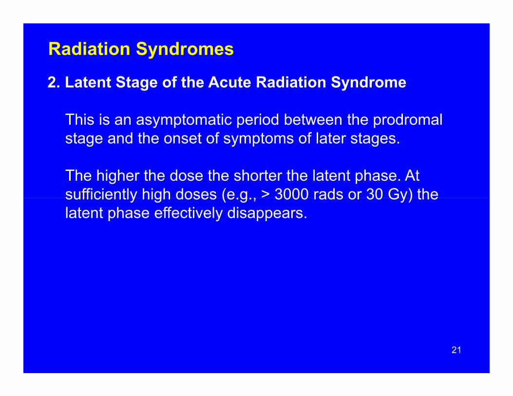
2. Latent Stage of the Acute Radiation Syndrome
This is an asymptomatic period between the prodromal stage and the onset of symptoms of later stages.
The higher the dose the shorter the latent phase. At sufficiently high doses (e.g., > 3000 rads or 30 Gy) the
Radiation Syndromes
21
sufficiently high doses (e.g., 3000 rads or 30 Gy) the latent phase effectively disappears.

3. Illness and/or Death Stage of the Acute Radiation Syndrome
Many of the characteristics of the prodromal phase might reoccur, e.g., nausea, vomiting, anorexia, fatigue. Additional symptoms might include fever, hemorrhaging
Radiation Syndromes
22

3. Illness and/or Death Stage of the Acute Radiation Syndrome
It is generally believed that without medical attention:
death is certain above 600 rads (6 Gy) 50% of exposed would die from 400 rads (4 Gy)
Radiation Syndromes
23
50% of exposed would die from 400 rads (4 Gy) 5% of exposed would die from 200 rads (2 Gy)
If the patient survives six weeks, recovery is very likely but not guaranteed.
If the dose exceeds 1000 rads (10 Gy) death is certain, most likely within 60 days.

Hematopoietic Syndrome
24

Hematopoietic Syndrome
General
The hematopoietic syndrome, sometimes referred to as the bone marrow syndrome, is produced by acute whole body doses of approximately 200 to 800 rads (2 – 8 Gy). Some authors place the range at 100 to 1000 rads and others specify the upper end of the range low as 600 rads.
25
p y pp g
Death, if it occurs, is primarily a result of damage to thehematopoietic (blood forming) organs: red bone marrow, lymph nodes and spleen.
Damage to other systems, notably the gastrointestinal tract, also plays a role.

Dose SymptomPercentage of exposed
experiencing symptom
Onset
post-exposure
50 100 rads
Anorexia 15 – 50% 3 – 18 hrs
5 30%
Symptoms as a Function of Dose
Hematopoietic Syndrome
26
50 – 100 rads
(0.5 – 1 Gy)Nausea
5 – 30%3 – 16 hrs
Vomiting 15- 20% 4 – 16 hrs

Dose SymptomPercentage of exposed
experiencing symptom
Onset
post-exposure
Anorexia 50 - 90% 3 – 18 hrs
Nausea 30 - 70% 3 – 16 hrs
Hematopoietic Syndrome
Symptoms as a Function of Dose
27
100 – 200 rads
(1 – 2 Gy)
Vomiting 10 - 50% 4 – 16 hrs
Fatigue and weakness
25 – 60% 3 – 24 hrs
Bleeding (mild)
10% 1 – 5 weeks
Fever and infection
10 – 50%2 days – 5 weeks

Dose SymptomPercentage of exposed
experiencing symptom
Onset
post-exposure
Anorexia 90 - 100% 1 – 48 hrs
Nausea 70 - 90% 1 – 48 hrs
V i i 0 90% 1 24 h
Hematopoietic Syndrome
Symptoms as a Function of Dose
28
200 – 350 rads
(2 – 3.5 Gy)
Vomiting 70 - 90% 1 – 24 hrs
Diarrhea < 10% 4 – 8 hrs
Fatigue and weakness
50 – 90% 2 hrs – 6 weeks
Bleeding 10 - 50% 1 – 5 weeks
Fever 10 - 80% 1 – 5 weeks
Infection 10 – 80% 2 – 5 weeks
Ulceration 30 3 – 5 weeks

Dose SymptomPercentage of exposed
experiencing symptom
Onset
post-exposure
Anorexia 100% 1 – 72 hrs
Nausea 90 - 100% 1 – 72 hrs
V i i 80 100% 1 24 h
Hematopoietic Syndrome
Symptoms as a Function of Dose
29
350 - 550 rads
(3.5 – 5.5 Gy)
Vomiting 80 - 100% 1 – 24 hrs
Diarrhea < 10% 3 – 8 hrs
Fatigue and weakness
90 – 100% 1 hr – 6 weeks
Headache 50% 4 – 24 hrs
Bleeding 50 - 100% 6 days – 6 weeks
Fever and Infection
80 - 100% 6 days – 6 weeks

Dose SymptomPercentage of exposed
experiencing symptom
Onset
post-exposure
Anorexia and nausea
100% 1 – 72 hrs
Vomiting 100% 1 – 48 hrs
Hematopoietic Syndrome
Symptoms as a Function of Dose
30
550 - 750 rads
(5.5 – 7.5 Gy)
Diarrhea > 10% 1 – 6 hrs
Fatigue and weakness
100% 1 hr – 2 weeks
Headache 80% 4 – 30 hrs
Bleeding,
fever and Infection
100% 10 – 14 days
Dizziness and
disorientation100% 4 – 48 hrs

Composition of Blood
Fluid (plasma)
Blood Cells: a. lymphocytes (white blood cells)
b. neutrophils (white blood cells)
Hematopoietic Syndrome
31
p ( )
c. platelets (cytoplasmic fragments)
d. erythrocytes (red blood cells)

Lymphocytes and the Effect of Radiation
Lymphocytes are a type of leukocyte (white blood cell) responsible for antibody production.
They are produced in the lymph nodes, the thymus and parts of the spleen.
Hematopoietic Syndrome
32
p p
Although mature lymphocytes are long lived and not divide, they are very radiosensitive and can be killed directly by radiation.

Lymphocytes and the Effect of Radiation
Within 15 minutes of a dose as low as 10 rads, the lymphocyte population can be seen to decrease.
The decrease in the white blood cell population is referred to as neutropenia.
Hematopoietic Syndrome
33
p
The rate of decrease in the number of lymphocytes can be used to estimate the dose.
Recovery of the lymphocyte population is slow.

Neutrophils and the Effect of Radiation
Neutrophils are the most common type of granulocytes, a type of leukocyte produced in the myelopoietic cell renewal system of the red bone marrow. They fight infection by engulfing foreign particles in the body.
The neutrophils are radioresistant but their life span is short
Hematopoietic Syndrome
34
The neutrophils are radioresistant, but their life span is short (less than one day). Damage to their radiosensitive precursors results in a measurable decrease in the number of neutrophils within a few days of the exposure.
Recovery of the neutrophil population is faster than that for lymphocytes.

Platelets and the Effect of Radiation
Platelets (aka thrombocytes) are cytoplasmic fragments produced by megakaryocytes in the bone marrow (also known as the thrombopoietic cell renewal system). They are not true cells but nevertheless play an important role in promoting the coagulation.
Hematopoietic Syndrome
35
p g g
Platelets and mature megakaryocytes are radioresistant, however the megakaryocyte precursor stem cells and the immature megakaryocytes are radiosensitive.

Platelets and the Effect of Radiation
The lifespan of platelets (8-9 days) is longer than that of the neutrophils which means they disappear more slowly.
The decrease in the platelet population is referred to as thrombocytopenia.
Hematopoietic Syndrome
36
y p

Erythrocytes and the Effect of Radiation
Erythrocytes (red blood cells) are responsible for carrying oxygen from the lungs to the various tissues of thebody.
Comparatively long-lived, they have an average life span of
Hematopoietic Syndrome
37
p y g y g pfour months (120 days).

Erythrocytes and the Effect of Radiation
Approximately one week after the exposure (above 10-20 rads, 0.1-0.2 Gy), a drop in the number of red blood cells will occur.
This decrease in erythrocytes is a result of damage to their
Hematopoietic Syndrome
38
y y gradiosensitive precursors, the stem cells of the red bone marrow, the erythropoietic cell renewal system.
For the victim to have any chance of survival, some of these stem cells must survive the exposure.

Lympocytes andNeutrophils x 1000
14
12
10
16 8
hro
cyte
s (g
ram
s)400
300
elet
s x
1000
Erythrocytes
Platelets
12 6
8 4
4 2
Ery
th
200
100
Pla
te
Days 0 10 20 30 40 50 60
Lymphocytes
Neutrophils
Prodromal phase
Latent period Manifest Illness Recovery
Dose 3 Gy

Blood Cells and the Effect of Radiation
Lymphocyte and Granulocyte Populations Approximately One Week After Exposure (x 109)
Dose Lymphocytes Granulocytes
100-200 rads (1-2 Gy) 0.8 - 1.5/liter > 2.0/liter
Hematopoietic Syndrome
40
( y)
200-400 rads (2-4 Gy) 0.5 - 0.8/liter 1.5 – 2.0/liter
400-600 rads (4-6 Gy) 0.3 - 0.5/liter 1.0 – 1.5/liter
600-800 rads (6-8 Gy) 0.1 – 0.3/liter < 0.5/liter
> 800 rads (>8 Gy) 0 – 0.1/liter < 0.1/liter
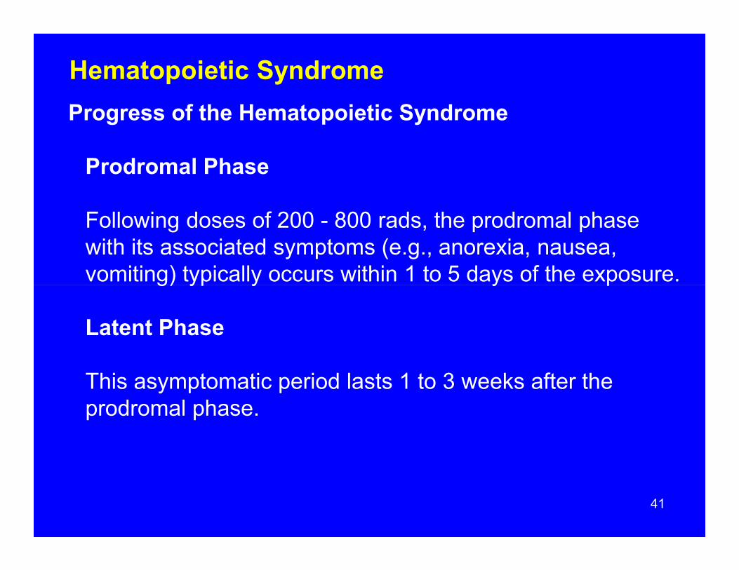
Progress of the Hematopoietic Syndrome
Prodromal Phase
Following doses of 200 - 800 rads, the prodromal phase with its associated symptoms (e.g., anorexia, nausea, vomiting) typically occurs within 1 to 5 days of the exposure.
Hematopoietic Syndrome
41
g) yp y y p
Latent Phase
This asymptomatic period lasts 1 to 3 weeks after the prodromal phase.

Progress of the Hematopoietic Syndrome
Illness and Death or Recovery
Following the latent phase a period of extreme illness begins. Symptoms include nausea, fatigue, anemia (brought about by the decrease in the red blood cell
Hematopoietic Syndrome
42
( g ypopulation), fever, epilation (hair loss), anorexia (loss of appetite), impaired wound healing and petechial (pinpoint) hemorrhaging on the skin caused by damage to the lining of capillaries.

Progress of the Hematopoietic Syndrome
Illness and Death or Recovery
Death, if it occurs, is typically within 2 to 6 weeks of the exposure. The most probable causes of death are hemorrhaging and infection.
Hematopoietic Syndrome
43
The hemorrhaging is caused by damage to the radiosensitive cells lining the fine blood vessels and is compounded by the reduced population of platelets.
Infection occurs because the intestinal bacteria penetrate the damaged lining of the gastrointestinal tract. At the same time, the body's ability to fight infection is reduced due to a decrease in white blood cell population..

Gastrointestinal Syndrome
44
Syndrome

Gastrointestinal Syndrome
General
The gastrointestinal (GI) syndrome is associated with acute whole body exposures from 800 rads (8 Gy) up to 3000 rads (30 Gy).
Estimates for the onset of the gastrointestinal syndrome
45
g yrange from as low as 600 up to 1000 rads (6-10 Gy).
Death results from both the damage to the lining of the gastrointestinal tract and damage to the hematopoietic system.
Description of Gastrointestinal Tract Lining
Much of the lining of the gastrointestinal tract is covered with small finger like projections called villi. The villi add to the effective surface area of the lining and thereby increase the capacity of the body to absorb nutrients.
Gastrointestinal Syndrome
46
The cells on the surface of the villi are constantly migrating towards the tip of the projections where they are sloughed off. Mitotically active stem cells at the base of the villi (the crypt area) replace those cells that are lost. Each crypt contains 30-40 stem cells.
The turnover rate of the epithelial cells is high - they have an average life span from 7 to 8 days.

Gastrointestinal Syndrome
Gastrointestinal Syndrome
47

Gastrointestinal Syndrome
Gastrointestinal Syndrome
48

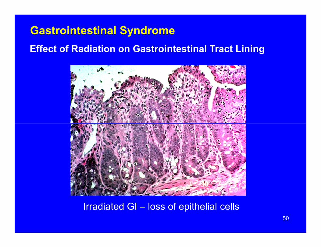
Effect of Radiation on Gastrointestinal Tract Lining
Sufficiently large acute exposures lead to the reproductive death of the rapidly dividing crypt cells. The cells covering the villi continue to be sloughed off but are no longer replaced.
Gastrointestinal Syndrome
49
This deterioration of the lining of the gastrointestinal tract then leads to a loss of body fluid, inadequate absorption of nutrients and infection from the intestinal flora (bacteria).
Above 1,000 - 1,200 rads (10-12 Gy) the crypt cells are completely destroyed. At this point, death is certain.
Effect of Radiation on Gastrointestinal Tract Lining
Gastrointestinal Syndrome
50
Irradiated GI – loss of epithelial cells

Effect of Radiation on Gastrointestinal Tract Lining
Gastrointestinal Syndrome
51
Irradiated GI

Progress of the Gastrointestinal Syndrome
Prodromal Phase
Within a couple of hours of the exposure the individual will demonstrate a sharp loss of appetite, upset stomach and apathy. Several hours later nausea, severe vomiting and
Gastrointestinal Syndrome
52
p y gdiarrhea (possibly bloody) will occur
Latent Phase
By the third day after the exposure the previous symptoms will have disappeared and the victim will appear healthy. This asymptomatic phase might last for 1-7 days.

Progress of the Gastrointestinal Syndrome
Illness
This can include nausea, vomiting, ileus (failure of intestinal tract to move contents), diarrhea, fever, sepsis (whole body inflammation due to infection) hemorrhaging,
Gastrointestinal Syndrome
53
( y ) g gapathy, anorexia and loss of weight.
Death usually occurs within 3 to 12 days of the exposure. Since the cell renewal mechanism of the gastrointestinal tract has been completely destroyed and cannot be replaced, death is inevitable.

Progress of the Gastrointestinal Syndrome
Illness
Causes of death include fluid and electrolyte losses due to the destruction of the lining of the GI tract. These fluid losses also account for the loss of weight, diarrhea and
f G
Gastrointestinal Syndrome
54
thickening of the blood associated with the GI syndrome. There is an inability to absorb nutrients.
A contributing cause of death is infection which can occur within 24 hours of the exposure as the endogenous bacteria that inhabit the GI tract invade the body across the damaged lining. Damage to the hematopoietic system simultaneously reduces the body's ability to cope with the infection.

Cerebrovascular(Central Nervous System)
55
(Central Nervous System) Syndrome
Cerebrovascular Syndrome
General
Associated with doses over 3000 rads (>30 Gy) to the whole-body.
Always fatal.
56
Immediate nausea, vomiting, anorexia, disorientation and prostration, and irreversible hypotension; blood pressure will be markedly unstable.
Within hours of exposure, the victim will be listless, drowsy, tremulous, convulsive, and ataxic. Will likely fall into a coma.
Death most likely will occur within 24 to 48 hours.

General
Direct damage to the brain might occur due to electrochemical inactivation of the nerve cells that results from damage to the cell membranes.
Most likely, death results from several causes, e.g.,
Cerebrovascular Syndrome
57
y gmeningitis (inflammation of the membranes covering the brain), myelitis (inflammation of the spinal cord), encephalitis (inflammation of the brain), and vascular damage.

General
The blood vessels of the brain are known to be damaged by large doses of radiation. Because this damage increases the permeability of the vessel walls, fluid from the blood leaks into the skull cavity (edema) and causes a buildup of pressure inside the skull. Perhaps death is partly due to the
Cerebrovascular Syndrome
58
p p p yincreased pressure on certain areas of the brain (e.g., the respiratory center).
Death might also involve the change in the blood supply to the brain.

Initial Effects Consequence to Organism
DIGESTIVE SYSTEMDecreased food intakeDecreased absorptionDiarrheaUlceration
HEMATOPOIETIC SYSTEM
Poor Nutrition
Fluid Loss
Electrolyte Loss
Infection
59
Decreased lymphocytesDecreased granulocytesDecreased plateletsDecreased erythrocytes
VASCULAR SYSTEMVascular fragility Increased capillary permeabilityObstruction of blood vessels
Hemorrhage
Anemia
Anoxia
Damage to More Resistant Tissue

Partial Body Exposures
60

Partial Body Exposures
Skin - Cutaneous Radiation Syndrome/Injury
Exposure limited to skin and adjacent underlying tissue. Exposure might be from low energy x-rays, contamination on skin, external source adjacent to body.
Immediate damage from acute exposures only occurs in the
61
g p yexposed region of skin. This might lead to indirect effects in adjacent tissues at a later time.
The magnitude of the effect is primarily dependent on the dose, but the characteristics of the exposed skin also play a role. Fair skin is more radiosensitive than darker skin. The skin of the hands, feet, scalp, and eyelids is more radiosensitive than that of the face, trunk, arms and legs.

Skin - Cutaneous Radiation Syndrome/Injury
The larger the area exposed (up to 400 cm2 or so) the greater the effect.
The term “cutaneous radiation syndrome” or “cutaneous radiation injury” replaces the older term “radiation burns” for
Partial Body Exposures
62
j y pdescribing these injuries.
There are two components to the cutaneous syndrome: the early phase during the initial few months following the exposure and the long term chronic phase.

Skin - Cutaneous Radiation Syndrome/Injury
Initial symptoms: itching, tingling, erythema.
Transient erythema (associated with itching) can occur within a few hours of exposure. This is followed by a latent, symptom-free phase lasting from a few days to several
Partial Body Exposures
63
y p p g yweeks.
After the latent phase, intense reddening, blistering, and ulceration of the irradiated site are visible.
It is possible that a third or fourth wave of erythema might occur over the ensuing months or possibly years.

Skin - Cutaneous Radiation Syndrome/Injury
As long as the basal layer of the skin is not destroyed, the skin will heal itself.
Large doses can cause permanent hair loss, damaged sweat glands, atrophy, fibrosis, decreased or increased skin
Partial Body Exposures
64
g p ypigmentation, and ulceration or necrosis of the exposed tissue.

Skin - Cutaneous Radiation Syndrome/Injury
Doses of 300-800 rads (3 - 8 Gy)
The minimum threshold for erythema (reddening of the skin). The effects can be compared to a first degree burn, i.e., sunburn. There is a reddening of the skin with some
Partial Body Exposures
65
gscaling (dry desquamation) possible.
This redness is caused by a dilation of capillaries justbeneath the surface of the epidermis - probably due to a release of histamine. No medical treatment is required and the skin will completely recover.
Skin - Cutaneous Radiation Syndrome/Injury
Doses of 300-800 rads (3 - 8 Gy)
In the first wave of erythema (if it occurs) the skin turns red 1 to 3 days after the exposure. The redness begins to fade by the end of the first week.
Partial Body Exposures
66
y
The second phase begins 2 to 3 weeks after the exposure and may last as long as one month. The second wave seems to be due to damage to the blood vessels. Perhaps the capillaries dilate to compensate for a decrease in oxygen reaching the tissue, a consequence of radiation damage to the small arterioles.

Skin - Cutaneous Radiation Syndrome/Injury
Doses of 1000 - 5000 rads (10 - 50 Gy)
The effects from doses in this range can be considered similar to those from second degree burns.
The first a e of er thema occ rs er shortl after
Partial Body Exposures
67
The first wave of erythema occurs very shortly after exposure while the second phase may begin within the first to second week.
The second phase involves dry desquamation and, at higher doses (> 2000 – 3000 rads ), wet desquamation. This loss of epidermis is due to the death of the germinal layer. With wet desquamation the loss of epidermis is associated with ulceration and the exudation of fluid.
Skin - Cutaneous Radiation Syndrome/Injury
Doses of 1000 - 5000 rads (10 - 50 Gy)
In many cases the affected area is sharply demarcated forming a clear contrast with the surrounding healthy tissue.
Treatment is aimed at keeping the tiss e clean pre enting
Partial Body Exposures
68
Treatment is aimed at keeping the tissue clean, preventing infection and reducing any pain or irritation that is present. Healing will take weeks to months. After recovery, theskin might be pigmented and more susceptible to injury.
Treatment might include the oral administration of antihistamines (e.g., Loratadine) which can reduce itching sensation and shorten the duration of the erythema. Topical steroids can also be used to alleviate the erythema.

Skin - Cutaneous Radiation Syndrome/Injury
Doses of 1000 - 5000 rads (10 - 50 Gy)
Partial Body Exposures
Twenty-four days after the exposure.
Blisters are breaking
69
gand dead skin is sloughing off.

Skin - Cutaneous Radiation Syndrome/Injury
Doses above 5000 rads (> 50 Gy)
The results can be compared to a third degree burn.
The associated pain may be intense.
Partial Body Exposures
70
p y
The germinal cells is destroyed and sufficient damage may be done for necrosis of the skin to result.
Infection and gangrene are among the short term concerns. Carcinogenesis is the long term worry.

Skin - Cutaneous Radiation Syndrome/Injury
Doses above 5000 rads (> 50 Gy)
Treatment may require skin grafts and/or amputation.
Delayed tissue death may occur as deep fibrosis and
Partial Body Exposures
71
y y pcollagen deposition gradually reduce the blood supplyto the tissue.
Rather than healing with time, the condition may get progressively worse.

Skin - Cutaneous Radiation Syndrome/Injury
Doses above 5000 rads (> 50 Gy)
Partial Body Exposures
This arteriogram shows the circulatory system in the hand of an individual who picked up an
72
p pindustrial radiography source three weeks earlier. The dose was large enough to destroy many blood vessels.

Skin - Cutaneous Radiation Syndrome/Injury
Doses above 5000 rads (> 50 Gy)
Partial Body Exposures
73
Day 6. Beginning of erythema
Day 12. Dark erythema with beginning of dry desquamation
Day 15. Tissue necrosis

Skin - Cutaneous Radiation Syndrome/Injury
Partial Body Exposures
74
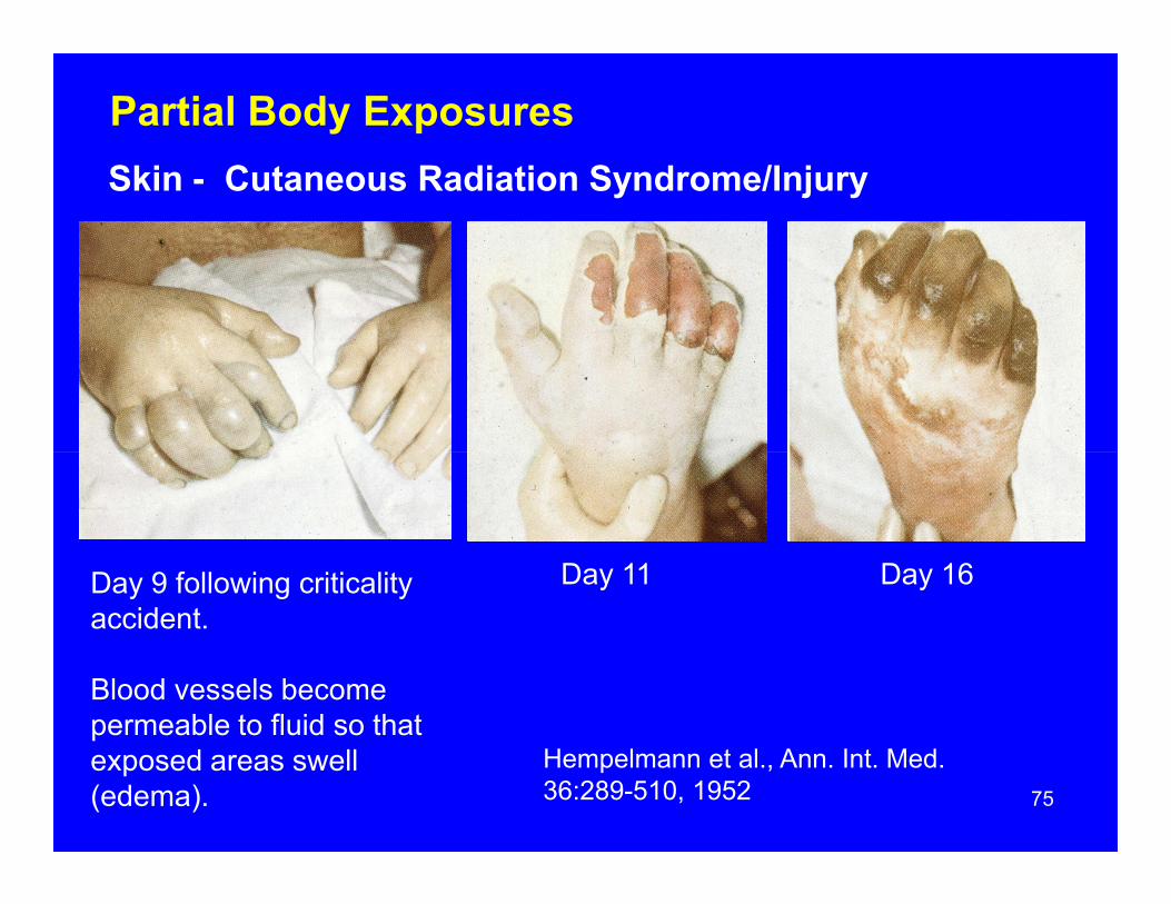
Day 9 following criticality accident.
Blood vessels become permeable to fluid so that exposed areas swell (edema). Hempelmann et al., Ann. Int. Med. 36:289-510, 1952
Skin - Cutaneous Radiation Syndrome/Injury
Partial Body Exposures
75
Day 9 following criticality accident.
Blood vessels become permeable to fluid so that exposed areas swell (edema).
Day 11 Day 16
Hempelmann et al., Ann. Int. Med. 36:289-510, 1952

Chronic Cutaneous Syndrome
The chronic phase of the cutaneous radiation syndrome can involve a gradual fibrosis brought about by the formation of collagenous tissue from the dermal and subcutaneousfibroblasts. There is a disappearance of fatty tissue and possibly blockage of blood vessels. Treatment might involve
f f f
Partial Body Exposures
76
the administration of interferon, Vitamin E and pentoxifylline. Small localized spider-like veins, possibly associated with an itching and feeling of warmth, might appear on the skin (Telangiectasis). In most cases, these veins can be removed with a laser treatment.
Keratoses might develop in the exposed area - small growths such as a wart. Since these might be precancerous, they might be excised or at least monitored.

Epilation – loss of hair
Acute doses from 300 to 600 rads (3 – 6 Gy) to the skin may lead to a temporary hair loss approximately three weeks after the exposure. The hair can be expected to regrow within one to two months after the exposure. The new hair may be of a different color (white or gray) and of a
ff
Partial Body Exposures
77
different texture.
Acute doses above 700 rads (7 Gy) or so will lead to a permanent hair loss within three weeks.
These results are typical for exposures to the beard and scalp area. Hair on other parts of the body is less easily affected.
Sterility In Males
10 rads (0.1 Gy) to the gonads can result in a slight decrease in the sperm count.
25 rads (0.25 Gy) can reduce the sperm count by 30% six weeks after exposure.
Partial Body Exposures
78
50 rads (0.5 Gy) can cause brief temporary sterility in many men. Recovery of the spermatogonia and the subsequent increase in the sperm count to normal levels may take 40 weeks.
250 rads (2.5 Gy) will lead to sterility for 1 to 2 years.
500 - 600 rads (5 – 6 Gy) results in permanent sterility.

Sterility in Females
100 – 200 rads (1 – 2 Gy) temporary sterility for 1 to 3 years
350 - 400 rads (3.5 – 4 Gy) destroys the primary and secondary oocytes and leads to permanent sterility.
Partial Body Exposures
79

Fibroatrophy
Months or years after exposures of several hundred gray, the exposed tissues can deteriorate and be replaced by fibrotic tissue. The result is a gradual loss of tissue function.
Partial Body Exposures
80

Health Physicist’s Role Following Acute Exposures
81
Following Acute Exposures

Immediate Response
First aid treatment for physical injuries should have the top priority.
If external contamination is possible, it should be looked for and dealt with.
Health Physicist’s Role following Acute Exposures
82
Consideration might be given to administering a mild sedative.

Dose Estimates
The health physicist does not prescribe medical treatment. That is the physician's responsibility.
Once the exposed individual is in the care of a physician, the health physicist's role is to help obtain an initial dose
Health Physicist’s Role following Acute Exposures
83
p y pestimate and to provide any assistance that is requested. The dose estimate can help determine the appropriate course to follow during the initial handling of the exposed individual.
A more detailed determination of the dose may be required later to help guide the medical treatment.

Assess Possible Intake
If there was a potential for radioactive material to get into the body, the health physicist might be involved in quantifying the potential intake. This assessment might involve some or all of the following:
In vitro bioassay:
Health Physicist’s Role following Acute Exposures
84
y
• Analysis of nasal swabs• Analysis of urine• Analysis of feces• Analysis of blood (possible but not standard practice)
In vivo bioassay:
• Whole body count

Assess Possible Intake
“Nasal swabs should always be promptly collected and analyzed, because a rule of thumb is that the anterior nares contain approximately 5% of the total inhaled activity, up to an hour or two after inhalation.” (Ricks et al 2002).
Health Physicist’s Role following Acute Exposures
85
Potentially contaminated clothing, wound dressing, etc. should be bagged, labelled and analyzed.

Dose Assessment
86

General
Volume 98 (No. 2), February 2010 of the Health Physics Journal contains the papers presented at the 8th International Symposium on EPR Dating and Dosimetry and 3rd Joint International Conference on Biodosimetry.
Dose Assessment
87
It is an excellent source of information concerning dose assessment following radiological incidents.

Initial Dose Estimate
• Observe and record all clinical responses. Although some of these (e.g., nausea, vomiting, etc.) may have psychological causes independent of the magnitude of the dose, it should first be assumed that they are a consequence of the exposure.
Dose Assessment
88
• If erythema occurs, obtain color photographs. These can help determine how localized the exposure was.
• Collect and process all personnel dosimeters but remember that this information may be misleading if the exposure was highly localized and/or the dosimeter was shielded by some part of the body. Obtain available information from area monitors.

Initial Dose Estimate
Dose Assessment
89Time to emesis (vomiting) after exposure

Initial Dose Estimate
• Obtain available information from area monitors.
• Carefully interview all witnesses as soon as possible. Obtain their names, addresses and phone numbers. Early interviews are important because memories are
f
Dose Assessment
90
short and witnesses may become unavailable if a weekend or holiday is near. When conducting the interview obtain information about the duration of the exposure, the position of the individual's body with respect to the source and the location of any objects that might have served as shields.

Lymphocyte Counts
Following an initial dose estimate of 20 to 100 rads (0.2-1 Gy), a blood sample might be taken the first day.
If the dose is estimated to be greater than 100 rads (> 1 Gy), blood samples might be taken every 6 to 12 hours
Dose Assessment
91
y) p g yduring the first two days after the exposure.
The absolute lymphocyte count is considered the best single indicator of the severity of the exposure. As such, lymphocyte counts will be performed on each blood sample.
The hematocrit, platelet count, and total white blood cell (WBC) count might also be determined.

Dose Assessment
Classical Andrews lymphocyte depletion curves and accompanying clinical severity ranges.
Lymphocyte Counts
92
Curves 1-4 correspond roughly to the following whole-body doses:
Curve 1 - 3.1 Gy Curve 2 - 4.4 Gy Curve 3 - 5.6 GyCurve 4 - 7.1 Gy.

Lymphocyte Counts
Goans et al (2001) described the following method for estimating the dose – it assumes an exponential decrease in the lymphocyte population during the first 48 hours.
Multiple lymphocyte counts are obtained in the first 48 hours following the exposure and the following equation is
Dose Assessment
93
hours following the exposure and the following equation is solved for K.
Lt = L0 e-Kt
Lt is the lymphocyte count at day tL0 is the lymphocyte count at the time of the exposure, t = 0t is the time after the exposure (days)K is the depletion rate constant (days-1)
Lymphocyte Counts
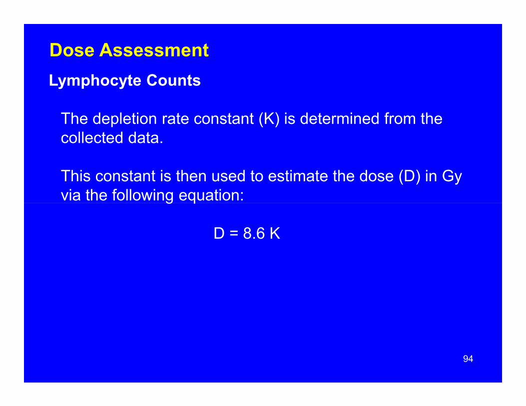
The depletion rate constant (K) is determined from the collected data.
This constant is then used to estimate the dose (D) in Gy via the following equation:
Dose Assessment
94
g q
D = 8.6 K

Cytogenetic Dosimetry “the gold standard”
Experts in cytogenetic dosimetry include the REACTS cytogenetics group at the Oak Ridge Institute for Science and Education, and the Department of Defense’s AFRRI group. The technique can be useful in situations where the exposure exceeds 10-20 rads (0.1-0.2 Gy).
Dose Assessment
95
p ( y)
Peripheral blood lymphocytes are obtained and cultured for a couple of days. The cells are arrested in mitosis and analyzed under a microscope. The average number of dicentric chromosomes per cell is determined.
The number of dicentrics per cell is related to the probable dose with a graph such as the following:

Dicentrics/cell
1.5
1.0
High LETRadiation
Low LET
Dose Assessment
96Dose (Gy)
0
0.5
0 1 2 3 4 5
Radiation

Electron Paramagnetic (Spin) Resonance
In this technique an electron paramagnetic resonance (EPR) spectrometer is used to estimate the number of free radicals produced in certain materials. The greater the radiation dose, the greater the number of free radicals.
In most materials the free radicals disappear almost
Dose Assessment
97
In most materials the free radicals disappear almost immediately after the exposure to radiation. However, in organic materials that contain little to no water (e.g., teeth, bones, hair, fingernails, sugar, etc.), free radicals can persist for weeks to years. As little as 10 mg of material is all that is required in the analysis.
EPR is only applicable if the exposures exceed 10 rads (0.1 Gy) or so, but it can do so long after the exposure.

Accident Reconstruction - Mathematical
It might be possible to assess the dose to the victim(s) mathematically.
Given a sufficiently straightforward situation (e.g., a uniform exposure to a gamma source), the calculations might be
Dose Assessment
98
p g ) gdone by hand.
A more sophisticated approach would be to estimate the dose via computer modeling. A “standard” mathematical human model might be used, or a voxel phantom might be constructed from a CT or MRI scan of the victim. In either case, the dose to the different “tissues” would be determined using a Monte Carlo analysis.

Accident Reconstruction - Physical
In some situations (e.g., sealed source exposure), it might be possible to physically duplicate the accident conditions.
A tissue equivalent phantom would be positioned at the same distance(s) from the source and for the same
Dose Assessment
99
( )period(s) of time as the victim. This phantom would have numerous dosimeters (e.g., TLDs) distributed throughout its volume. Analysis of the dosimeters would indicate the dose to different body tissues.
One of the most widely used phantoms for this purpose is the Alderson Rando phantom.
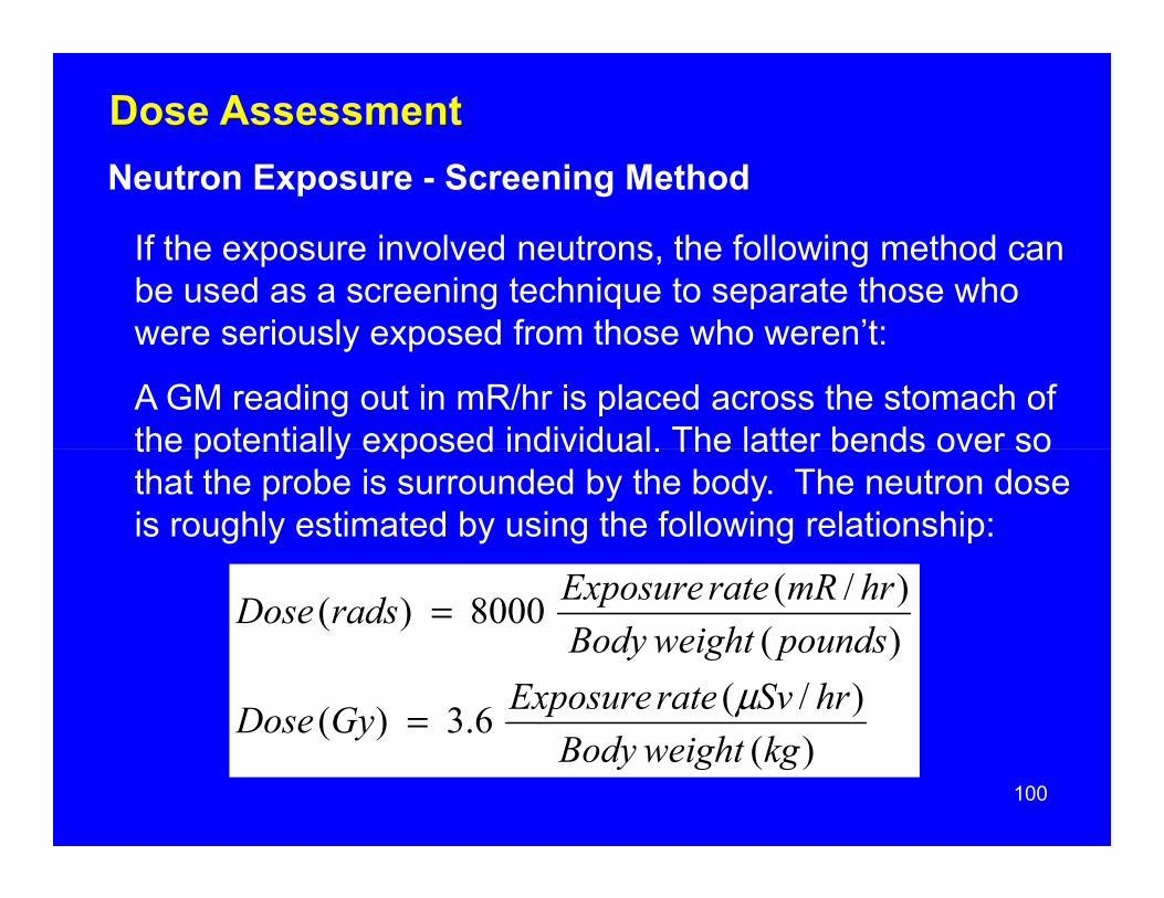
Neutron Exposure - Screening Method
If the exposure involved neutrons, the following method can be used as a screening technique to separate those who were seriously exposed from those who weren’t:
A GM reading out in mR/hr is placed across the stomach of the potentially exposed individual. The latter bends over so
Dose Assessment
100
the potentially exposed individual. The latter bends over so that the probe is surrounded by the body. The neutron dose is roughly estimated by using the following relationship:
)()/(6.3)(
)()/(8000)(
kgweightBodyhrSvrateExposureGyDose
poundsweightBodyhrmRrateExposureradsDose
μ=
=

Neutron Exposure - Screening Method
Ricks et al (2002) provided another quick and dirty way to estimate the neutron dose: “In terms of count rate, 70 counts per minute above background equals one rad ofneutron dose.”
Dose Assessment
101
Metal objects such as coins, rings, watches and belt buckles should be collected. Activation products should be quantified by gamma spectroscopy. An estimate of the neutron fluence (n/cm2) can then be made if the elemental composition of the target material is known. The estimate of the fluence is used to estimate the dose.

Neutron Exposure - Na-24 Concentration in Blood
To determine the neutron dose more precisely, the Na-24 concentration in the victim’s blood might be measured by gamma spectrometry. This concentration is decay corrected to the time of the exposure.
Dose Assessment
102
Given the Na-24 concentration in the blood, a graph such as the following might be used (from IAEA Technical Report Series #152) to estimate the dose due to thermal neutrons.

103

Medical Treatment:General
104
General

Hospitalization Guidance (from Ricks et al, 2002):
Dose less than 100 rads (< 1 Gy): Treat victims as outpatients.
Dose of 100 - 200 rads (1-2 Gy): While the victim could be treated as an outpatient, in most cases they will be hospitalized
Medical Treatment: General
105
most cases they will be hospitalized.
Dose of 200 – 400 rads (2-4 Gy): Hospitalization required, preferably in a hospital with an intensive care unit and hematology department.
Dose above 400 rads (>4 Gy): Victim should be placed in the intensive care unit of a hematology department.

General
The treatment depends on the symptoms. Nevertheless, the main effort is devoted to avoiding and fighting infection and assisting in the recovery of the blood forming tissues.
This might involve the use of antibiotics, erythrocyte and
Medical Treatment: General
106
g , y yplatelet transfusions, administration of cytokines (colony stimulating factors), and possibly bone marrow transplants.
During the prodromal phase, any nausea and diarrhea might be treated with Ondansetron.

Issues Regarding Infection
Since infections can appear soon after a large exposure (e.g., within 24 hours), nasal, mouth, throat, vaginal, skin, and urine cultures are made as soon as possible after the exposure.
Identified infections are treated immediately, e.g., oral
Medical Treatment: General
107
y, g ,mystatin to reduce Candida. Cultures will continue to be made twice a week.
The patient may be isolated in a laminar flow (bacteria free) room. If such a facility is unavailable, consideration may be given to sending the individual home; the chances of developing an infection at home are less than in a normal hospital setting.

Blood Cell Monitoring
Close watch is kept on the granulocyte and platelet levels.
If the granulocyte count drops below 1,500/mm3 the following steps may be taken: administration of oral antibiotics
daily antiseptic baths
Medical Treatment: General
108
trim and scrub finger and toe nails
If the granulocyte count drops below 750/mm3 and the infection is not responding, a transfusion of white blood cellsmay be performed.
If the platelet count falls below 40,000/mm3 and/or bleeding occurs injections of platelets may be warranted

Palliative Treatment
If the exposure exceeded 1000 rads (10 Gy), the patient is not expected to survive and treatment will be palliative.
Attempts will be made to provide relief from the symptoms rather than provide a “cure.” For example, the patient might
Medical Treatment: General
109
p p , p gbe administered pain killers, anti-depressant medication and be fed intravenously.

REACTS Guidance for Doses Exceeding 2 Gy ( 200 rads)
• Vomiting - use selective blocking of serotonin 5-T3 receptors or use 5-HT3 receptor antagonists.
• Consider initiating viral prophylaxis.• Consider tissue, blood typing.• Treat trauma.
Medical Treatment: General
110
• Consider prompt consultation with hematologist and radiation experts, re: dosimetry and prognosis, use of colony stimulating factors, stem cell transfusion, and other treatment options.
• Draw blood for chromosome analysis; use heparinized tube.
• Note areas of erythema and record on body chart. If possible, take photographs.

REACTS Guidance for Doses Exceeding 2 Gy ( 200 rads)
• SUPPORTIVE CARE in a CLEAN environment.• Prevention and treatment of infections.• Stimulation of hematopoiesis (use of growth factors, i.e.,
GCSF, GMCSF, interleukin 11).• Stem cell transfusions: cord blood, peripheral blood, or
Medical Treatment: General
111
bone marrow. Platelet transfusions if bleeding occurs or if platelet count too low.
• Psychological support.• Observe carefully for erythema (document locations), hair
loss, skin injury, mucositis, parotitis, weight loss, FEVER.• Consultation with experts in radiation accident
management is encouraged.

Hematopoietic Cell Transplantations and
112
and Colony Stimulating Factors

Hematopoietic Cell Transplantation
Hematopoietic cell transplantation consists of harvesting blood progenitor or stem cells from one individual for administration to the exposed victim. The donor and recipient should be matched as closely as possible.
Hematopoietic Cell Transplantation and Colony Stimulating Factors
113
The three most important sources of these cells:
bone marrow
peripheral blood progenitor cells
placental/umbilical-cord blood

Bone Marrow Transplants
With doses between 800 - 1200 rads (8-12 Gy), consideration might be given to a bone marrow transplant if a closely matched donor is available. Much above 1,000 rads (10 Gy), the crypt cells of the gastrointestinal tract are completely destroyed and attempts at salvaging the
Hematopoietic Cell Transplantation and Colony Stimulating Factors
114
p y y p g ghematopoietic system are pointless.
The bone marrow is collected from the posterior pelvic bones under anesthesia. Significant pain that lasts a few days is common with this procedure.
Since it takes 2 to 3 weeks for the transplanted marrow to develop sufficiently to do some good, the transplant should be performed within a week of the exposure.
Bone Marrow Transplants
The body may attempt to reject the transplanted tissue and in the process so weaken itself. However, if the bone marrow transplant is accepted for several weeks, it might provide enough granulocytes for the victim to survive until their own bone marrow recovers and the transplant is finally
Hematopoietic Cell Transplantation and Colony Stimulating Factors
115
their own bone marrow recovers and the transplant is finally rejected.
Of the 13 accident victims at Chernobyl who were given bone marrow transplants, two became long-term survivors. It has been speculated that the transplants may, in fact, have been a contributing cause of death in a few cases.

Peripheral Blood Progenitor (stem) Cell Transplant
These cells are collected directly from the donor’s bloodstream following their mobilization from the bone marrow cavity by the use of hematopoietic growth factors.
Their collection is usually four top five days after the
Hematopoietic Cell Transplantation and Colony Stimulating Factors
116
Their collection is usually four top five days after the mobilization. The donor is likely to experience some pain during the mobilization phase.

Placental/umbilical-cord Blood Transplant
This is the blood (60 to 180 mls) that remains in the umbilical cord and placenta following birth.
The red blood cells and plasma are often removed while the cord blood is frozen Advantages include the fact that there
Hematopoietic Cell Transplantation and Colony Stimulating Factors
117
cord blood is frozen. Advantages include the fact that there are no donor side effects (e.g., pain) and the fact that the cord blood - recipient HLA matching is less crucial.

Hematopoietic Growth Factors (cytokines)
Cytokines are naturally occurring proteins secreted by human leukocytes that stimulate the production of the progenitor cells in the hematopoietic tissue.
One important group of cytokines are referred to as colony
Hematopoietic Cell Transplantation and Colony Stimulating Factors
118
One important group of cytokines are referred to as colonystimulating factors (CSFs). The most important of these that can be produced in useful quantities are the granulocyte colony stimulating factor (G-CSF) and the granulocyte -macrophage stimulating factor (GM-CSF). Both have FDA approval are standard treatments for neutopenia.
Daily administration should begin soon after the exposure.

Hematopoietic Growth Factors (cytokines)
Another agent, recombinant human IL-11 ( interleukin-2), has been approved by the FDA for the treatment of thrombocytopenia as has TPO (thrombopoietin).
Other similar agents currently being evaluated include Peg
Hematopoietic Cell Transplantation and Colony Stimulating Factors
119
Other similar agents currently being evaluated include Peg-MGDF and SD/01 (sustained-duration G-CSF).
These agents are not without side effects. For example, the administration of G-CSF can slow down the recovery of the platelets.

Rescuing Victims of Acute Whole Body
120
Acute Whole Body Exposures
General
In the course of an accident involving acute whole body overexposures, decisions regarding rescue operations must be made quickly and on the basis of fragmentary information.
Rescuing Victims
121
NCRP Report 39 Guidance
Where it is necessary to search for and rescue injured persons as part of a life saving operation, the NCRP (in Report #39) at one time offered the following recommendations:

NCRP Report 39 Guidance
1. Rescuers should be volunteers or professional rescue personnel.
2. Rescue personnel should be familiar with the possible consequences of exposure.
Rescuing Victims
122
q p
3. Women capable of reproduction should not participate.
4. Where possible, volunteers should be over 45 years of age. [This recommendation does not take into account the risks to an elderly individual from the physical strainthat exhaustive rescue efforts might entail.]

NCRP Report 39 Guidance
5. The "planned" dose to the whole body should not exceed 100 rems [rads].
6. Hands and forearms may receive a total dose of 300 rems [rads].
Rescuing Victims
123
7. Such exposures should be limited to once in a lifetime.
8. Persons receiving such exposure should avoid procreation for several months.
9. The best available respiratory protection and protective clothing should be used if appropriate.

NCRP Report 116 Guidance
Only lifesaving activities justify doses much in excess of the annual worker limits.
If the actions do not involve lifesaving the recommended limit is 0.5 Sv (50 rems).
Rescuing Victims
124
For lifesaving purposes when 0.5 Sv might be exceeded, the volunteers should understand the potential for acute (early) effects and the lifetime increased risk of cancer.
The use of volunteers is desirable.
Older workers should be chosen whenever possible.

Department of Energy Regulations 10 CFR 835 Subpart N—Emergency Exposure Situations
§ 835.1301 General provisions.
(a) A general employee whose occupational dose has exceeded the numerical value of any of the limits specified in § 835.202 as a result of an authorized emergency exposure may be permitted to return to work
Rescuing Victims
125
in radiological areas during the current year providing that all of the following conditions are met:
(1) Approval is first obtained from the contractor management and the Head of the responsible DOE field organization;
(2) The individual receives counseling from radiological protection and medical personnel regarding the consequences of receiving additional occupational exposure during the year; and
(3) The affected employee agrees to return to radiological work.
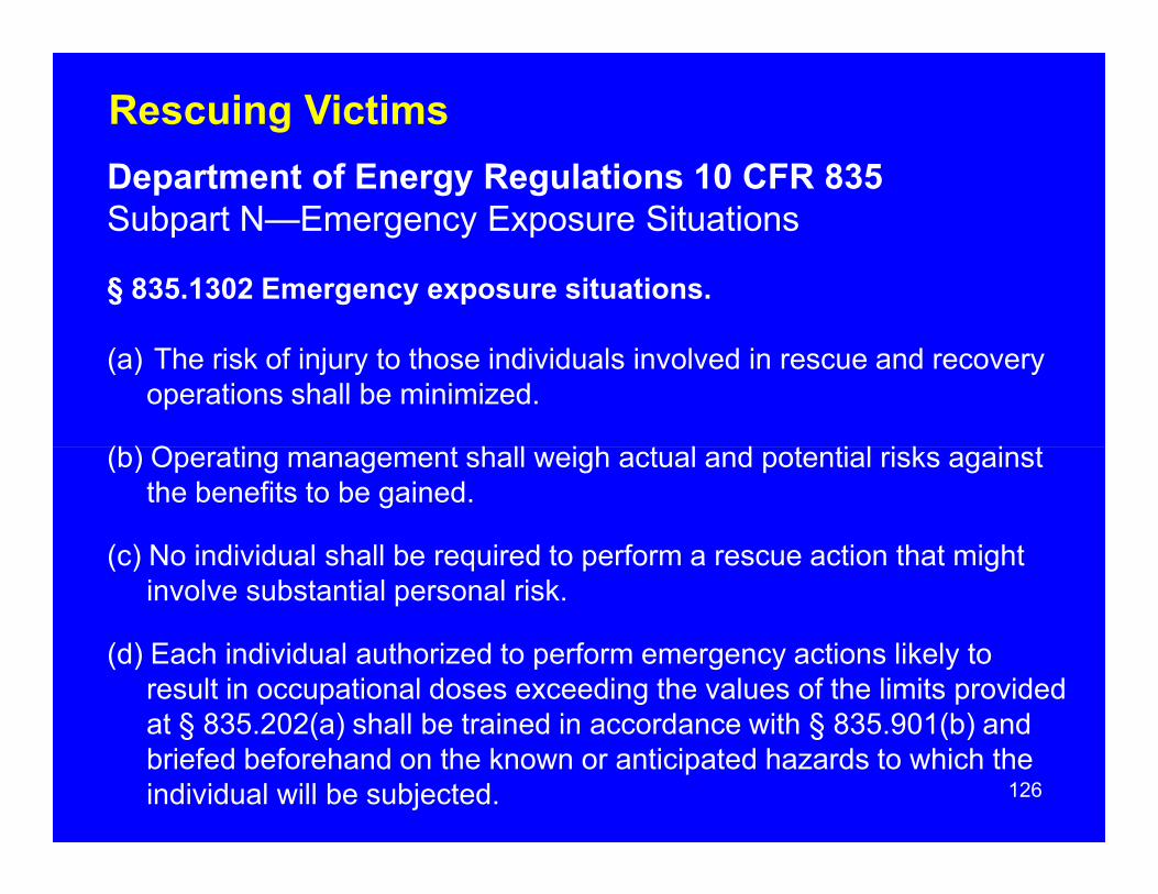
Department of Energy Regulations 10 CFR 835 Subpart N—Emergency Exposure Situations
§ 835.1302 Emergency exposure situations.
(a) The risk of injury to those individuals involved in rescue and recovery operations shall be minimized.
(b) O ti t h ll i h t l d t ti l i k i t
Rescuing Victims
126
(b) Operating management shall weigh actual and potential risks against the benefits to be gained.
(c) No individual shall be required to perform a rescue action that might involve substantial personal risk.
(d) Each individual authorized to perform emergency actions likely to result in occupational doses exceeding the values of the limits provided at § 835.202(a) shall be trained in accordance with § 835.901(b) and briefed beforehand on the known or anticipated hazards to which the individual will be subjected.

References
127

Goans, R.E., Holloway, E.C., Berger, M.E. and Ricks, R.C. Early Dose Assessment in Criticality Accidents, Health Physics 81 (4): 446-449; 2001.
Gusev, I.A., Guskova, A.K. and Mettler, F.A. Medical Management of Radiation Accidents 2nd edition, CRC Press Inc., Florida, 1990.
Mettler, F.A., Kelsey, C.A., and Ricks, R.C. Medical Management of R di ti A id t CRC P I Fl id 1990
References
128
Radiation Accidents CRC Press Inc., Florida, 1990.
Mettler, F.A. and Moseley, R.D. Medical Effects of Ionizing Radiation Grune & Stratton , Orland Florida, 1985.
Ricks, R.C., Berger, M.E., and O’Hara, M. The Medical Basis for Radiation-Accident Preparedness- the Clinical Care of Victims. Proc. Of the Fourth International REAC/TS Conference. Parthenon Pub. Group, Florida, 2002.
Swindon, T.N. Manual on the Medical Management of Individuals Involved in Radiation Accidents Technical Report 131, ARPANSA, August 2000.