Embed Size (px)
Citation preview

Each year, hundreds of thousands of adults are diagnosed with
Cervical Disc Degeneration, an upper spine condition that
can cause pain and numbness in the neck, shoulders, arms, and
even hands. This patient guide is intended to provide you with a
better understanding of cervical disc degeneration as well as an
overview of certain treatment options including Anterior
Cervical Discectomy and Fusion, or ACDF.
This guide is not intended as a substitute for an informed
discussion with your physician. If you have questions regarding this booklet, please write them
down so that your doctor or other health care professional can
answer them for you.
1
The Cervical Spine
The cervical spine is a complex system of bones, muscles,cartilage, and nerves designed to support the weight of the head while allowing movement in multiple directions. The cervical spine begins at the base of the skull and has seven small bones called vertebrae. It forms a protective pathway for your spinal cord and the nerve roots that carry signals to and from the brain, shoulders, arms and chest.
The Cervical Intervertebral Disc
Between each vertebra is a disc; a shock-absorbing pillow that helps maintain proper spacing, stability and motion within the cervical spine. Each disc has
T . , . , ^ , , a fibrous, tire-like outer band lop view of cervical vertebra and '
its structures (called the annulus fibrosus) that encases a central gel-like substance (called the nucleus pulposus).
What is the Cervical Spine?

Cervical Disc Degeneration
As we age, the discs in our cervical spine begin to flatten and wear down. When a disc flattens, it forces the vertebrae closer together, which can put added stress not only on the disc itself, but also on the surrounding joints, muscles, and nerves. This process is called Cervical Disc Degeneration, and can lead to several painful conditions.
Conditions Caused By Cervical Disc Degeneration
Herniated Disc
A Herniated Disc, known as a Herniated Nucleus Pulposus (HNP), occurs when the outer layer of the disc (the annulus fibrosus) tears or ruptures due to stress from the surrounding vertebrae. These tears can cause the disc's soft central core (the nucleus pulposus) to bulge out or even detach completely, putting pressure on the nearby nerves or spinal cord. This nerve pressure can cause symptoms of pain or weakness in specific parts of the body, depending on which nerves are being compressed.
Bone Spurs (Osteophytes)
Bone spurs, also called osteophytes, are small bony ridges that form on vertebrae as a result of increased stress on these bones. Usually, these spurs cause nothing more than an occasional stiff or sore neck. However, as with a HNP, bone spurs may press against nearby nerves or the spinal cord, causing symptoms of pain or weakness in specific parts of the body.
Herniated discs shown impinging on adjacent nerves and spinal corc
3
Symptoms of Cervical Disc Degeneration Although many people experience Cervical Disc Degeneration as a result of aging, few people experience severe symptoms.
Typically, Cervical Disc Degeneration symptoms are mild such as aches or stiffness in the neck and shoulder, as well as occasional headaches. However, Cervical Disc Degeneration symptoms can become severe when nerves are pinched due to a herniated disc or bone spurs. This can lead to painful conditions known as Cervical Radiculopath ynd CervicalMyelopathy.
• Cervical Radiculopathy - When spinal nerves are pinched, it can lead to pain, weakness, or numbness in the neck, shoulder, arms, and hands. Oftentimes, this feels like a shooting pain traveling down the arm.
• Cervical Myelopathy - Occasionally, the spinal cord itself is pinched, which can lead to severe pain or weakness in the arms and legs. This pain can cause difficulty walking as well as trouble using the hands.
Diagnosis
Your physician will conduct a history and physical examination to understand your symptoms and to determine if you have any nerve or spinal cord impairment caused by conditions related to Cervical Disc Degeneration.
Your posture, neck motion, reflexes, muscle strength and areas of pain are all evaluated during the examination. If Cervical Disc Degenerations suspected, your doctor may order an X-ray or MRI to evaluate your discs, nerves and spinal cord to help outline a course of treatment.
MRI & X-ray helps your physician detect any degeneration that could be causing pain
4

Treating Cervical Disc Degeneration
Non-Surgical Treatments
For many patients, non-surgical or conservative treatments will effectively relieve symptoms of Cervical Disc Degeneration. These treatments may include a combination of rest, physical therapy, or the use of painkillers or anti-inflammatory medications. If pain or numbness persists despite these treatments, surgical treatment options are considered.
Surgical Treatments
Artificial Cervical Disc Replacement
An investigational procedure called artificial cervical disc replacement is currently being evaluated to determine the safety and efficacy of a device that may offer some advantages over traditional spinal fusion. Artificial disc replacement surgery is very similar to Anterior Cervical Discectomy with Fusion. However, when the flattened disc is removed, the space between the vertebrae is filled with a specialized implant called an artificial disc, instead of a bone graft. The artificial disc is designed to restore spacing between the vertebrae while allowing some motion.
Single level artificial disc replacement
5
Fusion Cervical spinal fusion, also known as ACDF (Anterior Cervical Discectomy and Fusion) is the most common and standardized surgical treatment for cervical disc degeneration. During this procedure, the flattened disc is removed (called a discectomy) along with any bone spurs that are punching against the nerves. This process of relieving pressure on the spinal nerves is called decompression. Once the disc is removed, the space between the vertebrae is filled with a bone graft, or Allograft. Typically, a small titanium plate is also used to help stabilize the vertebrae. Over time, this bone graft will join together with the vertebrae to form one column of bone. This process is known as fusion. The goal of spinal fusion is to eliminate pain while restoring proper spacing between vertebrae.
A common concern about spinal fusion is loss of mobility. A single level fusion is rarely associated with significant loss of mobility that is noticeable to patients. Many patients observe that their neck mobility is actually improved after surgery due to relief from neck and/or arm pain. Resumption of normal activities can be expected within six to twelve weeks after surgery. Some patients return to work within several days of surgery or up to six weeks post-surgery. The amount of time it takes for proper healing of the bone graft varies for each patient depending on key factors such as health, age, and smoking status. These should be discussed with your doctor.
Herniated Disc Removed Single level Anterior Cervical Discectomy with Fusion (ACDF)
6
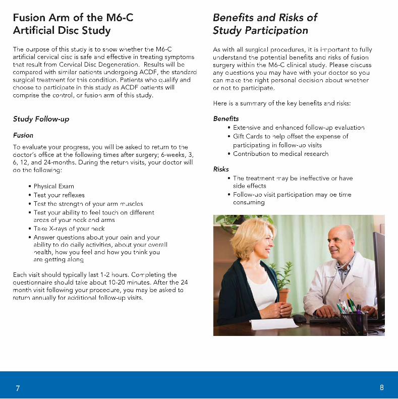
Fusion Arm of the M6-C Artificial Disc Study The purpose of this study is to show whether the M6-C artificial cervical disc is safe and effective in treating symptoms that result from Cervical Disc Degeneration. Results will be compared with similar patients undergoing ACDF, the standard surgical treatment for this condition. Patients who qualify and choose to participate in this study as ACDF patients will comprise the control, or fusion arm of this study.
Study Follow-up
Fusion
To evaluate your progress, you will be asked to return to the doctor's office at the following times after surgery; 6-weeks, 3, 6, 12, and 24-months. During the return visits, your doctor will do the following:
• Physical Exam • Test your reflexes • Test the strength of your arm muscles • Test your ability to feel touch on different
areas of your neck and arms • Take X-rays of your neck • Answer questions about your pain and your
ability to do daily activities, about your overall health, how you feel and how you think you are getting along
Each visit should typically last 1-2 hours. Completing the questionnaire should take about 10-20 minutes. After the 24 month visit following your procedure, you may be asked to return annually for additional follow-up visits.
'
Benefits and Risks of Study Participation
As with all surgical procedures, it is important to fully understand the potential benefits and risks of fusion surgery within the M6-C clinical study. Please discuss any questions you may have with your doctor so you can make the right personal decision about whether or not to participate.
Here is a summary of the key benefits and risks:
Benefits • Extensive and enhanced follow-up evaluation • Gift Cards to help offset the expense of
participating in follow-up visits • Contribution to medical research
Risks • The treatment may be ineffective or have
side effects • Follow-up visit participation may be time
consuming
8
Do I Qualify?
To take part in the Fusion Arm of the M6-C clinical study, you must meet certain criteria, some of which includes:
• Age 18 to 75 • Diagnosis of cervical radiculopathy
requiring treatment at 1 level • Failure to respond to non-surgical treatment • No previous anterior cervical spinal surgery • Able to attend follow-up progress visits with
your physician over a 24-month period and possibly longer
To help better determine if you qualify to participate in Fusion Arm of the M6-C clinical study, talk it over with your doctor.
9
Glossary of Terms
Allograft A bone graft taken from a bone bank (from organ donors) and stored under sterile conditions until needed.
Annulus Fibrosus The fibrous tire-like outer band of a natural disc that encases the central gel-like substance (called the nucleus pulposus).
Anterior Frontal position
Cervical Disc Located between each vertebrae. Helps maintain proper spacing, stability, and motion within the cervical spine. Each disc is comprised of a nucleus pulposus and annulus fibrosus.
Cervical Disc Degeneration Changes of the spine and its associated surrounding areas (intervertebral disc, spinal joints, etc.) that result from the natural aging process or injury that can limit the spine's mobility and stability.
Decompression A surgical treatment that involves relieving pressure on the spinal cord or nerve roots caused by a herniated disc, osteophytes, and bone spurs.
Discectomy The removal of part of or the entire intervertebral disc.
Fusion Joining together
Herniated Nucleus Pulposus (HNP) A disc herniates or ruptures when part of the central gel-like substance (nucleus) pushes through a tear in the tire-like outer band of the disc (annulus), putting pressure on the adjacent nerves or spinal cord. This migration of disc material beyond the edges of the vertebral body is referred to as Herniated Nucleus Pulposus (HNP). There are varying degrees of HNP that when identified are treated accordingly.
Nucleus Pulposus A gel-like substance in the center of the disc encased by a fibrous tire-like outer band (called the annulus fibrosus).
Radiculopathy Compression of one or more cervical nerve roots. Nerve root compression resulting from a herniated disc or bony spurs (osteophytes).
Vertebrae (Vertebral Body) Bony segments that form the spinal column of humans. The cervical (neck) vertebrae are the upper 7 vertebrae in the spinal column (the vertebral column). They are designated C1 through C7 from the top down.
10

/SpinaKinetics-Motion for Life1" MKT-0127 Rev. 1