Embed Size (px)
Citation preview
201
Copyright © 2014 The Korean Society of Plastic and Reconstructive SurgeonsThis is an Open Access article distributed under the terms of the Creative Commons Attribution Non-Commercial License (http://creativecommons.org/ licenses/by-nc/3.0/) which permits unrestricted non-commercial use, distribution, and reproduction in any medium, provided the original work is properly cited. www.e-aps.org
INTRODUCTION!e increase in restrictions to animal use and the "nancial con-straints of training in recent years have led to the development and spread of many non-living animal models for microsurgery simulation. Such models are numerous and include a huge spectrum such as rat cadavers, cryo-preseved rat aortas, chicken and turkey wings, leaves and grape skin, human cadaver vessels, and di#erent styles of plastic simulation materials [1-4]. Most of these non-living models are su$cient in enabling students to acquire the basic set of microsurgery skills, especially in the early
stages of training. At this beginner stage, the set of skills required includes a basic working knowledge of the surgical microscope, handling the microsurgery instruments and small sutures, and basic suturing and anastomosis techniques. Using non-living models for this purpose decreases the number of live animals used for training purposes, and gives students con"dence when working with living tissues.
However, the live rat animal model remains an indispensable model for many training microsurgical courses around the world. !e use of this model in microsurgery training stretches back to the early 1960’s, when pioneers such as Lee [5] identi-
!e Rat Model in Microsurgery Education: Classical Exercises and New HorizonsSandra Shurey1, Yelena Akelina2, Jose%e Legagneux3, Gerardo Malzone4, Lucian Jiga5, Ali Mahmoud Ghanem6
1Northwick Park Institute for Biomedical Research, Imperial College, London, UK; 2Microsurgery Research and Training Laboratory, Columbia University, New York, USA; 3Microsurgery Training and Research Lab, Paris School of Surgery, Paris, France; 4Department of Plastic Surgery Sapienza University, Rome, Italy; 5Pius Branzeu Centre for Laparoscopic Surgery and Microsurgery, Timisoara, Romania; 6Microvascular Anastomosis Simulation Hub, Barts and the London School of Medicine and Dentistry, London, UK
Correspondence:Ali Mahmoud GhanemAcademic Plastic Surgery Group, Centre for Cutaneous Research, Barts and the London School of Medicine and Dentistry, Blizard Institute, 4 New Ark Street, London E1 2AT, London, UK Tel: +44-20-7882-7173Fax: +44-20-7882-7171E-mail: [email protected]
Microsurgery is a precise surgical skill that requires an extensive training period and the supervision of expert instructors. The classical training schemes in microsurgery have started with multiday experimental courses on the rat model. These courses have offered a low threat supervised high fidelity laboratory setting in which students can steadily and rapidly progress. This simulated environment allows students to make and recognise mistakes in microsurgery techniques and thus shifts any related risks of the early training period from the operating room to the lab. To achieve a high level of skill acquisition before beginning clinical practice, students are trained on a comprehensive set of exercises the rat model can uniquely provide, with progressive complexity as competency improves. This paper presents the utility of the classical rat model in three of the earliest microsurgery training centres and the new prospects that this versatile and expansive training model offers.
Keywords Microsurgery / Models, educational / Rats / Laboratories
Received: 10 Feb 2013 • Revised: 29 Apr 2013 • Accepted: 14 May 2013pISSN: 2234-6163 • eISSN: 2234-6171 • http://dx.doi.org/10.5999/aps.2014.41.3.201 • Arch Plast Surg 2014;41:201-208
No potential conflict of interest relevant to this article was reported.
Topic

202
Shurey S et al. The rat model in microsurgery education
"ed the need for low cost surgical models that could meet the clinical needs of the day. He and subsequent researchers went on to develop organ transplant models in the rat to help address the current immunological issues at that time. It became evident that there was a need to transfer these skills to the clinical sector, as these new microsurgical techniques opened up new surgical possibilities. !is, in return, led to the establishment of micro-surgical training courses utilizing the rat model across both shores of the Atlantic and the expansion of training [6].
THE DEVELOPMENT OF RAT-MOD-EL MICROSURGERY TR AINING COURSES: A TALE OF THREE CITIESMany di#erent microsurgery training courses have been success-ful in training surgical specialists from different specialties and countries. Even though they di#er in their set-ups and programs, the basic curriculum is very similar, and we will brie&y describe three of the longest serving courses using rats as a classical train-ing model for microsurgery training: Paris School of Surgery (Paris, France), Northwick Park workshops (London, UK), and Columbia University (New York, USA).
!e Paris School of Surgery is the longest serving out of eigh-teen training courses on the record to date in France, all of which are either basic ‘certificate’ courses or advanced university ‘di-ploma’ courses. !e training lab was established in 1976 by Dr. Alain Gilbert, Gisèle Amichot, and Jose%e Legagneux as instruc-tors. !ere is an in-house course manual [7], and the pedagogi-cal approach designed at the beginning of the programme is still followed today. Currently, the course is directed by Professor Alain Masquelet. !e course accepts an average of 60 students a year for its basic certi"cate training, and 50 students can take the advanced course (university diploma). !e course teaches about 60'70 surgeons per year, and to date, the course has taught over 3,000 surgeons [8].
In the United Kingdom (working within Home O$ce guide-lines for the use of animals), basic microsurgery workshops were established at Northwick Park Institute of Biomedical Research in 1979 by Professor Colin Green and Sandra Shurey, and over 3,000 surgeons have benefited from this training to date. A course manual was also wri%en in-house to explain microsurgical theory and the use of the rat model, and to accompany the exer-cises incorporated into these workshops [9].
At Columbia University (CU), the Microsurgery Research and Training Lab was established in the early 1980s by Dr. Harold M Dick in the Department of Orthopedic Surgery, and now it is un-der the leadership of Dr. Melvin P. Rosenwasser and Dr. Yelena Akelina, training over 150 surgeons every year, from 12 special-
ties and more than 45 countries.The three centres’ basic courses are very similar, with slight
variations, and here we note the common educational philoso-phy and style that is used. At the very beginning of the course, introductory videos and short lectures from the instructors are used to put students in the right mindset for learning and prepare them for the di$cult and humbling experience of handling mi-crosurgery for the first time. New techniques and exercises are introduced with a gradual increase in di$culty in parallel to the rise of students’ con"dence and frustration management.
All of the courses emphasise the importance of the right at-titude and psychology set in achieving a successful outcome in microsurgery. All surgeons were "rst instructed on the use, care, and setup of the operating microscope, followed by basic inter-rupted suturing exercises on a rubber glove model to learn the difference between macro and micro-suturing. When this was mastered, the anaesthetised rat model was introduced.
In all courses, the rats are anesthetised intraperitoneally using a ketamine (75'95 mg/kg) and xylazine (5'8 mg/kg) cocktail and constantly monitored by the instructor and students for the depth of anesthesia. The rats’ groin area is surgically prepared by standard methods and the femoral vessels are exposed, clamped, transected, and then anastomosed with 8 interrupted sutures using 10-0 nylon according to the standard methods of Acland [10].
Classical exercises were developed to take surgeons through a series of microsurgical maneuvers that would develop their skills in one week involving 35 to 40 hours of hands-on experience. All of the basic exercises utilised the femoral artery and vein, the epigastric artery and vein, and the sciatic nerve (Fig. 1). Surgeons would work at low- to mid -range magni"cation ( ( 4' ( 15).
!e rat femoral area is used for basic exercises in preference to the aorta, carotid artery, or vena cava, as mistakes are easily man-aged and do not usually result in fatal hemorrhage. In the basic mi-crosurgery skills course (3 days in Paris vs. 5 days in London and New York), surgeons learn how to prepare and care for the opera-tive site, and how to prepare and care for micro–vessels ranging in size from 0.5 to 2 mm. !ey also perform interrupted suturing with 10/0 stitches on both individual end-to-end and end-to-side anastomoses using rat models of vein gra)ing and free &aps.
During the same time, students also learn how to safely apply clamps, the action to be taken if a clamp slips, and how to place stitches with a view to correct tension, bite sizes, and stitch place-ment according to the type of vessel, including how to remove stitches safely when necessary, as well as correctly interpreting patency.

Vol. 41 / No. 3 / May 2014
203
Course day Exercise taught
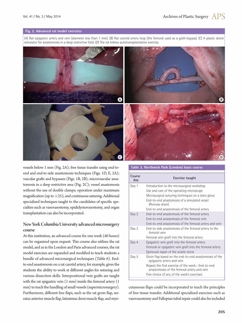
Day1 Introduction to the microsurgical workshop Use and care of the operating microscopeMicrosurgical suturing techniques on a latex gloveEnd-to-end anastomosis of a simulated vessel (Silicone tube)
Day 2 End-to-end anastomosis of the rat carotid arteryEnd-to-end anastomosis of the rat jugular veinEnd-to-end anastomosis of the rat abdominal aorta
Day 3 End-to-end anastomosis of the rat femoral arteryEnd-to-end anastomosis of the rat femoral veinInterpositional vein graft of the rat jugular vein into the abdominal aorta
Course dayRAT MODEL BASIC SKILLS COURSE COMPONENTSParis School of Surgery basic microsurgery courseIn the basic course, students undergo 3 days of training (20 hours in total). Eight hours of the course is dedicated to theoreti-cal lessons and 12 hours to practical training. During the course, the students learn how to use the microscope as well as six basic techniques of microsurgery, namely end-to-end anastomosis on synthetic and rat femoral vessels as well as vein gra) to arterial defects (Fig. 1A, B). !e training, however, is not su$cient for achieving competency. It is simply an introduction to microsur-gery and its challenging techniques. Table 1 highlights the train-ing exercises taught in this basic course.
London Northwick Park Institute for Medical Research (NPIMR) course!e Northwick Park basic microsurgery workshop runs over the course of one week (5 training days), utilizing the rat as its main training model. !e exercises taught in this basic course (Table 2) include end-to-end rat femoral artery (approximate average diameter of 1 mm) and vein (approximate average diameter of 2 mm) (Fig. 1A); rat femoral vein gra) into the femoral artery (2-mm vessel end-to-end into 1-mm vessel), which also involves surgeons learning how to cope with discrepant-sized vessels (Fig. 1B); end-to-side rat femoral artery into the femoral vein (1-mm vessel into 2-mm vessel) (Fig. 1C); rat groin &ap (Fig. 1D, E) and nerve repair using the rat sciatic nerve model (2-mm epineural repair) (Fig. 1F). Some students go on to perform epigastric vein gra) into femoral artery (1-mm vessel into 1-mm vessel, with a
di#erence in vessel wall thickness) or even raise a groin &ap on the epigastric artery (0.5 mm) and vein (1 mm) (Fig. 2A).
New York Columbia University basic microsurgery courseAt Columbia University (CU), the basic microsurgery training course is also taught over 5 working days (40 training hours). As in Paris and London, the CU basic skills course consists of ex-ercises for teaching the students basic microsurgery techniques such as end-to-end interrupted suturing of vessels using the rat femoral artery and vein model (Fig. 1A); interpositional vein gra) (Fig. 1B); end-to-side anastomosis of arteries and veins that gives the student the ability to perform both arteriotomy and venotomy (Fig. 1C); and peripheral epineural nerve repair (Fig. 1F). However, there are a few extra points emphasised during the Columbia University course: 1) End-to-end arterial anas-tomosis (1 mm) is taught in both the conventional way as well
Table 1. Paris School of Surgery basic microsurgery course
Fig. 1. Basic rat model exercises
(A) The femoral artery and vein in the rat model. (B) Interpositional vein graft: femoral vein into femoral artery. (C) End-to-side anastomosis: femoral artery into femoral vein. (D) End-to-side arterial and venous anastomosis (groin flap). (E) Rat superficial inferior epigastric artery flap. (F) Rat sciatic nerve epineural repair.
A B C
D E F
204
Shurey S et al. The rat model in microsurgery education
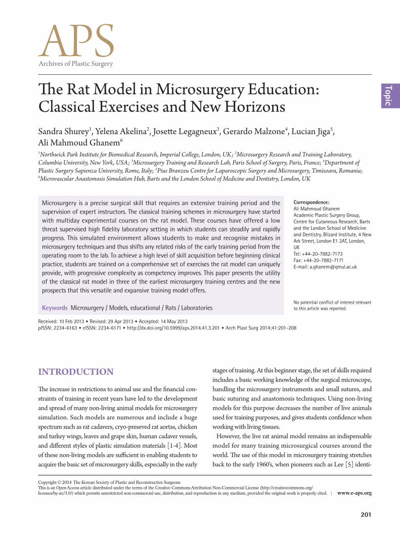
Course session Exercise taughtSession 1 Anaesthesia, analgesia and the rat model preparationSession 2 End-to-end anastomosis of the rat femoral arterySession 3 End-to-side anastomosis of the rat femoral artery to the femoral veinSession 4 End-to-end anastomosis of the rat abdominal aortaSession 5 End-to-end anastomosis of the rat carotid artery Session 6 End-to-end anastomosis of the rat external jugular vein Session 7 Interpositional vein graft of the external jugular vein into the carotid arterySession 8 Abdominal aorta bypass graft using the external jugular vein Session 9 Carotid artery collateral graft using the external jugular veinSession 10 Carotid artery bypass graft using a loop of the external jugular veinSession 11 End-to-side anastomosis of the carotid artery to the external jugular veinSession 12 End-to-side anastomosis of the rat aorta to the right iliac arterySession 13 End-to-side anastomosis of the rat portal vein to the inferior vena cavaSession 14 Ureter end-to-end anastomosisSession 15 Right kidney autotransplantation with end-to-end anastomosis of the renal artery and veinSession 16 Right kidney autotransplantation with end-to-side anastomosis of the renal artery to the aorta and the renal vein to the inferior vena cavaSession 17 Kidney transplantation (from left to right) Session 18 Lymphaticovenous anastomosis of the rat thoracic duct into the external jugular veinSession 19 End-to-end anastomosis of the rat tail arterySession 20 Epigastric vein graft into the femoral arterySession 21 Femoral artery bypass graft using the epigastric veinSession 22 End-to-end anastomosis of the rat epigastric artery and veinSession 23 Rat groin flap based on the end-to-end anastomoses of the epigastric artery and veinSession 24 Rat groin flap based on the end-to-side anastomoses of the femoral artery and veinSession 25 Groin flap based on the femoral vessels transposed to the carotid artery and jugular vein in the neckSession 26 Groin flap based on the femoral vessels transposed to the brachial vessels in the axillaSession 27 Epineural repair of the rat sciatic nerveSession 28 Rat limb transplantationSession 29 Rat fallopian tube end-to-end anastomosis and auto-graft repairSession 30 Rat cardiac transplantation
as by one-way-up or the posterior-wall-first technique. 2) The course advocates the use of the inguinal fat pad as a hemostasis tool at the end of each exercise to reduce the bleeding time and to demonstrate the e#ect of fat tissue in hemostasis [11]. 3) !e course stresses the need for the students to learn how to control anastomosis bleeding caused by gapping or uneven spacing with-out reapplying the approximating clamps in order to reduce the risk of thrombosis.
Table 3 highlights the training exercises taught in this basic course.
!T MODEL ADVANCED SKILLS COURSE COMPONENTSExperienced surgeons who want to further develop their skills are encouraged to take the advanced courses available at all three centres, which also make use of the rat model.
Paris School of Surgery advanced microsurgery courseIn this university diploma level advanced course, students are
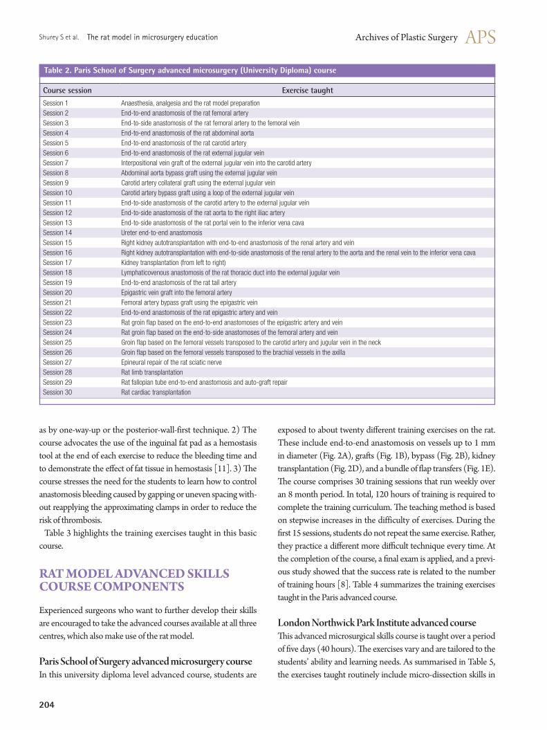
exposed to about twenty di#erent training exercises on the rat. These include end-to-end anastomosis on vessels up to 1 mm in diameter (Fig. 2A), gra)s (Fig. 1B), bypass (Fig. 2B), kidney transplantation (Fig. 2D), and a bundle of &ap transfers (Fig. 1E). !e course comprises 30 training sessions that run weekly over an 8 month period. In total, 120 hours of training is required to complete the training curriculum. !e teaching method is based on stepwise increases in the difficulty of exercises. During the "rst 15 sessions, students do not repeat the same exercise. Rather, they practice a di#erent more di$cult technique every time. At the completion of the course, a "nal exam is applied, and a previ-ous study showed that the success rate is related to the number of training hours [8]. Table 4 summarizes the training exercises taught in the Paris advanced course.
London Northwick Park Institute advanced course!is advanced microsurgical skills course is taught over a period of "ve days (40 hours). !e exercises vary and are tailored to the students’ ability and learning needs. As summarised in Table 5, the exercises taught routinely include micro-dissection skills in
Table 2. Paris School of Surgery advanced microsurgery (University Diploma) course
Vol. 41 / No. 3 / May 2014
205
vessels below 1 mm (Fig. 2A); free tissue transfer using end-to-end and end-to-side anastomosis techniques (Figs. 1D, E, 2A); vascular gra)s and bypasses (Figs. 1B, 2B); microvascular anas-tomosis in a deep restrictive area (Fig. 2C); vessel anastomosis without the use of double clamps; operation under maximum magni"cation (up to ( 25); and continuous suturing. Additional specialised techniques taught to the candidates of speci"c spe-cialties such as vasovasostomy, epididymovasostomy, and organ transplantation can also be incorporated.
New York Columbia University advanced microsurgery course At this institution, an advanced course for one week (40 hours) can be organized upon request. !is course also utilises the rat model, and as in the London and Paris advanced courses, the rat model exercises are expanded and modi"ed to teach students a bundle of advanced microsurgical techniques (Table 6). End-to-end anastomosis on a rat carotid artery, for example, gives the students the ability to work at di#erent angles for suturing and various dissection skills. Interpositional vein grafts are taught with the rat epigastric vein (1 mm) inside the femoral artery (1 mm) to teach the handling of small vessels (supermicrosurgery). Furthermore, di#erent free &aps, such as the rat groin &ap, ser-ratus anterior muscle &ap, latissimus dorsi muscle &ap, and myo-
cutaneous flaps could be incorporated to teach the principles of free tissue transfer. Additional specialised exercises such as vasovasostomy and Fallopian tubal repair could also be included
Course day Exercise taught
Day 1 Introduction to the microsurgical workshop Use and care of the operating microscopeMicrosurgical suturing techniques on a latex gloveEnd-to-end anastomosis of a simulated vessel (Penrose drain)End-to-end anastomosis of the femoral artery
Day 2 End-to-end anastomosis of the femoral arteryEnd-to-end anastomosis of the femoral veinEnd-to-end anastomosis of the femoral artery and vein
Day 3 End-to-side anastomosis of the femoral artery to the femoral veinFemoral vein graft into the femoral artery
Day 4 Epigastric vein graft into the femoral arteryFemoral or epigastric vein graft into the femoral artery Epineural repair of the sciatic nerve
Day 5 Groin flap based on the end-to-end anastomoses of the epigastric artery and veinRepeat the first exercise of the week:- End-to-end anastomoses of the femoral artery and veinFree choice of any of the week's exercises
Exercise taughtTable 3. Northwick Park (London) basic course
(A) Rat epigastric artery and vein (diameter less than 1 mm). (B) Rat carotid artery loop (the femoral used as a graft-bypass). (C) A plastic dome simulator for anastomosis in a deep restrictive field. (D) The rat kidney autotransplantation exercise.
Fig. 2. Advanced rat model exercises
A B
C D
206
Shurey S et al. The rat model in microsurgery education
Course day Exercise taught
Day 1 Introduction to mental preparation in microsurgery, use of the operating microscope, mechanics of microsurgical suturingMicrosurgical suturing techniques on a latex gloveEnd-to-end anastomosis of a simulated vessel (silastic tube)End-to-end anastomosis of the femoral artery
Day 2 End-to-end anastomosis of the rat femoral artery using the one-way-up techniqueEnd-to-end anastomosis of the rat femoral vein with interrupted suturing
Day 3 Interpositional vein graft using the femoral vein into a femoral artery defect End-to-end arterial and venous anastomosis without approximator clamps
Day 4 End-to-side anastomosis using the femoral artery to the femoral veinEnd-to-side anastomosis using the femoral vein to the femoral artery
Day 5 Epineural repair of the rat sciatic nerveExam: end-to-end arterial and venous anastomosis within a time limit
Table 5. Columbia University (New York) basic course
according to the student’s specialty and speci"c goals associated with her/his surgical practice.
!e following is a summary of the advanced exercises common to the three centres that they have developed to cover the skills training needs of the advanced trainees. !ese exercises can be an excellent foundation for higher skills necessary for clinical scenarios such as organ transplantation and complex chimeric &ap reconstruction.
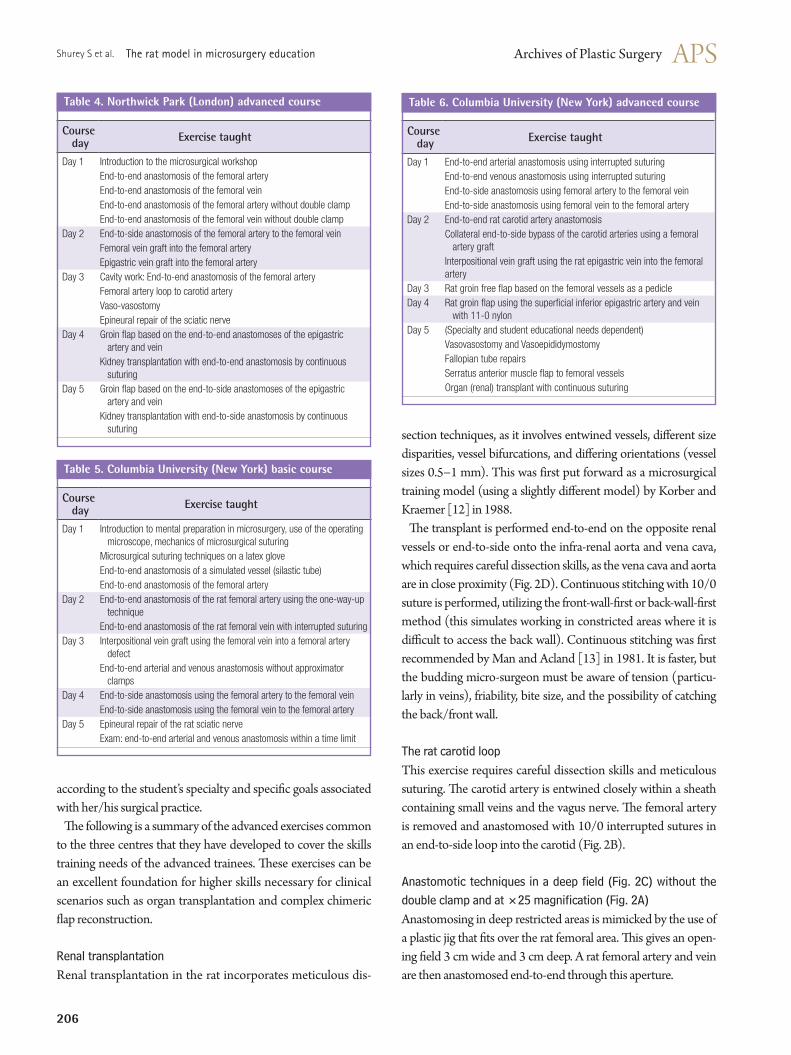
Renal transplantationRenal transplantation in the rat incorporates meticulous dis-
section techniques, as it involves entwined vessels, di#erent size disparities, vessel bifurcations, and di#ering orientations (vessel sizes 0.5'1 mm). This was first put forward as a microsurgical training model (using a slightly di#erent model) by Korber and Kraemer [12] in 1988.
!e transplant is performed end-to-end on the opposite renal vessels or end-to-side onto the infra-renal aorta and vena cava, which requires careful dissection skills, as the vena cava and aorta are in close proximity (Fig. 2D). Continuous stitching with 10/0 suture is performed, utilizing the front-wall-"rst or back-wall-"rst method (this simulates working in constricted areas where it is di$cult to access the back wall). Continuous stitching was "rst recommended by Man and Acland [13] in 1981. It is faster, but the budding micro-surgeon must be aware of tension (particu-larly in veins), friability, bite size, and the possibility of catching the back/front wall.
The rat carotid loop This exercise requires careful dissection skills and meticulous suturing. !e carotid artery is entwined closely within a sheath containing small veins and the vagus nerve. !e femoral artery is removed and anastomosed with 10/0 interrupted sutures in an end-to-side loop into the carotid (Fig. 2B).
Anastomotic techniques in a deep !eld (Fig. 2C) without the double clamp and at "25 magni!cation (Fig. 2A)Anastomosing in deep restricted areas is mimicked by the use of a plastic jig that "ts over the rat femoral area. !is gives an open-ing "eld 3 cm wide and 3 cm deep. A rat femoral artery and vein are then anastomosed end-to-end through this aperture.
Course day Exercise taught
Day 1 Introduction to the microsurgical workshopEnd-to-end anastomosis of the femoral arteryEnd-to-end anastomosis of the femoral veinEnd-to-end anastomosis of the femoral artery without double clampEnd-to-end anastomosis of the femoral vein without double clamp
Day 2 End-to-side anastomosis of the femoral artery to the femoral veinFemoral vein graft into the femoral arteryEpigastric vein graft into the femoral artery
Day 3 Cavity work: End-to-end anastomosis of the femoral arteryFemoral artery loop to carotid arteryVaso-vasostomyEpineural repair of the sciatic nerve
Day 4 Groin flap based on the end-to-end anastomoses of the epigastric artery and veinKidney transplantation with end-to-end anastomosis by continuous suturing
Day 5 Groin flap based on the end-to-side anastomoses of the epigastric artery and veinKidney transplantation with end-to-side anastomosis by continuous suturing
Course dayTable 4. Northwick Park (London) advanced course
Course day Exercise taught
Day 1 End-to-end arterial anastomosis using interrupted suturing End-to-end venous anastomosis using interrupted suturing End-to-side anastomosis using femoral artery to the femoral veinEnd-to-side anastomosis using femoral vein to the femoral artery
Day 2 End-to-end rat carotid artery anastomosis Collateral end-to-side bypass of the carotid arteries using a femoral artery graft Interpositional vein graft using the rat epigastric vein into the femoral artery
Day 3 Rat groin free flap based on the femoral vessels as a pedicleDay 4 Rat groin flap using the superficial inferior epigastric artery and vein
with 11-0 nylonDay 5 (Specialty and student educational needs dependent)
Vasovasostomy and VasoepididymostomyFallopian tube repairsSerratus anterior muscle flap to femoral vesselsOrgan (renal) transplant with continuous suturing
Table 6. Columbia University (New York) advanced course

Vol. 41 / No. 3 / May 2014
207
Some clinical centres do not use or have access to double clam-ps, so the anastomotic techniques taught do not utilise this, and the rat femoral artery and vein anastomosis can be completed by starting at the centre of the back wall "rst or using a 1,200 or 1,800 orientation.
For very small vessels of 1 mm and below, a higher magni"cation is needed. Trainees learn that at this level of magnification, the light is not so bright and the depth of "eld is much shorter, mean-ing that the anastomosis must be carried out in a horizontal plane.
Rat groin flap models involve end-to-end anastomosis of the epigastric artery and vein (Fig. 1A). This exercise can be per-formed utilising an end-to-side anastomosis of these vessels onto the femoral artery and vein. !e epigastric vein can also be anas-tomosed as a gra) into the epigastric artery.
Vasovasostomy and epididymovasostomyRat models of vasovasostomy and epididymovasostomy are good substitutes for clinical operations due to their relatively large size in the rat, and are used routinely in vasectomy reversal workshops for urologists in the UK [7].
NEW HORIZONS IN MICROSUR-GERY T!ININGFifty years after Sun Lee’s manual describing microsurgery techniques in the rat to address the needs of reconstructive sur-geons of that time, the clinical world is again in need of further microsurgical training interventions with the advent of new hori-zons in microsurgery, namely, the advent of perforator &aps and lymphatic anastomosis. !ese new emerging techniques utilise vessels of below 1 mm. !is has been termed ‘supra-microsurgery’ [14]. In addition, the use of microsurgical techniques is now be-ing incorporated into many new areas of the clinical specialties. In 1979, most new micro-surgeons were from the "eld of plastic surgery. In contrast, current students come from a variety of spe-cialties and include maxillo-facial, oral/otolaryngol ogy (ENT), orthopaedic, hand, general, urology, obstetrics, paediatric, car-diac, and transplant surgeons.
When surgeons gain experience in microsurgery, the horizons for utilising the rat model are virtually endless, given the possibili-ty to learn how to perform tail, toe, and organ transplants, to work with vessels with a size discrepancy, to work with video-assisted devices, to suture with continuous stitching, and other di$cult exercises [15-25]. New techniques with the robot-assisted micro-surgery open a new door to utilise the classic rat model for teach-ing surgeons from di#erent specialties to perform microsurgical procedures using robotic arms. Other techniques and procedures that can be utilised in the rat for microsurgical training (but are
yet to be permitted in the UK by current Home Office rules) include most of the organ transplants described by Lee [5] such as those of the liver, lung, small and large bowel, stomach, and pancreatico-duodenum. Additional exercises can include other free &aps, and muscle, bone, and limb transplantations.
CONCLUSIONSIt has been established that microsurgery training courses using rat models offer many advantages. They greatly enhance stu-dents’ surgical skills and provide the highest "delity simulator for clinical microsurgery. !e use of rat models in advanced courses under a supervised training programme provides an excellent simulation model for complex microsurgical reconstruction pro-cedures. As such, the rat model remains the best preparation for achieving high standards of competency in microsurgery. Ad-vances in microsurgical reconstruction demand new educational interventions. !e live rat is one of the most versatile models in microsurgery training courses worldwide, and 50 years a)er mi-crosurgery was "rst pioneered, the rat model is still irreplaceable for advanced microsurgery skill acquisition. Its prospects include future educational roles in perforator &ap techniques and robot-assisted microsurgery.
REFERENCES 1. Austin GT, Hammond FW, Schaberg SJ, et al. A laboratory
model for vascular microsurgery. J Oral Maxillofac Surg 1983; 41:450-5.
2. Kaufman T, Hurwitz DJ, Ballantyne DL. !e foliage leaf in microvascular surgery. Microsurgery 1984;5:57-8.
3. Weber D, Moser N, Rosslein R. A synthetic model for mi-crosurgical training: a surgical contribution to reduce the number of animal experiments. Eur J Pediatr Surg 1997;7: 204-6.
4. Lannon DA, Atkins JA, Butler PE. Non-vital, prosthetic, and virtual reality models of microsurgical training. Micro-surgery 2001;21:389-93.
5. Lee S. Historical events on development of experimental microsurgical organ transplantation. Yonsei Med J 2004;45: 1115-20.
6. Green CJ. Microsurgery in the clinic and laboratory. Lab Anim 1987;21:1-10.
7. Gilbert A, Legagneux J, Lapierre F. Apprentissage de la microchirurgie. In: Techniques chirurgicales: chirurgie plas-tique reconstructrice et esthetique (45-025). Paris: Encycl Med Chir; 1998. p.24.
8. Cabrol C, Gilbert A. Training of microsurgery in the labora-
208
Shurey S et al. The rat model in microsurgery education
tory of the Hopitaux de Paris. Chirurgie 1991;117:145-8. 9. Green C, Simpkin S; Clinical Research Centre (Harrow,
London, England). Basic microsurgical techniques: a labo-ratory manual. Harrow, Middlesex, UK: Surgical Research Group, MRC Clinical Research Centre; 1986.
10. Acland RD. Practice manual for microvascular surgery. St. Louis: Mosby; 1980.
11. Akelina Y, Danilo P. Endogenous adipose tissue as a hemo-static: use in microsurgery. Microsurgery 2008;28:192-6.
12. Korber KE, Kraemer BA. Heterotopic renal transplantation in the rat: an advanced microsurgical training exercise. Mi-crosurgery 1988;9:286-91.
13. Man D, Acland RD. Continuous-suture technique in micro-vascular end-to-end anastomosis. J Microsurg 1981;2:238-43.
14. Koshima I, Inagawa K, Urushibara K, et al. Supermicrosur-gical lymphaticovenular anastomosis for the treatment of lymphedema in the upper extremities. J Reconstr Microsurg 2000;16:437-42.
15. Bao JY. Rat tail: a useful model for microvascular training. Microsurgery 1995;16:122-5.
16. Crosby NL, Clapson JB, Buncke HJ, et al. Advanced non-animal microsurgical exercises. Microsurgery 1995;16:655-8.
17. Zhang F, Chin BT, Ho PR, et al. Rat tail replantation as a training model for microvascular procedures of digit replan-tation. Microsurgery 1998;18:364-7.
18. Akan M, Cakir B, Akoz T. “Open y” technique in vessel di-ameter discrepancy. Microsurgery 2006;26:506-14.
19. Blidisel A, Jiga L, Nistor A, et al. Video-assisted versus con-ventional microsurgical training: a comparative study in the rat model. Microsurgery 2007;27:446-50.
20. Galvao FH, Bacchella T, Cerqueira Machado M. Teaching intestinal transplantation in the rat for medical student. Mi-crosurgery 2007;27:277-81.
21. Miyamoto S, Okazaki M, Ohura N, et al. Comparative study of di#erent combinations of microvascular anastomoses in a rat model: end-to-end, end-to-side, and &ow-through anas-tomosis. Plast Reconstr Surg 2008;122:449-55.
22. Mofikoya BO, Ugburo AO, Bankole OB. Microvascular Anastomosis of Vessels Less Than 0.5 mm in Diameter: A Supermicrosurgery Training Model in Lagos, Nigeria. J Hand Microsurg 2011;3:15-7.
23. Sakrak T, Kose AA, Karabagli Y, et al. Rat tail revasculariza-tion model for advanced microsurgery training and research. J Reconstr Microsurg 2011;27:391-6.
24. Spetzger U, von Schilling A, Brombach T, et al. Training models for vascular microneurosurgery. Acta Neurochir Suppl 2011;112:115-9.
25. Yazici I, Cavusoglu T, Karakaya EI, et al. Microsurgical train-ing model for lymphaticovenous anastomosis in rat. Micro-surgery 2012;32:420-2.


![Critical evaluation of near-field seismic records in Greecelee.civil.ntua.gr/pdf/mathimata/texniki_seismo... · 19 Ευρωκώδικας8 [cen 2004]: Μέρος2 - Γέφυρες(συνέχεια)](https://img.pdfslide.us/doc/110x75/5ec67e098fda4a7c6a3c9cfe/critical-evaluation-of-near-field-seismic-records-in-19-8.jpg)