Embed Size (px)
Citation preview

E. Ann Steiner, MT(ASCP)SBB
Blood Bank Technical Specialist
Ortho Clinical Diagnostics
Why Do We Care?� Considering change? Considering automation?
� To better understand current methodology, for
use in troubleshooting, problem prevention
� Evaluation of current alternative method(s)
� Facilitate antibody identification

Serological Results That Trigger
an ABID study
�Certain parts of routine pretransfusion testing
�Certain parts of optional steps in
pretransfusion testing
�The results of diagnostic, or ‘special’, testing

Serological Results That Trigger
an ABID study
�Apparently ‘extra’ reaction in ABO reverse
�Positive antibody screen
�Positive crossmatch
�Positive inert control, IAT control, or DAT
�The results of diagnostic, or ‘special’, testing

Pretransfusion Testing�Test suspension of patient’s red blood cells
(RBC’s) with anti-A, -B, and -D
aka, the ABO/Rh forward group
�Test plasma/serum against known A1 and B
reagent RBC’s
aka, the ABO reverse group
�Test plasma (or serum) against Group O
reagent RBC’s of known phenotype
aka, the antibody screen or detection test

Optional Serological Steps �Test suspension of patient’s RBC’s with:
�Anti-A1, -A,B, second –D
� Inert control (reagent, autologous plasma)
�Test plasma/serum against known A2 RBC’s
�Run a 3 or 4-cell screen (rather than 2-cell)
� IAT autocontrol or Direct Antiglobulin Test (DAT)
�Perform a crossmatch
�For major ABO confirmation only
�For the detection of IAT reactive antibodies

‘Special’ Testing
� Studies to aid in diagnosis such as:
�Direct Antiglogulin Tests (DATs), with
Polyspecific and/or Monospecific AHG
�Preparation and testing of an eluate, with or
without the presence of a positive DAT
�Use of Polyspecific AHG & fresh serum in IAT
�Other special methods and modified testing
parameters

Diagnostic Studies
�Differential diagnostic investigations such as:
�Transfusion (Tx) Reaction
�Hemolytic Disease of the Fetus/Newborn
�Autoimmune Hemolytic Anemia
�Drug-related Hemolysis
�Graft vs host, other complications of solid
organ or stem cell/marrow transplantation

History of Antibody Detection�Review of antibodies:
� Identified subsequent to the detection of an
incompatible crossmatch (not attributed to
ABO incompatibility)
�Associated with a hemolytic tx reaction
� It was determined that the corresponding
antigens for >95% of these antibodies could be
located on RBC’s from 2-3 select individuals

What Really Is This Test Called
ANTIBODY DETECTION?�Historically – developed in the 1950’s to
lessen the incidence of antibodies detected at
time of crossmatching (XM) and therefore too
close to time of need for transfusion
�Recent past – simultaneous to time of XM,
intended to be, in some instances, more
sensitive and more predictable in antigenic
make-up, e.g., double dose expression of
certain antigens

What Really Is This Test Called
ANTIBODY DETECTION?�Current definition and use
�Detect the majority of antibodies likely to
cause significantly shortened RBC survival
�Done as replacement to crossmatch (with
certain defined exceptions)
�Minimum antigen make up defined by CFR
�Common practice to select RBCs whose
antigenic make up enhance sensitivity

Variables in Antibody Screens �Number of reagent RBC’s (for pretransfusion
patients, can NOT be pooled cells)
�Antigenic make up of these reagent RBC’s
�Methodology/enhancement
�Variables between and within a method: � Low Ionic Strength saline (LISS), Polyethylene
Glycol (PEG), Bovine Albumin, other
� Time, temperature
� Tube, solid-phase, column
FDA 21 CFR Requirements� For Reagent RBC’s for antibody detection:
Antigens that must be present
� For compatibility testing
�Time of sample collection relative to tx
� <3 days if recipient has been transfused or
pregnant in preceding 3 months
�Type of testing
� Capable of demonstrating incompatibility

AABB Standards�Method to detect clinically significant
antibodies:
�Defined as capable of producing a
significant adverse reaction
�Must include 37°C incubation and include
an antiglobulin test
�Reagent RBC’s must not be pooled

Antigens Required�D, C, c, E, and e
�K, k
� Fya, Fyb
� Jka, Jkb
� Lea, Leb
�M, N, S, s
�P1

Dichotomy Abounds�Want to find “clinically significant” and yet:
�Antigens are missing whose antibodies are
known to cause significant RBC destruction
� Doa, Kpa, Lan, (and dozens more)
�Antigens are present whose antibodies are
not generally associated with significant
RBC destruction
� M, N, Lea, Leb, P1 (and a few dozen more)

Dichotomy Abounds�Want to find “clinically significant” and yet:
�Antigens are present whose antibodies are
not generally associated with significant
RBC destruction
� M, N*, Lea, Leb, P1 (and a few dozen more)

There’s More…�Autoantibodies
�Generally a panagglutinin, but not always
�May be clinically significant
�May affect tx and donor selection
�May be warm/cold; IgM/IgG; combinations
�Passively-acquired antibodies
�May be only temporarily present, but
clinically significant during that time
�Sources include transplantation, donor
components, infusions (IVIG, RhIG, etc.)

Goals for Antibody Detection�Detect the necessary clinically significant
antibodies
�Avoid false positives or unreadable tests, i.e.,
have a high first pass yield
�Quick enough to meet our customers’ needs
�Reasonably low amount of plasma needed
�Cheap and easy to perform

Causes of False Positive or
Unreadable Results� False positives
�Unwanted antibodies, i.e., not considered
likely to be clinically significant
�Rouleaux
�Anomolous reactions such as antibodies to
preservatives and additives
�Unreadable results
�Contamination, fibrin, AHG neutralization, etc.

Maximizing Predictive Value:
Method Selection
C Generally Unwanted Considered Significant W
O Allo Anti-M, -N, -Lea, -Leb, -P1 Allo Anti-D, -C, -c, -E, -e, -K A
L Auto Anti-I, -i, -H, -HI -Jka, Jkb, Fya, Fyb, S, s R
D False or Unreadable Results AHG neutralized, etc. M
(IS, RT) Direct agglutination IAT (≈37°C)

Maximizing Predictive Value:
Method Performance
�Minimize the unwanted
�Maintain sample quality, avoid fibrin, cryo,
contamination, hemolysis, etc.
�Adhere to procedure guidelines, have a
quality staff training program
�Quality check reagents and equipment
�Avoid inadvertent cold exposure

Maximizing Predictive Value:
Method Selection
� Select what’s best for your setting
�Staff capable of proper performance
�Meets budget and time constraints
� Increase sensitivity?� Reagent RBC make-up
� Additional antigens
� Double-dose expression of selected antigens
� Length of 37°C incubation

Routine Alternative Methods
�Tube testing
� Low Ionic Strength Saline (LISS)
�Polyethylene Glycol (PEG)
�Bovine albumin, 22% or 30%
� Solid-phase (in micro titer plates [MTPs])
�RBC stroma adherence
�Protein A adherence
�Column technology
�Gel columns

Tube Methods
�Procedure steps in common
�Can employ 2 or 3 reagent RBC’s
� Incubation at 37°C for first stage of reactivity
– antibody uptake
�Enhancement reagent present during
incubation
� Include Indirect Antiglobulin Testing (IAT)
� IAT needs coated cells as quality check for
each negative result
Tube Methods�Variable steps in common
�Time at 37°C� Minimum required: manufacturer, regulations
� Minimum for maximum sensitivity
� Maximum to avoid risk of antibody elution
�RBC make up� Double-dose requirements, e.g., R1R1, R2R2,
Jk(a+b-), Fy(a+b-), etc.
� Presence of low incidence antigens, e.g., Kp(a+)
�Phases of testing: Imm. Spin, Room Temp.

Tube Methods�Variable steps in common
�Phases of reading: 37°C
�Optical aid for AHG reading, e.g.,� Microscope
� Viewlight
�Type of AHG employed� Monoclonal monospecific anti-IgG
� Detects subclasses 1, 2, and 3; not 4
� Polyclonal monospecific anti-IgG
� Polyspecific anti-AHG
� Monoclonal, Polyclonal, Blended

Tube Method - LISS�Delivery of LISS
�As an additive, i.e., drops from reagent vial
�As an RBC suspension medium
�Reactants
�Critical control of proportions, generally
equal parts of LISS and plasma� Quality checked pipettes may be necessary
�Additives may contain additional agents
�Can use anti-IgG or Poly AHG
�37°C incubation as short at 10 minutes

Tube Method - LISS� “Wanted” ↑ SensiWvity
�Kidd system antibodies
� “Wanted” ↓ SensiWvity
�Certain anti-K, esp. with shortened 37°C inc.� Personal experience: some anti-K ↑
� “Unwanted” ↑ SensiWvity
�Anti-M
�Certain cold autoantibodies� Esp. if serum and Poly AHG is used
� “Wanted” ↓ SensiWvity
�Can omit readings other than IgG

Tube Method - PEG�Delivered as an additive, i.e., drops from a
reagent vial
�Use of anti-IgG strongly recommended
�Do not read at phases other than IAT
�Non-specific aggregates may mimic
agglutination
�Precipitins may form in high protein samples
�Mimic agglutination
�Neutralize AHG (not washed away)
�37°C incubation as short at 10 minutes

Tube Method - PeG� “Wanted” ↑ SensiWvity
�Kidd system antibodies
� “Wanted” ↓ SensiWvity
�Some Knops-system antibodies
�Perhaps some Chido-Rodgers antibodies
� “Unwanted” ↑ SensiWvity
�Anti-P1
�Warm autoantibodies
� “Wanted” ↓ SensiWvity
�No readings other than IgG

Tube Method – Bovine Albumin�Delivered as an additive, i.e., drops from a
reagent vial
�Affects second-stage of agglutination
�No increase in antibody uptake
�Creates low-ionic environment
�Facilitates direct agglutination of IgM or IgG
� Lack of innate sensitivity prompts use of other
enhancing alternatives, e.g.,
�Serum + poly AHG
� Longer 37°C incubation: at least 20’, better 30’
Tube Method – Bovine Albumin� “Wanted” ↑ SensiWvity
�Rh, at least as direct agglutination
� “Wanted” ↓ SensiWvity
� Less AHG carry-over of cold IgM vs LISS/PeG
�Warm autoantibodies
� “Unwanted” ↑ SensiWvity
�Complement-binding cold autoantibodies
� “Unwanted” ↓ SensiWvity
�Pretty much everything

Other Methods�Performed in MTPs
�RBC adherence� Available with automation or manual testing
�Protein A adherence� Available with automation
�Performed by column agglutination technology
(CAT)� In the U.S., available as gel column, with
automation or manual testing
�All use smaller reactant volumes than tube

Solid Phase – RBC Stroma� Stroma of reagent RBC’s is bound to bottom
of U-shaped MTP
�Plasma and LISS are added
�37°C incubation (15-60’ followed by x4 wash)
� Indicator cells with affixed anti-IgG added
�Centrifugation
� Indicator cells adhere if stroma has
antibody affixed
�No adherence with negative test

Solid Phase – RBC Stroma� “Wanted” ↑ SensiWvity
�Kidd system antibodies
�However may not be in vivo significant
� “Wanted” ↓ SensiWvity
�Anti-K due to LISS
� “Unwanted” ↑ SensiWvity
�Warm autoantibodies
�Non-specific adherence
� “Wanted” ↓ SensiWvity
�No readings other than IgG
�Manual test may be difficult to read
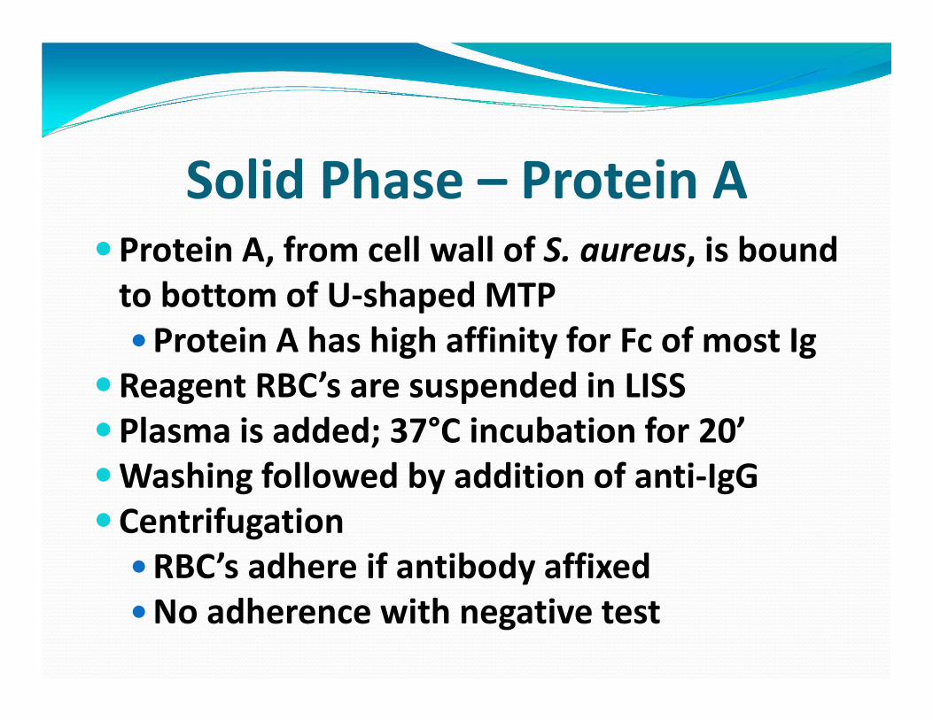
Solid Phase – Protein A�Protein A, from cell wall of S. aureus, is bound
to bottom of U-shaped MTP
�Protein A has high affinity for Fc of most Ig
�Reagent RBC’s are suspended in LISS
�Plasma is added; 37°C incubation for 20’
�Washing followed by addition of anti-IgG
�Centrifugation
�RBC’s adhere if antibody affixed
�No adherence with negative test

Solid Phase – Protein A� Sensitivity/Specificity
�Newer method, data incomplete
�AnWcipate ↓ AnW-K due to LISS
�Some reports indicate “non-specific”
reactions comparable to Stroma method
�AnWcipate ↑ warm autoanWbodies
� “Wanted” ↓ SensiWvity
�No readings other than IgG
�Manual test not available

Column Technology - Gel�Performed in molded plastic columns
�Contain small particles of gel + anti-IgG
�Particles serve as sieve to filter RBCs by size
� LISS suspended Reagent RBC’s added
�Plasma added, followed by 37°C inc. for 15’
�Controlled centrifugation
�Sensitized RBC’s react with IgG and trap
�Negative RBC’s centrifuge to the bottom
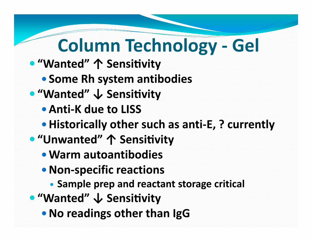
� “Wanted” ↑ SensiWvity
�Some Rh system antibodies
� “Wanted” ↓ SensiWvity
�Anti-K due to LISS
�Historically other such as anti-E, ? currently
� “Unwanted” ↑ SensiWvity
�Warm autoantibodies
�Non-specific reactions� Sample prep and reactant storage critical
� “Wanted” ↓ SensiWvity
�No readings other than IgG
Column Technology - Gel

Comparing & ContrastingWithin “same” method, e.g.,
� Source of LISS or PeG
�Other additives in LISS or PeG
�Variables of solid phase, e.g.,
�Parameters of buffer, indicator cells, etc.
�Variables of gel, e.g.,
�Manufacturer’s reagents cells vs. in-house
preparation

Comparing & Contrasting�No method is perfect or ideal
�All meet minimum requirements
�Many exceed minimum, but in different ways
�Gain of increased sensitivity (weak clinically
significant antibodies) may be at the cost of
decreased specificity (increased false positives)
�Antibody ID work is geared to characteristics of
antibody detection method