Embed Size (px)
Citation preview

Lianne Beck, MDAssistant ProfessorEmory Family Medicine
Define dysphagia Know the 2 main types and how to
differentiate them Learn the major causes of dysphagia Understand how to work up a patient with
dysphagia Become familiar with the treatment options

Dysphagia—difficulty with swallowing—is a common condition Reported by 7-10% of the general population aged over 50
years, 16% of the elderly Up to 25% of hospitalized patients
Oropharyngeal dysphagia, is even more common in the chronic-care setting; up to 60% of nursing-home occupants have feeding difficulties that include dysphagia.
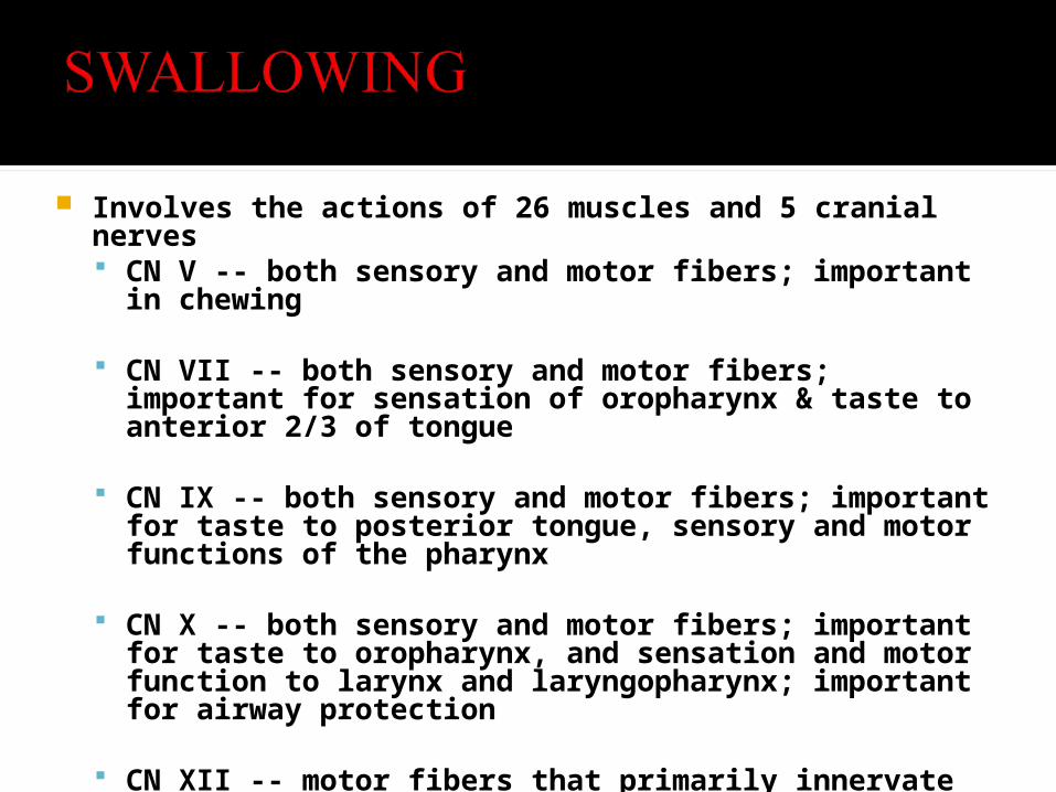
Involves the actions of 26 muscles and 5 cranial nerves CN V -- both sensory and motor fibers; important in chewing
CN VII -- both sensory and motor fibers; important for sensation of oropharynx & taste to anterior 2/3 of tongue
CN IX -- both sensory and motor fibers; important for taste to posterior tongue, sensory and motor functions of the pharynx
CN X -- both sensory and motor fibers; important for taste to oropharynx, and sensation and motor function to larynx and laryngopharynx; important for airway protection
CN XII -- motor fibers that primarily innervate the tongue
A normal adult swallows unconsciously 600 times a day


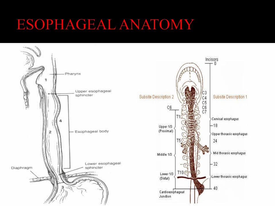
Upper one-third is composed of skeletal muscle
Distal two-thirds is smooth muscle
NO SEROSA
Outer longitudinal, inner circular muscle layer
Myenteric plexus of Auerbach, parasympathetic ganglion cells, interspersed among the muscle layers
Submucosa – blood vessels/lymphatics, myenteric plexus of Meissner (parasympathetic ganglion cells)
Mucosa – stratified squamous epithelium
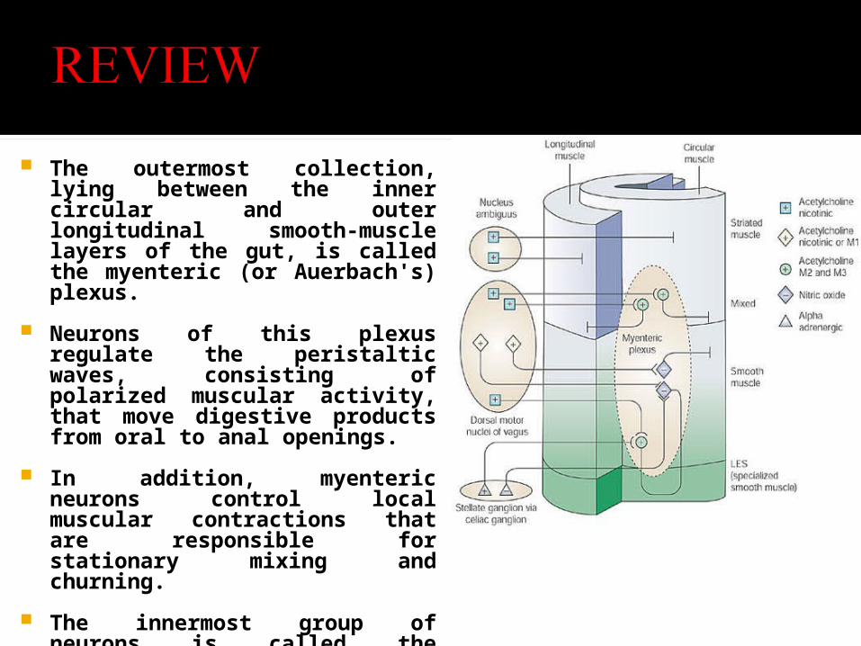
The outermost collection, lying between the inner circular and outer longitudinal smooth-muscle layers of the gut, is called the myenteric (or Auerbach's) plexus.
Neurons of this plexus regulate the peristaltic waves, consisting of polarized muscular activity, that move digestive products from oral to anal openings.
In addition, myenteric neurons control local muscular contractions that are responsible for stationary mixing and churning.
The innermost group of neurons is called the submucosal (or Meissner's) plexus. This group regulates the configuration of the luminal surface, controls glandular secretions, alters electrolyte and water transport, and regulates local blood flow
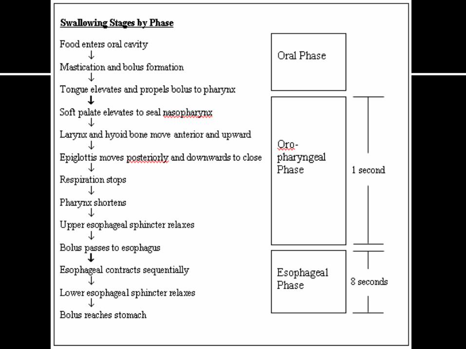
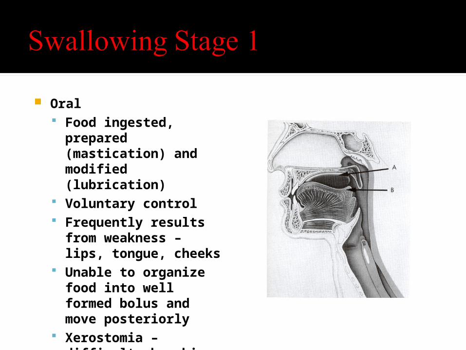
Oral Food ingested, prepared
(mastication) and modified (lubrication)
Voluntary control Frequently results from
weakness – lips, tongue, cheeks
Unable to organize food into well formed bolus and move posteriorly
Xerostomia – difficulty breaking down solids
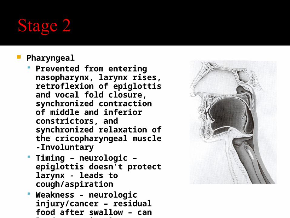
Pharyngeal Prevented from entering
nasopharynx, larynx rises, retroflexion of epiglottis and vocal fold closure, synchronized contraction of middle and inferior constrictors, and synchronized relaxation of the cricopharyngeal muscle -Involuntary
Timing – neurologic – epiglottis doesn’t protect larynx - leads to cough/aspiration
Weakness – neurologic injury/cancer – residual food after swallow – can lead to aspiration
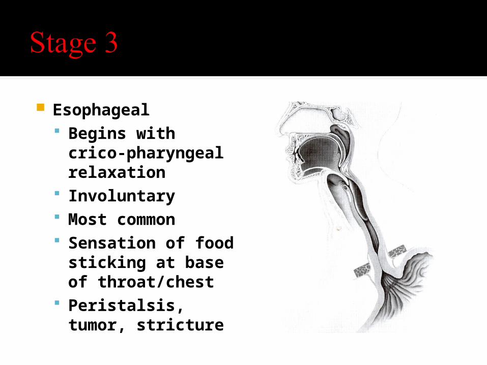
Esophageal Begins with crico-
pharyngeal relaxation Involuntary Most common Sensation of food
sticking at base of throat/chest
Peristalsis, tumor, stricture
Taking a careful history is vital for the evaluation of dysphagia.
The history will yield the likely underlying -pathophysiologic process
-anatomic site of the problem in most patients - 80%
Crucial for determining whether subsequently detected radiographic or endoscopic 'anomalies' are relevant or incidental.
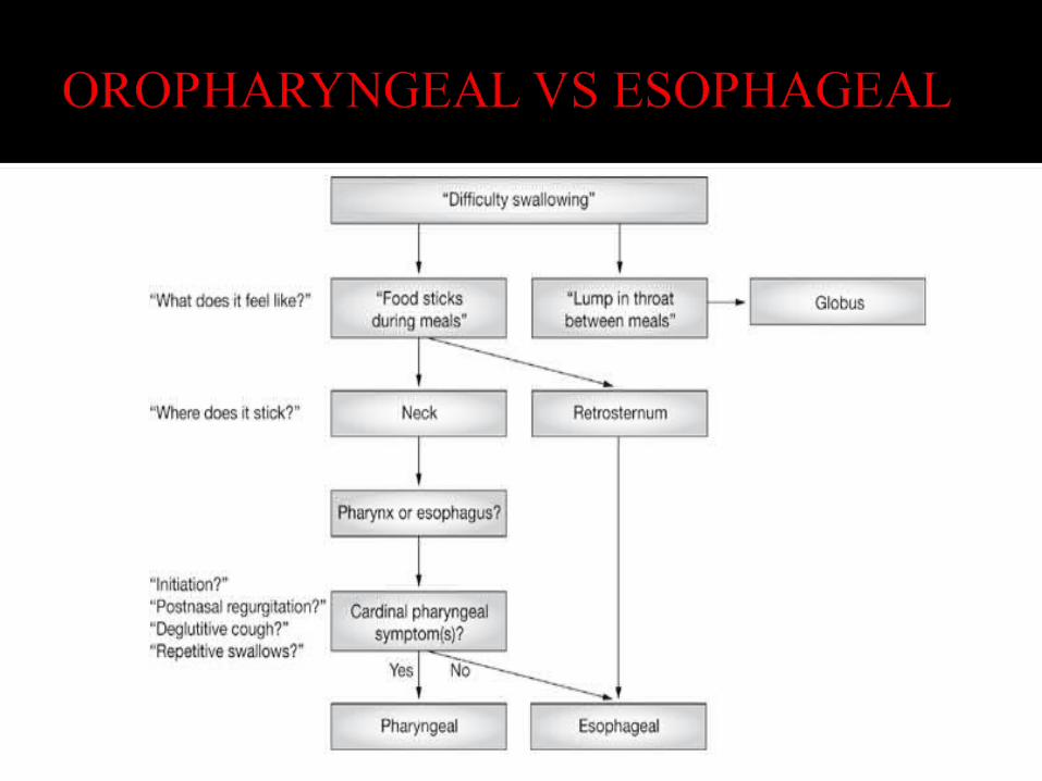
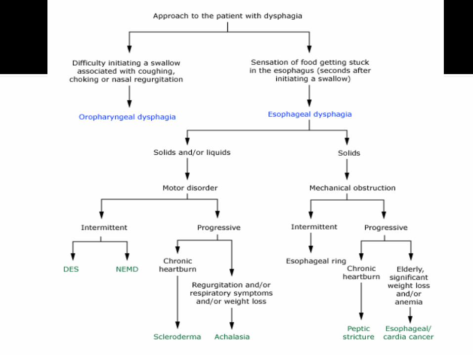
First, establish whether or not dysphagia is actually present Globus sensation (in b/w meals), Xerostomia-lose the lubrication properties and stimulus Odynophagia- pain w/swallowing, transient than dysphagia,
and persists only during the 15–30s that a bolus takes to traverse the esophagus.
Second, determine whether the site of the problem is esophageal or oropharyngeal.
Third , distinguish a structural abnormality from a motor disorder.
The history will also dictate whether the next diagnostic procedure should be endoscopy or barium swallow.
Retrosternal bolus hold-up indicates that the disorder lies within the esophagus.
However, the patient's perception of an apparent bolus hold-up in the neck has low diagnostic specificity, and cervical localization per se does not help the clinician to distinguish pharyngeal from esophageal causes of dysphagia.
Owing to viscerosomatic referral, in 30% of cases the perceived site of hold-up is above the suprasternal notch when the actual hold-up is within the esophageal.
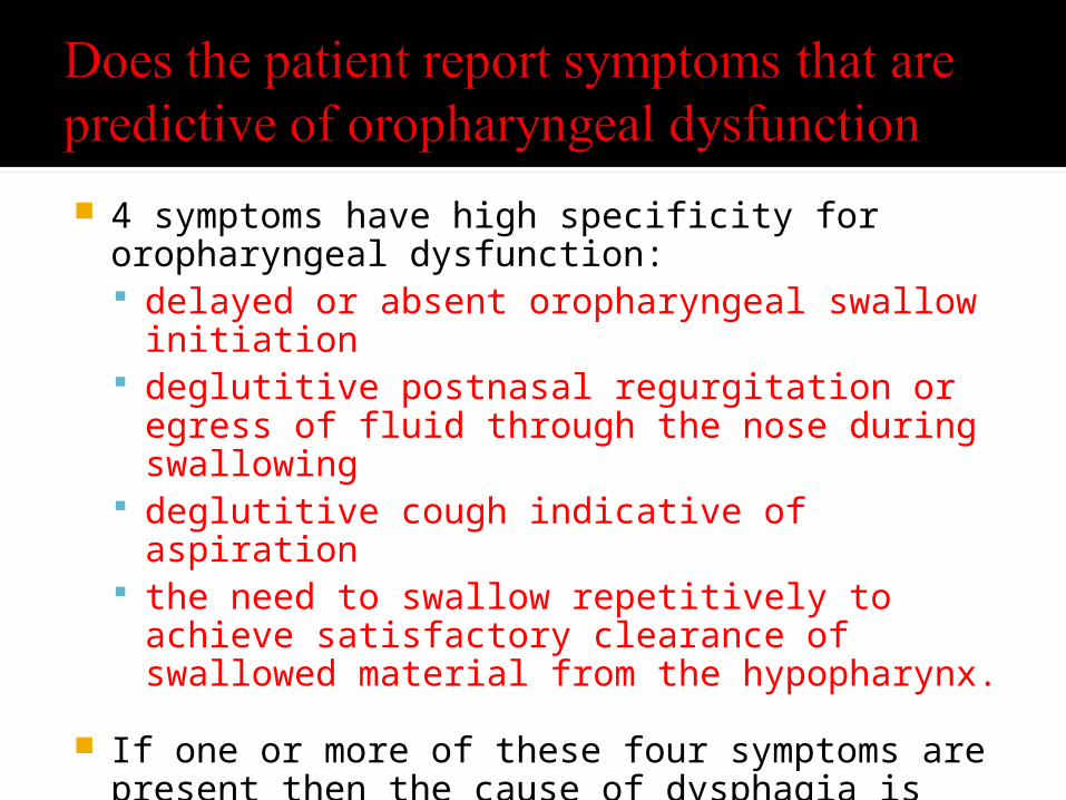
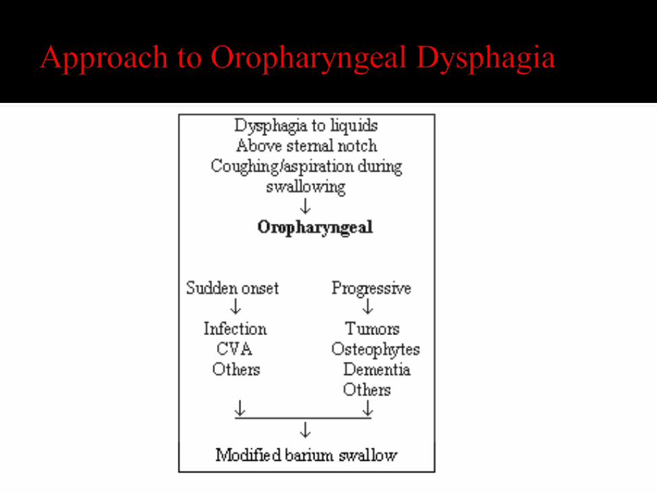
4 symptoms have high specificity for oropharyngeal dysfunction: delayed or absent oropharyngeal swallow initiation deglutitive postnasal regurgitation or egress of fluid through
the nose during swallowing deglutitive cough indicative of aspiration the need to swallow repetitively to achieve satisfactory
clearance of swallowed material from the hypopharynx.
If one or more of these four symptoms are present then the cause of dysphagia is probably oropharyngeal, either structural or neuromyogenic

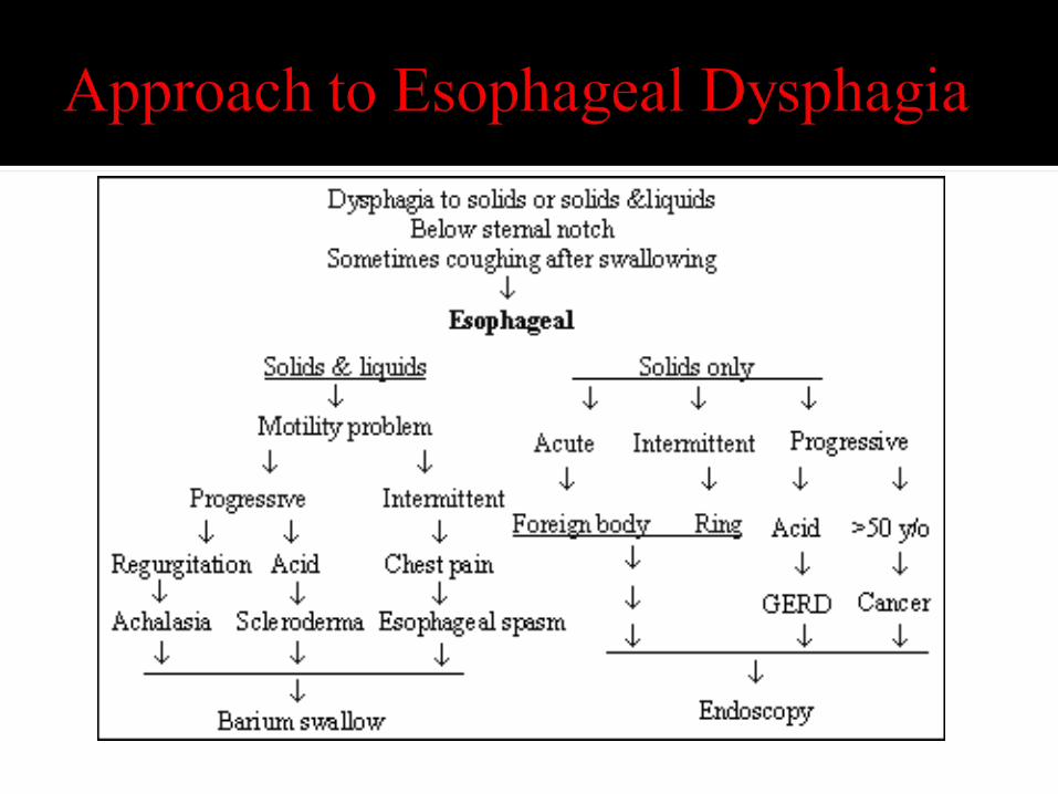
Progressive dysphagia -> Neuromuscular dysphagia
Sudden dysphagia -> Obstructive dysphagia, esophagitis
Difficulty initiating swallow -> Oropharyngeal dysphagia
Food "sticks" after swallow -> Esophageal dysphagia
Cough Early in swallow -> Neuromuscular dysphagia
Cough Late in swallow -> Obstructive dysphagia Weight loss In the elderly -> Carcinoma Weight loss with regurgitation -> Achalasia Progressive symptoms Heartburn -> Peptic
stricture, scleroderma Intermittent symptoms -> Rings and webs,
diffuse esophageal spasm, nutcracker esophagus
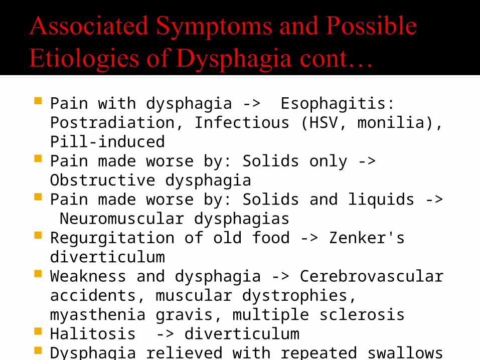
Pain with dysphagia -> Esophagitis: Postradiation, Infectious (HSV, monilia), Pill-induced
Pain made worse by: Solids only -> Obstructive dysphagia
Pain made worse by: Solids and liquids -> Neuromuscular dysphagias
Regurgitation of old food -> Zenker's diverticulum
Weakness and dysphagia -> Cerebrovascular accidents, muscular dystrophies, myasthenia gravis, multiple sclerosis
Halitosis -> diverticulum Dysphagia relieved with repeated swallows ->
Achalasia Dysphagia made worse with cold foods ->
Neuromuscular motility disorders
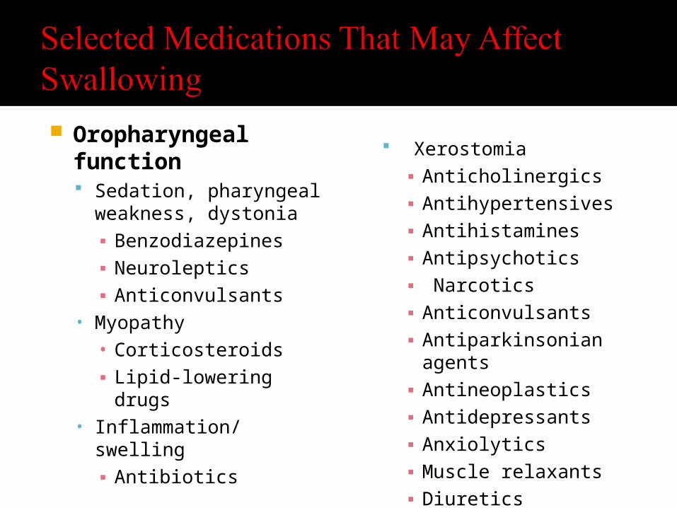
Oropharyngeal function Sedation, pharyngeal
weakness, dystonia ▪ Benzodiazepines▪ Neuroleptics▪ Anticonvulsants
• Myopathy • Corticosteroids▪ Lipid-lowering drugs
• Inflammation/swelling ▪ Antibiotics
Xerostomia ▪ Anticholinergics▪ Antihypertensives▪ Antihistamines▪ Antipsychotics▪ Narcotics▪ Anticonvulsants▪ Antiparkinsonian agents▪ Antineoplastics▪ Antidepressants▪ Anxiolytics▪ Muscle relaxants▪ Diuretics

Esophageal function Inflammation (resulting from irritation by pill)▪ Tetracycline, Doxycycline (Vibramycin)▪ Iron preparations▪ Quinidine▪ Nonsteroidal anti-inflammatory drugs▪ Potassium
Impaired motility or exacerbated gastroesophageal reflux▪ Anticholinergics▪ Calcium channel blockers▪ Theophylline▪ Nitrates
Esophagitis (related to immunosuppression) ▪ Corticosteroids

Structural/Obstructive Head or neck tumors Postsurgical/Radiation stenosis Cervical spondylosis Zenker's diverticulum Cricopharyngeal web Infectious (tonsilar hypertrophy/abscess) Extrinsic compression (goiter)
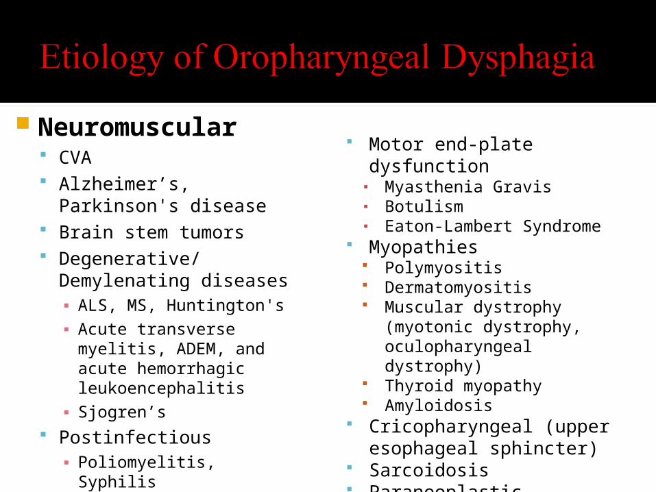
Neuromuscular CVA Alzheimer’s, Parkinson's disease Brain stem tumors Degenerative/Demylenating
diseases▪ ALS, MS, Huntington's▪ Acute transverse myelitis, ADEM,
and acute hemorrhagic leukoencephalitis
▪ Sjogren’s Postinfectious▪ Poliomyelitis, Syphilis
Peripheral nervous system▪ Peripheral neuropathy
Motor end-plate dysfunction▪ Myasthenia Gravis▪ Botulism▪ Eaton-Lambert Syndrome
Myopathies Polymyositis Dermatomyositis Muscular dystrophy (myotonic
dystrophy, oculopharyngeal dystrophy)
Thyroid myopathy Amyloidosis
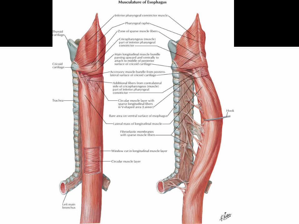
Cricopharyngeal (upper esophageal sphincter)
Sarcoidosis Paraneoplastic Syndromes
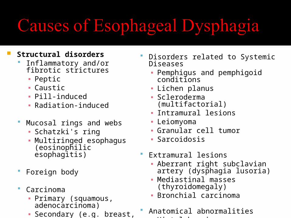
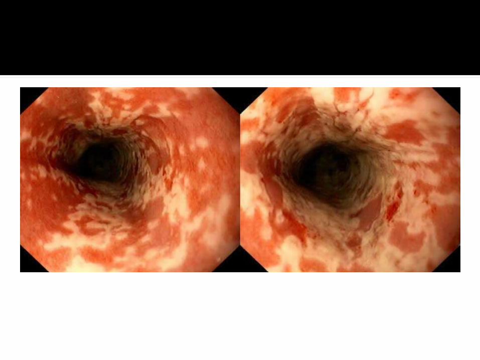
Structural disorders Inflammatory and/or fibrotic
strictures▪ Peptic ▪ Caustic ▪ Pill-induced ▪ Radiation-induced
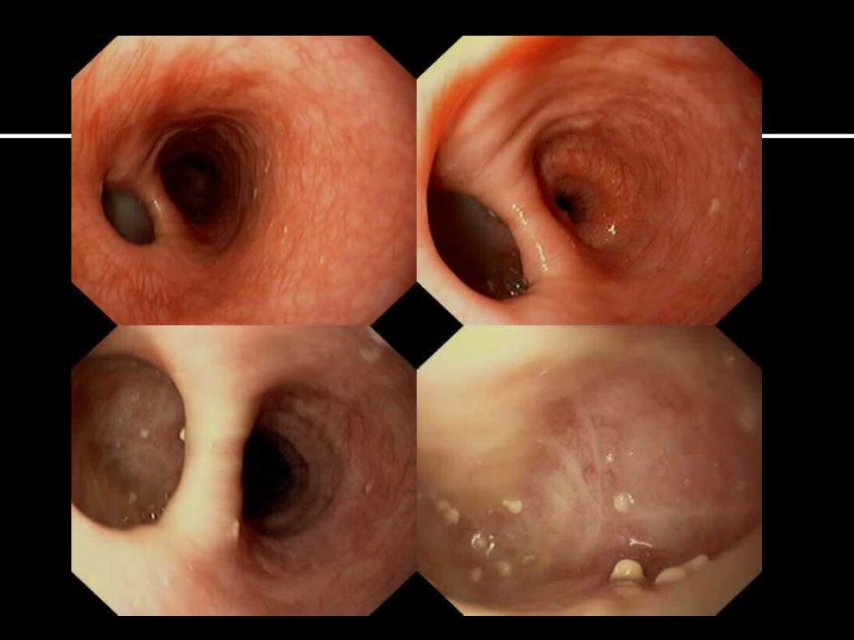
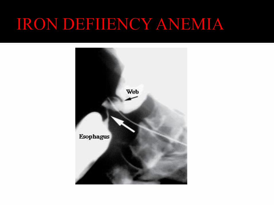
Mucosal rings and webs▪ Schatzki's ring ▪ Multiringed esophagus
(eosinophilic esophagitis)
Foreign body
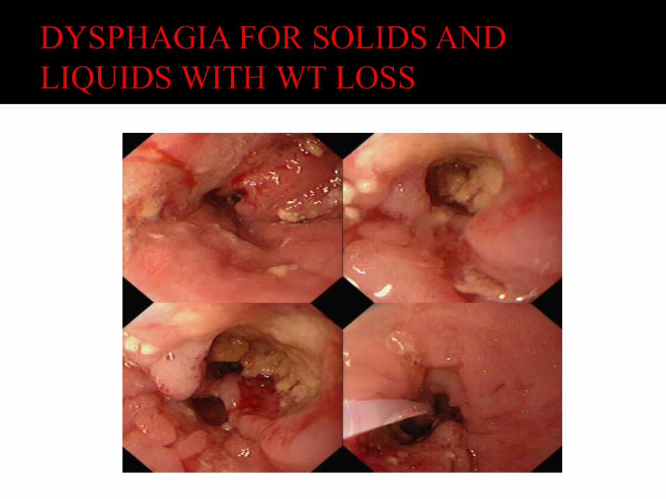
Carcinoma▪ Primary (squamous,
adenocarcinoma) ▪ Secondary (e.g. breast, melanoma)
Disorders related to Systemic Diseases▪ Pemphigus and pemphigoid conditions ▪ Lichen planus ▪ Scleroderma (multifactorial)▪ Intramural lesions▪ Leiomyoma ▪ Granular cell tumor▪ Sarcoidosis
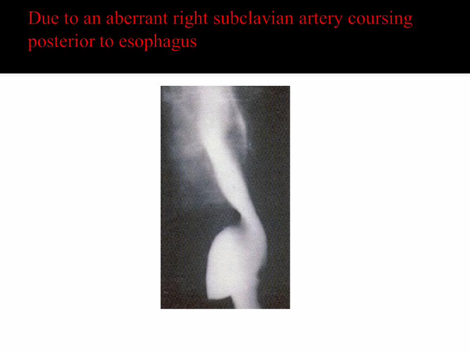
Extramural lesions▪ Aberrant right subclavian artery
(dysphagia lusoria) ▪ Mediastinal masses (thyroidomegaly) ▪ Bronchial carcinoma
Anatomical abnormalities▪ Hiatal hernia ▪ Esophageal diverticulum
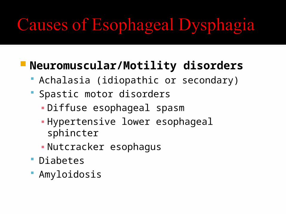
Neuromuscular/Motility disorders Achalasia (idiopathic or secondary) Spastic motor disorders▪ Diffuse esophageal spasm▪ Hypertensive lower esophageal sphincter▪ Nutcracker esophagus
Diabetes Amyloidosis

Is it a structural vs motility disorder?
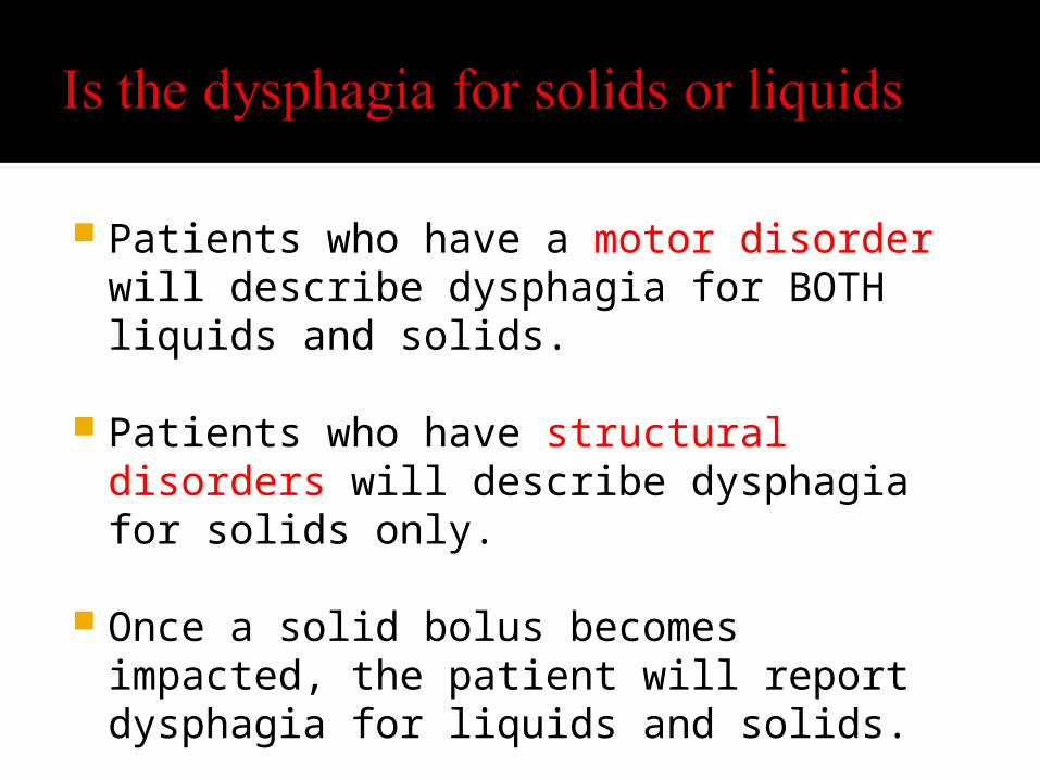
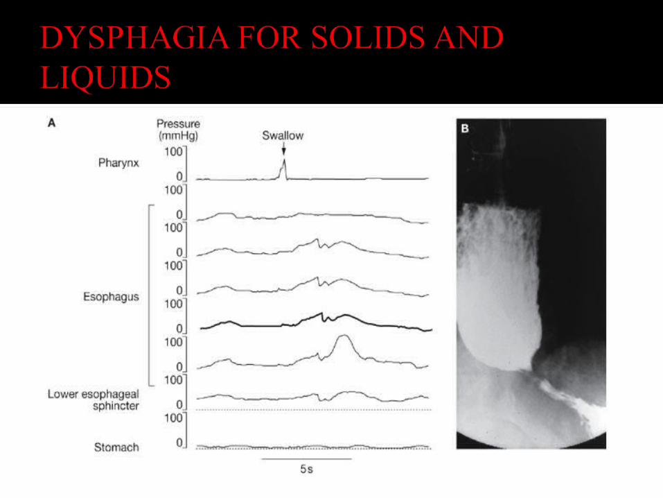
Patients who have a motor disorder will describe dysphagia for BOTH liquids and solids.
Patients who have structural disorders will describe dysphagia for solids only.
Once a solid bolus becomes impacted, the patient will report dysphagia for liquids and solids.
Three cardinal features of dysmotility dysphagia (for solids and liquids) chest pain and regurgitation
Regurgitation during meals, as well as spontaneous regurgitation between meals or at night, is highly suggestive of dysmotility.
Unlike regurgitation that is related to GERD, the regurgitated fluid in patients with esophageal dysmotility is generally not noxious to taste.
In addition, spasm or achalasia typically cause chest pain. Although this chest pain is frequently described as 'heavy' or 'crushing', it can be indistinguishable from the typical 'heartburn' of reflux.
The pain frequently occurs during meals, but it can be quite unpredictable and sporadic or nocturnal.
Sipping antacids or even water can relieve the pain related to dysmotility, which further confuses its distinction from reflux-related pain.
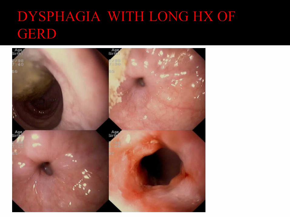
Slowly progressive, long-standing dysphagia, particularly against a background of reflux, is suggestive of a peptic stricture. Caveat - severity of heartburn correlates poorly with
esophageal mucosal damage. A short history of dysphagia—particularly with rapid
progression (weeks or months) and associated weight loss—is highly suggestive of esophageal cancer.
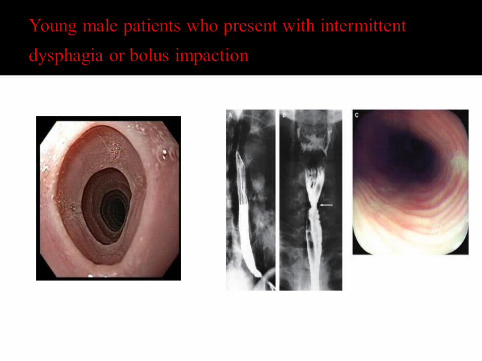
Long-standing, intermittent, non-progressive dysphagia
purely for solids is indicative of a fixed structural lesion such as a distal esophageal ring or proximal esophageal mucosal web.
If oropharyngeal dysphagia is suspected, evaluation for neuromuscular disorders is important.
Thorough neurological, head and neck exam Skin should be examined for features of connective tissue
disorders, particularly scleroderma and CREST syndrome. Muscle weakness or wasting might be evident if myositis is
present, and myositis can overlap with other connective tissue disorders that affect the esophagus.
Look for tremors, rigidity, fasciculations Signs of malnutrition, weight loss and pulmonary complications
from aspiration should be looked for.
CBC to screen for infectious or inflammatory conditions TFT’s may detect hypo- or hyperthyroid-associated causes of
dysphagia ( Grave's disease or thyroid carcinoma), Anti-acetylcholine antibodies to diagnose myasthenia gravis Muscular enzymes to diagnose myositis Autoimmune studies (ANA, RF, Anti-SSA, Anti-SSB, Anti-
Scl-70, anti-centromere) CT/MRI to evaluate for CVA, MS, tumors
Video Fluoroscopic Swallowing Study (VFSS) “Modified barium swallow", is the "gold standard" for diagnosing
oropharyngeal dysphagia. Dynamic test in which the patient is asked to swallow a variety of food
items of different consistencies covered with barium. A video fluoroscopic recording is made in both A/P and lateral views.
Allows for observation of bolus progress throughout the different stages of the swallowing process. The presence of pooling, delayed transit and laryngeal aspiration can be detected.
The dynamic nature of this study provides an opportunity to evaluate the response to certain correctional techniques (e.g., chin tucking) during the study.
This technique requires the cooperation of an alert patient, which is the most limiting factor to performing VFSS.
Video Endoscopic Swallowing Study (VESS) Direct visualization of the oropharynx in action with and without
swallowing, using a fiberoptic scope inserted nasally. This test is valuable when VFSS can not be performed and is usually
done by an otolaryngologist Barium swallow studies
Initial recommended test if esophageal dysphagia is suspected Suspected obstructive lesion (e.g., Schatzki's ring, tumor) Suspected esophageal motility disorder
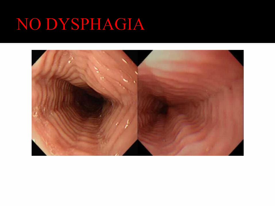
EGD Suspected acute obstructive lesion (impacted food bolus) Evaluation of the esophageal mucosa Confirmation of a positive barium study with biopsies or cytology
Manometry Abnormality not identified on barium study or by endoscopy

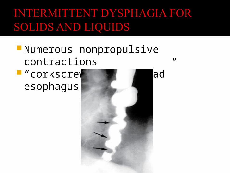
Numerous nonpropulsive contractions “corkscrew/ rosary bead” esophagus
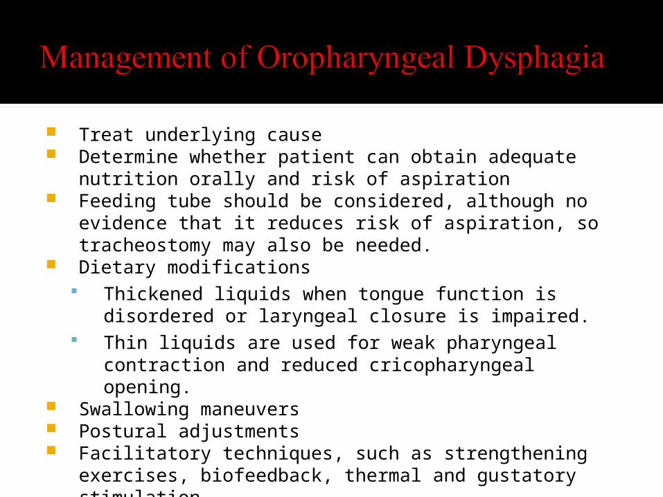
Treat underlying cause Determine whether patient can obtain adequate nutrition orally and risk
of aspiration Feeding tube should be considered, although no evidence that it reduces
risk of aspiration, so tracheostomy may also be needed. Dietary modifications
Thickened liquids when tongue function is disordered or laryngeal closure is impaired.
Thin liquids are used for weak pharyngeal contraction and reduced cricopharyngeal opening.
Swallowing maneuvers Postural adjustments Facilitatory techniques, such as strengthening exercises, biofeedback,
thermal and gustatory stimulation.
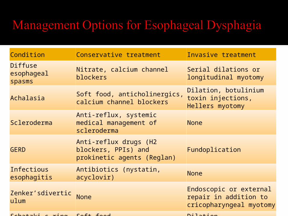
Condition Conservative treatment Invasive treatmentDiffuse esophageal spasms Nitrate, calcium channel blockers Serial dilations or
longitudinal myotomy
Achalasia Soft food, anticholinergics, calcium channel blockers
Dilation, botulinium toxin injections, Hellers myotomy
Scleroderma Anti-reflux, systemic medical management of scleroderma None
GERDAnti-reflux drugs (H2 blockers, PPIs) and prokinetic agents (Reglan)
Fundoplication
Infectious esophagitis Antibiotics (nystatin, acyclovir) None
Zenker’sdiverticulum None
Endoscopic or external repair in addition to cricopharyngeal myotomy
Schatzki_s ring Soft food Dilation
Omran L. Dyphagia. http://www.cyberounds.com/cmecontent/art76.html Speiker M. Evaluating Dysphagia. American Family Physician. June 15,
2000. Palmer J, Drennan J, Baba M. Evaluation and Treatment of Swallowing
Impairments. American Family Physician. April 15, 2000. www.medicine.nevada.edu/residency/lasvegas/.../DYSPHAGIA.ppt