Embed Size (px)
Citation preview

Dysfunctional potassium channel subunit interaction as a novelmechanism of long QT syndrome
Michael Hoosien, MD,* Mary Ellen Ahearn, MS,*† Robert J. Myerburg, MD,*‡ Thai V. Pham, PhD,y
Todd E. Miller, PhD,* Marcel J. Smets, XX,* Lisa Baumbach-Reardon, PhD,†J Ming-Lon Young, MD,z
Amjad Farooq, PhD,J Nanette H. Bishopric, MD, FACC, FAHA*yz
From the Departments of *Medicine, yNeurology, zPhysiology, yMolecular and Cellular Pharmacology, JBiochemistry andMolecular Biology and zPediatrics, University of Miami Miller School of Medicine, Miami, Florida.
BACKGROUND The slowly-activating delayed rectifier current IKs
contributes to repolarization of the cardiac action potential, and iscomposed of a pore-forming a-subunit, KCNQ1, and a modulatoryb-subunit, KCNE1. Mutations in either subunit can cause long QTsyndrome, a potentially fatal arrhythmic disorder. How KCNE1exerts its extensive control over the kinetics of IKs remainsunresolved
OBJECTIVE To evaluate the impact of a novel KCNQ1 mutation onIKs channel gating and kinetics
METHODS KCNQ1 mutations were expressed in Xenopus oocytes inthe presence and absence of KCNE1. Voltage clamping andMODELLER software were used to characterize and model channelfunction. Mutant and wt genes were cloned into FLAG, Myc and HAexpression vectors to achieve differential epitope tagging, andexpressed in HEK293 cells for immunohistochemical localizationand surface biotinylation assay.
RESULTS We identified 2 adjacent mutations, S338F and F339S, inthe KCNQ1 S6 domain in unrelated probands. The novel KCNQ1 S338Fmutation segregated with prolonged QT interval and torsade depointes; the second variant, F339S, was associated with fetalbradycardia and prolonged QT interval, but no other clinical events.S338F channels expressed in Xenopus oocytes had slightly increasedpeak conductance relative to wild type, with a more positive
This work was supported by the Florida Heart Research Institute (to DrBishopric and Dr Myerburg), the National Institutes of Health grants R01-HL71094 (to Dr Bishopric) and R01-GM083897 (to Dr Farooq), theAmerican Heart Association Florida/Puerto Rico Affiliate (to DrBaumbach-Reardon), the Fondation Leducq (Trans-Atlantic Network ofExcellence Grant, “Preventing Sudden Cardiac Death” 05-CVD-01; to DrMyerburg and Dr Bishopric), and an NIH T32 Training Grant inCardiovascular Signaling (to Dr Hoosien). Address reprint requests andcorrespondence: Dr Nanette H. Bishopric, Department of Medicine,University of Miami Miller School of Medicine, 1600 NW 10th Avenue,RMSB 6026, PO Box 016189 (R-189), Miami, FL 33101. E-mail address:[email protected].
1547-5271/$-see front matter B 2013 Heart Rhythm Society. All rights reserved.
activation voltage. F339S channels conducted minimal current.Unexpectedly, S338F currents were abolished by co-expression withintact WT KCNE1 or its C-terminus (aa63-129), despite normalmembrane trafficking and surface co-localization of KCNQ1 S338Fand wt KCNE1. Structural modeling indicated that the S338Fmutation specifically alters the interaction between the S6 domainof one KCNQ1 subunit and the S4-S5 linker of another, inhibitingvoltage-induced movement synergistically with KCNE1 binding.
CONCLUSIONS A novel KCNQ1 mutation specifically impairedchannel function in the presence of KCNE1. Our structural modelshows that this mutation effectively immobilizes voltage gating byan inhibitory interaction that is additive with that of KCNE1. Ourfindings illuminate a previously unreported mechanism for LQTS,and validate recent theoretical models of the closed state of theKCNQ1:KCNE1 complex.
KEYWORDS Arrhythmia; Ion channels; Long QT syndrome;Structural modeling; Genetics
ABBREVIATIONS IKs ¼ slowly activating delayed rectifier current;LQTS ¼ long QT syndrome; VS ¼ voltage sensor; WT ¼ wild type
(Heart Rhythm 2013;10:728–737) I 2013 Heart Rhythm Society.All rights reserved.
IntroductionCongenital long QT syndrome (LQTS) is an inheriteddisorder carrying a high risk of lethal cardiac arrhythmias.1,2
Most cases of LQTS are caused by loss-of-function mutationsin ion channels that regulate the repolarization phase of thecardiac action potential, including KCNQ1 (KVLQT1) andKCNE1 (Isk, MinK). These genes encode the a- andb-subunits of a heterotetrameric complex that produces theslowly activating delayed rectifier current IKs (Kv 7.1), anoutward potassium current important for the rapid phase(phase 3) of repolarization.3–5 Mutations in these same geneshave been linked to short QT syndrome and familial atrialfibrillation.6 Although KCNQ1 by itself can form voltage-gated ion channels, the distinctive IKs current requirescoassembly with KCNE1. Interaction with KCNE1 greatlyincreases the voltage required for IKs channel opening whileincreasing channel conductance, slowing the time course ofactivation and deactivation,3–5,7 and enabling adrenergic
http://dx.doi.org/10.1016/j.hrthm.2012.12.033

Syncope
Cardiacarrest
Long QTby EKG
80
65
2929 16
55
13 8 7 4
60
?Unknownheart problem
?
?
33KCNQ1 (S338F)
KCNQ1 (S338F) KCNQ1 (S338F) KCNQ1 (S338F)
KCNE1 (S38G)
KCNE1 (S38G) KCNE1 (S38G)
KCNE2 (Q9E)
KCNE2 (Q9E)
Stillbirth
92
37 33
64KCNE1 (D85N)
KCNE1 (D85N)
KCNE1 (D85N)KCNE1 (D85N) KCNE1 (D85N)
KCNE1 (S38G)KCNE1 (S38G)
KCNE1 (S38G)
KCNE1 (S38G)
KCNE1 (D85N)
1
1 2
1 2
3 4 5 6
3 4
1 2 3 4
V5
Figure 1 Pedigree (A) and 12-lead electrocardiogram (B) from proband 1 at 7 years of age.
729Hoosien et al Dysfunctional Binding of KCNQ1:KCNE1 Causes LQTS
regulation.8 How KCNE1 interacts with KCNQ1 to producethese extensive functional alterations is incompletelyunderstood.9,10
In this report, we analyze 2 mutations—KCNQ1 S338Fand F339S—occupying adjacent sites within the S6 trans-membrane domain. One of these (S338F) does not affectKCNQ1 channel function by itself, but causes a complete lossof conductance when coassembled with KCNE1 or the C-terminal residues of KCNE1 that physically associate withKCNQ1.10,11 The other mutant carries almost no current, andits voltage-dependent activation by KCNE1 is minimal and
extremely right-shifted.12,13 Structural modeling of theKCNQ1 homotetramer predicts that the variant phenylalanineat residue 338 in 1 subunit forms van der Waals attachments tothe S4-S5 helical linker of a neighboring subunit, impedingchannel opening. The combined inhibitory interaction ofKCNE1 and the S338F mutation effectively immobilizes theS4-S5 linker, rendering the channel unable to open atphysiologic voltages. Our findings elucidate a novel coassem-bly specific mechanism for LQTS and provide high-resolutionstructural and mechanistic insights into KCNQ1:KCNE1modulatory interaction and gating of IKs.

I:2 I:3
II:5
I:1
II:4II:2 II:3II:1KCNQ1 F339S
IIVI
V2
Figure 2 Pedigree (A) and 12-lead electrocardiogram (B) from proband 2.
Heart Rhythm, Vol 10, No 5, May 2013730
MethodsA detailed Methods supplement is available online.
SubjectsSubjects of this study were referred after presenting withsyncope and/or ventricular tachyarrhythmias with prolongedQT intervals and family histories of unexplained or suddencardiac deaths. Subjects were recruited and evaluated inaccordance with a human subject research protocol approvedby the University of Miami Institutional Review Board.
Mutation identificationDNA was isolated by using a commercial kit (PuregeneDNA Isolation Kit, Gentra Systems/Qiagen, Minneapolis,MN), and individual exons from 5 genes—KCNQ1, KCNH2,SCN5A, KCNE1, and KCNE2—associated with LQTS wereamplified with published primers14,15 by using the polymer-ase chain reaction.
ElectrophysiologyStage V oocytes were prepared from mature Xenopus laevisfollicular tissue (Nasco, Fort Atkinson, WI). Oocytes wereinjected with combinations of KCNQ1/KCNE1 cRNA, andcurrent responses were analyzed under 2-electrode voltageclamp by using an OpusXpress 6000A parallel oocytevoltage clamp system running OPUSXPRESS 1.1 andCLAMPFIT 9.1 software (molecular devices).
Epitope tagging and membrane expressionWe used differential epitope tagging to facilitate microscopicvisualization and biotinylation of membrane-bound mutantand wild-type (WT) a- and b-subunits. Details pertaining tothese experimental procedures can be found in the onlinesupplement.
Molecular modelingStructural models of WT and S338F/F339S mutants ofKCNQ1 in the closed state were built by using MODELLER
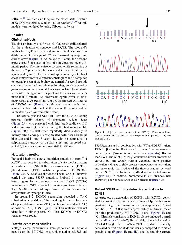
Figure 3 Adjacent novel mutations in the KCNQ1 S6 transmembranedomain. Partial KCNQ1 exon 7 DNA sequence from proband 1 (A) andproband 2 (B).
731Hoosien et al Dysfunctional Binding of KCNQ1:KCNE1 Causes LQTS
software.16 We used as a template the closed-state structureof KCNQ1 modeled by Sanders and co-workers.17,18 Atomicmodels were rendered by using Ribbons software.19
ResultsClinical subjectsThe first proband was a 7-year-old Caucasian child referredfor the evaluation of syncope and LQTS. The proband’smother had LQTS and received an implantable cardioverter-defibrillator at the age of 29 for recurrent syncope andcardiac arrest (Figure 1). At the age of 7 years, the probandexperienced 3 episodes of loss of consciousness over a 6-month period. The first episode occurred while swimming atthe age of 7 years when he was noted to have fixed pupils,apnea, and cyanosis. He recovered spontaneously after briefchest compression; an electroencephalogram and a computedtomography scan of the brain were normal. A second episodeoccurred 2 months later while swimming; an electrocardio-gram was reportedly normal. Four months later, he suddenlyfell while running around the pool and lost consciousness formore than a minute. An electrocardiogram revealed sinusbradycardia at 58 beats/min and a QT/corrected QT intervalof 510/505 ms (Figure 1). He was treated with beta-adrenergic blockade, and at the age of 8, he received animplantable cardioverter-defibrillator.
The second proband was a full-term infant with a strongpaternal family history of premature sudden death(Figure 2A), who presented with fetal bradycardia (o110)and a prolonged QT interval during the first month of life(Figure 2B); his half-sister reportedly died suddenly ininfancy while crying. He was treated with beta-adrenergicblockade and is now 8 years old, with no complaints ofpalpitations, syncope, or cardiac arrest and recorded cor-rected QT intervals ranging from 440 to 500 ms.
Molecular geneticsProband 1 harbored a novel transition mutation in exon 7 ofKCNQ1 that resulted in substitution of cytosine for thymineat position 1013 (NM 000218), causing a serine (TCT) tophenylalanine (TTT) switch at position 338 (S338F;Figure 3A). All relatives of proband 1 with long QT intervalscarried the same S338F mutation. Proband 1 was alsoheterozygous for a previously reported D85N (G253A)mutation in KCNE1, inherited from his asymptomatic father.Two S338F carrier siblings have had no documentedarrhythmias or syncope to date.
In proband 2, KCNQ1 sequencing revealed a T4Csubstitution at position 1016, resulting in the replacementof a phenylalanine codon (TTC) with a serine codon (TCC)at position 339 (F339S; Figure 3B). This mutation was notidentified in either parent. No other KCNQ1 or KCNE1variants were found.
Protein expression studiesVoltage clamp experiments were performed in Xenopusoocytes on the 2 KCNQ1 a-subunit mutations (S338F and
F339S), alone and in combination with WT and D85N-variantKCNE1 b-subunits. Background currents from endogenousoocyte a- and b-subunits were minimal (Figure 4A). Homo-meric WT- and S338F-KCNQ1 conducted similar amounts ofcurrent, but the S338F current exhibited more positiveactivation voltage, slightly greater conductance at activation,and more rapid inactivation of the time-dependent outwardcurrent. S338F also lacked a rapidly deactivating tail current(Figure 4A). In contrast, homomeric F339S channels hadextremely poor conductance at all voltages (Figure 4B).
Mutant S338F exhibits defective activation byKCNE1As expected, co-expression of KCNE1 with KCNQ1 gener-ated a current exhibiting typical features of IKs, with a morepositive voltage of activation and current amplitudes (mA) anddensities (mA/mF) that were approximately 40 times greaterthan that produced by WT KCNQ1 alone (Figures 4B and4C). Channels consisting of KCNE1 alone conducted a smallcurrent (Figures 4B and 4C). Remarkably, channels formed byS338F-KCNQ1 with WT-KCNE1 exhibited severelydepressed current amplitude and density compared with eitherprotein alone (Figures 4B and 4D), and the resulting current
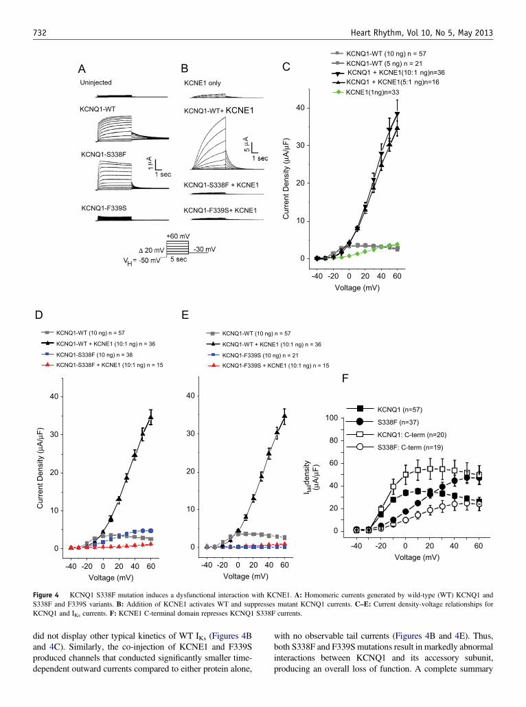
Uninjected KCNE1 only
KCNQ1-WT KCNQ1-WT+ KCNE1
KCNQ1-S338F
KCNQ1-S338F + KCNE1
KCNQ1-F339S KCNQ1-F339S+ KCNE1
Voltage (mV)
I taild
ensi
ty(µ
A/µ
F)
KCNQ1 (n=57)
S338F (n=37)
KCNQ1: C-term (n=20)
S338F: C-term (n=19)
0
20
40
-40 -20 0 20 40 60
60
80
100
-40 -20 0 20 40 60
0
10
20
30
40
KCNQ1 + KCNE1(10:1 ng)n=36KCNQ1 + KCNE1(5:1 ng)n=16KCNE1(1ng)n=33
Cur
rent
Den
sity
(µA
/µF)
Voltage (mV)
-40 -20 0 20 40 60
0
10
20
30
40
Voltage (mV)-40 -20 0 20 40 60
0
10
20
30
40
Voltage (mV)
Cur
rent
Den
sity
(µA
/µF)
KCNQ1-WT (10 ng) n = 57
KCNQ1-WT + KCNE1 (10:1 ng) n = 36
KCNQ1-F339S (10 ng) n = 21
KCNQ1-F339S + KCNE1 (10:1 ng) n = 15
KCNQ1-WT (10 ng) n = 57
KCNQ1-WT + KCNE1 (10:1 ng) n = 36
KCNQ1-S338F (10 ng) n = 38
KCNQ1-S338F + KCNE1 (10:1 ng) n = 15
KCNQ1-WT (10 ng) n = 57KCNQ1-WT (5 ng) n = 21
Figure 4 KCNQ1 S338F mutation induces a dysfunctional interaction with KCNE1. A: Homomeric currents generated by wild-type (WT) KCNQ1 andS338F and F339S variants. B: Addition of KCNE1 activates WT and suppresses mutant KCNQ1 currents. C–E: Current density-voltage relationships forKCNQ1 and IKs currents. F: KCNE1 C-terminal domain represses KCNQ1 S338F currents.
Heart Rhythm, Vol 10, No 5, May 2013732
did not display other typical kinetics of WT IKs (Figures 4Band 4C). Similarly, the co-injection of KCNE1 and F339Sproduced channels that conducted significantly smaller time-dependent outward currents compared to either protein alone,
with no observable tail currents (Figures 4B and 4E). Thus,both S338F and F339S mutations result in markedly abnormalinteractions between KCNQ1 and its accessory subunit,producing an overall loss of function. A complete summary

6
5
4
3
2
1
0
-40 -20 40 600 20
KCNQ1 (10 ng) n = 57KCNQ1 (5 ng) n = 21S338F (10 ng) n = 37KCNQ1:S338F (5:5ng) n = 12F339S (10ng) n = 21
50
40
30
20
10
0
-40 -20 40 600 20
KCNE1 (1 ng) n = 33
KCNQ1:KCNE1 (5:1 ng) n = 16KCNQ1:KCNE1 (10:1 ng) n = 29
S338F:KCNE1 (10:1 ng) n = 15 F339S:KCNE1 (10:1 ng) n = 15
µA/µ
F
4
3
2
1
0
-40 -20 40 600 20
KCNE1 (1 ng) n = 33S338F:KCNE1 (10:1 ng) n = 15 F339S:KCNE1 (10:1 ng) n = 15
-40 -20 40 600 20
KCNQ1:KCNE1 (10:1 ng) n = 29KCNQ1:KCNE1 (5:1 ng) n = 16 S338F:KCNE1 (10:1 ng) n = 15KCNQ1:S338F:KCNE1 (5:5:1 ng) n = 12KCNQ1:S338F:KCNE1 (9:1:1 ng) n = 10
50
40
30
20
10
0
Voltage (mV)
µA/µ
F
-40 -20 40 600 20
50
40
30
20
10
0
KCNE1 (1 ng) n = 33
D85N (1 ng) n = 17KCNQ1:D85N (10:1 ng) n = 20S338F:D85N (10:1 ng) n = 8
S338F:KCNE1 (10:1 ng) n = 15 F339S:KCNE1 (10:1 ng) n = 15
KCNQ1:KCNE1 (10:1 ng) n = 29
Voltage (mV)
µA/µ
F
Figure 5 Dominant negative effects of KCNQ1 S338F and F339s mutations. A: Current density-voltage relationships for wild-type (WT), mutant, and mixeda-subunits. B: Effect of KCNE1 co-expression on WT and mutant KCNQ1 current density. C: Inhibitory effect of KCNQ1 mutants on KCNE1-generated currents.D: Dominant negative effect of S338F on WT KCNQ1/IKs current. E: Differential effect of KCNE1 D85N variant on WT and S338F mutant KCNQ1/IKs currents.
733Hoosien et al Dysfunctional Binding of KCNQ1:KCNE1 Causes LQTS
of current properties for all channel stoichiometries isprovided in Supplementary Table 1.
Co-expression of a KCNE1 C-term fragment (aa63-129)previously shown to physically interact with the S6 domainof KCNQ111 had a similar negative effect on S338F currentamplitude (Figure 4F, n ¼ 19; Supplementary Table 1).Taken together, these data indicate a specific role forKCNE1:KCNQ1 contact in the manifestation of the KCNQ1S338F defect.
Dominant-negative effect of KCNQ1heterotetramersAs is shown in Figure 5A, homomeric S338F had a greaterpeak current density than did WT KCNQ1, but a morepositive activation voltage, while homomeric F339S currentdensity was extremely low (Figure 5A). Co-expression ofS338F with WT KCNQ1 at a 1:1 ratio generated a currentdensity that was greatly reduced compared with eitherprotein expressed separately (Figures 5B and 5C), suggest-ing the formation of heterotetramers with reduced coopera-tivity or stability and/or inability to interact with endogenousoocyte KCNE1-like proteins.
Dominant negative effect of KCNQ1 mutants onactivation by KCNE1To confirm the dominant negative effect of the S338Fmutation, we expressed both WT and mutant a-subunits at
varying ratios in the presence of KCNE1 (Figure 5D). WhenS338F and WT KCNQ1 were co-expressed at approximatelyequal stoichiometry, almost no activation by KCNE1 wasobserved, suggesting that the depressive effect of the S338Fmutation is dominant.
Functional interaction of KCNE1 D85N mutant withKCNQ1 WT and S338F mutantsIn addition to the S338F mutation, proband 1 had a rarevariant in KCNE1, D85N (rs1805128; MAF¼ 0.8%–2.5%),previously associated with drug-induced LQTS.20–23
Accordingly, we analyzed channels containing variouscombinations of WT and mutant a- and b-subunits. WTKCNE1 and D85N (Figure 5E) generated identical smallcurrents in the absence of co-expressed a-subunits (o1 mA/mF at 20 mV), likely through interaction with endogenousXenopus a-subunits. However, the D85 variant activated WTKCNQ1 channels with reduced efficacy compared to WTKCNE1. S338F had identical inhibitory interactions withboth WT and variant KCNE1 (Figure 5E).
S338F mutant traffics normally in the presence ofKCNE1To explore the possibility of trafficking defects, cell surfacebiotinylation experiments were performed in HEK293 cellstransfected with combinations of FLAG-tagged WT-KCNQ1, Myc-tagged S338F-KCNQ1, and HA-tagged

KCNQ1 wt-FLAGKCNE1-HAAnti-FLAG
Anti-HA
KCNQ1 wt-FLAGKCNQ! S338F- Myc
KCNE1-HAAnti-FLAGAnti-MycAnti-HA
++++
+-+
+-+
++-
-++
75 kd
15 kd
Q1
E1
75 kd
15 kd
NT Unb BWT S338F
--++
Q1
E1
Figure 6 KCNQ1 S338F-KCNE1 channels trafficnormally to plasma membrane. A: Biotinylationidentifies membrane-localized wild-type (WT)KCNQ1-KCNE1 channels. B ¼ bound fraction ofcell lysates; NT ¼ nontransfected; Unb ¼ unboundfraction of cell lysates; WT ¼ KCNQ1 WT-FLAG.B: Equivalent membrane localization of WT andS338F mutant KCNQ1 in the presence of KCNE1.Left lane: Biotinylated fraction of WT KCNQ1-FLAG and KCNE1-HA-transfected HEK293 cells.Right lane: Biotinylated fraction of KCNQ1-S338F-MYC and KCNE1-HA-transfected HEK293 cells.C–F: Membrane colocalization of WT and S338Fmutant KCNQ1 in the presence of KCNE1. C:Membrane localization of WT KCNQ1-FLAG inthe presence of WT KCNE1-HA. Arrows denotefluorescent signal at the cell surface; star denotessignal within the intracellular compartment. Thenucleus has been counterstained with 40,6-diami-dino-2-phenylindole (blue). D: Membrane colocali-zation of KCNQ1 S338F and KCNE1. Green denotesS338F KCNQ1-MYC; red denotes WT KCNE1-HA.Colocalization (yellow) can be seen at the cell surface(arrow) and within the interior of the cell (star). E:Membrane colocalization of KCNQ1 S338F andKCNE1 in the presence of WT KCNQ1. Red denotesanti-HA (KCNE1); green denotes anti-MYC (S338FKCNQ1). F: Membrane colocalization of KCNQ1S338F and KCNQ1 WT in the presence of KCNE1.Triple-transfected cells as in panel E were imaged forS338F (MYC; green) and WT KCNQ1 (FLAG; red).Again note signal colocalization at the cell membrane(arrow) and, to a lesser extent, within the cell interior(star).
Heart Rhythm, Vol 10, No 5, May 2013734
KCNE1 (see Supplemental Methods). As expected, WTQ1:E1 channels appeared as bands at 75 and 100 kDa(KCNQ1) and 15 kDa (KCNE1), which were enriched at themembrane surface (Figure 6A). Equivalent membraneenrichment was seen for S338F Q1:E1 channels(Figure 6B), arguing against a KCNE1-induced traffickingdefect. Confirming these findings, immunocytochemistrydemonstrated colocalization of all 3 proteins at the cellsurface, alone and in combination (Figure 6C–6F). WTKCNQ1 was observed at the cell surface when co-expressedwith KCNE1 (Figure 6C), with some intracellular stainingreflecting high channel overexpression as noted elsewhere.24
KCNQ1 S338F similarly colocalized with KCNE1 at theplasma membrane, both in the absence (Figure 6D) and inthe presence (Figures 6E and 6F) of WT KCNQ1. Theseresults additionally exclude the possibility that the dominantnegative effect of the S338F mutant on WT channels resultsfrom a trafficking defect arising in heteromeric channels.
Structural modeling of KCNQ1 WT and S338F mutantinteractions with KCNE1We used MODELLER software to build closed-state struc-tures of the WT and S338F/F339S variants of KCNQ1channels, assuming a tetramer with a fourfold axis of

Figure 7 Structural modeling of KCNQ1 mutant channels. A and B: Model of wild-type (WT) KCNQ1 channel in the closed state. A (Bottom view): Asvisualized along an axis perpendicular to the plane of the cell membrane from the cytosolic side. Dashed circles and connecting arrows show the direction ofrotation of the VS domain during channel opening. Red dot denotes the central pore. B (Side view): In the plane of the cell membrane. Blue, green, cyan, andpurple denote individual monomers; gray denotes the S4-S5 helical linker connecting the VS domain (helices S1-S4) to the pore-forming domain (helices S5/P/S6) within each monomer; red arrow denotes the path of potassium ions. C and D: Impact of the S338F variant on the KCNQ1 channel structure. In parts C andD, the interface of 2 monomers (cyan and green) is highlighted within the tetrameric KCNQ1. Gray denotes the S4-S5 helical linker. C: Model of KCNQ1 WT inthe closed state. D: Model of KCNQ1 S338F in the closed state. Red denotes side-chain moieties of residues S338/F338 in the S6 helix, which are showninteracting through van der Waals forces with W248/L251 residues in the S4-S5 helical linker of an adjacent monomer. E and F: Impact of the F339S variant onthe KCNQ1 channel structure. In these models, only 1 monomer is shown (blue) so as to highlight interhelical interactions within each monomer. Gray denotesthe S4-S5 helical linker; red denotes side-chain moieties of F339/S339 and T265/I268 involved in intramolecular van der Waals contacts between S5 and S6helices. E: Model of KCNQE1 WT in the closed state. F: Model of KCNQ1 F339S in the closed state.
735Hoosien et al Dysfunctional Binding of KCNQ1:KCNE1 Causes LQTS
symmetry (Figure 7A); each monomer contains an N-terminal voltage sensor (VS) domain and a C-terminalpore-forming domain.17 The pore domain of each monomercrosses over its neighbor in an orthogonal fashion, creatingthe central ion-conducting channel. Because the extracellularopening of the pore always remains open, gating of thecytosolic vestibule is key to controlling ion flow (Figure 7B).
Importantly, the VS and pore domains are connected by ashort S4-S5 helical linker that is critical for transmittingvoltage-induced conformational changes in the VS to openingof the pore. At the cytosolic gate, the S6 C-terminal hingesoutward, enforced by a proline at residue 343, creating anopening for ions.17,25 Our model predicts that in the closedstate (Figure 7A), the S6 C hinge is forced to straighten by

Heart Rhythm, Vol 10, No 5, May 2013736
steric hindrance applied downward by the S4-S5 helical linker.In this conformation, the interwoven S6 helices close off thecytosolic vestibule of the pore. During membrane depolariza-tion, the VS domains undergo a synchronized rotation aboutan axis perpendicular to the membrane plane17,25 (see rota-tional arrow in Figure 7A). This movement pulls the S4-S5helical linker away from S6, allowing the S6 helix to bend atits hinge point at residue P343 and splay outward from thecentral axis of the channel. This bending results in the openingof the cytosolic pore to Kþ ions (Figure 7B).
Both S338 and F339 residues are located within thefunctionally critical S6 helix and lie just 1 helical turn abovethe hinge at residue P343. Structural modeling of thetetrameric channel revealed that S338 in 1 monomer liesclose to residues W248 and L251 within the S4-S5 linker ofthe neighboring monomer (Figure 7C and SupplementaryFile 2). Thus, the substitution of a small polar serine with amore bulky and apolar phenylalanine at this position willlikely result in the formation of a tight network of van derWaals contacts among these residues (Figure 7D andSupplementary File 3). In particular, overlapping of electronclouds of the benzyl ring of F338 with those of the indolemoiety of W248 and the aliphatic side chain of L152 wouldbe highly energetically favorable. The result is to create a newstable interaction between the S4-S5 linker of 1 monomer andthe S6 helix of another that does not exist in the WT tetramer.Such an interaction would increase the energy required todisplace the S4-S5 linker away from S6, and channel openingwould thus require a higher membrane potential.
Structural modeling of KCNQ1 WT and F339S mutantinteractionsAs described above, opening of the inner channel pore involvesbending and outward splaying of the S6 helices at the hingecentered at position P343. During depolarization, the rotation ofthe VS domains (Figure 7A) applies torque to the S5 helix thatis transmitted to S6 just above the hinge, promoting its outwardbending and opening of the channel. Transmission of thistorque requires a strong network of van der Waals adhesivecontacts between the antiparallel S5 and S6 helices (Figure 7A).One strong adhesive contact is formed by sandwiching thebenzyl ring of F339 in S6 between the aliphatic side chains ofT265 and I268 in S5 (Figure 7E and Supplementary File 2). Ourmodel shows that the substitution of the bulky apolar phenyl-alanine with a small polar serine in the F339S mutant woulddisrupt this contact, uncouple the S6 C-terminus from voltage-dependent opening forces (Figure 7F and Supplementary File4), and irreversibly inactivate the KCNQ1 channel.
DiscussionOur observations identify a novel KCNQ1 mutation thatqualitatively alters the effect of its accessory subunit KCNE1from a positive modulation to a negative one, therebyprofoundly depressing the current density of IKs over a widerange of voltages. In the absence of KCNE1, homomericS338F forms functional channels, exhibiting a positive shift
in voltage-dependent activation, reminiscent of the rightwardshift in the voltage dependence of channel activation thatoccurs when KCNQ1 complexes with KCNE1.26 Our modelshows that the S338F substitution causes this rightward shiftby creating a new “sticky” intermolecular bond between S6and the S4-S5 linker that impedes voltage-induced movementof the linker away from the pore. Since this movement isrequired for pore opening, as shown by molecular dynamicssimulations17,27,28 (see Supplementary Files 2 and 3), greaterenergy is required to open the channel, which is in excellentagreement with our electrophysiologic data.
Our model further explains the catastrophic impact ofadding KCNE1 to S338F homotetramers. Cysteine mappingand mutagenesis have identified multiple points of contactbetween KCNQ1 and KCNE1, notably between the E1transmembrane domain and the Q1 S6 domain (includingresidues S338 and F339), between the E1 transmembranedomain and the Q1 S1 domain of a different subunit, andbetween the E1 extracellular domain and the S1-S2 linker ofQ1.10,26,27,29 Importantly, a recent structural model suggeststhat KCNE1 contacts the S4-S5 helix from the side opposite tothat pressing on the S6 helix. This contact impedes the S4-S5linker movement and thereby increases the voltage requiredfor pore opening.17,30 The “sandwiching” of the S4-S5 linkerbetween the new sticky interior phenylalanine and the exteriorKCNE1 C-terminus effectively immobilizes the S4-S5 helicallinker, rendering the channel unable to open.
Another interesting observation is that heterotetramers ofWT and S338F channels conduct much less current thanhomotetramers of either protein. Again, our model predictsthat the mutant phenylalanine of 1 monomer interacts withresidues of a neighboring subunit, so that WT KCNQ1gating or interaction with endogenous oocyte KCNE-likeproteins could be affected by the presence of 1 or moreS338F subunits within the same complex. Alternatively, thepresence of the large substitution at aa338 could impair the4-sided symmetry of the heterotetrameric pore structure.
The adjacent F339S mutation from proband 2 results in anonfunctional channel. It, like the S338F mutant, exerteddominant-negative effects on WT channel function, provid-ing strong support for the concept that it is translated andassembled into complexes with WT KCNQ1. Although wecannot exclude a coassembly induced trafficking defect, ourstructural model predicts that the F339S mutation disrupts acritical strong contact between S5 and S6 (Figure 7F andSupplementary File 4) that is highly likely to result inmisfolding and, consequently, either irreversible inactivationof the channel or failure to form functional channels at thesurface. Our functional and structural data are consistent withand expand on those of Yang et al,13 who previouslyidentified the F339S mutation and localized it to a criticalinterhelical position in S6. It is intriguing to speculate thatthe lack of clinical events in proband 2 is related to thepresence of a nonfunctioning channel, as opposed to thedysfunctional channel in the family of proband 1.
The apparent lack of impact of coexisting rare KCNE1 andKCNE2 variants in the family of proband 1 is noteworthy,

737Hoosien et al Dysfunctional Binding of KCNQ1:KCNE1 Causes LQTS
although the small number of individuals prevents conclusiveinterpretation.21,23,31 The D85N variant lies near a KCNQ1-interacting D76 residue11 and has been associated with greaterQT interval length (þ10.5 ms).32,33 Both KCNE1 D85N andKCNE2 Q9E variants have been implicated in drug-inducedLQTS and torsades de pointes,20–23 and D85N is frequentlyidentified as a second variant in patients with LQTS with othermutations.34 In our studies, the presence of the D85N varianthad no effect—positive or negative—on S338F channelconductance, possibly because the inhibitory interactionbetween KCNE1 and KCNQ1 involves other contacts orbecause the negative effect of S338F conceals the more subtleeffects of KCNE1 D85N on IKs.
One potential limitation of these studies is the presence ofcompeting effects of endogenous KCNQ1-like and KCNE1-like channels previously identified in X. laevis oocytes.However, the background currents contributed by thesechannels were minimal throughout these experiments, andit is unlikely that such currents masked important differencesbetween the WT and mutant channels or had mutation-specific impact on the exogenous channels.
The novel insight provided by our study is the identi-fication of a mutation that causes abnormal bonding betweena-subunit amino acids within the tetramer, the effect ofwhich is magnified by the stabilizing effect of KCNE1binding. Our findings further reinforce the importance forchannel gating and kinetics of direct contacts betweenpositions S338 and F339 in Q1 with E1 transmembraneresidues, as reported by others,26,35 and additionally providea new view of steric interactions among subunits of thehomotetramer and accessory subunits that affect the energyrequirements for gating.
AcknowledgmentsWe are grateful to Lijing You for her technical support and toDr Charles Luetje for providing access to the OpusXpresssystem. We also thank Dr Peter Larsson for his reading andcomments on the manuscript.
AppendixSupporting dataSupplementary data cited in this article is available onlineversion at http://dx.doi.org/10.1016/j.hrthm.2012.12.033.
References1. Modell SM, Lehmann MH. The long QT syndrome family of cardiac ion
channelopathies: a HuGE review. Genet Med 2006;8:143–155.2. Moss AJ, Shimizu W, Wilde AA, et al. Clinical aspects of type-1 long-QT
syndrome by location, coding type, and biophysical function of mutationsinvolving the KCNQ1 gene. Circulation 2007;115:2481–2489.
3. Sanguinetti MC, Curran ME, Zou A, et al. Coassembly of K(V)LQT1 and minK(IsK) proteins to form cardiac I(Ks) potassium channel. Nature 1996;384:80–83.
4. Barhanin J, Lesage F, Guillemare E, Fink M, Lazdunski M, Romey G.K(V)LQT1 and lsK (minK) proteins associate to form the I(Ks) cardiac potassiumcurrent. Nature 1996;384:78–80.
5. Nerbonne JM, Kass RS. Molecular physiology of cardiac repolarization. PhysiolRev 2005;85:1205–1253.
6. Chen YH, Xu SJ, Bendahhou S, et al. KCNQ1 gain-of-function mutation infamilial atrial fibrillation. Science 2003;299:251–254.
7. Osteen JD, Sampson KJ, Kass RS. The cardiac IKs channel, complex indeed. ProcNatl Acad Sci U S A 2010;107:18751–18752.
8. Marx SO, Kurokawa J, Reiken S, et al. Requirement of a macromolecularsignaling complex for b-adrenergic receptor modulation of the KCNQ1-KCNE1potassium channel. Science 2002;295:496–499.
9. Nakajo K, Ulbrich MH, Kubo Y, Isacoff EY. Stoichiometry of the KCNQ1-KCNE1 ion channel complex. Proc Natl Acad Sci U S A 2010;107:18862–18867.
10. Chung DY, Chan PJ, Bankston JR, et al. Location of KCNE1 relative to KCNQ1in the IKs potassium channel by disulfide cross-linking of substituted cysteines.Proc Natl Acad Sci U S A 2009;106:743–748.
11. Chen J, Zheng R, Melman YF, McDonald TV. Functional interactions betweenKCNE1 C-terminus and the KCNQ1 channel. PLoS One 2009;4:5143.
12. Chung SK, MacCormick JM, McCulley CH, et al. Long QT and Brugadasyndrome gene mutations in New Zealand. Heart Rhythm 2007;4:1306–1314.
13. Yang T, Chung SK, Zhang W, et al. Biophysical properties of 9 KCNQ1mutations associated with long-QT syndrome. Circ Arrhythm Electrophysiol2009;2:417–426.
14. Splawski I, Shen J, Timothy KW, Vincent GM, Lehmann MH, Keating MT.Genomic structure of three long QT syndrome genes: KVLQT1, HERG, andKCNE1. Genomics 1998;51:86–97.
15. Syrris P, Murray A, Carter ND, McKenna WM, Jeffery S. Mutation detection inlong QT syndrome: a comprehensive set of primers and PCR conditions. J MedGenet 2001;38:705–710.
16. Martı-Renom MA, Stuart AC, Fiser A, Sanchez R, Melo F, Sali A. Comparativeprotein structure modeling of genes and genomes. Annu Rev Biophys BiomolStruct 2000;29:291–325.
17. Smith JA, Vanoye CG, George AL Jr, Meiler J, Sanders CR. Structural models forthe KCNQ1 voltage-gated potassium channel. Biochemistry 2007;46:14141–14152.
18. Long SB, Campbell EB, Mackinnon R. Crystal structure of a mammalianvoltage-dependent Shaker family Kþ channel. Science 2005;309:897–903.
19. Carson M. Ribbons 2.0. J Appl Crystallogr 1991;24:958–961.20. Paulussen AD, Gilissen RA, Armstrong M, et al. Genetic variations of KCNQ1,
KCNH2, SCN5A, KCNE1, and KCNE2 in drug-induced long QT syndromepatients. J Mol Med 2004;82:182–188.
21. Salisbury B, Judson RS, Pungliya M, et al. AB47-4: the single nucleotidepolymorphism D85N-KCNE1 is associated with both congenital and drug-induced long QT [abstract]. Heart Rhythm 2006;3:S98.
22. Kaab S., Crawford D.C., Sinner M.F., et al. A large candidate gene surveyidentifies the KCNE1 D85N polymorphism as a possible modulator of drug-induced torsades de pointes. Circ Cardiovasc Genet 2011; Epub ahead of print.
23. Nishio Y, Makiyama T, Itoh H, et al. D85N, a KCNE1 polymorphism, is adisease-causing gene variant in long QT syndrome. J Am Coll Cardiol 2009;54:812–819.
24. Grunnet M, Jespersen T, Rasmussen HB, et al. KCNE4 is an inhibitory subunit tothe KCNQ1 channel. J Physiol 2002;542:119–130.
25. Kang C, Tian C, Sonnichsen FD, et al. Structure of KCNE1 and implications for howit modulates the KCNQ1 potassium channel. Biochemistry 2008;47:7999–8006.
26. Panaghie G, Tai KK, Abbott GW. Interaction of KCNE subunits with theKCNQ1 Kþ channel pore. J Physiol 2006;570:455–467.
27. Melman YF, Um SY, Krumerman A, Kagan A, McDonald TV. KCNE1 binds tothe KCNQ1 pore to regulate potassium channel activity. Neuron 2004;42:927–937.
28. Jensen MØ, Jogini V, Borhani DW, Leffler AE, Dror RO, Shaw DE. Mechanismof voltage gating in potassium channels. Science 2012;336:229–233.
29. Xu X, Jiang M, Hsu KL, Zhang M, Tseng GN. KCNQ1 and KCNE1 in the IKs
channel complex make state-dependent contacts in their extracellular domains.J Gen Physiol 2008;131:283–291.
30. Van Horn WD, Vanoye CG, Sanders CR. Working model for the structural basisfor KCNE1 modulation of the KCNQ1 potassium channel. Curr Opin Struct Biol2011;21:283–291.
31. Ackerman MJ, Tester DJ, Jones GS, Will ML, Burrow CR, Curran ME. Ethnicdifferences in cardiac potassium channel variants: implications for geneticsusceptibility to sudden cardiac death and genetic testing for congenital longQT syndrome. Mayo Clin Proc 2003;78:1479–1487.
32. Gouas L, Nicaud V, Berthet M, et al. Association of KCNQ1, KCNE1, KCNH2and SCN5A polymorphisms with QTc interval length in a healthy population. EurJ Hum Genet 2005;13:1213–1222.
33. Marjamaa A, Newton-Cheh C, Porthan K, et al. Common candidate gene variantsare associated with QT interval duration in the general population. J Intern Med2009;265:448–458.
34. Westenskow P, Splawski I, Timothy KW, Keating MT, Sanguinetti MC.Compound mutations: a common cause of severe long-QT syndrome. Circulation2004;109:1834–1841.
35. Strutz-Seebohm N, Pusch M, Wolf S, et al. Structural basis of slow activationgating in the cardiac IKs channel complex. Cell Physiol Biochem 2011;27:443–542.