Embed Size (px)
Citation preview

Dynamin-mediated Nephrin Phosphorylation RegulatesGlucose-stimulated Insulin Release in Pancreatic Beta Cells*
Received for publication, June 7, 2012 Published, JBC Papers in Press, June 20, 2012, DOI 10.1074/jbc.M112.389452
Jongmin Jeon‡§, Ingo Leibiger¶, Tilo Moede¶, Britta Walter§, Christian Faul§, Dony Maiguel‡§, Rodrigo Villarreal‡§,Johanna Guzman‡§, Per-Olof Berggren‡¶, Peter Mundel�, Camillo Ricordi‡, Sandra Merscher-Gomez§,and Alessia Fornoni‡§1
From the ‡Diabetes Research Institute and the §Division of Nephrology and Hypertension, Department of Medicine, University ofMiami L. Miller School of Medicine, Miami, Florida 33136, the ¶Rolf Luft Research Center for Diabetes and Endocrinology,Karolinska Institutet, Stockholm SE-171-77, Sweden, and the �Harvard Medical School and Department of Medicine, MassachusettsGeneral Hospital, Boston, Massachusetts 02114
Background: Nephrin is an immunoglobulin-like protein that facilitates insulin release by pancreatic beta cells.Results:Nephrin phosphorylation at tyrosine residues responsible for SH2 domain binding is a Dynamin-dependent phenom-enon, and it is necessary for glucose-stimulated insulin release in insulinoma cells and human islets.Conclusion: Dynamin-dependent Nephrin phosphorylation is necessary for glucose-stimulated insulin secretion.Significance: Pharmacological modulation of Nephrin phosphorylation may facilitate pancreatic beta cell function.
We have previously demonstrated a role for Nephrin in glu-cose stimulated insulin release (GSIR).Wenowhypothesize thatNephrin phosphorylation is required for GSIR and thatDynamin influences Nephrin phosphorylation and function.MIN6-C3 Nephrin-deficient pancreatic beta cells and humanislets were transfected with WT-Nephrin or with a mutantNephrin in which the tyrosine residues responsible for SH2domain binding were substituted with phenylalanine (3YF-Nephrin). GSIR and live images of Nephrin and vesicle traffick-ing were studied. Immunoprecipitation experiments and over-expression of WT-Dynamin or dominant negative Dynaminmutant (K44A-Dynamin) in WT-Nephrin, 3YF-Nephrin, orNephrin siRNA-transfected cells were utilized to study Neph-rin-Dynamin interaction. In contrast to WT-Nephrin or to sin-gle tyrosine mutants, 3YF-Nephrin did not positively affectGSIR and led to impaired cell-cell contacts and vesicle traffick-ing. K44A-Dynamin prevented the effect of Nephrin onGSIR inthe absence of protein-protein interaction between Nephrinand Dynamin. Nephrin gene silencing abolished the positiveeffects of WT-Dynamin on GSIR. The effects of protamine sul-fate and vanadate on Nephrin phosphorylation and GSIR werestudied in MIN6 cells and human islets. WT-Nephrin phos-phorylation after glucose occurred at Tyr-1176/1193 andresulted in improved GSIR. On the contrary, protamine sulfate-induced phosphorylation at Tyr-1176/1193/1217 was associ-
ated with Nephrin degradation and impaired GSIR. Vanadate,which prevented Nephrin dephosphorylation after glucosestimulation, improved GSIR in human islets and MIN6 cells. Inconclusion, Dynamin-dependent Nephrin phosphorylationoccurs in response to glucose and is necessary for Nephrin-me-diated augmentation of GSIR. Pharmacological modulation ofNephrin phosphorylation may thus facilitate pancreatic betacell function.
We have previously shown that Nephrin, an immunoglobu-lin-like transmembrane protein originally identified in kidneypodocytes (1), is also expressed by pancreatic beta cells where itfacilitates insulin release and vesicle formation (2). In the kid-ney, Nephrinmodulates actin polymerization and cellularmor-phology of podocytes, which are highly specialized glomerularepithelial cells with numerous foot processes bridged by anextracellular structure known as the slit diaphragm (3, 4). At theslit diaphragm, Nephrin engages in homophilic interactionswith other Nephrin molecules and in heterophilic interactionswith other slit diaphragmproteins fromneighboring podocytes(5). Because such interaction results in the modulation ofNephrin-dependent signaling (6), it is likely that Nephrin maycontribute to cell-cell communication, an important feature ofhighly specialized cells such as pancreatic beta cell (7–9). Animportant role of Nephrin phosphorylation in governing cellu-lar phase transitions (10) and in governing actin dynamics andlamellipodia formation (11, 12) has been recently reported.Upon homophilic or heterophilic interaction, tyrosine phos-phorylation of Nephrin controls the interaction of Nephrinwith the SH2-SH3 domain-containing adaptor proteins Nck1andNck2 (13, 14) andwith PI3K (15–17), thus influencing actinpolymerization and cellular morphology (18). Although Neph-rin phosphorylation at different sites seems to have amajor rolein the formation of complexes that either facilitate or preventNephrin trafficking (19), the physiological role of Nephrinphosphorylation remains unclear. Inhibition of Nephrin phos-
* This work was supported, in whole or in part, by National Institutes of HealthGrants DK82636, DK70460, and U42 RR016603. This work was also sup-ported by American Diabetes Association Grant 7-09-JF-23, the ForestCounty Potawatomi Community Foundation, the Max and Yetta KarasikFamily Foundation, the Diabetes Research Institute Foundation, the Swed-ish Research Council, the Knut and Alice Wallenberg Foundation, Torstenand Ragnar Söderberg’s Foundation, the Swedish Diabetes Association,the Family Erling-Persson Foundation, the Peggy and Harold Katz FamilyFoundation, and the City of Hope, Duarte, CA.Author’s Choice—Final version full access.
1 To whom correspondence should be addressed: Diabetes Research Insti-tute and Division of Nephrology and Hypertension, University of MiamiMiller School of Medicine, 1450 NW 10th Ave., Miami, FL 33136. Tel.: 305-243-6558; Fax: 305-243-4404; E-mail: [email protected].
THE JOURNAL OF BIOLOGICAL CHEMISTRY VOL. 287, NO. 34, pp. 28932–28942, August 17, 2012Author’s Choice © 2012 by The American Society for Biochemistry and Molecular Biology, Inc. Published in the U.S.A.
28932 JOURNAL OF BIOLOGICAL CHEMISTRY VOLUME 287 • NUMBER 34 • AUGUST 17, 2012
by guest on May 25, 2018
http://ww
w.jbc.org/
Dow
nloaded from
by guest on May 25, 2018
http://ww
w.jbc.org/
Dow
nloaded from
by guest on May 25, 2018
http://ww
w.jbc.org/
Dow
nloaded from
phorylation by blockade of protein-tyrosine phosphatase 1B(PTP1b) has also been demonstrated to be detrimental to podo-cyte function (20). Although decreased Nephrin tyrosine phos-phorylation has been described in both human and experimen-tal proteinuria (21, 22), exogenous inducers of Nephrinphosphorylation can also cause proteinuria (23, 24). Nephrinphosphorylation can be experimentally induced by protaminesulfate (23), anti-Nephrin antibodies (24), podocin and the Srckinase Fyn (24–27), Nephrin interaction with Neph1 (28), orNephrin homophilic interaction (29). However, the nature ofphysiological stimuli ofNephrin phosphorylation remains to bedetermined. The recent evidence that hyperglycemia causes aPKC�-dependent Nephrin internalization (30) suggests animportant role of glucose as a stimulus for Nephrin phosphor-ylation and trafficking. If and how Nephrin phosphorylationaffects pancreatic beta cell function remains to be determined.Dynamin is a large guanosine triphosphatase (GTPase) that
plays an important role in vesicle formation (31), actin remod-eling (32–38), Nck interaction (39), glucose-stimulated ATPproduction (40), and insulin granule exocytosis (41). Further-more, Dynamin dependent raft-mediated endocytosis isrequired for Nephrin internalization and proper signaling inpodocytes (23). The similarities in Nephrin and Dynamin func-tion, together with the evidence that Dynamin can affect Neph-rin phosphorylation (23) have prompted us to investigatewhetherNephrin andDynamin share common functions in theprocess of glucose-stimulated insulin release (GSIR)2 in pan-creatic beta cells. In particular, we studied the ability ofDynamin to influence Nephrin phosphorylation, Nephrin traf-ficking, and Nephrin augmentation of GSIR, and we investi-gated how the pharmacological modulation of Nephrin phos-phorylation may affect GSIR in human islets and MIN6 cells.
EXPERIMENTAL PROCEDURES
Cell Culture and Islet Culture—Both MIN6 cells and theNephrin-deficient non-glucose-responsive C3 subclones (giftof Dr. A. Thomas) were cultured in 25 mM glucose DMEM(Invitrogen) (42). Human islets from cadaveric donors withresearch consent were obtained through the Islet Cell ResourceDistribution system or were isolated at the local Human CellProcessing facility (43). Bright field microscopy at 20� magni-fication was utilized to screen the morphology of MIN6 celllines transfected with wild type Nephrin and mutated Nephrinas described below. When indicated, human islets and MIN6cellswere incubatedwith a selective inhibitor of Src family tyro-sine kinases (PP2, 1 �M; Sigma) overnight prior to glucosestimulation.Western Blotting (WB) and Immunoprecipitation—For WB
in MIN6 cells, a polyclonal guinea pig anti-Nephrin antibody(1:5000, C-terminal, Fitzgerald Laboratories, Concord, MA)and rabbit anti-phospho-Nephrin antibodies (1:2000, Tyr(P)-1176/1193 and 1:500 Tyr(P)-1217; Epitomics, Burlingame, CA)were utilized. ForWB in human islets, 3.3K human islets (IEQ)were lysed in 0.5% CHAPS buffer (20 mM Tris, pH 7.5, 500 mM
NaCl, and 0.5% CHAPS (w/v)). The lysates were run on 4–20%Mini-PROTEAN TGX precast polyacrylamide gels (Bio-Rad)and transferred to PVDF membranes (Millipore). The follow-ing antibodies were serially used: guinea pig anti-Nephrin(1:2000,1h,Fitzgerald), rabbit anti-guinea pig (1:3000, 1 h;Abcam), biotin goat anti-rabbit (1:3000, 1 h; Abcam) andstreptavidin-peroxidase (1:1000, 15 min; Sigma). Washingbuffer (TBS-T � 0.05% Tween 20) and blocking buffer (TBS-T� 5%milk orTBS-T� 1%BSA)were used. The biotin-blockingsystem (Dako) was also applied after incubation of rabbit anti-guinea pig. Co-immunoprecipitation studies were performedusing HEK293 cells co-transfected with FLAG-Nephrin andGFP-tagged Nck1 or podocin. FLAG-Pin1 served as a negativecontrol. Subconfluent cells grown on a 10-cm dish were trans-fected with 2 �g of cDNA using 6 �l of FuGENE 6 reagent(Roche Applied Science). The cells were incubated for 2 days at37 °C after transfection. Protein lysates were collected in Tritonbuffer (50 mM Tris-HCl, 150 mM NaCl, and 1% Triton X-100),and FLAG fusion proteinswere immunoprecipitatedwith 50�lof FLAG-M2 beads (Sigma). Bound proteins were eluted with50 �l of FLAG-peptide (Sigma), followed by standard SDS-polyacrylamide gel electrophoresis (Bio-Rad). ForWestern blotdetection, rabbit anti-FLAG (1:50,000; Sigma), rabbit anti-GFP(1:1000; Clontech), and rabbit anti-Dynamin2 (1:40,000; NovusBiologicals, Littleton, CO) were used.Cell Transfection, Infection, and siRNA—Fusion proteins of
GFP with human WT-Nephrin or with mutated forms ofhuman Nephrin were inserted into pcDNA3. In the mutatedforms ofNephrin, the three tyrosine residues (at positions 1176,1193, and 1217) responsible for SH2 domain binding weremutated to phenylalanine, either separately (Y1176F, Y1193F,and Y1217F) or all three at once (3YF-Nephrin). All of the con-structs were used to generate stably transfectedMIN6 cell linesusing sterile sorting and antibiotic selection as described (2).The pcDNA3-GFP vector was used as negative control. Fourseparate clones were studied. For Nephrin gene silencing, apool of four siRNAs (On Target Plus SMART pool; Dharma-con, Lafayette, CO) was transfected, and efficiency was deter-mined as previously described (2). Adenovirus carrying WT-Dynamin or the Dynamin mutant K44A (K44A-Dynamin, adominant negativemutant that lacksGTPase activity; gift ofDr.S. Sever) (44) was applied to MIN6 cells stably transfected witheither WT-Nephrin, 3YF-Nephrin, Nephrin siRNA, or theirrespective controls. Nephrin overexpression was achieved bylentiviral transduction. GFP-tagged WT-Nephrin and 3YF-Nephrin were cloned into the VVPW lentiviral vector (gift ofDr. G. Luca Gusella). GFP alone in the VVPW vector was usedas a control. 100% confluent HEK 293T cells were transfectedwith the VVPW and the packing and envelope plasmids,psPAX2 and pCMV-VSVG (Addgene, Cambridge, MA), in aratio of 3:2:1 using FuGENE 6, according to the manufacturer’sprotocol. After 16 h, the medium was changed to Dulbecco’smodified Eagle’s medium and 10% fetal bovine serum contain-ing penicillin and streptomycin. After 24 or 48 h of incubation,lentivirus-containing supernatant was harvested and stored at4 °C. The pooled supernatant was centrifuged (600 � g/5 min),filtered through a 0.45-�m filter, and aliquoted at�80 °C. Viraltiters were determined by serial dilution,Western blotting, and
2 The abbreviations used are: GSIR, glucose-stimulated insulin release; K44A-Dynamin, a dominant negative Dynamin mutant; PS, protamine sulfate;TIRF, total internal reflection fluorescence; WB, Western blotting.
Nephrin Phosphorylation Affects Insulin Release
AUGUST 17, 2012 • VOLUME 287 • NUMBER 34 JOURNAL OF BIOLOGICAL CHEMISTRY 28933
by guest on May 25, 2018
http://ww
w.jbc.org/
Dow
nloaded from

immunofluorescence. Isolated human islets (n � 4) wereinfected with the lentivirus for 16 h. At day 4, the infected isletswere used for insulin secretion experiments.Cell Viability—The cells expressing Nephrin wild-type or
mutated constructs were trypsinized after 72 h in culture,stained with 7-amino-actinomycin D (Invitrogen) as a markerfor cell death, and analyzed by flow cytometry, and the datawere expressed as percentages of 7-amino-actinomycin D pos-itive cells.Confocal Images of Nephrin Localization and Trafficking—
For immunocytochemistry, WT-Nephrin-overexpressing cellswere fixed with 4% paraformaldehyde and counterstained withrhodamine-labeled phalloidin and DAPI (Invitrogen). Thesame cells were utilized to study WT-Nephrin and 3YF-Neph-rin localization at base line and in response to different stimuli.Images ofWT-Nephrin-overexpressing cells exposed to 11mM
glucose for 20 min at 37 °C, protamine sulfate (PS, 300 �g/mlfor 20 min), and PP2 (1 �M) administered overnight prior toglucose stimulation were acquired with a Leica SP5-confocal-DMI6000 microscope. Staining of GFP-Nephrin-transfectedcells with N-(3-Triethylammoniumpropyl)-4-(6-(4-(Diethyl-amino)Phenyl)Hexatrienyl)Pyridinium Dibromide (FM4-64)styryl dye (Invitrogen) was performed to quantify the percent-age of FM positive plasma membrane that was also positive forGFP-Nephrin using the National Institutes of Health ImageJsoftware.Live Images of Vesicle Trafficking—For live cell imaging, the
cells were grown on 15-mm coverglasses and mounted in anopen face chamber. An inflow and an outflowport connected toa pump allowed for the continuous perifusion 0.5mMglucose inHBSS first, followed by stimulationwith 11mMglucose up to 20min at 37 °C. The rate of vesicle endocytosis and exocytosis wasdetermined based on an established method utilizing 1 �g/mlof the lipophilic styryl dye FM4-64 (45). Image acquisitionswere performed using a Leica SP5 confocal DMI6000 micro-scope. For the quantification of FM dye intensity over timefollowing glucose stimulation, an acquisition of 10 differentcells per conditionwas performed, and imageswere analyzed byImageJ.Total Internal Reflection Fluorescence (TIRF) Microscopy—
The cells were grown on 25-mm coverslips and incubated inHBSS with 0.5 mM glucose and analyzed by TIRF microscopyusing aZeissAxiovert 200Mmicroscope (Carl Zeiss, NewYork,NY) equipped with a �Plan-Fluar �100/1.45 oil objective, aTIRF-slider, a LASOS 77 laser for excitation, and anAxioCamHS camera for image capture. For detection of GFPfluorescence, the following filter sets from Zeiss were used:excitation, 488/10 nm; dichroic, 492 nm; and emission, 520/35nm. The cells were randomly selected and analyzed for 5min toobtain the base-line change in fluorescence caused by a combi-nation of photobleaching andNephrin trafficking. New cells onthe same coverslip were then imaged after addition of 16.7 mM
glucose at 60 s. To mimic the effect of addition of fluid to thechamber, HBSS was added at 90 s for the untreated cells.Changes in fluorescence intensity were evaluated using ImageJ,and the data were expressed as f/f0 plot diagram of themeans�S.D. of three independent experiments.
Static Incubation and Perifusions—Analyses of insulin pro-duction at base line and after glucose stimulation were per-formed as previously described (46). Briefly, control, NephrinsiRNA, or Nephrin (WT and mutants) overexpressing MIN6cells were utilized for the in vitro analysis of GSIR after stimu-lation with 11 mM glucose (expressed as the ratio of insulinsecreted at 11 mM glucose to 0.5 mM glucose). Insulin secretionwas studied by ELISA (Mercodia, SW) and normalized to DNAcontent (Quanti-iT PicoGreen dsDNA assay kit; Invitrogen).For perifusion studies, 110 human islets/condition were loadedonmicrocolumns connected to an inflow and anoutflowport ofa customized perifusion system (Biorep,Miami, FL). Islets wereperifused with medium of defined composition (3 mM glucose,11mM glucose and 25mMKCl), and the samples were collectedevery 2min for insulin determination. For experiments with PS(30 and 300 �g/ml) or vanadate (0.1 or 1mM), human islets andMIN6 cells were incubated for 30 min at low glucose concen-trationmodifiedHEPES buffer (125mMNaCl, 5.9mMKCl, 2.56mM CaCl2, 1.2 mM MgCl2, 25 mM HEPES, and 1 mg/ml BSA)with any of the above agents prior to glucose stimulation. Forstatic incubation studies, insulin secretion was measured in 25size-matched human islets isolated from four different donorsat day 4 after viral infection.Groups of 25 size-matched infectedislets of control, WT-Nephrin, or 3YF-Nephrin were seriallyincubated in 3mM glucose for 1.5 h and then 11mM glucose for1.5 h. Insulin content was determined using an Insulin ELISAkit (Mercodia) and normalized to total cellular protein meas-ured by the Bio-Rad DC protein assay kit.Statistical Analysis—The results are represented as the
means with standard deviation of three to eight independentexperiments. When one-way analysis of variance showed sta-tistical significance, the results were compared using t test afterTukey’s correction formultiple comparisons (Graph Pad Prismsoftware). Statistical significance was set at p � 0.05.
RESULTS
Glucose-induced Nephrin Phosphorylation Is Responsible forNephrin Trafficking and Insulin Secretion—Because Nephrintrafficking plays an important role in GSIR (2), and Nephrintrafficking is phosphorylation-dependent and induced byhyperglycemia (30), we investigatedwhether 11mMglucose caninduce Nephrin phosphorylation when compared with 0.5 mM
inMIN6-C3 overexpressingWT-Nephrin or 3YF-Nephrin.Wewere able to detect increased Nephrin phosphorylation at tyro-sine residues 1176 and 1193 after exposure to high glucose (11mM versus 0.5 mM) in WT-Nephrin-overexpressing cells (Fig.1A). PS treatment (300 �g/ml for 20 min) utilized as positivecontrol led to a significant induction of Nephrin phosphoryla-tion at tyrosine residues 1176, 1193, and 1217. Because thesethree residues have been reported to mediate SH2 domainbinding (13), we testedwhether overnight incubationwith 1�M
of the Src kinase inhibitor PP2 prior to exposure to 11 mM
glucose would preventNephrin phosphorylation.Wewere ableto demonstrate a complete prevention of Nephrin phosphory-lation, confirming that Nephrin phosphorylation is mediatedby Src. Nephrin phosphorylation at residue 1176, 1193, or 1217did not occur in 3YF-Nephrin-overexpressing cells. To under-stand the role of Nephrin tyrosine phosphorylation in the proc-
Nephrin Phosphorylation Affects Insulin Release
28934 JOURNAL OF BIOLOGICAL CHEMISTRY VOLUME 287 • NUMBER 34 • AUGUST 17, 2012
by guest on May 25, 2018
http://ww
w.jbc.org/
Dow
nloaded from

ess of GSIR, we investigated whether overexpression ofmutated Nephrin containing individual tyrosine to phenylala-nine substitutions at position 1176, 1193, or 1217 (Y1176F,Y1193F, or Y1217F) or combined (3YF) would affect GSIR. Asexpected, overexpression of WT-Nephrin resulted in theincrease in GSIR (Fig. 1B). However, transfection of the cellswith the single mutant forms of Nephrin was not sufficient toaffect GSIR, whereas GSIR was impaired when the triplemutant form was utilized. These data indicate that Nephrinphosphorylation at more than one tyrosine residue is requiredfor proper GSIR. Cells transfected with WT-Nephrin or withany of the mutants were viable when compared with controlcells (Fig. 1C). Because we previously described an important
role ofNephrin trafficking in response to glucose (2), we furtherinvestigated whether the pharmacological modulation ofNephrin phosphorylation would affect Nephrin trafficking inMIN6 cells. We were able to demonstrate that PS (300 �g/mlfor 20 min) induced WT-Nephrin internalization similar towhat we have described for glucose (1), whereas treatment with1 �M PP2 overnight prior to stimulation with 11 mM glucoseprevented such phenomenon (Fig. 1D). Interestingly, 3YF-Nephrin was primarily localized to intracellular compartmentsand did not traffic in response to any of the stimuli utilized. Thelocalization of 3YF-Nephrin to intracellular compartments andnot to the plasma membrane suggests that Nephrin phosphor-ylationmay be essential for both Nephrin endocytosis and exo-
FIGURE 1. Nephrin phosphorylation affects responsiveness to glucose and cell morphology. A, the degree of Nephrin phosphorylation at tyrosineresidues 1176/1193 and 1217 was studied by WB in MIN6-C3 subclones (glucose unresponsive) overexpressing WT-Nephrin or 3YF-Nephrin. The cells werestimulated with glucose (11 mM), with PS (300 �g/ml) or PP2 (1 �M) in combination with 11 mM glucose. Although both glucose and PS induced phosphory-lation of WT-Nephrin, PS induced phosphorylation of Nephrin only at residue Tyr-1217. PP2 and transfection with 3YF-Nephrin prevented Nephrin phos-phorylation. B, static incubation experiments were performed in MIN6-C3 cells transfected with an empty vector (CTRL), WT-Nephrin (WT-N), single tyrosineresidues mutants (Y1193F, Y1176F, and Y1217F), or 3YF-Nephrin (3YF-N) and demonstrated that Nephrin phosphorylation is essential for GSIR. ***, p � 0.001,n � 6. C, bar graph analysis of cell viability (% of 7-amino-actinomycin D (7AAD) positive cells) in MIN6-C3 cells transfected with an empty vector (CTRL),WT-Nephrin (WT-N), single tyrosine residues mutants (Y1193F, Y1176F, and Y1217F), or 3YF-Nephrin (3YF-N). No effects on cell viability were observed. D,Nephrin trafficking after 11 mM glucose, PS (300 �g/ml), and PP2 (1 �M) stimulation was studied in WT-Nephrin or 3YF-Nephrin-overexpressing cells. Althoughglucose and PS induced WT-Nephrin internalization, PP2 treatment resulted in a peripheral WT-Nephrin distribution. On the contrary, 3YF-Nephrin waslocalized in the intracellular compartments and was not affected by glucose, PS, or PP2. Bar graph analyses of the co-localization of WT-Nephrin and 3YF-Nephrin with the FM dye 4-64 utilized to stain plasma membranes are also shown. **, p � 0.01; ***, p � 0.001. E, WT-Nephrin-overexpressing cells presentedwith a normal morphology by light microscopy, whereas 3YF-Nephrin-overexpressing cells seemed more scattered and were characterized by impaired cell tocell contacts. Confocal images of WT-Nephrin-overexpressing cells showing a localization of WT-Nephrin primarily on the cell surface and at sites of cell to cellcontact, whereas 3YF-Nephrin seemed to be primarily localized to intracellular compartments. 3YF-Nephrin-overexpressing cells did not show any cell to cellcontacts. Phalloidin staining (bottom) was performed to demonstrate that cortical F-actin distribution seen in WT-Nephrin-overexpressing cells was impairedin 3YF-Nephrin-overexpressing cells. Three independent experiments with imaging of 5–10 cells each were performed. F, bar graph analysis for insulinsecretion in human islets isolated from four different donors transfected with an empty vector (control, CTRL), WT-Nephrin (WT-N), or 3YF-Nephrin (3YF-N)exposed to either 3 or 11 mM glucose. *, p � 0.05. G, representative Western blot demonstrating efficiency of transfection of WT-Nephrin and 3YF-Nephrin inhuman islets.
Nephrin Phosphorylation Affects Insulin Release
AUGUST 17, 2012 • VOLUME 287 • NUMBER 34 JOURNAL OF BIOLOGICAL CHEMISTRY 28935
by guest on May 25, 2018
http://ww
w.jbc.org/
Dow
nloaded from
cytosis. Because proper GSIR in pancreatic beta cells requirescell to cell contact, andNephrin contributes to the formation ofa slit diaphragm between two interdigitating podocytes in thekidney, we tested whether contacts between neighboring cellswere affected by Nephrin. We comparedWT-Nephrin to 3YF-Nephrin-overexpressing cells cultured in 0.5 mM glucose for2 h. We were able to demonstrate that althoughWT-Nephrin-transfected cells establish side to side contacts, such contactswere compromised in cells overexpressing 3YF-Nephrin,which grew in a scattered fashion. Furthermore, 3YF-Nephrin-overexpressing cells lost the cortical actin distribution that istypical forWT-Nephrin-overexpressing cells (Fig. 1E). Overall,these data support a role for Nephrin and Nephrin phosphory-lation in the modulation of pancreatic beta cell morphology,communication, and function. To validate our findings inhuman islets, islet cell preparations from four different donorswere utilized for lentivirus-dependent transfection with eitherWT-Nephrin or 3YF-Nephrin or empty vector control, andstatic incubation experiments were performed to investigateinsulin release at both 3 and 11 mM glucose. Although Nephrintransfection resulted in the expected augmentation of insulinrelease in response to 11mM glucose, this phenomenon did notoccur in 3YF-Nephrin-transfected human islets (Fig. 1F). WBanalysis confirmed Nephrin transfection in human islets (Fig.1G).Nephrin Phosphorylation Positively Influences Vesicle For-
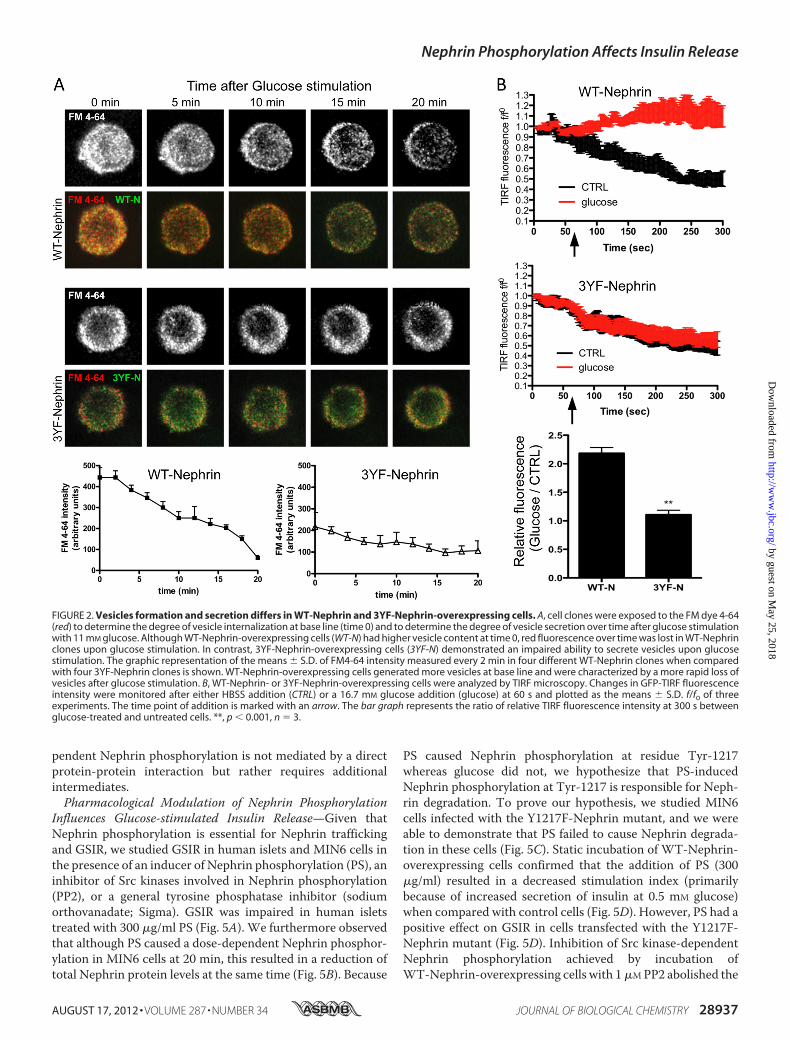
mation and Secretion—We performed live cell imaging ofWT-and 3YF-Nephrin-overexpressing cells stained with FM dye4-64 to visualize endocytosis and exocytosis of vesicles. Therelative level of vesicle endocytosis was measured by determin-ing the total fluorescence achieved after complete vesicles exo-cytosis via a single stimulation with 25 mM KCl followed byincubation with 1 �g/ml of FM4-64 for 15 min as described(45). After three washes with calcium-free PBS, fluorescenceimages were acquired at base line and after subsequent stimu-lation with 11 mM glucose to assess vesicle exocytosis. Imageswere taken every 5 min for a total of 20 min, and the loss offluorescence measured during stimulation was utilized to indi-cate the rate and amount of vesicle exocytosis. Although WT-Nephrin-overexpressing cells had a higher base-line fluores-cence, suggesting a higher endocytotic activity, stimulationwith glucose led to a more rapid loss of fluorescence in WT-Nephrin-overexpressing cells when comparedwith 3YF-Neph-rin-overexpressing cells, where the fluorescence intensityremained mainly unchanged over time (Fig. 2A). Next, we per-formed TIRF microscopy to study WT-Nephrin and 3YF-Nephrin trafficking in response to glucose. The fluorescencesignal obtained by TIRF imaging represents GFP-Nephrinwithin 150–200 nmof the coverslip, i.e., in or in close proximityto the plasma membrane. In untreated cells, the fluorescencesignal decayed over time mostly because of photobleaching(Fig. 2B, untreated) independently of the expressed Nephrinform. Glucose stimulation reversed this decay and even led to asignificant increase in TIRF-fluorescence in WT-Nephrin-overexpressing cells. However, this effect was absent in 3YF-Nephrin-overexpressing cells. The difference in TIRF betweenuntreated and glucose-stimulated cells at the end of the exper-iment was 2.2-fold for WT-Nephrin cells and only 1.1-fold for
3YF-Nephrin, indicating thatWT-Nephrinwas recruited to theplasma membrane, whereas 3YF-Nephrin was not.Nephrin Is a Downstream Effector of Dynamin in Glucose-
stimulated Insulin Release—Dynamin is a master regulator ofvesicle trafficking and actin remodeling (24–32), and it pro-motes GSIR (33). Because those functions are similar to whatwe have described for Nephrin (1), we investigated whether adecrease in Nephrin expression in MIN6 cells could alter theeffect of Dynamin on GSIR. Although WT-Dynamin inducedthe expected augmentation of GSIR (41), WT-Dynamin over-expression in Nephrin siRNA-treatedMIN6 cells did not resultin an augmentation of GSIR, similar to what was observed withthe dominant negative Dynamin mutant (K44A-Dynamin; Fig.3A).We also performedGSIR experiments in cells overexpress-ingWT-Dynamin or K44A-Dynamin mutants that were stablytransfected with WT-Nephrin, 3YF-Nephrin, or GFP alone.WT-Nephrin did not overcome the negative effect of K44A-Dynamin onGSIR, suggesting that Dynamin is essential to pro-mote Nephrin-mediated GSIR. Likewise, overexpression ofWT-Dynamin was not sufficient to overcome the inhibitoryeffect of 3YF-Nephrin on GSIR (Fig. 3B). We therefore investi-gated whether Dynamin is essential to promote Nephrin phos-phorylation. Although the induction of Nephrin phosphoryla-tion by high glucose (11 mM) and PS (300 �g/ml) wereconserved in WT-Dynamin-overexpressing cells, this effectwas abolished in K44A-Dynamin-overexpressing cells (Fig. 3,CandD). Overall, these data suggest that DynaminGTPase activ-ity is necessary for Nephrin phosphorylation and function.Nephrin Trafficking Requires Dynamin GTPase Activity but
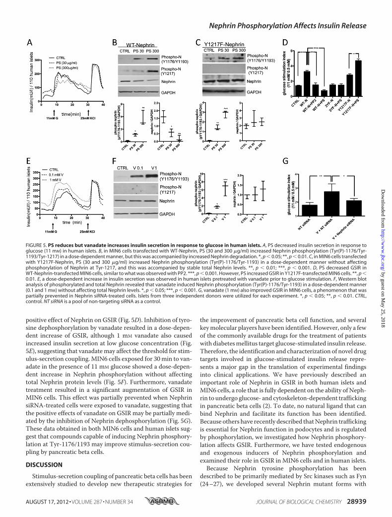
Does Not Involve a Nephrin Dynamin Protein-ProteinInteraction—WT-Nephrin-overexpressing cells were infectedwith either WT-Dynamin or K44A-Dynamin. WT-Dynamin-overexpressing cells conserved the ability of WT-Nephrin tointernalize in response to glucose andPS (as shown for untrans-fected cells in Fig. 1D). On the contrary, K44A-Dynamin-over-expressing cells resulted in deficient WT-Nephrin traffickingfrom the plasma membrane to intracellular compartments inresponse to either stimulus (Fig. 4A). Given the presence of afunctional interaction between Nephrin and Dynamin andbased on the ability of Dynamin to bind several modulators ofactin cytoskeleton remodeling (32–38), we tested whetherNephrin and Dynamin could physically interact. HEK293 cells,which express endogenousDynamin (Fig. 4B), were transfectedwith FLAG-Nephrin or FLAG-Pin1. FLAG-Pin1 was used as anegative control. Co-transfections with GFP-Podocin or GFP-Nck1 were utilized as positive controls. After immunoprecipi-tation of FLAG fusion proteins, bound proteins were elutedwith a FLAG peptide, and eluates were analyzed for the pres-ence of FLAG andGFP proteins as well as Dynamin byWesternblotting. Although FLAG-Nephrin was able to co-immunopre-cipitate GFP-Nck1 and GFP-Podocin, FLAG-Nephrin did notinteract with endogenous Dynamin. Overexpression of WT-Dynamin in FLAG-Nephrin expressing cells did also not resultin the co-immunoprecipitation of both proteins (data notshown). As expected, FLAG-Pin1 did not bind GFP-Nck1,GFP-Podocin, or Dynamin (Fig. 4B), underlining the specificityof the binding assay. These results suggest that Nephrin is adownstream target of Dynamin function and thatDynamin-de-
Nephrin Phosphorylation Affects Insulin Release
28936 JOURNAL OF BIOLOGICAL CHEMISTRY VOLUME 287 • NUMBER 34 • AUGUST 17, 2012
by guest on May 25, 2018
http://ww
w.jbc.org/
Dow
nloaded from
pendent Nephrin phosphorylation is not mediated by a directprotein-protein interaction but rather requires additionalintermediates.Pharmacological Modulation of Nephrin Phosphorylation
Influences Glucose-stimulated Insulin Release—Given thatNephrin phosphorylation is essential for Nephrin traffickingand GSIR, we studied GSIR in human islets and MIN6 cells inthe presence of an inducer of Nephrin phosphorylation (PS), aninhibitor of Src kinases involved in Nephrin phosphorylation(PP2), or a general tyrosine phosphatase inhibitor (sodiumorthovanadate; Sigma). GSIR was impaired in human isletstreated with 300 �g/ml PS (Fig. 5A). We furthermore observedthat although PS caused a dose-dependent Nephrin phosphor-ylation in MIN6 cells at 20 min, this resulted in a reduction oftotal Nephrin protein levels at the same time (Fig. 5B). Because
PS caused Nephrin phosphorylation at residue Tyr-1217whereas glucose did not, we hypothesize that PS-inducedNephrin phosphorylation at Tyr-1217 is responsible for Neph-rin degradation. To prove our hypothesis, we studied MIN6cells infected with the Y1217F-Nephrin mutant, and we wereable to demonstrate that PS failed to cause Nephrin degrada-tion in these cells (Fig. 5C). Static incubation of WT-Nephrin-overexpressing cells confirmed that the addition of PS (300�g/ml) resulted in a decreased stimulation index (primarilybecause of increased secretion of insulin at 0.5 mM glucose)when compared with control cells (Fig. 5D). However, PS had apositive effect on GSIR in cells transfected with the Y1217F-Nephrin mutant (Fig. 5D). Inhibition of Src kinase-dependentNephrin phosphorylation achieved by incubation ofWT-Nephrin-overexpressing cells with 1�MPP2 abolished the
FIGURE 2. Vesicles formation and secretion differs in WT-Nephrin and 3YF-Nephrin-overexpressing cells. A, cell clones were exposed to the FM dye 4-64(red) to determine the degree of vesicle internalization at base line (time 0) and to determine the degree of vesicle secretion over time after glucose stimulationwith 11 mM glucose. Although WT-Nephrin-overexpressing cells (WT-N) had higher vesicle content at time 0, red fluorescence over time was lost in WT-Nephrinclones upon glucose stimulation. In contrast, 3YF-Nephrin-overexpressing cells (3YF-N) demonstrated an impaired ability to secrete vesicles upon glucosestimulation. The graphic representation of the means � S.D. of FM4-64 intensity measured every 2 min in four different WT-Nephrin clones when comparedwith four 3YF-Nephrin clones is shown. WT-Nephrin-overexpressing cells generated more vesicles at base line and were characterized by a more rapid loss ofvesicles after glucose stimulation. B, WT-Nephrin- or 3YF-Nephrin-overexpressing cells were analyzed by TIRF microscopy. Changes in GFP-TIRF fluorescenceintensity were monitored after either HBSS addition (CTRL) or a 16.7 mM glucose addition (glucose) at 60 s and plotted as the means � S.D. f/f0 of threeexperiments. The time point of addition is marked with an arrow. The bar graph represents the ratio of relative TIRF fluorescence intensity at 300 s betweenglucose-treated and untreated cells. **, p � 0.001, n � 3.
Nephrin Phosphorylation Affects Insulin Release
AUGUST 17, 2012 • VOLUME 287 • NUMBER 34 JOURNAL OF BIOLOGICAL CHEMISTRY 28937
by guest on May 25, 2018
http://ww
w.jbc.org/
Dow
nloaded from
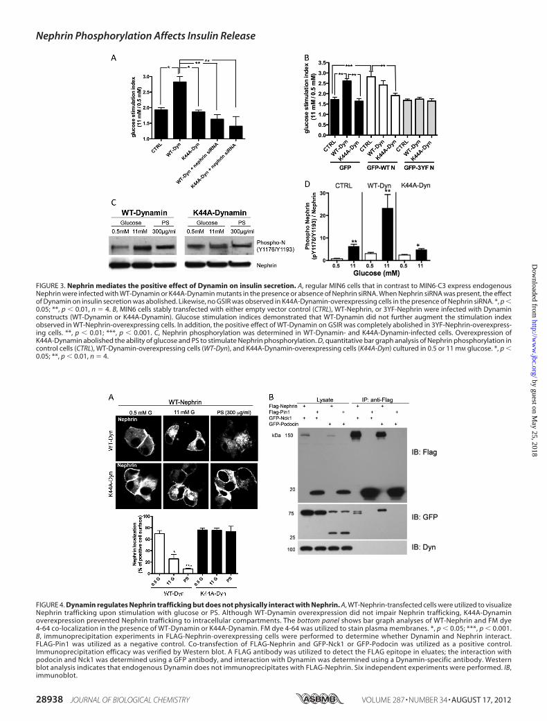
FIGURE 3. Nephrin mediates the positive effect of Dynamin on insulin secretion. A, regular MIN6 cells that in contrast to MIN6-C3 express endogenousNephrin were infected with WT-Dynamin or K44A-Dynamin mutants in the presence or absence of Nephrin siRNA. When Nephrin siRNA was present, the effectof Dynamin on insulin secretion was abolished. Likewise, no GSIR was observed in K44A-Dynamin-overexpressing cells in the presence of Nephrin siRNA. *, p �0.05; **, p � 0.01, n � 4. B, MIN6 cells stably transfected with either empty vector control (CTRL), WT-Nephrin, or 3YF-Nephrin were infected with Dynaminconstructs (WT-Dynamin or K44A-Dynamin). Glucose stimulation indices demonstrated that WT-Dynamin did not further augment the stimulation indexobserved in WT-Nephrin-overexpressing cells. In addition, the positive effect of WT-Dynamin on GSIR was completely abolished in 3YF-Nephrin-overexpress-ing cells. **, p � 0.01; ***, p � 0.001. C, Nephrin phosphorylation was determined in WT-Dynamin- and K44A-Dynamin-infected cells. Overexpression ofK44A-Dynamin abolished the ability of glucose and PS to stimulate Nephrin phosphorylation. D, quantitative bar graph analysis of Nephrin phosphorylation incontrol cells (CTRL), WT-Dynamin-overexpressing cells (WT-Dyn), and K44A-Dynamin-overexpressing cells (K44A-Dyn) cultured in 0.5 or 11 mM glucose. *, p �0.05; **, p � 0.01, n � 4.
FIGURE 4. Dynamin regulates Nephrin trafficking but does not physically interact with Nephrin. A, WT-Nephrin-transfected cells were utilized to visualizeNephrin trafficking upon stimulation with glucose or PS. Although WT-Dynamin overexpression did not impair Nephrin trafficking, K44A-Dynaminoverexpression prevented Nephrin trafficking to intracellular compartments. The bottom panel shows bar graph analyses of WT-Nephrin and FM dye4-64 co-localization in the presence of WT-Dynamin or K44A-Dynamin. FM dye 4-64 was utilized to stain plasma membranes. *, p � 0.05; ***, p � 0.001.B, immunoprecipitation experiments in FLAG-Nephrin-overexpressing cells were performed to determine whether Dynamin and Nephrin interact.FLAG-Pin1 was utilized as a negative control. Co-transfection of FLAG-Nephrin and GFP-Nck1 or GFP-Podocin was utilized as a positive control.Immunoprecipitation efficacy was verified by Western blot. A FLAG antibody was utilized to detect the FLAG epitope in eluates; the interaction withpodocin and Nck1 was determined using a GFP antibody, and interaction with Dynamin was determined using a Dynamin-specific antibody. Westernblot analysis indicates that endogenous Dynamin does not immunoprecipitates with FLAG-Nephrin. Six independent experiments were performed. IB,immunoblot.
Nephrin Phosphorylation Affects Insulin Release
28938 JOURNAL OF BIOLOGICAL CHEMISTRY VOLUME 287 • NUMBER 34 • AUGUST 17, 2012
by guest on May 25, 2018
http://ww
w.jbc.org/
Dow
nloaded from
positive effect of Nephrin on GSIR (Fig. 5D). Inhibition of tyro-sine dephosphorylation by vanadate resulted in a dose-depen-dent increase of GSIR, although 1 mM vanadate also causedincreased insulin secretion at low glucose concentration (Fig.5E), suggesting that vanadatemay affect the threshold for stim-ulus-secretion coupling. MIN6 cells exposed for 30min to van-adate in the presence of 11 mM glucose showed a dose-depen-dent increase in Nephrin phosphorylation without affectingtotal Nephrin protein levels (Fig. 5F). Furthermore, vanadatetreatment resulted in a significant augmentation of GSIR inMIN6 cells. This effect was partially prevented when NephrinsiRNA-treated cells were exposed to vanadate, suggesting thatthe positive effects of vanadate on GSIR may be partially medi-ated by the inhibition of Nephrin dephosphorylation (Fig. 5G).These data obtained in both MIN6 cells and human islets sug-gest that compounds capable of inducing Nephrin phosphory-lation at Tyr-1176/1193 may improve stimulus-secretion cou-pling by pancreatic beta cells.
DISCUSSION
Stimulus-secretion coupling of pancreatic beta cells has beenextensively studied to develop new therapeutic strategies for
the improvement of pancreatic beta cell function, and severalkeymolecular players have been identified.However, only a fewof the commonly available drugs for the treatment of patientswith diabetesmellitus target glucose-stimulated insulin release.Therefore, the identification and characterization of novel drugtargets involved in glucose-stimulated insulin release repre-sents a major gap in the translation of experimental findingsinto clinical applications. We have previously described animportant role of Nephrin in GSIR in both human islets andMIN6 cells, a role that is fully dependent on the ability of Neph-rin to undergo glucose- and cytoskeleton-dependent traffickingin pancreatic beta cells (2). To date, no natural ligand that canbind Nephrin and facilitate its function has been identified.Because others have recently described thatNephrin traffickingis essential for Nephrin function in podocytes and is regulatedby phosphorylation, we investigated how Nephrin phosphory-lation affects GSIR. Furthermore, we have tested endogenousand exogenous inducers of Nephrin phosphorylation andexamined their role in GSIR inMIN6 cells and in human islets.Because Nephrin tyrosine phosphorylation has been
described to be primarily mediated by Src kinases such as Fyn(24–27), we developed several Nephrin mutant forms with
FIGURE 5. PS reduces but vanadate increases insulin secretion in response to glucose in human islets. A, PS decreased insulin secretion in response toglucose (11 mM) in human islets. B, in MIN6 cells transfected with WT-Nephrin, PS (30 and 300 �g/ml) increased Nephrin phosphorylation (Tyr(P)-1176/Tyr-1193/Tyr-1217) in a dose-dependent manner, but this was accompanied by increased Nephrin degradation. *, p � 0.05; **, p � 0.01. C, in MIN6 cells transfectedwith Y1217F-Nephrin, PS (30 and 300 �g/ml) increased Nephrin phosphorylation (Tyr(P)-1176/Tyr-1193) in a dose-dependent manner without affectingphosphorylation of Nephrin at Tyr-1217, and this was accompanied by stable total Nephrin levels. **, p � 0.01; ***, p � 0.001. D, PS decreased GSIR inWT-Nephrin-transfected MIN6 cells, similar to what was observed with PP2. ***, p � 0.001. However, PS increased GSIR in Y1217F-transfected MIN6 cells. **, p �0.01. E, a dose-dependent increase in insulin secretion was observed in human islets pretreated with vanadate prior to glucose stimulation. F, Western blotanalysis of phosphorylated and total Nephrin revealed that vanadate induced Nephrin phosphorylation (Tyr(P)-1176/Tyr-1193) in a dose-dependent manner(0.1 and 1 mM) without affecting total Nephrin levels. *, p � 0.05; ***, p � 0.001. G, vanadate (1 mM) also improved GSIR in MIN6 cells, a phenomenon that waspartially prevented in Nephrin siRNA-treated cells. Islets from three independent donors were utilized for each experiment. *, p � 0.05; **, p � 0.01. CTRL,control. NT siRNA is a pool of non-targeting siRNA as a control.
Nephrin Phosphorylation Affects Insulin Release
AUGUST 17, 2012 • VOLUME 287 • NUMBER 34 JOURNAL OF BIOLOGICAL CHEMISTRY 28939
by guest on May 25, 2018
http://ww
w.jbc.org/
Dow
nloaded from

tyrosine to phenylalanine substitutions at three major residuesthat have been shown to be crucial for Nephrin-mediated reg-ulation of the actin cytoskeleton. Because single mutants didnot significantly affect GSIR (Fig. 1B), we have primarily uti-lized a triple mutant form to demonstrate that glucose- andPS-dependent Nephrin phosphorylation can be abrogated byusing this mutant form of Nephrin (3YF-Nephrin; Fig. 1A).MIN6 C3 clones overexpressing 3YF-Nephrin showedimpaired GSIR and were not capable of forming cell-cell con-tacts that have been described to be essential for the augmen-tation of GSIR in cultured pancreatic beta cells (7–9). Further-more, Nephrin trafficking from and to the plasma membranewas impaired in these cells (Fig. 1D) without affecting cell via-bility (Fig. 1C). The scattered distribution of 3YF-Nephrin-overexpressing cells with decreased cell to cell contacts and thereorganization of the actin cytoskeleton including the loss ofcortical actin (Fig. 1E) supports a primary role of Nephrin as anactive regulator of actin cytoskeleton remodeling in pancreaticbeta cells, similar to what has been described in podocytes (13,14). Human islets infected with 3YF-Nephrin mutants also didnot experience increases in insulin release as normally observedafter infection withWT-Nephrin (Fig. 1F). It has been assumedfor over a decade sinceNephrin has been discovered thatNeph-rin localizes exclusively to the plasma membrane where it ful-fills its primary role in the formation of the slit diaphragm byengaging in homophilic interactions with adjacent Nephrinmolecules (5, 24) and heterophilic interactions with otherstructural components of the S.D (16, 47–49). Instead, themore dynamic aspects of Nephrin trafficking are a relativelynew concept. It has been reported that Nephrin endocytosis inpodocytes occurs under pathological conditions that involvethe impairment of the slit diaphragm integrity (19). We dem-onstrated herein that phosphorylation-dependent Nephrintrafficking is essential under physiological conditions to grantGSIR and to facilitate proper secretory vesicle formation andexocytosis in pancreatic beta cells (Fig. 2). This is in line withthe evidence that a novel role of Nephrin in vesicular dockinghas been described in podocytes, where the C terminus ofNephrin interacts with the v-SNARE protein VAMP-2 (50),which is also found on themembrane of insulin secretory gran-ules in pancreatic beta cells (51). In pancreatic beta cells, glu-cose transiently modulates the cortical actin organization anddisrupts the interaction of F-actin with the t-SNARE complexat the plasmamembrane to facilitate glucose-stimulated insulinsecretion (52). However, the relationship between SNARE-me-diated exocytosis and actin reorganization remains to be fullyunderstood. The pathway accounting for the regulated target-ing of vesicles to their cognate t-SNAREs has not been estab-lished, and a functional Nephrin/actin interaction could repre-sent the molecular link between GTPases and SNAREs. Actinreorganization and actin binding to t- and v-SNARE proteinshas been shown to be dependent onDynamin-2 (33, 41), a largeGTPase that is functionally coupled to insulin granule exocyto-sis and that facilitates GSIR through a yet unknownmechanism(41). Because Dynamin is essential for the maintenance ofpodocyte structure and function (44), and it has recently beendemonstrated that Dynamin affects Nephrin internalization(23), we tested whether Dynamin couldmediate Nephrin phos-
phorylation and function in pancreatic beta cells. Glucose-in-duced Nephrin phosphorylation and insulin release wereimpaired in cells overexpressing a dominant negative Dynaminmutant form (Fig. 3, B–D), and Nephrin deficiency abolishedthe positive effect of Dynamin on GSIR (Fig. 3A), suggestingthat Dynamin is a keymediator in the signaling pathway linkingglucose stimulation to Nephrin phosphorylation. BecauseDynamin is degraded by Cathepsin L, a proteolytic enzyme thatis elevated in diabetes (44), it may be interesting to determinewhether Cathepsin L inhibitors may facilitate GSIR. Alterna-tively, identification of smallmolecules or ligands that can stim-ulate Nephrin phosphorylation could be tested as drugs for theaugmentation of GSIR. However, our data with PS and van-adate suggest that the role of Nephrin phosphorylation is morecomplex than originally expected, because inducers of Nephrinphosphorylation such as PS may result in a reduction of totalNephrin and therefore negatively affect islet cell function (Fig.5). It is therefore likely that the phosphorylation states of dif-ferent tyrosine residueswithin theC-terminal portion ofNeph-rin are responsible for the formation of different signaling com-plexes, which may lead to either Nephrin degradation or tophysiological Nephrin trafficking. In fact, a positive effect of PSon GSIR that is associated with preservation of total Nephrin isobserved in cells transfected with the Y1217F-Nephrin mutant(Fig. 5, C and D), strongly supporting a key role of Nephrinphosphorylation at Tyr-1217 as a prerequisite for Nephrindegradation.In conclusion, we have demonstrated an important role of
Dynamin-dependent Nephrin phosphorylation in stimulus-se-cretion coupling in pancreatic beta cells. A definitive role forNephrin in pancreatic beta cell function remains to be estab-lished through ongoing metabolic studies in patients withNephrin mutations and through the phenotypic analysis ofmice carrying a conditional deletion of the Nephrin gene inpancreatic beta cells. Nevertheless, our study offers the ratio-nale to screen drug libraries for the identification of modifiersof Nephrin phosphorylation that might be pharmacologicallyused to improve pancreatic beta cell function.
Acknowledgment—We thank Dr. George W. Burke (Department ofSurgery, University of Miami, Miami, FL) for careful review of themanuscript.
REFERENCES1. Kestilä, M., Lenkkeri, U., Männikkö, M., Lamerdin, J., McCready, P.,
Putaala, H., Ruotsalainen, V., Morita, T., Nissinen,M., Herva, R., Kashtan,C. E., Peltonen, L., Holmberg, C., Olsen, A., and Tryggvason, K. (1998)Positionally cloned gene for a novel glomerular protein–Nephrin–is mu-tated in congenital nephrotic syndrome.Mol. Cell 1, 575–582
2. Fornoni, A., Jeon, J., Varona Santos, J., Cobianchi, L., Jauregui, A., Inve-rardi, L., Mandic, S. A., Bark, C., Johnson, K., McNamara, G., Pileggi, A.,Molano, R. D., Reiser, J., Tryggvason, K., Kerjaschki, D., Berggren, P. O.,Mundel, P., and Ricordi, C. (2010) Nephrin is expressed on the surface ofinsulin vesicles and facilitates glucose-stimulated insulin release.Diabetes59, 190–199
3. Ruotsalainen, V., Ljungberg, P., Wartiovaara, J., Lenkkeri, U., Kestilä, M.,Jalanko, H., Holmberg, C., and Tryggvason, K. (1999) Nephrin is specifi-cally located at the slit diaphragm of glomerular podocytes. Proc. Natl.Acad. Sci. U.S.A. 96, 7962–7967
4. Tryggvason, K. (1999) Unraveling the mechanisms of glomerular ultrafil-
Nephrin Phosphorylation Affects Insulin Release
28940 JOURNAL OF BIOLOGICAL CHEMISTRY VOLUME 287 • NUMBER 34 • AUGUST 17, 2012
by guest on May 25, 2018
http://ww
w.jbc.org/
Dow
nloaded from

tration. Nephrin, a key component of the slit diaphragm. J. Am. Soc. Neph-rol. 10, 2440–2445
5. Khoshnoodi, J., Sigmundsson, K., Ofverstedt, L. G., Skoglund, U., Obrink,B., Wartiovaara, J., and Tryggvason, K. (2003) Nephrin promotes cell-celladhesion through homophilic interactions.Am. J. Pathol. 163, 2337–2346
6. Heikkilä, E., Ristola, M., Havana, M., Jones, N., Holthöfer, H., andLehtonen, S. (2011) Trans-interaction of Nephrin and Neph1/Neph3 in-duces cell adhesion that associates with decreased tyrosine phosphoryla-tion of Nephrin. Biochem. J. 435, 619–628
7. Jaques, F., Jousset, H., Tomas, A., Prost, A. L., Wollheim, C. B., Irminger,J. C., Demaurex,N., andHalban, P. A. (2008)Dual effect of cell-cell contactdisruption on cytosolic calcium and insulin secretion. Endocrinology 149,2494–2505
8. Hauge-Evans, A. C., Squires, P. E., Persaud, S. J., and Jones, P. M. (1999)Pancreatic beta-cell-to-beta-cell interactions are required for integratedresponses to nutrient stimuli. Enhanced Ca2� and insulin secretory re-sponses of MIN6 pseudoislets. Diabetes 48, 1402–1408
9. Wojtusciszyn, A., Armanet,M.,Morel, P., Berney, T., and Bosco, D. (2008)Insulin secretion from human beta cells is heterogeneous and dependenton cell-to-cell contacts. Diabetologia 51, 1843–1852
10. Li, P., Banjade, S., Cheng, H. C., Kim, S., Chen, B., Guo, L., Llaguno, M.,Hollingsworth, J. V., King, D. S., Banani, S. F., Russo, P. S., Jiang, Q. X.,Nixon, B. T., and Rosen, M. K. (2012) Phase transitions in the assembly ofmultivalent signalling proteins. Nature 483, 336–340
11. George, B., Verma, R., Soofi, A. A., Garg, P., Zhang, J., Park, T. J., Giardino,L., Ryzhova, L., Johnstone, D. B., Wong, H., Nihalani, D., Salant, D. J.,Hanks, S. K., Curran, T., Rastaldi, M. P., and Holzman, L. B. (2012) Crk1/2-dependent signaling is necessary for podocyte foot process spreading inmouse models of glomerular disease. J. Clin. Invest. 122, 674–692
12. Venkatareddy,M., Cook, L., Abuarquob, K., Verma, R., andGarg, P. (2011)Nephrin regulates lamellipodia formation by assembling a protein com-plex that includes Ship2, filamin and lamellipodin. PLoS One 6, e28710
13. Jones, N., Blasutig, I.M., Eremina, V., Ruston, J.M., Bladt, F., Li, H., Huang,H., Larose, L., Li, S. S., Takano, T., Quaggin, S. E., and Pawson, T. (2006)Nck adaptor proteins link Nephrin to the actin cytoskeleton of kidneypodocytes. Nature 440, 818–823
14. Verma, R., Kovari, I., Soofi, A., Nihalani, D., Patrie, K., and Holzman, L. B.(2006) Nephrin ectodomain engagement results in Src kinase activation,Nephrin phosphorylation, Nck recruitment, and actin polymerization.J. Clin. Invest. 116, 1346–1359
15. Zhu, J., Sun, N., Aoudjit, L., Li, H., Kawachi, H., Lemay, S., and Takano, T.(2008) Nephrin mediates actin reorganization via phosphoinositide 3-ki-nase in podocytes. Kidney Int. 73, 556–566
16. Huber, T. B., Hartleben, B., Kim, J., Schmidts, M., Schermer, B., Keil, A.,Egger, L., Lecha, R. L., Borner, C., Pavenstädt,H., Shaw,A. S.,Walz, G., andBenzing, T. (2003) Nephrin and CD2AP associate with phosphoinositide3-OH kinase and stimulate AKT-dependent signaling.Mol. Cell Biol. 23,4917–4928
17. Simons,M., Schwarz, K., Kriz,W.,Miettinen, A., Reiser, J.,Mundel, P., andHolthöfer, H. (2001) Involvement of lipid rafts in Nephrin phosphoryla-tion and organization of the glomerular slit diaphragm.Am. J. Pathol. 159,1069–1077
18. Tryggvason, K., Pikkarainen, T., and Patrakka, J. (2006) Nck links Nephrinto actin in kidney podocytes. Cell 125, 221–224
19. Quack, I., Rump, L. C., Gerke, P., Walther, I., Vinke, T., Vonend, O.,Grunwald, T., and Sellin, L. (2006) �-Arrestin2 mediates Nephrin endo-cytosis and impairs slit diaphragm integrity. Proc. Natl. Acad. Sci. U.S.A.103, 14110–14115
20. Aoudjit, L., Jiang, R., Lee, T. H., New, L. A., Jones, N., and Takano, T.(2011) Podocyte protein, Nephrin, is a substrate of protein tyrosine phos-phatase 1B. J. Signal Transduct. 2011, 376543
21. Uchida, K., Suzuki, K., Iwamoto, M., Kawachi, H., Ohno, M., Horita, S.,andNitta, K. (2008) Decreased tyrosine phosphorylation of Nephrin in ratand human nephrosis. Kidney Int. 73, 926–932
22. Fan, Q., Xing, Y., Ding, J., andGuan, N. (2009) Reduction in VEGF proteinand phosphorylated Nephrin associated with proteinuria in adriamycinnephropathy rats. Nephron. Exp. Nephrol. 111, e92-e102
23. Qin, X. S., Tsukaguchi, H., Shono, A., Yamamoto, A., Kurihara, H., and
Doi, T. (2009) Phosphorylation of Nephrin triggers its internalization byraft-mediated endocytosis. J. Am. Soc. Nephrol. 20, 2534–2545
24. Lahdenperä, J., Kilpeläinen, P., Liu, X. L., Pikkarainen, T., Reponen, P.,Ruotsalainen, V., and Tryggvason, K. (2003) Clustering-induced tyrosinephosphorylation of Nephrin by Src family kinases. Kidney Int. 64,404–413
25. Li, H., Lemay, S., Aoudjit, L., Kawachi, H., and Takano, T. (2004) SRC-family kinase Fyn phosphorylates the cytoplasmic domain of Nephrin andmodulates its interaction with podocin. J. Am. Soc. Nephrol. 15,3006–3015
26. Verma, R., Wharram, B., Kovari, I., Kunkel, R., Nihalani, D., Wary, K. K.,Wiggins, R. C., Killen, P., and Holzman, L. B. (2003) Fyn binds to andphosphorylates the kidney slit diaphragm component Nephrin. J. Biol.Chem. 278, 20716–20723
27. Liu, X. L., Doné, S. C., Yan, K., Kilpeläinen, P., Pikkarainen, T., and Tryg-gvason, K. (2004) Defective trafficking of Nephrin missense mutants res-cued by a chemical chaperone. J. Am. Soc. Nephrol. 15, 1731–1738
28. Garg, P., Verma, R., Nihalani, D., Johnstone, D. B., and Holzman, L. B.(2007) Neph1 cooperates with Nephrin to transduce a signal that inducesactin polymerization.Mol. Cell Biol. 27, 8698–8712
29. Barletta, G. M., Kovari, I. A., Verma, R. K., Kerjaschki, D., and Holzman,L. B. (2003) Nephrin and Neph1 co-localize at the podocyte foot processintercellular junction and form cis hetero-oligomers. J. Biol. Chem. 278,19266–19271
30. Quack, I., Woznowski, M., Potthoff, S. A., Palmer, R., Königshausen, E.,Sivritas, S., Schiffer, M., Stegbauer, J., Vonend, O., Rump, L. C., and Sellin,L. (2011) PKC � mediates �-arrestin2-dependent Nephrin endocytosis inhyperglycemia. J. Biol. Chem. 286, 12959–12970
31. Praefcke, G. J., and McMahon, H. T. (2004) The Dynamin superfamily.Universalmembrane tubulation and fissionmolecules?Nat. Rev.Mol. CellBiol. 5, 133–147
32. Krueger, E. W., Orth, J. D., Cao, H., and McNiven, M. A. (2003) A Dy-namin-cortactin-Arp2/3 complex mediates actin reorganization ingrowth factor-stimulated cells.Mol. Biol. Cell 14, 1085–1096
33. Schafer, D. A. (2004) Regulating actin dynamics atmembranes. A focus onDynamin. Traffic 5, 463–469
34. Merrifield, C. J., Feldman, M. E., Wan, L., and Almers, W. (2002) Imagingactin and Dynamin recruitment during invagination of single clathrin-coated pits. Nat. Cell Biol. 4, 691–698
35. McNiven, M. A., Kim, L., Krueger, E. W., Orth, J. D., Cao, H., and Wong,T. W. (2000) Regulated interactions between Dynamin and the actin-binding protein cortactin modulate cell shape. J. Cell Biol. 151, 187–198
36. Orth, J. D., Krueger, E. W., Cao, H., andMcNiven, M. A. (2002) The largeGTPase Dynamin regulates actin comet formation and movement in liv-ing cells. Proc. Natl. Acad. Sci. U.S.A. 99, 167–172
37. Lee, E., and De Camilli, P. (2002) Dynamin at actin tails. Proc. Natl. Acad.Sci. U.S.A. 99, 161–166
38. Gu, C., Yaddanapudi, S., Weins, A., Osborn, T., Reiser, J., Pollak, M.,Hartwig, J., and Sever, S. (2010) Direct Dynamin-actin interactions regu-late the actin cytoskeleton. EMBO J. 29, 3593–3606
39. Kim, Y., and Chang, S. (2006) Ever-expanding network of Dynamin-inter-acting proteins.Mol. Neurobiol. 34, 129–136
40. Zhang, Z., Wakabayashi, N., Wakabayashi, J., Tamura, Y., Song, W. J.,Sereda, S., Clerc, P., Polster, B. M., Aja, S. M., Pletnikov, M. V., Kensler,T.W., Shirihai, O. S., Iijima,M., Hussain, M. A., and Sesaki, H. (2011) TheDynamin-related GTPase Opa1 is required for glucose-stimulated ATPproduction in pancreatic beta cells.Mol. Biol. Cell 22, 2235–2245
41. Min, L., Leung, Y. M., Tomas, A., Watson, R. T., Gaisano, H. Y., Halban,P. A., Pessin, J. E., andHou, J. C. (2007) Dynamin is functionally coupled toinsulin granule exocytosis. J. Biol. Chem. 282, 33530–33536
42. Miyazaki, J., Araki, K., Yamato, E., Ikegami, H., Asano, T., Shibasaki, Y.,Oka, Y., and Yamamura, K. (1990) Establishment of a pancreatic betacell line that retains glucose-inducible insulin secretion. Special refer-ence to expression of glucose transporter isoforms. Endocrinology 127,126–132
43. Ricordi, C., and Strom, T. B. (2004) Clinical islet transplantation. Ad-vances and immunological challenges. Nat. Rev. Immunol. 4, 259–268
44. Sever, S., Altintas, M. M., Nankoe, S. R., Möller, C. C., Ko, D., Wei, C.,
Nephrin Phosphorylation Affects Insulin Release
AUGUST 17, 2012 • VOLUME 287 • NUMBER 34 JOURNAL OF BIOLOGICAL CHEMISTRY 28941
by guest on May 25, 2018
http://ww
w.jbc.org/
Dow
nloaded from

Henderson, J., del Re, E. C., Hsing, L., Erickson, A., Cohen, C. D., Kretzler,M., Kerjaschki, D., Rudensky, A., Nikolic, B., and Reiser, J. (2007) Proteo-lytic processing of Dynamin by cytoplasmic cathepsin L is a mechanismfor proteinuric kidney disease. J. Clin. Invest. 117, 2095–2104
45. Gaffield, M. A., and Betz, W. J. (2006) Imaging synaptic vesicle exocytosisand endocytosis with FM dyes. Nat. Protoc. 1, 2916–2921
46. Fornoni, A., Pileggi, A., Molano, R. D., Sanabria, N. Y., Tejada, T., Gonza-lez-Quintana, J., Ichii, H., Inverardi, L., Ricordi, C., and Pastori, R. L. (2008)Inhibition of c-jun N terminal kinase (JNK) improves functional beta cellmass in human islets and leads to AKT and glycogen synthase kinase-3(GSK-3) phosphorylation. Diabetologia 51, 298–308
47. Liu, G., Kaw, B., Kurfis, J., Rahmanuddin, S., Kanwar, Y. S., andChugh, S. S.(2003) Neph1 and Nephrin interaction in the slit diaphragm is an impor-tant determinant of glomerular permeability. J. Clin. Invest. 112, 209–221
48. Huber, T. B., Kottgen, M., Schilling, B., Walz, G., and Benzing, T. (2001)Interaction with podocin facilitates Nephrin signaling. J. Biol. Chem. 276,41543–41546
49. Lehtonen, S., Lehtonen, E., Kudlicka, K., Holthöfer, H., and Farquhar,M. G. (2004) Nephrin forms a complex with adherens junction proteinsand CASK in podocytes and in Madin-Darby canine kidney cells express-ing Nephrin. Am. J. Pathol. 165, 923–936
50. Coward, R. J., Welsh, G. I., Koziell, A., Hussain, S., Lennon, R., Ni, L.,Tavaré, J. M., Mathieson, P. W., and Saleem, M. A. (2007) Nephrin iscritical for the action of insulin on human glomerular podocytes.Diabetes56, 1127–1135
51. Wheeler,M. B., Sheu, L., Ghai,M., Bouquillon, A., Grondin, G.,Weller, U.,Beaudoin, A. R., Bennett, M. K., Trimble, W. S., and Gaisano, H. Y. (1996)Characterization of SNARE protein expression in beta cell lines and pan-creatic islets. Endocrinology 137, 1340–1348
52. Thurmond, D. C., Gonelle-Gispert, C., Furukawa, M., Halban, P. A., andPessin, J. E. (2003) Glucose-stimulated insulin secretion is coupled to theinteraction of actin with the t-SNARE (target membrane solubleN-ethyl-maleimide-sensitive factor attachment protein receptor protein) com-plex.Mol. Endocrinol. 17, 732–742
Nephrin Phosphorylation Affects Insulin Release
28942 JOURNAL OF BIOLOGICAL CHEMISTRY VOLUME 287 • NUMBER 34 • AUGUST 17, 2012
by guest on May 25, 2018
http://ww
w.jbc.org/
Dow
nloaded from
Ricordi, Sandra Merscher-Gomez and Alessia FornoniRodrigo Villarreal, Johanna Guzman, Per-Olof Berggren, Peter Mundel, Camillo
Jongmin Jeon, Ingo Leibiger, Tilo Moede, Britta Walter, Christian Faul, Dony Maiguel,Release in Pancreatic Beta Cells
Dynamin-mediated Nephrin Phosphorylation Regulates Glucose-stimulated Insulin
doi: 10.1074/jbc.M112.389452 originally published online June 20, 20122012, 287:28932-28942.J. Biol. Chem.
10.1074/jbc.M112.389452Access the most updated version of this article at doi:
Alerts:
When a correction for this article is posted•
When this article is cited•
to choose from all of JBC's e-mail alertsClick here
http://www.jbc.org/content/287/34/28932.full.html#ref-list-1
This article cites 52 references, 22 of which can be accessed free at
by guest on May 25, 2018
http://ww
w.jbc.org/
Dow
nloaded from

VOLUME 287 (2012) PAGES 28932–28942DOI 10.1074/jbc.A112.389452
Dynamin-mediated Nephrin phosphorylation regulates glucose-stimulated insulin release in pancreatic beta cells.Jongmin Jeon, Ingo Leibiger, Tilo Moede, Britta Walter, Christian Faul, Dony Maiguel, Rodrigo Villarreal, Johanna Guzman, Per-Olof Berggren, Peter Mundel,Camillo Ricordi, Sandra Merscher-Gomez, and Alessia Fornoni
PAGE 28937:
Theauthors havebecomeawareof severalmistakes inFig. 2A. In the second row, the second and thirdpanels are identical, and the fourth and fifth panelsare also the same. In the third row, the third and fourth panels are repeated. In reviewing the errors, the authors caught another mistake with theblack-and-white digital subtraction images shown in the first and third rows. The figure and figure legend claim to show FM4-64, which is incorrect. Thecorrected Fig. 2A is shown below. The duplicated images in the second and third rows have replaced with ones taken at 5-, 8-, 12-, and 18-minute timepoints. The black-and-white representative images in the first and third rows are now digital subtraction images of the GFP signal forWT-N and 3YF-N.Data shown in the quantitative analysis were also checked for completeness and have been confirmed to be correct. The authors regret any confusion theincorrect figure may have caused. This correction does not influence the validity of the results and conclusions of the original article.
FIGURE 2. Vesicle formation and secretion differs in WT-Nephrin- and YF-Nephrin-overexpressing cells. A, cell clones were exposed to the FM dye 4-64 (red) todetermine the degree of vesicle internalization at base line (time 0) and to determine the degree of vesicle secretion over time after glucose stimulation with 11 mM
glucose. WT-Nephrin (WT-N)-overexpressing cells had higher vesicle content at time 0, and red fluorescence over time was lost in WT-Nephrin clones upon glucosestimulation. In contrast, 3YF-Nephrin (3YF-N)-overexpressing cells demonstrated an impaired ability to secrete vesicles upon glucose stimulation. The graphic repre-sentation of the means and S.D. of FM4–64 intensity measured every 2 min in four different WT-N clones when compared with four 3YF-N clones is shown. WT-Nephrin-overexpressing cells generated more vesicles at base line and were characterized by a more rapid loss of vesicles after glucose stimulation.
THE JOURNAL OF BIOLOGICAL CHEMISTRY VOL. 288, NO. 2, p. 1277, January 11, 2013© 2013 by The American Society for Biochemistry and Molecular Biology, Inc. Published in the U.S.A.
JANUARY 11, 2013 • VOLUME 288 • NUMBER 2 JOURNAL OF BIOLOGICAL CHEMISTRY 1277
ADDITIONS AND CORRECTIONS
Authors are urged to introduce these corrections into any reprints they distribute. Secondary (abstract) services are urged to carry notice ofthese corrections as prominently as they carried the original abstracts.