Embed Size (px)
Citation preview

Available online at www.sciencedirect.com
Dynamics of the translational machineryAlexey Petrov1, Guy Kornberg1, Sean O’Leary1, Albert Tsai1,2,Sotaro Uemura1,3 and Joseph D Puglisi1
The recent growth in single molecule studies of translation has
provided an insight into the molecular mechanism of ribosomal
function. Single molecule fluorescence approaches allowed
direct observation of the structural rearrangements occurring
during translation and revealed dynamic motions of the
ribosome and its ligands. These studies demonstrated how
ligand binding affects dynamics of the ribosome, and the role of
the conformational sampling in large-scale rearrangements
intrinsic to translation elongation. The application of time-
resolved cryo-electron microscopy revealed new
conformational intermediates during back-translocation
providing an insight into ribosomal dynamics from an
alternative perspective. Recent developments permitted
examination of conformational and compositional dynamics of
the ribosome in real-time through multiple cycles of elongation
at the single molecule level. The zero-mode waveguide
approach allowed direct observation of the compositional
dynamics of tRNA occupancy on the elongating ribosome. The
emergence of single molecule in vivo techniques provided
insights into the mechanism and regulation of translation at the
organismal level.
Addresses1 Department of Structural Biology, Stanford University School of
Medicine, Stanford, CA 94305-5126, USA2 Department of Applied Physics, Stanford University, Stanford, CA
94305-4090, USA3 Japan Science and Technology Agency, 4-1-8, Honcho, Kawaguchi,
Saitama 332-0012, Japan
Corresponding author: Puglisi, Joseph D ([email protected])
Current Opinion in Structural Biology 2011, 21:137–145
This review comes from a themed issue on
Protein-nucleic acid interactions
Edited by Kiyoshi Nagai and Song Tan
0959-440X/$ – see front matter
# 2010 Elsevier Ltd. All rights reserved.
DOI 10.1016/j.sbi.2010.11.007
IntroductionThe study of translation has been revolutionized over the
past decades by the remarkable structural biology on the
ribosome and its ligand complexes. These structures have
revealed how the ribosomal particles assemble from RNA
and proteins, how transfer RNA ligands are bound and
suggested mechanisms for the catalysis of peptide bond
formation (reviewed in [1–3]). Recent structures have
www.sciencedirect.com
shown how GTPase factors (EF-Tu and EF-G) interact
with the 50S subunit to stimulate their enzymatic activity
to enhance function in tRNA delivery or translocation of
tRNA–mRNA complexes with respect to the ribosome
[4��,5�,6–8]. Together with biochemical and mechanistic
data, a coherent structural mechanism for the basic steps
of translation can be outlined. Yet despite this progress,
dynamic data are needed to link these structural pictures
to a time-resolved pathway of translation.
Single-molecule fluorescence methods have provided
rich detail on the dynamics of ligand–ligand, ligand–ribosome, and ribosomal particle dynamics. By using
fluorescence resonance energy transfer (FRET) on single
ribosomes, conformational dynamics have been probed
on various ribosomal complexes containing fluorescent
labels. Original experiments focused on the dynamic
pathway of tRNA delivery to the ribosome, and sub-
sequent tRNA dynamics within the hybrid state after
peptide bond formation. More recent experiments have
mapped dynamics between ribosomal proteins and
tRNA, ribosomal proteins and factors, and within the
ribosome particle itself. Here we review recent progress
in single-molecule fluorescence investigations of trans-
lation. We emphasize the interplay of structural and
dynamic data through the application of electron micro-
scopy, and recent experiments that allow tracking of
translation in real time both in vitro and in vivo.
Structural approaches to resolveconformational dynamicsCryo electron microscopy and X-ray crystallography have
obtained static structural snapshots of ribosomes in a
variety of functionally relevant conformations
[9,10��,11–13], whereas molecular dynamics simulations
have suggested the molecular motions involved in the
transition of the ribosome between these distinct states
[14–17]. Recently, several groups have combined time-
resolved methods with single-particle EM to provide
experimental conformational trajectories. Fischer et al.[18��] investigated the dynamics of translocation using
time-resolved electron microscopy. Their study charac-
terized the global conformational changes associated with
thermally driven ribosomal back-translocation. Ribo-
somes assembled in a post-translocation state, with cog-
nate deacylated E-site tRNAfMet and P-site fMet-Val-
tRNAVal (E/P state), were allowed to convert spon-
taneously to their A/P states. The experimentally ident-
ified ribosome configurations included ones consistent
with the previously observed ‘classical’ and ‘hybrid’
Current Opinion in Structural Biology 2011, 21:137–145

138 Protein-nucleic acid interactions
Figure 1
(a) E P A
E P A
E P A
E P A
Pre1 Pre2
E P A
E P A
Pre3
E P A
E P A
Pre4
E P A
E P A
E P A
E P A
Post3
Post1 Post2 Post3
Post2
K642
K1K2K50.12
K11.9
K24.0
K31.2
K40.30
Post1
E P A
E P A
E P A
E P A
Pre5
Pre1 Pre2 Pre3 Pre5Pre4
(b)
Current Opinion in Structural Biology
(a) Conformations of the (retro-)translocating ribosome identified by cryo-EM. The individual complexes are classified by tRNA position. A
reconstruction of a representative sub-state from the ensemble of sub-states for each pre-translocational and post-translocational state (pre1 to pre5
and post1 to post3) is shown, along with a schematic representation of tRNA position. (b) Thermodynamics of translocation based on population
analysis of the states identified by cryo-EM. The data indicate that conversion globally from the pre- to post-translocation state defines the observed
rate of translocation, while sub-states within the post-category and pre-category are in rapid equilibrium with each other. Reproduced with
modifications and with permission from Fischer et al. [18��].
states. In addition, a number of intermediate and pre-
viously unobserved sub-states were identified (Figures 1
and 2). Kinetic analysis indicated that the rate-limiting
step for back-translocation (measured at 0.8 min�1 under
these conditions) is the conversion of the post-transloca-
tion complex to the pre-translocation complex.
The study also illustrated that tRNA and ribosome
motion, particularly 30S rotation, are coupled in the
thermally driven back-translocation process. The energy
landscape for these coupled motions was shown to be
relatively flat, leading to the conclusion that the overall
process involves rapid sampling of the various confor-
mational sub-states by the translocating ribosome. The
data suggest that a number of parallel dynamic pathways
exist by which translocation can occur. The study also
showed that rotation of the 30S subunit, which impacts on
tRNA motion, was more dynamic at 37 8C. The results
were interpreted to provide further evidence that the
ribosome acts as a Brownian ratchet. However, the influ-
ence of EF-G on the distribution and accessibility of the
Current Opinion in Structural Biology 2011, 21:137–145
individual conformational sub-states was not investigated
and awaits future study.
Williamson and co-workers used cryoEM to track the
assembly of the 30S subunit from ribosomal RNA and
proteins [19��]. Using a method called discovery single
particle analysis, they initiated assembly of 30S subunits,
and rapidly trapped intermediates at different time points
(1–100 min) through negative staining. They could struc-
turally classify the different assembly intermediates, and
compare the observed conformations with their prior
assembly kinetics and thermodynamics measurements.
The results showed a heterogeneous, parallel mechanism
of subunit assembly, and demonstrate the power of inte-
grated biophysical and structural imaging of the trans-
lation machinery.
Probing conformational dynamics of theribosome with smFRETConformational changes within the ribosome have been
measured using single-molecule FRET. The efficiency
www.sciencedirect.com

Dynamics in translation Petrov et al. 139
Figure 2
6o
Current Opinion in Structural Biology
Intersubunit rotation. Cryo-EM and X-ray crystallography methods
revealed ribosomes in distinct conformations that differ by the relative
orientation of the ribosomal subunits. 70S ribosome form is shown in top
down view from the 30S subunit. 50S is shown in dark blue and 30S in
un-rotated state is shown in light grey. 30S in rotated state is shown with
the black outline. During elongation cycle 30S subunit rotates �68relative to the large subunit. Upon aminoacyl-tRNA selection and
peptidyl transfer the 30S subunit rotates clockwise. The ribosome
rotates counterclockwise upon translocation.
of FRET is highly sensitive to the distance (1/R6, where Ris inter-dye transition dipole distance) between the fluor-
ophores in a range of 20–80 A within the >200 A riboso-
mal particle. FRET is thus well suited to monitoring the
conformation and structural dynamics of the ribosome
and factors during translation. The ability of single-mol-
ecule FRET methods to observe stochastic events pro-
vides a window onto dynamics otherwise invisible in bulk
experiments. One powerful example involved exper-
iments that measured rapid tRNA fluctuations in the
A-site during tRNA selection [20,21]. The cognate tRNA
has a reduced dissociation rate over near-cognate tRNAs
from the A-site both before and after GTP hydrolysis by
EF-Tu. Accordingly, rapid conformational sampling by
tRNAs ensure that near-cognate tRNAs are quickly
shuttled into unstable binding conformations, thereby
only selecting the cognate tRNA for accommodation.
A variety of structural perspectives have explored con-
formational dynamics within the ribosome during the pre-
translocational and post-translocational phases of trans-
lation. There is general agreement that before transloca-
tion the ribosome shifts into an unlocked state
www.sciencedirect.com
characterized by rapid tRNA fluctuations between a
classical and two hybrid tRNA configurations [22], and
by dynamic movements of the subunits [23��]. The
translational machinery modulates the equilibria of these
fluctuations to guide the ribosome and tRNAs into a
desirable configuration for translocation. Wang et al.demonstrated that EF-G fluctuates between two binding
conformations on the ribosome [24]. Such fluctuations
only occur when the ribosome is in the pre-translocation
state; this suggests that ribosome unlocking allows EF-G
to access conformational states not otherwise available.
There is additional evidence that EF-G binding to the
unlocked ribosome shifts the equilibria of tRNA
dynamics, perhaps with the help of L1 stalk movements,
towards the hybrid conformations [25��]. Furthermore,
aminoglycosides, which shift the equilibrium of tRNA
conformations back towards the classical state, decrease
the EF-G-catalyzed translocation rate [26]. Taken
together, these results suggest that EF-G, beyond driving
translocation through GTP hydrolysis, also sets the stage
for translocation by guiding the tRNAs into hybrid con-
formations favorable for translocation.
In the unlocked state, the ribosome becomes highly
dynamic and these fluctuations must converge into the
correct configuration to prepare for translocation
[25��,27,28]. Cornish et al. [29�] observed three distinct
conformational states of the L1 stalk and that the occu-
pancy of the 50S tRNA E-site determines which of those
three states are accessible to the L1 stalk. Such a finding
suggests that at least some of these conformational
changes are in fact linked and is consistent with the
cryo-EM and X-ray data and molecular dynamics simu-
lations.
To investigate the conformational dynamics of translation
termination, Sternberg et al. employed FRET between P-
site tRNA and RF1 (release factor 1) to probe how release
factors alter the equilibrium between two global riboso-
mal states that strongly resemble the locked and unlocked
states during elongation [30]. Thus RF1 stabilizes the
state analogous to a locked ribosome while RF3 binding
favors the opposite state, suggesting that global confor-
mational rearrangements of the ribosome are a central
mechanism exploited throughout all phases of translation
by different initiation, elongation, and release factors.
Real-time tracking of ribosomal via inter-subunit smFRETConformational rearrangements are a central theme in the
dynamics of translation, underscoring the importance of a
fluorescence signal that monitors the global confor-
mational changes of the ribosome. Using specifically
labeled 30S-Cy3 and 50S-Cy5 subunits [31], Marshall
et al. characterized an inter-subunit FRET signal that
reports on the global conformations of the ribosome and
successfully applied that signal to study the role of GTP
Current Opinion in Structural Biology 2011, 21:137–145

140 Protein-nucleic acid interactions
Figure 3
6(FK)
GTPTu
2
0 40 80 120 160
0
200
400
600
800
1,000
Flu
ores
cenc
e in
tens
ity
Time (s)
M
Tu
RotatedLow FRET
Non-RotatedHigh FRET
Non-RotatedHigh FRET
Initiation
F1 F2 F6F5F4F3
(a)
(b)
Den
sity
1 2 3 4 5 6 7 8 9 10 11 12
Codons translated
6(FK)+ Erythromycin
0
0.2
0.4
0.6
0.8
1.0
1.2
(c) (d)
0
0.2
0.4
0.6
0.8
1.0
1.2
1 2 3 4 5 6 7 8 9 100
2
4
6
8
10
Codon
mea
n lif
etim
e (s
)
High FRET
1 2 3 4 5 6 7 8 9 100
2
4
6
8
Codon
mea
n lif
etim
e (s
)
Low FRET
(e) (f)
1 2 3 4 5 6 7 8 9 10 11 12
Codons translated
M F F M F F F F F
G
Current Opinion in Structural Biology
6F mRNAF1M F2 F3 F4 F5]] ] ] ] ] ]F6
GTP
Current Opinion in Structural Biology 2011, 21:137–145 www.sciencedirect.com

Dynamics in translation Petrov et al. 141
hydrolysis by IF2 upon subunit joining [32]. The authors
demonstrated that IF2 accelerates subunit joining and
that GTP hydrolysis by IF2 guides ribosomes joined in an
unproductive low FRET state into an elongation-compe-
tent high FRET state.
The intersubunit FRET signal alternates between a high
FRET state and a low FRET state on an elongating
ribosome [33�]. The transition from the high to the low
FRET state occurs upon peptide-bond formation and is
consistent with unlocking of the ribosome from the
tightly locked state that preserves reading frame on the
mRNA to a highly dynamic state in preparation for
translocation [22,23��,27,29�,34]. The subsequent reverse
transition from the low FRET state back to the high
FRET state requires GTP hydrolysis by EF-G, but not
EF-G dissociation, and likely corresponds to the re-lock-
ing of the ribosome upon translocation, where the local
conformational dynamics of the ribosome are suppressed.
Both FRET transitions do not occur spontaneously, thus
indicating that the ribosome harnesses the free energies
of peptidyl transfer and GTP hydrolysis, respectively, for
two rearrangements to translocate during elongation
[33�]. Thus, the inter-subunit FRET signal provides a
method to track global ribosomal conformation during the
elongation cycle.
Aitken et al. used the inter-subunit FRET signal to
monitor multiple rounds of elongation (Figure 3a and
b) [35��]. The maximum number of observed high-low-
high FRET cycles produced by a single elongating ribo-
some corresponds to the number of codons in the trans-
lated mRNA (Figure 3c). Withholding a necessary tRNA
ternary complex prevents progression past the cognate
codon and arrests the FRET cycling. Therefore a cycle of
high-low-high FRET transitions corresponds to a success-
ful cycle of elongation, and multiple elongation cycles can
be accurately tracked.
The inter-subunit FRET signal provides a powerful
method to monitor the global conformational dynamics
of an elongating ribosome codon-by-codon in real-time.
The authors showed that ribosomal translocation rates, as
revealed by the low FRET lifetimes at each codon,
increase as the ribosome translates the first several codons
(Figure 3e and f). These data suggest improved proces-
sivity of translation as the ribosome moves from initiation
to elongation, assuming a similar dissociation rate of
translation complexes form mRNAs during translation.
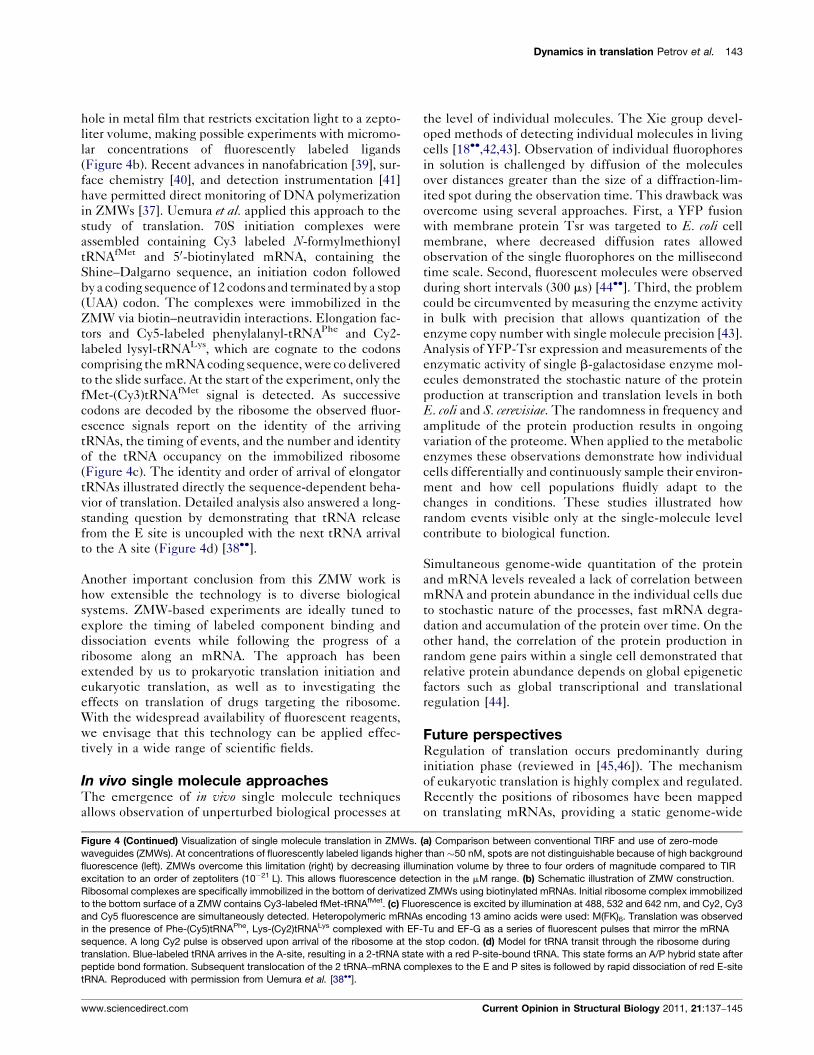
Figure 3 (Continued) Intersubunit FRET. (a) Single-molecule translation assa
to surface-immobilized Cy3-labeled 30S PICs. (b) Immobilization with an mRN
conformation during multiple rounds of elongation via the intersubunit FRET
initiation and is followed by multiple cycles of high-low-high FRET, each repo
(c) Number of FRET cycles observed on mRNAs encoding six alternating FK
nascent chain at codon 7. Mean lifetime estimates for the (e) high-FRET (lo
codons of the 12F mRNA. Error bars denote 95% confidence intervals from
rate beyond initial codons, as shown by the decreasing low FRET (unlocked
www.sciencedirect.com
Thus, the early steps of ribosomal translocation are
particularly slow, and could represent a point of potential
regulation. The mechanistic origins for slow translocation
at the initial codons are not due to disruption of Shine–Dalgarno pairing, but may be related to the length or
chemical identity of the growing nascent chain. Further
investigation is required.
The mechanisms of ribosome-targeting antibiotics have
been explored by these single-molecule fluorescence
methods. Fusidic acid, spectinomycin, and viomycin
effectively inhibited translation of multiple codons, and
all showed distinct and codon-specific effects on the
lifetimes of the FRET states [35��]. The observed effects
agree well with the expected mechanisms of the anti-
biotics and the cumulative evidence that these drugs act
during elongation. Additionally, the authors observed a
significant reduction in the number of ribosomes under-
going more than six FRET cycles in the presence of
erythromycin, providing direct, real-time evidence that
erythromycin blocks the peptide exit tunnel at a position
where a polypeptide of seven amino acids would reach
(fMet plus six additional amino acids) (Figure 3c and d).
The inter-subunit FRET signal will allow mechanistic
exploration of many ribosome-directed antibiotics during
real-time translation.
Full translation in zero-mode waveguidesNew approaches have allowed direct tracking of translation
under physiological conditions. Zero-mode waveguides
(ZMWs) are an emerging platform for the investigation
of processes using single-molecule fluorescence [36,37].
Uemura et al. have employed new ZMW technology to
monitor timing of arrival and departure of tRNAs in real
time on the ribosome [38��]. Traditional total internal
reflection fluorescence (TIRF) microscopy limits fluores-
cently labeled tRNA concentrations to�50 nM because of
background fluorescence from unbound labeled tRNA
(Figure 4a). When tRNA is present at such low concen-
trations, the long delays between sequential tRNA arrival
events can cause molecules to become photobleached,
rendering them undetectable. The signals of interest con-
sequently vanish prematurely during translation at rates
supported by TIRF microscopy. Additionally, artificially
long waiting times allow the ribosome to explore stable
non-productive states.
ZMWs provide an elegant solution to these problems. Each
ZMW consists of a �50–200 nm diameter nanofabricated
y. Cy5-labeled 50S subunits, ternary complexes and EF-G are delivered
A coding for six phenylalanines (6F) permits the observation of ribosome
signal. The arrival of FRET corresponds to 50S subunit joining during
rting on ribosome unlocking and locking during one round of elongation.
pairs. (d) Erythromycin stalls single translating ribosomes by blocking the
cked) and (f) low-FRET (unlocked) states of the ribosome at the first ten
single-exponential fits. There is an apparent increase in the translocation
) state lifetime. Reproduced with permission from Aitken et al. [35��].
Current Opinion in Structural Biology 2011, 21:137–145

142 Protein-nucleic acid interactions
Figure 4
Flu
ores
cenc
e in
tens
ity
00
2000
1500
1000
500
20 100806040
Time (sec)
(a)
(b)
TuGTP
GTC
G
tRNA dissociationfrom E site
Rapid
G
GTP GDP
(c)
Laser inLaser out
~ 100 nm
~ 100 nm
Fluorescent ligand concentratio50nM~5000nM
(d)
Current Opinion in Structural Biology
GTP
GDP GDP
Total internal reflection
Laser in
Aluminium
100 nm
High background fluorescence Single molecule fluorescence
Zero Mode WaveguidesConventional TIRF
Current Opinion in Structural Biology 2011, 21:137–145 www.sciencedirect.com
Dynamics in translation Petrov et al. 143
hole in metal film that restricts excitation light to a zepto-
liter volume, making possible experiments with micromo-
lar concentrations of fluorescently labeled ligands
(Figure 4b). Recent advances in nanofabrication [39], sur-
face chemistry [40], and detection instrumentation [41]
have permitted direct monitoring of DNA polymerization
in ZMWs [37]. Uemura et al. applied this approach to the
study of translation. 70S initiation complexes were
assembled containing Cy3 labeled N-formylmethionyl
tRNAfMet and 50-biotinylated mRNA, containing the
Shine–Dalgarno sequence, an initiation codon followed
by a coding sequence of 12 codons and terminated by a stop
(UAA) codon. The complexes were immobilized in the
ZMW via biotin–neutravidin interactions. Elongation fac-
tors and Cy5-labeled phenylalanyl-tRNAPhe and Cy2-
labeled lysyl-tRNALys, which are cognate to the codons
comprising the mRNA coding sequence, were co delivered
to the slide surface. At the start of the experiment, only the
fMet-(Cy3)tRNAfMet signal is detected. As successive
codons are decoded by the ribosome the observed fluor-
escence signals report on the identity of the arriving
tRNAs, the timing of events, and the number and identity
of the tRNA occupancy on the immobilized ribosome
(Figure 4c). The identity and order of arrival of elongator
tRNAs illustrated directly the sequence-dependent beha-
vior of translation. Detailed analysis also answered a long-
standing question by demonstrating that tRNA release
from the E site is uncoupled with the next tRNA arrival
to the A site (Figure 4d) [38��].
Another important conclusion from this ZMW work is
how extensible the technology is to diverse biological
systems. ZMW-based experiments are ideally tuned to
explore the timing of labeled component binding and
dissociation events while following the progress of a
ribosome along an mRNA. The approach has been
extended by us to prokaryotic translation initiation and
eukaryotic translation, as well as to investigating the
effects on translation of drugs targeting the ribosome.
With the widespread availability of fluorescent reagents,
we envisage that this technology can be applied effec-
tively in a wide range of scientific fields.
In vivo single molecule approachesThe emergence of in vivo single molecule techniques
allows observation of unperturbed biological processes at
Figure 4 (Continued) Visualization of single molecule translation in ZMWs.
waveguides (ZMWs). At concentrations of fluorescently labeled ligands highe
fluorescence (left). ZMWs overcome this limitation (right) by decreasing illum
excitation to an order of zeptoliters (10�21 L). This allows fluorescence dete
Ribosomal complexes are specifically immobilized in the bottom of derivatize
to the bottom surface of a ZMW contains Cy3-labeled fMet-tRNAfMet. (c) Fluo
and Cy5 fluorescence are simultaneously detected. Heteropolymeric mRNAs
in the presence of Phe-(Cy5)tRNAPhe, Lys-(Cy2)tRNALys complexed with EF
sequence. A long Cy2 pulse is observed upon arrival of the ribosome at the
translation. Blue-labeled tRNA arrives in the A-site, resulting in a 2-tRNA state
peptide bond formation. Subsequent translocation of the 2 tRNA–mRNA com
tRNA. Reproduced with permission from Uemura et al. [38��].
www.sciencedirect.com
the level of individual molecules. The Xie group devel-
oped methods of detecting individual molecules in living
cells [18��,42,43]. Observation of individual fluorophores
in solution is challenged by diffusion of the molecules
over distances greater than the size of a diffraction-lim-
ited spot during the observation time. This drawback was
overcome using several approaches. First, a YFP fusion
with membrane protein Tsr was targeted to E. coli cell
membrane, where decreased diffusion rates allowed
observation of the single fluorophores on the millisecond
time scale. Second, fluorescent molecules were observed
during short intervals (300 ms) [44��]. Third, the problem
could be circumvented by measuring the enzyme activity
in bulk with precision that allows quantization of the
enzyme copy number with single molecule precision [43].
Analysis of YFP-Tsr expression and measurements of the
enzymatic activity of single b-galactosidase enzyme mol-
ecules demonstrated the stochastic nature of the protein
production at transcription and translation levels in both
E. coli and S. cerevisiae. The randomness in frequency and
amplitude of the protein production results in ongoing
variation of the proteome. When applied to the metabolic
enzymes these observations demonstrate how individual
cells differentially and continuously sample their environ-
ment and how cell populations fluidly adapt to the
changes in conditions. These studies illustrated how
random events visible only at the single-molecule level
contribute to biological function.
Simultaneous genome-wide quantitation of the protein
and mRNA levels revealed a lack of correlation between
mRNA and protein abundance in the individual cells due
to stochastic nature of the processes, fast mRNA degra-
dation and accumulation of the protein over time. On the
other hand, the correlation of the protein production in
random gene pairs within a single cell demonstrated that
relative protein abundance depends on global epigenetic
factors such as global transcriptional and translational
regulation [44].
Future perspectivesRegulation of translation occurs predominantly during
initiation phase (reviewed in [45,46]). The mechanism
of eukaryotic translation is highly complex and regulated.
Recently the positions of ribosomes have been mapped
on translating mRNAs, providing a static genome-wide
(a) Comparison between conventional TIRF and use of zero-mode
r than �50 nM, spots are not distinguishable because of high background
ination volume by three to four orders of magnitude compared to TIR
ction in the mM range. (b) Schematic illustration of ZMW construction.
d ZMWs using biotinylated mRNAs. Initial ribosome complex immobilized
rescence is excited by illumination at 488, 532 and 642 nm, and Cy2, Cy3
encoding 13 amino acids were used: M(FK)6. Translation was observed
-Tu and EF-G as a series of fluorescent pulses that mirror the mRNA
stop codon. (d) Model for tRNA transit through the ribosome during
with a red P-site-bound tRNA. This state forms an A/P hybrid state after
plexes to the E and P sites is followed by rapid dissociation of red E-site
Current Opinion in Structural Biology 2011, 21:137–145

144 Protein-nucleic acid interactions
snapshot of translation [47��]. Translation initiation is
governed by seven initiation factors and involves mRNA
50-cap recognition, scanning and start codon selection.
Alternative initiation pathways allow distinct translational
responses. To allow application of the single molecule
approaches to investigation of eukaryotic translation,
Petrov and Puglisi successfully labeled small and large
ribosomal subunits of the yeast ribosome and demon-
strated their utility for single molecule fluorescence and
force studies [48�]. The future application of single-
molecule methods will reveal molecular details of the
eukaryotic translation.
References and recommended readingPapers of particular interest, published within the period of review,have been highlighted as:
� of special interest
�� of outstanding interest
1. Schmeing TM, Ramakrishnan V: What recent ribosomestructures have revealed about the mechanism of translation.Nature 2009, 461:1234-1242.
2. Agirrezabala X, Frank J: Elongation in translation as a dynamicinteraction among the ribosome, tRNA, and elongation factorsEF-G and EF-Tu. Q Rev Biophys 2009, 42:159-200.
3. Korostelev A, Ermolenko DN, Noller HF: Structural dynamics ofthe ribosome. Curr Opin Chem Biol 2008, 12:674-683.
4.��
Voorhees RM, Schmeing TM, Kelley AC, Ramakrishnan V: Themechanism for activation of GTP hydrolysis on the ribosome.Science 2010, 330:835-838.
Authors have determined the crystal structure of 70S:EF-Tu:Trp-tRNATrp:GDPCP complex to 3.2 A resolution. This structure, and that ofthe 70S:EF-Tu:ribosomal complex after GTP hydrolysis [5�] suggest auniversal mechanism for ribosomal GTPase activation and GTP hydrolysis.
5.�
Schmeing TM, Voorhees RM, Kelley AC, Gao YG, Murphy FVt,Weir JR, Ramakrishnan V: The crystal structure of the ribosomebound to EF-Tu and aminoacyl-tRNA. Science 2009, 326:688-694.
Describes the structure of the 70S:EF-Tu:Trp-tRNATrp:GDP complexstalled with kirromycin and paromomycin to 3.6 and 3.8 A resolution.
6. Schuette JC, Murphy FVt, Kelley AC, Weir JR, Giesebrecht J,Connell SR, Loerke J, Mielke T, Zhang W, Penczek PA et al.:GTPase activation of elongation factor EF-Tu by the ribosomeduring decoding. EMBO J 2009, 28:755-765.
7. Villa E, Sengupta J, Trabuco LG, LeBarron J, Baxter WT,Shaikh TR, Grassucci RA, Nissen P, Ehrenberg M, Schulten Ket al.: Ribosome-induced changes in elongation factor Tuconformation control GTP hydrolysis. Proc Natl Acad Sci USA2009, 106:1063-1068.
8. Li W, Agirrezabala X, Lei J, Bouakaz L, Brunelle JL, Ortiz-Meoz RF,Green R, Sanyal S, Ehrenberg M, Frank J: Recognition ofaminoacyl-tRNA: a common molecular mechanism revealedby cryo-EM. EMBO J 2008, 27:3322-3331.
9. Schuwirth BS, Borovinskaya MA, Hau CW, Zhang W, Vila-Sanjurjo A, Holton JM, Cate JH: Structures of the bacterialribosome at 3.5 A resolution. Science 2005, 310:827-834.
10.��
Valle M, Zavialov A, Sengupta J, Rawat U, Ehrenberg M, Frank J:Locking and unlocking of ribosomal motions. Cell 2003,114:123-134.
Upon EF-G binding to pre-translocating ribosomes 30S ribosomal sub-unit undergo �68 rotational movement relative to 50S. The rotation isaccompanied by �20 A movement of the L1 stalk. The observed motionprovides insight into global ribosomal dynamics during elongation.
11. Ban N, Nissen P, Hansen J, Moore PB, Steitz TA: The completeatomic structure of the large ribosomal subunit at 2.4 Aresolution. Science 2000, 289:905-920.
Current Opinion in Structural Biology 2011, 21:137–145
12. Agrawal RK, Penczek P, Grassucci RA, Burkhardt N, Nierhaus KH,Frank J: Effect of buffer conditions on the position of tRNA onthe 70 S ribosome as visualized by cryoelectron microscopy.J Biol Chem 1999, 274:8723-8729.
13. Yusupov MM, Yusupova GZ, Baucom A, Lieberman K, Earnest TN,Cate JH, Noller HF: Crystal structure of the ribosome at 5.5 Aresolution. Science 2001, 292:883-896.
14. Whitford PC, Geggier P, Altman RB, Blanchard SC, Onuchic JN,Sanbonmatsu KY: Accommodation of aminoacyl-tRNA into theribosome involves reversible excursions along multiplepathways. RNA 2010, 16:1196-1204.
15. Sanbonmatsu KY: Energy landscape of the ribosomal decodingcenter. Biochimie 2006, 88:1053-1059.
16. Sanbonmatsu KY: Alignment/misalignment hypothesis fortRNA selection by the ribosome. Biochimie 2006, 88:1075-1089.
17. Sanbonmatsu KY, Joseph S, Tung CS: Simulating movement oftRNA into the ribosome during decoding. Proc Natl Acad SciUSA 2005, 102:15854-15859.
18.��
Fischer N, Konevega AL, Wintermeyer W, Rodnina MV, Stark H:Ribosome dynamics and tRNA movement by time-resolvedelectron cryomicroscopy. Nature 2010, 466:329-333.
Dynamic snapshots of the 70S ribosome during retro-translocation wereobtained by time-resolved electron cryo-microscopy, identifying a num-ber of previously unobserved pre-translocation and post-translocationsub-states. 30S-tRNA interactions were shown to be important for thecontrol of global translocation dynamics.
19.��
Mulder AM, Yoshioka C, Beck AH, Bunner AE, Milligan RA,Potter CS, Carragher B, Williamson JR: Visualizing ribosomebiogenesis: parallel assembly pathways for the 30S subunit.Science 2010, 330:673-677.
Authors employed time-resolved electron microscopy to elucidate path-ways of 30S subunit assembly. The results showed that 30S assemblyoccurs via multiple compositional pathways. This study demonstrates theutility of structural imaging for resolving alternative pathways in translation.
20. Lee TH, Blanchard SC, Kim HD, Puglisi JD, Chu S: The role offluctuations in tRNA selection by the ribosome. Proc Natl AcadSci USA 2007, 104:13661-13665.
21. Geggier P, Dave R, Feldman MB, Terry DS, Altman RB, Munro JB,Blanchard SC: Conformational sampling of aminoacyl-tRNAduring selection on the bacterial ribosome. J Mol Biol 2010,399:576-595.
22. Munro JB, Altman RB, O’Connor N, Blanchard SC: Identificationof two distinct hybrid state intermediates on the ribosome. MolCell 2007, 25:505-517.
23.��
Cornish PV, Ermolenko DN, Noller HF, Ha T: Spontaneousintersubunit rotation in single ribosomes. Mol Cell 2008,30:578-588.
The intersubunit rotation was observed by utilizing single moleculespectroscopy. After peptidyl transfer ribosomes undergo spontaneousoscillations between two states. The observed fluctuations correspond tothe hybrid and classical states. EF-G binding stabilizes rotational con-formation of the ribosome.
24. Wang Y, Qin H, Kudaravalli RD, Kirillov SV, Dempsey GT, Pan D,Cooperman BS, Goldman YE: Single-molecule structuraldynamics of EF-G–ribosome interaction during translocation.Biochemistry 2007, 46:10767-10775.
25.��
Fei J, Bronson JE, Hofman JM, Srinivas RL, Wiggins CH,Gonzalez RL Jr: Allosteric collaboration between elongationfactor G and the ribosomal L1 stalk directs tRNA movementsduring translation. Proc Natl Acad Sci USA 2009, 106:15702-15707.
Authors observed dynamics of the L1 stalk fluctuation during transloca-tion. Together with [10��,29�,27] this paper highlights the role of ribosomaldynamics during large-scale ribosomal movements.
26. Feldman MB, Terry DS, Altman RB, Blanchard SC:Aminoglycoside activity observed on single pre-translocationribosome complexes. Nat Chem Biol 2009, 6:54-62.
27. Fei J, Kosuri P, MacDougall DD, Gonzalez RL Jr: Coupling ofribosomal L1 stalk and tRNA dynamics during translationelongation. Mol Cell 2008, 30:348-359.
www.sciencedirect.com

Dynamics in translation Petrov et al. 145
28. Munro JB, Altman RB, Tung CS, Sanbonmatsu KY, Blanchard SC:A fast dynamic mode of the EF-G-bound ribosome. EMBO J2009, 29:770-781.
29.�
Cornish PV, Ermolenko DN, Staple DW, Hoang L, Hickerson RP,Noller HF, Ha T: Following movement of the L1 stalk betweenthree functional states in single ribosomes. Proc Natl Acad SciUSA 2009, 106:2571-2576.
By employing smFRET authors demonstrated that L1 stalk could adoptthree distinct conformational states. Findings suggest that L1 stalkmovement modulates tRNA movement and dissociation.
30. Sternberg SH, Fei J, Prywes N, McGrath KA, Gonzalez RL Jr:Translation factors direct intrinsic ribosome dynamics duringtranslation termination and ribosome recycling. Nat Struct MolBiol 2009, 16:861-868.
31. Dorywalska M, Blanchard SC, Gonzalez RL, Kim HD, Chu S,Puglisi JD: Site-specific labeling of the ribosome for single-molecule spectroscopy. Nucleic Acids Res 2005, 33:182-189.
32. Marshall RA, Aitken CE, Puglisi JD: GTP hydrolysis by IF2 guidesprogression of the ribosome into elongation. Mol Cell 2009,35:37-47.
33.�
Marshall RA, Dorywalska M, Puglisi JD: Irreversible chemicalsteps control intersubunit dynamics during translation. ProcNatl Acad Sci USA 2008, 105:15364-15369.
The authors observed global locking and unlocking of the ribosome viainter-subunit FRET.
34. Kim HD, Puglisi JD, Chu S: Fluctuations of transfer RNAsbetween classical and hybrid states. Biophys J 2007,93:3575-3582.
35.��
Aitken CE, Puglisi JD: Following the intersubunit conformationof the ribosome during translation in real time. Nat Struct MolBiol 2010, 17:793-800.
The authors employed intersubunit ratcheting signal to monitor globalribosomal dynamics throughout initiation followed by multiple elongationcycles. Multiple continuous cycles of elongation were observed at thesingle molecule level in real-time. The signal was employed to distinguishthe mechanisms of ribosome-targeting antibiotics.
36. Levene MJ, Korlach J, Turner SW, Foquet M, Craighead HG,Webb WW: Zero-mode waveguides for single-moleculeanalysis at high concentrations. Science 2003, 299:682-686.
37. Eid J, Fehr A, Gray J, Luong K, Lyle J, Otto G, Peluso P, Rank D,Baybayan P, Bettman B et al.: Real-time DNA sequencing fromsingle polymerase molecules. Science 2009, 323:133-138.
38.��
Uemura S, Aitken CE, Korlach J, Flusberg BA, Turner SW,Puglisi JD: Real-time tRNA transit on single translatingribosomes at codon resolution. Nature 2010, 464:1012-1017.
The authors utilized a ZMW approach to observe multiple rounds of tRNAtransit on single translating ribosomes in real-time. The number of tRNAmolecules simultaneously bound to each ribosome was directly deter-mined by observing fluorescently labeled tRNAs. The codon identity and
www.sciencedirect.com
number of translated codons could be directly inferred from the tRNAsignals. The authors demonstrated that E-site tRNA release is uncoupledfrom A-site tRNA binding.
39. Foquet M, Samiee KT, Kong X, Chauduri BP, Lundquist PM,Turner SW, Freudenthal J, Roitman DB: Improved fabrication ofzero-mode waveguides for single-molecule detection. J ApplPhys 2008, 103:034301.
40. Korlach J, Marks PJ, Cicero RL, Gray JJ, Murphy DL, Roitman DB,Pham TT, Otto GA, Foquet M, Turner SW: Selective aluminumpassivation for targeted immobilization of single DNApolymerase molecules in zero-mode waveguidenanostructures. Proc Natl Acad Sci USA 2008, 105:1176-1181.
41. Lundquist PM, Zhong CF, Zhao P, Tomaney AB, Peluso PS,Dixon J, Bettman B, Lacroix Y, Kwo DP, McCullough E et al.:Parallel confocal detection of single molecules in real time.Opt Lett 2008, 33:1026-1028.
42. Yu J, Xiao J, Ren X, Lao K, Xie XS: Probing gene expression inlive cells, one protein molecule at a time. Science 2006,311:1600-1603.
43. Cai L, Friedman N, Xie XS: Stochastic protein expression inindividual cells at the single molecule level. Nature 2006,440:358-362.
44.��
Taniguchi Y, Choi PJ, Li GW, Chen H, Babu M, Hearn J, Emili A,Xie XS: Quantifying E. coli proteome and transcriptome withsingle-molecule sensitivity in single cells. Science 2010,329:533-538.
The authors quantitatively measured proteome-wide transcription andtranslation in vivo with single molecule precision. The authors demon-strated how stochastic biological processes contribute to the rapid cellresponse to the external stimuli.
45. Livingstone M, Atas E, Meller A, Sonenberg N: Mechanismsgoverning the control of mRNA translation. Phys Biol 2010,7:021001.
46. Jackson RJ, Hellen CU, Pestova TV: The mechanism ofeukaryotic translation initiation and principles of itsregulation. Nat Rev Mol Cell Biol 2010, 11:113-127.
47.��
Ingolia NT, Ghaemmaghami S, Newman JR, Weissman JS:Genome-wide analysis in vivo of translation withnucleotide resolution using ribosome profiling. Science 2009,324:218-223.
Ribosome position on mRNA was determined with nucleotide resolution.The genome-wide approach provides a snapshot of the ribosome posi-tion on actively translated mRNAs.
48.�
Petrov A, Puglisi JD: Site-specific labeling of Saccharomycescerevisiae ribosomes for single-molecule manipulations.Nucleic Acids Res 2010, 38:e143.
Large and small subunits of eukaryotic ribosome were specificallylabeled. The authors demonstrated utility of the labeled ribosomes forsingle molecule spectrometry and force experiments.
Current Opinion in Structural Biology 2011, 21:137–145