Embed Size (px)
Citation preview

Dynamics of neural recruitment surrounding the spontaneous arising ofthoughts in experienced mindfulness practitioners
Melissa Ellamil a, Kieran C.R. Fox a, Matthew L. Dixon a, Sean Pritchard b, Rebecca M. Todd a,c,Evan Thompson d, Kalina Christoff a,c,⁎a Department of Psychology, University of British Columbia, 2136 West Mall, Vancouver, British Columbia V6T 1Z4, Canadab School of Psychology, Fielding Graduate University, 2020 De la Vina Street, Santa Barbara, CA 93105, United Statesc Centre for Brain Health, University of British Columbia, 2215 Wesbrook Mall, Vancouver, British Columbia V6T 1Z3, Canadad Department of Philosophy, University of British Columbia, 1866 Main Mall, Vancouver, British Columbia V6T 1Z1, Canada
a b s t r a c ta r t i c l e i n f o
Article history:Received 22 December 2015Revised 12 April 2016Accepted 14 April 2016Available online 23 April 2016
Thoughts arise spontaneously in ourmindswith remarkable frequency, but tracking the brain systems associatedwith the early inception of a thought has proved challenging. Herewe addressed this issue by taking advantage ofthe heightened introspective ability of experienced mindfulness practitioners to observe the onset of their spon-taneously arising thoughts.We found subtle differences in timing among themany regions typically recruited byspontaneous thought. In some of these regions, fMRI signal peaked prior to the spontaneous arising of a thought—most notably in themedial temporal lobe and inferior parietal lobule. In contrast, activation in themedial pre-frontal, temporopolar, mid-insular, lateral prefrontal, and dorsal anterior cingulate cortices peaked together withor immediately following the arising of spontaneous thought.We propose that brain regions that show anteced-ent recruitmentmay be preferentially involved in the initial inception of spontaneous thoughts, while those thatshow later recruitmentmay be preferentially involved in the subsequent elaboration andmetacognitive process-ing of spontaneous thoughts. Our findings highlight the temporal dynamics of neural recruitment surroundingthe emergence of spontaneous thoughts andmay help account for some of spontaneous thought's peculiar qual-ities, including its wild diversity of content and its links to memory and attention.
© 2016 The Authors. Published by Elsevier Inc. This is an open access article under the CC BY license(http://creativecommons.org/licenses/by/4.0/).
Keywords:Spontaneous thoughtDefault mode networkMedial temporal lobeNeural antecedentsfMRI
1. Introduction
Where do thoughts come from? One of themost intriguing yet leastunderstood aspects of the humanmind is its tendency to spontaneouslygive rise to words, images, and emotions that flow effortlessly from onetopic to another. There is a stunning ubiquity of such spontaneouslyarising mental content in people's lives: thoughts that occur withoutour deliberate control take up asmuch as one-third of ourmental expe-rience (Klinger and Cox 1987).
The last decade marked an upsurge of investigations into a numberof closely related mental phenomena, often investigated using termssuch as ‘mind-wandering’, ‘stimulus-independent thought’, or ‘task-un-related thought’ (Christoff, 2012). Psychological research has focused onthe conditions that facilitate these kinds of thought processes, showingthat their frequency increases when task demands are low or when ex-ternal sensory stimulation is weak (Smallwood and Schooler, 2006). Inparallel, neuroscientific investigations have focused on identifying the
brain regions that are consistently recruited during such conditions.The majority of investigations have placed particular emphasis on re-cruitment of the default mode network (for review, see Christoff,2012), although a number of executive control network regions are re-cruited with equal consistency (Christoff et al., 2009a; Fox et al., 2015).While these investigations have greatly improved our understanding ofwhich brain regions are engagedwhen spontaneous thought is ongoing,we still have virtually no knowledge of how these various brain regionscontribute to the different components of a spontaneous thought expe-rience and, in particular, which brain regions support the early incep-tion of spontaneous thoughts (Smallwood, 2013).
The goal of the present study, therefore, was to investigate key brainregions associated with the arising of spontaneous thoughts. To do this,we set out to examine brain recruitment that occurs immediately priorto the onset of a spontaneous thought. The central difficulty in investi-gating this question lies in identifying the precise temporal onset ofspontaneously arising mental content. There are currently no third-person behavioral measures that provide such information reliably,making it necessary for scientists to use first-person introspective re-ports fromparticipants. Althoughmost individuals can provide fairly re-liable descriptions of the basic contents of their thoughts (Hurlburt and
NeuroImage 136 (2016) 186–196
⁎ Corresponding author at: Department of Psychology, University of British Columbia,2136 West Mall, Vancouver, British Columbia V6T 1Z4, Canada.
E-mail address: [email protected] (K. Christoff).
http://dx.doi.org/10.1016/j.neuroimage.2016.04.0341053-8119/© 2016 The Authors. Published by Elsevier Inc. This is an open access article under the CC BY license (http://creativecommons.org/licenses/by/4.0/).
Contents lists available at ScienceDirect
NeuroImage
j ourna l homepage: www.e lsev ie r .com/ locate /yn img

Schwitzgebel, 2007), their ability to report on themental processes thatgive rise to thought content is generally considered to be poor (NisbettandWilson, 1977). This apparent lack of introspective ability, combinedwith the need for reliance on introspective measures, has created amethodological knot that has tied the hands of researchers interestedin differentiating the neural systems associatedwith the early formationof spontaneous thoughts.
The ability to detect the arising of spontaneous thoughts, however,can vary across individuals (Seli et al., 2015) as well as within thesame individual (Zedelius et al., 2015). This raises the possibilitythat the methodological difficulty described earlier can be tackled byemploying participants with heightened introspective skills, a methodknown as neurophenomenology (Fazelpour and Thompson, 2015;Varela, 1996). In this approach, participants with introspective traininggive first-person reports about their mental processes that are then re-lated tomeasures of neural activity (Lutz et al., 2002). One form ofmen-tal training that specifically focuses on observing the spontaneousarising of one's own thoughts is mindfulness practice (Lutz et al.,2008; Tang et al., 2015). Mindfulness training has been shown to en-hance attention in numerous ways, including a greater ability to noticesubtle or rapid events, such as increased line length discrimination at vi-sual threshold during a vigilance task (MacLean et al., 2010; Sahdraet al., 2011; Zanesco et al., 2013) and improved second target detectionduring an attentional blink task (Slagter et al., 2007). Specifically, intro-spective ability appears to be enhanced through mindfulness training,as shown by increased metacognitive memory confidence judgments(Baird et al., 2014) and greater accuracy in emotional self-awareness rel-ative to no training (Sze et al., 2010). Thus,we employed a group of highlyexperiencedmindfulness practitioners,who tracked the arising of sponta-neous thoughts during fMRI scanning, to capitalize on this potentiallyheightened introspective ability andmore specifically examine the neuralantecedents of spontaneous mental content as well as the full suite ofneural recruitment before, during, and after spontaneous thought reports.
We hypothesized that the medial temporal lobe would show prefer-ential recruitment during the initial generation of spontaneous thoughtas it is often recruited during rest (Buckner et al., 2008; Christoff et al.,2004; Stark and Squire, 2001) and has been associated with memory re-trieval (Squire et al., 2004), future thinking (Addis et al., 2009), andmen-tal simulation (Hassabis et al., 2007), which form a large portion ofspontaneous thought content (Klinger, 2009; Klinger and Cox, 1987).We also hypothesized that the medial and lateral prefrontal cortices,also consistently recruited during rest (Christoff et al., 2009a; Fox et al.,2015), would show increased recruitment after the initial generationof spontaneous thought as they have been associated more with theevaluation and monitoring of self-generated thought content (Buckneret al., 2008; Dixon and Christoff, 2012; Dixon and Christoff, 2014; Dixonet al., 2014; Fox and Christoff, 2014; Miller and Cohen, 2001).
2. Materials and methods
2.1. Participants
Eighteen participants (8 male and 10 female; M = 49.91 years old,SD=11.17, range=29.39–68.42) took part in the experiment. The par-ticipants were long-term, expert mindfulness meditators with morethan 3000 h of lifetime meditation experience and at least 1 h of dailypractice in the Mahasi Vipassana tradition (M = 8338.60 h, SD =5989.98, range = 3174–23,700). The number of hours reported didnot include practice in other traditions (e.g., Zen meditation, Transcen-dental meditation, Goenka or body scanning meditation, Yoga, Tai Chi,Qi Gong). Eligibility screening consisted of the administration of a med-itation experience questionnaire and a phone interview by S.P., a formerMahasi monk. The screening ensured consistency of the meditationtechnique across participants and attainment of extensive knowledgeof and experience with the meditation technique through regular prac-tice and attendance of several long-term intensive retreats.
The participants were recruited from Vipassanameditation commu-nities in Vancouver (BC, Canada), Vancouver Island (BC, Canada),Boulder (CO, USA), San Francisco (CA, USA), and Seattle (WA, USA).All participants had normal or corrected-to-normal vision with no MRIcontraindications and no current psychiatric medication use. Fourteenwere right-handed and 4 were left-handed, but all used their righthand to respond during the experiment. From an original sample of22 participants, three participants were excluded from the analysesdue to excessive motion (2 with N5° pitch rotation, and 1 with N5°pitch rotation and N5 mm in the z-direction). One participant was ex-cluded from the analyses due to technical problems with behavioraldata recording. All protocols were approved by the University of BritishColumbia (UBC; Vancouver, BC, Canada) Clinical Research Ethics Boardand the UBCMRI Research Center. All participants gave informedwrittenconsent prior to participating and received payment as compensation.
2.2. Procedure
One to two days prior to the actual scanning session, participants en-gaged in a practice session identical to the actual scanning procedure ina mock scanner environment in order to become acclimatized to thetask, button pressing, and scanner noises. Participants alternated be-tween 30 s blocks of monitoring thoughts that arose spontaneously(thought condition, Fig. 1A) and 30 s blocks of monitoring words thatappeared onscreen (word condition, Fig. 1B). During both blocks, partic-ipants attended to the rising and falling of the abdomen (i.e., breathing).Participants reported with a first button press (index finger) to indicatewhen a thought arose or when aword appeared onscreen (see Stimuli),andwith a secondbutton press to indicatewhat type of thought orwordit was (index finger = image or symbol, middle finger = narrative orinner speech, ring finger = emotion, pinky finger = body sensation).Each word stayed onscreen until the first button press. The first buttonpress was followed by one asterisk (*) onscreen for 250ms and the sec-ond button press was followed by two asterisks (**) onscreen for250 ms to signal successful button presses. Each participant completedthree 9-min task runs. Each run consisted of 8 thought blocks and 8word blocks; each participant therefore completed 24 thought blocksand 24 word blocks in total. Blocks were separated by interstimulus in-tervals (ISIs) with jittered durations (randomly chosen from 250 ms,500 ms, or 750 ms), with each ISI appearing an equal number of times.
The instructionswere designed to be consistentwith a typical mind-fulness practice session (Sayadaw, 1985, 2002): the first and secondbutton presses roughly corresponded to the standard steps of (i) notingthe occurrence of thought and then (ii) labeling its content. In thethought condition, participants were instructed to report on the occur-rence of mental events that took attention away from one's breathing,or became more prominent in attention than breathing. This enabledthe assessment ofmental content arisingwithout being explicitly elicitedby external cues in the environment. In the word condition, participantswere instructed to briefly think about the definition of each word pre-sented on the screen before responding. This enabled the assessment ofmental content elicited by external cues in the environment.
The onsets and categories of words presented within each wordblockwere selected in real-time by a computer algorithm that matchedthe timing onsets and categories of words to the timing onsets and cat-egories of spontaneous thoughts reported in the immediately precedingthought block. Thus, each pair of thought andword blocks had the samenumber of events, and these events had the same content categoriesand appeared at the same time within the block.
2.3. Stimuli
Thewords presented duringwordmonitoring blockswere randomlychosen from four lists that corresponded to the categories of thoughtsroutinely identified during mindfulness meditation (Sayadaw, 1985,2002) and covered a large portion of spontaneous thought content
187M. Ellamil et al. / NeuroImage 136 (2016) 186–196

(Fox et al., 2013): (i) image or symbol, (ii) narrative or inner speech, (iii)emotion, or (iv) body sensation. Words in the image list consisted of 30nouns (e.g., mountain, beach, rain, sun, pet) selected from the MedicalResearch Council (MRC) Psycholinguistics Database (Wilson, 1988),which had imageability, concreteness, and familiarity ratings of500–700 (on scales of 100 = very low to 700 = very high) to ensureease of visualization. Words in the narrative list consisted of 30 nouns(e.g., work, money, family, goals, health) selected from the EdinburghAssociative Thesaurus (EAT) (Kiss et al., 1973), which were associatedwith the types of current concerns that people tend to have and thatare thought to be major determinants of spontaneous thought content(Klinger, 2009; Klinger and Cox, 1987). These included home andhousehold matters; employment and finance; partner, family, and rela-tives; friends and acquaintances; love, intimacy, and sexual matters;self-changes; education and training; health and medical matters; spir-itual matters; and hobbies, pastimes, and recreation (Klinger and Cox,2004). Words in the emotion list consisted of 30 adjectives (e.g., calm,happy, sad, afraid,worried) also selected from the EAT that were associ-ated with various emotions (e.g., happiness, sadness, anger, disgust,fear, surprise). Words in the body sensation list consisted of 30 nounsand adjectives similarly selected from the EAT that were associatedwith various body sensations (e.g., warmth, tickle, vibration, pressure,pain). Each word contained 3–10 letters and 1–3 syllables. The wordsand fixation cross appeared as gray text on a black background. Thetask and stimuli were implemented and presented using E-Prime 2.0(Psychology Software Tools, Sharpsburg, PA, USA).
2.4. Data acquisition
Functional and structural MRI data were collected using a 3.0 TeslaPhilips Intera MRI scanner (Best, Netherlands) with a standard headcoil. Head movement was restricted using foam padding around thehead. T2*-weighted functional images were acquired parallel to the
anterior commissure/posterior commissure (AC/PC) line using asingle-shot gradient echo-planar sequence (EPI; repetition time[TR] = 2 s, echo time [TE] = 30 ms, flip angle [FA] = 90°, field of view[FOV] = 240 ! 240 ! 143 mm, matrix size =80 ! 80, SENSE factor =1.0). A total of 265 functional volumes were acquired for each taskrun, each including 36 interleaved axial slices (3 mm thick with 1 mmskip) covering the entire brain. Before functional imaging, an inversionrecovery prepared T1-weighted structural volume was acquired in thesame slice locations and orientation as the functional images using afast spin-echo sequence (TR = 2 s, TE = 10 ms, FA = 90°, FOV =224! 224! 143mm, acquisitionmatrix size= 240! 235, reconstruct-ed matrix size = 480 ! 480, inversion delay [IR] = 800 ms, spin-echoturbo factor = 5).
2.5. Data preprocessing
fMRI data for each participant were preprocessed and analyzedusing SPM8 (Statistical Parametric Mapping, Wellcome Department ofImaging Neuroscience, London, UK). Slice timing correction was per-formed using sinc interpolation and resampling with the middle(18th) slice as a reference point. All functional volumes were realignedto the first volume to correct for between-scan motion. The structuralvolume was coregistered to the mean functional image and segmentedto extract a gray matter image. The segmented structural volumewas then spatially normalized to a gray matter image of the MontrealNeurological Institute (MNI) template and resliced to a voxel size of2 ! 2 ! 2 mm. The derived spatial transformations were applied tothe realigned functional volumes to bring them into standardized MNIspace. Finally, the functional volumes were smoothed with an 8-mmfull-width at half-maximum (FWHM) isotropic Gaussian kernel tocompensate for residual between-subject variability after spatial nor-malization and to permit application of Gaussian random field theoryfor corrected statistical inference (Friston et al., 1994). To ensure that
Fig. 1. Experimental procedure. Participants alternated between (A) spontaneous thought detection blocks (30 s) and (B) matched word detection blocks (30 s). Participants indicatedwith a first button press as soon as they noticed a thought spontaneously arising, which served as the experimental measure of the thought's onset, and with a second button presswhat category of thought it was, which allowed online matching of the subsequent word detection block. Each word block was constructed in real-time to correspond to the timingand categories of spontaneous thoughts reported during the immediately preceding thought block, allowing a closely matched comparison between neural recruitment associatedwith spontaneously arising and perceptually triggered mental content.
188 M. Ellamil et al. / NeuroImage 136 (2016) 186–196
statistical analysis was performed for all brain regions, including thosewhere the signal might have been low due to susceptibility artifacts, amask was created by averaging and thresholding at N.80 the firstpreprocessed functional volume from all participants andwas explicitlyspecified during model estimation at the individual level. To removelow-frequency drift in the blood oxygen-level dependent (BOLD) signal,the data were high-pass filtered using an upper cut-off period of 128 s.No global scaling was performed.
2.6. General linear model
Condition effects at each voxel were estimated according to the gen-eral linear model for the whole-brain analyses. The model included (a)the observed time series of intensity values, which represented the de-pendent variable; (b) covariates modeling session-specific effects(i.e., the six head movement parameters), later treated as confounds;and (c) regressor functions constructed by convolving condition-specific stick functions with a synthetic hemodynamic response func-tion. The regressor functions were constructed to model each of thethought (timage, tnarrative, temotion, and tsensation), word (wimage, wnarrative,wemotion, and wsensation), and button press events and were comparedusing pairwise contrasts for each participant. Group random-effectsanalyses were then performed for each contrast. The resulting T mapswere subsequently transformed to the unit normal Z distribution to cre-ate a statistical parametric map for each contrast. The threshold for sig-nificance was set at Z N 2.576 and p b .05 family-wise error (FWE)cluster corrected for multiple comparisons.
Onsets for the thought andword events were specified at 4 s prior tothe corresponding first button press. A period of 6 s (3 TRs) yieldedsimilar results as a period of 4 s but excluded more trials (i.e., thosethat did not occur 6 s or more after a previous event).
2.7. Time course extraction
Percent signal change time courses for brain regions showing greateractivation during thought events compared toword eventswere extract-ed for each participant using the MarsBaR toolbox in SPM8 (MARSeilleBoîte À Région d'Intérêt) (Brett et al., 2002). The extraction volumeswere 4-mm radius spheres centered on local maxima from the group-level contrasts. Eight finite impulse response (FIR) functions wereused, one for each peristimulus time point within a trial window of16 s following the onset of a thought or word event (specified at 4 sbefore the first button press). This allowed the estimation of a regionof interest's response over time without making assumptions about itsshape and thus the assessment of activation differences across trialphases, compared to modeling the long duration of the hemodynamicresponse function (Dale, 1999; Henson and Friston, 2007; Ollingeret al., 2001).
2.8. Spatiotemporal partial least squares
Changes in neural recruitment over timeduring thought events com-pared to word events were assessed using spatiotemporal partial leastsquares. This whole-brain, multivariate technique identifies the tempo-ral evolution of spatial patterns of activity correlated with differentconditions across the length of an event and is not dependent on as-sumptions about the shape and duration of the hemodynamic responsefunction (Lin et al., 2003; Lobaugh et al., 2001; McIntosh et al., 2004).The non-rotated task version of spatiotemporal Partial Least Squares(PLS) was used, which allows the specification of a priori non-orthogonal contrasts (instead of the rotated data-driven version thatidentifies orthogonal latent variables). One contrast was entered to ex-amine differences in the pattern of neural recruitment across regionsand over time between thought andword events (thought Nword) with-in a trial windowof 16 s (8 TRs) following the onset of a thought orwordtrial (specified at 4 s before the first button press). The statistical
significance of the results at p b .05 was computed using permutationtesting with 500 permutations. Correction for multiple comparisonswas not necessary as the whole spatiotemporal pattern was tested inone analytical step instead of in a series of voxel-wise statistical tests.The reliability of the results was computed using bootstrap estimationof standard errors with 300 iterations. Reliable voxels were signifiedby bootstrap ratios greater than 2.576, which is approximately equalto a Z-score with p b .01.
3. Results
3.1. Frequency distribution and reaction times of reports
Participants reported an average of 67.33 thoughts (SD = 35.02)throughout all scanning sessions and an average of 2.81 thoughts(SD = 1.21) per thought block. However, only an average of 44.67thoughts (SD = 15.03), or 73.80% of the thoughts reported, occurred4 s or more after the previous thought report and were thus includedin the analyses. Of the thoughts included, 18.66% (M = 8.33, SD =10.28) were images, 37.31% (M = 16.67, SD = 5.55) were narrative,12.94% (M = 5.78, SD = 5.86) were emotions, and 31.09% (M =13.89, SD=8.80)were body sensations. A two-way repeatedmeasuresANOVA at the ! = .05 level indicated that the number of thought re-ports differed significantly across the four thought types [F(3,51) =7.01, p b .001], but not across the three scanning sessions [F(2,34) =0.23, p N .05] or their interaction [F(6102) = 0.75, p N .05]. Follow-upmultiple paired t-tests with a Bonferroni correction (! = .05/6 =.0083) revealed that there were significantly more narrative reports[t(17) = 6.82, p b .001] and body sensation reports [t(17) = 4.39,p b .001] than emotion reports. The participants' age in years andhours of meditation experience were not significantly correlated,at ! = .05, with the overall number of thoughts reported or with thedifferent types of thoughts reported. In addition, participants respondedwith the first button press an average of 1145.28 ms (SD= 403.71 ms)after the presentation of a word. They categorized a word with the sec-ond button press an average of 971.96 ms (SD = 352.21 ms) after thefirst button press. They categorized a thought with the second buttonpress an average of 707.75 ms (SD = 358.16 ms) after the first buttonpress signaling the arising of a thought.
3.2. Overall neural recruitment during spontaneous thought reports
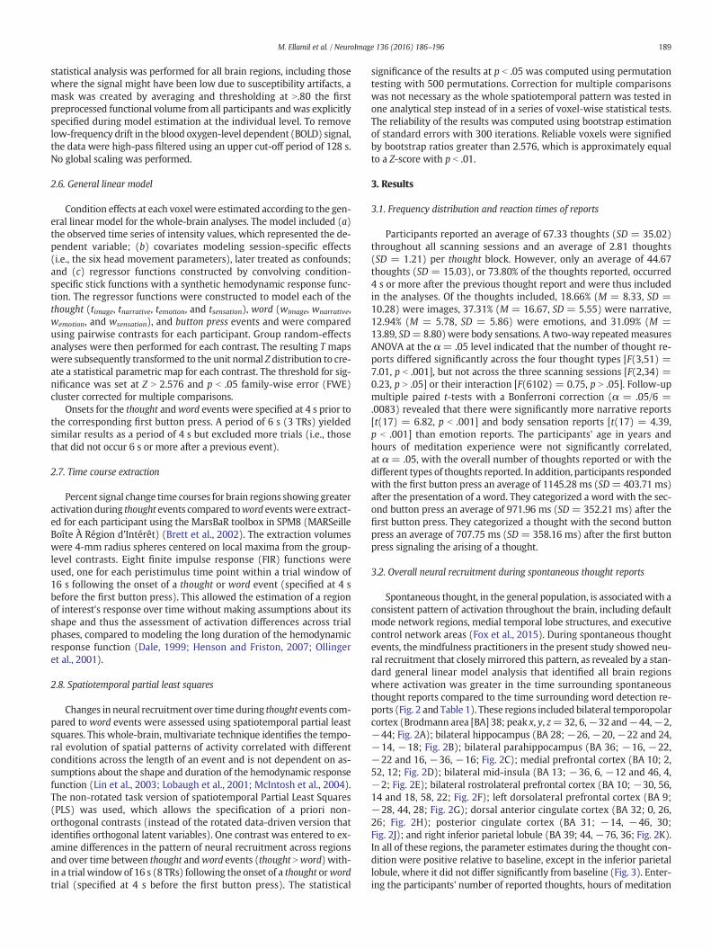
Spontaneous thought, in the general population, is associated with aconsistent pattern of activation throughout the brain, including defaultmode network regions, medial temporal lobe structures, and executivecontrol network areas (Fox et al., 2015). During spontaneous thoughtevents, the mindfulness practitioners in the present study showed neu-ral recruitment that closelymirrored this pattern, as revealed by a stan-dard general linear model analysis that identified all brain regionswhere activation was greater in the time surrounding spontaneousthought reports compared to the time surrounding word detection re-ports (Fig. 2 and Table 1). These regions included bilateral temporopolarcortex (Brodmann area [BA] 38; peak x, y, z=32, 6,!32 and!44,!2,!44; Fig. 2A); bilateral hippocampus (BA 28;!26,!20,!22 and 24,!14, !18; Fig. 2B); bilateral parahippocampus (BA 36; !16, !22,!22 and 16, !36, !16; Fig. 2C); medial prefrontal cortex (BA 10; 2,52, 12; Fig. 2D); bilateral mid-insula (BA 13; !36, 6, !12 and 46, 4,!2; Fig. 2E); bilateral rostrolateral prefrontal cortex (BA 10; !30, 56,14 and 18, 58, 22; Fig. 2F); left dorsolateral prefrontal cortex (BA 9;!28, 44, 28; Fig. 2G); dorsal anterior cingulate cortex (BA 32; 0, 26,26; Fig. 2H); posterior cingulate cortex (BA 31; !14, !46, 30;Fig. 2J); and right inferior parietal lobule (BA 39; 44, !76, 36; Fig. 2K).In all of these regions, the parameter estimates during the thought con-dition were positive relative to baseline, except in the inferior parietallobule, where it did not differ significantly from baseline (Fig. 3). Enter-ing the participants' number of reported thoughts, hours of meditation
189M. Ellamil et al. / NeuroImage 136 (2016) 186–196

experience, and age in years as covariates in the analysis did not alterthe significance of the results and yielded similar activation maps.
3.3. Neural recruitment prior to spontaneous thought reports
To specifically identify the neural antecedents of spontaneousthought, we computed the average time courses of activation in eachof the brain regions identified in the standard general linearmodel anal-ysis (Fig. 2 and Table 1). The spontaneous arising of thoughtwas associ-ated with activation in default mode network, medial temporal lobe,and paralimbic brain regions that preceded the onset of thought by sev-eral seconds. The specific brain regionswhere activation peaked prior toreports of spontaneous thought (Fig. 4) comprised themedial temporallobe, including bilateral hippocampus (BA 28;!26,!20,!22 and 24,!14, !18; Fig. 4A,B) and bilateral parahippocampus (BA 36; !16,!22,!22 and 16,!36,!16; Fig. 4C,D), the posterior cingulate cortex
(BA 31;!14,!46, 30; Fig. 4E), the right inferior parietal lobule (BA 39;44,!76, 36; Fig. 4F), the rostral anterior cingulate cortex (BA 24; 0, 36,14; Fig. 4G), and the right posterior insula (BA 13; 36,!20, 16; Fig. 4H).
3.4. Neural recruitment during and following spontaneous thought reports
Time courses of activation in the brain regions identified in the stan-dard general linear model analysis (Fig. 2 and Table 1) also revealed re-cruitment of additional areas simultaneous with and subsequent tospontaneous thought detection. The brain regions where activationpeaked during reports of spontaneous thought (Fig. 5) comprised themedial prefrontal cortex (BA 10; 2, 52, 12; Fig. 5A), right rostrolateralprefrontal cortex (BA 10; 18, 58, 22; Fig. 5B), bilateral temporopolar cor-tex (BA 38;!44,!2,!44 and 32, 6,!32; Fig. 5C,D), and bilateralmid-insula (BA 13; !36, 6, !12 and 46, 4, !2; Fig. 5E,F). In addition, thebrain regions where activation peaked after reports of spontaneousthought (Fig. 6) included the left dorsolateral prefrontal cortex (BA 9;!28, 44, 28; Fig. 6A), left rostrolateral prefrontal cortex (BA 10; !30,56, 14; Fig. 6B), and dorsal anterior cingulate cortex (BA 32; 0, 26, 26;Fig. 6C).
3.5. Neural recruitment over time during spontaneous thought reports
The progression of neural recruitment during the time surroundingspontaneous thought reports, compared to the time surrounding worddetection reports, was examined using non-rotated task spatiotemporalpartial least squares, with the contrast being significant at p b .001.The positive saliences, or regions in which increases and decreases
Fig. 2. Overall neural recruitment during spontaneous thought reports. Relative to matched word trials, spontaneous thought trials were associated with greater activation in(A) temporopolar cortex, (B) hippocampus, (C) parahippocampus, (D) medial prefrontal cortex, (E) mid-insula, (F) rostrolateral prefrontal cortex, (G) left dorsolateral prefrontalcortex, (H) dorsal anterior cingulate cortex, (J) posterior cingulate cortex, and (K) right inferior parietal lobule. Onsets for the spontaneous thought and matched word trials werespecified at 4 s before the corresponding button press. All activations were significant at p b .05, family-wise error cluster-corrected for multiple comparisons. Results are displayed inneurological orientation, with the right hemisphere depicted on the right, on the average high-resolution structural image.
Table 1Activation peaks for thought events (thought N word).
Region MNI coordinates
L/R/M BA x y z Voxels Z value
FrontalRostral ACC M 24 0 36 14 659 5.02Dorsal ACC M 32 0 26 26 178 4.25Superior frontal gyrus (MPFC) M 10 2 52 12 33 3.71Middle frontal gyrus (RLPFC) L 10 !30 56 14 81 3.90Middle frontal gyrus (RLPFC) R 10 18 58 22 357 3.46Middle frontal gyrus (DLPFC) L 9 !28 44 28 251 4.51Mid-insula L 13 !36 6 !12 55 3.28Mid-insula R 13 46 4 !2 505 4.86Posterior insula R 13 36 !20 16 32 3.50Supplementary motor area M 6 0 !26 66 165 3.14
ParietalPosterior cingulate cortex M 31 !14 !46 30 55 3.15Inferior parietal lobule R 39 44 !76 36 30 4.08Superior parietal lobule L 5 !22 !38 58 31 2.86Superior parietal lobule R 5 20 !42 56 117 2.88Postcentral gyrus L 43 !50 !4 4 81 3.81Postcentral gyrus R 43 52 !10 14 33 4.21
TemporalHippocampus L 28 !26 !20 !22 33 4.74Hippocampus R 28 24 !14 !18 81 4.03Parahippocampus L 36 !16 !22 !22 33 3.75Parahippocampus R 36 16 !36 !16 184 4.13Temporopolar cortex L 38 !44 !2 !44 122 3.52Temporopolar cortex R 38 32 6 !32 61 3.52Superior temporal gyrus L 22 !54 !10 6 33 3.55Superior temporal gyrus R 22 60 !2 0 113 5.06
SubcorticalDentate of cerebellum M – !14 !48 !34 47 3.34Posterior cerebellum L – !48 !62 !36 232 3.92
Note. All activations were significant at p b .05 FWE cluster corrected.
Fig. 3. Parameter estimates for activated brain regions. Histograms represent averageparameter estimates for the thought and word conditions in 4-mm radius spherescentered on local maxima from the overall neural recruitment (Fig. 2 and Table 1). Errorbars represent the standard error of the mean.
190 M. Ellamil et al. / NeuroImage 136 (2016) 186–196

in activation were associated with the positively weighted condition(i.e., thought trials), are shown in Fig. 7 and listed in Table 2. The left hip-pocampus (BA 28; !14, !36, 0; Fig. 7A), bilateral parahippocampus(BA 36; !20, !20, !22 and 14, !38, !6; Fig. 7B), and right inferiorparietal lobule (BA 39/40; 50, !62, 46; Fig. 7C) were engagedbefore reports of spontaneous thoughts (TR 2). Meanwhile, the righttemporopolar cortex (BA 38; 46, 2, !38; Fig. 7D), right posterior andmid-insula (BA 13; 48,!10, 4 and 38, 4,!10; Fig. 7E), medial prefron-tal cortex (BA 11;!8, 38,!14; Fig. 7F), posterior cingulate cortex (BA31; 0,!60, 26; Fig. 7G), left inferior parietal lobule (BA 39;!46,!74,32), and left dorsolateral prefrontal cortex (BA 9;!22, 40, 44)were en-gaged during reports of spontaneous thoughts (TR 3). Finally, the rightdorsolateral prefrontal cortex (BA 9; 28, 38, 30; Fig. 7J), bilateralrostrolateral prefrontal cortex (BA 10; !22, 58, 24 and 24, 54, 24),and dorsal anterior cingulate cortex (BA 24; !2, 18, 24; Fig. 7H) wereengaged after reports of spontaneous thoughts (TR 4).
4. Discussion
Here we investigated the key neural regions associated withthe early inception of spontaneous thoughts. By taking advantage ofthe introspective ability of mindfulness practitioners, the presentstudy was able to examine the brain regions associated with the arisingof spontaneous thought throughout the brain. Using this novelneurophenomenology-based approach (Fazelpour and Thompson,2015; Lutz et al., 2002; Varela, 1996), we were able to observe subtledifferences in timing among the many regions typically recruited byspontaneous thought (Fox et al., 2015). Only in some of these brainareas did neural activation peak prior to the spontaneous arising of athought–most notably in themedial temporal lobe and inferior parietallobule. How do these findings relate to previous research andwhat rolemight these neural antecedents play in the generation of spontaneousthought?
Fig. 4. Activation peaks occurring prior to spontaneous thought detection were observed in (A–D) medial temporal lobe, including hippocampus and parahippocampus, (E) posteriorcingulate cortex, (F) right inferior parietal lobule, (G) rostral anterior cingulate cortex, and (H) right posterior insula. The graphs represent average percent signal change in 4-mmradius spheres centered on local maxima from the overall neural recruitment (Fig. 2 and Table 1). The gray bars indicate the 4-s time period before the reported spontaneous thoughtonset, while the hand icons mark the time point corresponding to the button press. The error bars represent the standard error of the mean.
191M. Ellamil et al. / NeuroImage 136 (2016) 186–196

Fig. 5.Activation peaks occurring during spontaneous thought detectionwere observed in (A)medial prefrontal cortex, (B) right rostrolateral prefrontal cortex, (C,D) temporopolar cortex,and (E,F) mid-insula. The graphs represent average percent signal change in 4-mm radius spheres centered on local maxima from the overall neural recruitment (Fig. 2 and Table 1). Thegray bars indicate the 4-s time period before the reported spontaneous thought onset, while thehand iconsmark the time point corresponding to the buttonpress. The error bars representthe standard error of the mean.
Fig. 6. Activation peaks occurring subsequent to spontaneous thought detection were observed in (A) left dorsolateral prefrontal cortex, (B) left rostrolateral prefrontal cortex, and(C) dorsal anterior cingulate. The graphs represent average percent signal change in 4-mm radius spheres centered on local maxima from the overall neural recruitment (Fig. 2 andTable 1). The gray bars indicate the 4-s time period before the reported spontaneous thought onset, while the hand icons mark the time point corresponding to the button press. Theerror bars represent the standard error of the mean.
192 M. Ellamil et al. / NeuroImage 136 (2016) 186–196

Recruitment of the medial temporal lobe several seconds before re-ports of spontaneously arising thoughts is consistent with previousfindings from single-cell recordings in humans showing that firingrates in medial temporal lobe neurons can peak up to several secondsbefore memories spontaneously come to mind (Burke et al., 2014;Gelbard-Sagiv et al., 2008), with only medial temporal lobe antecedentactivity correlated with memory performance (Burke et al., 2014).Furthermore, electrical stimulation of medial temporal lobe structureshas been found to evoke thought-like or dream-like experiences farmore frequently than stimulation of other regions (Fish et al., 1993;Selimbeyoglu and Parvizi, 2010). However, in contrast to these single-cell findings from patients with epilepsy, the present results demon-strate a key role for the medial temporal lobe during the generation ofspontaneous thought in a neurologically healthy sample.
The antecedent medial temporal lobe activation that we observedmay reflect spontaneously occurring reactivation and recombinationof memory traces – a process originally described during sleep butalso more recently identified during waking rest (Gelbard-Sagiv et al.,2008). The medial temporal lobe is commonly associated with episodicmemory retrieval (Squire et al., 2004), but has also been found to sup-port thinking about the future (Schacter and Addis, 2009), imaginingnovel situations (Hassabis et al., 2007), and generating creative ideas(Ellamil et al., 2012). It is also consistently recruited in the absence ofexperimental tasks or during “rest” (Buckner et al., 2008; Christoffet al., 2004; Stark and Squire, 2001). Medial temporal lobe-driven spon-taneous reactivation and recombination of memory traces may influ-ence cortical activation patterns (O'Reilly et al., 2014), in the processgiving rise to spontaneously retrieved old episodicmemories or sponta-neously generated novel mental simulations.
Memories or novel simulations are often said to “come to us” — aphrase reflecting the common subjective experience of mental contentarising in a bottom-up fashion and capturing our attention reflexivelyin the process. The neural antecedents identified in the present studyare consistent with this subjective experience. Bottom-up attentionalorienting towards retrieved memory content has been closely linkedto inferior parietal lobule recruitment (Cabeza et al., 2008). The
Fig. 7. Progression over time of brain activation associatedwith spontaneous thought reports, compared to word detection report (thought Nword), based on spatiotemporal Partial LeastSquares (PLS) analyses. During spontaneous thought trials, the (A) hippocampus, (B) parahippocampus, and (C) inferior parietal lobulewere engaged before the buttonpress (TR 2), whilethe (D) temporopolar cortex, (E) insula, (F)medial prefrontal cortex, and (G) posterior cingulate cortexwere engaged during the button press (TR 3), and the (H) dorsal anterior cingulatecortex and (J) lateral prefrontal cortexwere engaged after the button press (TR 4). Results are displayed in neurological orientation,with the right hemisphere depicted on the right, on theaverage high-resolution structural image and thresholded using a bootstrap ratio (BSR) of 2.576 (equivalent to p b .01) and a spatial extent of 50 voxels.
Table 2Activation over time associatedwith thought events (thought Nword), based on spatiotem-poral Partial Least Squares (PLS) analysis.
Region MNI coordinates
L/R/M BA x y z BSR TRs
FrontalMedial orbital gyrus (MPFC) M 11 !8 38 !14 9.52 3*, 4Dorsal ACC M 24 !2 18 24 4.11 3, 4*Middle frontal gyrus (DLPFC) L 9 !22 40 44 6.68 2, 3*, 4Middle frontal gyrus (DLPFC) R 9 28 38 30 4.79 2, 3, 4*Middle frontal gyrus (RLPFC) L 10 !22 58 24 3.69 3, 4*Middle frontal gyrus (RLPFC) R 10 24 54 24 3.23 3, 4*Mid-insula R 13 38 4 !10 4.27 3*, 4Posterior insula R 13 48 !10 4 5.26 3*, 4Supplementary motor area M 6 4 32 62 3.68 2, 3*
ParietalPosterior cingulate cortex M 31 0 !60 26 5.58 2, 3*,4Inferior parietal lobule L 39 !46 !74 32 6.63 2, 3*, 4Inferior parietal lobule R 39/40 50 !62 46 7.58 2*, 3, 4
TemporalHippocampus L 28 !14 !36 0 5.51 2*, 3Parahippocampus L 36 !20 !20 !22 3.23 2*, 3Parahippocampus R 36 14 !38 !6 4.57 2*, 3Temporopolar cortex R 38 46 2 !38 4.54 3*, 4Superior temporal gyrus L 22 !64 0 8 4.57 3*Superior temporal gyrus R 22 62 !6 8 5.08 3, 4*
OccipitalCuneus M 19 4 !92 28 6.27 2*, 3
SubcorticalAnterior cerebellum R – 28 !32 !30 4.74 2*, 3Posterior cerebellum L – !46 !64 !38 4.02 2, 3*Posterior cerebellum R – 46 !66 !36 5.01 2*, 3
Note. For each cluster, the TRs of activation are noted, and the peak of activation(from which the bootstrap ratio and coordinates were taken) is indicated by an asterisk.All clusters had bootstrap ratios (BSR) greater than 2.576 (equivalent to p b .01) andhad a spatial extent of at least 50 voxels.
193M. Ellamil et al. / NeuroImage 136 (2016) 186–196

activation of the inferior parietal lobule prior to reports of spontaneousthoughts suggests that reflexive attentional orienting towards sponta-neously arising mental content may form an integral early part of theprocess of spontaneous thought generation. At the same time, the ante-cedent recruitment of paralimbic areas such as the posterior cingulatecortex, rostral anterior cingulate cortex, and posterior insulamay reflectthe bottom-up tuning of attention towards salient features in the inter-nal mental stream, including spontaneously generated memories(Leech and Sharp, 2014), affective states (Lane et al., 1997), or body sen-sations (Craig, 2002).
Some of the neural antecedents observed in the present study – suchas activations in the inferior parietal lobule and posterior cingulate cor-tex – fall clearly within the boundaries of the default mode network(Fig. S1). Other antecedent activations, however – such as, notably,the posterior insula – would be considered well beyond even a liberalboundary of this network (Fig. S1). This suggests that the early inceptionof spontaneous thought may not be exclusively linked to default modenetwork functions (for related arguments, see Christoff, 2012; Foxet al., 2015). However, while the present results show antecedent co-activation of brain regions both within and beyond the default modenetwork, the question of how these brain regions interact and supportthe early inception of spontaneous thought remains an important sub-ject for further research.
While recruitment of the medial prefrontal cortex, temporopolarcortex, mid-insula, lateral prefrontal cortex, and dorsal anterior cingu-late cortex are consistently associatedwithmind-wandering and relatedspontaneous thought processes (Fox et al., 2015), none of the anteced-ent activations identified in the present study were in these brain re-gions. This is consistent with the idea that these brain regions may bemore involved in the subsequent elaboration or evaluation of spontane-ous thought rather than its initial generation (Dixon et al., 2014; Fox andChristoff, 2014). The medial prefrontal cortex, temporopolar cortex, andmid-insula have been linked to the binding of emotional and physiolog-ical information to external stimuli as well as internal experiences(Craig, 2011; Ochsner andGross, 2008; Olson et al., 2007). Their recruit-mentmay therefore reflect the automatic affective appraisals of sponta-neously generated hippocampal-neocortical firing patterns. Meanwhile,the lateral prefrontal cortex and dorsal anterior cingulate cortex areknown to support metacognitive processing and higher-order abstractthought (Carter et al., 1999; Christoff et al., 2009b; Christoff et al.,2001; Fox and Christoff, 2014; Miller and Cohen, 2001). Their recruit-ment may thus be associated with the metacognitive processing ofspontaneously arising mental content, such as their explicit evaluation,categorization, or monitoring. Spontaneous activations in medialtemporal and paralimbic structures may serve as inputs to heteromodalassociation areas, in the process giving rise to internally directedcognition — similar to the way activations in primary and unimodal as-sociation regions serve as inputs to heteromodal association areas, in theprocess giving rise to externally directed cognition (Dixon et al., 2014).Our results are consistent with such a model, but to test this more pre-cisely it would be necessary to examine the generation of spontaneousthoughts usingmodalities with higher temporal resolution such as elec-troencephalography (EEG) or human intracranial recordings.
To what extent do experienced mindfulness meditators differ fromnon-meditators in terms of the neural processes they recruit in associa-tion with spontaneously arising thoughts? The recruitment of medialtemporal lobe, default mode, and executive control regions duringspontaneous thought in the present study closely parallels the patternof brain activation consistently associated with mind-wandering andspontaneous thought in prior work using non-meditators and condi-tions that do not require a high level of metacognitive awareness suchas “rest” periods and thought probes during easy tasks (Fox et al.,2015). Furthermore, the present findings of medial temporal loberecruitment preceding spontaneous thought reports in meditators isconsistent with similar recruitment prior to spontaneously arisingmemories in non-meditators (Burke et al., 2014; Gelbard-Sagiv et al.,
2008). Mindfulness training has previously been shown to influencethe recruitment of default mode, executive control, and viscerosomaticstructures, whichmay reflect their importance formonitoring processesduring mindfulness practice (Bærentsen et al., 2010; Brefczynski-Lewiset al., 2007; Ives-Deliperi et al., 2011). However, the amount of medita-tion experience across participants in our study did not influence thepattern of brain recruitment during spontaneous thought: this patternremained the same after the amount (i.e., hours) of meditation experi-ence was entered as a covariate of no interest in the fMRI analysis.Thus, our overall findings suggest that the pattern of brain recruitmentidentified during spontaneous thought in thepresent study is not neces-sarily specific tomindfulness practitioners butmay instead reflect moregeneral neural processes supporting spontaneous thought.
A number of previous neurophenomenological studies have inte-grated first-person subjective reports with third-person neural mea-sures. For example, Libet and colleagues (Libet, 1985; Libet et al.,1983) assessed neural recruitment prior to the volitional urge to act.In contrast, the present study assessed neural recruitment duringthe spontaneous arising of thoughts. Given that some thoughts are asso-ciatedwith the urge to actwhile others are not, the phenomenon exam-ined here is orthogonal to that assessed by the Libet experiments. Inaddition, Hasenkamp and colleagues (Hasenkamp and Barsalou, 2012;Hasenkamp et al., 2012) examined neural recruitment around thetime when the participant noticed becoming fully absorbed in a trainof thought and compared different attentional states duringmeditation(i.e., mind wandering, awareness, shifting, and focusing). In contrast,the present study examined neural recruitment around the time whenthe participant detected the initial arising of an individual thought andcompared differently generated mental content (i.e., spontaneouslyarising versus perceptually triggered). Thus, the present study madeuse of neurophenomenology to investigate the spontaneous onset ofthoughts— a distinct aspect of thought that had remained unexaminedso far.
Despite the relatively low temporal resolution of fMRI signal, thepresent results revealed consistent temporal differences in the dynam-ics of recruitment surrounding the spontaneous arising of thought.Further research will be necessary to determine the precise links be-tween the timing of neural recruitment and the cognitive elements ofearly thought inception. More specifically, the present findings can becorroborated and further expanded by future investigations employingmethods with higher temporal resolution such as magnetoencephalog-raphy (MEG) or large-scale human intracranial recordings (e.g., Burkeet al., 2014), in combination with more refined phenomenologicalreporting techniques (e.g., Petitmengin and Lachaux, 2013). This combi-nation of approaches and methodological developments is already be-ginning to set the stage for the systematic exploration of the neuralbasis of spontaneous thought — a phenomenon that until recentlyremained largely off-limits for scientific exploration, but is nowwell po-sitioned to become part of the neuroscientific mainstream.
5. Conclusions
John Lennon famously sang, “Thoughts meander like a restless windinside a letter box. They tumble blindly as they make their way acrossthe universe.” What neural systems could give rise to such a wildlydivergent stream of consciousness? The prominent hippocampal re-cruitment among the neural antecedents identified here suggests an in-triguing possibility. In contrast to the connectivity of other corticalareas, where short-distance synapses to nearby neurons predominateand long-distance connections are rare, neurons within a large part ofthe hippocampus are equally likely to connect to nearby or distantneighbors (Buzsaki, 2006). This highly variablemicrocircuitrymay facil-itate the creation of arbitrary or unlikely connections between groups ofneurons that otherwise encode distinct memories or experiences(Buzsaki, 2006). This capacity for novel connections at the synapticlevel might be linked to the variability of spontaneous thought at the
194 M. Ellamil et al. / NeuroImage 136 (2016) 186–196

subjective level. Although the hippocampus is only one region amongthose identified here as neural antecedents of spontaneous thought,its role as an indexing system connecting many other brain areas(Teyler and Rudy, 2007) suggests that divergent patterns of hippocam-pal activation could lead to similarly divergent patterns of activation inother regions throughout the brain. While the specific neural systemsunderlying the restless nature of our minds remain a mystery, the pres-ent findings bring us one step closer to understanding the neural basisof our thoughts' universal meanderings.
Supplementary data to this article can be found online at http://dx.doi.org/10.1016/j.neuroimage.2016.04.034.
Acknowledgments
This work was supported by grants to K.C. from the Canadian Insti-tutes of Health Research (grantsMOP-115197) and theNatural Sciencesand Engineering Research Council of Canada (grants number RGPIN327317-11), and by a Mind and Life Institute Francisco J. VarelaResearch Award to M.E.
References
Addis, D.R., Pan, L., Vu, M.A., Laiser, N., Schacter, D.L., 2009. Constructive episodic simula-tion of the future and the past: distinct subsystems of a core brain network mediateimagining and remembering. Neuropsychologia 47, 2222–2238.
Bærentsen, K.B., Stødkilde-Jørgensen, H., Sommerlund, B., Hartmann, T., Damsgaard-Madsen, J., Fosnæs, M., Green, A.C., 2010. An investigation of brain processessupporting meditation. Cogn. Process. 11, 57–84.
Baird, B., Mrazek, M.D., Phillips, D.T., Schooler, J.W., 2014. Domain-specific enhance-ment of metacognitive ability following meditation training. J. Exp. Psychol. 143,1972–1979.
Brefczynski-Lewis, J.A., Lutz, A., Schaefer, H.S., Levinson, D.B., Davidson, R.J., 2007. Neuralcorrelates of attentional expertise in long-term meditation practitioners. Proc. Natl.Acad. Sci. 104, 11483–11488.
Brett, M., Anton, J.L., Valabregue, R., Poline, J.B., 2002. MarsBaR: region of interest analysisusing an SPM toolbox. Human Brain Mapping Annual Meeting.
Buckner, R.L., Andrews-Hanna, J.R., Schacter, D.L., 2008. The brain's default network:anatomy, function, and relevance to disease. Ann. N. Y. Acad. Sci. 1124, 1–38.
Burke, J.F., Sharan, A.D., Sperling, M.R., Ramayya, A.G., Evans, J.J., Healey, M.K., Beck, E.N.,Davis, K.A., Lucas, T.H., Kahana, M.J., 2014. Theta and high-frequency activity markspontaneous recall of episodic memories. J. Neurosci. 34, 11355–11365.
Buzsaki, G., 2006. Rhythms of the Brain (Oxford, New York).Cabeza, R., Ciaramelli, E., Olson, I.R., Moscovitch, M., 2008. The parietal cortex and episodic
memory: an attentional account. Nat. Rev. Neurosci. 9, 613–625.Carter, C.S., Botvinick, M.M., Cohen, J.D., 1999. The contribution of the anterior cingulate
cortex to executive processes in cognition. Rev. Neurosci. 10, 49–57.Christoff, K., 2012. Undirected thought: neural determinants and correlates. Brain Res.
1428, 51–59.Christoff, K., Prabhakaran, V., Dorfman, J., Zhao, Z., Kroger, J.K., Holyoak, K.J., Gabrieli, J.D.E.,
2001. Rostrolateral prefrontal cortex involvement in relational integration duringreasoning. NeuroImage 14, 1136–1149.
Christoff, K., Ream, J.M., Gabrieli, J.D.E., 2004. Neural basis of spontaneous thoughtprocesses. Cortex 40, 623–630.
Christoff, K., Gordon, A.M., Smallwood, J., Smith, R., Schooler, J.W., 2009a. Experiencesampling during fMRI reveals default network and executive system contributionsto mind wandering. Proc. Natl. Acad. Sci. 8719-8724.
Christoff, K., Keramatian, K., Gordon, A.M., Smith, R., Mädler, B., 2009b. Prefrontalorganization of cognitive control according to levels of abstraction. Brain Res.1286, 94–105.
Craig, A.D., 2002. How do you feel? Interoception: the sense of the physiological conditionof the body. Nat. Rev. Neurosci. 3, 655–666.
Craig, A.D., 2011. Significance of the insula for the evolution of human awareness of feel-ings from the body. Ann. N. Y. Acad. Sci. 1225, 72–82.
Dale, A.M., 1999. Optimal experimental design for event-related fMRI. Hum. Brain Mapp.8, 109–114.
Dixon, M.L., Christoff, K., 2012. The decision to engage cognitive control is driven byexpected reward-value: Neural and behavioral evidence. PLoS ONE 7 12, e51637.
Dixon, M.L., Christoff, K., 2014. The lateral prefrontal cortex and complex value-basedlearning and decision making. Neurosci. Biobehav. Rev. 45, 9–18.
Dixon, M.L., Fox, K.C., Christoff, K., 2014. A framework for understanding the relation-ship between externally and internally directed cognition. Neuropsychologia 62,321–330.
Ellamil, M., Dobson, C., Beeman, M., Christoff, K., 2012. Evaluative and generative modesof thought during the creative process. NeuroImage 59, 1783–1794.
Fazelpour, S., Thompson, E., 2015. The Kantian brain: brain dynamics from a neuro-phenomenological perspective. Curr. Opin. Neurobiol. 31, 223–229.
Fish, D.R., Gloor, P., Quesney, F.L., Olivier, A., 1993. Clinical responses to electrical brainstimulation of the temporal and frontal lobes in patients with epilepsy: pathophysi-ological implications. Brain 116, 397–414.
Fox, K.C.R., Christoff, K., 2014. Metacognitive facilitation of spontaneous thought processes:when metacognition helps the wandering mind find its way. In: Fleming, S.M., Frith,C.D. (Eds.), The Cognitive Neuroscience of Metacognition. Springer, Berlin, pp. 293–319.
Fox, K.C.R., Nijeboer, S., Solomonova, E., Domhoff, G.W., Christoff, K., 2013. Dreaming asmind wandering: evidence from functional neuroimaging and first-person contentreports. Front. Hum. Neurosci. 7, 412.
Fox, K.C.R., Spreng, R.N., Ellamil, M., Andrews-Hanna, J.R., Christoff, K., 2015. The wander-ing brain: meta-analysis of functional neuroimaging studies of mind-wandering andrelated spontaneous thought processes. NeuroImage 111, 611–621.
Friston, K.J., Jezzard, P., Turner, R., 1994. Analysis of functional MRI time-series. Hum.Brain Mapp. 1, 153–171.
Gelbard-Sagiv, H., Mukamel, R., Harel, M., Malach, R., Fried, I., 2008. Internally generatedreactivation of single neurons in human hippocampus during free recall. Science 322,96–101.
Hasenkamp, W., Barsalou, L.W., 2012. Effects of meditation experience on functional con-nectivity of distributed brain networks. Front. Hum. Neurosci. 6.
Hasenkamp, W., Wilson-Mendenhall, C.D., Duncan, E., Barsalou, L.W., 2012. Mind wan-dering and attention during focused meditation: a fine-grained temporal analysis offluctuating cognitive states. NeuroImage 59, 750–760.
Hassabis, D., Kumaran, D., Maguire, E.A., 2007. Using imagination to understand the neu-ral basis of episodic memory. J. Neurosci. 27, 14365–14374.
Henson, R.N.A., Friston, K.J., 2007. Convolution models for fMRI. In: Friston, K.J.,Ashburner, J.T., Kiebel, S., Nichols, T., Penny,W. (Eds.), Statistical Parametric Mapping:the Analysis of Functional Brain Images. Academic Press, London, pp. 178–192.
Hurlburt, R.T., Schwitzgebel, E., 2007. Describing Inner Experience? Proponent MeetsSkeptic. MIT, Cambridge.
Ives-Deliperi, V.L., Solms, M., Meintjes, E.M., 2011. The neural substrates of mindfulness:an fMRI investigation. Soc. Neurosci. 6, 231–242.
Kiss, G.R., Armstrong, C., Milroy, R., Piper, J., 1973. An associative thesaurus of English andits computer analysis. In: Aitken, A.J., Bailey, R.W., Hamilton-Smith, N. (Eds.), TheComputer and Literary Studies. University Press, Edinburgh, pp. 153–165.
Klinger, E., 2009. Daydreaming and fantasizing: thought flow and motivation. In:Markman, K.D., Klein, W.M.P., Suhr, J.A. (Eds.), The Handbook of Imagination andMental Simulation. Psychology Press, New York, pp. 225–239.
Klinger, E., Cox, W.M., 1987. Dimensions of thought flow in everyday life. Imag. Cogn.Pers. 7, 105–128.
Klinger, E., Cox, W.M., 2004. Motivation and the theory of current concerns. In: Klinger, E.,Cox, W.M. (Eds.), The Handbook of Motivational Counseling: Concepts, Approaches,and Assessment. Wiley & Sons, United Kingdom, pp. 3–27.
Lane, R.D., Fink, G.R., Chau, P.M.L., Dolan, R.J., 1997. Neural activation during selectiveattention to subjective emotional responses. Neuroreport 8, 3969–3972.
Leech, R., Sharp, D.J., 2014. The role of the posterior cingulate cortex in cognition and dis-ease. Brain 137, 12–32.
Libet, B., 1985. Unconscious cerebral initiative and the role of conscious will in voluntaryaction. Behav. Brain Sci. 8, 529–539.
Libet, B., Gleason, C.A., Wright, E.W., Pearl, D.K., 1983. Time of conscious intention to act inrelation to onset of cerebral activity (readiness-potential). Brain 106, 623–642.
Lin, F.-H., McIntosh, A.R., Agnew, J.A., Eden, G.F., Zeffiro, T.A., Belliveau, J.W., 2003. Multi-variate analysis of neuronal interactions in the generalized partial least squaresframework: simulations and empirical studies. NeuroImage 20, 625–642.
Lobaugh, N.J., West, R., McIntosh, A.R., 2001. Spatiotemporal analysis of experimental dif-ferences in event-related potential data with partial least squares. Psychophysiology38, 517–530.
Lutz, A., Lachaux, J.-P., Martinerie, J., Varela, F.J., 2002. Guiding the study of brain dynamicsby using first-person data: synchrony patterns correlate with ongoing consciousstates during a simple visual task. Proc. Natl. Acad. Sci. 99, 1586–1591.
Lutz, A., Slagter, H.A., Dunne, J.D., Davidson, R.J., 2008. Attention regulation and monitor-ing in meditation. Trends Cogn. Sci. 12, 163–169.
MacLean, K.A., Ferrer, E., Aichele, S.R., Bridwell, D.A., Zanesco, A.P., Jacobs, T.L., King, B.G.,Rosenberg, E.L., Sahdra, B.K., Shaver, P.R., Wallace, B.A., Mangun, G.R., Saron, C.D.,2010. Intensive meditation training improves perceptual discrimination andsustained attention. Psychol. Sci. 21, 829–839.
McIntosh, A.R., Chau, W.K., Protzner, A.B., 2004. Spatiotemporal analysis of event-relatedfMRI data using partial least squares. NeuroImage 23, 764–775.
Miller, E.K., Cohen, J.D., 2001. An integrative theory of prefrontal cortex function. Annu.Rev. Neurosci. 24, 167–202.
Nisbett, R.E., Wilson, T.D., 1977. Tellingmore thanwe can know: verbal reports on mentalprocesses. Pscyh. Rev. 84, 231–259.
O'Reilly, R.C., Bhattacharyya, R., Howard, M.D., Ketz, N., 2014. Complementary learningsystems. Cogn. Sci. 38, 1229–1248.
Ochsner, K.N., Gross, J.J., 2008. Cognitive emotion regulation insights from social cognitiveand affective neuroscience. Curr. Dir. Psychol. Sci. 17, 153–158.
Ollinger, J.M., Shulman, G.L., Corbetta, M., 2001. Separating processes within a trial inevent-related functional MRI: I. Method NeuroImage 13, 210–217.
Olson, I.R., Plotzker, A., Ezzyat, Y., 2007. The enigmatic temporal pole: a review of findingson social and emotional processing. Brain 130, 1718–1731.
Petitmengin, C., Lachaux, J.-P., 2013. Microcognitive science: bridging experiential andneuronal microdynamics. Front. Hum. Neurosci. 7, 10.3389.
Sahdra, B.K., MacLean, K.A., Ferrer, E., Shaver, P.R., Rosenberg, E.L., Jacobs, T.L., Zanesco,A.P., King, B.G., Aichele, S.R., Bridwell, D.A., 2011. Enhanced response inhibition dur-ing intensive meditation training predicts improvements in self-reported adaptivesocioemotional functioning. Emotion 11, 299.
Sayadaw, M., 1985. The Progress of Insight Through the Stages of Purification. BuddhistPublication Society, Sri Lanka.
Sayadaw, M., 2002. The Fundamentals of VipassanaMeditation. Buddhadhamma Founda-tion, Thailand.
195M. Ellamil et al. / NeuroImage 136 (2016) 186–196

Schacter, D.L., Addis, D.R., 2009. On the nature of medial temporal lobe contributionsto the constructive simulation of future events. Philos. Trans. R. Soc. B Biol. Sci. 364,1245–1253.
Seli, P., Jonker, T.R., Cheyne, J.A., Cortes, K., Smilek, D., 2015. Can research participantscomment authoritatively on the validity of their self-reports of mind wanderingand task engagement? J. Exp. Psychol. 41, 703–709.
Selimbeyoglu, A., Parvizi, J., 2010. Electrical stimulation of the human brain: perceptualand behavioral phenomena reported in the old and new literature. Front. Hum.Neurosci. 4, 46.
Slagter, H.A., Lutz, A., Greischar, L.L., Francis, A.D., Nieuwenhuis, S., Davis, J.M., Davidson,R.J., 2007. Mental training affects distribution of limited brain resources. PLoS Biol.5, e138.
Smallwood, J., 2013. Distinguishing how from why the mind wanders: a process-occurrence framework for self-generatedmental activity. Psychol. Bull. 139, 519–535.
Smallwood, J., Schooler, J.W., 2006. The restless mind. Psychol. Bull. 132, 946–958.Squire, L.R., Stark, C.E.L., Clark, R.E., 2004. The medial temporal lobe. Annu. Rev. Neurosci.
27, 279–306.Stark, C.E.L., Squire, L.R., 2001. When zero is not zero: the problem of ambiguous baseline
conditions in fMRI. Proc. Natl. Acad. Sci. 98, 12760–12766.
Sze, J.A., Gyurak, A., Yuan, J.W., Levenson, R.W., 2010. Coherence between emotionalexperience and physiology: does body awareness training have an impact? Emotion10, 803–814.
Tang, Y.-Y., Holzel, B.K., Posner, M.I., 2015. The neuroscience of mindfulness meditation.Nat. Rev. Neurosci. 16, 213–225.
Teyler, T.J., Rudy, J.W., 2007. The hippocampal indexing theory and episodic memory:updating the index. Hippocampus 17, 1158–1169.
Varela, F.J., 1996. Neurophenomenology: a methodological remedy for the hard problem.J. Conscious. Stud. 3, 330–349.
Wilson, M., 1988. MRC psycholinguistic database: machine-usable dictionary, version2.00. Behav. Res. Methods 20, 6–10.
Zanesco, A.P., King, B., MacLean, K., Saron, C.D., 2013. Executive control and felt concentra-tive engagement following intensive meditation training. Front. Hum. Neurosci. 7,10.3389.
Zedelius, C.M., Broadway, J.M., Schooler, J.W., 2015. Motivating meta-awareness of mindwandering: a way to catch the mind in flight? Conscious. Cogn. 36, 44–53.
196 M. Ellamil et al. / NeuroImage 136 (2016) 186–196