Embed Size (px)
Citation preview
Dynamics and Stability of Individual Base Pairs in Two HomologousRNA-DNA Hybrids†
Yuegao Huang, Congju Chen,‡ and Irina M. Russu*
Department of Chemistry and Molecular Biophysics Program, Wesleyan University, Middletown, Connecticut 06459‡Present address: Department of Radiology, Columbia University Medical Center, New York, NY 10032
Received January 15, 2009. Revised Manuscript Received March 12, 2009
ABSTRACT: Nuclear magnetic resonance spectroscopy and proton exchange have been used tocharacterize two RNA-DNA hybrids from the tR2 intrinsic transcription terminator site of phageλ. The hybrids have the same base sequence [50-GGCGCAGGCC(T/U)(T/U)CC-30/50-GGAAGGCC(T/U)GCGCC-30] but differ from each other by an interchange of DNA and RNA strands. Theopening of single base pairs in the two hybrids is characterized by measuring the rates of exchange ofimino protons with solvent protons as a function of the concentration of a proton acceptor (ammoniabase) at 10 �C. The free energy change in the opening reaction provides ameasure of the stability of thebase pair, while the rates of opening and closing define the base pair dynamics. The results demonstratethat, within the same base sequence context, dA-rU base pairs are less stable than dT-rA base pairs.The differences in stability are enhanced when two dA-rU base pairs are located next to each other inthe hybrid structure. For the G-C base pairs, the rates of opening and closing and the stability areaffected by the base sequence context and by the nature of the sugar moiety attached to the guanine.The dominant feature of the base sequence is the proximity of the dA-rU base pair, which destabilizestheG-C base pair when the guanine is located on theDNA strand. TwoG-C base pairs (namely, thosein the fourth and 10th positions) exhibit large differences in their opening and closing rates between thetwo hybrids, while maintaining the same stability. These results provide the first demonstration that,for RNA-DNA hybrid structures with the same base sequence, the opening dynamics and thestability of individual base pairs are strongly influenced by the chemical nature of each strand.
RNA-DNA hybrid structures play important rolesin the transmission and processing of biological informa-tion (1). The hybrid between templateDNAandmRNA isthe key intermediate in the expression of genetic informa-tion. Hybrid structures between RNA and DNA strandsserve as initiation sites for the synthesis of Okazaki frag-ments inDNAreplication and as sites for the elongation oftelomeric repeats by telomerase. The complementaritybetween bases in the RNA-DNA hybridization reactionis also the basis for a variety of applications, such asidentification of specific sites of genomes by in situ hybri-dization, the antisense technology, and microarray geneexpression profiling.The numerous biological roles of RNA-DNA hybrids
have prompted great interest in characterization of thestructures and the physical properties of these duplexes.Crystallographic, spectroscopic, and molecular dynamicsstudies have demonstrated that the structures of hybrids
encompass a large domain in the conformational space,close to the canonical A-form, but often incorporatingfeatures of the B-form (2-11). Thermodynamic analyseshave revealed that the stability of hybrid structuresis strongly influenced by the base sequence (12-14).Furthermore, the stability depends on the nature of thebases in each strand. This property has first been demon-strated for RNA-DNA hybrid duplexes with polypurineand polypyrimidine strands: the hybrid with a polypurineDNA strand is significantly less stable than the hybridwith a polypurine RNA strand (15-17). Molecular dy-namics simulations have shown that the nature of eachstrand also affects the fluctuations of the bases and thephosphodiester backbone: the fluctuations are enhancedwhen the polypurine sequence is present in the DNAstrand (11). Together, these strand-specific alterationsin stability and molecular flexibility may contributeto the recognition and cleavage of RNA-DNA hybridsby RNase H (10, 11).The work presented in this paper addresses the question
of the stability and dynamics of individual base pairs inRNA-DNA hybrids of mixed, but homologous, base
*Towhom correspondence should be addressed. E-mail: [email protected]. Telephone: (860) 685-2428. Fax: (860) 685-2211.
†This work was supported by a grant from theNational Institutesof Health (GM077188).
Biochemistry 2009, 48, 3988–3997DOI:10.1021/bi900070f
© 2009 American Chemical SocietyPublished on Web 3/19/2009pubs.acs.org/Biochemistry
3988
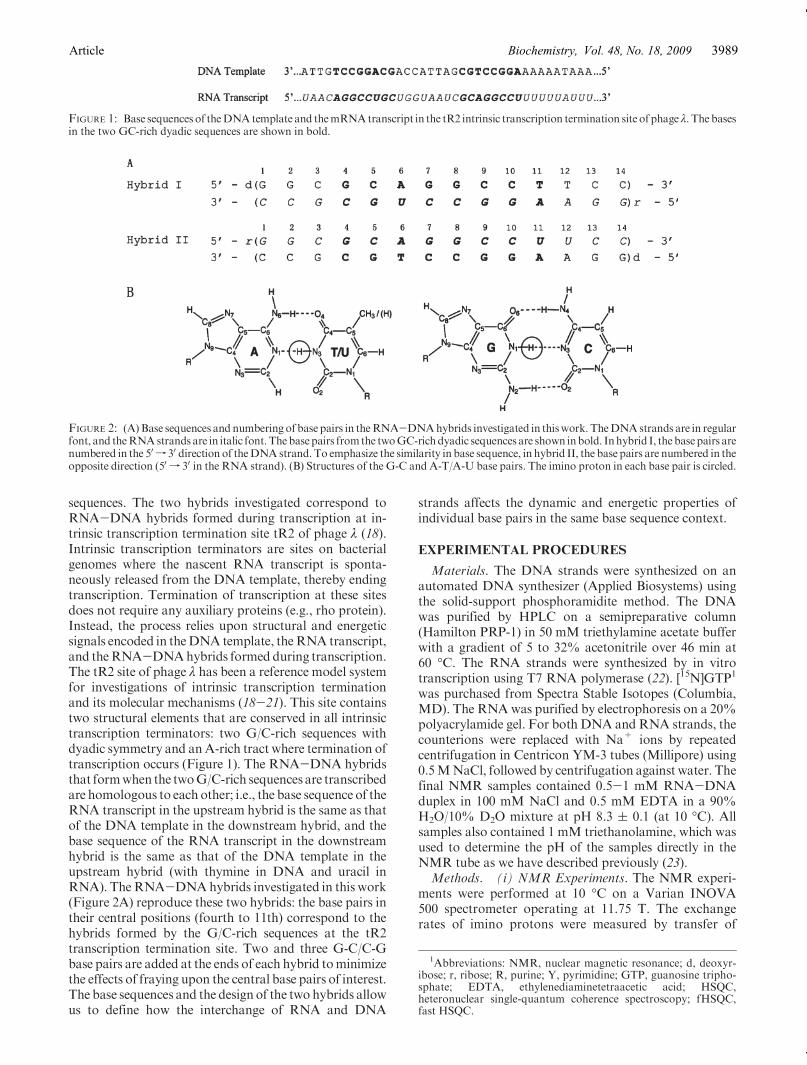
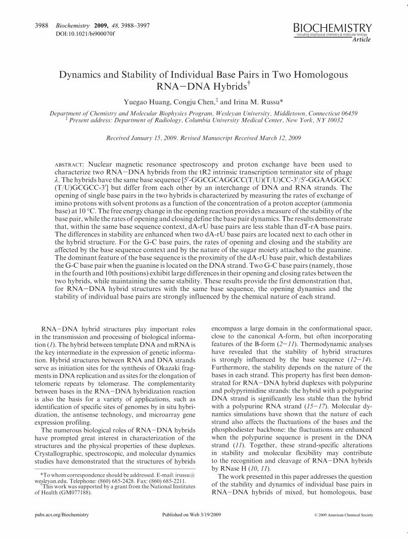
sequences. The two hybrids investigated correspond toRNA-DNA hybrids formed during transcription at in-trinsic transcription termination site tR2 of phage λ (18).Intrinsic transcription terminators are sites on bacterialgenomes where the nascent RNA transcript is sponta-neously released from the DNA template, thereby endingtranscription. Termination of transcription at these sitesdoes not require any auxiliary proteins (e.g., rho protein).Instead, the process relies upon structural and energeticsignals encoded in theDNA template, theRNA transcript,and theRNA-DNAhybrids formed during transcription.The tR2 site of phage λ has been a reference model systemfor investigations of intrinsic transcription terminationand its molecular mechanisms (18-21). This site containstwo structural elements that are conserved in all intrinsictranscription terminators: two G/C-rich sequences withdyadic symmetry and anA-rich tract where termination oftranscription occurs (Figure 1). The RNA-DNA hybridsthat formwhen the twoG/C-rich sequences are transcribedare homologous to each other; i.e., the base sequence of theRNA transcript in the upstream hybrid is the same as thatof the DNA template in the downstream hybrid, and thebase sequence of the RNA transcript in the downstreamhybrid is the same as that of the DNA template in theupstream hybrid (with thymine in DNA and uracil inRNA). TheRNA-DNAhybrids investigated in this work(Figure 2A) reproduce these two hybrids: the base pairs intheir central positions (fourth to 11th) correspond to thehybrids formed by the G/C-rich sequences at the tR2transcription termination site. Two and three G-C/C-Gbase pairs are added at the ends of each hybrid tominimizethe effects of fraying upon the central base pairs of interest.The base sequences and the design of the two hybrids allowus to define how the interchange of RNA and DNA
strands affects the dynamic and energetic properties ofindividual base pairs in the same base sequence context.
EXPERIMENTAL PROCEDURES
Materials. The DNA strands were synthesized on anautomated DNA synthesizer (Applied Biosystems) usingthe solid-support phosphoramidite method. The DNAwas purified by HPLC on a semipreparative column(Hamilton PRP-1) in 50 mM triethylamine acetate bufferwith a gradient of 5 to 32% acetonitrile over 46 min at60 �C. The RNA strands were synthesized by in vitrotranscription using T7 RNA polymerase (22). [15N]GTP1
was purchased from Spectra Stable Isotopes (Columbia,MD). The RNAwas purified by electrophoresis on a 20%polyacrylamide gel. For both DNA andRNA strands, thecounterions were replaced with Na+ ions by repeatedcentrifugation in Centricon YM-3 tubes (Millipore) using0.5MNaCl, followed by centrifugation against water. Thefinal NMR samples contained 0.5-1 mM RNA-DNAduplex in 100 mM NaCl and 0.5 mM EDTA in a 90%H2O/10% D2O mixture at pH 8.3 ( 0.1 (at 10 �C). Allsamples also contained 1 mM triethanolamine, which wasused to determine the pH of the samples directly in theNMR tube as we have described previously (23).Methods. (i) NMR Experiments. The NMR experi-
ments were performed at 10 �C on a Varian INOVA500 spectrometer operating at 11.75 T. The exchangerates of imino protons were measured by transfer of
FIGURE 1: Base sequences of theDNAtemplate and themRNAtranscript in the tR2 intrinsic transcription termination site of phage λ. The basesin the two GC-rich dyadic sequences are shown in bold.
FIGURE 2: (A)Base sequences and numbering of base pairs in theRNA-DNAhybrids investigated in thiswork. TheDNAstrands are in regularfont, and theRNAstrands are in italic font. Thebasepairs fromthe twoGC-richdyadic sequences are shown inbold. Inhybrid I, the basepairs arenumbered in the 50 f 30 direction of theDNAstrand. To emphasize the similarity in base sequence, in hybrid II, the base pairs are numbered in theopposite direction (50 f 30 in the RNA strand). (B) Structures of the G-C and A-T/A-U base pairs. The imino proton in each base pair is circled.
1Abbreviations: NMR, nuclear magnetic resonance; d, deoxyr-ibose; r, ribose; R, purine; Y, pyrimidine; GTP, guanosine tripho-sphate; EDTA, ethylenediaminetetraacetic acid; HSQC,heteronuclear single-quantum coherence spectroscopy; fHSQC,fast HSQC.
Article Vol. 48, No. 18, 2009Biochemistry, 3989
magnetization from water. The water proton resonancewas selectively inverted using a Gaussian 180� pulse(5.8 ms) followed by a variable delay for the exchangeof magnetization between water and imino protons. Agradient of 0.21 G/cm was applied during the exchangedelay to prevent the effects of radiation damping upon therecovery of watermagnetization to equilibrium.A secondselective pulse on water was applied to bring the watermagnetization back onto the z-axis before observation.Twenty-five values of the exchange delay in the rangefrom 1 to 800 ms were used in each experiment. Theexchange rates were calculated from the dependenceof the intensity of the imino proton resonance on theexchange delay as we described previously (24). Forthe unlabeled samples, the observation was with theJump-and-Return pulse sequence (25). For the 15N-labeled samples, the observation was with the one-dimen-sional (1D) version of the fast HSQC (fHSQC) pulsesequence (26) to edit the resonances of protons attachedto 15N. A modified fHSQC pulse sequence (27) was usedto filter out the resonances of protons attached to 15N andretain all other proton resonances. The highest exchangerates that can be accurately measured by these methodsare ∼70-80 s-1. The 1H-1H NOESY spectra wereobtained using the Watergate pulse sequence (28) with amixing time of 250 ms.
(ii) Optical Melting. The optical melting curves wereobtained on a Beckman DU650 spectrophotometer at260 nm. The concentration of each RNA-DNA hybridwas 1.5 μM in 100 mM NaCl.
(iii) Imino Proton Exchange in Nucleic Acid Duplexes.The exchange of imino protons in nucleic acids is a two-step process. In the first step, the base containing theimino proton (Figure 2B) flips out of the structure into anopen state in which the barriers to exchange are removed.In this open state, the imino proton is accessible to protonacceptors, and the hydrogen bond in which the protonparticipates breaks. The second step is the actual transferof the proton to an acceptor, such as OH- and NH3. Theexchange rate observed experimentally is given as (24, 29)
kex ¼ kopkex;open
kcl þ kex;openð1Þ
where kop and kcl are the rates of opening and closing,respectively, of the base containing the imino proton andkex,open is the rate of exchange from the open state. Therates of opening and closing define the equilibrium con-stant of the opening reaction:
Kop ¼ kop
kclð2Þ
This equilibrium constant is related to the free energychange in the opening reaction by
ΔGop ¼ -RT ln Kop ð3ÞwhereT is the absolute temperature andR is the universalgas constant.The rate of exchange from the open state depends on
the concentration of proton acceptor B as
kex;open ¼ RkB½B� ð4Þ
where kB is the rate constant for the transfer of the iminoproton to proton acceptor B in isolated nucleotides and Ris a factor that accounts for differences in proton transferrates between isolated nucleotides and open base pairs.Previous studies have shown that, for nucleic acid du-plexes, R has a value close to unity (30). The rate constantkB is calculated from the pKa value of the imino group(pKNH) and the pKa of the proton acceptor (pKB) as (31)
kB ¼ kcoll � 1
1 þ 0:27� 10-pKB þpKNH þ0:8ð5Þ
where kcoll is the rate of diffusion-controlled collisionbetween the imino group and the proton acceptor in theopen state of the base pair.The equation describing the dependence of the ex-
change rate observed experimentally on the concentra-tion of proton acceptor is obtained by inserting eq 4 ineq 1 (with R = 1):
kex ¼ kopkB½B�kcl þ kB½B� ð6Þ
Two kinetic regimes for imino proton exchange can bedistinguished depending on how the rate of exchangefrom the open state compares with the rate of closing.When the concentration of proton acceptor is sufficientlyhigh to make the exchange from the open state very fast(kex,open. kcl, EX1 regime), the exchange is limited by therate of base opening. In this case, eq 6 becomes
kex ¼ kop ð7ÞAt low concentrations of proton acceptor, the rate ofexchange from the open state is much smaller than therate of base pair closing (kex,open , kcl, EX2 regime). Inthis regime, the observed exchange rate depends linearlyon the concentration of proton acceptor:
kex ¼ Kopkex;open ¼ KopkB½B� ð8ÞIn this work, we have used ammonia base (NH3) as the
imino proton acceptor in the exchange. Previous workfrom this and other laboratories has shown that, due to itssmall size and its lack of charge, ammonia base is theacceptor of choice for proton exchange studies of nucleicacids (24, 29). The rate constant for transfer of a proton toNH3 was calculated from eq 5 as 8.8 � 108 M-1 s-1 forboth guanine and uracil and 4.1 � 108 M-1 s-1 forthymine at 10 �C. The concentration of ammonia baseNH3 was calculated from the total ammonia concentra-tion C0 and the pH as
½B� ¼ C0 � 10-pK=ð10-pH þ 10-pKÞ ð9ÞThe pH was measured at each ammonia concentration,directly in the NMR tube, using the proton resonances oftriethanolamine (23). The pK value of ammonia at 10 �Cis 9.73 (32).
RESULTS
Our characterization of base pair dynamics and ener-getics in the two RNA-DNA hybrids relies upon theexchange of imino protons with solvent protons. Thelocation of imino protons in the structures of the base
Biochemistry, Vol. 48, No. 18, 2009 Huang et al.3990
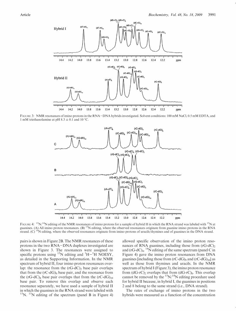
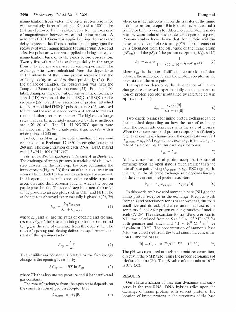
pairs is shown in Figure 2B. TheNMR resonances of theseprotons in the two RNA-DNA duplexes investigated areshown in Figure 3. The resonances were assigned tospecific protons using 15N editing and 1H-1H NOESY,as detailed in the Supporting Information. In the NMRspectrum of hybrid II, four imino proton resonances over-lap: the resonance from the (rG-dC)2 base pair overlapsthat from the (rC-dG)9 base pair, and the resonance fromthe (rG-dC)4 base pair overlaps that from the (rC-dG)10base pair. To remove this overlap and observe eachresonance separately, we have used a sample of hybrid IIinwhich the guanines in theRNA strandwere labeledwith15N. 15N editing of the spectrum (panel B in Figure 4)
allowed specific observation of the imino proton reso-nances of RNA guanines, including those from (rG-dC)2and (rG-dC)4.
14N editing of the same spectrum (panel C inFigure 4) gave the imino proton resonances from DNAguanines [including those from (rC-dG)9 and (rC-dG)10] aswell as those from thymines and uracils. In the NMRspectrumofhybrid I (Figure 3), the iminoproton resonancefrom (dG-rC)2 overlaps that from (dG-rC)8. This overlapcannot be removed by the 15N/14N editing procedure usedfor hybrid II because, in hybrid I, the guanines in positions2 and 8 belong to the same strand (i.e., DNA strand).The rates of exchange of imino protons in the two
hybrids were measured as a function of the concentration
FIGURE 3: NMR resonances of imino protons in the RNA-DNAhybrids investigated. Solvent conditions: 100mMNaCl, 0.5 mMEDTA, and1 mM triethanolamine at pH 8.3 ( 0.1 and 10 �C.
FIGURE 4: 15N/14N editing of the NMR resonances of imino protons for a sample of hybrid II in which the RNA strand was labeled with 15N atguanines. (A) All imino proton resonances. (B) 15N editing, where the observed resonances originate from guanine imino protons in the RNAstrand. (C) 14N editing, where the observed resonances originate from imino protons of uracils/thymines and of guanines in the DNA strand.
Article Vol. 48, No. 18, 2009Biochemistry, 3991
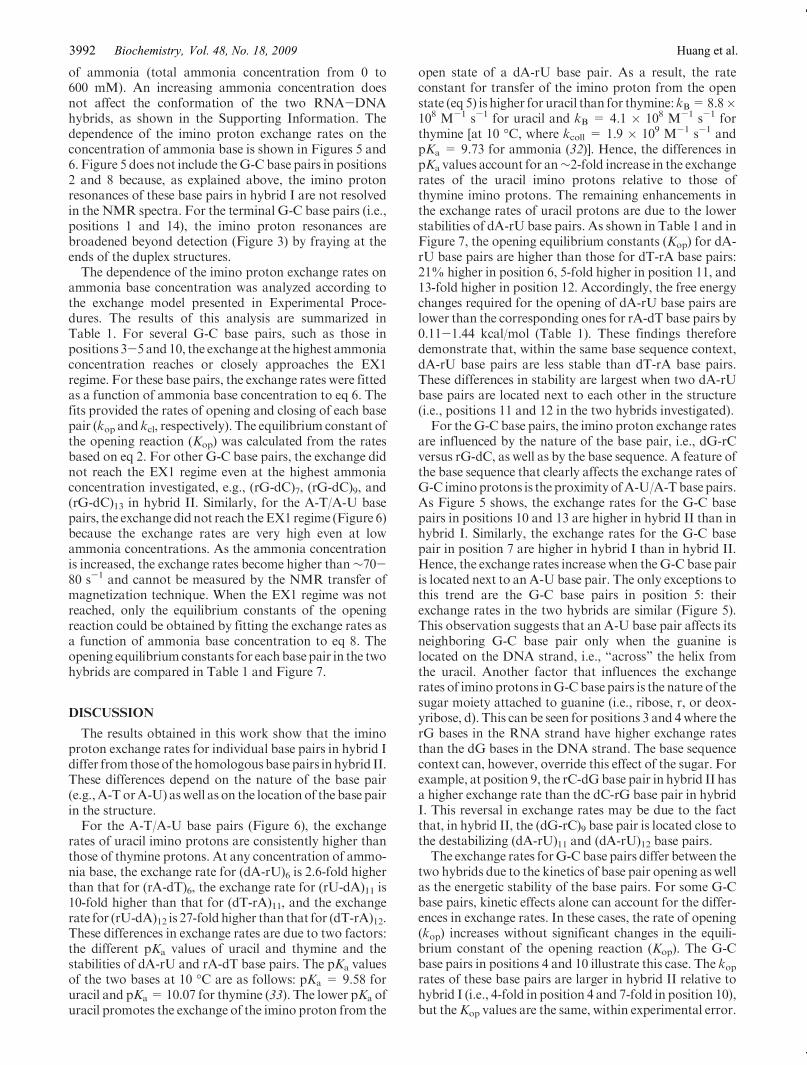
of ammonia (total ammonia concentration from 0 to600 mM). An increasing ammonia concentration doesnot affect the conformation of the two RNA-DNAhybrids, as shown in the Supporting Information. Thedependence of the imino proton exchange rates on theconcentration of ammonia base is shown in Figures 5 and6. Figure 5 does not include theG-C base pairs in positions2 and 8 because, as explained above, the imino protonresonances of these base pairs in hybrid I are not resolvedin the NMR spectra. For the terminal G-C base pairs (i.e.,positions 1 and 14), the imino proton resonances arebroadened beyond detection (Figure 3) by fraying at theends of the duplex structures.The dependence of the imino proton exchange rates on
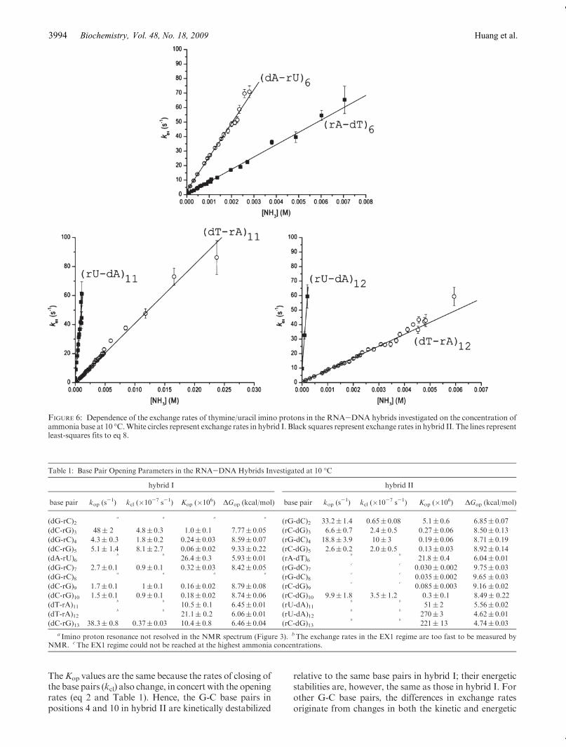
ammonia base concentration was analyzed according tothe exchange model presented in Experimental Proce-dures. The results of this analysis are summarized inTable 1. For several G-C base pairs, such as those inpositions 3-5and10, the exchangeat the highest ammoniaconcentration reaches or closely approaches the EX1regime. For these base pairs, the exchange rates were fittedas a function of ammonia base concentration to eq 6. Thefits provided the rates of opening and closing of each basepair (kop and kcl, respectively). The equilibrium constant ofthe opening reaction (Kop) was calculated from the ratesbased on eq 2. For other G-C base pairs, the exchange didnot reach the EX1 regime even at the highest ammoniaconcentration investigated, e.g., (rG-dC)7, (rG-dC)9, and(rG-dC)13 in hybrid II. Similarly, for the A-T/A-U basepairs, the exchangedidnot reach theEX1 regime (Figure 6)because the exchange rates are very high even at lowammonia concentrations. As the ammonia concentrationis increased, the exchange rates become higher than∼70-80 s-1 and cannot be measured by the NMR transfer ofmagnetization technique. When the EX1 regime was notreached, only the equilibrium constants of the openingreaction could be obtained by fitting the exchange rates asa function of ammonia base concentration to eq 8. Theopening equilibrium constants for eachbase pair in the twohybrids are compared in Table 1 and Figure 7.
DISCUSSION
The results obtained in this work show that the iminoproton exchange rates for individual base pairs in hybrid Idiffer from those of the homologous base pairs inhybrid II.These differences depend on the nature of the base pair(e.g.,A-TorA-U) aswell as on the location of the base pairin the structure.For the A-T/A-U base pairs (Figure 6), the exchange
rates of uracil imino protons are consistently higher thanthose of thymine protons. At any concentration of ammo-nia base, the exchange rate for (dA-rU)6 is 2.6-fold higherthan that for (rA-dT)6, the exchange rate for (rU-dA)11 is10-fold higher than that for (dT-rA)11, and the exchangerate for (rU-dA)12 is 27-fold higher than that for (dT-rA)12.These differences in exchange rates are due to two factors:the different pKa values of uracil and thymine and thestabilities of dA-rU and rA-dT base pairs. The pKa valuesof the two bases at 10 �C are as follows: pKa = 9.58 foruracil and pKa= 10.07 for thymine (33). The lower pKa ofuracil promotes the exchange of the imino proton from the
open state of a dA-rU base pair. As a result, the rateconstant for transfer of the imino proton from the openstate (eq 5) is higher for uracil than for thymine:kB=8.8�108 M-1 s-1 for uracil and kB = 4.1 � 108 M-1 s-1 forthymine [at 10 �C, where kcoll = 1.9 � 109 M-1 s-1 andpKa = 9.73 for ammonia (32)]. Hence, the differences inpKa values account for an∼2-fold increase in the exchangerates of the uracil imino protons relative to those ofthymine imino protons. The remaining enhancements inthe exchange rates of uracil protons are due to the lowerstabilities of dA-rU base pairs. As shown in Table 1 and inFigure 7, the opening equilibrium constants (Kop) for dA-rU base pairs are higher than those for dT-rA base pairs:21% higher in position 6, 5-fold higher in position 11, and13-fold higher in position 12. Accordingly, the free energychanges required for the opening of dA-rU base pairs arelower than the corresponding ones for rA-dT base pairs by0.11-1.44 kcal/mol (Table 1). These findings thereforedemonstrate that, within the same base sequence context,dA-rU base pairs are less stable than dT-rA base pairs.These differences in stability are largest when two dA-rUbase pairs are located next to each other in the structure(i.e., positions 11 and 12 in the two hybrids investigated).For theG-C base pairs, the imino proton exchange rates
are influenced by the nature of the base pair, i.e., dG-rCversus rG-dC, as well as by the base sequence. A feature ofthe base sequence that clearly affects the exchange rates ofG-C iminoprotons is the proximityofA-U/A-Tbasepairs.As Figure 5 shows, the exchange rates for the G-C basepairs in positions 10 and 13 are higher in hybrid II than inhybrid I. Similarly, the exchange rates for the G-C basepair in position 7 are higher in hybrid I than in hybrid II.Hence, the exchange rates increase when theG-C base pairis located next to anA-U base pair. The only exceptions tothis trend are the G-C base pairs in position 5: theirexchange rates in the two hybrids are similar (Figure 5).This observation suggests that an A-U base pair affects itsneighboring G-C base pair only when the guanine islocated on the DNA strand, i.e., “across” the helix fromthe uracil. Another factor that influences the exchangerates of imino protons inG-Cbase pairs is the nature of thesugar moiety attached to guanine (i.e., ribose, r, or deox-yribose, d). This can be seen for positions 3 and 4where therG bases in the RNA strand have higher exchange ratesthan the dG bases in the DNA strand. The base sequencecontext can, however, override this effect of the sugar. Forexample, at position 9, the rC-dGbase pair in hybrid II hasa higher exchange rate than the dC-rG base pair in hybridI. This reversal in exchange rates may be due to the factthat, in hybrid II, the (dG-rC)9 base pair is located close tothe destabilizing (dA-rU)11 and (dA-rU)12 base pairs.The exchange rates forG-C base pairs differ between the
two hybrids due to the kinetics of base pair opening as wellas the energetic stability of the base pairs. For some G-Cbase pairs, kinetic effects alone can account for the differ-ences in exchange rates. In these cases, the rate of opening(kop) increases without significant changes in the equili-brium constant of the opening reaction (Kop). The G-Cbase pairs in positions 4 and 10 illustrate this case. The koprates of these base pairs are larger in hybrid II relative tohybrid I (i.e., 4-fold in position 4 and 7-fold in position 10),but theKop values are the same, within experimental error.
Biochemistry, Vol. 48, No. 18, 2009 Huang et al.3992
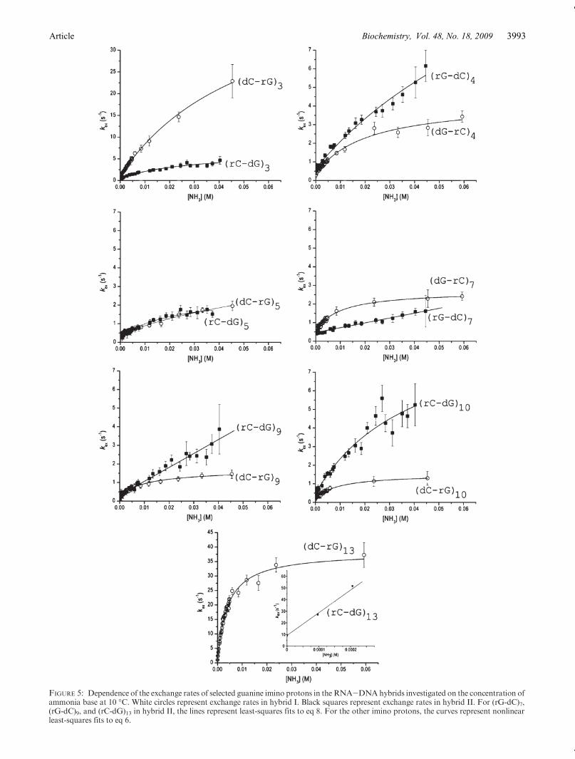
FIGURE 5: Dependence of the exchange rates of selected guanine imino protons in the RNA-DNAhybrids investigated on the concentration ofammonia base at 10 �C. White circles represent exchange rates in hybrid I. Black squares represent exchange rates in hybrid II. For (rG-dC)7,(rG-dC)9, and (rC-dG)13 in hybrid II, the lines represent least-squares fits to eq 8. For the other imino protons, the curves represent nonlinearleast-squares fits to eq 6.
Article Vol. 48, No. 18, 2009Biochemistry, 3993
TheKop values are the same because the rates of closing ofthe base pairs (kcl) also change, in concert with the openingrates (eq 2 and Table 1). Hence, the G-C base pairs inpositions 4 and 10 in hybrid II are kinetically destabilized
relative to the same base pairs in hybrid I; their energeticstabilities are, however, the same as those in hybrid I. Forother G-C base pairs, the differences in exchange ratesoriginate from changes in both the kinetic and energetic
FIGURE 6: Dependence of the exchange rates of thymine/uracil imino protons in the RNA-DNA hybrids investigated on the concentration ofammonia base at 10 �C.White circles represent exchange rates in hybrid I. Black squares represent exchange rates in hybrid II. The lines representleast-squares fits to eq 8.
Table 1: Base Pair Opening Parameters in the RNA-DNA Hybrids Investigated at 10 �C
hybrid I hybrid II
base pair kop (s-1) kcl (�10-7 s-1) Kop (�106) ΔGop (kcal/mol) base pair kop (s
-1) kcl (�10-7 s-1) Kop (�106) ΔGop (kcal/mol)
(dG-rC)2a a a a
(rG-dC)2 33.2( 1.4 0.65( 0.08 5.1( 0.6 6.85( 0.07
(dC-rG)3 48( 2 4.8( 0.3 1.0( 0.1 7.77( 0.05 (rC-dG)3 6.6( 0.7 2.4( 0.5 0.27( 0.06 8.50( 0.13
(dG-rC)4 4.3( 0.3 1.8( 0.2 0.24( 0.03 8.59( 0.07 (rG-dC)4 18.8( 3.9 10( 3 0.19( 0.06 8.71( 0.19
(dC-rG)5 5.1( 1.4 8.1( 2.7 0.06( 0.02 9.33( 0.22 (rC-dG)5 2.6( 0.2 2.0( 0.5 0.13( 0.03 8.92( 0.14
(dA-rU)6b b
26.4( 0.3 5.93( 0.01 (rA-dT)6b b
21.8( 0.4 6.04( 0.01
(dG-rC)7 2.7( 0.1 0.9( 0.1 0.32( 0.03 8.42( 0.05 (rG-dC)7c c
0.030( 0.002 9.75( 0.03
(dG-rC)8a a a a
(rG-dC)8c c
0.035( 0.002 9.65 ( 0.03
(dC-rG)9 1.7( 0.1 1( 0.1 0.16( 0.02 8.79( 0.08 (rC-dG)9c c
0.085( 0.003 9.16 ( 0.02
(dC-rG)10 1.5( 0.1 0.9( 0.1 0.18( 0.02 8.74( 0.06 (rC-dG)10 9.9( 1.8 3.5( 1.2 0.3( 0.1 8.49( 0.22
(dT-rA)11b b
10.5( 0.1 6.45( 0.01 (rU-dA)11b b
51( 2 5.56( 0.02
(dT-rA)12b b
21.1( 0.2 6.06( 0.01 (rU-dA)12b b
270( 3 4.62( 0.01
(dC-rG)13 38.3( 0.8 0.37( 0.03 10.4( 0.8 6.46( 0.04 (rC-dG)13b b
221( 13 4.74( 0.03
a Imino proton resonance not resolved in the NMR spectrum (Figure 3). bThe exchange rates in the EX1 regime are too fast to be measured byNMR. cThe EX1 regime could not be reached at the highest ammonia concentrations.
Biochemistry, Vol. 48, No. 18, 2009 Huang et al.3994
parameters of the opening reaction. For example, the koprate for (dC-rG)3 in hybrid I is 7-fold higher than that for(rC-dG)3 in hybrid II. The corresponding increase inKop isonly 4-fold because the kcl rate is also increased ∼2-fold(Table 1). Another example is that of theG-C base pairs atposition 9. The kop rate for (rC-dG)9 in hybrid II could notbe determined because the exchange of the imino protondid not reach the EX1 regime at the highest ammoniaconcentrations investigated. Nevertheless, the data shownin Figure 5 clearly indicate that the kop rate for this basepair should be higher than ∼4 s-1. This represents anincreaseof at least 2-fold relative to thekop rateof (dC-rG)9in hybrid I. In spite of this increase in the opening rate, theequilibrium constantKop for (rC-dG)9 in hybrid II is 2-foldsmaller than that for (dC-dG)9 in hybrid I. These oppositechanges in kop and Kop are due to the fact that the kcl ratefor (rC-dG)9 in hybrid II is at least 4-fold higher than thatfor (dC-dG)9 inhybrid I. In summary, forG-Cbasepairs, adifference in stability between the two hybrids is observedwhen the change in the opening rate exceeds or is smallerthan the change in the closing rate.When the change in theopening rate parallels that in the closing rate, the stabilityof the G-C base pair remains unaffected.Differences in the stability of homologous RNA-DNA
hybrids have been previously observed in optical meltingexperiments (12, 13, 15-17, 34). In contrast to our ap-proach presented here, which provides the stability ofindividual base pairs, optical melting experimentsmeasurethe overall stability of the hybrid duplex structure. In theseprevious studies, the largest differences in stability wereobserved for RNA-DNA hybrids that contained allpurine (R) and all pyrimidine (Y) bases in each strand.Specifically, the rR-dY hybrids (i.e., purines in the RNAstrandandpyrimidines in theDNAstrand) aremore stablethan the homologous dR-rY hybrids (13, 15-17). Kooland co-workers have elucidated the structural origin ofthese energetic differences for a RNA-DNA hybrid con-taining an adenine-rich strand and a thymine/uracil-richstrand (34). Using appropriate chemical modifications ofthe hybrid, they have shown that each C5 methyl group of
thymine enhances the overall stability of the hybrid by anaverage of 0.17-0.27 kcal/mol (at 37 �C) relative to thehomologous hybrid in which thymine is replaced withuracil. The origin of this effect is the methyl-inducedenhancement of the stacking of thymine onto neighboringbases (11, 34). The ribose 20-OH group also has a stabiliz-ing effect but only when placed in the purine-rich strand;for example, substitution of a dA-rU base pair with a rA-dU base pair stabilizes the structure by 0.32 kcal/mol (34).These findings offer important insights into the structuralorigin of the differences in stability between dA-rU andrA-dT base pairs observed in this work. They suggest thatrA-dTbase pairs are stabilized relative to dA-rUbase pairsdue to the presence of the C5methyl group in thymine andto the location of the 20-OH group in the purine.For homologous RNA-DNA hybrids of mixed base
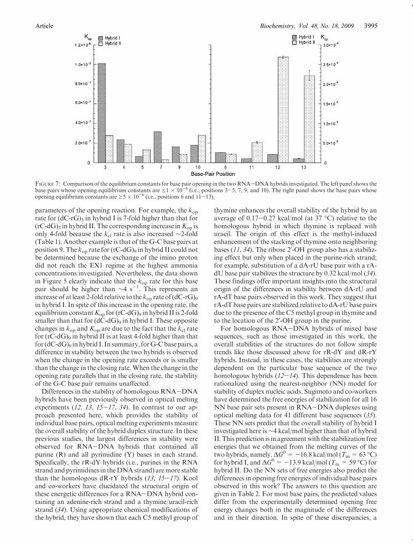
sequences, such as those investigated in this work, theoverall stabilities of the structures do not follow simpletrends like those discussed above for rR-dY and dR-rYhybrids. Instead, in these cases, the stabilities are stronglydependent on the particular base sequence of the twohomologous hybrids (12-14). This dependence has beenrationalized using the nearest-neighbor (NN) model forstability of duplex nucleic acids. Sugimoto and co-workershave determined the free energies of stabilization for all 16NN base pair sets present in RNA-DNA duplexes usingoptical melting data for 41 different base sequences (35).These NN sets predict that the overall stability of hybrid Iinvestigated here is∼4 kcal/mol higher than that of hybridII. This prediction is in agreementwith the stabilization freeenergies that we obtained from the melting curves of thetwo hybrids, namely,ΔG0=-16.8 kcal/mol (Tm=63 �C)for hybrid I, andΔG0=-13.9 kcal/mol (Tm= 59 �C) forhybrid II. Do the NN sets of free energies also predict thedifferences in opening free energies of individual base pairsobserved in this work? The answers to this question aregiven in Table 2. For most base pairs, the predicted valuesdiffer from the experimentally determined opening freeenergy changes both in the magnitude of the differencesand in their direction. In spite of these discrepancies, a
FIGURE 7: Comparison of the equilibrium constants for base pair opening in the twoRNA-DNAhybrids investigated. The left panel shows thebase pairs whose opening equilibrium constants are e1 � 10-6 (i.e., positions 3-5, 7, 9, and 10). The right panel shows the base pairs whoseopening equilibrium constants are g5 � 10-6 (i.e., positions 6 and 11-13).
Article Vol. 48, No. 18, 2009Biochemistry, 3995
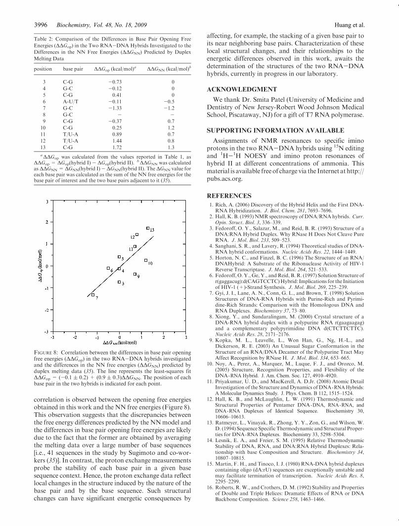
correlation is observed between the opening free energiesobtained in this work and the NN free energies (Figure 8).This observation suggests that the discrepancies betweenthe free energy differences predicted by the NNmodel andthe differences in base pair opening free energies are likelydue to the fact that the former are obtained by averagingthe melting data over a large number of base sequences[i.e., 41 sequences in the study by Sugimoto and co-wor-kers (35)]. In contrast, the proton exchange measurementsprobe the stability of each base pair in a given basesequence context. Hence, the proton exchange data reflectlocal changes in the structure induced by the nature of thebase pair and by the base sequence. Such structuralchanges can have significant energetic consequences by
affecting, for example, the stacking of a given base pair toits near neighboring base pairs. Characterization of theselocal structural changes, and their relationships to theenergetic differences observed in this work, awaits thedetermination of the structures of the two RNA-DNAhybrids, currently in progress in our laboratory.
ACKNOWLEDGMENT
We thank Dr. Smita Patel (University of Medicine andDentistry of New Jersey-Robert Wood Johnson MedicalSchool, Piscataway, NJ) for a gift of T7 RNA polymerase.
SUPPORTING INFORMATION AVAILABLE
Assignments of NMR resonances to specific iminoprotons in the two RNA-DNA hybrids using 15N editingand 1H-1H NOESY and imino proton resonances ofhybrid II at different concentrations of ammonia. Thismaterial is available freeof charge via the Internet at http://pubs.acs.org.
REFERENCES
1. Rich, A. (2006) Discovery of the Hybrid Helix and the First DNA-RNA Hybridization. J. Biol. Chem. 281, 7693–7696.
2. Hall, K. B. (1993)NMR spectroscopy ofDNA/RNAhybrids. Curr.Opin. Struct. Biol. 3, 336–339.
3. Fedoroff, O. Y., Salazar, M., and Reid, B. R. (1993) Structure of aDNA:RNA Hybrid Duplex. Why RNase H Does Not Cleave PureRNA. J. Mol. Biol. 233, 509–523.
4. Sanghani, S. R., and Lavery, R. (1994) Theoretical studies of DNA-RNA hybrid conformations. Nucleic Acids Res. 22, 1444–1449.
5. Horton, N. C., and Finzel, B. C. (1996) The Structure of an RNA/DNAHybrid: A Substrate of the Ribonuclease Activity of HIV-1Reverse Transcriptase. J. Mol. Biol. 264, 521–533.
6. Fedoroff, O.Y., Ge, Y., andReid, B. R. (1997) Solution Structure ofr(gaggacug):d(CAGTCCTC)Hybrid: Implications for the Initiationof HIV-1 (+)-Strand Synthesis. J. Mol. Biol. 269, 225–239.
7. Gyi, J. I., Lane, A. N., Conn, G. L., and Brown, T. (1998) SolutionStructures of DNA-RNA Hybrids with Purine-Rich and Pyrimi-dine-Rich Strands: Comparison with the Homologous DNA andRNA Duplexes. Biochemistry 37, 73–80.
8. Xiong, Y., and Sundaralingam, M. (2000) Crystal structure of aDNA-RNA hybrid duplex with a polypurine RNA r(gaagaagag)and a complementary polypyrimidine DNA d(CTCTTCTTC).Nucleic Acids Res. 28, 2171–2176.
9. Kopka, M. L., Leavelle, L., Won Han, G., Ng, H.-L., andDickerson, R. E. (2003) An Unusual Sugar Conformation in theStructure of an RNA/DNA Decamer of the Polypurine Tract MayAffect Recognition by RNase H. J. Mol. Biol. 334, 653–665.
10. Noy, A., Perez, A., Marquez, M., Luque, F. J., and Orozco, M.(2005) Structure, Recognition Properties, and Flexibility of theDNA 3RNA Hybrid. J. Am. Chem. Soc. 127, 4910–4920.
11. Priyakumar, U. D., and MacKerell, A. D.Jr. (2008) Atomic DetailInvestigation of the Structure andDynamics ofDNA 3RNAHybrids:A Molecular Dynamics Study. J. Phys. Chem. B 112, 1515–1524.
12. Hall, K. B., and McLaughlin, L. W. (1991) Thermodynamic andStructural Properties of Pentamer DNA 3DNA, RNA 3RNA, andDNA 3RNA Duplexes of Identical Sequence. Biochemistry 30,10606–10613.
13. Ratmeyer, L., Vinayak, R., Zhong, Y. Y., Zon, G., and Wilson, W.D. (1994) Sequence Specific Thermodynamic and Structural Proper-ties for DNA 3RNA Duplexes. Biochemistry 33, 5298–5304.
14. Lesnik, E. A., and Freier, S. M. (1995) Relative ThermodynamicStability of DNA, RNA, and DNA:RNA Hybrid Duplexes: Rela-tionship with base Composition and Structure. Biochemistry 34,10807–10815.
15. Martin, F. H., and Tinoco, I. J. (1980) RNA-DNA hybrid duplexescontaining oligo (dA:rU) sequences are exceptionally unstable andmay facilitate termination of transcription. Nucleic Acids Res. 8,2295–2299.
16. Roberts, R.W., and Crothers, D.M. (1992) Stability and Propertiesof Double and Triple Helices: Dramatic Effects of RNA or DNABackbone Composition. Science 258, 1463–1466.
Table 2: Comparison of the Differences in Base Pair Opening Free
Energies (ΔΔGop) in the Two RNA-DNA Hybrids Investigated to the
Differences in the NN Free Energies (ΔΔGNN) Predicted by Duplex
Melting Data
position base pair ΔΔGop (kcal/mol)a ΔΔGNN (kcal/mol)b
3 C-G -0.73 0
4 G-C -0.12 0
5 C-G 0.41 0
6 A-U/T -0.11 -0.5
7 G-C -1.33 -1.2
8 G-C - -9 C-G -0.37 0.7
10 C-G 0.25 1.2
11 T/U-A 0.89 0.7
12 T/U-A 1.44 0.8
13 C-G 1.72 1.3
aΔΔGop was calculated from the values reported in Table 1, asΔΔGop = ΔGop(hybrid I) - ΔGop(hybrid II). bΔΔGNN was calculatedas ΔΔGNN = ΔGNN(hybrid I)- ΔGNN(hybrid II). The ΔGNN value foreach base pair was calculated as the sum of the NN free energies for thebase pair of interest and the two base pairs adjacent to it (35).
FIGURE 8: Correlation between the differences in base pair openingfree energies (ΔΔGop) in the two RNA-DNA hybrids investigatedand the differences in the NN free energies (ΔΔGNN) predicted byduplex melting data (35). The line represents the least-squares fitΔΔGop = (-0.1 ( 0.2) + (0.9 ( 0.3)ΔΔGNN. The position of eachbase pair in the two hybrids is indicated for each point.
Biochemistry, Vol. 48, No. 18, 2009 Huang et al.3996
17. Gyi, J. I., Conn, G. L., Lane, A. N., and Brown, T. (1996)Comparison of the Thermodynamic Stabilities and Solution Con-formations of DNA-RNA Hybrids Containing Purine-Rich andPyrimidine-Rich Strands with DNA and RNADuplexes. Biochem-istry 35, 12538–12548.
18. Wilson, K. S., and von Hippel, P. H. (1994) Stability of EscherichiacoliTranscriptionComplexesNear an Intrinsic Terminator. J.Mol.Biol. 244, 36–51.
19. von Hippel, P. H. (1998) An Integrated Model of the TranscriptionComplex in Elongation, Termination, and Editing. Science 281,660–665.
20. Larson, M. H., Greenleaf, W. J., Landick, R., and Block, S. M.(2008) Applied Force Reveals Mechanistic and Energetic Details ofTranscription Termination. Cell 132, 971–982.
21. Datta, K., and von Hippel, P. H. (2008) Direct SpectroscopicStudy of Reconstituted Transcription Complexes Reveals ThatIntrinsic Termination is Driven Primarily by Thermodynamic De-stabilization of the Nucleic Acid Framework. J. Biol. Chem. 283,3537–3549.
22. Milligan, J. F., Groebe, D. R., Witherell, G. W., and Uhlenbeck, O.C. (1987) Oligoribonucleotide synthesis using T7 RNA polymeraseand synthetic DNA templates. Nucleic Acids Res. 15, 8783–8798.
23. Chen, C., and Russu, I. M. (2004) Sequence-Dependence of theEnergetics of Opening of AT Base Pairs in DNA. Biophys. J. 871–7.
24. Russu, I. M. (2004) Probing Site-Specific Energetics in Proteins andNucleic Acids by Hydrogen Exchange and NMR Spectroscopy.Methods Enzymol. 379, 152–175.
25. Plateau, P., and Gueron, M. (1982) Exchangeable proton NMRwithout base-line distortion, using new strong-pulse sequences. J.Am. Chem. Soc. 104, 7310–7311.
26. Mori, S., Abeygunawardana, C., Johnson,M.O., and van Zijl, P. C.M. (1995) Improved Sensitivity of HSQC Spectra of Exchanging
Protons at Short Interscan Delays Using a new Fast HSQC(FHSQC) Detection Scheme That Avoids Water Saturation. J.Magn. Reson., Ser. B 108, 94–98.
27. Chen, C., Jiang, L., Michalczyk, R., and Russu, I. M. (2006)Structural Energetics and Base-Pair Opening Dynamics in Sarcin-Ricin Domain RNA. Biochemistry 45, 13606–13613.
28. Lippens, G., Dhalluin, C., and Wieruszeski, J.-M. (1995) Use of awater flip-back pulse in the homonuclear NOESY experiment. J.Biomol. NMR 5, 327–331.
29. Gueron,M., andLeroy, J.-L. (1995) Studies of Base PairKinetics byNMR Measurement of Proton Exchange. Methods Enzymol. 261,383–413.
30. Gueron,M., Charretier, E., Hagerhorst, J., Kochoyan,M., Leroy, J.L., and Moraillon, A. (1990) Applications of Imino Proton Ex-change to Nucleic Acid Kinetics and Structures. In Structure &Methods (Sarma, R. H., and Sarma, M. H., Eds.) pp 113-137,Adenine Press, Albany, NY.
31. Benight,A. S., Schurr, J.M., Flynn, P. F., Reid, B.R., andWemmer,D. E. (1988) Melting of a Self-complementary DNA Minicircle.Comparison of Optical Melting Theory with Exchange Broadeningof the Nuclear Magnetic Resonance Spectrum. J. Mol. Biol. 200,377–399.
32. Weast, R. C. (1987) CRCHandbook of Chemistry and Physics, 67thed., CRC Press, Boca Raton, FL.
33. Ts’o, P.O. P. (1974) Basic Principles inNucleicAcidChemistry, Vol.I, Academic Press, New York.
34. Wang, S., and Kool, E. T. (1995) Origins of the Large Differences inStability of DNA and RNA Helices: C-5 Methyl and 2’-HydroxylEffects. Biochemistry 34, 4125–4132.
35. Wu, P., Nakano, S., and Sugimoto, N. (2002) Temperaturedependence of thermodynamic properties for DNA/DNAand RNA/DNA duplex formation. Eur. J. Biochem. 269, 2821–2830.
Article Vol. 48, No. 18, 2009Biochemistry, 3997