Embed Size (px)
Citation preview
REVIEW
Dynamic molecular linkers of thegenome: the first decade of SMC proteinsAna Losada1 and Tatsuya Hirano2,3
1Spanish National Cancer Center (CNIO), Madrid E-28029, Spain; 2Cold Spring Harbor Laboratory, Cold Spring Harbor,New York 11724, USA
Structural maintenance of chromosomes (SMC) proteinsare chromosomal ATPases, highly conserved from bac-teria to humans, that play fundamental roles in manyaspects of higher-order chromosome organization anddynamics. In eukaryotes, SMC1 and SMC3 act as thecore of the cohesin complexes that mediate sister chro-matid cohesion, whereas SMC2 and SMC4 function asthe core of the condensin complexes that are essentialfor chromosome assembly and segregation. Anothercomplex containing SMC5 and SMC6 is implicated inDNA repair and checkpoint responses. The SMC com-plexes form unique ring- or V-shaped structures withlong coiled-coil arms, and function as ATP-modulated,dynamic molecular linkers of the genome. Recent stud-ies shed new light on the mechanistic action of theseSMC machines and also expanded the repertoire of theirdiverse cellular functions. Dissecting this class of chro-mosomal ATPases is likely to be central to our under-standing of the structural basis of genome organization,stability, and evolution.
The chromosome is at the heart of all genetic processes.Its precise duplication and segregation are arguably themost important goal of the mitotic cell cycle, and pro-grammed expression of its content, either genetic or epi-genetic, is central to the development of an organism.While the astonishing advancement of genome biologyin recent years has provided an advanced starting pointfor virtually all areas in biology, it does not solve an oldproblem in chromosome biology: How is the genomicDNA folded, organized, and segregated in the tiny spaceof a cell? The discovery of structural maintenance ofchromosomes (SMC) proteins, almost a decade ago, pro-vided a decisive clue to solve this longstanding question,and led to the identification of cohesin and condensins,two representative classes of SMC-containing complexes
in eukaryotes. The proposed actions of cohesin and con-densins offer a plausible, if not complete, explanation forthe sudden appearance of thread-like “substances” (thechromosomes) and their longitudinal splitting duringmitosis, first described by Walther Flemming (1882).Remarkably, SMC proteins are conserved among thethree phyla of life, indicating that the basic strategy ofchromosome organization may be evolutionarily con-served from bacteria to humans. An emerging theme isthat SMC proteins are dynamic molecular linkers of thegenome that actively fold, tether, and manipulate DNAstrands. Their diverse functions range far beyond chro-mosome segregation, and involve nearly all aspects ofchromosome behavior including chromosome-wide orlong-range gene regulation and DNA repair. In this re-view article, we summarize our current understanding ofSMC proteins with a major focus on studies publishedduring the past three years. We start by describing thearchitecture and mechanistic actions of SMC proteincomplexes, and then discuss how the concerted actionsof cohesin and condensin support the faithful segrega-tion of chromosomes during mitosis and meiosis. Fi-nally, emerging studies of a third SMC complex in eu-karyotes and of bacterial SMC protein complexes are dis-cussed.
SMC protein complexes: a common theme with manyvariations
Basic architecture and enzymology of SMC proteins
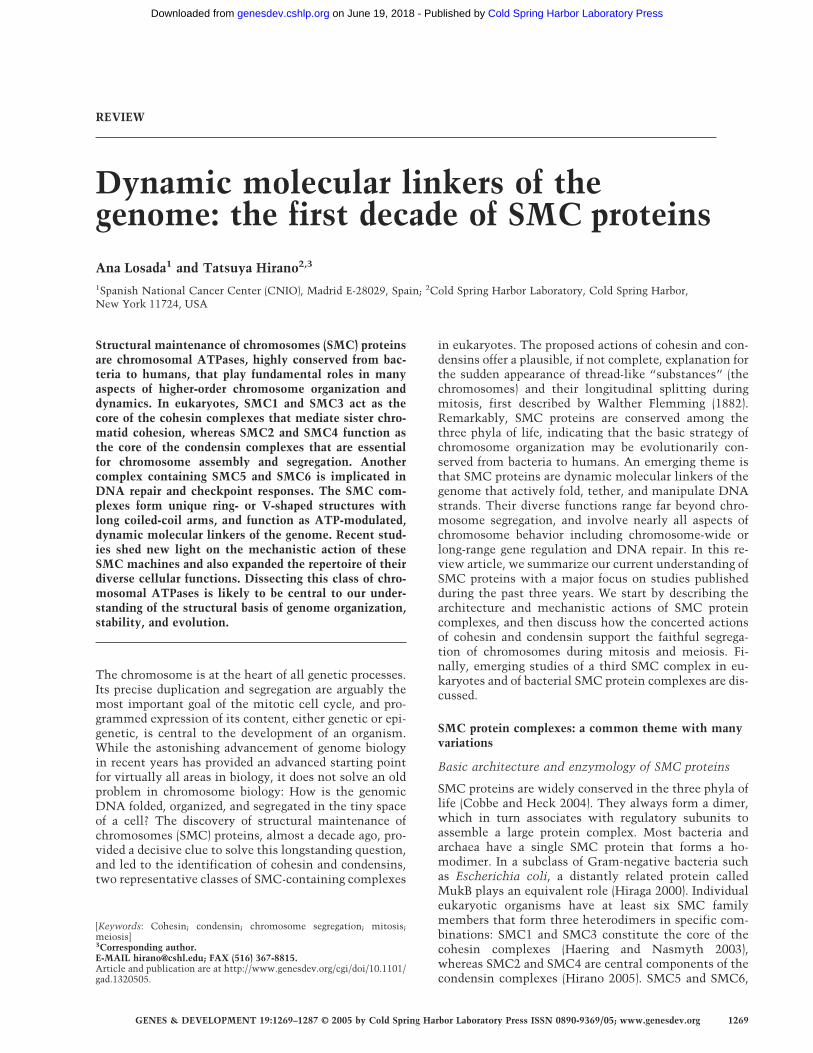
SMC proteins are widely conserved in the three phyla oflife (Cobbe and Heck 2004). They always form a dimer,which in turn associates with regulatory subunits toassemble a large protein complex. Most bacteria andarchaea have a single SMC protein that forms a ho-modimer. In a subclass of Gram-negative bacteria suchas Escherichia coli, a distantly related protein calledMukB plays an equivalent role (Hiraga 2000). Individualeukaryotic organisms have at least six SMC familymembers that form three heterodimers in specific com-binations: SMC1 and SMC3 constitute the core of thecohesin complexes (Haering and Nasmyth 2003),whereas SMC2 and SMC4 are central components of thecondensin complexes (Hirano 2005). SMC5 and SMC6,
[Keywords: Cohesin; condensin; chromosome segregation; mitosis;meiosis]3Corresponding author.E-MAIL [email protected]; FAX (516) 367-8815.Article and publication are at http://www.genesdev.org/cgi/doi/10.1101/gad.1320505.
GENES & DEVELOPMENT 19:1269–1287 © 2005 by Cold Spring Harbor Laboratory Press ISSN 0890-9369/05; www.genesdev.org 1269
Cold Spring Harbor Laboratory Press on June 19, 2018 - Published by genesdev.cshlp.orgDownloaded from

whose primary sequences are slightly divergent fromthose of SMC1–SMC4, are part of a complex implicatedin DNA repair and checkpoint responses (Lehmann2005).
Each SMC subunit is self-folded by antiparallel coiled-coil interactions, creating a rod-shaped molecule with anATP-binding cassette (ABC)-like “head” domain at oneend and a “hinge” domain at the other. Two SMC sub-units then associate with each other through their hingedomains, producing a V-shaped dimer (Fig. 1A, panel a;Melby et al. 1998; Anderson et al. 2002). This basic fold-ing scheme seems applicable to all SMC dimers (Haeringet al. 2002; Hirano and Hirano 2002). It is important tonote that the SMC dimer is a huge molecule: Eachcoiled-coil arm is ∼50 nm long, a length equivalent tothat of 150 bp of double-stranded DNA (dsDNA). Howdoes this two-armed structure interact with DNA andmanipulate its conformation? As has been predictedfrom crystal structures of other ABC ATPases (Hopfneret al. 2000; Smith et al. 2002), recent studies demon-
strate that ATP binding to the SMC head domains drivesthe formation of a nucleotide-sandwiched dimer (Fig. 1A,panel b; Haering et al. 2004; Lammens et al. 2004). Bio-chemical work using the Bacillus subtilis SMC proteinshows that ATP binding and hydrolysis modulate en-gagement and disengagement of the head domains, re-spectively, and thereby play a crucial role in the dynamicinteraction between SMC proteins and DNA (Hirano etal. 2001; Hirano and Hirano 2004). Dimerization at thehinge domain is very tight and is ATP-independent, andmutational analysis shows that this domain acts as anessential determinant of SMC–DNA interactions (Hi-rano and Hirano 2002). Moreover, evidence is availablethat different SMC dimers interact with each other inboth energy-dependent and -independent manners (Hi-rano et al. 2001; Sakai et al. 2003; Stray and Lindsley2003; Hirano and Hirano 2004), implying that their ac-tion is highly dynamic and complex. Thus, much re-mains to be learned regarding even the basic enzymologyof SMC proteins.
Figure 1. Architecture and subunit composition of SMC protein complexes. (A) Each SMC subunit self-folds by antiparallel coiled-coil interactions and forms a hinge domain at one end and an ATP-binding head domain, composed of its N- and C-terminal sequences,at the other end. (Panel a) A hinge–hinge interaction between two subunits mediates dimerization, thereby producing a V-shapedmolecule. (Panel b) Two ATP molecules (red) are sandwiched between two SMC head domains and induce their engagement. Sub-sequent disengagement is triggered by ATP hydrolysis. The head–head engagement may occur either intramolecularly within a dimeror intermolecularly between different dimers. (B, panel a) An SMC1–SMC3 heterodimer functions as the core of the cohesin complex,which contains two other non-SMC subunits, Scc1/Mcd1/Rad21 and Scc3/SA. An SMC2–SMC4 heterodimer acts as the core ofcondensin I (panel b) and condensin II (panel c). The CAP-D2, CAP-G, CAP-D3, and CAP-G2 subunits contain HEAT repeats, whereasthe CAP-H and CAP-H2 subunits belong to the kleisin family of proteins, like the Scc1 subunit of cohesin. (Panel a) Two cohesinregulators, Scc2 and Pds5, share HEAT repeats, although they are not tightly associated with cohesin. (Panel d) An SMC5–SMC6heterodimer functions as part of a complex that contains Nse1, Nse2, Nse3, and Nse4. (Panel e) The B. subtilis SMC protein complexis composed of an SMC homodimer, a kleisin subunit (ScpA), and another small subunit called ScpB.
Losada and Hirano
1270 GENES & DEVELOPMENT
Cold Spring Harbor Laboratory Press on June 19, 2018 - Published by genesdev.cshlp.orgDownloaded from
SMC
proteins
and
chrom
osome
dynam
ics
GE
NE
S&
DE
VE
LO
PM
EN
T1271
C
old Spring H
arbor Laboratory Press
on June 19, 2018 - Published by
genesdev.cshlp.orgD
ownloaded from

Cohesins: ring-shaped linkers composed of SMC1and SMC3
In the cohesin complex, the SMC1–SMC3 heterodimerassociates with the non-SMC subunits Scc1 (also knownas Mcd1/Rad21) and Scc3/SA (Table 1; Fig. 1B, panel a).Subunit–subunit interaction assays have shown that co-hesin forms a tripartite ring in which the open-V struc-ture of the SMC heterodimer is closed by the simulta-neous binding of the N- and C-terminal regions of Scc1to the head domains of SMC3 and SMC1, respectively(Haering et al. 2002). Such a structure is consistent withthe electron micrographs of cohesin complexes purifiedfrom Xenopus laevis eggs or human cells (Anderson et al.2002). Scc1 is now classified as a member of a superfam-ily of proteins termed “kleisins”, which include the con-densin subunits CAP-H and CAP-H2 (see below; Table 1;Schleiffer et al. 2003). The C terminus of Scc1 forms awinged helix, a folding motif found in many DNA-bind-ing proteins, and binds to the “outer” surface of theSmc1 head domain (Haering et al. 2004). Functional data,however, suggest that the winged helix motif of Scc1does not interact directly with DNA.
The ring-like structure of cohesin has led to the pro-
posal that the complex may hold sister chromatids to-gether by embracing two DNA duplexes within itscoiled-coil arms (Haering et al. 2002). This ring model isappealing because it nicely explains how proteolyticcleavage of the Scc1 subunit of the complex might openthe ring and thereby trigger sister chromatid separationat the anaphase onset (Fig. 2A; Uhlmann et al. 1999).Some predictions of this model have been successfullytested by genetic experiments in Saccharomyces cerevi-siae; for example, opening the ring by cleavage of a siteengineered in the coiled-coil arm of SMC3 promotes re-lease of cohesin from chromosomes (Gruber et al. 2003),and an “open” complex that lacks a coiled-coil domainor one of the heads fails to bind to chromatin (Weitzer etal. 2003). Nevertheless, the model remains to be testedcritically in vitro using purified or reconstituted compo-nents. Biochemical analysis of cohesin components isstill at a primitive stage: The purified complex displays amodest affinity for DNA or chromatin, and no ATP-de-pendent in vitro activity has been reported to date(Losada and Hirano 2001a; Sakai et al. 2003; Kagansky etal. 2004). Future studies should test whether a singlecohesin complex can indeed accommodate two DNA du-plexes within its coiled-coil arms, and determine how
Figure 2. Hypothetical actions of cohesin and condensin. (A) According to the ring model, cohesin embraces DNA within itscoiled-coil arms. The linkage between duplicated DNAs is established during DNA replication through an unknown mechanism.Cleavage of the Scc1 subunit (yellow) opens the ring and releases the chromatids for separation in anaphase. (B) Condensin may utilizethe energy of ATP hydrolysis to create a chiral (positively supercoiled) loop. Alternatively, multiple condensins may associate witheach other to make a DNA loop. Such association could be mediated by intermolecular head–head engagement (as shown here) orintermolecular coiled-coil interactions (not shown). For simplicity, no regulatory subunits are depicted in this cartoon. (C) Speculativemodel of how progressive release of cohesin and cooperative action of condensin lead to the assembly of a metaphase chromosomewith two resolved sister chromatids. For simplicity, no distinction is made between condensins I and II.
Losada and Hirano
1272 GENES & DEVELOPMENT
Cold Spring Harbor Laboratory Press on June 19, 2018 - Published by genesdev.cshlp.orgDownloaded from
the mechanical cycle of cohesin may be coupled to thecatalytic cycle of the SMC subunits.
Condensins: V-shaped linkers composedof SMC2–SMC4
SMC2 and SMC4 constitute the core subunits of con-densin. Vertebrate cells possess two different condensincomplexes, condensins I and II, that are distinguished bytheir unique, yet related, sets of non-SMC regulatorysubunits (Table 1; Fig. 1B, panels b,c; Ono et al. 2003;Yeong et al. 2003). Two of them, CAP-D2 and CAP-G incondensin I, and CAP-D3 and CAP-G2 in condensin II,contain HEAT repeats, a highly degenerate repeatingmotif implicated in protein–protein interactions (Neu-wald and Hirano 2000). CAP-H and CAP-H2 belong tothe kleisin family of proteins (Schleiffer et al. 2003). Theregulatory subunits of condensins bind to the head do-main(s) of the SMC heterodimer (Anderson et al. 2002;Yoshimura et al. 2002). Unlike the ring-like cohesincomplex whose hinge is wide open, condensin I shows aV-shaped structure with the coiled-coil arms of the SMCheterodimer placed close together (Anderson et al. 2002).These remarkably different arm conformations of cohe-sin and condensin probably contribute to their special-ized biochemical and physiological functions.
The phylogeny of condensin subunits sheds new lighton the evolution of chromosome architecture. All of thenon-SMC subunits of condensin I are highly conservedfrom yeast to humans, with the notable exceptions of thenematodes Caenorhabditis elegans and Caenorhabditisbriggsae (Table 1; Ono et al. 2003). The non-SMC sub-units of condensin II are found in plants and vertebratesbut not in yeast. The apparent loss of condensin I in C.elegans and C. briggsae is puzzling but may be related totheir unique, holocentric chromosome structure (Hag-strom et al. 2002; Stear and Roth 2002). Alternatively, anancient condensin I complex in these organisms mayhave lost its mitotic functions during evolution and be-come specialized in dosage compensation, a process thatequalizes expression of X-linked genes in the two sexes(Table 1; for review, see Hagstrom and Meyer 2003).
The holocomplex of condensin I, purified from eitherXenopus eggs or HeLa cells, has the ability to introducepositive superhelical tension into dsDNA in an ATP-hydrolysis-dependent manner (e.g., Kimura and Hirano1997). Most recently, a single-DNA-molecule nanoma-nipulation technique using magnetic tweezers hasshown that condensin I is able to physically compactDNA in the presence of hydrolyzable ATP (Strick et al.2004). The compaction reaction occurs in a highly dy-namic and reversible fashion, possibly involving a loop-ing mechanism. Both the supercoiling/knotting activityobserved in biochemical assays and the compaction ofsingle-DNA molecules are stimulated by cdk1-depen-dent phosphorylation of the non-SMC subunits (Kimuraet al. 1998; Strick et al. 2004), suggesting that they mayplay a direct role in driving chromosome assembly dur-ing mitosis. Despite this progress, a number of questionsremain to be answered regarding the action of con-
densins. In particular, it will be important to determinewhether a single condensin complex may be sufficient tomediate some of these reactions (Bazett-Jones et al. 2002)or whether cooperative interactions of multiple conden-sin complexes may be crucial (Fig. 2B; Strick et al. 2004).It will also be important to compare and contrast theaction of condensins and cohesin in the same set of func-tional assays (e.g., Losada and Hirano 2001a; Sakai et al.2003).
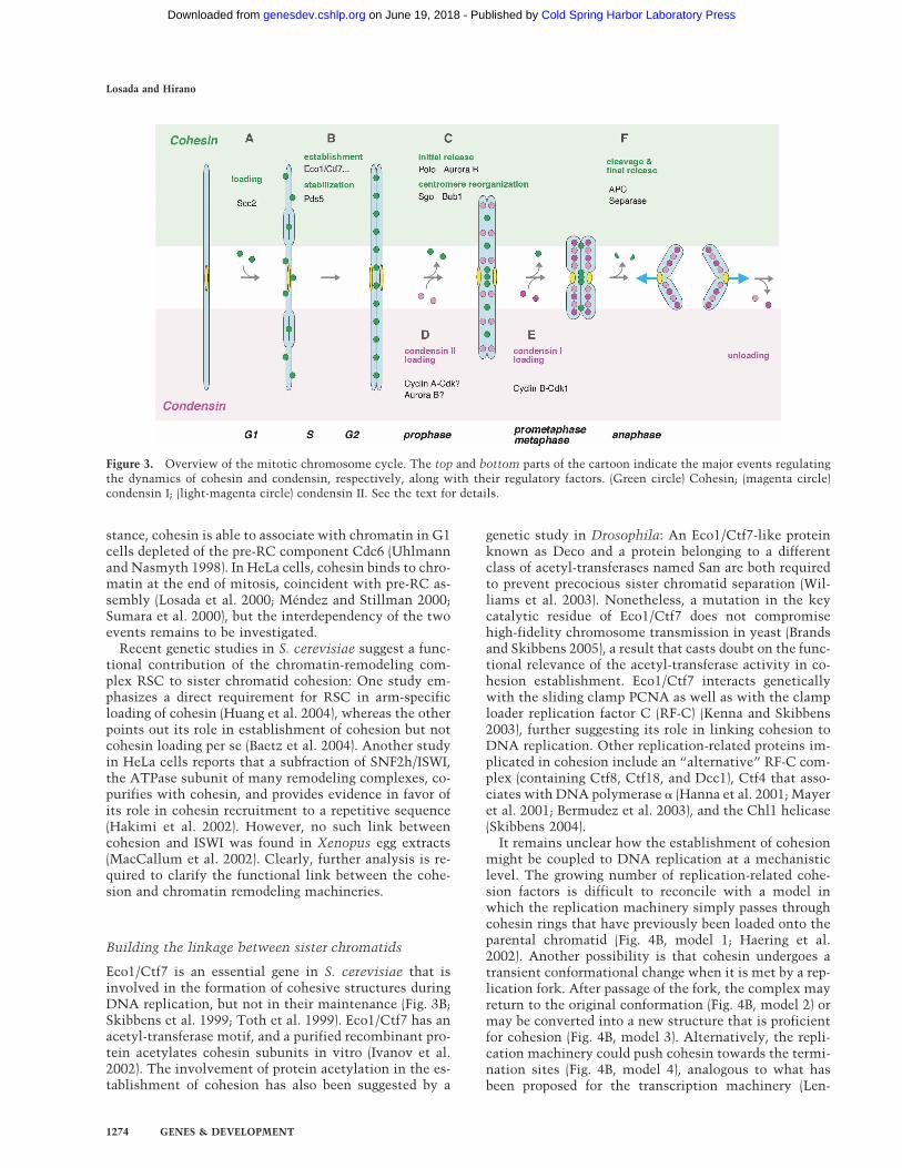
The chromosome cycle supported by cohesinand condensins: an overview
A substantial body of evidence has accumulated over thepast decade that the concerted action of cohesin and con-densins contributes to the faithful segregation of chro-mosomes during the mitotic cell cycle (Fig. 3). In short,cohesin establishes sister chromatid cohesion betweenduplicating DNAs in S phase. A large structural reorga-nization of chromosomes starts in prophase, with initialrelease of cohesin and progressive loading of condensins,and culminates in the formation of metaphase chromo-somes with well-resolved sister chromatids. This pro-cess, sister chromatid resolution, is a prerequisite of thefinal separation of sister chromatids that is triggered byproteolytic cleavage of cohesin at the onset of anaphase.The dynamic behavior of cohesin and condensins mustbe tightly regulated under the control of the cell cyclemachinery and, not surprisingly, a large number of spe-cialized factors participate in this regulation (Fig. 3). Inthe following four sections, we discuss the series ofevents that ensure the segregation of mitotic chromo-somes in a temporal order. Whenever possible, we ex-plore the mechanistic connection between the observ-able cytological events and the underlying molecularevents.
Establishing, mobilizing, and stabilizing sisterchromatid cohesion
Loading cohesin onto chromatin
A protein known as Scc2/Mis4 is required for loading ofcohesin onto chromatin (Fig. 3A; Ciosk et al. 2000; To-monaga et al. 2000). In C. elegans, TIM-1, a paralog of theDrosophila melanogaster clock gene timeless, performsa similar task (Chan et al. 2003). Scc2 and TIM-1 areHEAT-repeat proteins that physically interact with co-hesin (Arumugam et al. 2003; Chan et al. 2003). It hasbeen suggested that Scc2 may promote hydrolysis ofATP bound to cohesin’s SMC heads and thereby stimu-late the opening of the ring to allow loading onto chro-matin (Fig. 4A; Arumugam et al. 2003). In Xenopus eggextracts, loading of Scc2 on chromatin, and therefore ef-ficient loading of cohesin, depends on the assembly ofprereplication complex (pre-RC) but not on the initiationof DNA replication (Gillespie and Hirano 2004; Takaha-shi et al. 2004). This requirement could be a mechanismunique to early embryonic cells. In S. cerevisiae, for in-
SMC proteins and chromosome dynamics
GENES & DEVELOPMENT 1273
Cold Spring Harbor Laboratory Press on June 19, 2018 - Published by genesdev.cshlp.orgDownloaded from
stance, cohesin is able to associate with chromatin in G1cells depleted of the pre-RC component Cdc6 (Uhlmannand Nasmyth 1998). In HeLa cells, cohesin binds to chro-matin at the end of mitosis, coincident with pre-RC as-sembly (Losada et al. 2000; Méndez and Stillman 2000;Sumara et al. 2000), but the interdependency of the twoevents remains to be investigated.
Recent genetic studies in S. cerevisiae suggest a func-tional contribution of the chromatin-remodeling com-plex RSC to sister chromatid cohesion: One study em-phasizes a direct requirement for RSC in arm-specificloading of cohesin (Huang et al. 2004), whereas the otherpoints out its role in establishment of cohesion but notcohesin loading per se (Baetz et al. 2004). Another studyin HeLa cells reports that a subfraction of SNF2h/ISWI,the ATPase subunit of many remodeling complexes, co-purifies with cohesin, and provides evidence in favor ofits role in cohesin recruitment to a repetitive sequence(Hakimi et al. 2002). However, no such link betweencohesion and ISWI was found in Xenopus egg extracts(MacCallum et al. 2002). Clearly, further analysis is re-quired to clarify the functional link between the cohe-sion and chromatin remodeling machineries.
Building the linkage between sister chromatids
Eco1/Ctf7 is an essential gene in S. cerevisiae that isinvolved in the formation of cohesive structures duringDNA replication, but not in their maintenance (Fig. 3B;Skibbens et al. 1999; Toth et al. 1999). Eco1/Ctf7 has anacetyl-transferase motif, and a purified recombinant pro-tein acetylates cohesin subunits in vitro (Ivanov et al.2002). The involvement of protein acetylation in the es-tablishment of cohesion has also been suggested by a
genetic study in Drosophila: An Eco1/Ctf7-like proteinknown as Deco and a protein belonging to a differentclass of acetyl-transferases named San are both requiredto prevent precocious sister chromatid separation (Wil-liams et al. 2003). Nonetheless, a mutation in the keycatalytic residue of Eco1/Ctf7 does not compromisehigh-fidelity chromosome transmission in yeast (Brandsand Skibbens 2005), a result that casts doubt on the func-tional relevance of the acetyl-transferase activity in co-hesion establishment. Eco1/Ctf7 interacts geneticallywith the sliding clamp PCNA as well as with the clamploader replication factor C (RF-C) (Kenna and Skibbens2003), further suggesting its role in linking cohesion toDNA replication. Other replication-related proteins im-plicated in cohesion include an “alternative” RF-C com-plex (containing Ctf8, Ctf18, and Dcc1), Ctf4 that asso-ciates with DNA polymerase � (Hanna et al. 2001; Mayeret al. 2001; Bermudez et al. 2003), and the Chl1 helicase(Skibbens 2004).
It remains unclear how the establishment of cohesionmight be coupled to DNA replication at a mechanisticlevel. The growing number of replication-related cohe-sion factors is difficult to reconcile with a model inwhich the replication machinery simply passes throughcohesin rings that have previously been loaded onto theparental chromatid (Fig. 4B, model 1; Haering et al.2002). Another possibility is that cohesin undergoes atransient conformational change when it is met by a rep-lication fork. After passage of the fork, the complex mayreturn to the original conformation (Fig. 4B, model 2) ormay be converted into a new structure that is proficientfor cohesion (Fig. 4B, model 3). Alternatively, the repli-cation machinery could push cohesin towards the termi-nation sites (Fig. 4B, model 4), analogous to what hasbeen proposed for the transcription machinery (Len-
Figure 3. Overview of the mitotic chromosome cycle. The top and bottom parts of the cartoon indicate the major events regulatingthe dynamics of cohesin and condensin, respectively, along with their regulatory factors. (Green circle) Cohesin; (magenta circle)condensin I; (light-magenta circle) condensin II. See the text for details.
Losada and Hirano
1274 GENES & DEVELOPMENT
Cold Spring Harbor Laboratory Press on June 19, 2018 - Published by genesdev.cshlp.orgDownloaded from

gronne et al. 2004; see below). Replication of the lastshort stretch of DNA where cohesin accumulates couldbe managed without additional factors but be completedmost efficiently with help of replication-related cohe-sion factors such as the alternative RF-C complex. Thiswould explain why many of these factors are nonessen-tial for cell viability in yeast. Setting up a system inwhich replication-coupled cohesion can be reconstitutedin vitro would surely help test these models.
Mobilizing and stabilizing cohesin
High-resolution mapping of cohesin-binding sites inyeast has revealed a preference of cohesin for sites ofconvergent transcription (Glynn et al. 2004; Lengronneet al. 2004). It is possible that some characteristic ofthese sites (e.g., specific histone modifications or higher-order chromatin structure) favors the binding of cohesin(Glynn et al. 2004). A more intriguing proposal is thatthe transcription machinery “pushes” the cohesin com-plexes until they reach the sites where RNA polymer-ases traveling in opposite directions meet (Lengronne etal. 2004). This model is supported by the observationthat transcriptional activity changes the pattern of cohe-sin-binding sites. It remains to be determined, however,whether cohesin does actually slide along DNA orwhether it dissociates from the initial loading sites and
reassociates again farther down. In scc2 mutants, cohe-sin is still able to associate with chromatin (perhaps inan abortive manner) but is unable to relocate (Lengronneet al. 2004). Scc2 might therefore function not only inthe productive loading step but also in the dynamic mo-bilization or conformational change of cohesin during orafter DNA replication.
Pds5 (also known as BimD or Spo76) is another proteinthat modulates the dynamic association of cohesin withchromatin (Fig. 3B). While this class of proteins is highlyconserved from yeast to humans, the phenotypes ob-served in Pds5-deficient cells are somewhat variableamong different organisms. For instance, Pds5 is essen-tial for viability and is required to maintain cohesion inunperturbed mitosis in S. cerevisiae (Stead et al. 2003),whereas it is not essential in Schizosaccharomycespombe and cohesion defects are observed only after pro-longed G2/M arrest (Tanaka et al. 2001; Wang et al.2002). The idea that Pds5 is important to reinforce co-hesion is also supported by several studies in other or-ganisms (Storlazzi et al. 2003; Wang et al. 2003). Verte-brate cells have two Pds5-like proteins, Pds5A andPds5B, and mild cohesion defects are observed when thelevel of these proteins is decreased (Losada et al. 2005).Unexpectedly, metaphase chromosomes assembled in aPds5-depleted Xenopus egg extract retain an elevatedamount of cohesin, suggesting that vertebrate Pds5 couldnot only stabilize cohesion in interphase but also partici-
Figure 4. Regulation of cohesin dynamicsduring interphase. (A) Scc2 promotes load-ing of cohesin onto the parental chromatidbefore DNA replication. It may also be in-volved in transient unloading or mobiliza-tion of cohesin. Scc2 could mediate thesefunctions by facilitating disengagement ofthe SMC head domains of cohesin. Twoconformations of cohesin, with engagedand disengaged head domains, are repre-sented in green and orange, respectively. (B)Speculative models for the establishmentof cohesion during DNA replication. Thereplication machinery (yellow oval) couldsimply pass through the cohesin ring(model 1). Alternatively, passage of the rep-lication fork could impose a conforma-tional change in cohesin. The altered cohe-sin complex may return to the original con-formation (model 2) or may generate anovel cohesive structure (model 3). Model 4proposes that the replication forks push thecohesin complexes so that they accumulateat termination sites. Replication of the re-maining short stretch of DNA could be fa-cilitated by specialized factors such as thealternative RF-C. Subsequent action of thetranscriptional machinery could then relocate cohesin from the termination sites to regions of convergent transcription (Glynn et al.2004; Lengronne et al. 2004). (C) Pds5 (red oval) stabilizes cohesion, possibly by acting as a “closer” of the cohesin ring and/or bypromoting intermolecular interactions between adjacent cohesin complexes. Pds5 could mediate these functions by suppressingdisengagement of the SMC head domains of cohesin. In vertebrates, Pds5 may have an additional role in destabilizing cohesion andhelping efficient release of cohesin in early mitosis. These apparently opposite actions of Pds5 could be regulated by cell cycle-specificmodifications (represented by color change).
SMC proteins and chromosome dynamics
GENES & DEVELOPMENT 1275
Cold Spring Harbor Laboratory Press on June 19, 2018 - Published by genesdev.cshlp.orgDownloaded from
pate in its efficient dissolution in early mitosis (Fig. 4C).From a mechanistic point of view, it is interesting tonote that, like Scc2, Pds5 contains multiple HEAT re-peats (e.g., Neuwald and Hirano 2000). One possibility isthat Scc2 and Pds5 use the same motif to modulate theSMC ATPase cycle of cohesin, but that they do so inopposite ways. For example, Scc2 could destabilize ormobilize cohesin’s interaction with chromatin by actingas its “opener”, whereas Pds5 may stabilize this interac-tion by acting as a “closer”. The precise balance of theactions of Scc2 and Pds5 must be regulated tightly by cellcycle-specific modifications, such as sumoylation orphosphorylation, of each component (Stead et al. 2003;Gillespie and Hirano 2004; Losada et al. 2005). The ex-istence of this intricate regulatory mechanism maypartly be responsible for the rather complex and diversephenotypes displayed by Pds5-deficient cells among dif-ferent organisms.
Resolving and restructuring sister chromatids in earlymitosis
Release of cohesin from chromosome arms
In vertebrates, most cohesin dissociates from chromatinat prophase, and only a small population, enriched in thepericentromeric region, remains on chromosomes bymetaphase (Losada et al. 1998, 2000; Waizenegger et al.2000). This step does not entail proteolysis of cohesinand is regulated by two mitotic kinases, polo and auroraB (Fig. 3C; Losada et al. 2002; Sumara et al. 2002;Giménez-Abián et al. 2004). Experiments in Xenopus eggextracts depleted of both polo and aurora B kinasesshowed that this initial release of cohesin is not requiredfor condensin-mediated chromosome compaction. In-stead, it is crucial for proper resolution of sister chroma-tids (Losada et al. 2002). Hauf et al. (2005) identified mi-tosis-specific phosphorylation sites in the cohesin sub-units SA2 and Scc1 in HeLa cells, and generated stablecell lines expressing nonphosphorylatable versions ofboth proteins (SA2-12xA and SCC1-9xA). While expres-sion of SCC1-9xA did not have a measurable effect, pro-phase release of cohesin complexes containing SA2-12xA was severely impaired. Interestingly, the separase-mediated pathway removed the high level of cohesin leftall along the chromosomes, leading to apparently normalsegregation in anaphase. Under this experimental condi-tion, however, endogenous cohesin complexes contain-ing wild-type SA2 (and SA1) are released with normaltiming, making it difficult to address the functional im-portance of cohesin release in prophase.
Two-step loading of condensins
Release of cohesin from chromosome arms largely coin-cides with loading of condensins from prophase throughprometaphase. Recent studies show that the spatial andtemporal distributions of condensins I and II are differ-entially regulated during the cell cycle in HeLa cells (Hi-
rota et al. 2004; Ono et al. 2004). Condensin II is pre-dominantly nuclear during interphase, whereas conden-sin I is sequestered in the cytoplasm from interphasethrough prophase, and gains access to chromosomes onlyafter the nuclear envelope breaks down in prometaphase.It was proposed that sequential activation of cyclinA–cdk1 and cyclin B–cdk1 could be responsible for thesuccessive loading of condensin II and condensin I, re-spectively (Fig. 3D,E; Ono et al. 2004; Hirano 2005). Howcondensins are actually loaded on chromosomes is notfully understood. While the loading of cohesin requiresthe HEAT repeat protein Scc2, such a specialized loadingfactor has not yet been identified for condensins. It istempting to speculate that the two intrinsic subunitscontaining HEAT repeats (e.g., CAP-D2 and CAP-G forcondensin I) may perform a function analogous to Scc2.In fact, they are among the primary targets of cdk1-de-pendent phosphorylation, and their essential role in cellcycle-dependent loading of condensin I has been docu-mented in both Xenopus egg extracts (Kimura and Hi-rano 2000) and tissue culture cells (Ball et al. 2002).Moreover, it is most likely that the cdk1-dependentphosphorylation directly stimulates the activity of con-densins in vivo, as has been demonstrated in vitro(Kimura et al. 1998).
Condensins and chromosome architecture
A recent study combining light and electron microscopyin mammalian tissue culture cells suggests that thestructural changes of chromosomes in early and mid-mitosis may be mechanistically distinct (Kireeva et al.2004). A simple prediction from all emerging observa-tions is that condensin II initiates the early stage of con-densation by “hierarchical folding.” Upon nuclear enve-lope breakdown (NEBD), condensin I could collaboratewith condensin II to shape, resolve, and stabilize chro-mosomes by forming an “axial glue” structure withinthe chromatids. Nearly all subunits of condensin I havebeen found among the major components of a biochemi-cally defined fraction known as the chromosome scaffold(Hudson et al. 2003; Maeshima and Laemmli 2003; Gass-mann et al. 2005). At the level of light microscopy, con-densins I and II apparently alternate along the axis ofmetaphase chromatids (Ono et al. 2003, 2004). The dif-ferential distribution of the two condensin complexesappears to be unique to individual chromosomes (T. Onoand T. Hirano, unpubl.), providing a possible molecularexplanation for classical chromosome banding. It will beof great interest to determine how the differential distri-bution of condensins I and II may be specified in a chro-mosome-specific manner.
Consistent with the predictions described above, chro-mosome condensation within the prophase nucleus isdelayed in cells depleted of condensin II-specific sub-units, but not in those depleted of condensin I-specificsubunits (Hirota et al. 2004; Ono et al. 2004). By meta-phase, depletion of condensin I- or condensin II-specificsubunits produces a distinct, highly characteristic defectin chromosome architecture, whereas depletion of the
Losada and Hirano
1276 GENES & DEVELOPMENT
Cold Spring Harbor Laboratory Press on June 19, 2018 - Published by genesdev.cshlp.orgDownloaded from
SMC core subunits causes the severest defect (Ono et al.2003). Distinct roles of condensins I and II in chromo-some assembly have also been demonstrated convinc-ingly in Xenopus egg cell-free extracts. It remains to beestablished, however, exactly how the two condensincomplexes participate in the shaping and structuralmaintenance of metaphase chromatids (Gassmann et al.2004; Hirano 2005). The extent of defects observed incondensin-deficient cells varies among different condi-tions and different organisms, thereby leaving room fordivergent interpretations (Hagstrom et al. 2002; Coelhoet al. 2003; Hudson et al. 2003; Chan et al. 2004; Dej etal. 2004; Hirota et al. 2004; Watrin and Legagneux 2005).Critical assignment of the role of individual subunits invivo, along with a better understanding of their func-tions in vitro, will be essential in the future.
Sister chromatid resolution promoted by cohesinrelease and condensins’ action
The two events discussed above, release of cohesin fromchromosome arms and loading of condensins, are bothrequired for proper assembly of metaphase chromosomesthat are competent for segregation in anaphase. Figure2C depicts a highly speculative view of how cohesin andcondensins might behave and work during chromosomeassembly. While cohesin release and condensin loadingcan be uncoupled experimentally in Xenopus egg ex-tracts (Losada et al. 1998, 2002), it is reasonable to specu-late that the two events are linked at a mechanistic level.In fact, a modest delay in cohesin release is observed inHeLa cells depleted of a condensin I subunit (Hirota et al.2004). The third component important for sister chroma-tid resolution is topoisomerase II (topo II), an enzymethat catalyzes the transient passage of two DNA du-plexes. It has been hypothesized that positive supercoil-ing or chiral looping of DNA supported by condensinsmay facilitate topo II-mediated decatenation of sisterDNAs (Hirano 2000). Alternatively, condensin-mediatedassembly of sister chromatid axes could provide a drivingforce that pushes the equilibrium of topo II action to-ward decatenation (e.g., Maeshima and Laemmli 2003;Kireeva et al. 2004). The two mechanisms are not mu-tually exclusive, because axial distribution of topo II inmetaphase chromatids depends on functional con-densins (Coelho et al. 2003). We also speculate that thecollaborative action of condensins and topo II may bepowerful enough to “drive” the resolution of sister chro-matids in the absence of spindle forces (Paliulis andNicklas 2004; Machin et al. 2005). Such a spindle-inde-pendent mechanism might also underlie SMC-mediatedchromosome segregation in bacterial cells, as has beensuggested before (Hirano 2002).
Building sister centromeres/kinetochores in earlymitosis
The fundamental contribution of cohesin and con-densins to chromosome segregation is not restricted tochromosome arms. Emerging lines of evidence stronglysuggest that they play important roles in assembling cen-
tromeric heterochromatin that helps create tight cohe-sion during mitosis, and in orienting sister kinetochoresto allow proper attachment to the spindle (Fig. 3C–E).
Heterochromatin environment
Classical cytological observations suggest that sisterchromatids are more tightly associated at heterochro-matic regions (e.g., González et al. 1991), leading to thespeculation that the particular structure or compositionof heterochromatin could enhance recruitment of cohe-sin (Losada and Hirano 2001b). In fact, it was shown thatthe heterochromatin protein (HP)-1 homolog Swi6 bindsto methylated Lys 9 of histone H3 and promotes cohesinloading at the centromeric repeats in S. pombe (Bernardet al. 2001; Nonaka et al. 2002). More recent studies haverevealed that the RNA interference machinery regulatesthe establishment of heterochromatin, which in turn re-cruits cohesin to this region, in both S. pombe (Hall et al.2003; Schramke and Allshire 2003) and vertebrate cells(Fukagawa et al. 2004). Intriguingly, even in S. cerevisiaethat apparently lacks centromere-proximal heterochro-matin, a functional centromere induces increased asso-ciation of cohesin in a surrounding region spanning 20–50 kb (Weber et al. 2004). This observation implicatesthe existence of additional mechanisms to ensure stron-ger cohesion at centromeres.
Role of Sgo/Mei-S332 in centromeric cohesion:protection or active reconstruction?
The distinct regulation of arm and centromeric cohesionis even more crucial in meiosis than in mitosis. In meio-sis, arm cohesion is dissolved in anaphase I, while cen-tromeric cohesion persists until metaphase II (for review,see Nasmyth 2001). Unlike mitosis, both steps of cohe-sion dissolution in meiosis involve separase-mediatedcleavage of cohesin, but the question is the same in bothcases: What protects centromeric cohesin when arm co-hesion is dissolved? A pioneering study in Drosophilaidentified a centromeric protein, known as Mei-S332,that may perform this job (Kerrebrock et al. 1995). Morerecently, independent genetic screens in S. pombe and S.cerevisiae have “rediscovered” proteins related to Mei-S332, leading to the definition of the shugoshin (or Sgo)family of proteins (Katis et al. 2004; Kitajima et al. 2004;Marston et al. 2004; Rabitsch et al. 2004). Although thecontribution of Sgo/Mei-S332 proteins to mitotic chro-mosome segregation is modest in Drosophila or yeast,depletion of Sgo1, one of the two human members of thisfamily, from HeLa cells causes premature separation ofsister chromatids during mitosis (Salic et al. 2004; Tanget al. 2004; Kitajima et al. 2005). This phenotype is par-tially suppressed when cohesion is reinforced by express-ing a nonphosphorylatable form of the cohesin subunitSA2 (McGuiness et al. 2005), consistent with a model inwhich Sgo1 protects centromeric cohesin from release inprophase (Fig. 5A). The spindle checkpoint protein Bub1regulates Sgo1 localization at centromeres, and in its ab-sence, Sgo1 and Scc1 are no longer enriched at this region
SMC proteins and chromosome dynamics
GENES & DEVELOPMENT 1277
Cold Spring Harbor Laboratory Press on June 19, 2018 - Published by genesdev.cshlp.orgDownloaded from
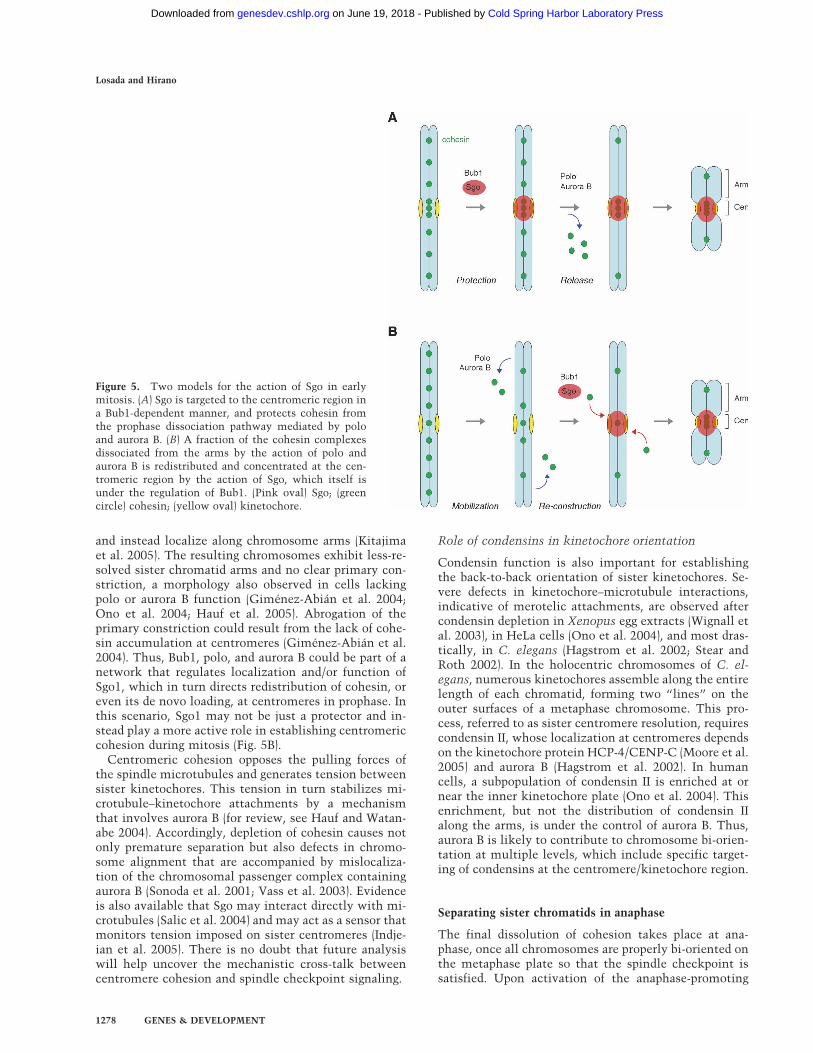
and instead localize along chromosome arms (Kitajimaet al. 2005). The resulting chromosomes exhibit less-re-solved sister chromatid arms and no clear primary con-striction, a morphology also observed in cells lackingpolo or aurora B function (Giménez-Abián et al. 2004;Ono et al. 2004; Hauf et al. 2005). Abrogation of theprimary constriction could result from the lack of cohe-sin accumulation at centromeres (Giménez-Abián et al.2004). Thus, Bub1, polo, and aurora B could be part of anetwork that regulates localization and/or function ofSgo1, which in turn directs redistribution of cohesin, oreven its de novo loading, at centromeres in prophase. Inthis scenario, Sgo1 may not be just a protector and in-stead play a more active role in establishing centromericcohesion during mitosis (Fig. 5B).
Centromeric cohesion opposes the pulling forces ofthe spindle microtubules and generates tension betweensister kinetochores. This tension in turn stabilizes mi-crotubule–kinetochore attachments by a mechanismthat involves aurora B (for review, see Hauf and Watan-abe 2004). Accordingly, depletion of cohesin causes notonly premature separation but also defects in chromo-some alignment that are accompanied by mislocaliza-tion of the chromosomal passenger complex containingaurora B (Sonoda et al. 2001; Vass et al. 2003). Evidenceis also available that Sgo may interact directly with mi-crotubules (Salic et al. 2004) and may act as a sensor thatmonitors tension imposed on sister centromeres (Indje-ian et al. 2005). There is no doubt that future analysiswill help uncover the mechanistic cross-talk betweencentromere cohesion and spindle checkpoint signaling.
Role of condensins in kinetochore orientation
Condensin function is also important for establishingthe back-to-back orientation of sister kinetochores. Se-vere defects in kinetochore–microtubule interactions,indicative of merotelic attachments, are observed aftercondensin depletion in Xenopus egg extracts (Wignall etal. 2003), in HeLa cells (Ono et al. 2004), and most dras-tically, in C. elegans (Hagstrom et al. 2002; Stear andRoth 2002). In the holocentric chromosomes of C. el-egans, numerous kinetochores assemble along the entirelength of each chromatid, forming two “lines” on theouter surfaces of a metaphase chromosome. This pro-cess, referred to as sister centromere resolution, requirescondensin II, whose localization at centromeres dependson the kinetochore protein HCP-4/CENP-C (Moore et al.2005) and aurora B (Hagstrom et al. 2002). In humancells, a subpopulation of condensin II is enriched at ornear the inner kinetochore plate (Ono et al. 2004). Thisenrichment, but not the distribution of condensin IIalong the arms, is under the control of aurora B. Thus,aurora B is likely to contribute to chromosome bi-orien-tation at multiple levels, which include specific target-ing of condensins at the centromere/kinetochore region.
Separating sister chromatids in anaphase
The final dissolution of cohesion takes place at ana-phase, once all chromosomes are properly bi-oriented onthe metaphase plate so that the spindle checkpoint issatisfied. Upon activation of the anaphase-promoting
Figure 5. Two models for the action of Sgo in earlymitosis. (A) Sgo is targeted to the centromeric region ina Bub1-dependent manner, and protects cohesin fromthe prophase dissociation pathway mediated by poloand aurora B. (B) A fraction of the cohesin complexesdissociated from the arms by the action of polo andaurora B is redistributed and concentrated at the cen-tromeric region by the action of Sgo, which itself isunder the regulation of Bub1. (Pink oval) Sgo; (greencircle) cohesin; (yellow oval) kinetochore.
Losada and Hirano
1278 GENES & DEVELOPMENT
Cold Spring Harbor Laboratory Press on June 19, 2018 - Published by genesdev.cshlp.orgDownloaded from
complex or cyclosome (APC/C), the cysteine proteaseseparase is freed from securin and cleaves Scc1, therebytriggering sister chromatid separation (Fig. 3F; Uhlmannet al. 1999). Phosphorylation of Scc1 by polo facilitatesits cleavage by separase in vitro, but this modificationmay not be essential in vivo (Alexandru et al. 2001;Hornig and Uhlmann 2004; Hauf et al. 2005). As a puta-tive protector of centromeric cohesion, Sgo1’s functionmay also be inactivated or down-regulated at the ana-phase onset. In fact, Sgo1 is a substrate of the APC/C invertebrates (Salic et al. 2004), but the precise timing orfunctional importance of its degradation remains to bedetermined. In Drosophila, delocalization of Sgo/Mei-S332 from centromeres in anaphase requires two differ-ent pathways: One involves separase function (Lee et al.2004) and the other involves phosphorylation by polo(Clarke et al. 2005). However, polo mutants are able toseparate their chromosomes despite persistence of Mei-S332 at centromeres, suggesting that the postulated in-activation of this protein may require neither its removalfrom centromeres nor its degradation.
According to an oversimplified view emphasized inearly studies, arm cohesion is completely dissolved bymetaphase while centromeric cohesion is released at theonset of anaphase (e.g., Waizenegger et al. 2000). This isclearly not the case in unperturbed mitosis: Arm cohe-sion is gradually lost in anaphase after sister centromeresseparate and sister chromatids move toward oppositepoles of the cell (Giménez-Abián et al. 2004; Paliulis andNicklas 2004). A series of recent studies further suggeststhat certain chromosomal domains separate at laterstages in anaphase, possibly through unique mecha-nisms. For example, the segregation of rDNA in S. cer-evisiae occurs in mid-anaphase and requires Cdc14, aprotein phosphatase that is activated by the FEAR (four-teen early anaphase release) network (D’Amours et al.2004; Sullivan et al. 2004; Wang et al. 2004). Condensinis recruited to the rDNA locus in anaphase in a Cdc14-dependent manner and mediates the condensation andresolution of rDNA at this stage (Lavoie et al. 2004). Thesegregation defect observed in cdc14 mutants is not re-lieved by inactivation of cohesin, suggesting that a dis-tinct form of linkage exists at this locus. In mammaliancells, the separation of sister telomeres may use anothermechanism involving tankyrase 1, a telomeric proteinthat has a poly(ADP-ribose) polymerase motif (Dynekand Smith 2004). In this case, it remains to be estab-lished whether the persistent linkage of telomeres ob-served in tankyrase 1-deficient cells is independent ofcohesin.
Cohesin and condensins in meiotic chromosomesegregation
In meiosis, two consecutive chromosome-segregationevents follow a single round of DNA replication. Ho-mologous chromosomes separate in meiosis I, while sis-ter chromatids separate in meiosis II. This fact posesspecific requirements for the regulation of cohesion andhas most likely guided the emergence of meiosis-specific
isoforms of the cohesin subunits Scc1 (known as Rec8),Scc3/SA, and even SMC1 (see Table 1). In S. pombemeiosis, Rec8 forms two different complexes: One con-taining Scc3 (referred to as Psc3 in this organism) local-izes to the vicinity of centromeres, whereas the othercontaining the meiosis-specific version of Scc3 (knownas Rec11) is found along the arms (Kitajima et al. 2003).Thus, meiotic isoforms may contribute to the differen-tial susceptibility of the cohesin complex to cleavageby separase in meiosis I. Additional meiosis-specificfunctions have been hypothesized for meiotic cohesins;e.g., they could favor interhomolog invasion over inva-sion of the sister chromatid, thereby facilitating chias-mata formation (Martston and Amon 2004). Mammalshave a meiosis-specific isoform of SMC1, known asSMC1� (Table 1). In mice, a cohesin complex(es) con-taining the canonical SMC1 (SMC1�) is most likely re-sponsible for establishing cohesion in premeiotic Sphase, whereas Smc1� is detected on chromosomesonly after zygotene. Smc1�-deficient mice are sterile inboth sexes, displaying defects in synapsis, recombina-tion, and maintenance of cohesion both in chromosomearms and at centromeres (Revenkova et al. 2004). Rec8was also detected at the axial elements of the synaptone-mal complex (SC) in rat spermatocytes before SMC1�and SMC3, implying that the core and the regulatorysubunits of the complex may be targeted to chromo-somes separately (Eijpe et al. 2003). Support for this ideaalso comes from the observation that loss of TIM-1 func-tion in C. elegans prevents localization of Rec8, butnot of SMC1 and SMC3, to meiotic prophase chromo-somes (Chan et al. 2003). Future analysis should deter-mine the precise dynamics of the different cohesin com-plexes coexisting in meiotic cells, and their specific con-tributions to meiotic chromosome functions (e.g., Parraet al. 2004).
Condensin subunits also play crucial roles in thestructural and functional organization of meiotic chro-mosomes. In S. cerevisiae, condensin subunits localizeto the axial core of pachytene chromosomes and contrib-ute to their axial compaction and individualization (Yuand Koshland 2003). The SC is not properly assembled incondensin mutants, leading to defects in homolog pair-ing and processing of double-strand breaks (DSBs). Evi-dence is also available that condensin participates in theresolution of recombination-dependent linkages be-tween homologs in meiosis I and perhaps in the segre-gation of sister chromatids in meiosis II as well (Yu andKoshland 2003). A requirement for condensin functionin both meiosis I and meiosis II is consistent with resultsfrom Arabidopsis (Siddiqui et al. 2003) and C. elegans(Chan et al. 2004). Unlike in S. cerevisiae, the condensinsubunits associate with chromosomes only after exitfrom pachytene in C. elegans. This difference may berelated to the fact that S. cerevisiae and C. elegans con-tain only condensin I or condensin II, respectively. How-ever, the non-SMC components of the dosage compen-sation complex in C. elegans are also required for mei-otic (but not mitotic) chromosome segregation (Lieb etal. 1996), providing an additional level of complexity to
SMC proteins and chromosome dynamics
GENES & DEVELOPMENT 1279
Cold Spring Harbor Laboratory Press on June 19, 2018 - Published by genesdev.cshlp.orgDownloaded from

this problem. Clearly, a number of questions remain tobe addressed about the role of condensins in meiosis. Forexample, are there meiosis-specific condensin subunits?In vertebrates and plants, do condensins I and II differ-entially localize to meiotic chromosomes and performnonoverlapping functions in their recombination andsegregation?
Expanding roles of cohesin and condensins outsidechromosome segregation
Cohesin and DNA repair
In late S and G2 phases, when two sister chromatids areavailable, cells prefer to repair DSBs by homologous re-combination (HR). Results from studies of yeast (Birken-bihl and Subramani 1992; Sjögren and Nasmyth 2001)and vertebrate cells (Sonoda et al. 2001) suggested thatDSB repair is impaired in the absence of cohesin. It wasalso shown that cohesin accumulates at sites of laser-induced DNA damage in an Mre11/Rad50-dependentmanner in mammalian cells (J.S. Kim et al. 2002). Tworecent studies in S. cerevisiae refined this idea by reveal-ing that cohesin subunits are recruited to a region of∼100 kb surrounding a single DSB (Ström et al. 2004;Ünal et al. 2004). This DSB-induced recruitment of co-hesin is Scc2-dependent and requires phosphorylation ofH2AX by the DNA damage checkpoint kinases Mec1/ATM and Tel1/ATR (Ünal et al. 2004). Most impor-tantly, cohesin loaded in response to DSBs establishes ade novo linkage between the damage chromatid and itsundamaged sister, thereby facilitating DSB repair (Strömet al. 2004). This “excess” amount of cohesin may needto be removed to complete the repair process, possiblythrough a separase-dependent mechanism (Nagao et al.2004). In mammalian cells, SMC1 is phosphorylated byATM in response to ionizing irradiation (IR) (S.-T. Kim etal. 2002; Yazdi et al. 2002). Murine cells expressing anonphosphorylatable form of SMC1 show decreased sur-vival only after DNA damage, suggesting that this modi-fication of cohesin is required for its role in DNA repairbut not for its essential role in cohesion (Kitagawa et al.2004). It is nonetheless possible that cohesion may beroutinely reinforced during or after S phase through thisDSB-induced loading mechanism, as DSBs can arisenaturally during DNA replication. Furthermore, if therepeated sequences of heterochromatin were more proneto stalled forks and DSBs than the single-copy sequencesof euchromatin, then DSB-induced loading of cohesincould provide a means to generate a higher density ofcohesin in heterochromatin. In any case, these new stud-ies provide a fresh view on the loading and action ofcohesin, which appear to be much more dynamic thanwas anticipated.
Cohesin regulators and development
Reduced dosage of the Scc2 ortholog Nipped-B nega-tively affects the activation of the homeotic cut gene by
a distant enhancer in Drosophila (Rollins et al. 1999).The observation that Nipped-B and cohesin have oppo-site effects on this long-range regulation supports themodel that Scc2/Nipped-B acts both as a loader and as anunloader of cohesin, and thereby facilitates enhancer-promoter communication (Rollins et al. 2004). Certaindevelopmental genes could be particularly sensitive tothe presence of cohesin nearby their promoters and thusto reduced levels of Scc2. This might explain why mu-tation of one copy of the human Nipped-B like (NIPBL)gene cause Cornelia de Lange syndrome, a developmen-tal disorder characterized by growth and cognitive retar-dation (Krantz et al. 2004; Tonkin et al. 2004). It remainsto be determined, however, whether these developmen-tal defects indeed derive from misregulation of cohesinor whether Scc2 may have a cohesin-independent func-tion that affects the dynamics of other transcriptionalregulators. Interestingly, a very recent study shows thatmutations in ESCO2, the gene encoding one of the hu-man holomogs of Eco1/Ctf7, cause Roberts syndrome, arecessive disorder also characterized by growth retarda-tion and craniofacial anomalies. In this case, centro-meric cohesion defects have been observed in the chro-mosomes of affected individuals (Vega et al. 2005).
Condensins, checkpoint responses, and gene repression
A potential involvement of condensin in the DNA dam-age checkpoint was suggested by a genetic study of S.pombe that describes a condensin mutant being unableto activate the checkpoint kinase Cds1/Chk2 in the pres-ence of hydroxyurea (Aono et al. 2002). However, themolecular mechanism underlying this observation re-mains to be determined. The role of condensin subunitsin transcriptional repression has been described in S.cerevisiae (Bhalla et al. 2002; Machin et al. 2004) andDrosophila (Lupo et al. 2001; Dej et al. 2004; Jager et al.2005). In Arabidopsis, reduced expression of SMC2causes a defect in seed or meristem development (Liu etal. 2002; Siddiqui et al. 2003). Although global defects inchromatin structure, especially at heterochromatin orrepetitive regions of the genome, may be sufficient toaccount for these diverse phenotypes, more specific in-volvement of condensin subunits in transcriptional regu-lation cannot be excluded. For example, a recent paperreported that a subfraction of condensin may interactwith epigenetic machineries such as a DNA methyl-transferase in mammalian cells (Geiman et al. 2004). InC. elegans, a specialized condensin-like complex isknown to function as a major regulator of dosage com-pensation (for review, see Hagstrom and Meyer 2003). Itis of considerable interest to determine the mechanismby which this dosage compensation complex (DCC) re-configures the X chromosome to confer the twofold (andonly twofold) chromosome-wide repression of gene ex-pression. Further analysis of this system should help re-veal the potential involvement of the canonical con-densins in mitotic gene repression, and determine howsome genes might partially escape such repression (Xinget al. 2005).
Losada and Hirano
1280 GENES & DEVELOPMENT
Cold Spring Harbor Laboratory Press on June 19, 2018 - Published by genesdev.cshlp.orgDownloaded from

The third man: linkers for DNA repair composedof SMC5 and SMC6
Eukaryotes have a third SMC complex that is composedof the SMC5–SMC6 heterodimer and four non-SMC sub-units, Nse1–Nse4 (Table 1; McDonald et al. 2003; Har-very et al. 2004; Morikawa et al. 2004; Sergeant et al.2005). The cellular function of this complex is not fullyunderstood, but it is related to the DNA damage re-sponse. In fact, the gene encoding SMC6/Rad18 wasoriginally identified in a genetic screen for radiosensitivemutants in S. pombe (Lehmann et al. 1995). Geneticstudies have shown that hypomorphic mutations ofother subunits of the complex also cause hypersensitiv-ity to DNA damage (e.g., McDonald et al. 2003), andfurther suggest a role of the complex in HR-mediatedrepair as well as in meiosis (Morikawa et al. 2004; Pe-bernard et al. 2004). Establishment of the G2 checkpointafter IR seems normal in smc6/rad18 mutant cells, butthey exit the arrest without having repaired the DNAdamage (Harvery et al. 2004). Thus, accumulation of un-repaired DNA damage after multiple rounds of divisionmay account for the cell lethality observed in this mu-tant (Lehmann 2005). Most recently, a specific role ofSMC5 and SMC6 in the segregation of repetitive chro-mosome regions was reported (Torres-Rosell et al. 2005).
Despite a wealth of genetic analyses in yeast, the bio-chemical characterization of the SMC5–SMC6 complexis just starting to emerge (Sergeant et al. 2005). In S.pombe, SMC5 and SMC6 dimerize through their hingedomains, like other SMC proteins. Nse2 binds to thecoiled-coil domain of SMC5, which in turn recruits asubcomplex composed of Nse1, Nse3, and Nse4/Rad62,most likely through an Nse2–Nse3 interaction (Fig. 1B,panel d). It is important to note that the proposed archi-tecture of the SMC5–SMC6 complex differs significantlyfrom those of cohesin and condensins (Fig. 1B, panelsa–c). In S. cerevisiae, two additional subunits (YML023Cand Kre29) were identified in the same complex (Zhaoand Blobel 2005) or in a second complex containingSMC5 and SMC6 (Table 1; Hazbun et al. 2003).
The primary structure of the non-SMC subunits of theSMC5–SMC6 complex provides important clues to theirpossible functions. Nse1 contains a RING-finger motifthat is conserved in E3 ubiquitin ligases (Fujioka et al.2002; McDonald et al. 2003). Nse2 has another RING-finger motif, characteristic of SUMO ligases, and is ableto sumoylate in vitro some subunits of the complex, in-cluding SMC6 (Andrews et al. 2005). A mutation in theRING-finger motif abolishes the in vitro sumoylationactivity and decreases the level of SMC6 sumoylation invivo. These mutant cells are sensitive to DNA damagingagents but are viable, suggesting that the sumoylationactivity of the SMC5–SMC6 complex is important for itsfunction in DNA repair, but not critical for its essentialfunction. Moreover, a mutation in the SUMO ligase do-main of S. cerevisiae Nse2/Mms21 leads to formation ofirregular nucleoli and defects in telomere functions(Zhao and Blobel 2005). These phenotypes could resultfrom defective sumoylation of proteins, other than the
SMC5–SMC6 complex, involved in maintaining nucleo-lar and telomere structure. Alternatively, the SMC5–SMC6 complex may have a role in preventing promiscu-ous recombination between repeated sequences so thatregions containing DNA repeats such as rDNA or telo-meres would be particularly sensitive to loss of its func-tion. At least two other SMC-related complexes, cohesinand the Rad50-containing complex MRX, have a role inrecombinational repair. Why does the cell need so many“similar” complexes for the same job? Defining and con-trasting the mechanisms by which these SMC com-plexes contribute to DNA repair will be an importantgoal of future research.
Bacterial SMC linkers
Recent technical improvements in cell imaging com-bined with powerful bacterial genetics have uncovered anumber of similarities in the chromosome segregationmachineries of bacteria and eukaryotes (for review, seeSherratt 2003). The appreciation of SMC proteins as ma-jor chromosome organizers from bacteria to humans isone of the best examples. Disruption of the smc gene inBacillus subtilis causes decondensation and mis-segre-gation of chromosomes (e.g., Britton et al. 1998), indicat-ing that the bacterial SMC protein shares related, if notidentical, functions with the eukaryotic SMC complexesin vivo. More recent studies show that the bacterial SMCdimer forms a complex with two regulatory subunitscalled ScpA and ScpB (Fig. 1B, panel e; Mascarenhas et al.2002; Soppa et al. 2002; Volkov et al. 2003; Hirano andHirano 2004). ScpA belongs to the kleisin superfamily,further extending the similarity between bacterial andeukaryotic SMC complexes (Schleiffer et al. 2003). It ismost likely that the SMC–ScpA–ScpB complex contrib-utes to chromosome segregation by “pulling” duplicatedDNA strands to opposite poles of the cell using a mecha-nism that may involve DNA supercoiling (Lindow et al.2002a). These results imply that the bacterial SMC com-plex may be much closer to condensins than cohesin.Nevertheless, evidence is also available that B. subtilisSMC (or its distant relative MukB in E. coli) may havecohesin-like functions such as keeping together thenewly replicated sister DNAs (Sunako et al. 2001; Lin-dow et al. 2002b) or promoting DNA repair (Dervyn et al.2004). From an evolutionary point of view, bacterialSMC proteins belong to the main branch of the SMCfamily that includes SMC1, SMC2, SMC3, and SMC4but not SMC5 or SMC6 (Cobbe and Heck 2004). Thus,the bacterial SMC could be the common ancestor of con-densins and cohesin. Further analysis of these primitiveforms of SMC protein complex will continue to makegreat contributions to our understanding of the basicmechanisms of SMC action as well as the evolution ofthe SMC-mediated segregation machinery.
Future directions
A decade has passed since the first set of research papersreported the identification of SMC proteins and their
SMC proteins and chromosome dynamics
GENES & DEVELOPMENT 1281
Cold Spring Harbor Laboratory Press on June 19, 2018 - Published by genesdev.cshlp.orgDownloaded from
crucial involvement in higher-order chromosome orga-nization and segregation. Subsequent work has extendedour knowledge about their fundamental roles in manyaspects of chromosome functions and revealed the essen-tial features of their unique architecture. What might bethe major challenges in the coming years? First, we areonly beginning to get a glimpse of the mechanism ofaction of SMC protein complexes. We wish to know, forexample, whether a single cohesin complex is indeedable to hold two sister chromatids within its coiled-coilspace, and how the postulated enzymatic and structuralfunctions of condensins might be coordinated andcoupled to their ATPase cycle. Second, a genome-widemapping of preferred binding sites of these complexes, ashas been initiated in yeast, must be applied to more com-plex genomes including that of humans. Advanced im-aging approaches should complement such efforts to de-cipher the dynamics of cohesin and condensins duringthe cell cycle or other events such as DNA repair. Third,it has become increasingly clear that analyses of SMCproteins in a variety of systems create a fertile play-ground for exploring the common themes and variationsin chromosome architecture and dynamics. It will con-tinue to be important to compare and contrast monocen-tric and holocentric chromosomes, mitosis and meiosis,and the eukaryotic and bacterial systems. Other criticalquestions to be addressed include the potential cross-talk of SMC proteins with the epigenetic machinery, andthe essential function of the SMC5–SMC6 complex inmaintaining genome stability. There is no doubt thatanswering these questions will not only advance our un-derstanding of chromosome biology but will also have agreat impact on other areas such as cancer biology, ge-nome biology, and evolutionary biology.
Acknowledgments
We thank members of the Hirano laboratory for critically read-ing the manuscript, A. Lehmann for his comments on Table 1,and many colleagues in the field for stimulating discussions.Work in the authors’ laboratories is supported by grants fromthe Spanish Ministry of Science and Education and FundaciónCaja Madrid (to A.L.) and from the National Institutes of Health(to T.H.).
References
Alexandru, G., Uhlmann, F., Mechtler, K., Poupart, M.-A., andNasmyth, K. 2001. Phosphorylation of the cohesin subunitScc1 by Polo/Cdc5 kinase regulates sister chromatid cohe-sion in yeast. Cell 105: 459–472.
Anderson, D.E., Losada, A., Erickson, H.P., and Hirano, T. 2002.Condensin and cohesin display different arm conformationswith characteristic hinge angles. J. Cell Biol. 156: 419–424.
Andrews, E.A., Palecek, J., Sergeant, J., Taylor, E., Lehmann,A.R., and Watts, F.Z. 2005. Nse2, a component of theSmc5–6 complex, is a SUMO ligase required for the responseto DNA damage. Mol. Cell. Biol. 25: 185–196.
Aono, N., Sutani, T., Tomonaga, T., Mochida, S., and Yanagida,M. 2002. Cnd2 has dual roles in mitotic condensation andinterphase. Nature 417: 197–202.
Arumugam, P., Gruber, S., Tanaka, K., Haering, C.H., Mechtler,K., and Nasmyth, K. 2003. ATP hydrolysis is required forcohesin’s association with chromosomes. Curr. Biol. 13:1941–1953.
Baetz, K.K., Krogan, N.J., Emili, A., Greenblatt, J., and Hieter, P.2004. The ctf13-30/CTF13 genomic haploinsufficiency modi-fier screen identifies the yeast chromatin remodeling com-plex RSC, which is required for the establishment of sisterchromatid cohesion. Mol. Cell. Biol. 24: 1232–1244.
Ball Jr., A.R., Schmiesing, J.A., Zhou, C., Gregson, H.C., Okada,Y., Doi, T., and Yokomori, K. 2002. Identification of a chro-mosome-targeting domain in the human condensin subunitCNAP1/hCAP-D2/Eg7. Mol. Cell. Biol. 22: 5769–5781.
Barker, P.A. and Salehi, A. 2002. The MAGE proteins: Emergingroles in cell cycle progression, apoptosis, and neurogeneticdisease. J. Neurosci. Res. 67: 705–712.
Bazett-Jones, D.P., Kimura, K., and Hirano, T. 2002. Efficientsupercoiling of DNA by a single condensin complex as re-vealed by electron spectroscopic imaging. Mol. Cell 9: 1183–1190.
Bermudez, V.P., Maniwa, Y., Tappin, I., Ozato, K., Yokomori,K., and Hurwitz, J. 2003. The alternative Ctf18–Dcc1–Ctf8-replication factor C complex required for sister chromatidcohesion loads proliferating cell nuclear antigen onto DNA.Proc. Natl. Acad. Sci. 100: 10237–10242.
Bernard, P., Maure, J.-F., Partridge, J.F., Genier, S., Javerzat, J.-P.,and Allshire, R.C. 2001. Requirement of heterochromatinfor cohesion at centromeres. Science 294: 2539–2542.
Bhalla, N., Biggins, S., and Murray, A.W. 2002. Mutation ofYCS4, a budding yeast condensin subunit, affects mitoticand nonmitotic chromosome behavior. Mol. Biol. Cell 13:632–645.
Birkenbihl, R.P. and Subramani, S. 1992. Cloning and charac-terization of rad21, an essential gene of Schizosaccharomy-ces pombe involved in DNA double-strand-break repair.Nucl. Acids Res. 20: 6605–6611.
Brands, A. and Skibbens, R.V. 2005. Ctf7p/Eco1p exhibits acet-yltransferase activity—But does it matter? Curr. Biol. 15:R50–R51.
Britton, R.A., Lin, D.C.-H., and Grossman, A.D. 1998. Charac-terization of a prokaryotic SMC protein involved in chromo-some partitioning. Genes & Dev. 12: 1254–1259.
Cai, X., Dong, F., Edelmann, R.E., and Makaroff, C.A. 2003. TheArabidopsis SYN1 cohesin protein is required for sister chro-matid arm cohesion and homologous chromosome pairing. J.Cell Sci. 116: 2999–3007.
Chan, R.C., Chan, A., Jeon, M., Wu, T.F., Pasqualone, D.,Rougvie, A.E., and Meyer, B.J. 2003. Chromosome cohesionis regulated by a clock gene paralogue TIM-1. Nature 423:1002–1009.
Chan, R.C., Severson, A.F., and Meyer, B.J. 2004. Condensinrestructures chromosomes in preparation for meiotic divi-sions. J. Cell Biol. 167: 613–625.
Ciosk, R., Shirayama, M., Shevchenko, A., Tanaka, T., Toth, A.,Shevchenko, A., and Nasmyth, K. 2000. Cohesin’s bindingto chromosomes depends on a separate complex consistingof Scc2 and Scc4 proteins. Mol. Cell 5: 243–254.
Clarke, A.S., Tang, T.T., Ooi, D.L., and Orr-Weaver, T.L. 2005.POLO kinase regulates the Drosophila centromere cohesionprotein Mei-S332. Dev. Cell 8: 53–64.
Cobbe, N. and Heck, M.M.S. 2004. The evolution of SMC pro-teins: Phylogenetic analysis and structural implications.Mol. Biol. Evol. 21: 332–347.
Coelho, P., Queiroz-Mechado, J., and Sunkel, C.E. 2003. Con-densin-dependent localisation of topoisomerase II to an axialchromosomal structure is required for sister chromatid reso-
Losada and Hirano
1282 GENES & DEVELOPMENT
Cold Spring Harbor Laboratory Press on June 19, 2018 - Published by genesdev.cshlp.orgDownloaded from
lution during mitosis. J. Cell Sci. 116: 4763–4776.D’Amours, D., Stegmeier, F., and Amon, A. 2004. Cdc14 and
condensin controls the dissolution of cohesin-independentchromosome linkages at repeated DNA. Cell 117: 455–469.
Dej, K.J., Ahn, C., and Orr-Weaver, T.L. 2004. Mutations in theDrosophila condensin subunit dCAP-G: Defining the role ofcondensin for chromosome condensation in mitosis andgene expression in interphase. Genetics 168: 895–906.
Dervyn, E., Noirot-Gros, M.-F., Mervelet, P., McGovern, S.,Ehrlich, S.D., Polard, P., and Noirot, P. 2004. The bacterialcondensin/cohesin-like protein complex acts in DNA repairand regulation of gene expression. Mol. Microbiol. 51: 1629–1640.
Dynek, J.N. and Smith, S. 2004. Resolution of sister telomereassociation is required for progression through mitosis. Sci-ence 304: 97–100.
Eijpe, M., Offenberg, H., Jessberger, R., Revenkova, E., and Heyt-ing, C. 2003. Meiotic cohesin REC8 marks the axial ele-ments of rat synaptonemal complexes before cohesinsSMC1� and SMC3. J. Cell Biol. 160: 657–670.
Flemming, W. 1882. Zellsubstantz, Kern und Zelltheilung.F.C.W. Vogel, Leipzig, Germany.
Fujioka, Y., Kimata, Y., Nomaguchi, K., Watanabe, K., andKohno, K. 2002. Identification of a novel non-structuralmaintenance of chromosomes (SMC) component of theSMC5/SMC6 complex involved in DNA repair. J. Biol.Chem. 277: 21585–21591.
Fukagawa, T., Nogami, M., Yoshikawa, M., Ikeno, M., Okazaki,T., Takami, Y., Nakayama, T., and Oshimura, M. 2004.Dicer is essential for formation of the heterochromatinstructure in vertebrate cells. Nat. Cell Biol. 6: 784–791.
Gassmann, R., Vagnarelli, P., Hudson, D., and Earnshaw, W.C.2004. Mitotic chromosome formation and the condensinparadox. Exp. Cell Res. 296: 35–42.
Gassmann, R., Henzing, A.J., and Earnshaw, W.C. 2005. Novelcomponents of human mitotic chromosomes identified byproteomic analysis of the chromosome scaffold fraction.Chromosoma 113: 385–397.
Geiman, T.M., Sankpal, U.T., Robertson, A.K., Chen, Y., Ma-zumdar, M., Heale, J.T., Schmiesing, J.A., Kim, W., Yoko-mori, K., Zhao, Y., et al. 2004. Isolation and characterizationof a novel DNA methyltransferase complex linkingDNMT3B with components of the mitotic chromosomecondensation machinery. Nucl. Acids Res. 32: 2716–2729.
Gillespie, P.J. and Hirano, T. 2004. Scc2 couples replication li-censing to sister chromatid cohesion in Xenopus egg ex-tracts. Curr. Biol. 14: 1598–1603.
Giménez-Abián, J.F., Sumara, I., Hirota, T., Hauf, S., Gerlich,D., De la Torre, C., Ellenberg, J., and Peters, J.-M. 2004.Regulation of sister chromatid cohesion between chromo-some arms. Curr. Biol. 14: 1187–1193.
Glynn, E.F., Megee, P.C., Yu, H.-G., Mistrot, C., Unal, E., Kosh-land, D., DeRisi, J.L., and Gerton, J.L. 2004. Genome-widemapping of the cohesin complex in the yeast Saccharomycescerevisiae. PLoS Biol. 2: e259.
González, C., Casal Jimenez, J., Ripoll, P., and Sunkel, C.E.1991. The spindle is required for the process of sister chro-matid separation in Drosophila neuroblasts. Exp. Cell Res.192: 10–15.
Gruber, S., Haering, C.H., and Nasmyth, K. 2003. Chromosomalcohesin forms a ring. Cell 112: 765–777.
Haering, C.H. and Nasmyth, K. 2003. Building and breakingbridges between sister chromatids. BioEssays 25: 1178–1191.
Haering, C.H., Löwe, J., Hochwagen, A., and Nasmyth, K. 2002.Molecular architecture of SMC proteins and the yeast cohe-
sin complex. Mol. Cell 9: 773–788.Haering, C.H., Schoffegger, D., Nishino, T., Helmhart, W., Nas-
myth, K., and Löwe, J. 2004. Structure and stability of cohe-sin’s Smc1–kleisin interaction. Mol. Cell 15: 951–964.
Hagstrom, K.A. and Meyer, B.J. 2003. Condensin and cohesin:More than chromosome compactor and glue. Nat. Rev.Genet. 4: 520–534.
Hagstrom, K.A., Holmes, V.F., Cozzarelli, N.R., and Meyer, B.J.2002. C. elegans condensin promotes mitotic chromosomearchitecture, centromere organization, and sister chromatidsegregation during mitosis and meiosis. Genes & Dev.16: 729–742.
Hakimi, M.-A., Bochar, D.A., Schmiesing, J.A., Dong, Y., Barak,O.G., Speicher, D.W., Yokomori, K., and Schiekhattar, R.2002. A chromatin remodeling complex that loads cohesinonto human chromosomes. Nature 418: 994–998.
Hall, I.M., Noma, K., and Grewal, S.I. 2003. RNA interferencemachinery regulates chromosome dynamics during mitosisand meiosis in fission yeast. Proc. Natl. Acad. Sci. 100: 193–198.
Hanna, J.S., Kroll, E.S., Lundblad, V., and Spencer, F.A. 2001.Saccharomyces cerevisiae CTF18 and CTF4 are required forsister chromatid cohesion. Mol. Cell. Biol. 21: 3144–3158.
Harvery, S.H., Sheedy, D.M., Cuddihy, A.R., and O’Connell,M.J. 2004. Coordination of DNA damage responses via theSmc5/Smc6 complex. Mol. Cell. Biol. 24: 662–674.
Hauf, S. and Watanabe, Y. 2004. Kinetochore orientation in mi-tosis and meiosis. Cell 119: 317–327.
Hauf, S., Roitinger, E., Koch, B., Dittrich, C.M., Mechtler, K.,and Peters, J.-M. 2005. Dissociation of cohesin from chro-mosome arms and loss of arm cohesion during early mitosisdepends on phosphorylation of SA2. PLoS Biol. 3: e69.
Hazbun, T.R., Malmstrom, L., Anderson, S., Graczyk, B.J., Fox,B., Riffle, M., Sundin, B.A., Aranda, J.D., McDonald, W.H.,Chiu, C.H., et al. 2003. Assigning function to yeast proteinsby integration of technologies. Mol. Cell 12: 1353–1365.
Heidmann, D., Horn, S., Heidmann, S., Schleiffer, A., Nasmyth,K., and Lehner, C.F. 2004. The Drosophila meiotic kleisinC(2)M functions before the meiotic divisions. Chromosoma113: 177–187.
Hiraga, S. 2000. Dynamic localization of bacterial and plasmidchromosomes. Annu. Rev. Genet. 34: 21–59.
Hirano, T. 2000. Chromosome cohesion, condensation andseparation. Annu. Rev. Biochem. 69: 115–144.
———. 2002. The ABCs of SMC proteins: Two-armed ATPasesfor chromosome condensation, cohesion and repair. Genes& Dev. 16: 399–414.
———. 2005. Condensins: Organizing and segregating the ge-nome. Curr. Biol. 15: R265–R275.
Hirano, M. and Hirano, T. 2002. Hinge-mediated dimerizationof SMC protein is essential for its dynamic interaction withDNA. EMBO J. 21: 5733–5744.
———. 2004. Positive and negative regulation of SMC–DNAinteractions by ATP and accessory proteins. EMBO J. 23:2664–2673.
Hirano, M., Anderson, D.E., Erickson, H.P., and Hirano, T.2001. Bimodal activation of SMC ATPase by intra- and inter-molecular interactions. EMBO J. 20: 3238–3250.
Hirota, T., Gerlich, D., Koch, B., Ellenberg, J., and Peters, J.M.2004. Distinct functions of condensin I and II in mitoticchromosome assembly. J. Cell Sci. 117: 6435–6445.
Hopfner, K.-P., Karcher, A., Shin, D.S., Craig, L., Arthur, L.M.,Carney, J.P., and Tainer, J.A. 2000. Structural biology ofRad50 ATPase: ATP-driven conformational control in DNAdouble-strand break repair and the ABC–ATPase superfam-ily. Cell 101: 789–800.
SMC proteins and chromosome dynamics
GENES & DEVELOPMENT 1283
Cold Spring Harbor Laboratory Press on June 19, 2018 - Published by genesdev.cshlp.orgDownloaded from
Hornig, N.C. and Uhlmann, F. 2004. Preferential cleavage ofchromatin-bound cohesin after targeted phosphorylation byPolo-like kinase. EMBO J. 23: 3144–3153.
Huang, J., Hsu, J.-M., and Laurent, B.C. 2004. The RSC nucleo-some-remodeling complex is required for cohesin’s associa-tion with chromosome arms. Mol. Cell 13: 739–750.
Hudson, D.F., Vagnarelli, P., Gassmann, R., and Earnshaw, W.C.2003. Condensin is required for nonhistone protein assem-bly and structural integrity of vertebrate chromosomes. Dev.Cell 5: 323–336.
Indjeian, V.B., Stern, B.M., and Murray, A.W. 2005. The centro-meric protein Sgo1 is required to sense lack of tension onmitotic chromosomes. Science 307: 130–133.
Ivanov, D., Schleiffer, A., Eisenhaber, F., Mechtler, K., Haering,C.H., and Nasmyth, K. 2002. Eco1 is a novel acetyltransfer-ase that can acetylate proteins involved in cohesion. Curr.Biol. 12: 323–328.
Jager, H., Rauch, M., and Heidmann, S. 2005. The Drosophilamelanogaster condensin subunit Cap-G interacts with thecentromere-specific histone H3 variant CID. Chromosoma113: 350–361.
Kagansky, A., Freeman, L., Lukyanov, D., and Strunnikov, A.2004. Histone tail-independent chromatin binding activityof recombinant cohesin holocomplex. J. Biol. Chem. 279:3382–3388.
Katis, V.L., Galova, M., Rabitsch, K.P., Gregan, J., and Nasmyth,K. 2004. Maintenance of cohesin at centromeres after meio-sis I in budding yeast requires a kinetochore-associated pro-tein related to MEI-S332. Curr. Biol. 14: 560–572.
Kenna, M.A. and Skibbens, R.V. 2003. Mechanical link betweencohesion establishment and DNA replication: Ctf7p/Eco1p,a cohesion establishment factor, associates with three dif-ferent replication factor C complexes. Mol. Cell. Biol. 23:2999–3007.
Kerrebrock, A.W., Moore, D.P., Wu, J.S., and Orr-Weaver, T.L.1995. Mei-S332, a Drosophila protein required for sisterchromatid cohesion, can localize to meiotic centromere re-gions. Cell 83: 247–256.
Kim, J.S., Krasieva, T.B., LaMorte, V., Taylor, A.M., and Yoko-mori, K. 2002. Specific recruitment of human cohesin tolaser-induced DNA damage. J. Biol. Chem. 277: 45149–45153.
Kim, S.-T., Xu, B., and Kastan, M.B. 2002. Involvement of thecohesin protein, Smc1, in Atm-dependent and independentresponses to DNA damage. Genes & Dev. 16: 560–570.
Kimura, K. and Hirano, T. 1997. ATP-dependent positive super-coiling of DNA by 13S condensin: A biochemical implica-tion for chromosome condensation. Cell 90: 625–634.
———. 2000. Dual roles of the 11S regulatory subcomplex in con-densin functions. Proc. Natl. Acad. Sci. 97: 11972–11977.
Kimura, K., Hirano, M., Kobayashi, R., and Hirano, T. 1998.Phosphorylation and activation of 13S condensin by Cdc2 invitro. Science 282: 487–490.
Kireeva, N., Lakonishok, M., Kireev, I., Hirano, T., and Bel-mont, A.S. 2004. Visualization of early chromosome conden-sation: A hierarchical folding, axial glue model of chromo-some structure. J. Cell Biol. 166: 775–785.
Kitagawa, R., Bakkenist, C.J., McKinnon, P.J., and Kastan, M.B.2004. Phosphorylation of SMC1 is a critical downstreamevent in the ATM–NBS1–BRCA1 pathway. Genes & Dev.18: 1423–1438.
Kitajima, T., Yokobayashi, S., Yamamoto, M., and Watanabe, Y.2003. Distinct cohesin complexes organize meiotic chromo-some domains. Science 300: 1152–1155.
Kitajima, T.S., Kawashima, S.A., and Watanabe, Y. 2004. Theconserved kinetochore protein shugoshin protects centro-
meric cohesion during meiosis. Nature 427: 510–517.Kitajima, T.S., Hauf, S., Ohsugi, M., Yamamoto, T., and Watan-
abe, Y. 2005. Human Bub1 defines the persistent cohesionsite along the mitotic chromosome by affecting shugoshinlocalization. Curr. Biol. 15: 353–359.
Krantz, I.D., McCallum, J., DeScipio, C., Kaur, M., Gillis, L.A.,Yaeger, D., Jukofsky, L., Wasserman, N., Bottani, A., Morris,C.A., et al. 2004. Cornelia de Lange syndrome is caused bymutations in NIPBL, the human homolog of Drosophila me-lanogaster Nipped-B. Nat. Genet. 36: 631–635.
Lammens, A., Schele, A., and Hopfner, K.-P. 2004. Structuralbiochemistry of ATP-driven dimerization and DNA-stimu-lated activation of SMC ATPases. Curr. Biol. 14: 1778–1782.
Lavoie, B.D., Hogan, E., and Koshland, D. 2004. In vivo require-ments for rDNA chromosome condensation reveal two cell-cycle-regulated pathways for mitotic chromosome folding.Genes & Dev. 18: 76–87.
Lee, J.Y., Dej, K.L., Lopez, J.M., and Orr-Weaver, T.L. 2004. Con-trol of centromere localization of the MEI-S332 cohesionprotection protein. Curr. Biol. 14: 1277–1283.
Lehmann, A.R. 2005. The role of SMC proteins in the responsesto DNA damage. DNA repair (Amst.) 4: 309–314.
Lehmann, A.R., Walicka, M., Griffiths, D.J.F., Murray, J.M.,Watts, F.Z., McCready, S., and Carr, A.M. 1995. The rad18gene of Schizosaccharomyces pombe defines a new sub-group of the SMC superfamily involved in DNA repair. Mol.Cell. Biol. 15: 7067–7080.
Lengronne, A., Katou, Y., Mori, S., Yokobayashi, S., Kelly, G.P.,Itoh, T., Watanabe, Y., Shirahige, K., and Uhlmann, F. 2004.Cohesin relocation from sites of chromosomal loading toplaces of convergent transcription. Nature 430: 573–578.
Lieb, J.D., Capowski, E.E., Meneely, P., and Meyer, B.J. 1996.DPY-26, a link between dosage compensation and meioticchromosome segregation in the nematode. Science 274:1732–1736.
Lindow, J.C., Britton, R.A., and Grossman, A.D. 2002a. Struc-tural maintenance of chromosomes protein of Bacillus sub-tilis affects supercoiling in vivo. J. Bacteriol. 184: 5317–5322.
Lindow, J.C., Kuwano, M., Moriya, S., and Grossman, A.D.2002b. Subcellular localization of the Bacillus subtilis struc-tural maintenance of chromosomes (SMC) protein. Mol. Mi-crobiol. 46: 997–1009.
Liu, C.-M., McElver, J., Tzafrir, I., Joosen, R., Wittich, P., Patton,D., Van Lammeren, A.A.M., and Meinke, D. 2002. Conden-sin and cohesin knockouts in Arabidopsis exhibit a titanseed phenotype. Plant J. 29: 405–415.
Losada, A. and Hirano, T. 2001a. Intermolecular DNA interac-tions stimulated by the cohesin complex in vitro: Implica-tions for sister chromatid cohesion. Curr. Biol. 11: 268–272.
———. 2001b. Shaping the metaphase chromosome: Coordina-tion of cohesion and condensation. BioEssays 23: 924–935.
Losada, A., Hirano, M., and Hirano, T. 1998. Identification ofXenopus SMC protein complexes required for sister chroma-tid cohesion. Genes & Dev. 12: 1986–1997.
Losada, A., Yokochi, T., Kobayashi, R., and Hirano, T. 2000.Identification and characterization of SA/Scc3p subunits inthe Xenopus and human cohesin complexes. J. Cell Biol.150: 405–416.
Losada, A., Hirano, M., and Hirano, T. 2002. Cohesin release isrequired for sister chromatid resolution, but not for conden-sin-mediated compaction, at the onset of mitosis. Genes &Dev. 16: 3004–3016.
Losada, A., Yokochi, T., and Hirano, T. 2005. Functional con-tribution of Pds5 to cohesin-mediated cohesion in humancells and Xenopus egg extracts. J. Cell Sci. (in press). [Epub
Losada and Hirano
1284 GENES & DEVELOPMENT
Cold Spring Harbor Laboratory Press on June 19, 2018 - Published by genesdev.cshlp.orgDownloaded from
ahead of print doi: 10.1242/jcs.02355.]Lupo, R., Breiling, A., Bianchi, M.E., and Orlando, V. 2001. Dro-
sophila chromosome condensation proteins topoisomeraseII and barren colocalize with polycomb and maintain Fab-7PRE silencing. Mol. Cell 7: 127–136.
MacCallum, D.E., Losada, A., Kobayashi, R., and Hirano, T.2002. ISWI remodeling complexes in Xenopus egg extracts:Identification as major chromosomal components that areregulated by INCENP-aurora B. Mol. Biol. Cell 13: 25–39.
Machin, F., Paschos, K., Jarmuz, A., Torres-Rosell, J., Pade, C.,and Aragon, L. 2004. Condensin regulates rDNA silencingby modulating nucleolar Sir2p. Curr. Biol. 14: 125–130.
Machin, F., Torres-Rosell, J., Jarmuz, A., and Aragon, L. 2005.Spindle-independent condensation-mediated segregation ofyeast ribosomal DNA in late anaphase. J. Cell Biol. 168: 209–219.
Maeshima, K. and Laemmli, U.K. 2003. A two-step scaffoldingmodel for mitotic chromosome assembly. Dev. Cell 4: 467–480.
Marston, A.L. and Amon, A. 2004. Meiosis: Cell-cycle controlsshuffle and deal. Nat. Rev. Mol. Cell Biol. 5: 983–997.
Marston, A.L., Tham, W.H., Shah, H., and Amon, A. 2004. Agenome-wide screen identifies genes required for centro-meric cohesion. Science 303: 1367–1370.
Mascarenhas, J., Soppa, J., Strunnikov, A.V., and Graumann,P.L. 2002. Cell cycle-dependent localization of two novelprokaryotic chromosome segregation and condensation pro-teins in Bacillus subtilis that interact with SMC protein.EMBO J. 21: 3108–3118.
Mayer, M.L., Gygi, S.P., Aebersold, R., and Hieter, P. 2001. Iden-tification of RFC(Ctf18p, Ctf8p, Dcc1p): An alternative RFCcomplex required for sister chromatid cohesion in S. cerevi-siae. Mol. Cell 7: 959–970.
McDonald, W.H., Pavlova, Y., Yates III, J.R., and Boddy, M.N.2003. Novel essential DNA repair proteins Nse1 and Nse2are subunits of the fission yeast Smc5–Smc6 complex. J.Biol. Chem. 278: 45460–45467.
McGuinness, B., Hirota, T., Kudo, N.R., Peters, J.-M., and Nas-myth, K. 2005. Shugoshin prevents dissociation of cohesinfrom centromeres during mitosis in vertebrate cells. PLoSBiol. 3: e86.
Melby, T.E.G., Ciampaglio, C.N., Briscoe, G., and Erickson,H.P. 1998. The symmetrical structure of structural mainte-nance of chromosomes (SMC) and MukB proteins: Long, an-tiparallel coiled coils, folded at a flexible hinge. J. Cell Biol.142: 1595–1604.
Méndez, J. and Stillman, B. 2000. Chromatin association of hu-man ORC, Cdc6 and MCM proteins during the cell cycle:Assembly of pre-replication complexes in late mitosis. Mol.Cell. Biol. 20: 8602–8612.
Mito, Y., Sugimoto, A., and Yamamoto, M. 2003. Distinct de-velopmental function of two C. elegans homologs of thecohesin subunit Scc1/Rad21. Mol. Biol. Cell 14: 2399–2409.
Moore, L.L., Stanvitch, G., Roth, M.B., and Rosen, D. 2005.HCP-6/CENP-C promotes the prophase timing of centro-mere resolution by enabling the centromere association ofHCP-6 in Caenorhabditis elegans. Mol. Cell. Biol. 25: 2583–2592.
Morikawa, H., Morishita, T., Kawane, S., Iwasaki, H., Carr,A.M., and Shinagawa, H. 2004. Rad62 protein functionallyand physically associates with the Smc5/Smc6 protein com-plex and is required for chromosome integrity and recombi-nation repair in fission yeast. Mol. Cell. Biol. 24: 9401–9413.
Nagao, K., Adachi, Y., and Yanagida, M. 2004. Separase-medi-ated cleavage of cohesin at interphase is required for DNArepair. Nature 430: 1044–1048.
Nasmyth, K. 2001. Disseminating the genome: Joining, resolv-ing, and separating sister chromatids during mitosis andmeiosis. Annu. Rev. Genet. 35: 673–745.
Neuwald, A.F. and Hirano, T. 2000. HEAT repeats associatedwith condensins, cohesins and other chromosome-relatedcomplexes. Genome Res. 10: 1445–1452.
Nonaka, N., Kitajima, T., Yokobayashi, S., Xiao, G., Yamamoto,M., Grewal, S.I.S., and Watanabe, Y. 2002. Recruitment ofcohesin to heterochromatic regions by Swi6/HP1 in fissionyeast. Nat. Cell Biol. 4: 89–93.
Ono, T., Losada, A., Hirano, M., Myers, M.P., Neuwald, A.F.,and Hirano, T. 2003. Differential contributions of condensinI and condensin II to mitotic chromosome architecture invertebrate cells. Cell 115: 109–121.
Ono, T., Fang, Y., Spector, D., and Hirano, T. 2004. Spatial andtemporal regulation of condensins I and II in mitotic chro-mosome assembly in human cells. Mol. Biol. Cell 15: 3296–3308.
Paliulis, L.V. and Nicklas, R.B. 2004. Micromanipulation ofchromosomes reveals that cohesion release during cell divi-sion is gradual and does not require tension. Curr. Biol.14: 2124–2129.
Parra, M.T., Viera, A., Gómez, R., Page, J., Benavente, R., San-tos, J.L., Rufas, J.S., and Suja, J.A. 2004. Involvement of thecohesin Rad21 and SCP3 in monopolar attachment of sisterkinetochores during mouse meiosis I. J. Cell Sci. 117: 1221–1234.
Pebernard, S., McDonald, W.H., Pavlova, Y., Yates III, J.R., andBoddy, M.N. 2004. Nse1, Nse2, and a novel subunit of theSmc5–Smc6 complex, Nse3, play a crucial role in meiosis.Mol. Biol. Cell 15: 4866–4876.
Rabitsch, K.P., Gregan, J., Schleiffer, A., Javerzat, J.-P., Eisen-haber, F., and Nasmyth, K. 2004. Two fission yeast homologsof Drosophila Mei-S332 are required for chromosome segre-gation during meiosis I and II. Curr. Biol. 14: 287–301.
Revenkova, E., Eijpe, M., Heyting, C., Hodges, C.A., Hunt, P.A.,Liebe, B., Scherthan, H., and Jessberger, R. 2004. CohesinSMC1� is required for meiotic chromosome dynamics, sisterchromatid cohesion and DNA recombination. Nat. CellBiol. 6: 555–562.
Rollins, R.A., Morcillo, P., and Dorsett, D. 1999. Nipped-B, aDrosophila homologue of chromosomal adherins, partici-pates in activation by remote enhancers in the cut and ul-trabithorax genes. Genetics 152: 577–593.
Rollins, R.A., Korom, M., Aulner, N., Martens, A., and Dorsett,D. 2004. Drosophila Nipped-B protein supports sister chro-matid cohesion and opposes the stromalin/Scc3 cohesionfactor to facilitate long-range activation of the cut gene. Mol.Cell. Biol. 24: 3100–3111.
Sakai, A., Hizume, K., Sutani, T., Takeyasu, K., and Yanagida,M. 2003. Condensin but not cohesin SMC heterodimer in-duces DNA reannealing through protein–protein assembly.EMBO J. 22: 2764–2775.
Salic, A., Waters, J.C., and Mitchison, T.J. 2004. Vertebrate shu-goshin links sister centromere cohesion and kinetochore mi-crotubule stability in mitosis. Cell 118: 567–578.
Schleiffer, A., Kaitna, S., Maurer-Stroh, S., Glotzer, M., Nas-myth, K., and Eisenhaber, F. 2003. Kleisins: A superfamily ofbacterial and eukaryotic SMC protein partners. Mol. Cell11: 571–575.
Schramke, V. and Allshire, R. 2003. Hairpin RNAs and retro-transposon LTRs effect RNAi and chromatin-based gene si-lencing. Science 301: 1069–1074.
Sergeant, J., Taylor, E., Palecek, J., Fousteri, M., Andrews, E.A.,Sweeney, S., Shinagawa, H., Watts, F.Z., and Lehmann, A.R.2005. Composition and architecture of the Schizosaccharo-
SMC proteins and chromosome dynamics
GENES & DEVELOPMENT 1285
Cold Spring Harbor Laboratory Press on June 19, 2018 - Published by genesdev.cshlp.orgDownloaded from
myces pombe Rad18 (Smc5–6) complex. Mol. Cell. Biol.25: 172–184.
Sherratt, D.J. 2003. Bacterial chromosome dynamics. Science301: 780–785.
Siddiqui, N.U., Stronghill, P.E., Dengler, R.E., Hasenkampf, C.,A., and Riggs, C.D. 2003. Mutations in Arabidopsis conden-sin genes disrupt embryogenesis, meristem organization andsegregation of homologous chromosomes during meiosis.Development 130: 3283–3295.
Sjögren, C. and Nasmyth, K. 2001. Sister chromatid cohesion isrequired for postreplicative double-strand break repair inSaccharomyces cerevisiae. Curr. Biol. 11: 991–995.
Skibbens, R.V. 2004. Chl1p, a DNA helicase-like protein in bud-ding yeast, functions in sister-chromatid cohesion. Genetics166: 33–42.
Skibbens, R.V., Corson, L.B., Koshland, D., and Hieter, P. 1999.Ctf7p is essential for sister chromatid cohesion and linksmitotic chromosome structure to the DNA replication ma-chinery. Genes & Dev. 13: 307–319.
Smith, P.C., Karpowich, N., Millen, L., Moody, J.E., Rosen, J.,Thomas, P.J., and Hunt, J.F. 2002. ATP binding to the motordomain from an ABC transporter drives formation of anucleotide sandwich dimer. Mol. Cell 10: 139–149.
Sonoda, E., Matsusaka, T., Morrison, C., Vagnarelli, P., Hoshi,O., Ushiki, T., Nojima, K., Fukagawa, T., Waizenegger, I.C.,Peters, J.-M., et al. 2001. Scc1/Rad21/Mcd1 is required forsister chromatid cohesion and kinetochore function in ver-tebrate cells. Dev. Cell 1: 759–770.
Soppa, J., Kobayashi, K., Noirot-Gros, M.-F., Oesterhelt, D.,Ehrlich, S.D., Dervyn, E., Ogasawara, N., and Moriya, S.2002. Discovery of two novel families of proteins that areproposed to interact with prokaryotic SMC proteins, andcharacterization of the Bacillus subtilis family membersScpA and ScpB. Mol. Microbiol. 45: 59–71.
Stead, K., Aguilar, C., Hartman, T., Drexel, M., Meluh, P., andGuacci, V. 2003. Pds5p regulates the maintenance of sisterchromatid cohesion and is sumoylated to promote the dis-solution of cohesion. J. Cell Biol. 163: 729–741.
Stear, J.H. and Roth, M.B. 2002. Characterization of HCP-6, a C.elegans protein required to prevent chromosome twistingand merotelic attachment. Genes & Dev. 16: 1498–1508.
Storlazzi, A., Tesse, S., Gargano, S., James, F., Kleckner, N., andZickler, D. 2003. Meiotic double-strand breaks at the inter-face of chromosome movement, chromosome remodeling,and reductional division. Genes & Dev. 17: 2675–2687.
Stray, J.E. and Lindsley, J.E. 2003. Biochemical analysis of theyeast condensin Smc2/4 complex: An ATPase that promotesknotting of circular DNA. J. Biol. Chem. 278: 26238–26248.
Strick, T.R., Kawaguchi, T., and Hirano, T. 2004. Real-time de-tection of single-molecule DNA compaction by condensin I.Curr. Biol. 14: 874–880.
Ström, L., Lindroos, H.B., Shirahige, K., and Sjögren, C. 2004.Postreplicative recruitment of cohesin to double-strandbreaks is required for DNA repair. Mol. Cell 16: 1003–1015.
Sullivan, M., Higuchi, T., Katis, V.L., and Uhlmann, F. 2004.Cdc14 phosphatase induces rDNA condensation and re-solves cohesin-independent cohesion during budding yeastanaphase. Cell 117: 471–482.
Sumara, I., Vorlaufer, E., Gieffers, C., Peters, B.H., and Peters,J.-M. 2000. Characterization of vertebrate cohesin com-plexes and their regulation in prophase. J. Cell Biol. 151:749–762.
Sumara, I., Vorlaufer, E., Stukenberg, P.T., Kelm, O., Reder-mann, N., Nigg, E.A., and Peters, J.-M. 2002. The dissocia-tion of cohesin from chromosomes in prophase is regulatedby Polo-like kinase. Mol. Cell 9: 515–525.
Sunako, Y., Onogi, T., and Hiraga, S. 2001. Sister chromatidcohesion of Escherichia coli. Mol. Microbiol. 42: 1233–1241.
Takahashi, T.S., Yiu, P., Chou, M.F., Gygi, S., and Walter, J.C.2004. Recruitment of Xenopus Scc2 and cohesin to chroma-tin requires the pre-replication complex. Nat. Cell Biol.6: 991–996.
Tanaka, K., Hao, Z., Kai, M., and Okayama, H. 2001. Establish-ment and maintenance of sister chromatid cohesion in fis-sion yeast by a unique mechanism. EMBO J. 20: 5779–5790.
Tang, Z., Sun, Y., Harley, S.E., Zou, H., and Yu, H. 2004. HumanBub1 protects centromeric sister-chromatid cohesionthrough Shugoshin during mitosis. Proc. Natl. Acad. Sci.101: 18012–18017.
Tomonaga, T., Nagao, K., Kawasaki, Y., Furuya, K., Murakami,A., Morishita, J., Yuasa, T., Sutani, T., Kearsey, S.E., Uhl-mann, F., et al. 2000. Characterization of fission yeast cohe-sin: Essential anaphase proteolysis of Rad21 phosphorylatedin the S phase. Genes & Dev. 14: 2757–2770.
Tonkin, E.T., Wang, T.J., Lisgo, S., Bamshad, M.J., and Strachan,T. 2004. NIPBL, encoding a homolog of fungal Scc2-typesister chromatid cohesion proteins and fly Nipped-B, is mu-tated in Cornelia de Lange syndrome. Nat. Genet. 36: 636–641.
Torres-Rosell, J., Machin, F., Farmer, S., Jarmuz, A., Eydmann,T., Dalgaard, J.Z., and Aragón, L. 2005. SMC5 and SMC6genes are required for the segregation of repetitive chromatinregions. Nat. Cell Biol. 7: 412–419.
Toth, A., Ciosk, R., Uhlmann, F., Galova, M., Schleiffer, A., andNasmyth, K. 1999. Yeast cohesin complex requires a con-served protein, Eco1p(Ctf7), to establish cohesion betweensister chromatids during DNA replication. Genes & Dev.13: 320–333.
Uhlmann, F. and Nasmyth, K. 1998. Cohesion between sisterchromatids must be established during DNA replication.Curr. Biol. 8: 1095–1101.
Uhlmann, F., Lottspeich, F., and Nasmyth, K. 1999. Sister-chro-matid separation at anaphase onset is promoted by cleavageof the cohesin subunit Scc1. Nature 400: 37–42.
Ünal, E., Arbel-Eden, A., Sattler, U., Shroff, R., Lichten, M.,Haber, J.E., and Koshland, D. 2004. DNA damage responsepathway uses histone modification to assemble a double-strand break-specific cohesion domain. Mol. Cell 16: 991–1002.
Vass, S., Cotterill, S., Valdeolmillos, A.M., Barbero, J.L., Lin, E.,Warren, W.D., and Heck, M.M.S. 2003. Depletion of Drad21/Scc1 in Drosophila cells leads to instability of the cohesincomplex and disruption of mitotic progression. Curr. Biol.13: 208–218.
Vega, H., Waisfisz, Q., Gordillo, M., Sakai, N., Yanagihara, I.,Yamaga, M., van Gosliga, D., Kayserili, H., Xu, C., Ozono,K., et al. 2005. Roberts syndrome is caused by mutations inESCO2, a human homolog of yeast ECO1 that is essential forthe establishment of sister chromatid cohesion. Nat. Genet.37: 468–470.
Volkov, A., Mascarenhas, J., Andrei-Selmer, C., Ulrich, H.D.,and Graumann, P.L. 2003. A prokaryotic condensin/cohesin-like complex can actively compact chromosomes from asingle position on the nucleoid and binds to DNA as a ring-like structure. Mol. Cell. Biol. 23: 5638–5650.
Waizenegger, I.C., Hauf, S., Meinke, A., and Peters, J.-M. 2000.Two distinct pathways remove mammalian cohesin fromchromosome arms in prophase and from centromeres in ana-phase. Cell 103: 399–410.
Wang, S.-W., Read, R.L., and Norbury, C.J. 2002. Fission yeastPds5 is required for accurate chromosome segregation andfor survival after DNA damage or metaphase arrest. J. Cell
Losada and Hirano
1286 GENES & DEVELOPMENT
Cold Spring Harbor Laboratory Press on June 19, 2018 - Published by genesdev.cshlp.orgDownloaded from
Sci. 115: 587–598.Wang, F., Yoder, J., Antoshechkin, I., and Han, M. 2003. Cae-
norhabditis elegans EVL-14/PDS-5 and SCC-3 are essentialfor sister chromatid cohesion in meiosis and mitosis. Mol.Cell. Biol. 23: 7698–7707.
Wang, B.-D., Yong-Gonzalez, V., and Strunnikov, A.V. 2004.Cdc14p/FEAR pathway controls segregation of nucleolus inS. cerevisiae by facilitating condensin targeting to rDNAchromatin in anaphase. Cell Cycle 3: 960–967.
Watrin, E. and Legagneux, V. 2005. Contribution of hCAP-D2, anon-SMC subunit of condensin I, to chromosome and chro-mosomal protein dynamics during mitosis. Mol. Cell. Biol.25: 740–750.
Weber, S.A., Gerton, J.L., Polancic, J.E., DeRisi, J.L., Koshland,D., and Megee, P.C. 2004. The kinetochore is an enhancer ofpericentric cohesin binding. PLoS Biol. 2: e260.
Weitzer, S., Lehane, C., and Uhlmann, F. 2003. A model for ATPhydrolysis-dependent binding of cohesin to DNA. Curr. Biol.13: 1930–1940.
Wignall, S.M., Deehan, R., Maresca, T.J., and Heald, R. 2003.The condensin complex is required for proper spindle assem-bly and chromosome segregation in Xenopus egg extracts. J.Cell Biol. 161: 1041–1051.
Williams, B.C., Garrett-Engele, C.M., Li, Z., Williams, E.V.,Rosenman, E.D., and Goldberg, M.L. 2003. Two putativeacetyltransferases, san and deco, are required for establishingsister chromatid cohesion in Drosophila. Curr. Biol. 13:2025–2036.
Xing, H., Wilkerson, D.C., Mayhew, C.N., Lubert, E.J., Skaggs,H.S., Goodson, M.L., Hong, Y., Park-Sarge, O.K., and Sarge,K.D. 2005. Mechanism of hsp70i gene bookmarking. Science307: 421–423.
Yazdi, P.T., Wang, Y., Zhao, S., Patel, N., Lee, E.Y.-H.P., andQin, J. 2002. SMC1 is a downstream effector in the ATM/NBS1 branch of the human S-phase checkpoint. Genes &Dev. 16: 571–582.
Yeong, F.M., Hombauer, H., Wendt, K.S., Hirota, T., Mudrak, I.,Mechtler, K., Loregger, T., Marchler-Bauer, A., Tanaka, K.,Peters, J.-M., et al. 2003. Identification of a subunit of a novelkleisin-�/SMC complex as a potential substrate of proteinphosphatase 2A. Curr. Biol. 13: 2058–2064.
Yoshimura, S.H., Hizume, K., Murakami, A., Sutani, T.,Takeyasu, K., and Yanagida, M. 2002. Condensin architec-ture and interaction with DNA: Regulatory non-SMC sub-units bind to the head of SMC heterodimer. Curr. Biol.12: 508–513.
Yu, H.-G. and Koshland, D.E. 2003. Meiotic condensin is re-quired for proper chromosome compaction, SC assembly,and resolution of recombination-dependent chromosomelinkages. J. Cell Biol. 163: 937–947.
Zhao, X. and Blobel, G. 2005. A SUMO ligase is part of a nuclearmultiprotein complex that affects DNA repair and chromo-somal organization. Proc. Natl. Acad. Sci. 102: 4777–4782.
SMC proteins and chromosome dynamics
GENES & DEVELOPMENT 1287
Cold Spring Harbor Laboratory Press on June 19, 2018 - Published by genesdev.cshlp.orgDownloaded from

10.1101/gad.1320505Access the most recent version at doi: 19:2005, Genes Dev.
Ana Losada and Tatsuya Hirano proteinsDynamic molecular linkers of the genome: the first decade of SMC
References
http://genesdev.cshlp.org/content/19/11/1269.full.html#ref-list-1
This article cites 173 articles, 82 of which can be accessed free at:
License
ServiceEmail Alerting
click here.right corner of the article or
Receive free email alerts when new articles cite this article - sign up in the box at the top
Cold Spring Harbor Laboratory Press
Cold Spring Harbor Laboratory Press on June 19, 2018 - Published by genesdev.cshlp.orgDownloaded from


![Connectors and Linkers[1]](https://img.pdfslide.us/doc/110x75/552fd66f550346dd568b45ae/connectors-and-linkers1.jpg)