Embed Size (px)
Citation preview
Hongchun Li, Nanaocha Sharma, Ignacio General, Gideon Schreiber and Ivet Bahar
Department of Computational and Systems BiologySchool of Medicine, University of Pittsburgh, Pittsburgh
&Department of Biomolecular Sciences
Weizmann Institute of Science, Rehovot, Israel
Dynamic Modulation of Binding Affinity as a Mechanism to Regulate Interferon Signaling

The tyrosine kinase 2 (Tyk2) and Janus family kinases (Jak1) associated with IFNAR1 and IFNAR2, respectively, transphosphorylate each other, and phosphorylate-specific tyrosine residues of IFNAR1 and IFNAR2 (red dots).
Upon phosphorylation, STAT1 and STAT2 form homo- and heterodimers, which translocate into the nucleus to activate transcription.
Immunological Reviews (2012) 250, 317-334
Type I interferons (IFNs) and receptors (IFNAR1 and IFNAR2) and their signaling network
IFNs are members of the cytokine family mediating diverse biological responses: resistance to viral infections, modulation of cell survival, promotion of antitumor activities, regulation of immune response.
Weak, transient binding to a few receptors is sufficient to induce robust activities (such as antiviral).
Tight and prolonged binding to a large number of IFN receptors is required to induce the tunable activities (e.g., antiproliferative) .
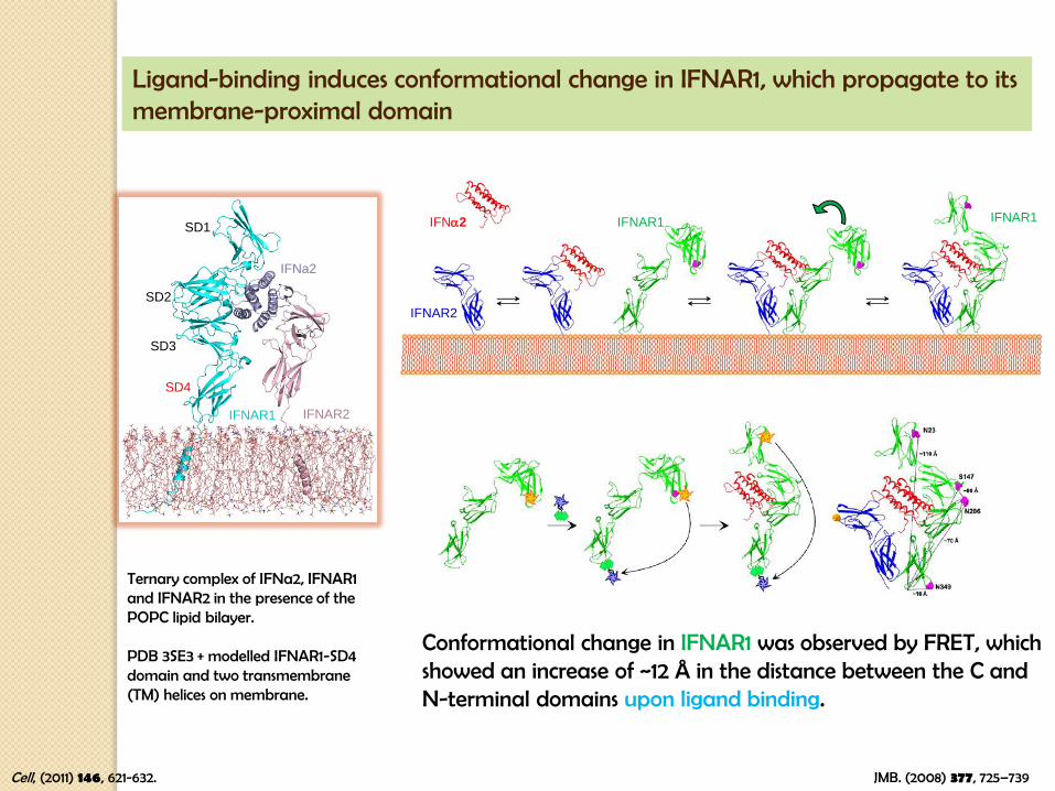
Ligand-binding induces conformational change in IFNAR1, which propagate to its membrane-proximal domain
IFNAR2
IFNa2 IFNAR1 IFNAR1
JMB. (2008) 377, 725–739
Conformational change in IFNAR1 was observed by FRET, which showed an increase of ~12 Å in the distance between the C and N-terminal domains upon ligand binding.
SD1
SD2
SD3
SD4
IFNa2
IFNAR2IFNAR1
Ternary complex of IFNa2, IFNAR1 and IFNAR2 in the presence of the POPC lipid bilayer.
PDB 3SE3 + modelled IFNAR1-SD4 domain and two transmembrane (TM) helices on membrane.
Cell, (2011) 146, 621-632.

Intrinsic ability of bound form IFNAR1 on membranes to adapt its conformation to functional interactions (observed in experiments)
SD4
SD3
Coupled movements of the ternary complex and membrane
The SD3 and SD4 domains moved close to each other. The distance between residues N23 (red sphere) and T407 (magenta sphere) decreased by ~10 Å. IFNAR1, IFNa2 and IFNAR2 in the ternary complex are colored cyan, light blue and pink, respectively.
membrane ANM mode 6

86.6 Å
107.4 Å
Mouse (bound) Human (bound)MD snapshot
MD
sim
ula
tion
s
Human (bound) Human (unbound)
Nat Immuol, (2013) 14, 901-907.
SD4
SD2SD1 Human IFNAR1
Mouse IFNAR1
SD3
Cry
sta
l st
ruct
ures
RMSD: 10.2 Å
SD4
SD3SD2
SD1
Mouse IFNAR1ANM mode 3
Human IFNAR1ANM mode 1Alo
ng A
NM
m
odes
RMSD: 6.5 Å
Human IFNAR1, ANM mode 1
IFNAR1 – Extreme Flexibility/Adaptability due to modularity
A B
ANM mode 4RMSD 4.1 -> 2.3 Å
109.5 Å
97.2 Å
mode 1
109.5 Å
100.4 Å
mode 2
Bound crystal structure / Unbound crystal structure / ANM-prediction, starting from unbound

Blue circles highlight sequentially distant inter-subdomain interactions. They are subject to large ANM distance fluctuations that permit them to come into close proximity to form disulfide bridges even if in the equilibrium state they are not so close to each other, and they are located on opposite sides of a GNM hinge.
GNM-based identification of IFNAR1 hinge sites
Q100
E199
Q302
A B
SD1
SD2SD3
SD4
F238
G133
I19V187
L134R241
I162
E293
K164N207
K220L306
Residue
Resid
ue
Inter-residue distance fluctuationsC D
GN
M M
od
e 2
Residue Residue
GN
M M
od
e 1
E199
K240L247
Q100
Q302
B
Q213-
Q215
C328-
N331
GNM hinge residues (blue space-filling) at the interface
Membrane ANM mode 6
Dr. Ignacio General
Universidad Nacional de San MartinBuenos Aires, Argentina
GNM: http://gnm.csb.pitt.eduANM: http://anm.csb.pitt.edu

I162E293 (green) and K164N207 (blue)
K220L306 (blue)
G133F238 (purple), L134R241 (green) and I19V187 (blue)
SD1 --> SD4
Cry
sta
l str
uctu
res
Alo
ng A
NM
modes
9.4 Å
8.1 Å
Mode 1 I162-E293Mode 4 G133-F238
14.1 Å
9.1 Å
Mode 4 L134-R241
8.2 Å
6.7 Å
I162
E293
I162
E293
G133F238
G133F238
L134
R241
L134
R241
The structural mobility of IFNAR1 allows selected residues pairs to come into proximity and form disulfide bridges.

Residue pairs located on opposite sides of GNM hinges selected for cross-linking experiments and functional assays
Set I – residue pairs predicted by ANM soft modes to alter global dynamics if cross-linked
Subdo-mains
Res 1 Res 2 Dista (Å)ANM results
Mode MaxVar (Å)b Max d (Å) Min d (Å) E-Min dc(Å) E2-Min dd (Å)
SD2-SD3
G133 F238 14.1 4 5.1 17.0 11.9 11.6 9.1
L134 R241 8.2 4 2.1 9.4 7.3 7.4 6.7
I162 E293 9.4 1 2.7 10.8 8.1 8.3 8.1
K164 N207 13.7 1 4.5 16.1 11.6 8.9 8.4
SD3-SD4 K220 L306 14.9 2 4.6 17.4 12.9 14.5 12.6
SD1-SD2 I19 V187 14.2 4 5.2 17.1 11.9 10.7 9.7
Set II – pairs located in the vicinity of hinge centers
SD2-SD3Y163 E293 7.6 2 0.7 8.2 7.5 6.0 6.2
E111 F290 7.5 1 1.3 8.2 6.9 5.8 5.6
SD1-SD2D15 T123 6.5 2 0.3 6.7 6.4 5.9 6.0
D16 V127 6.4 4 0.2 6.6 6.4 6.5 6.6
SD3-SD4 C268 C328 6.8 4 0.4 7.1 6.7 8.2 8.3a Distances between the two residues (based on Ca atoms) in the human IFNAR1 crystal structure, unbound form (PDB id: 3s98) with modeled SD4b MaxVar is the difference between the maximal and minimal distances (Max d and Min d) between the two residues in the examined mode (based on Ca atoms) using the scaling parameter a = 27; c E-Min d is the value for Min d after energy minimization; d E2-Min d is the closest separation attained after a 2nd round of displacement along the same modes and energy minimization.

WT
95.4 Å
SD
4
N349
W347
A SD34B
Q268C
Q328C
107.7 Å
N349
W347
N23
T407
Disulfide bridge formation between 268-328 (SD34) and its restrictive effect on the dynamics of IFNAR1 SD4.
SD4 movement is severely restricted in the double mutant SD34 (B), compared to the WT (A).
White : initial IFNAR1 conformationCyan : MD sampled IFNAR1 conformation
The distances (W347-N349) in SD34 (gray) are in low level (strong quenching effects), compared to the WT (black).
Dr. Gideon Schreiber
Dept. Biomolecular SciencesWeizmann Institute of Science
The 268-328 disulfide bond hinders de-quenching of the fluorescence of Trp 347 upon IFN binding, suggesting inhibition of SD4 movement upon IFN binding.

The cross-links (G133-F238 and L134-R241) with increasing distance from the global hinge center have stronger effects to the binding, due to increased moment arm.
Conclusion:
Dynamics modulates binding affinity, which in turn, modulates biological activity.
Hinge center
Thanks
Dr. Gideon Schreiber
Dept. Biomolecular Sciences
Weizmann Institute of Science
Dr. Ignacio General
Universidad Nacional de San Martin
Buenos Aires, Argentina
Funding
NIH grant P41 GM103712.
Acknowledgments
Dr. Nanaocha Sharma
Dept. Biomolecular Sciences
Weizmann Institute of Science
Dr. Hongchun Li
Computational & Systems Biology Dept.School of MedicineUniversity of Pittsburgh

The change observed experimentally between the unbound (green) and bound (cyan) structures of IFNAR1 agrees with the changes intrinsically favored by ANM.
The diagram colored orange is obtained by starting from the unbound form (green) and deforming it along its intrinsically accessible ANM mode 4.
ANM Mode 4
Intrinsic ability of IFNAR1 to adapt its conformation to functional interactions (observed in experiments) upon movements along its structure-encoded global modes.
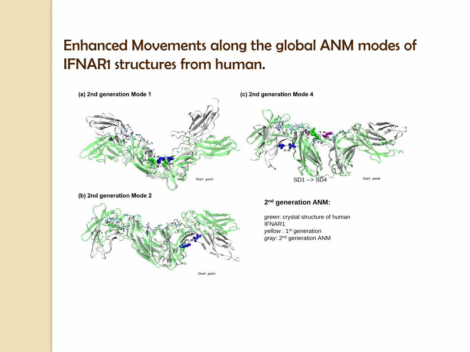
2nd generation ANM:
green: crystal structure of human
IFNAR1
yellow : 1st generation
gray: 2nd generation ANM
Enhanced Movements along the global ANM modes of IFNAR1 structures from human.
SD1 --> SD4