Embed Size (px)
Citation preview

Dynamic Gene Expression Profiling Using aMicrofabricated Living Cell Array
Deanna M. Thompson,† Kevin R. King,† Kenneth J. Wieder, Mehmet Toner, Martin L. Yarmush, andArul Jayaraman*
Center for Engineering in Medicine/Department of Surgery, Massachusetts General Hospital, Harvard Medical School, andShriners Hospitals for Children, Boston, Massachusetts 02114
We describe the development of a microfluidic platformfor continuous monitoring of gene expression in live cells.This optically transparent microfluidic device integrateshigh-throughput molecular stimulation with nondestruc-tive monitoring of expression events in individual livingcells, hence, a living cell array (LCA). Several concentra-tions of a soluble molecular stimulus are generated in anupstream microfluidic network and used to stimulatedownstream reporter cells, each containing a green fluo-rescence reporter plasmid for a gene of interest. Cellularfluorescence is continuously monitored and quantified toinfer the expression dynamics of the gene being studied.We demonstrate this approach by profiling the activationof the transcription factor NF-KB in HeLa S3 cells inresponse to varying doses of the inflammatory cytokineTNF-r. The LCA platform offers a unique opportunity tosimultaneously control dynamic inputs and measuredynamic outputs from adherent mammalian cells in ahigh-throughput fashion. This approach to profiling ex-pression dynamics, in conjunction with complementarytechniques such as DNA microarrays, will help provide amore complete picture of the dynamic cellular responseto diverse soluble stimuli.
Technologies such as Northern blots and reverse transcriptionpolymerase chain reaction have been extensively applied to themonitoring of gene expression in cells and tissues for numerousbiological investigations. Recent developments in DNA microarraytechnology have further expanded the scope of these investiga-tions by enabling the simultaneous monitoring of several genes.1-4
While these techniques provide snapshots of changes in geneexpression at single time points, they are not ideal for investigatingtime-dependent behavior. Often, to approximate continuous-timemeasurements, multiple cell populations are destructively mea-sured at several discrete time points. A technique allowingcontinuous, nondestructive monitoring would complement current
genomics technologies by providing a more dynamic picture ofgene expression. Green fluorescent protein (GFP) technologies5
have recently emerged to allow noninvasive measurements of cellfunction and cell responses;6-8 however, GFP-based expressionstudies are typically performed in traditional single-dish ormultiwell formats to monitor a small number of stimulationconditions.
Microfluidics is an inherently scalable technology, offering aunique opportunity to simultaneously screen a wide range ofexperimental conditions with relative ease (different concentra-tions, combinations, and temporal profiles of molecular inducers,inhibitors, and modulators). Poly(dimethylsiloxane) (PDMS) mi-crofluidic devices9,10 are ideal for cell-based applications involvingfluorescent protein expression because of their well-establishedbiocompatibility and optical transparency. They have been appliedto several biological studies including chemotaxis,11 cell and small-molecule patterning,12,13 cell sorting,14,15 and biochemical separa-tions.16 Recent advances in cell-based microfluidics have enabledadherent mammalian cells to be cultured in microchannels fordays to weeks;17 however, functional measurements such as geneexpression have not been demonstrated.
* To whom correspondence should be addressed: (phone) (617) 371-4874;(fax) (617) 573-9471; (e-mail) [email protected].
† Both authors contributed equally.(1) DeRisi, J.; Penland, L.; Brown, P. O.; Bittner, M. L.; Meltzer, P. S.; Ray, M.;
Chen, Y.; Su, Y. A.; Trent, J. M. Nat. Genet. 1996, 14, 457-460.(2) Brown, P. O.; Botstein, D. Nat. Genet. 1999, 21, 33-37.(3) Diehn, M.; Eisen, M. B.; Botstein, D.; Brown, P. O. Nat. Genet. 2000, 25,
58-62.(4) Roth, C. M.; Yarmush, M. L. Annu. Rev. Biomed. Eng. 1999, 1, 265-297.
(5) Ding, G. J.; Fischer, P. A.; Boltz, R. C.; Schmidt, J. A.; Colaianne, J. J.; Gough,A.; Rubin, R. A.; Miller, D. K. J. Biol. Chem. 1998, 273, 28897-28905.
(6) Carroll, J. A.; Stewart, P. E.; Rosa, P.; Elias, A. F.; Garon, C. F. Microbiology2003, 149, 1819-1828.
(7) Li, H. Y.; Ng, E. K.; Lee, S. M.; Kotaka, M.; Tsui, S. K.; Lee, C. Y.; Fung, K.P.; Waye, M. M. J. Cell. Biochem. 2001, 80, 293-303.
(8) Li, J.; Xu, H.; Herber, W. K.; Bentley, W. E.; Rao, G. Biotechnol. Bioeng.2002, 79, 682-693.
(9) McDonald, J. C.; Duffy, D. C.; Anderson, J. R.; Chiu, D. T.; Wu, H.; Schueller,O. J.; Whitesides, G. M. Electrophoresis 2000, 21, 27-40.
(10) Anderson, J. R.; Chiu, D. T.; Jackman, R. J.; Cherniavskaya, O.; McDonald,J. C.; Wu, H.; Whitesides, S. H.; Whitesides, G. M. Anal. Chem. 2000, 72,3158-3164.
(11) Li Jeon, N.; Baskaran, H.; Dertinger, S. K.; Whitesides, G. M.; Van de Water,L.; M, T. Nat. Biotechnol. 2002, 20, 828-836.
(12) Folch, A.; Toner, M. Biotechnol. Prog. 1998, 14, 388-392.(13) Takayama, S.; Ostuni, E.; LeDuc, P.; Naruse, K.; Ingber, D. E.; Whitesides,
G. M. Nature 2000, 411, 1016.(14) Fu, A. Y.; Chou, H. P.; Spence, C.; Arnold, F. H.; Quake, S. R. Anal. Chem.
2000, 74, 2451-2457.(15) Cho, B. S.; Schuster, T. G.; Zhu, X.; Chang, D.; Smith, G. D.; Takayama, S.
Anal. Chem. 2003, 75, 1671-1675.(16) Ricco, A. J.; Boone, T. D.; Fan, Z. H.; Gibbons, I.; Matray, T.; Singh, S.;
Tan, H.; Tian, T.; Williams, S. J. Biochem. Soc. Trans. 2002, 30, 73-78.(17) King, K. R.; Terai, H.; Wang, C. C.; Vacanti, J. P.; Borenstein, J. T.
Microfluidics for Tissue Engineering Microvasculature: Endothelial CellCulture. Fifth International Conference on Miniaturized Chemical andBiochemical Analysis Systems, Monterey, CA, October 21-25, 2001;pp 247-249.
Anal. Chem. 2004, 76, 4098-4103
4098 Analytical Chemistry, Vol. 76, No. 14, July 15, 2004 10.1021/ac0354241 CCC: $27.50 © 2004 American Chemical SocietyPublished on Web 06/09/2004

In this report, we describe the development of a functionalgenomics tool that combines GFP reporter technology andmicrofabrication for dynamic gene expression profiling. This tool,the living cell array (LCA) (Figure 1A), can be used for simulta-neously stimulating and monitoring the time course of geneexpression in living cells. The feasibility of studying dynamic geneexpression using the LCA is demonstrated by profiling theactivation of a transcription factor (NF-κB) in response to eightdifferent doses of a cytokine (TNF-R). The resultant fluorescenceof the entire population is monitored with single-cell resolution,allowing direct measurement of population dynamics and biologi-cal heterogeneity in the culture. The LCA has the potential tosignificantly impact investigations where various gene expressionevents and their interactions need to be studied in a time-dependent manner with individual cell resolution.
EXPERIMENTAL PROTOCOLSMicrofluidic Design. The LCA consists of an upstream
dilution module that generates a range of stimulus concentrationsand a downstream cell culture module where EGFP reporter celllines are grown in individual chambers (Figure 1B) of a singlemicrofluidic network. The dilution module, based on the fluidicsof a microgradient generator previously described by Li Jeon et
al.,11 consists of a highly interconnected network that generateseight outlet concentrations spanning two inlet concentrations.Successive steps of diffusive mixing between adjacent laminar flowstreams occur in long (50 × 75 × 10000 µm) channels to ensurecomplete mixing at the relevant flow rates. Each outlet streamthen feeds into a downstream array of 800 µm × 500 µm cellchambers (Figure 1B).
Device Fabrication. Microchannels and cell chambers werefabricated in PDMS (Dow Corning, Corning, NY) using softlithography and rapid prototyping techniques.9 Inlets and outletswere drilled with a blunted and beveled syringe needle, and theresulting PDMS microfluidic network was irreversibly bonded toa glass slide assisted by oxygen plasma surface treatment (150mTorr, 50 W, 20 s), creating a sterile optically transparent devicefor cell culture and gene expression profiling.
Bioreactor Design. The supporting bioreactor consisted ofthe microfluidic devices, medium-containing reservoirs for gravity-driven flow, and associated tubing. Inlet and outlet reservoirs weredrilled and outfitted with plastic connectors (Small Parts Inc.,Miami Lakes, FL). Inlet reservoirs were tapped at the bottom toallow medium outflow without air incorporation or bubble genera-tion. Outlet reservoirs were tapped near the top to drain cultureeffluent without suffering back-pressure buildup. All reservoirswere outfitted with sterile filter caps to allow continuous pressureequilibration for the entire system while maintaining sterility.
GFP Reporter Cell Line. HeLa S3 cells (ATCC, Rockville,MD.) were grown in high-glucose Dulbecco’s modified Eaglesmedium (Invitrogen, Gaithersburg, MD) supplemented with 10%bovine calf serum and 100 units/mL penicillin per 100 µg/µLstreptomycin in a humidified 5% CO2 incubator at 37 °C. PlasmidpNF-κB/CMVmind2EGFP (referred to as pd2NF-κB) was con-structed as described elsewhere.18 Briefly, promoter, responseelement, and d2EGFP reporter gene (EGFP with a 2-h half-life)were excised from a Clontech Living Colors plasmid (Clontech,Paolo Alto, CA) and cloned into an expression vector. Cells wereelectroporated, selected for plasmid integration, sorted for maxi-mum responsiveness with minimal background fluorescence,18 andsubcloned to obtain the stable reporter cell line (HeLa-NF).
Cell Seeding. Microfluidic bioreactors were autoclave steril-ized, rinsed with PBS, and degassed by driving trapped air throughthe walls of the gas-permeable device. Fluidic networks wereprecoated with 50ng/mL fibronectin (Sigma Chemical) overnightto promote HeLa-NF cell attachment. Excess fibronectin wasremoved by rinsing with PBS, and devices were seeded with cells.A suspension of HeLa-NF cells (3 × 10⁶ cells/mL) was injectedthrough the device outlet and allowed to settle under staticconditions for ∼18 h. The device was aseptically connected tothe remainder of the bioreactor to provide sterile gravity-drivenmedium flow, and the entire microfluidic reactor was moved toan incubated stage on a fluorescence microscope to perform thestimulus-response assay. During the assay, cells were continu-ously perfused with fresh medium (with no phenol red) at ∼1.0µL/min to replenish critical metabolites and remove potentiallytoxic wastes. Static control experiments were prepared by seeding2 × 104 HeLa-NF cells into 12-well tissue culture dishes (Costar),and experiments were performed at ∼40-60% confluency. All
(18) Wieder, K. J.; Thompson, D. M.; Wang, S.; Zia, C.; Foley, A. M.; Yarmush,M. L.; Jayaraman, A. Biochem. Biophys. Res. Commun. Submitted.
Figure 1. Living cell array (LCA) (A) Schematic representation ofthe LCA device. Medium containing a soluble mediator enters thedevice, delivers nutrients, stimulates cells cultured in the cell cham-bers, and exits into a waste stream. Gene expression dynamics areobtained by time-lapse imaging and quantified using image analysissoftware. (B) The microfluidic network design and micrographs of thedilution and cell cultivation modules are shown. The transparentPDMS device allows visualization of the TNF-R gradient (doped withfluorescein dye) and EGFP reporter cells. The microfluidic channelsin the upstream dilution module are 50 µm in width and 50 µm inheight and generate several concentrations of the stimulus bycontinuous-flow diffusive mixing of adjacent laminar flow streams. Thevarious concentrations are delivered to the downstream array ofculture chambers, each 800 µm long and 500 µm wide.
Analytical Chemistry, Vol. 76, No. 14, July 15, 2004 4099

experiments were performed on an incubated stage (37 °C and5% CO2) of a Zeiss Axiovert 200 microscope (Carl Zeiss Inc.,Thornwood, NY).
Induction of NF-KB in the LCA. HeLa-NF cells were grownto ∼40% confluence in either the LCA or standard tissue culturedishes and continuously stimulated with 10 ng/mL TNF-R. Phasecontrast and fluorescence images were obtained every 10-60 minat preprogrammed position using a 20× objective (Carl Zeiss).Microfluidic dose-response experiments were performed bydelivering growth medium containing 0.1 mg/mL RITC-dextran(Sigma) and 10 ng/mL TNF-R through one inlet and unmodifiedmedium through the other. Inlet reservoirs were maintained atelevated heights with respect to the outlet reservoir and adjustedto achieve the desired concentrations. Continuous-flow dilutionswere monitored throughout each experiment by fluorescenceimaging of comparable molecular weight (17 200) RITC-dextranindicator. The total flow rate was ∼1 µL/min. Phase contrast andfluorescence images were captured every 30-60 min at threepositions per TNF-R concentration.
Image Analysis. Fluorescent images were analyzed usingMetamorph V 6.0r4 (Universal Imaging Corp., Downington, PA).The average fluorescence of each cell region was corrected formedium and device fluorescence as well as illumination fluctua-tions and nonuniformities by subtracting the average localbackground fluorescence at each time point. The result was scaledby the region area and divided by the number of cells in the regionto determine an average intensity per cell in the region. Thedifference between the initial and final average intensity per cellwas plotted to compare the response to the various concentrationsof TNF-R.
RESULTS AND DISCUSSIONDevelopment of the LCA. We present a LCA platform that
combines microfluidic solution handling with GFP reportertechnology to continuously and noninvasively profile gene expres-sion dynamics in living cells. The specific LCA device describedhere generates several concentrations of a soluble mediator andstimulates GFP reporter cells cultured in the device. Activationof gene expression results in the induction of fluorescence thatis continuously monitored to quantify gene expression dynamics(Figure 1A). The LCA contains two components: (1) a dilutionmodule of microfluidic channels and (2) multiple cell culturechambers integrated in a single device as shown in Figure 1B.
The dilution module of the LCA device described in this studywas designed to generate eight concentrations of a solublemediator such as the cytokine TNF-R. Fluorescent RITC-dextran(17 200) was used as a detectable indicator for the comparablemolecular weight TNF-R (17 000) used in this study. Thefluorescence was monitored throughout the entire dilution moduleto verify complete diffusive mixing. The final concentrationentering the downstream cell culture chambers was monitoredby fluorescence microscopy to quantify the stimulus and was foundto remain steady throughout the experiment. Figure 2A showsthe corresponding TNF-R concentration profile ranging from 0to 10 ng/mL generated using the dilution module. The dilutionmodule in the current LCA prototype can be used to generate alinear range of concentrations of any soluble molecular mediatorspanning the two inlet concentrations. By varying the inletconcentration of the mediator, it is possible to precisely andreproducibly generate a range of well-defined concentrations of a
Figure 2. Characterization of the LCA dilution and cell culture modules. (A) Generation of TNF-R concentrations in the dilution module. Arange of concentrations (0, 0.36, 1.39, 2.85, 5.06, 7.24, 9.04, and 10 ng/mL) of RITC-conjugated dextran (17.2 kDa) were generated in thedilution module of the LCA as described in Experimental Protocols. Fluorescence micrographs of the junction between the dilution and cellculture modules are shown. The fluorescence images were quantified and the concentration of TNF-R entering each cell culture chamber wasinferred. (B) Viability of HeLa-NF cells in the cell culture chambers. HeLa-NF cells were seeded in the device at three different cell densities (1× 10⁶, 5 × 10⁶, and 10 × 106 cells/mL) and allowed to attach in the LCA device for 24 h. Cells were stained with the LIVE/DEAD viability stainand observed on an inverted microscope with a 20× objective to assess viability. In the figure, live cells are stained green and dead cells arestained red.
4100 Analytical Chemistry, Vol. 76, No. 14, July 15, 2004

soluble molecular mediator such as the cytokine TNF-R. The datapresented here demonstrate the ability to generate a range ofconcentrations in the dilution module and deliver them todownstream preseeded cells.
Microfluidic Cell Culture. The PDMS/glass chamber wascoated with fibronectin to promote cell attachment to the device.Based on previous work in our laboratory, this extracellularmatrix protein maximized HeLa-NF cell attachment when com-pared to laminin and collagen (not shown). For the HeLa-NF cellsused in this work, overnight incubation with 50-100 ng/mLfibronectin was sufficient to support uniform cell spreading andmaintenance of morphology similar to that observed on tissueculture plastic. In the absence of surface modification, however,few cells attached.
Cells were introduced into the device via the inlet and allowedto attach under static conditions. Due to the large surface area-to-volume ratios in the device, a concentrated cell suspension ((1-10) × 106 cells/mL) was required. This concentration, ∼2-foldgreater than conventional tissue culture protocols, allows efficientseeding by delivering a large number of cells into the channelsof the small-volume device. Seeded devices were placed in tissueculture incubators for 24 h to allow for proper cell attachmentand spreading prior to stimulation with TNF-R.
The suitability of the cell chambers for seeding and maintainingcells was determined using cell viability measurements. HeLa-NF cells were seeded in the LCA cell chambers as described inExperimental Protocols and allowed to grow under flow conditionsfor 24 h prior to viability measurements using the LIVE/DEADstain (Molecular Probes, Eugene, OR). Our data show greaterthan 90% viability over a 10-fold range of cell seeding densities
((1-10) × 106 cells/mL; Figure 2B). Furthermore, HeLa celldivision was also routinely observed in the LCA cell chambers,suggesting that the microfabricated LCA provided an environmentconducive to cell proliferation (not shown).
Expression Dynamics in the LCA versus Static TissueCulture. We demonstrated the feasibility of monitoring geneexpression dynamics in the LCA device by profiling the inductionof NF-κB in HeLa-NF reporter cells and comparing to standardtissue culture formats. Cells were seeded in the LCA as describedin Experimental Protocols and periodically monitored for 24 h toobtain a stable baseline fluorescence measurement prior toinduction of NF-κB. NF-κB was stimulated in HeLa-NF cells byperfusing with medium containing 10 ng/mL TNF-R. The fluo-rescence profile was monitored over time and showed an increasein fluorescence relative to the baseline value (Figure 3A). Incomparison, no significant fluorescence signal was observed forHeLa-NF cells in the absence of TNF-R stimulation (not shown).The fluorescence profile was quantified using image analysis andcompared to that observed for HeLa-NF cells stimulated with 10ng/mL TNF-R in a six-well tissue culture plate (i.e., staticincubation). The temporal fluorescence profiles in the LCAmirrored those observed in standard tissue culture, with similarincreases in fluorescence relative to unstimulated controls occur-ring by 2 h (Figure 3B).
Cells cultivated in the LCA experience shear stress of ∼0.5dyn/cm2 (based on a flow rate of 1 µL/min and chamberdimensions of 500-µm width and 50-µm height) due to thecontinuous perfusion of medium, as compared to cells in statictissue culture formats. This value is below the range of shear
Figure 3. Induction of EGFP fluorescence in the LCA. (A) HeLa-NF cells were grown in the LCA and stimulated with 10 ng/mL TNF-R. Phaseand fluorescence images were captured on a Zeiss Inverted microscope every 10 min using a 20× objective. Representative fluorescenceimages from a single region in the LCA are shown at various time points during the course of the experiment. (B) The temporal fluorescenceprofiles from TNF-R stimulated HeLa-NF cells in the LCA at a flow rate of 1.0 µL/min and in standard tissue culture dishes were determined. Thetime course of fluorescence was found to be similar in both culture platforms. Normalized data are shown to facilitate comparison.
Analytical Chemistry, Vol. 76, No. 14, July 15, 2004 4101
stress values reported to disrupt cell function.19 This is especiallyimportant for NF-κB, as its responsiveness to shear stress is welldocumented.20 The similar fluorescence profiles between the LCAand static culture suggest minimal shear induction.
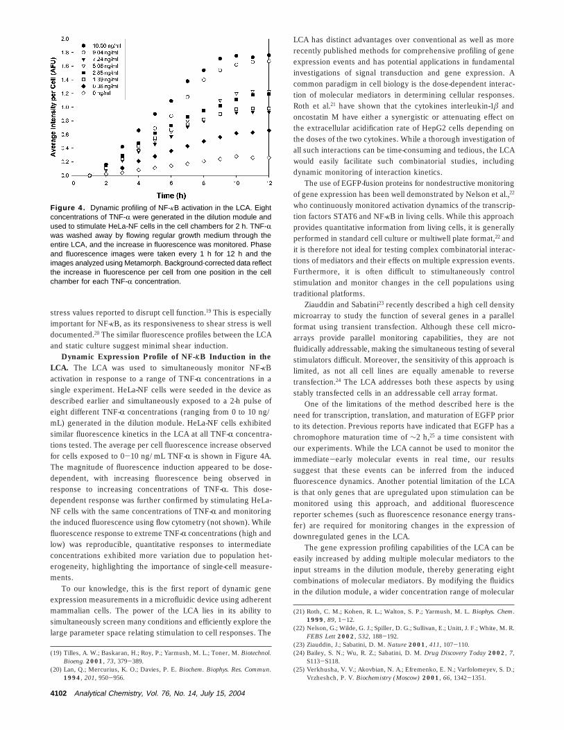
Dynamic Expression Profile of NF-KB Induction in theLCA. The LCA was used to simultaneously monitor NF-κBactivation in response to a range of TNF-R concentrations in asingle experiment. HeLa-NF cells were seeded in the device asdescribed earlier and simultaneously exposed to a 2-h pulse ofeight different TNF-R concentrations (ranging from 0 to 10 ng/mL) generated in the dilution module. HeLa-NF cells exhibitedsimilar fluorescence kinetics in the LCA at all TNF-R concentra-tions tested. The average per cell fluorescence increase observedfor cells exposed to 0-10 ng/mL TNF-R is shown in Figure 4A.The magnitude of fluorescence induction appeared to be dose-dependent, with increasing fluorescence being observed inresponse to increasing concentrations of TNF-R. This dose-dependent response was further confirmed by stimulating HeLa-NF cells with the same concentrations of TNF-R and monitoringthe induced fluorescence using flow cytometry (not shown). Whilefluorescence response to extreme TNF-R concentrations (high andlow) was reproducible, quantitative responses to intermediateconcentrations exhibited more variation due to population het-erogeneity, highlighting the importance of single-cell measure-ments.
To our knowledge, this is the first report of dynamic geneexpression measurements in a microfluidic device using adherentmammalian cells. The power of the LCA lies in its ability tosimultaneously screen many conditions and efficiently explore thelarge parameter space relating stimulation to cell responses. The
LCA has distinct advantages over conventional as well as morerecently published methods for comprehensive profiling of geneexpression events and has potential applications in fundamentalinvestigations of signal transduction and gene expression. Acommon paradigm in cell biology is the dose-dependent interac-tion of molecular mediators in determining cellular responses.Roth et al.21 have shown that the cytokines interleukin-1â andoncostatin M have either a synergistic or attenuating effect onthe extracellular acidification rate of HepG2 cells depending onthe doses of the two cytokines. While a thorough investigation ofall such interactions can be time-consuming and tedious, the LCAwould easily facilitate such combinatorial studies, includingdynamic monitoring of interaction kinetics.
The use of EGFP-fusion proteins for nondestructive monitoringof gene expression has been well demonstrated by Nelson et al.,22
who continuously monitored activation dynamics of the transcrip-tion factors STAT6 and NF-κB in living cells. While this approachprovides quantitative information from living cells, it is generallyperformed in standard cell culture or multiwell plate format,22 andit is therefore not ideal for testing complex combinatorial interac-tions of mediators and their effects on multiple expression events.Furthermore, it is often difficult to stimultaneously controlstimulation and monitor changes in the cell populations usingtraditional platforms.
Ziauddin and Sabatini23 recently described a high cell densitymicroarray to study the function of several genes in a parallelformat using transient transfection. Although these cell micro-arrays provide parallel monitoring capabilities, they are notfluidically addressable, making the simultaneous testing of severalstimulators difficult. Moreover, the sensitivity of this approach islimited, as not all cell lines are equally amenable to reversetransfection.24 The LCA addresses both these aspects by usingstably transfected cells in an addressable cell array format.
One of the limitations of the method described here is theneed for transcription, translation, and maturation of EGFP priorto its detection. Previous reports have indicated that EGFP has achromophore maturation time of ∼2 h,25 a time consistent withour experiments. While the LCA cannot be used to monitor theimmediate-early molecular events in real time, our resultssuggest that these events can be inferred from the inducedfluorescence dynamics. Another potential limitation of the LCAis that only genes that are upregulated upon stimulation can bemonitored using this approach, and additional fluorescencereporter schemes (such as fluorescence resonance energy trans-fer) are required for monitoring changes in the expression ofdownregulated genes in the LCA.
The gene expression profiling capabilities of the LCA can beeasily increased by adding multiple molecular mediators to theinput streams in the dilution module, thereby generating eightcombinations of molecular mediators. By modifying the fluidicsin the dilution module, a wider concentration range of molecular
(19) Tilles, A. W.; Baskaran, H.; Roy, P.; Yarmush, M. L.; Toner, M. Biotechnol.Bioeng. 2001, 73, 379-389.
(20) Lan, Q.; Mercurius, K. O.; Davies, P. E. Biochem. Biophys. Res. Commun.1994, 201, 950-956.
(21) Roth, C. M.; Kohen, R. L.; Walton, S. P.; Yarmush, M. L. Biophys. Chem.1999, 89, 1-12.
(22) Nelson, G.; Wilde, G. J.; Spiller, D. G.; Sullivan, E.; Unitt, J. F.; White, M. R.FEBS Lett 2002, 532, 188-192.
(23) Ziauddin, J.; Sabatini, D. M. Nature 2001, 411, 107-110.(24) Bailey, S. N.; Wu, R. Z.; Sabatini, D. M. Drug Discovery Today 2002, 7,
S113-S118.(25) Verkhusha, V. V.; Akovbian, N. A.; Efremenko, E. N.; Varfolomeyev, S. D.;
Vrzheshch, P. V. Biochemistry (Moscow) 2001, 66, 1342-1351.
Figure 4. Dynamic profiling of NF-κB activation in the LCA. Eightconcentrations of TNF-R were generated in the dilution module andused to stimulate HeLa-NF cells in the cell chambers for 2 h. TNF-Rwas washed away by flowing regular growth medium through theentire LCA, and the increase in fluorescence was monitored. Phaseand fluorescence images were taken every 1 h for 12 h and theimages analyzed using Metamorph. Background-corrected data reflectthe increase in fluorescence per cell from one position in the cellchamber for each TNF-R concentration.
4102 Analytical Chemistry, Vol. 76, No. 14, July 15, 2004

mediators can also be tested. The flexibility of rapid prototypingfor device fabrication makes it possible to further modify thecurrent LCA prototype design to increase the number of stimula-tion conditions or factors tested. For one, the number of inputstreams can be increased to screen complex combinations ofmediators in a single experiment. Similarly, it is also possible tomodify the device such that more than one type of reporter cellline is cultivated in the cell chambers, thereby enabling multiplemolecular events to be monitored and compared simultaneously.Additionally, the combinations of molecular mediators can bedelivered according to a defined temporal profile, ranging fromshort pulses (∼1 min) to continuous stimulation. Integration ofthese features into the LCA would enable complex expressionprofiling measurements from a small set of experiments, whichwould be difficult to obtain using standard tissue culture formats.
Dynamic profiling is not only limited to monitoring transcrip-tion factor activation but is also equally applicable to the study ofother gene expression aspects such as promoter activity and
protein-protein interactions. The incorporation of noninvasive andcontinuous monitoring as well as parallel processing in the LCAhas significant implications for fundamental investigations of cellbiology. These include applications involving drug discovery,toxicology, and biosensor development where continuous moni-toring of time-dependent gene expression and efficient evaluationof multiple conditions are important.
ACKNOWLEDGMENTThis work was partially supported by grants from the Whitaker
Foundation (RG-01-0117) and the Shriners Hospital for Children(8650) to A.J.. The authors acknowledge use of facilities at theMicroscale Core at CEM and the Morphology Core at SBH.
Received for review December 3, 2003. Accepted April 28,2004.
AC0354241
Analytical Chemistry, Vol. 76, No. 14, July 15, 2004 4103