Embed Size (px)
Citation preview
J Physiol 569.1 (2005) pp 75–89 75
Dynamic conformational changes of extracellular S5–Plinkers in the hERG channel
Min Jiang1, Mei Zhang1, Innokenty V. Maslennikov2, Jie Liu1, Dong-Mei Wu1, Yuliya V. Korolkova2,Alexander S. Arseniev2, Eugene V. Grishin2 and Gea-Ny Tseng1
1Department of Physiology, Virginia Commonwealth University, Richmond, VA 23298, USA2Shemyakin-Ovchinnikov Institute of Bioorganic Chemistry, Russian Academy of Sciences, Moscow, 117997 Russia
The hERG channel has an unusually long ‘S5–P linker’ (residues 571–613) that lines the outermouth of the pore. Previously, we have shown that residues along this S5–P linker are criticalfor the fast-inactivation process and K+ selectivity of the hERG channel. Here we used severalapproaches to probe the structure of this S5–P linker and its interactions with other domainsof the hERG channel. Circular dichroism and NMR analysis of a synthetic hERG S5–P linkerpeptide suggested that this linker is quite dynamic: its central region (positions 583–593) can beunstructured or helical, depending on whether it is immersed in an aqueous phase or in contactwith a hydrophobic environment. Cysteine introduced into positions 583–597 of the S5–P linkercan form intersubunit disulphide bonds, and at least four of them (at 584, 585, 588 and 589)can form disulphide bonds with counterparts from neighbouring subunits. We propose that thefour S5–P linkers in a hERG channel can engage in dynamic conformational changes duringchannel gating, and interactions between S5–P linkers from neighbouring subunits contributeimportantly to channel inactivation.
(Resubmitted 27 June 2005; accepted after revision 2 September 2005; first published online 8 September 2005)Corresponding author G.-N. Tseng: Department of Physiology, Virginia Commonwealth University, 1101 E. MarshallStreet, Richmond, VA 23298, USA. Email: [email protected]
Human ether-a-go-go related gene (hERG) encodes thepore-forming subunit of rapid delayed rectifier (IKr)channels in human heart (Sanguinetti et al. 1995). Ithas a uniquely fast and voltage-sensitive inactivationprocess (Spector et al. 1996; Smith et al. 1996). Thisfast-inactivation process, along with the slow activationand deactivation rates of the hERG/IKr channel, creates itsinward rectification property (Spector et al. 1996): thereis little outward current through the hERG/IKr channelsduring phase 2 of cardiac action potential due to channelinactivation, but a resurgence of outward current occursduring phase 3 when the hERG/IKr channels recover frominactivation. This outward hERG/IKr current duringphase 3 of cardiac action potentials serves anantiarrhythmic function by preventing prematuredepolarization from triggering extrasystole.
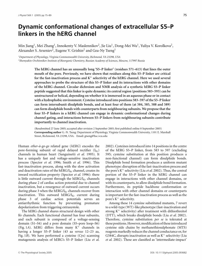
The hERG channel shares structural design with otherKv channels. Each functional channel has four subunits,and each subunit is composed of a voltage-sensingdomain (S1–S4) and a pore domain (S5–pore loop–S6)(Fig. 1A). hERG differs from many K+ channels inhaving a longer S5–P linker (43 aa versus 12–23 aa,Fig. 1B). We have performed a cysteine (Cys) scanningmutagenesis analysis of hERG’s S5–P linker (Liu et al.
2002). Cysteines introduced into 14 positions in the centreof the hERG S5–P linker, from 583 to 597 (excluding595; cysteine substitution at this position produces anon-functional channel) can form disulphide bonds.Disulphide bond formation produces a uniform mutantphenotype: disruption of the fast-inactivation process andthe pore’s K+ selectivity (Liu et al. 2002). Thus, the centralportion of the S5–P linker in the hERG channel canengage in interactions with other channel domains, orwith its counterparts, to allow disulphide bond formation.Furthermore, its peptide backbone conformation orinteraction with other channel domains or counterpartsis important for the fast-inactivation process as well as thepore’s K+ selectivity.
Among these 14 cysteine substituted mutants, 7 revertto a wild-type (WT)-like phenotype (fast-inactivation andstrong K+ selectivity) after treatment with dithiothreitol(DTT), which breaks disulphide bonds (Liu et al. 2002).Therefore, cysteine substitution per se is tolerated atthese positions. However, modification of these introducedcysteine side chains by methanethiosulphonate (MTS)reagents markedly reduces the channel conductance or, forG584C, disrupts inactivation as well as K+ selectivity (Liuet al. 2002). These are classified as ‘intermediate-impact’
C© The Physiological Society 2005 DOI: 10.1113/jphysiol.2005.093682
76 M. Jiang and others J Physiol 569.1
positions, as opposed to ‘low-impact’ positions whereneither cysteine substitution nor subsequent MTSmodification has much impact on the channel function(colour coded blue and black in the hERG sequence inFig. 1B). The other seven cysteine-substituted mutantsmaintain the mutant phenotype after DTT treatment.This suggests that side chain properties at these positionsare critical for the fast-inactivation process and thepore’s K+ selectivity of the hERG channel, so thatcysteine substitution is not tolerated. These are designated‘high-impact’ positions. Position 595 is also a high-impactposition. The high-impact positions along the 583–597segment and those in other regions of the S5–P and P–S6linkers are colour coded red in the hERG sequence inFig. 1B.
Why is the central portion of the S5–P linker so criticalfor the fast-inactivation process and K+ selectivity ofthe hERG channel? Inactivation in the hERG channel issimilar to the C-type inactivation in the Shaker channel(Hoshi et al. 1991; Smith et al. 1996; Herzberg et al. 1998):
Figure 1. The amino acid sequence lining the outer mouth region of hERG suggests unique structureand functionA, two-dimensional diagram of a voltage-gated K+ (Kv) channel subunit, with the following domains marked:transmembrane segments (S1 to S6), pore-loop and the two extracellular linkers lining the outer mouth (S5–P andP–S6 linkers). S5 and S6 correspond to TM1 and TM2 (or outer and inner helices) of 2-transmembrane domain K+channels such as KcsA and MthK, and S5–P linker is equivalent to ‘turrets’ in the K+ channel crystal structures.B, alignment of amino acid sequences of KcsA, KvAP, MthK, Shaker and hERG from the end of S5 (TM1) to themiddle of S6 (TM2). The K+ channel signature sequence that lines the selectivity filter is highlighted by a dottedrectangle. The 583–597 segment of hERG’s S5–P linker is shown as an inset. In the hERG sequence, red and bluecolours denote high- and intermediate-impact positions based on the effects of cysteine substitution on the channelfunction (Liu et al. 2002). C, crystal structure of KcsA, showing 2 diagonal subunits with 3 K+ ions coordinated bythe selectivity filter. TM1, TM2, turret and selectivity filter are marked.
inactivation results from conformational changes aroundthe selectivity filter that prevent ion conduction throughthe pore. The channel pore’s selectivity for K+ ions is alsomainly determined by the selectivity filter (Fig. 1B). Inthe available K+ channel crystal structures (Doyle et al.1998; Jiang et al. 2002; Jiang et al. 2003), the turrets(corresponding to hERG’s S5–P linker) are immersed inthe extracellular solution, making no direct contact withthe selectivity filter (Fig. 1C). However, our data describedabove suggest that this may not be the case for hERG. In thisstudy, we take several approaches to examine the secondarystructure of the 583–597 segment and to explore itsinteraction with other channel domains: (1) circulardichroism (CD) spectral analysis and spatial structurecalculations based on nuclear magnetic resonance (NMR)data are used to directly probe the secondary structure andside chain mobility of a synthetic hERG S5–P linker peptidethat includes the 583–597 segment and its flanking regions,and (2) site-specific mutations in conjunction withbiochemical and electrophysiological analysis are used to
C© The Physiological Society 2005

J Physiol 569.1 Atomic proximity between hERG’s S5–P linkers 77
probe side chain interactions in this segment of an intactchannel and how these interactions impact on channelfunction.
Methods
Circular dichroism (CD) and nuclear magneticresonance (NMR) spectroscopy of a synthetic hERGS5–P linker peptide
Peptide synthesis. A 25-mer ‘hERG S5–P linker’ peptidecorresponding to hERG residues 578–603 was synthesizedby Obrigen (San Diego, CA, USA). The peptide waspurified by reverse phase HPLC and composition of thepurification product was verified by mass spectroscopy.
CD spectroscopy. Sample of hERG S5–P linker peptide(74 µm) in 4.44 mm diphenylamine carboxylic acid (DPC)(peptide/DPC = 1/60) or in 5.92 mm sodium dodecylsulphate (SDS) (peptide/SDS = 1/80) was prepared inH2O and pH was titrated to 3.5. CD spectra were obtainedusing spectropolarimeter J-715 (Jasco, Easton, MD, USA)in the wavelength range 185–250 nm at 22◦C with a0.02 cm quartz cell.
NMR spectroscopy. NMR sample of 0.6 mm hERGS5–P linker peptide in 10% D2O or of 0.35 mm peptidein 21 mm DPC (peptide/DPC = 1/60, in 10% D2O)was used. 1H NMR spectra (double quantum-filteredcorrelated spectroscopy (DQF-COSY) (Rance et al.1983), total correlation spectroscopy (TOCSY) (Bax &Davis, 1985) with mixing times (τm) of 40 and 80 ms,and Overhauser enhancement spectroscopy (NOESY)(Jeener et al. 1979) with τm of 100 and 200 ms) wereacquired using a 600 MHz spectrometer (Unity 600,Varian, Palo Alto, CA, USA) at 30◦C, pH 3.5. NMRdata were processed with VNMR (Varian software),and analysed with the XEASY program (Bartels et al.1995). Proton resonance assignment of hERG S5–P linkerpeptide in DPC was performed by standard procedure(Wuthrich, 1986) using the XEASY program. Threehundred and eighty-six cross-peaks in the NOESY(τm = 100 ms) spectrum of peptide in DPC were assignedunambiguously. Cross-peak intensities of peptide in DPCwere measured in 100 ms NOESY spectrum using theXEASY program. The interproton distance constraintswere derived from NOESY cross-peak volumes via‘1/r6’-calibration, using the CALIBA module of theDYANA program (Guntert et al. 1997). Vicinal spin–spincoupling constants 3JHN-Cα H were determined from thefine structure along the ω2 axis of the non-overlappedNOESY cross-peaks between the corresponding amideproton (at the ω2 frequency) and any proton of otherresidues (at the ω1 frequency). 3JHCα -Cβ H values weremeasured in DQF-COSY or TOCSY (τm = 40 ms) spectra.Torsion angle constraints were derived from spin–spin
coupling constants and local nuclear Overhauserenhancement (NOE) distance constraints usingGRIDSEARCH module of the DYANA program(Guntert et al. 1997).
Structure calculation. Spatial structure calculation ofhERG S5–P linker peptide in DPC was performed usingthe DYANA program (Guntert et al. 1997). Twenty beststructures were selected based on the criterion of smallDYANA target function out of 100 calculated structuresconforming to experimental constraints. Hydrogen bondswere identified from an analysis of these structures usingdistance and angle criteria (Baker & Hubburd, 1984).Visual analysis of structures and preparation of figureswere performed using the MOLMOL program (Koradiet al. 1996).
Mutagenesis and oocyte voltage clamp experiments
The cysteine mutations of the hERG channel have beendescribed before (Liu et al. 2002). Oocytes were isolatedfrom Xenopus laevis. Animal usage is reviewed annually,and approved, by the Institutional Animal Care andUse Committee of Virginia Commonwealth University.Isolated oocytes were incubated in an ND96-basedmedium (mm: NaCl 96, KCl 2, CaCl2 1.8, MgCl2 1, Hepes5, sodium pyruvate 2.5, pH 7.5, supplemented with 10%horse serum and penicillin/streptomycin) at 16◦C. Fiveto twelve hours after isolation, each oocyte was injectedwith cRNA solution using a Drummond digital micro-dispenser. Oocytes were incubated in the above mediumat 16◦C, and studied 2–4 days after cRNA injection.Membrane currents were recorded from whole oocytesusing the ‘2-cushion pipette’ voltage clamp method ina low-Cl ND96 solution (Cl− in ND96 replaced bymethanesulphonate) to reduce interference from endo-genous Cl− channels (Schreibmayer et al. 1994). Voltageclamp was done at room temperature (24–26◦C) withOC-725B or OC-725C amplifier (Warner Instruments,MA, USA). Voltage clamp protocol generation and dataacquisition were controlled by pCLAMP5.5 via a 12-bitD/A and A/D converter (DMA, Axon Instruments, UnionCity, CA, USA). Current data were low-pass filtered at1 kHz (Frequency Devices Inc., Haverhill, MA, USA) andstored on disks for off-line analysis. CdCl2 stock solution(1, 10 or 100 mm) was added to the bath solution to reachdesired final concentrations (0.2–100 µm).
Immunoblot and immunocytochemistry
Oocytes were injected with cRNA and incubated asdescribed above. Three days after cRNA injection, oocytes(15–25 oocytes per group) were incubated with 20 mmN-ethylmaleimide (NEM) in Tris–EDTA (TE) buffercontaining protease inhibitor cocktail (Sigma) at roomtemperature for 15 min. Oocytes were then homogenized
C© The Physiological Society 2005
78 M. Jiang and others J Physiol 569.1
with a loose-fit glass grinder on ice. The homogenateswere centrifuged at 3000 g for 15 min to remove debris,and the supernatants were overlaid on 15% sucrosecushion (in TE buffer containing 20 mm NEM andprotease inhibitor cocktail) and centrifuged at 175 000 gfor 75 min at 4◦C. The pellets (enriched membranefractions) were resuspended in phosphate-buffered saline(PBS) containing 103 mm KCl and protease inhibitorcocktail and solubilized by incubating with 1% SDS atroom temperature for 2 h followed by sonication. Eachsample was divided into two aliquots. One was added2× sample buffer containing 125 mm DTT (reducing)and the other was added 2× sample buffer withoutDTT (non-reducing) Both were boiled for 5 min beforeloading on non-reducing SDS–5% polyacrylamide gel.After electrophoresis, proteins were blotted onto apolyvinylidene difluoride (PVDF) membrane(Amersham, Piscataway, NJ, USA). The PVDF membranewas blocked in PBS with 5% non-fat dried milk–0.1%Tween 20 for 1 h at room temperature and then incubatedwith anti-erg1 antibody (Alomone Labs Ltd, Jerusalem,Israel) at 4◦C overnight. This was followed by three10 min rinses in PBS with 0.1% Tween 20. The membranewas then incubated with alkaline phosphatase-conjugated
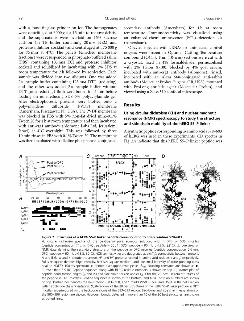
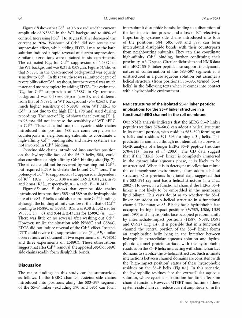
Figure 2. Structures of a hERG S5–P linker peptide corresponding to hERG residues 578–603A, circular dichroism spectra of the peptide in pure aqueous solution, and in DPC or SDS micelles(peptide concentration 74 µM, DPC : peptide = 60 : 1, SDS : peptide = 80 : 1, pH 3.5, 22◦C). B, overview ofNMR data defining the secondary structure of the peptide in DPC micelles (peptide concentration 0.6 mM,DPC : peptide = 60 : 1, pH 3.5, 30◦C). NOE connectivities are designated as dAB(i,j): connectivity between protonsA and B (N, α and β denote the amide, Hα and Hβ protons) located in amino acid residues i and j, respectively.Full-size square denotes high intensity, half-size square medium, and line small intensity of corresponding crosspeak in NOESY 100 ms spectrum. � denote overlapped cross-peaks. 3JNα coupling constants are shown as •,if lower than 5.5 Hz. Peptide sequence along with hERG residue numbers is shown on top. C, scatter plot ofpeptide bond torsion angles (ϕ and ψ ) and side chain torsion angles (χ1) for the 20 best DYANA structures ofthe peptide in DPC micelles. Peptide sequence is shown at the bottom, and hERG position numbers are shownon top. Dashed box denotes the helix region (583–593), and ∗ marks W585, L589 and D591 in the helix regionwith flexible side chain orientation. D, stereoview of the 20 best structures of the hERG S5–P linker peptide in DPCmicelles superimposed on the backbone atoms of the 583–593 region. Backbone and side chain heavy atoms ofthe 580–596 region are shown. Hydrogen bonds, detected in more than 10 of the 20 best structures, are shownas dotted lines.
secondary antibody (Amersham) for 1 h at roomtemperature. Immunoreactivity was visualized usingan enhanced-chemiluminescence (ECL) detection kit(Amersham).
Oocytes injected with cRNAs or uninjected controloocytes were frozen in Optimal Cutting Temperaturecompound (OCT). Thin (10-µm) sections were cut witha cryostat, fixed in 4% formaldehyde, permeabilizedwith 2% Triton X-100, blocked by 4% goat serum,incubated with anti-erg1 antibody (Alomone), rinsed,incubated with an Alexa 568-conjugated anti-rabbitantibody (Molecular Probes, Eugene, OR, USA), mountedwith ProLong antifade agent (Molecular Probes), andviewed using a Zeiss 510 confocal microscope.
Results
Using circular dichroism (CD) and nuclear magneticresonance (NMR) spectroscopy to study the structureand side chain mobility of the hERG S5–P linker
A synthetic peptide corresponding to amino acids 578–603of hERG was used in these experiments. CD spectra inFig. 2A indicate that this hERG S5–P linker peptide was
C© The Physiological Society 2005
J Physiol 569.1 Atomic proximity between hERG’s S5–P linkers 79
unstructured in a pure aqueous solution, but possessed∼30% helical structure in detergent micelles (DPC orSDS). Figure 2B summarizes NMR data used to locatethe peptide’s helical structure when placed in DPCmicelles. Spatial structure calculations showed that theN-terminal (corresponding to hERG residues 578–582)and the C-terminal (594–603) regions were unstructured.However, the segment from 583 to 593 was helical, with583–590 forming an α-helix and 591–593 forming a 310
helix (Fig. 2C and D). Although we cannot rule out thepossibility of solvent-induced formation of an α-helicalstructure, the NMR analysis in conjunction with ourprevious functional data (Liu et al. 2002) makes acompelling case for the notion that in a functional hERGchannel the 583–593 segment can form an α-helix at leastin some gating states of the channel.
This helix is amphipathic, with one face occupied byhydrophobic residues (W585, L586, L589 and I593) andthe other face occupied by hydrophilic residues (H587,N588, D591 and Q592). The conformations of all sidechains in the helical region have been unambiguouslydetermined. Higher dispersion in χ 1 values for W585 andL589, as well as D591, indicates that these side chains mayhave increased mobility (Fig. 2C).
Western blot analysis reveals that cysteine sidechains introduced into the 583–597 segment canform intersubunit disulphide bonds
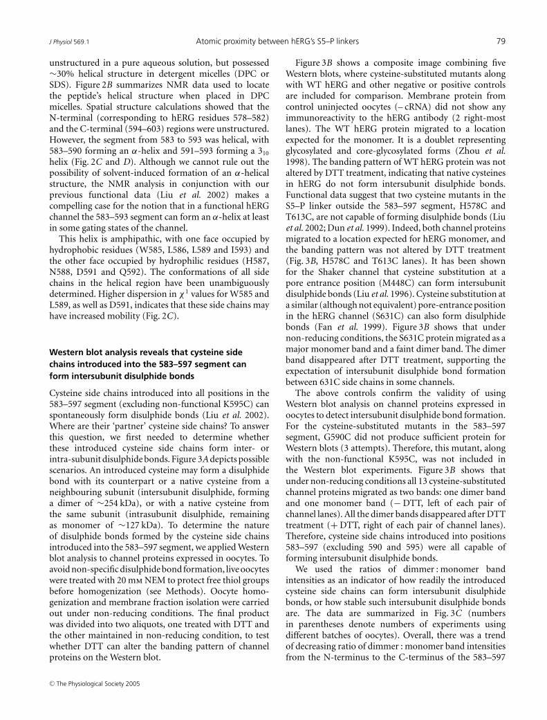
Cysteine side chains introduced into all positions in the583–597 segment (excluding non-functional K595C) canspontaneously form disulphide bonds (Liu et al. 2002).Where are their ‘partner’ cysteine side chains? To answerthis question, we first needed to determine whetherthese introduced cysteine side chains form inter- orintra-subunit disulphide bonds. Figure 3A depicts possiblescenarios. An introduced cysteine may form a disulphidebond with its counterpart or a native cysteine from aneighbouring subunit (intersubunit disulphide, forminga dimer of ∼254 kDa), or with a native cysteine fromthe same subunit (intrasubunit disulphide, remainingas monomer of ∼127 kDa). To determine the natureof disulphide bonds formed by the cysteine side chainsintroduced into the 583–597 segment, we applied Westernblot analysis to channel proteins expressed in oocytes. Toavoid non-specific disulphide bond formation, live oocyteswere treated with 20 mm NEM to protect free thiol groupsbefore homogenization (see Methods). Oocyte homo-genization and membrane fraction isolation were carriedout under non-reducing conditions. The final productwas divided into two aliquots, one treated with DTT andthe other maintained in non-reducing condition, to testwhether DTT can alter the banding pattern of channelproteins on the Western blot.
Figure 3B shows a composite image combining fiveWestern blots, where cysteine-substituted mutants alongwith WT hERG and other negative or positive controlsare included for comparison. Membrane protein fromcontrol uninjected oocytes (– cRNA) did not show anyimmunoreactivity to the hERG antibody (2 right-mostlanes). The WT hERG protein migrated to a locationexpected for the monomer. It is a doublet representingglycosylated and core-glycosylated forms (Zhou et al.1998). The banding pattern of WT hERG protein was notaltered by DTT treatment, indicating that native cysteinesin hERG do not form intersubunit disulphide bonds.Functional data suggest that two cysteine mutants in theS5–P linker outside the 583–597 segment, H578C andT613C, are not capable of forming disulphide bonds (Liuet al. 2002; Dun et al. 1999). Indeed, both channel proteinsmigrated to a location expected for hERG monomer, andthe banding pattern was not altered by DTT treatment(Fig. 3B, H578C and T613C lanes). It has been shownfor the Shaker channel that cysteine substitution at apore entrance position (M448C) can form intersubunitdisulphide bonds (Liu et al. 1996). Cysteine substitution ata similar (although not equivalent) pore-entrance positionin the hERG channel (S631C) can also form disulphidebonds (Fan et al. 1999). Figure 3B shows that undernon-reducing conditions, the S631C protein migrated as amajor monomer band and a faint dimer band. The dimerband disappeared after DTT treatment, supporting theexpectation of intersubunit disulphide bond formationbetween 631C side chains in some channels.
The above controls confirm the validity of usingWestern blot analysis on channel proteins expressed inoocytes to detect intersubunit disulphide bond formation.For the cysteine-substituted mutants in the 583–597segment, G590C did not produce sufficient protein forWestern blots (3 attempts). Therefore, this mutant, alongwith the non-functional K595C, was not included inthe Western blot experiments. Figure 3B shows thatunder non-reducing conditions all 13 cysteine-substitutedchannel proteins migrated as two bands: one dimer bandand one monomer band (− DTT, left of each pair ofchannel lanes). All the dimer bands disappeared after DTTtreatment (+ DTT, right of each pair of channel lanes).Therefore, cysteine side chains introduced into positions583–597 (excluding 590 and 595) were all capable offorming intersubunit disulphide bonds.
We used the ratios of dimmer : monomer bandintensities as an indicator of how readily the introducedcysteine side chains can form intersubunit disulphidebonds, or how stable such intersubunit disulphide bondsare. The data are summarized in Fig. 3C (numbersin parentheses denote numbers of experiments usingdifferent batches of oocytes). Overall, there was a trendof decreasing ratio of dimmer : monomer band intensitiesfrom the N-terminus to the C-terminus of the 583–597
C© The Physiological Society 2005

80 M. Jiang and others J Physiol 569.1
segment (denoted by the dashed curve), although N588C,L589C and Q592C formed stronger dimer bands thansuggested by the trend.
Cysteines introduced into four positions along the583–597 segment can form intersubunit disulphidebonds with their counterparts from neighbouringsubunits
We further explored whether the intersubunit disulphidebonds were formed between counterparts of introducedcysteine side chains, or between introduced and native
Figure 3. Probing intersubunit disulphide bond formation by Western blot analysis of channel proteinsexpressed in oocytesA, diagram of 2 hERG subunits, each marked with an introduced cysteine (Cys) in the S5–P linker and 5 native Cys.The introduced cysteine may form an intersubunit disulphide bond with its counterpart or a native cysteine froma neighbouring subunit (forming a dimer), or an intrasubunit disulphide bond with a native cysteine (molecularsize ∼ monomer). B, representative Western blot images of membrane proteins prepared from oocytes injectedwith subunit cRNAs marked on top. ‘– cRNA’ denote uninjected oocytes. Membrane fraction for each channelprotein without or with DTT treatment (– or + DTT) was run side-by-side on non-reducing SDS polyacrylamidegels. The positions of dimer and monomer (glycosylated and core-glycosylated forms) are marked on the left. Inthis and Fig. 5, the amounts of protein loaded in different lanes are not calibrated, causing differences in the overallband intensities. C, ratios of dimmer : monomer band intensities determined by densitometry. The monomer bandintensities include both glycosylated and core-glycosylated forms. Numbers in parentheses refer to numbers ofWestern blot measurements from different batches of oocytes. The dashed curve traces the general trend ofdecreasing dimmer : monomer ratio from N- to C-termini.
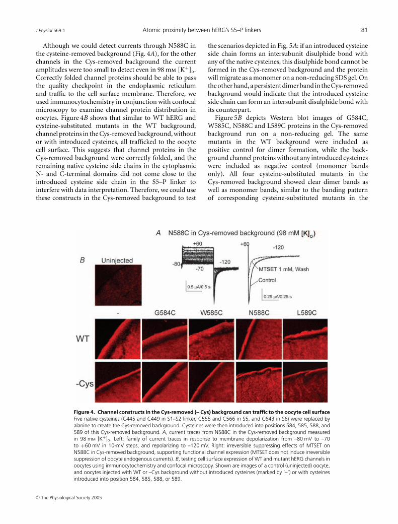
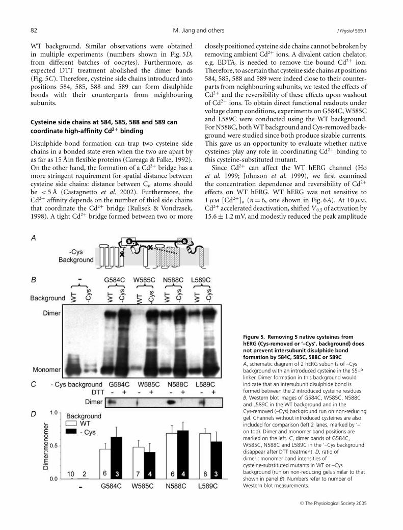
cysteine side chains. Four positions with a high tendencyof dimer formation were chosen for this analysis: 584, 585,588 and 589. There are a total of 24 native cysteine sidechains in each hERG subunit. Among them, five are in theextracellular region or in the transmembrane segments,and thus could potentially form disulphide bonds withthe introduced cysteines. These are C445 and C449 in theS1–S2 linker, C555 and C566 in S5, and C643 in S6. All fivenative cysteines were substituted with alanine, creating a‘Cys-removed’ hERG background (labelled as ‘– Cys back-ground’ in Figs 4 and 5). Cysteine side chains were thenreintroduced into this Cys-removed background at 584,585, 588 and 589.
C© The Physiological Society 2005
J Physiol 569.1 Atomic proximity between hERG’s S5–P linkers 81
Although we could detect currents through N588C inthe cysteine-removed background (Fig. 4A), for the otherchannels in the Cys-removed background the currentamplitudes were too small to detect even in 98 mm [K+]o.Correctly folded channel proteins should be able to passthe quality checkpoint in the endoplasmic reticulumand traffic to the cell surface membrane. Therefore, weused immunocytochemistry in conjunction with confocalmicroscopy to examine channel protein distribution inoocytes. Figure 4B shows that similar to WT hERG andcysteine-substituted mutants in the WT background,channel proteins in the Cys-removed background, withoutor with introduced cysteines, all trafficked to the oocytecell surface. This suggests that channel proteins in theCys-removed background were correctly folded, and theremaining native cysteine side chains in the cytoplasmicN- and C-terminal domains did not come close to theintroduced cysteine side chain in the S5–P linker tointerfere with data interpretation. Therefore, we could usethese constructs in the Cys-removed background to test
Figure 4. Channel constructs in the Cys-removed (– Cys) background can traffic to the oocyte cell surfaceFive native cysteines (C445 and C449 in S1–S2 linker, C555 and C566 in S5, and C643 in S6) were replaced byalanine to create the Cys-removed background. Cysteines were then introduced into positions 584, 585, 588, and589 of this Cys-removed background. A, current traces from N588C in the Cys-removed background measuredin 98 mM [K+]o. Left: family of current traces in response to membrane depolarization from –80 mV to –70to +60 mV in 10-mV steps, and repolarizing to –120 mV. Right: irreversible suppressing effects of MTSET onN588C in Cys-removed background, supporting functional channel expression (MTSET does not induce irreversiblesuppression of oocyte endogenous currents). B, testing cell surface expression of WT and mutant hERG channels inoocytes using immunocytochemistry and confocal microscopy. Shown are images of a control (uninjected) oocyte,and oocytes injected with WT or –Cys background without introduced cysteines (marked by ‘–’) or with cysteinesintroduced into position 584, 585, 588, or 589.
the scenarios depicted in Fig. 5A: if an introduced cysteineside chain forms an intersubunit disulphide bond withany of the native cysteines, this disulphide bond cannot beformed in the Cys-removed background and the proteinwill migrate as a monomer on a non-reducing SDS gel. Onthe other hand, a persistent dimer band in the Cys-removedbackground would indicate that the introduced cysteineside chain can form an intersubunit disulphide bond withits counterpart.
Figure 5B depicts Western blot images of G584C,W585C, N588C and L589C proteins in the Cys-removedbackground run on a non-reducing gel. The samemutants in the WT background were included aspositive control for dimer formation, while the back-ground channel proteins without any introduced cysteineswere included as negative control (monomer bandsonly). All four cysteine-substituted mutants in theCys-removed background showed clear dimer bands aswell as monomer bands, similar to the banding patternof corresponding cysteine-substituted mutants in the
C© The Physiological Society 2005
82 M. Jiang and others J Physiol 569.1
WT background. Similar observations were obtainedin multiple experiments (numbers shown in Fig. 5D,from different batches of oocytes). Furthermore, asexpected DTT treatment abolished the dimer bands(Fig. 5C). Therefore, cysteine side chains introduced intopositions 584, 585, 588 and 589 can form disulphidebonds with their counterparts from neighbouringsubunits.
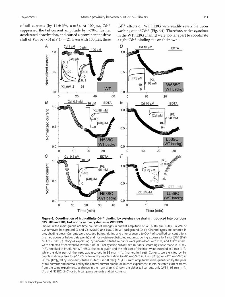
Cysteine side chains at 584, 585, 588 and 589 cancoordinate high-affinity Cd2+ binding
Disulphide bond formation can trap two cysteine sidechains in a bonded state even when the two are apart byas far as 15 A
�
in flexible proteins (Careaga & Falke, 1992).On the other hand, the formation of a Cd2+ bridge has amore stringent requirement for spatial distance betweencysteine side chains: distance between Cβ atoms shouldbe < 5 A
�
(Castagnetto et al. 2002). Furthermore, theCd2+ affinity depends on the number of thiol side chainsthat coordinate the Cd2+ bridge (Rulisek & Vondrasek,1998). A tight Cd2+ bridge formed between two or more
Figure 5. Removing 5 native cysteines fromhERG (Cys-removed or ‘–Cys’, background) doesnot prevent intersubunit disulphide bondformation by 584C, 585C, 588C or 589CA, schematic diagram of 2 hERG subunits of –Cysbackground with an introduced cysteine in the S5–Plinker. Dimer formation in this background wouldindicate that an intersubunit disulphide bond isformed between the 2 introduced cysteine residues.B, Western blot images of G584C, W585C, N588Cand L589C in the WT background and in theCys-removed (–Cys) background run on non-reducinggel. Channels without introduced cysteines are alsoincluded for comparison (left 2 lanes, marked by ‘–’on top). Dimer and monomer band positions aremarked on the left. C, dimer bands of G584C,W585C, N588C and L589C in the ‘–Cys background’disappear after DTT treatment. D, ratio ofdimer : monomer band intensities ofcysteine-substituted mutants in WT or –Cysbackground (run on non-reducing gels similar to thatshown in panel B). Numbers refer to number ofWestern blot measurements.
closely positioned cysteine side chains cannot be broken byremoving ambient Cd2+ ions. A divalent cation chelator,e.g. EDTA, is needed to remove the bound Cd2+ ion.Therefore, to ascertain that cysteine side chains at positions584, 585, 588 and 589 were indeed close to their counter-parts from neighbouring subunits, we tested the effects ofCd2+ and the reversibility of these effects upon washoutof Cd2+ ions. To obtain direct functional readouts undervoltage clamp conditions, experiments on G584C, W585Cand L589C were conducted using the WT background.For N588C, both WT background and Cys-removed back-ground were studied since both produce sizable currents.This gave us an opportunity to evaluate whether nativecysteines play any role in coordinating Cd2+ binding tothis cysteine-substituted mutant.
Since Cd2+ can affect the WT hERG channel (Hoet al. 1999; Johnson et al. 1999), we first examinedthe concentration dependence and reversibility of Cd2+
effects on WT hERG. WT hERG was not sensitive to1 µm [Cd2+]o (n = 6, one shown in Fig. 6A). At 10 µm,Cd2+ accelerated deactivation, shifted V 0.5 of activation by15.6 ± 1.2 mV, and modestly reduced the peak amplitude
C© The Physiological Society 2005
J Physiol 569.1 Atomic proximity between hERG’s S5–P linkers 83
of tail currents (by 14 ± 3%, n = 5). At 100 µm, Cd2+
suppressed the tail current amplitude by ∼70%, furtheraccelerated deactivation, and caused a prominent positiveshift of V 0.5 by ∼34 mV (n = 2). Even with 100 µm, these
Figure 6. Coordination of high-affinity Cd2+ binding by cysteine side chains introduced into positions585, 588 and 589, but not by native cysteines in WT hERGShown in the main graphs are time courses of changes in current amplitude of WT hERG (A), N588C in WT- orCys-removed background (B and C), W585C and L589C in WT-background (D–F). Channel types are denoted ingrey shading areas. Currents were recoded before, during and after exposure to Cd2+ of specified concentrations(marked above or below data points) and, for cysteine-substituted mutants, during exposure to 1 mM EDTA (B–E)or 1 mM DTT (F). Oocytes expressing cysteine-substituted mutants were pretreated with DTT, and Cd2+ effectswere detected after extensive washout of DTT. For cysteine-substituted mutants, recordings were made in 98 mM
[K+]o (marked in inset). For WT hERG, the main graph and the left part of the inset were recorded in 2 mM [K+]owhile the right part of the inset was recorded in 98 mM [K+]o (marked in inset). Currents were elicited by 1-sdepolarization pulses to +60 mV followed by repolarization to –60 mV (WT, in 2 mM [K+]o) or –120 mV (WT, in98 mM [K+]o, all cysteine-substituted mutants, in 98 mM [K+]o). Current amplitudes were quantified by the peakof tail currents and normalized by the control current amplitude in each experiment. Insets: selected current tracesfrom the same experiments as shown in the main graphs. Shown are either tail currents only (WT in 98 mM [K+]o(A), and N588C (B–C) or both test pulse currents and tail currents.
Cd2+ effects on WT hERG were readily reversible uponwashing out of Cd2+ (Fig. 6A). Therefore, native cysteinesin the WT hERG channel were too far apart to coordinatea tight Cd2+ binding site on their own.
C© The Physiological Society 2005
84 M. Jiang and others J Physiol 569.1
Figure 6B shows that Cd2+ at 0.5 µm reduced the currentamplitude of N588C in the WT background to 40% ofcontrol. Increasing [Cd2+] to 10 µm further decreased thecurrent to 20%. Washout of Cd2+ did not reverse thesuppression effect, while adding EDTA 1 mm to the bathsolution induced a rapid reversal of current suppression.Similar observations were obtained in six experiments.The estimated IC50 for Cd2+ suppression of N588C inthe WT background was 0.31 ± 0.09 µm. Figure 6C showsthat N588C in the Cys-removed background was equallysensitive to Cd2+. In this case, there was a limited degree ofreversibility after Cd2+ washout, but the reversal was muchfaster and more complete by adding EDTA. The estimatedIC50 for Cd2+ suppression of N588C in Cys-removedbackground was 0.38 ± 0.09 µm (n = 5), not differentfrom that of N588C in WT background (P = 0.563). Themuch higher sensitivity of N588C versus WT hERG toCd2+ is not due to the high [K+]o (98 mm) used duringrecordings. The inset of Fig. 6A shows that elevating [K+]o
to 98 mm did not increase the sensitivity of WT hERGto Cd2+. These data indicate that cysteine side chainsintroduced into position 588 can come very close tocounterparts in neighbouring subunits to coordinate ahigh-affinity Cd2+ binding site, and native cysteines arenot involved in Cd2+ binding.
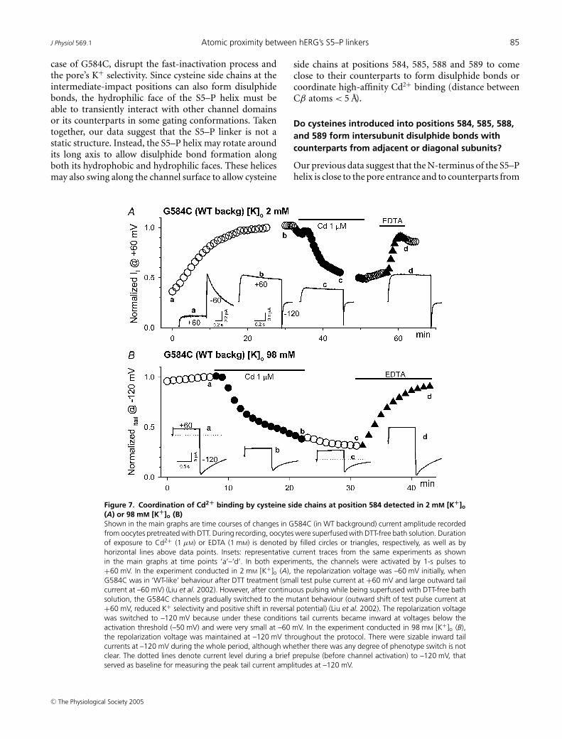
Cysteine side chains introduced into another positionon the hydrophilic face of the S5–P helix, 584, couldalso coordinate a high-affinity Cd2+ binding site (Fig. 7).The effects could not be reversed by washing out Cd2+,but required EDTA to chelate the bound Cd2+ ions. Thepotency of Cd2+ to suppress G584C appeared independentof [K+]o (IC50 = 0.65 ± 0.08 µm and 1.69 ± 0.81 µm, in 98and 2 mm [K+]o, respectively, n = 4 each, P = 0.343).
Figure 6D and E shows that cysteine side chainsintroduced into positions 585 and 589 on the hydrophobicface of the S5–P helix could also coordinate Cd2+ binding,although the binding affinity was lower than that of Cd2+
binding to N588C or G584C: IC50 was 9.38 ± 1.42 µm forW585C (n = 6) and 9.44 ± 2.43 µm for L589C (n = 11).There was little or no reversal after washing out Cd2+.However, unlike the situation with N588C and G584C,EDTA did not induce reversal of the Cd2+ effect. Instead,DTT could reverse the suppression effect (Fig. 6F , similarobservations are obtained in two experiments on W585Cand three experiments on L589C). These observationssuggest that after Cd2+ removal, the apposed 585C or 589Cside chains readily form disulphide bonds.
Discussion
The major findings in this study can be summarizedas follows. In the hERG channel, cysteine side chainsintroduced into positions along the 583–597 segmentof the S5–P linker (excluding 590 and 595) can form
intersubunit disulphide bonds, leading to a disruption ofthe fast-inactivation process and a loss of K+ selectivity.Importantly, cysteine side chains introduced into fourof the positions, 584, 585, 588 and 589, can formintersubunit disulphide bonds with their counterpartsfrom neighbouring subunits. They can also coordinatehigh-affinity Cd2+ binding, further confirming theirproximity in 3-D space. Circular dichroism and NMR dataof a hERG S5–P linker peptide also support the dynamicnature of conformation of the 583–597 segment: it isunstructured in a pure aqueous solution but assumes ahelical structure (from positions 583–593, termed ‘S5–Phelix’ in the following text) when it comes into contactwith a hydrophobic environment.
NMR structures of the isolated S5–P linker peptide:implications for the S5–P linker structure in afunctional hERG channel in the cell membrane
Our NMR analysis indicates that the hERG S5–P linkerpeptide (residues 578–603) can adopt a helical structurein its central portion, with residues 583–590 forming anα-helix and residues 591–593 forming a 310 helix. Thisprediction is similar, although not identical, to a previousNMR analysis of a longer hERG S5–P peptide (residues570–611) (Torres et al. 2003). The CD data suggestthat if the hERG S5–P linker is completely immersedin the extracellular aqueous phase, it is likely to beunstructured. When it is in detergent micelles that mimicthe cell membrane environment, it can adopt a helicalstructure. Our previous functional data suggested thatthe 583–594 segment has a helical structure (Liu et al.2002). However, in a functional channel the hERG S5–Plinker is not likely to be embedded in the membranelipid bilayer. This casts doubt as to whether the S5–Plinker can adopt an α-helical structure in a functionalchannel. The putative S5–P helix has a hydrophobic faceoccupied by high-impact positions (W585, L586, L589and I593) and a hydrophilic face occupied predominantlyby intermediate-impact positions (H587, N588, D591and Q592) (Fig. 8A). It is possible that in a functionalchannel the central portion of the S5–P linker formsan amphipathic helix lying in the interface betweenhydrophilic extracellular aqueous solution and hydro-phobic channel protein surface, with the hydrophobicresidues on the S5–P helix interacting with channel surfacedomains to stabilize the α-helical structure. Such intimateinteractions between channel domains are consistent withthe ‘high-impact position’ status of these hydrophobicresidues on the S5–P helix (Fig. 8A). In this scenario,the hydrophilic residues face the extracellular aqueoussolution, where cysteine substitution has little effects onchannel function. However, MTSET modification of thesecysteine side chain can reduce current amplitude, or in the
C© The Physiological Society 2005
J Physiol 569.1 Atomic proximity between hERG’s S5–P linkers 85
case of G584C, disrupt the fast-inactivation process andthe pore’s K+ selectivity. Since cysteine side chains at theintermediate-impact positions can also form disulphidebonds, the hydrophilic face of the S5–P helix must beable to transiently interact with other channel domainsor its counterparts in some gating conformations. Takentogether, our data suggest that the S5–P linker is not astatic structure. Instead, the S5–P helix may rotate aroundits long axis to allow disulphide bond formation alongboth its hydrophobic and hydrophilic faces. These helicesmay also swing along the channel surface to allow cysteine
Figure 7. Coordination of Cd2+ binding by cysteine side chains at position 584 detected in 2 mM [K+]o(A) or 98 mM [K+]o (B)Shown in the main graphs are time courses of changes in G584C (in WT background) current amplitude recordedfrom oocytes pretreated with DTT. During recording, oocytes were superfused with DTT-free bath solution. Durationof exposure to Cd2+ (1 µM) or EDTA (1 mM) is denoted by filled circles or triangles, respectively, as well as byhorizontal lines above data points. Insets: representative current traces from the same experiments as shownin the main graphs at time points ‘a’–‘d’. In both experiments, the channels were activated by 1-s pulses to+60 mV. In the experiment conducted in 2 mM [K+]o (A), the repolarization voltage was –60 mV initially, whenG584C was in ‘WT-like’ behaviour after DTT treatment (small test pulse current at +60 mV and large outward tailcurrent at –60 mV) (Liu et al. 2002). However, after continuous pulsing while being superfused with DTT-free bathsolution, the G584C channels gradually switched to the mutant behaviour (outward shift of test pulse current at+60 mV, reduced K+ selectivity and positive shift in reversal potential) (Liu et al. 2002). The repolarization voltagewas switched to –120 mV because under these conditions tail currents became inward at voltages below theactivation threshold (–50 mV) and were very small at –60 mV. In the experiment conducted in 98 mM [K+]o (B),the repolarization voltage was maintained at –120 mV throughout the protocol. There were sizable inward tailcurrents at –120 mV during the whole period, although whether there was any degree of phenotype switch is notclear. The dotted lines denote current level during a brief prepulse (before channel activation) to –120 mV, thatserved as baseline for measuring the peak tail current amplitudes at –120 mV.
side chains at positions 584, 585, 588 and 589 to comeclose to their counterparts to form disulphide bonds orcoordinate high-affinity Cd2+ binding (distance betweenCβ atoms < 5 A
�
).
Do cysteines introduced into positions 584, 585, 588,and 589 form intersubunit disulphide bonds withcounterparts from adjacent or diagonal subunits?
Our previous data suggest that the N-terminus of the S5–Phelix is close to the pore entrance and to counterparts from
C© The Physiological Society 2005

86 M. Jiang and others J Physiol 569.1
other subunits (Liu et al. 2002). We also propose that theS5–P helix is orientated with its C-terminus pointing awayfrom the pore entrance. This arrangement is consistentwith the trend of intersubunit disulphide bond stabilitysuggested by Fig. 3C: the N-terminal end of S5–P helixis closer to each other than the C-terminal end andcan more readily form intersubunit disulphide bonds. Ifthe S5–P helices are arranged around the central porein 4-fold symmetry, then the distance between cysteineside chains at equivalent positions on the S5–P helices isshorter between adjacent subunits than between diagonalsubunits. Although we do not have high-resolution datato definitely conclude one way or the other, the availableinformation does suggest that these cysteine side chainsare more likely to from intersubunit disulphide bondswith counterparts from adjacent subunits than fromthe diagonal subunit. Data from G584C also supportthis scenario. This channel manifests WT-like behaviourwhen the thiol side chains are in reduced state, butswitches to the mutant behaviour (disruption of fastinactivation and K+ selectivity) when the thiol sidechains form disulphide bonds. Therefore, we can trackdisulphide bond formation during the course of anexperiment. Figure 7A shows that after DTT washout,G584C gradually switches to the mutant behaviour,indicating disulphide bond formation. This process of‘phenotype-switch’ reaches a steady state in about 30 min.
Figure 8. Working models for the structure–function relationship of S5–P helices in the hERG channelA, helical wheel plot of hERG’s S5–P helix (residues 583–593), viewed from the N-terminus, with residues closerto the viewer drawn larger than those closer to the C-terminus. The hydrophobic and hydrophilic faces aremarked. High- and intermediate-impact positions are colour coded red and blue, respectively. The 4 positionswhere cysteine substitution can form intersubunit disulphide bonds with counterparts are highlighted by whitelettering on coloured background. B, three models of S5–P helix orientation and possible role in the inactivationprocess of the hERG channel. Channels are shown in cell membrane (boundaries denoted by dashed lines) withextracellular domains on top. The subunit closer to the viewer is removed to reveal the putative relationshipbetween the S5–P helices and the pore, which has a narrow selectivity filter and a large inner cavity. Cylindersrepresent S5–P helices, with their N-termini juxtaposed to the pore entrance with positive signs to denote thehelical dipole potential. The ball-and-sticks represent cysteine side chains introduced into the S5–P helices that canform intersubunit disulphide bond with counterparts from adjacent subunits. Arrows denote molecular motionsof the S5–P helices needed to allow cysteine side chains at equivalent positions from adjacent subunits to formintersubunit disulphide bonds. For more discussion, see text.
Adding Cd2+ 1 µm at this point can effectively suppressthe current, and washing out Cd2+ does not reverse thesuppressing effect until EDTA is applied. This indicatesthat Cd2+ is coordinated by at least two cysteine sidechains in close proximity (although histidine side chainscan also coordinate Cd2+ ions). Although this observationrepresents the averaged behaviour of all G584C channelsin the oocyte cell membrane, the data are consistentwith the notion that disulphide bonds are formedbetween 584C from two adjacent subunits (thus allowingcurrent through the pore). Furthermore, formation of onedisulphide bond within a channel between two adjacentsubunits does not perturb the outer mouth conformationsignificantly, so that the remaining two free thiolgroups can still coordinate a high affinity Cd2+ bindingsite.
Why is the S5–P linker so critical for thefast-inactivation process of the hERG channel?
In Fig. 8B, we propose three models for how S5–P helices ina functional hERG channel interact with each other, andhow these S5–P helices may contribute to the channel’sfast-inactivation process. Although a transition of the S5–Plinker conformation between helical and coil is possibleduring hERG gating, this possibility is not included in the
C© The Physiological Society 2005
J Physiol 569.1 Atomic proximity between hERG’s S5–P linkers 87
simplified models depicted in Fig. 8B. In the left panelof Fig. 8B, the S5–P helices are orientated perpendicularto the plane of the cell membrane. Relatively smallasymmetric rotations of S5–P helices along their longaxes can allow intersubunit disulphide bond formationbetween cysteine side chains at equivalent positions fromadjacent or even diagonal subunits. These S5–P helicescome close to each other during channel inactivation,preventing K+ ion flux through the pore by sterichindrance or by a hydrophobic seal formed between hydro-phobic residues along the S5–P helices. In the middlepanel, the S5–P helices are orientated parallel to the planeof the cell membrane. Asymmetric rotations of S5–Phelices along their long axes, as well as relatively large-scalesideway swinging motion along the channel surface, areneeded to allow cysteine side chains at equivalent positionsof adjacent subunits to form intersubunit disulphidebonds. Channel inactivation occurs when all four helicespoint their N-terminal positive helical dipole (Hol, 1985)toward the pore entrance, creating a local electrostaticpotential unfavourable to K+ ion flux through the pore.In the right panel, the S5–P helices are tilted and sinkinto the channel pore to interact with the selectivityfilter. Asymmetric rotations along their long axes as wellas small-scale sideways swinging are needed to allowintersubunit disulphide bond formation between cysteineside chains at equivalent positions of adjacent subunits.Channel inactivation is envisioned to be due to two factors:(a) local electrostatic potential due to the N-terminalpositive helical dipole of the four S5–P helices (Hol,1985), and (b) interactions between the S5–P helices andthe pore-loops, causing conformational changes aroundthe selectivity filter that shut down K+ flux through thepore.
The advantage of the first model is that relatively smallmolecular motions can allow intersubunit disulphidebond formation between counterparts from adjacent ordiagonal subunits. However, the disadvantage of thismodel is that it does not explain how the S5–P helicesare stabilized in an aqueous environment. In the secondand third models, the S5–P helices are stabilized by theinteraction between residues on its hydrophobic faceand the channel surface domain. The third model hasthe added advantages over the second model in thatsmaller sideways swinging motion allows intersubunitdisulphide bond formation between cysteines at equivalentpositions of two adjacent subunits, and the proposedinteraction between the S5–P helix and the pore loopcan better explain why the pore’s K+ selectivity isdisrupted by mutations at high-impact positions alongthe S5–P helix. We have proposed that relative tothe Shaker channel, there are fewer hydrogen bondsformed between residues around the outer mouth of thehERG channel (Fan et al. 1999). This may result in a
‘floppy’ outer mouth structure, able to accommodate theinsertion of the S5–P helices as proposed in the thirdmodel.
Technical consideration: validity of usingcysteine-substituted mutants or cysteine-removedbackground channels to probe the structure–functionrelationship of hERG’s S5–P linker
A critical issue for data interpretation in these experimentsis whether the cysteine-substituted mutants maintain thenative conformation of the channel’s outer vestibule. Thisis the case for cysteine substitution at intermediate-impactpositions, because these channels maintain the WTphenotype as long as the thiol side chains are in thereduced state. For cysteine substitution at the high-impactpositions, although the inactivation process and K+
selectivity are disrupted even when the thiol side chainsare reduced, 5 of the channels (L586C, N588C, L589C,D591C and I593C) maintain a high sensitivity to apeptide toxin, ErgTx1, similar to that of the WT hERGchannel (mean IC50 range 2.3–10.7 nm, versus 7.2 nmfor WT hERG) (Pardo-Lopez et al. 2002). Since sucha high toxin binding affinity requires the maintenanceof multiple contact points between toxin and thechannel’s outer vestibule, the data argue that these fivecysteine-substituted mutants at the high impact positionsdo not drastically alter the outer mouth conformation.The remaining two cysteine substituted mutants (W585Cand G590C) manifest markedly reduced ErgTx1 bindingaffinity (IC50 > 100 nm) (Pardo-Lopez et al. 2002). Theyare therefore less informative because of the uncertainty asto whether they maintain the native conformation of theouter vestibule.
Currents through channels in the Cys-removed back-ground (5 native cysteines replaced by alanines, Fig. 4legend) are very small or not detectable. This is likely tobe due to the removal of C566 in S5, because removingthe other four native cysteines, singly or in combination,does not significantly reduce the current amplitude (datanot shown). Although the mechanism for the detrimentaleffect of C566A on hERG channel function is not clear,we argue that the transmembrane topology of channelsin the Cys-removed background is maintained as the WThERG. This is based on the observation that channels inthe Cys-removed background can traffic to the oocytecell surface, similar to channels in the WT background(Fig. 4B). This is possible only if these channel proteins arecorrectly folded so that they can exit the ER checkpoint. Itis important to point out that in the oocyte Western blotexperiments, all free thiol groups in intact and functionalchannels are blocked by 20 mm NEM before homo-genization (see Methods). Disulphide bonds detected inWestern blot experiments represent those formed in the
C© The Physiological Society 2005
88 M. Jiang and others J Physiol 569.1
in situ intact channels. Therefore, our data rule outthe possibility that for channels in the Cys-removedbackground the remaining native cysteine side chains(in the cytoplasmic domain) can form disulphidebonds with those cysteine side chains introduced intothe extracellular S5–P linker and interfere with datainterpretation.
Our data cannot distinguish between two possibilities:cysteine side chains introduced into the 583–597 segmentform disulphide bonds with specific partners, or withmultiple partners in different gating conformations. Inthe Shaker channel, cysteine side chains introduced intoseveral positions in S4 can form disulphide bonds withcysteine introduced into the same position of S5 in agating state-dependent manner (Gandhi et al. 2003).Furthermore, whether cysteines introduced into positionsother than 584, 585, 588 and 589 can form intersubunitdisulphide bonds with counterparts from neighbouringsubunits needs to be tested.
References
Baker EN & Hubburd RE (1984). Hydrogen bonding inglobular proteins. Prog Biophys Mol Biol 44, 97–179.
Bartels C, Xia TH, Billeter M, Guntert P & Wuthrich K (1995).The program XEASY for computer-supported NMR spectralanalysis of biological macromolecules. J Biomol NMR 6,1–10.
Bax A & Davis DG (1985). MLEV-17-based two-dimensionalhomonuclear magnetization transfer spectroscopy. J MagnReson 65, 355–366.
Careaga CL & Falke JJ (1992). Thermal motions of surfaceα-helices in the d-galactose chemosensory receptor.Detection by disulfide trapping. J Mol Biol 226,1219–1235.
Castagnetto JM, Hennessy SW, Roberts VA, Getzoff ED, TainerJA & Pique ME (2002). MDB: the mtalloprotein databaseand browser at the Scripps Research Institute. Nucl Acids Res30, 379–382.
Doyle DA, Cabral JM, Pfuetzner RA, Kuo A, Gulbis JM, CohenSL, Chait BT & MacKinnon R (1998). The structure of thepotassium channel: molecular basis of K+ conduction andselectivity. Science 280, 69–77.
Dun W, Jiang M & Tseng G-N (1999). Allosteric effects ofmutations in the extracellular S5-P loop on the gating andion permeation properties of hERG. Pflugers Arch 439,141–149.
Fan J-S, Jiang M, Dun W, McDonald TV & Tseng G-N (1999).Effects of outer mouth mutations on hERG channelfunction: a comparison with similar mutations in Shaker.Biophys J 76, 3128–3140.
Gandhi CS, Clark E, Loots E, Pralle A & Isacoff EY (2003). Theorientation and molecular movement of a K+ channelvoltage-sensing domain. Neuron 40, 515–525.
Guntert P, Mumenthaler C & Wuthrich K (1997). Torsionangle dynamics for NMR structure calculation with a newprogram DYANA. J Mol Biol 273, 283–298.
Herzberg IM, Trudeau MC & Robertson GA (1998). Transfer ofrapid inactivation and sensitivity to the class IIIantiarrhythmic drug E-4031 from HERG to M-eag channels.J Physiol 511, 3–14.
Ho W-K, Kim I, Lee CO, Youm JB, Lee SH & Earm YE (1999).Blockade of HERG channels expressed in Xenopus laevisoocytes by external divalent cations. Biophys J 76,1959–1971.
Hol WG (1985). Effects of the alpha-helix dipole upon thefunctioning and structure of proteins and peptides. AdvBiophys 19, 133–165.
Hoshi T, Zagotta WN & Aldrich RW (1991). Two types ofinactivation on Shaker K+ channels: effects of alterations inthe carboxy-terminal region. Neuron 7, 547–556.
Jeener J, Meier GH, Backman P & Ernst RR (1979).Investigation of exchange processes by two-dimensionalNMR spectroscopy. J Chem Phys 71, 4546–4553.
Jiang Y, Lee A, Chen J, Cadene M, Chait BT & MacKinnon R(2002). Crystal structure and mechanism of a calcium-gatedpotassium channel. Nature 417, 515–522.
Jiang Y, Lee A, Chen J, Ruta V, Cadene M, Chait BT &MacKinnon R (2003). X-ray structure of a voltage-dependent K+ channel. Nature 423, 33–41.
Johnson JP, Balser JR & Bennett PB (1999). Ehancement ofHERG K+ currents by Cd2+ destabilization of the inactivatedstate. Biophys J 77, 2534–2541.
Koradi R, Billeter M & Wuthrich K (1996). MOLMOL: aprogram for display and analysis of macromolecularstructures. J Mol Graphics 14, 51–55.
Liu Y, Jurman ME & Yellen G (1996). Dynamic rearrangementof the outer mouth of a K+ channel during gating. Neuron16, 859–867.
Liu J, Zhang M, Jiang M & Tseng G-N (2002). Structural andfunctional role of the extracellular S5-P linker in the HERGpotassium channel. J Gen Physiol 120, 723–737.
Pardo-Lopez L, Zhang M, Liu J, Jiang M, Possani LD & TsengG-N (2002). Mapping the binding site of a HERG-specificpeptide toxin (ErgTx) to the channel’s outer vestibule. J BiolChem 277, 16403–16411.
Rance M, Sorensen OW, Bodenhausen G, Wagner C, Ernst RR& Wuthrich K (1983). Improved spectral resolution in COSY1H-NMR spectra of protein via double quantum filter.Biochem Biophys Res Comm 117, 479–485.
Rulisek L & Vondrasek J (1998). Coordination geometries ofselected transition metal ions (Co2+, Ni2+, Cu2+, Zn2+,Cd2+, and Hg2+) in metalloproteins. J Inorganic Biochem 71,115–127.
Sanguinetti MC, Jiang C, Curran ME & Keating MT (1995).A mechanistic link between an inherited and an acquiredcardiac arrhythmia: HERG encodes the IKr potassiumchannel. Cell 81, 299–307.
Schreibmayer W, Lester HA & Dascal N (1994). Voltageclamping of Xenopus laevis oocytes utilizing agarose-cushionelectrodes. Pflugers Arch 426, 453–458.
Smith PL, Baukrowitz T & Yellen G (1996). The inwardrectification mechanism of the HERG cardiac potassiumchannel. Nature 379, 833–836.
Spector PS, Curran ME, Zou A, Keating MT & Sanguinetti MC(1996). Fast inactivation causes rectification of the IKr
channel. J Gen Physiol 107, 611–619.
C© The Physiological Society 2005
J Physiol 569.1 Atomic proximity between hERG’s S5–P linkers 89
Torres AM, Bansal P, Sunde M, Clarke CE, Bursill JA, Smith DJ,Bauskin A, Breit SN, Campbell TJ, Alewood PF, Kuchel PW& Vandenberg JI (2003). Structure of the HERG K+ channelS5P extracellular linker: role of an amphipathic α-helix inC-type inactivation. J Biol Chem 278, 42136–42148.
Wuthrich K (1986). NMR of Proteins and Nucleic Acids. Wiley,New York.
Zhou Z, Gong QYeB, Fan Z, Makielski JC, Robertson GA &January CT (1998). Properties of HERG channels stablyexpressed in HEK 293 cells studied at physiologicaltemperature. Biophys J 74, 230–241.
Acknowledgements
This work was supported by HL 46451 and HL 67840 fromNIH/NHLBI and a grant-in-aid award from AHA/Mid-AtlanticAffiliate (GNT), and by Russian Ministry of Education andScience and Russian Basic Research Foundation. The authorswould like to thank Dr H. R. Guy (National Cancer Institute ofNational Institutes of Health) for helpful discussions along thecourse of this project.
C© The Physiological Society 2005


![Connectors and Linkers[1]](https://img.pdfslide.us/doc/110x75/552fd66f550346dd568b45ae/connectors-and-linkers1.jpg)