Embed Size (px)
Citation preview

Dyclonine rescues frataxin deficiency in animalmodels and buccal cells of patients withFriedreich’s ataxia
Sunil Sahdeo1, Brian D. Scott1, Marissa Z. McMackin1, Mittal Jasoliya1, Brandon Brown2, Heike
Wulff2, Susan L. Perlman3, Mark A. Pook4 and Gino A. Cortopassi1,∗
1Department of Molecular Biosciences, School of Veterinary Medicine, 2Department of Pharmacology, School of
Medicine, University of California, Davis, CA 95616, USA, 3Department of Neurology, University of California School of
Medicine,LosAngeles,CA90095,USAand 4SchoolofHealthSciencesandSocialCare,BrunelUniversity,UxbridgeUB8
3PH, UK
Received May 13, 2014; Revised July 2, 2014; Accepted August 4, 2014
Inherited deficiency in the mitochondrial protein frataxin (FXN) causes the rare disease Friedreich’s ataxia (FA),for which there is no successful treatment. We identified a redox deficiency in FA cells and used this to model thedisease. We screened a 1600-compound library to identify existing drugs, which could be of therapeutic benefit.We identifiedthe topicalanestheticdyclonineasprotective.Dyclonine increased FXNtranscriptandFXNproteindose-dependently in FA cells and brains of animal models. Dyclonine also rescued FXN-dependent enzymedeficiencies in the iron–sulfur enzymes, aconitase and succinate dehydrogenase. Dyclonine induces the Nrf2[nuclear factor (erythroid-derived 2)-like 2] transcription factor, which we show binds an upstream responseelement in the FXN locus. Additionally, dyclonine also inhibited the activity of histone methyltransferase G9a,known to methylate histone H3K9 to silence FA chromatin. Chronic dosing in a FA mouse model prevented aperformance decline in balance beam studies. A human clinical proof-of-concept study was completed ineight FA patients dosed twice daily using a 1% dyclonine rinse for 1 week. Six of the eight patients showed anincrease in buccal cell FXN levels, and fold induction was significantly correlated with disease severity.Dyclonine represents a novel therapeutic strategy that can potentially be repurposed for the treatment of FA.
INTRODUCTION
Friedreich’s ataxia (FA) is a severe neurodegenerative diseasethat is the most common autosomal recessive inherited move-ment disorder (1). There is no cure or effective treatment forFA. The disease causes degeneration and demyelination indorsal root ganglion (DRG) neurons and spinocerebellar tracts,resulting in movement and speech disorders (2,3). Symptomsbegin between the ages of 5–15 years, and patients are oftenwheelchair bound within 10–15 years of diagnosis. Earlydeath is common and usually occurs from cardiac complications(4). FA is caused by a decrease in the mitochondrial protein fra-taxin (FXN gene), which has been shown to have roles in iron–sulfur cluster synthesis, iron transfer and antioxidant defense(1,5–8). The decrease in FXN stems from an accumulation of
GAA triplet repeats in the first intron of the gene through inher-itance (9–11). Here, we report on screening and identificationof a potential therapeutic for FA. We previously identified anti-oxidant defects in DRG cells of an FA mouse model (12). Usingthis to develop a specific hypothesis for disease pathogenesis, wedesigned a cellular disease model for FA in order to identifya potential treatment. The cell model identified dyclonine, andwe have shown that it induces FXN in cells, animal modelsand FA patients through a novel mechanism. Dyclonine isan oral anesthetic used to provide topical anesthesia to mucousmembranes through sodium channel inhibition (13). It is theactive ingredient in Sucrets, an over-the-counter throatlozenge, and has been in use for over 50 years (14). Additionally,dyclonine is used during dental examinations and procedures tonumb mucous membranes of the mouth (15).
∗To whom correspondence should be addressed. Tel: +1 5307549665; Fax: +1 5307549342; Email: [email protected]
# The Author 2014. Published by Oxford University Press.This is an Open Access article distributed under the terms of the Creative Commons Attribution License (http://creativecommons.org/licenses/by/4.0/),which permits unrestricted reuse, distribution, and reproduction in any medium, provided the original work is properly cited.
Human Molecular Genetics, 2014 1–15doi:10.1093/hmg/ddu408
HMG Advance Access published August 25, 2014 at B
runel University on O
ctober 29, 2014http://hm
g.oxfordjournals.org/D
ownloaded from

Figure 1. High-throughput screening reveals that dyclonine protects FA patient fibroblasts from diamide stress. (A) Effect of antioxidant inhibitors on 50B11 cellviability with FXN knockdown. Eleven inhibitors of thiol-related antioxidants were tested in an siRNA-mediated, FXN-deficient 50B11 DRG cell line [10 mM anti-mycin A, 1 mM auranofin, 100 mM BSO, 100 mM carmustine, 10 mM diamide, 0.1% diethyl maleate (DEM), 0.1% ethanol, 0.03% H2O2, 1 mM L-glutathione(L-GSH), 0.1% phenethyl isothiocyanate (PEITC), 100 mM dichloronitrobenzene (DCNB) and 1 mM N-methyl protoporphyrin (NMP)]. Cell viability was measuredwith Calcein-AM after 24 h and normalized to untreated control (n ¼ 3). Increased sensitivity to cell death was induced by inhibitors of thiol-related antioxidantsdiamide and auranofin in FXN knockdown cells compared with AllStars non-targeting siRNA negative control. (B) FXN-dependent sensitivity to diamide is dose-dependent in 50B11 cells. Cell viability was measured with Calcein-AM after 24 h of treatment with 3–300 mM diamide and normalized to untreated control (n ¼ 3).(C) Friedreich’s patient cells are sensitive to diamide. To confirm these effects in patient cells with low FXN, we tested 100 mM diamide in fibroblasts and
2 Human Molecular Genetics, 2014
at Brunel U
niversity on October 29, 2014
http://hmg.oxfordjournals.org/
Dow
nloaded from

RESULTS
FXN-deficient cells are sensitive to the thioredoxin-oxidantdiamide
There is ample support for dysregulated antioxidant defenses inFA (12,16–18). We screened that12 inhibitors of thiol antioxi-dants were screened in FXN-deficient DRG neural cells, chosensince the DRGs are the primary site of disease pathogenesis(Fig. 1A and B) .Measuring viable cells with Calcein-AM stain-ing, the thioredoxin-oxidant diamide produced the greatest sen-sitivity (19). To confirm this phenotype in patient cells, we foundthat FXN-deficient FA fibroblasts and lymphoblasts were alsoclearly more sensitive to diamide treatment than healthy controls(Fig. 1C). Diamide is a specific thiol oxidant that causes oxida-tive damage and results in cell death (20,21). In the FA patientfibroblasts, a robust high-throughput screening assay based ondiamide sensitivity was developed, with an average Z′-score of0.7. We then screened a 1600-compound library of drugs thathave been approved for use in human trials, and identified pro-tective drugs (Fig. 1D). After the initial screen, multiple roundsof hit confirmation, and then concentration response curves, 33drugs were found that reproducibly protected the patient cellsfrom the thiol stress induced by diamide (Supplementary Mater-ial, Table S1). Of the drugs found to protect FA fibroblasts fromdiamide-induced oxidative stress, dyclonine was one of the mostpotent (Fig. 1E).
Dyclonine induces FXN in patient cells and in animal modeltissues
Multiple mechanisms of action of protection from diamide werepossible. One is reactivation of FXN expression, since its defici-ency mediated the sensitivity to diamide. Thus, all 33 confirmedthat screening hits including dyclonine were tested for theirability to induce FXN protein levels in the absence of diamide(Supplementary Material, Table S1). Of the compounds foundto increase FXN in FA patient cells, we eliminated some becauseof safety concerns (i.e. cotinine and nifursol), and others becauseof their primary use is in veterinary medicine (i.e. oxfendazole)(22–24). We chose to follow-up with dyclonine because of itspotency of diamide protection, reproducibility of FXN inductionand safe use in humans for decades. FA patient lymphoblaststreated with dyclonine show clear induction of FXN proteinafter 48 h of dyclonine exposure (Fig. 2A). Additionally, FXNmRNA transcript measured by RT-PCR is also dose-dependentlyincreased after 24 h drug treatment in FA lymphoblasts (Fig. 2B).We observed induction of FXN in multiple animal models afterintraperitoneal or oral dosing. FA-YG8 [hFXN+/2 with FXN
(GAA)190 expansion; mFxn2/2] transgenic mice (8) dosed for1 week once daily with dyclonine i.p. or p.o. showed a clearinduction of FXN protein in cerebellar lysate (Fig. 2C).
In addition, to explore the effects of dyclonine in an additionalmodel with chronic dosing, we treated FA-PandKIKO mice[mFxn+/2 with FXN (GAA)230 expansion; mFxn2/2] with25 mg/kg dyclonine for 4 weeks p.o. This higher dose waschosen in an attempt to ascertain the maximum induction ofFXN within the limits of dyclonine solubility. Thus, showingdyclonine can reverse the in vivo FXN protein defect in an add-itional FA model at a higher dose and chronic duration (Fig. 2D).These chronic studies resulted in no observable changes in sed-ation, feeding behavior or gross pathology. We observed theseeffects on FXN in multiple additional tissues, including heartand liver (Supplementary Material, Fig. S1).
Dyclonine activates the Nrf2 pathway
Of the 33 drugs that protected FA patient cells from diamidestress, some drugs were reported antioxidant response element(ARE)/nuclear factor (erythroid-derived 2)-like 2 (Nrf2) indu-cers in literature such as ebselen (25). Nrf2 is a transcriptionfactor that responds to oxidative and thiol stress by bindingAREs and driving the expression of multiple antioxidant andanti-inflammatory target genes including heme oxygenase 1(HMOX1), NAD(P)H dehydrogenase (quinone) (NQO1) andglutathione peroxidase (GPX4) (26,27). We explored the activityof all 33 drugs as ARE/Nrf2 inducers for their ability to stimulateARE-luciferase reporter system activity (Supplementary Mater-ial, Table S1), including dyclonine which dose-dependentlydrove induction of ARE-luciferase (Fig. 3A). Remarkably, mul-tiple drugs that increased ARE-luciferase expression were alsofound to be FXN inducers (Supplementary Material, Table S1).In addition, after oral dyclonine dosing in the FA-YG8 mousemodel of FA drives a dose-dependent increase in known Nrf2-target proteins in cerebellar lysate including heme oxygenase(HO1), NQO1 and GPX4 (Fig. 3B). Thus, dyclonine is an activa-tor of the ARE/Nrf2 pathway.
Evolutionarily conserved Nrf2-binding sites (AREs) exist inthe FXN gene and are functional Nrf2-binding sites
We studied the mechanism of induction of FXN by dycloninewith regard to Nrf2. If dyclonine induces FXN through theNrf2 pathway, then there should be active Nrf2-binding sites,i.e. AREs (28) within the FXN locus that are triggered by dyclo-nine exposure. Seven potential ARE sites were found between20 kb upstream and 5 kb downstream of the FXN locus
lymphoblasts and found that patient cells were more sensitive to diamide compared with healthy control cells. (D) Results of high-throughput screen for drugs thatprotect from diamide toxicity. Significance is shown comparing healthy volunteer and FA patient lines grouped together. This cell-based assay in FA patient fibroblastcell line 1134 was furtheroptimized for high-throughput screening in 96-well plates,with a mean Z′-value of 0.75 (n ¼ 25).This platformwas used to screena libraryof1600 drugs that have been approved for clinical use. FA fibroblasts were pretreated with 10 mM test compound, DMSO (negative control) or 300 mM dithiothreitol(DTT) (positive control) for 24 h and followed by 100 mM diamide for 24 h. Cell viability was measured with Calcein-AM. Screening data (diamide + all drugs) arethe mean of two replicates and presented as fold above DMSO + diamide control. Arrow indicates dyclonine response. Mean + SD for the 1600 drugs was 1+0.3;mean + SEM was 1+0.01. Compounds that rescued from diamide toxicity greater than mean + 2× SD advanced to secondary screening, which included replicationof protective effect in a concentration-dependent manner, 0.01–10 mM. (E) An example of dose-dependent protection by dyclonine. Dyclonine was added to FAfibroblast line 1134 for 24 h before 100 mM diamide treatment, and Calcein-AM viability is shown as fold above DMSO + diamide control. Intrinsic effect:2.1+0.49-fold above DMSO + diamide control; EC50: 0.36+0.25 mM (n ¼ 3). (F) Chemical structure of dyclonine. The plotted data in A, B, C and E displaymean responses and error bars represent SD (n ¼ 3–4). ∗P , 0.05, ∗ ∗P , 0.001 relative to control, t-test.
Human Molecular Genetics, 2014 3
at Brunel U
niversity on October 29, 2014
http://hmg.oxfordjournals.org/
Dow
nloaded from

Figure 2. Dyclonine induces FXN in cultured FA patient cells and FA mouse model cerebellum in vivo. (A) Dyclonine increases FXN protein expression in FA patientlymphoblast cell lines. To test if the mechanism of protection from diamide toxicity for dyclonine was through an increase in FXN protein levels, FA lymphoblastswere treated with 0.3–10 mM dyclonine or vehicle control (0.1% DMSO). Total protein was collected after 48 h, and lysates were probed by western blot analysis forFXN expression and normalized tob-actin. The plotted data represent the mean fold change in FXN protein in drug-treated cells, normalized to vehicle control. Errorbars represent SEM. ∗∗P , 0.01, t-test (n ¼ 2–15). A representative blot is shown for FA patient lymphoblast line 15850 treated with 10 mM dyclonine for 48 h. (B)Dyclonine increases FXN transcript levels in FA patient lymphoblasts. FA lymphoblast cell line 14518 was treated with 3–30 mM dyclonine or vehicle control (0.1%DMSO). RNA was analyzed after 24 h by RT-PCR with expression with primers for FXN normalized to b-actin. The plotted data represent the mean fold change inFXN transcript in drug-treated cells normalized to vehicle control. Error bars represent SEM. ∗P , 0.05, ∗∗P , 0.01, (n ¼ 4), t-test. (C) Dyclonine increases FXN
4 Human Molecular Genetics, 2014
at Brunel U
niversity on October 29, 2014
http://hmg.oxfordjournals.org/
Dow
nloaded from
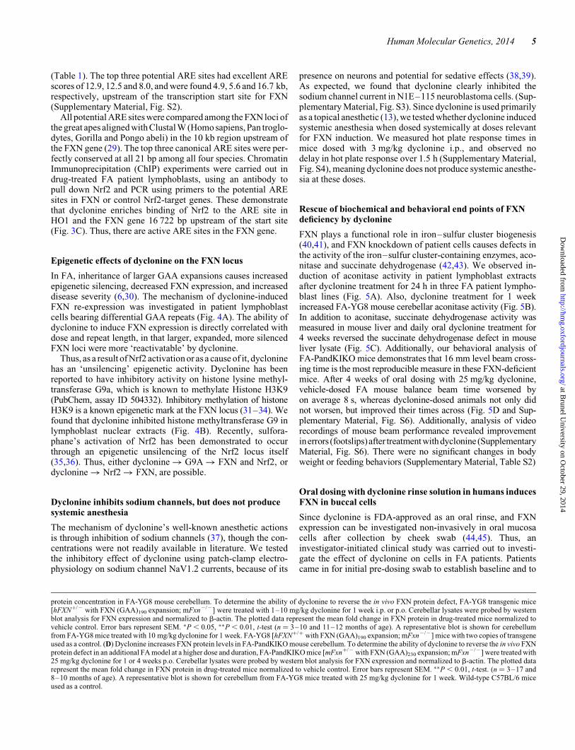
(Table 1). The top three potential ARE sites had excellent AREscores of 12.9, 12.5 and 8.0, and were found 4.9, 5.6 and 16.7 kb,respectively, upstream of the transcription start site for FXN(Supplementary Material, Fig. S2).
All potential ARE sites were compared among the FXN loci ofthe great apes aligned with Clustal W (Homo sapiens, Pan troglo-dytes, Gorilla and Pongo abeli) in the 10 kb region upstream ofthe FXN gene (29). The top three canonical ARE sites were per-fectly conserved at all 21 bp among all four species. ChromatinImmunoprecipitation (ChIP) experiments were carried out indrug-treated FA patient lymphoblasts, using an antibody topull down Nrf2 and PCR using primers to the potential AREsites in FXN or control Nrf2-target genes. These demonstratethat dyclonine enriches binding of Nrf2 to the ARE site inHO1 and the FXN gene 16 722 bp upstream of the start site(Fig. 3C). Thus, there are active ARE sites in the FXN gene.
Epigenetic effects of dyclonine on the FXN locus
In FA, inheritance of larger GAA expansions causes increasedepigenetic silencing, decreased FXN expression, and increaseddisease severity (6,30). The mechanism of dyclonine-inducedFXN re-expression was investigated in patient lymphoblastcells bearing differential GAA repeats (Fig. 4A). The ability ofdyclonine to induce FXN expression is directly correlated withdose and repeat length, in that larger, expanded, more silencedFXN loci were more ‘reactivatable’ by dyclonine.
Thus, as a result of Nrf2 activation or as a cause of it, dycloninehas an ‘unsilencing’ epigenetic activity. Dyclonine has beenreported to have inhibitory activity on histone lysine methyl-transferase G9a, which is known to methylate Histone H3K9(PubChem, assay ID 504332). Inhibitory methylation of histoneH3K9 is a known epigenetic mark at the FXN locus (31–34). Wefound that dyclonine inhibited histone methyltransferase G9 inlymphoblast nuclear extracts (Fig. 4B). Recently, sulfora-phane’s activation of Nrf2 has been demonstrated to occurthrough an epigenetic unsilencing of the Nrf2 locus itself(35,36). Thus, either dyclonine � G9A � FXN and Nrf2, ordyclonine � Nrf2 � FXN, are possible.
Dyclonine inhibits sodium channels, but does not producesystemic anesthesia
The mechanism of dyclonine’s well-known anesthetic actionsis through inhibition of sodium channels (37), though the con-centrations were not readily available in literature. We testedthe inhibitory effect of dyclonine using patch-clamp electro-physiology on sodium channel NaV1.2 currents, because of its
presence on neurons and potential for sedative effects (38,39).As expected, we found that dyclonine clearly inhibited thesodium channel current in N1E–115 neuroblastoma cells. (Sup-plementary Material, Fig. S3). Since dyclonine is used primarilyas a topical anesthetic (13), we tested whether dyclonine inducedsystemic anesthesia when dosed systemically at doses relevantfor FXN induction. We measured hot plate response times inmice dosed with 3 mg/kg dyclonine i.p., and observed nodelay in hot plate response over 1.5 h (Supplementary Material,Fig. S4), meaning dyclonine does not produce systemic anesthe-sia at these doses.
Rescue of biochemical and behavioral end points of FXNdeficiency by dyclonine
FXN plays a functional role in iron–sulfur cluster biogenesis(40,41), and FXN knockdown of patient cells causes defects inthe activity of the iron–sulfur cluster-containing enzymes, aco-nitase and succinate dehydrogenase (42,43). We observed in-duction of aconitase activity in patient lymphoblast extractsafter dyclonine treatment for 24 h in three FA patient lympho-blast lines (Fig. 5A). Also, dyclonine treatment for 1 weekincreased FA-YG8 mouse cerebellar aconitase activity (Fig. 5B).In addition to aconitase, succinate dehydrogenase activity wasmeasured in mouse liver and daily oral dyclonine treatment for4 weeks reversed the succinate dehydrogenase defect in mouseliver lysate (Fig. 5C). Additionally, our behavioral analysis ofFA-PandKIKO mice demonstrates that 16 mm level beam cross-ing time is the most reproducible measure in these FXN-deficientmice. After 4 weeks of oral dosing with 25 mg/kg dyclonine,vehicle-dosed FA mouse balance beam time worsened byon average 8 s, whereas dyclonine-dosed animals not only didnot worsen, but improved their times across (Fig. 5D and Sup-plementary Material, Fig. S6). Additionally, analysis of videorecordings of mouse beam performance revealed improvementinerrors (footslips)after treatmentwithdyclonine (SupplementaryMaterial, Fig. S6). There were no significant changes in bodyweight or feeding behaviors (Supplementary Material, Table S2)
Oral dosing with dyclonine rinse solution in humans inducesFXN in buccal cells
Since dyclonine is FDA-approved as an oral rinse, and FXNexpression can be investigated non-invasively in oral mucosacells after collection by cheek swab (44,45). Thus, aninvestigator-initiated clinical study was carried out to investi-gate the effect of dyclonine on cells in FA patients. Patientscame in for initial pre-dosing swab to establish baseline and to
protein concentration in FA-YG8 mouse cerebellum. To determine the ability of dyclonine to reverse the in vivo FXN protein defect, FA-YG8 transgenic mice[hFXN+/2 with FXN (GAA)190 expansion; mFxn2/2] were treated with 1–10 mg/kg dyclonine for 1 week i.p. or p.o. Cerebellar lysates were probed by westernblot analysis for FXN expression and normalized to b-actin. The plotted data represent the mean fold change in FXN protein in drug-treated mice normalized tovehicle control. Error bars represent SEM. ∗P , 0.05, ∗∗P , 0.01, t-test (n ¼ 3–10 and 11–12 months of age). A representative blot is shown for cerebellumfrom FA-YG8 mice treated with 10 mg/kg dyclonine for 1 week. FA-YG8 [hFXN+/+ with FXN (GAA)190 expansion; mFxn2/2] mice with two copies of transgeneused as a control. (D) Dyclonine increases FXN protein levels in FA-PandKIKO mouse cerebellum. To determine the ability of dyclonine to reverse the in vivo FXNprotein defect in an additional FA model at a higher dose and duration, FA-PandKIKO mice [mFxn+/2 with FXN (GAA)230 expansion; mFxn2/2] were treated with25 mg/kg dyclonine for 1 or 4 weeks p.o. Cerebellar lysates were probed by western blot analysis for FXN expression and normalized to b-actin. The plotted datarepresent the mean fold change in FXN protein in drug-treated mice normalized to vehicle control. Error bars represent SEM. ∗∗P , 0.01, t-test. (n ¼ 3–17 and8–10 months of age). A representative blot is shown for cerebellum from FA-YG8 mice treated with 25 mg/kg dyclonine for 1 week. Wild-type C57BL/6 miceused as a control.
Human Molecular Genetics, 2014 5
at Brunel U
niversity on October 29, 2014
http://hmg.oxfordjournals.org/
Dow
nloaded from

Figure 3. Dyclonine drives an induction of Nrf2 through the AREs in FXN gene. (A) Dyclonine increases the expression of ARE-luciferase reporter gene. To explorethe mechanism of FXN induction by dyclonine, effects on the nrf2-target ARE were evaluated in a reporter HeLa cell line transduced with ARE-luciferase. Cells weretreated with 1.25–10 mM dyclonine or vehicle control (0.1% DMSO). After 24 h, cells were lysed and luciferase activity was measured on a plate reader. The plotteddata represent the mean fold change in luminescence in drug-treated cells normalized to vehicle control. (+) control ¼ 5 mM sulforaphane. Error bars represent SEM.∗∗P , 0.01, t-test (n ¼ 3). (B) Dyclonine increases nrf2-target protein expression in the FA mouse cerebellum. FA-YG8 transgenic mice [hFXN+/2 with FXN(GAA)190 expansion; mFxn2/2] were treated with 1–10 mg/kg dyclonine for 1 week p.o. Cerebellar lysates were probed by western blot analysis for nrf2-targetproteins HO1, NQO1, and GPX4 expression and normalized tob-actin. The plotted data represent the mean fold change in FXN protein in drug-treated mice normal-ized to vehiclecontrol. Error bars represent SEM. ∗P , 0.05, ∗∗P , 0.01, t-test. (n ¼ 3). A representative blot is shown for cerebellumfrom FA-YG8mice treated with10 mg/kg dyclonine for 1 week. (C) Dyclonine induces nrf2 binding to ARE sites in Fxn and Hmox1. Multiple ARE sites were found upstream of FXN gene (Sup-plementary Material, Fig. S2). The top two candidates 5597 and 16722 bp upstream were selected from position weight. ChIP assays were performed to determine thebinding of Nrf2 to these sites and the promoter of Hmox1 as a positive control. FA lymphoblasts (GM14518, GM15850 and GM16220) were treated with vehicle (0.1%DMSO), 5 mM dyclonine or 5 mM of the nrf2-inducer dimethyl-fumarate for 24 h. Chromatin was immunoprecipitated with anti-nrf2 antibody or anti-IgG negativecontrol and by PCR for target loci. The plotted data represent the mean fold enrichment of PCR product with Nrf2 pulldown compared with IgG control. Thisshows increased amplification of regions of DNA for both the ARE sites, 16 722 bp upstream of fxn and Hmox1 after dyclonine treatment. Error bars representSEM. ∗P , 0.05, ∗∗P , 0.01, t-test (n ¼ 5 individual pulldowns per condition, n ¼ 3 per experiments).
6 Human Molecular Genetics, 2014
at Brunel U
niversity on October 29, 2014
http://hmg.oxfordjournals.org/
Dow
nloaded from

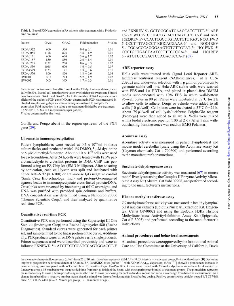
receive dyclonine. Patients or caregivers dosed with dyclonine byan oral rinse for 6–7 days twice daily, and then came in for a finalcheek swabbing for buccal cells. These blinded samples werethen sent for analysis. FXN protein was analyzed by the clinicallyvalidated dipstick immunoassay (44,45). We modified thesemethods by further by normalizing FXN to mitochondrial cyto-chrome oxidase expression, i.e. FXN/Complex IV (CIV). Fourrepeats of the dipstick immunoassay were performed per patientsample. Of the 8 patients who received dyclonine, six experiencedFXN increase (ranging from 60 to 480% induction), and twoexperienced no induction (Table 2). Interestingly, patients withgreater neurological impairment as measured by the Friedreich’sataxia rating scale (FARS) (46,47), experienced greater FXNinduction (Fig. 6A). Other correlations that were also significantincluded the Functional Disability Score (FDS) and Z2 ataxiafunctional composite score (Fig. 6B and C) (48). While the correl-ation of FXN response with GAA repeat length was not significant(Supplementary Material, Fig. 6D), this trend could underlay thedifferential response seen in Figure 4A.
DISCUSSION
FA is caused by inheritance of (GAA)n expansions in intron 1 ofthe nuclear FXN gene that decreases its expression to about 20%of normal. All consequences of Friedreich’s are thought to resultfrom this epigenetic silencing of FXN (5,6,11,49). There is cur-rently no approved or effective therapy for this ultimately lethaldisease that currently affects approximately 6000 in the USA and20 000 in Europe (1). We identified a biochemical deficiency inFriedreich’s patient cells to design and perform a drug screen,and have identified a potential treatment for the disease thatrescues FXN deficiency as well as downstream consequencesin cells, animal models and in FA patients.
Our previous microarray of Friedreich’s mouse model DRGsindicated defects in thioredoxin reductase, thiol-antioxidanttranscripts and a deficiency in Nrf2 activity in FXN knockdowncells (12). Also, a deficiency in Nrf2 activation was noted in Frie-dreich’s fibroblasts (18). Taken together, these data suggestedthat FXN deficiency may cause deficiency in Nrf2 activationor the related mitochondrial thioredoxin reductase pathway(50), which leads to decreased mitochondrial antioxidant protec-tion, increased reactive oxygen species, inflammation and neu-rodegeneration (51).
Given the data on thiol antioxidant deficiency in mouse DRG(12), and previous data on antioxidant deficiency in FA fibroblasts(12,16,17), we tested poisons of thiol antioxidants in a FXN-deficient DRG cell line (Fig. 1A). In these cells, diamide andauranofin were more toxic to FXN knockdown cells comparedwith controls, whereas hydrogen peroxide (H2O2) and buthioninesulfoxamine (BSO) displayed little to no difference. This wasinteresting because diamide oxidizes thioredoxin (52), and aura-nofin is an inhibitor of thioredoxin reductase (53). These datasupport the idea that, in the contextof thedorsal rootganglia, thior-edoxin reductase is an important antioxidant system. Others haveshown that, in neural mitochondria, thioredoxin reductase is thedominant antioxidant system (54,55). We examined FA patientcells and also observed increased cell death after diamide treat-ment which was reversed by the reductant dithiothreitol (DTT),confirming a thiol-oxidant mechanism (Fig. 1C).
To expedite the search for clinical candidates, we took a repur-posing approach and examined a collection of 1600 drugs withmostly known pharmacokinetic and safety profiles. Our initialscreen identified 100 protective hits in a range of drug classes,of which 33 were found to be reproducibly dose-dependent,for a hit rate of �2.5% (Supplementary Material, Table S1).One of the strongest hit in terms of efficacy and potency at pro-tecting from diamide stress was dyclonine, the active agent inSucrets. Dyclonine was initially characterized as an anti-epileptic (56), but through its sodium channel inhibitoryproperties it has been used for anesthesia since 1955 (13). Dyclo-nine’s protective effect could potentially be due to multiplemechanisms, including increased mitochondrial biogenesis,Fe/S cluster biogenesis, iron binding or others. Since all FA con-sequences are the result of deficient FXN expression, weexplored whether dyclonine was able to induce FXN in FApatient cells and the animal model (Fig. 2). We discovered an in-duction of FXN protein, and this was supported further withdose-dependent increases in FXN mRNA by RT-PCR. Thishas implications for therapy, since dyclonine rescues theprotein deficiency that causes the disease in FA lymphoblastsand fibroblasts. In animal models of FA, we observed a clear2-fold induction in FXN protein in two mouse models, in mul-tiple tissues including cerebellum, liver and heart (Fig. 2C andD, and Supplementary Material, Fig. S1), two of which (cerebel-lum and heart) are major sites of disease pathogenesis. Ouranimal studies included intraperitoneal and oral dosing, with
Table 1. ARE sites in human FXN gene
Position Position relativeto FXN
Direction DNA strand Sequence PWM score Evolutionarilyconserved (95% CI)
71633757 216 722 Antisense Non-template ACCATGTGACAATGCCCACTT 7.80 Yes71634506 215 973 Sense Template CCTGGGTGACAGAGCAAGACT 12.30 No71636687 213 792 Antisense Non-template AGGAACTGACTCCGCACAAGA 10.30 No71639782 210 697 Antisense Non-template AGTAGATGACCCAGCTGATAG 8.90 No71643186 27293 Sense Template ACAGTGTGACTATGCTCGACT 10.20 No71644882 25597 Antisense Non-template GACTCATGACTCAGCCAGTCC 12.50 Yes71645605 24874 Antisense Template GACCGGTGACTTTGCAAGTTA 12.90 Yes71651446 967 Antisense Template AACCTTTGACTGGGCTGGAAA 4.20 No71654599 4120 Sense Template GGGATGTGACGGGGCTGCGTC 1.20 No
FXN gene starts at 71650479 and ends at 71715094(64 615 bp). GAA-repeat insertion is 1731 bp downstream of FXN exon-1 start site. Position weight matrix (PWM)score from Wang et al. (28). Searched for the regular expression of ARE consensus sequence: TGAC[ACTG]{3}GC from 20 kb upstream to 5 kb downstream of FXN.
Human Molecular Genetics, 2014 7
at Brunel U
niversity on October 29, 2014
http://hmg.oxfordjournals.org/
Dow
nloaded from

drug exposures from 4 days to 4 weeks. In all, we have done 12independent in vivo studies with dyclonine with between 3 and 6mice per group, and each study supported an induction in FXN bydyclonine. Since FA patients are asymptomatic when their FXNlevels are 40–50% of normal levels, an induction of 2-foldshould boost patient FXN levels into this range, and alleviateconsequences of FXN deficiency (57).
Early on in our development efforts, we noted that a few of thedrugs we found to protect from diamide were also Nrf2 inducersin literature. Nrf2 translocates to the nucleus in response to oxi-dative stress (and especially thiol stress) and binds to AREs intarget genes (27). Dyclonine had never been described toinduce Nrf2, and we demonstrated a dose-dependent stimula-tion, and that multiple Nrf2-target proteins are elevated aftertreatment with dyclonine in both FA patient cells and animalmodel tissues (Fig. 3B). To investigate if the induction of FXNwas Nrf2-mediated, we used bioinformatics to identify AREsequences in the FXN locus, finding three ARE sites upstreamof the transcription start site of FXN that are completely con-served across ape evolution (Table 1 and Supplementary Mater-ial, Fig. S2). The functionality of ARE sites was verified by ChIPand pull down with an Nrf2 antibody (Fig. 3C). Thus the induc-tion of FXN is likely the result of recruitment of Nrf2 to adjacentARE sites. Data also suggest that other Nrf2 inducers will reacti-vate FXN expression.
We observe that dyclonine has a differential effect on FXNre-expression from alleles of different expansion length. Specif-ically, longer GAA alleles, which experience greater epigene-tic silencing, are more ‘reactivatable’ by dyclonine in cells(Fig. 4A), and this is consistent with what is observed in patients(Fig. 6C). There is precedent for Nrf2 activation having epigen-etic effects on genes (35,58–60). There is also recent precedentthat the classical Nrf2-inducer sulforaphane may be workingthrough epigenetic unsilencing of the Nrf2 locus (35,36). Dyclo-nine has been reported to have inhibitory activity on histonelysine methyltransferase G9a, which is known to methylatehistone H3K9 (PubChem, assay ID 504332). Inhibitory methy-lation of histone H3K9 is a known epigenetic mark at the FXNlocus (31–34). And we observed experimentally that dyclonineinhibited histone methyltransferase G9 in lymphoblast nuclearextracts (Fig. 4B). These data support either of two mechanisms,i.e. that dyclonine induces Nrf2, and Nrf2 through its epigeneticunsilencing effects reactivates FXN; or that dyclonine sup-presses histone methyltransferase G9, which in turn reactivatesboth the FXN locus and the Nrf2 locus.
Since dyclonine is an anesthetic whose pain amelioratingeffects are mediated through sodium channel inhibition, therecould be concern of the sedative potential of inhibiting neuronalNaV1.2 sodium channels (61). Additionally, sodium channelblockers are used to treat cardiac arrhythmias and alteration ofheart rate would be a concern prior to treatment FA patientswith systemic dyclonine. Since there were no data in the litera-ture about the specific concentrations at which this occurred,we examined inhibition of NaV1.2 currents using patch-clampelectrophysiology. As expected, there was clear dose-dependentinhibition with an IC50 of �6 mM. This IC50 is 30-fold higherthan our EC50 for protection from diamide in FA fibroblasts,and we observe FXN induction in FA lymphoblasts with 1 mMdyclonine. Additionally, after 4 weeks of chronic daily dosingin mice, no sedation or adverse phenotype was observed.
To verify that there was no systemic peripheral anesthesiafrom ingestion of a topical anesthetic at the doses that induceFXN reactivation, we measured hot plate response and foundno delay. Finally, we tested �20 sodium channel inhibitors oranesthetics including lidocaine, bupivicaine and propofol andnone were significantly active in the diamide protection assay.Taken together, this reduces the concern that the protective
Figure 4. Dyclonine exerts epigenetic effects. (A) Dyclonine induction of FXNtranscript levels in healthy and FA patient lymphoblast cells with varyingGAA-repeat length. Healthy and FA patient lymphoblasts (GM16216,GM16197 and GM14518) were treated with 30 mM dyclonine or vehiclecontrol (0.1% DMSO). RNA was analyzed after 24 h by RT-PCRwith expressionwith primers for FXN normalized to b-actin. GAA, number of GAA repeats onFXN allele with less insertions. The plotted data represent the mean foldchange in FXN transcript in drug-treated cells normalized to vehicle control forthat cell line. Error bars represent SEM. ∗P , 0.05, ∗∗P , 0.01, t-test (n ¼ 4).The induction of Fxn mRNA increases with GAA repeat length. (B) Dyclonineinhibits histone methyltransferase activity. Histone methyltransferase G9a(G9aHMTase) activity, which specifically methylates histone H3K9, was mea-sured in nuclear extracts of healthy lymphoblast cells treated with drug for60 min. Negative controls are 0.1% DMSO vehicle-treated cells; positivecontrols are cells treated with G9aHMTase-specific inhibitor 1 mM Bix01294.The plotted data represent the mean absorbance at 450 nm which reflectsG9aHMTase activity. Error bars represent SEM. ∗P , 0.05, ∗∗P , 0.01, t-test.(n ¼ 4).
8 Human Molecular Genetics, 2014
at Brunel U
niversity on October 29, 2014
http://hmg.oxfordjournals.org/
Dow
nloaded from

actions of dyclonine are mediated through sodium channelblockade, since the doses required for FXN activation arelower than those for sodium channel engagement.
The most well-defined downstream function for mammalianFXN is in iron–sulfur cluster biogenesis (40,42,62), and dyclo-nine treatment induced iron–sulfur cluster enzyme activity inanimal and cell models (Fig. 5A–C). We also examined severalneurobehavioral parameters in multiple FA mouse models,including open field activity, grip strength, treadscan andmotor coordination on the level beam task. The most sensitivebehavioral test was time to cross a level beam. Cerebellardefects in FA patients lead to motor coordination difficulties,and the level beam test mimics the walking score in the FARSataxia score (47). We observed a significant improvement inthis parameter after 4 weeks of oral dyclonine dosing.
Since dyclonine is FDA-approved as an oral rinse, its prescrip-tion and dispensing was allowed in an IRB-approved trial. Tenpatients were recruited, but 2 did not complete the treatmentand stopped 2–3 days into the study, likely because of the tasteor mouth numbing that persisted for up to 1 h after each dosing.Of the remaining patients, 6 of the 8 showed an increase in FXNafter 1 week. Additionally, both healthy volunteers responded todyclonine with an increase in FXN (Table 2). The correlationswith neurological scores are highly significant, and suggestthat dyclonine produces a greater induction in patients withmore severe FXN deficiency (Fig. 6).
FXN deficiency causes oxidative stress and subsequent celldeath in dorsal root ganglia and cardiac tissue (3). Oxidativestress is suspected to be important in multiple neurodegenerativediseases (63–65). Here, we describe a well-known anestheticdrug dyclonine that protects FA patient cells from diamide-induced oxidative stress, increases FXN and rescues primarybiochemical end points of the disease in cell and animal models,and appears to do so involving the Nrf2/ARE pathway. Limita-tions of this work include translation of beneficial effects seenin mice to humans, as well as the hope that FXN engagementafter topical exposure with a rinse solution of dyclonine willmodel results after a systemic dosing regimen in FA patients.Although there are a number of questions that need to be addres-sed for these findings to be clinically applied to patients, includ-ing a better understanding of pharmacokinetics, optimal dosingformulation for oral bioavailability and cardiovascular toxicol-ogy, dyclonine represents a potential novel treatment strategyfor the disease.
MATERIALS AND METHODS
Cell lines
Human control and FA patient fibroblasts and lymphoblasts wereobtained from Coriell Institute for Medical Research repository.Patient lymphoblast cell lines used: GM16214, GM04079,GM16205, GM16243, GM15850, GM16220 and GM16197.Patient fibroblast lines used: 1133, 1134.
Diamide screening assay
Patient fibroblasts were grown in MEM media (Life Technolo-gies Corp.) supplemented with 15% fetal bovine serum in t225tissue culture flasks and kept below 70% confluency. Cells were
trypsinized and density determined using a Vi-Cell counter(Beckman Coulter Corp). Five thousand fibroblast cells werethen aliquoted into 96-well poly-D-lysine-coated black/clearculture plates (Becton Dickinson Corp.) in growth mediawithout antibiotics in a volume of 180 ml. Cells were allowedto adhere for 3–4 h at 378C. Drugs (10 mM stock in dimethylsulfoxide, DMSO) were dispensed into assay plate wells afteran intermediate dilution in PBS, giving a final DMSO concentra-tion of 0.1% using an electronic multichannel pipetter (BioHit,Sartorius Corp.). Pharmakon drug library consisted of 1600compounds (Microsource Discovery Systems, Inc.) in 96 wellplates at stock concentration of 10 mM in DMSO. Test com-pounds were all be tested at 10 mM final assay concentration inprimary screen. Eight wells each of 300 mM dithiothreitol(Sigma-Aldrich Corp.) or 0.1% DMSO were used as positiveor negative controls, respectively. Cells were then incubated at378C with 5% CO2 overnight. After 24 h, diamide (Sigma-Aldrich Corp.) was added to all wells at a final concentrationof 125 mM from a 100 mM stock solution prepared in DMSOand allowed to incubate at 378C with 5% CO2 overnight(14–18 h). Plates were then washed with PBS, supplementedwith 1 mM Calcein-AM (Molecular Probes, Invitrogen Corp.)and incubated at room temperature for 45 min. Cells wereagain washed with PBS to remove residual dye, and read onBMG PolarStar Optima with 485 excitation and 520 emissionwavelengths (BMG LabTech).
Western blot analysis
Cells were lysed using lysis buffer (Promega Corp.) supplemen-ted with a complete protease inhibitor cocktail (Roche AppliedScience) and phenylmethylsulfonyl fluoride (Sigma-AldrichCorp.). Tissues were further homogenized using 0.5 mm glassbeads in a Bullet Blender high-throughput homogenizer (NextAdvance, Inc.). After pelleting cellular debris by spinning at16 000 rpm at 48C for 15 min, protein was quantified by Brad-ford assay (66). For western blotting, 40–50 mg protein wasadded per lane of 4–12% Bis-Tris gels (Invitrogen Corp.).Primary antibodies were diluted in Odyssey blocking buffer(LI-COR Biosciences). Antibodies used included: anti-FXN(provided by Franco Taroni M.D., Istituto Besta), anti-HO1 (#sc-10789, Santa Cruz Biotechnology, Inc.), anti-NQO1(#3187, Cell Signaling Technologies), anti-Gpx4 (#ab125066,AbCam), anti-nrf2 (sc-25820, Santa Cruz) and anti-actin(#A2668, Sigma). Direct conjugated secondary antibodies (anti-rabbit IRdye800Cw and anti-mouse IRdye680 from LI-COR)were used to detect and quantify the signal of primary antibodiesand imaged using a LI-COR Odyssey.
Bioinformatics and phylogenetic analysis of FXN locus
A position weight matrix was used to identify ARE sequences inFXN based on previously described functional ARE sequences(28). A pre-filter was used with the consensus sequence ofnnnnnnTGACnnnGCnnnnnn to identify potential ARE sites.The position weight matrix was used to calculate the score foreach of the pre-filtered hits. Phylogenetic footprinting wasthen done using Clustalw2, and a multiple alignment wasdone between the great apes (Homo sapiens, Pan troglodytes,
Human Molecular Genetics, 2014 9
at Brunel U
niversity on October 29, 2014
http://hmg.oxfordjournals.org/
Dow
nloaded from

Figure 5. Dyclonine recovers downstream effects of FXN deficiency in vitro and in vivo. (A) Dyclonine increases aconitase activity in FA patient lymphoblast celllines. FA lymphoblasts (GM16205, GM16197 and GM16243) were treated with 10 mM dyclonine or vehicle control (0.1% DMSO). Cell pellets were collected after48 h, and aconitase activity was measured in lysates over 90 min to ensure linear range was captured. A healthy volunteer lymphoblast cell line was used as a control.The plotted data represent the mean rate change in fluorescence (dF/dt) from 25 to 30 min. Error bars represent SEM. ∗P , 0.05, t-test. (n ¼ 3). (B) Dyclonineincreases aconitase activity in FA mouse model cerebellum in vivo. FA-YG8 transgenic mice [hFXN+/2 with FXN (GAA)190 expansion; mFxn2/2] were treatedwith 5 mg/kg dyclonine for 1 week p.o. Aconitase activity was measured in cerebellar lysates over 90 min to ensure linear range was captured. Positive controlswere vehicle-treated FA-YG8 transgenic mice [hFXN+/+ with FXN (GAA)190 expansion; mFxn2/2]. The plotted data represent the mean rate change in fluorescence(dF/dt) from 15 to 20 min.Error bars represent SEM. ∗P , 0.05, t-test (n ¼ 4 mice per group,11–12 months ofage). (C) Dyclonine increases succinatedehydrogenaseactivity in FA mouse model liver in vivo. FA-PandKIKO mice [mFxn+/2 with FXN (GAA)230 expansion; mFxn2/2] were treated with 25 mg/kg dyclonine for 1 weekp.o. Succinate dehydrogenase activity was measured in liver lysates over 90 min. Positive controls were vehicle-treated WT C57/Bl6 mice. The plotted data represent
10 Human Molecular Genetics, 2014
at Brunel U
niversity on October 29, 2014
http://hmg.oxfordjournals.org/
Dow
nloaded from
Gorilla and Pongo abeli) in the region upstream of the FXNgene (29).
Chromatin immunoprecipitation
Patient lymphoblasts were seeded at 0.5 × 10⁶/ml in tissueculture flasks, and incubated with 0.1% DMSO, 5 mM dyclonineor 5 mM dimethyl-fumarate. About �10 × 106 cells were usedfor each condition. After 24 h, cells were treated with 18.5% par-aformaldehyde to crosslink proteins to DNA. ChIP was per-formed using an EZ-Chip kit (EMD Millipore). After shearingby sonication, each cell lysate was split and incubated witheither Anti-Nrf2 (#H-300) or anti-mouse IgG negative control(Santa Cruz Biotechnology, Inc.) and protein-G-conjugatedagarose beads to immunoprecipitate cross-linked protein/DNA.Crosslinks were reversed by incubating at 658C overnight, andDNA was purified with provided spin columns and buffers.DNA concentration was determined using a Nanodrop 2000c(Thermo Scientific Corp.), and then analyzed by quantitativereal-time PCR.
Quantitative real-time PCR
Quantitative PCR was performed using the Superscript III OneStep kit (Invitrogen Corp) in a Roche Lightcycler 480 (RocheDiagnostics). Standard curves were generated for each primerset, and samples fitted to the linear portion of the curve. Addition-ally,PCRproductswere runonDNAgels toverifysingle products.Primer sequences used were described previously and were asfollows: FXNFWD 5′- ATCTTCTCCATCCAGTGGACCT-3′
and FXNREV 5′- GCTGGGCATCAAGCATCTTTT-3′; ARE16223FWD 5′- CCTGCCGTACTCAGTCCTTC-3′ and ARE16223REV 5′- CCACTCGGCTGTACTGTCTG-3′; NQO1FWD5′-CCCTTTTAGCCTTGGCACGAAA-3′ and NQO1REV5′- TGCACCCAGGGAAGTGTGTTGTAT-3′; HO1FWD 5′
CCCTGCTGAGTAATCCTTTCCCGA-3′ and HO1REV5′- ATGTCCCGACTCCAGACTCCA-3′ (67).
ARE reporter assay
HeLa cells were treated with Cignal Lenti Reporter ARE-luciferase lentiviral reagent (SABiosciences, Cat # CLS-2020L) and underwent selection with 1 mg/ml of puromycin togenerate stable cell line. Hela-ARE stable cells were washedwith PBS and 1× EDTA, and plated in phenol-free DMEMmedia supplemented with 10% FBS in white wall/bottom96-well plates in 90 ml. Plates were incubated at 378C for 2 hto allow cells to adhere. Drugs or vehicle were added to allwells (10 ml/well). Cell plates were incubated at 378C for 24 h.About 75 ml/well of cell lysis/luciferase Bright-Glo reagent(Promega) were then added to all wells. Wells were mixedwith a biohit electronic pipettor (100 ml 2×). After 5 min with-out shaking, luminescence was read on BMG Polarstar.
Aconitase assay
Aconitase activity was measured in patient lymphoblast andmouse model cerebellar lysate using the Aconitase Assay Kit(Cayman chemicals, Cat # 700600) and performed accordingto the manufacturer’s instructions.
Succinate dehydrogenase assay
Succinate dehydrogenase activity was measured (67) in mousemodel liver lysate using the Complex II Enzyme Activity Micro-plate Assay Kit (Abcam, Cat # ab109908) and performed accord-ing to the manufacturer’s instructions.
Histone methyltransferase assay
G9 methyltransferase activity was measured in healthy lympho-blast nuclear extracts (Epiquik Nuclear Extraction Kit, Epigen-tek, Cat # OP-0002) and using the EpiQuik H3K9 HistoneMethyltransferase Activity/Inhibition Assay Kit (Epigentek,Cat # P-3003) and performed according to the manufacturer’sinstructions.
Animal procedures and behavioral assessments
All animal procedures wereapprovedby the Institutional AnimalCare and Use Committee at the University of California, Davis
Table 2. Buccal FXN expression in FA patients after treatment with a 1% dyclo-nine oral rinse
Patient GAA1 GAA2 Fold induction P-value
FRDA4522 600 500 0.4+0.1 0.01FRDA0055 1178 826 4.8+1.9 0.01FRDA0171 600 600 3.6+1.7 0.02FRDA0157 850 850 2.6+1.4 0.01FRDA0235 1122 230 0.6+0.3 0.02FRDA4539 1045 670 1.6+0.5 0.07FRDA4579 1133 71 1.3+0.6 0.31FRDA4576 800 800 1.8+0.6 0.04HV0001 ND ND 5.2+1.9 0.02HV0002 ND ND 1.7+0.3 0.01
Patients and controls were dosed for 1 week with a 1% dyclonine oral rinse, twicedaily for 30 s. Buccal cell samples were taken by clinician use swabs and blindedprior to analysis. GAA1 and GAA2 refer to the number of GAA repeats in bothalleles of the patient’s FXN gene (ND, not determined). FXN was measured inblinded samples using dipstick immunoassay normalized to complex IVexpression. Fold induction is a value post-treatment divided by pre-treatmentFXN/CIV+SD (n ¼ 4 measurements per patient).P-value determined by the t-test.
the mean rate change in fluorescence (dF/dt) from 25 to 30 min. Error bars represent SEM. ∗P , 0.05, t-test (n ¼ 4 mice per group, 8–9 months of age). (D) Dyclonineimproves progressive behavioral defect of FA mice. FA-PandKIKO mice [mFxn+/2 with FXN (GAA)230 expansion; mFxn2/2] showed a pronounced increase inbeam crossing time compared with WT controls at 13 months of age. FA-PandKIKO mice were treated with 25 mg/kg dyclonine or vehicle for 4 weeks p.o.Latency to cross a 16 mm beam was the recorded time from start to finish of the beam, with the experimenter blinded to treatment groups. The plotted data representthe mean latency to cross a beam post-dosing minus the time to cross pre-dosing for each individual mouse and serve as a change from baseline measurement. As achange from baseline, a negative number means that the animal was faster after dosing than it was before dosing. Positive controls were vehicle-treated WT C57/Bl6mice. ∗P , 0.05, t-test (n ¼ 7–9 mice per group, 12–14 months of age).
Human Molecular Genetics, 2014 11
at Brunel U
niversity on October 29, 2014
http://hmg.oxfordjournals.org/
Dow
nloaded from

with adherence to the NIH Guide for the Care and Use of Labora-tory Animals. The age of mice used for all studies was between 6and 14 months and was age-matched within experiments. Drugdosing was done i.p. formulated in a DMSO and PBS mixture,and p.o. formulated in a DMSO, water and peanut butter mixture.Behavior testing was performed at the University of California,Davis Mouse Behavioral Analysis Laboratory. A colony ofYG8R B6.Cg-Fxntm1Mkn Tg(FXN)YG8Pook/J mice was estab-lished at UC Davis (Jackson Labs, # 012253) (68). For ourtesting, we used both hemizygous mice [FA-YG8 (hFXN+/2
with FXN (GAA)190 expansion; mFxn2/2)] containing oneallele of the mutant FXN transgene and one KO (and thus the
least amount of FXN) and homozygous mice [FA-YG8(hFXN+/+ with FXN (GAA)190 expansion; mFxn2/2], contain-ing two alleles of the transgene (and thus higher levels of FXN),and wild-type (WT) mice. In addition, the B6.Cg-Fxntm1Mkn
Fxntm1Pand/J strain was also established and used (JacksonLabs, # 014162), and is referred to as FA-PandKIKO mice[mFxn+/2 with FXN (GAA)230 expansion; mFxn2/2] (69,70).Both of these mice are bred on a C57BL/6J background.
Level beam performance was measured by placing mice on a100 cm long level beam with a line marked 10 cm from each endas the start and end lines. A 60 W desk lamp was used as an aver-sive stimulus at the starting end of the beam, and an enclosed
Figure 6. Dyclonine inductionof FXN in buccal cells of FA patients correlateswith disease severity. FA patients were dosed with a 1% dyclonine oral rinse, twice dailyfor 30 s. Buccal cells were collected by cheek swabbing before and after 1 week of dosing. Cell lysates were probed by dipstick immunoassay for analysis of FXNexpression and normalized to mitochondrial complex IV (n ¼ 4 replicate experiments of FXN analysis per patient, summarized in Table 1). The plotted data representthe mean fold change in FXN protein in dyclonine-treated FA patient buccal cells normalized to expression for each individual patient before drug dosing (healthyvolunteers not shown). This fold FXN induction is plotted against (A) FARS score, (B) FDS, (C) Z2 ataxia functional composite score and (D) FXN gene GAA-repeatlength for each FA patient who participated in this clinical study (obtained through a natural history database). For A and B, a higher score indicates greater diseaseseverity. For C, a lower score indicates greater disease severity. A P-value of ,0.05 is considered significant, Pearson’s correlation coefficient.
12 Human Molecular Genetics, 2014
at Brunel U
niversity on October 29, 2014
http://hmg.oxfordjournals.org/
Dow
nloaded from

shelter remained at the end of the beam. A training beam of21 mm diameter was used for three training trials, with at least10 min of rest allowed for each animal between beam crosses.Subsequently, three trials were conducted for 21, 16 and 9 mmwide beams. Time was recorded for each animal to cross fromstart to finish. Additionally, a video camera recorded everytrial and was later processed for quantification of foot slips aserrors. Experimenter who recorded times and errors wasblinded to treatment groups.
Clinical human buccal cell collection
Eight FA patients and two healthy volunteers were dosed with a1% dyclonine oral rinse 2× daily for 30 s for 7 days. Sampleswere collected prior to and at the end of 1 week dosing. Buccalcells were collected from patients and healthy controls usingMasterAmp Buccal Swab brushes (Epicentre, Illumina Corp.).Swabs were gently twirled against the inside of the right cheekfor 30 s. The swab was then removed from the mouth anddipped into a tube containing 500 ml of ice cold extractionbuffer (AbCam, #ab109877). The swab brush was placedgently in the tube of buffer for �10 s to dislodge the cells.Tubes were frozen at 2208C until all samples received soFXN measurement could be performed at one time.
FXN immunoassay protocol
FXN expression in buccal cells was measured using a commer-cially dipstick immunoassay for FXN (AbCam, #ab109877) andmitochondrial complex IV (AbCam, #ab109881) expressionaccording to the manufacturer’s instructions. Buccal cells werelysed using extraction buffer supplied with the assay kit and cel-lular debris pelleted. Protein concentration was measured byBradford assay. About 10 mg of peripheral blood mononu-cleated cell protein in 25 ml of extraction buffer was mixedwith 25 ml of blocking buffer and added to individual wells ona 96-well plate with gold-conjugated mAb at the bottom ofeach well. Samples were measured as duplicates. After 5 min in-cubation at room temperature, dipsticks were added into thewell and allowed transfer onto the membrane, where FXN orcomplex IV was immunocaptured. This capture was quantifiedon dried dipsticks using a Hamamatsu dipstick reader(AbCam, #MS1000), and raw mABS (milli-Absorbance)values for FXN were normalized to complex IV raw values.
Statistical analysis
Data are presented as mean+SD or SEM, and the significanceof the difference between groups was evaluated with the Stu-dent’s t-test (two-tailed) or Pearson’s correlation coefficient. AP-value of ,0.05 was considered significant. Curve fits weredone using GraphPad prism nonlinear regression analysis witha variable slope. Additionally, a 95% confidence interval wasused in bioinformatics analysis of ARE sites in FXN gene.
Study approval
The human clinical study was reviewed and approved by theInvestigational Review Board of the University of California,Los Angeles (proof of concept of dyclonine in buccal cells of
FA patients, UCLA IRB#13-000478). All patients were pro-vided written informed consent prior to initiation of the study.All animal studies and procedures were approved by theInstitutional Animal Care and Use Committee at the Universityof California, Davis.
SUPPLEMENTARY MATERIAL
Supplementary Material is available at HMG online.
ACKNOWLEDGEMENTS
We thank Billy Zhang, Emma Karey, UC Davis Mouse BehaviorAnalysis Laboratory, Mari Golub, Peter Takenuchi, Todd Tolen-tino, Maria Casado, Franco Taroni and Daniel McKemie fortheir many contributions and assistance.
Conflict of Interest statement. The authors have no competing fi-nancial interests. S.S. and G.A.C. are inventors on a pendingpatent on identification of agents useful for the treatment ofFriedreich’s ataxia.
FUNDING
This work was supported by funding from the Friedreich’sAtaxia Research Alliance, the National Institutes of Health(PHS NIH RO1 NS077777-15, PO1 AG025532 and R01EY012245) and the NIGMS-funded Pharmacology TrainingProgram (T32GM099608). Funding to pay the Open Accesspublication charges for this article was provided by the NIH.
REFERENCES
1. Campuzano, V., Montermini, L., Lutz, Y., Cova, L., Hindelang, C.,Jiralerspong, S., Trottier, Y., Kish, S.J., Faucheux, B. and Trouillas, P.(1997) Frataxin is reduced in Friedreich ataxia patients and is associated withmitochondrial membranes. Hum. Mol. Genet., 6, 1771–1780.
2. Koeppen, A.H., Michael, S.C., Knutson, M.D., Haile, D.J., Qian, J., Levi, S.,Santambrogio, P., Garrick, M.D. and Lamarche, J.B. (2007) The dentatenucleus in Friedreich’s ataxia: the role of iron-responsive proteins. ActaNeuropathol. (Berl.), 114, 163–173.
3. Koeppen, A.H., Morral, J.A., Davis, A.N., Qian, J., Petrocine, S.V., Knutson,M.D., Gibson, W.M., Cusack, M.J. and Li, D. (2009) The dorsal rootganglion in Friedreich’s ataxia. Acta Neuropathol. (Berl.), 118, 763–776.
4. Albano, L.M.J., Nishioka, S.A.D., Moyses, R.L., Wagenfuhr, J., Bertola, D.,Sugayama, S.M.M. and Chong, A.K. (2002) Friedreich’s ataxia: cardiacevaluation of 25 patients with clinical diagnosis and literature review. Arq.Bras. Cardiol., 78, 448–451.
5. Durr, A., Cossee, M., Agid, Y., Campuzano, V., Mignard, C., Penet, C.,Mandel, J.-L., Brice, A. and Koenig, M. (1996) Clinical and geneticabnormalities in patients with Friedreich’s ataxia. N. Engl. J. Med., 335,1169–1175.
6. Filla, A., De Michele, G., Cavalcanti, F., Pianese, L., Monticelli, A.,Campanella, G. and Cocozza, S. (1996) The relationship betweentrinucleotide (GAA) repeat length and clinical features in Friedreich ataxia.Am. J. Hum. Genet., 59, 554.
7. Lu, C. and Cortopassi, G. (2007) Frataxin knockdown causes loss ofcytoplasmic iron–sulfur cluster functions, redox alterations and induction ofheme transcripts. Arch. Biochem. Biophys., 457, 111–122.
8. Al-Mahdawi, S., Pinto, R.M., Varshney, D., Lawrence, L., Lowrie, M.B.,Hughes, S., Webster, Z., Blake, J., Cooper, J.M. and King, R. (2006) GAArepeat expansion mutation mouse models of Friedreich ataxia exhibitoxidative stress leading to progressive neuronal and cardiac pathology.Genomics, 88, 580–590.
Human Molecular Genetics, 2014 13
at Brunel U
niversity on October 29, 2014
http://hmg.oxfordjournals.org/
Dow
nloaded from

9. Condo, I., Ventura,N., Malisan, F.,Tomassini, B. and Testi, R. (2006)A poolof extramitochondrial frataxin that promotes cell survival. J. Biol. Chem.,281, 16750–16756.
10. Montermini, L., Richeter, A., Morgan, K., Justice, C.M., Julien, D.,Castellotti, B., Mercier, J., Poirier, J., Capozzoli, F. and Bouchard, J.P.(1997) Phenotypic variability in Friedreich ataxia: role of the associatedGAA triplet repeat expansion. Ann. Neurol., 41, 675–682.
11. De Biase, I., Chutake, Y.K., Rindler, P.M. and Bidichandani, S.I. (2009)Epigenetic silencing in Friedreich ataxia is associated with depletion ofCTCF (CCCTC-binding factor) and antisense transcription. PLoS ONE, 4,e7914.
12. Shan, Y., Schoenfeld, R.A., Hayashi, G., Napoli, E., Akiyama, T., IodiCarstens, M., Carstens, E.E., Pook, M.A. and Cortopassi, G.A. (2013)Frataxin deficiency leads to defects in expression of antioxidants and Nrf2expression in dorsal root ganglia of the Friedreich’s ataxia YG8R mousemodel. Antioxid Redox Signal, 19, 1481–1493.
13. Shelmire, B., Gastineau, F. and Shields, T.L. (1955) Evaluation of a newtopical anesthetic, dyclonine hydrochloride. Arch. Dermatol., 71, 728.
14. Florestano, H. and Bahler, M. (1956) Antimicrobial properties of dycloninehydrochloride, a new topical anesthetic. J. Am. Pharm. Assoc., 45, 320–325.
15. Sinha, B., Pattabhi, V., Nethaji, M. and Gabe, E. (1987) Structure of a localanaesthetic: dyclonine hydrochloride. Acta Crystallogr C, 43, 360–361.
16. Yang, J., Cavadini, P., Gellera, C., Lonnerdal, B., Taroni, F. and Cortopassi,G. (1999) The Friedreich’s ataxia mutation confers cellular sensitivity tooxidant stress which is rescued by chelators of iron and calcium andinhibitors of apoptosis. Hum. Mol. Genet., 8, 425–430.
17. Chantrel-Groussard, K., Geromel, V., Puccio, H., Koenig, M., Munnich, A.,Rotig, A. and Rustin, P. (2001) Disabled early recruitment of antioxidantdefenses in Friedreich’s ataxia. Hum. Mol. Genet., 10, 2061–2067.
18. Paupe, V., Dassa, E.P., Goncalves, S., Auchere, F., Lonn, M., Holmgren, A.and Rustin, P. (2009) Impaired nuclear Nrf2 translocation undermines theoxidative stress response in Friedreich ataxia. PLoS ONE, 4, e4253.
19. Kosower, N.S. and Kosower, E.M. (1995) Diamide: an oxidant probe forthiols. Methods Enzymol., 251, 123.
20. Kosower, N.S., Kosower, E.M., Wertheim, B. and Correa, W.S. (1969)Diamide, a new reagent for the intracellular oxidation of glutathione to thedisulfide. Biochem. Biophys. Res. Commun., 37, 593–596.
21. Liu, L., Trimarchi, J.R. and Keefe, D.L. (1999) Thiol oxidation-inducedembryonic cell death in mice is prevented by the antioxidant dithiothreitol.Biol. Reprod., 61, 1162–1169.
22. Verdon, E., Couedor, P. and Sanders, P. (2007) Multi-residue monitoring forthe simultaneous determination of five nitrofurans (furazolidone,furaltadone, nitrofurazone, nitrofurantoine, nifursol) in poultry muscletissue through the detection of their five major metabolites (AOZ, AMOZ,SEM, AHD, DNSAH) by liquid chromatography coupled to electrospraytandem mass spectrometry—in-house validation in line with CommissionDecision 657/2002/EC. Anal. Chim. Acta, 586, 336–347.
23. Sangster, N., Rickard, J., Hennessy, D., Steel, J. and Collins, G. (1991)Disposition of oxfendazole in goats and efficacy compared with sheep. Res.
Vet. Sci., 51, 258–263.
24. Riah, O., Dousset, J.-C., Courriere, P., Stigliani, J.-L., Baziard-Mouysset, G.and Belahsen, Y. (1999) Evidence that nicotine acetylcholine receptors arenot the main targets of cotinine toxicity. Toxicol. Lett., 109, 21–29.
25. Kim, S.-J., Park, C., Han, A.L., Youn, M.-J., Lee, J.-H., Kim, Y., Kim, E.-S.,Kim, H.-J., Kim, J.-K. and Lee, H.-K. (2009) Ebselen attenuatescisplatin-induced ROS generation through Nrf2 activation in auditory cells.Hear. Res., 251, 70–82.
26. Kensler, T.W., Wakabayashi, N. and Biswal, S. (2007) Cell survivalresponses to environmental stresses via the Keap1-Nrf2-ARE pathway.Annu. Rev. Pharmacol. Toxicol., 47, 89–116.
27. Osburn, W.O. and Kensler, T.W. (2008) Nrf2 signaling: an adaptiveresponse pathway for protection against environmental toxic insults. Mutat
Res, 659, 31–39.28. Wang, X., Tomso, D.J., Chorley, B.N., Cho, H.-Y., Cheung, V.G.,
Kleeberger, S.R. and Bell, D.A. (2007) Identification of polymorphicantioxidant response elements in the human genome. Hum. Mol. Genet., 16,1188–1200.
29. Larkin, M., Blackshields, G., Brown, N., Chenna, R., McGettigan, P.A.,McWilliam, H., Valentin, F., Wallace, I.M., Wilm, A. and Lopez, R. (2007)Clustal W and Clustal X version 2.0. Bioinformatics, 23, 2947–2948.
30. Bidichandani, S.I., Ashizawa, T. and Patel, P.I. (1998) The GAAtriplet-repeat expansion in Friedreich ataxia interferes with transcription and
may be associated with an unusual DNA structure. Am. J. Hum. Genet., 62,111–121.
31. Sandi, C., Pinto, R.M., Al-Mahdawi, S., Ezzatizadeh, V., Barnes, G., Jones,S., Rusche, J.R., Gottesfeld, J.M. and Pook, M.A. (2011) Prolongedtreatment with pimelic o-aminobenzamide HDAC inhibitors ameliorates thedisease phenotype of a Friedreich ataxia mouse model. Neurobiol. Dis., 42,496–505.
32. Al-Mahdawi, S., Pinto, R.M., Ismail, O., Varshney, D., Lymperi, S., Sandi,C., Trabzuni, D. and Pook, M. (2008) The Friedreich ataxia GAA repeatexpansion mutation induces comparable epigenetic changes in human andtransgenic mouse brain and heart tissues. Hum. Mol. Genet., 17, 735–746.
33. Saveliev, A., Everett, C., Sharpe, T., Webster, Z. and Festenstein, R. (2003)DNA triplet repeats mediate heterochromatin-protein-1-sensitivevariegated gene silencing. Nature, 422, 909–913.
34. Greene, E., Mahishi, L., Entezam, A., Kumari, D. and Usdin, K. (2007)Repeat-induced epigenetic changes in intron 1 of the frataxin gene and itsconsequences in Friedreich ataxia. Nucleic Acids Res., 35, 3383–3390.
35. Paredes-Gonzalez, X., Fuentes,F., Su, Z.Y. and Kong, A.N. (2014) Apigeninreactivates Nrf2 anti-oxidative stress signaling in mouse skin epidermalJB6 P+ cells through epigenetics modifications. AAPS j., 16, 727–735.
36. Zhang, C., Su, Z.-Y., Khor, T.O., Shu, L. and Kong, A.-N.T. (2013)Sulforaphane enhances Nrf2 expression in prostate cancer TRAMP C1 cellsthrough epigenetic regulation. Biochem. Pharmacol., 85, 1398–1404.
37. Tella, S.R. and Goldberg, S.R. (1998) Monoamine transporter and sodiumchannel mechanisms in the rapid pressor response to cocaine. Pharmacol.Biochem. Behav., 59, 305–312.
38. Catterall, W.A. and Mackie, K. (1996) Local Anesthetics. Goodman &Gilman‘s The Pharmacological Basis of Therapeutics, 9th edn.McGraw-Hill, New York, pp. 331–347.
39. Lai, H.C. and Jan, L.Y. (2006) The distribution and targeting of neuronalvoltage-gated ion channels. Nat. Rev. Neurosci., 7, 548–562.
40. Bridwell-Rabb, J., Winn, A.M. and Barondeau, D.P. (2011) Structure–function analysis of Friedreich’s ataxia mutants reveals determinants offrataxin binding and activation of the Fe–S assembly complex.Biochemistry, 50, 7265–7274.
41. Shan, Y., Napoli, E. and Cortopassi, G. (2007) Mitochondrial frataxininteracts with ISD11 of the NFS1/ISCU complex and multiple mitochondrialchaperones. Hum. Mol. Genet., 16, 929–941.
42. Tan, G., Napoli, E., Taroni, F. and Cortopassi, G. (2003) Decreasedexpression of genes involved in sulfur amino acid metabolism infrataxin-deficient cells. Hum. Mol. Genet., 12, 1699–1711.
43. Shan, Y. and Cortopassi, G. (2012) HSC20 interacts with frataxin and isinvolved in iron–sulfur cluster biogenesis and iron homeostasis. Hum. Mol.Genet., 21, 1457–1469.
44. Willis, J.H., Isaya, G., Gakh, O., Capaldi, R.A. and Marusich, M.F. (2008)Lateral-flow immunoassay for the frataxin protein in Friedreich’s ataxiapatients and carriers. Mol. Genet. Metab., 94, 491–497.
45. Deutsch, E.C., Santani, A.B., Perlman, S.L., Farmer, J.M., Stolle, C.A.,Marusich, M.F. and Lynch, D.R. (2010) A rapid, noninvasive immunoassayfor frataxin: utility in assessment of Friedreich ataxia. Mol. Genet. Metab.,101, 238–245.
46. Schmitz-Hubsch, T., du Montcel, S.T., Baliko, L., Berciano, J., Boesch, S.,Depondt, C., Giunti, P., Globas, C., Infante, J. and Kang, J.-S. (2006) Scalefor the assessment and rating of ataxia development of a new clinical scale.Neurology, 66, 1717–1720.
47. Fahey, M., Corben, L., Collins, V., Churchyard, A. and Delatycki, M. (2007)How is disease progress in Friedreich’s ataxia best measured? A study of fourrating scales. J. Neurol. Neurosurg. Psychiatry, 78, 411–413.
48. Friedman, L.S., Farmer, J.M., Perlman, S., Wilmot, G., Gomez, C.M.,Bushara, K.O., Mathews, K.D., Subramony, S., Ashizawa, T. and Balcer,L.J. (2010) Measuring the rate of progression in Friedreich ataxia:implications for clinical trial design. Mov. Disord., 25, 426–432.
49. Chutake, Y.K., Costello, W.N., Lam, C. and Bidichandani, S.I. (2014)Altered nucleosome positioning at the transcription start site and deficienttranscriptional initiation in Friedreich ataxia. J. Biol. Chem., 289, 15194–15202.
50. Kim, Y.-C., Masutani, H., Yamaguchi, Y., Itoh, K., Yamamoto, M. andYodoi, J. (2001) Hemin-induced activation of the thioredoxin gene by Nrf2.A differential regulation of the antioxidant responsive element by a switch ofits binding factors. J. Biol. Chem., 276, 18399–18406.
51. Lu, C., Schoenfeld, R., Shan, Y., Tsai, H.-J., Hammock, B. and Cortopassi,G. (2009) Frataxin deficiency induces Schwann cell inflammation and death.Biochim. Biophys. Acta, 1792, 1052–1061.
14 Human Molecular Genetics, 2014
at Brunel U
niversity on October 29, 2014
http://hmg.oxfordjournals.org/
Dow
nloaded from

52. Hashemy, S.I. and Holmgren, A. (2008) Regulation of the catalytic activityand structure of human thioredoxin 1 via oxidation and S-nitrosylation ofcysteine residues. J. Biol. Chem., 283, 21890–21898.
53. Cox, A.G., Brown, K.K., Arner, E.S. and Hampton, M.B. (2008) Thethioredoxin reductase inhibitor auranofin triggers apoptosis through a Bax/Bak-dependent process that involves peroxiredoxin 3 oxidation. Biochem.
Pharmacol., 76, 1097–1109.
54. Drechsel, D.A. and Patel, M. (2010) Respiration-dependent H2O2 removal inbrain mitochondria via the thioredoxin/peroxiredoxin system. J. Biol.
Chem., 285, 27850–27858.
55. Lopert, P., Day, B.J. and Patel, M. (2012) Thioredoxin reductase deficiencypotentiates oxidative stress, mitochondrial dysfunction and cell death indopaminergic cells. PLoS ONE, 7, e50683.
56. Weaver, L.C., Richards, A.B. and Abreu, B.E. (1960) Central nervoussystem effects of a local anesthetic dyclonine. Toxicol. Appl. Pharmacol., 2,616–627.
57. Selak, M.A., Lyver, E., Micklow, E., Deutsch, E.C., Onder, O., Selamoglu,N., Yager, C., Knight, S., Carroll, M. and Daldal, F. (2011) Blood cells fromFriedreich ataxia patients harbor frataxin deficiency without a loss ofmitochondrial function. Mitochondrion, 11, 342–350.
58. Huang, Y., Khor, T.O., Shu, L., Saw, C.L.-L., Wu, T.-Y., Suh, N., Yang, C.S.and Kong, A.-N.T. (2012) A g-tocopherol-rich mixture of tocopherols
maintains Nrf2 expression in prostate tumors of TRAMP mice via epigeneticinhibition of CpG methylation. J. Nutr., 142, 818–823.
59. Yu, S., Khor, T.O., Cheung, K.-L., Li, W., Wu, T.-Y., Huang, Y., Foster,B.A., Kan, Y.W. and Kong, A.-N. (2010) Nrf2 expression is regulated byepigenetic mechanisms in prostate cancer of TRAMP mice. PLoS ONE, 5,e8579.
60. Kalinin, S., Polak, P.E., Lin, S.X., Braun, D., Guizzetti, M., Zhang, X.,Rubinstein, I. and Feinstein, D.L. (2013) Dimethyl fumarate
regulates histone deacetylase expression in astrocytes. J. Neuroimmunol.,263, 13–19.
61. Frank, H.Y. and Catterall, W.A. (2003) Overview of the voltage-gatedsodium channel family. Genome Biol., 4, 207.
62. Tan, G., Chen, L.-S., Lonnerdal, B., Gellera, C., Taroni, F.A. and Cortopassi,G.A. (2001) Frataxin expression rescues mitochondrial dysfunctions inFRDA cells. Hum. Mol. Genet., 10, 2099–2107.
63. Streck, E.L., Czapski, G.A. and da Silva, C.G. (2013) Neurodegeneration,mitochondrial dysfunction, and oxidative stress. Oxidative Med. Cell.Longevity, doi:10.1155/2013/826046.
64. Subramaniam, S.R. and Chesselet, M.-F. (2013) Mitochondrial dysfunctionand oxidative stress in Parkinson’s disease. Prog. Neurobiol., 106, 17–32.
65. Wang, X. and Michaelis, E.K. (2010) Selective neuronal vulnerability tooxidative stress in the brain. Front. Aging Neurosci., 2, 1–22.
66. Kruger, N.J. (1994) The Bradford method for protein quantitation.Basic protein and peptide protocols, Humana Press, New York, pp. 9–15.Methods in Molecular BiologyTM, 32, pp. 9–15.
67. Chorley, B.N., Campbell, M.R., Wang, X., Karaca, M., Sambandan, D.,Bangura, F., Xue, P., Pi, J., Kleeberger, S.R. and Bell, D.A. (2012)Identification of novel NRF2-regulated genes by ChIP-Seq: influence onretinoid X receptor alpha. Nucleic Acids Res., 40, 7416–7429.
68. Al-Mahdawi, S., Pinto, R.M., Ruddle, P., Carroll, C., Webster, Z. and Pook,M. (2004) GAA repeat instability in Friedreich ataxia YAC transgenic mice.Genomics, 84, 301–310.
69. Cossee, M., Puccio, H., Gansmuller, A., Koutnikova, H., Dierich, A.,LeMeur, M., Fischbeck, K., Dolle, P. and Kœnig, M. (2000) Inactivation ofthe Friedreich ataxia mouse gene leads to early embryonic lethality withoutiron accumulation. Hum. Mol. Genet., 9, 1219–1226.
70. Miranda, C.J., Santos, M.M., Ohshima, K., Smith, J., Li, L., Bunting, M.,Cossee, M., Koenig, M., Sequeiros, J. and Kaplan, J. (2002) Frataxin knockinmouse. FEBS Lett., 512, 291–297.
Human Molecular Genetics, 2014 15
at Brunel U
niversity on October 29, 2014
http://hmg.oxfordjournals.org/
Dow
nloaded from