Embed Size (px)
Citation preview
Page 1/23
A study of bladder �lling consistency before andduring the radiotherapy for pelvic cancerXin Feng
Sun Yat-sen University Cancer CenterQianyi Chen
Sun Yat-sen University Cancer CenterSijuan Huang
Sun Yat-sen University Cancer CenterJiaying Wu
Sun Yat -sen University Cancer CenterWanjia Zheng
Sun Yat-sen University Cancer CenterYaning Li
Sun Yat-sen University Cancer CenterJunyun Li
Sun Yat-sen University Cancer CenterCunxiao Li
Sun Yat-sen University Cancer CenterFeng Chi
Sun Yat-sen University Cancer CenterXinping Cao
Sun Yat-sen University Cancer CenterXin Yang ( [email protected] )
Xinhua College of Sun Yat-sen University https://orcid.org/0000-0001-7864-8518
Research
Keywords: Pelvic cancer, Bladder �lling, Bladder volume
Posted Date: October 28th, 2020
DOI: https://doi.org/10.21203/rs.3.rs-96693/v1
License: This work is licensed under a Creative Commons Attribution 4.0 International License. Read Full License

Page 2/23
AbstractBackground: The purpose of this study was to analyze the consistency of bladder �lling before andduring radiotherapy for pelvic cancer patients.
Methods: Before radiotherapy, 105 patients followed a strict bladder protocol of consuming 540mL ofwater immediately after emptying the bladder. Bladderscan device BVI 9400 was used after emptyingbladder and measured every 30 minutes until the bladder volume achieve 400mL. When emptying, halfan hour and 1 hour after drinking, chief complaint of urgency, the actual bladder volume andcorresponding time were described as: U0, t0; U0.5, t0.5; U1, t1; Ut, t; UT, T. During radiotherapy, 18 patientswere randomly selected from 105 patients. They were instructed to keep the same pattern of suppressingurination during the following radiotherapy. The chief complaint of urgency during radiotherapy wasobserved and recorded. The relative bladder volume was as close as possible to (50%,155%).
Results: Before radiotherapy, patients were no statistically difference between U1 (P=0.177) and UT
(P=0.052) in the Immobilization, Simulation CT scan, X-ray Simulation. Bladder volume was concentratedat 298-526mL. There was no statistical difference between Ut and UT in X-ray Simulation(P=0.198).Patients emptied bladder volume for 75.2±49.9min, Ut=331.2±140.3mL. During radiotherapy, 18 (18/105)patients received a total of 450 bladder volume (18*25). UP and UT were statisticall different (P<0.05),and the difference was 17.81%. The overall relative bladder volume was negatively correlated with thenumber of radiotherapy (r = -0.5726, p = 0.0028) and the consistency rate was 82.89% (373/450). 15patients (15/18) passed, and the consistency rate was 96% (360/375). 3 patients (3/18) failed, and theconsistency rate was 17.33% (13/75). The consistency rate had no linear correlations with age (P =0.2741).
Conclusions: U1 was consistent in the Immobilization, Simulation CT scan, X-ray Simulation and duringinter-fraction radiotherapy. This indicates that the consistency range is reasonable. Relative bladdervolume between (50%,155%) can determine whether the bladder �lling of patients with pelvic cancer isconsistent with the planned bladder volume before radiotherapy.
BackgroundReproducibility of target volume position is a fundamental component of external beam radiotherapy atany site. It is of particular importance where a dose escalated regimen is being employed and where thesurrounding organs are both dose and volume sensitive. Thus, consistency in target and organ at risk(OAR) position from planning to treatment is an important basic principle of radiotherapy [1]. The scopeof pelvic tumors is relatively large, such as cervical cancer, prostate cancer, rectal cancer, etc.
Preoperative chemoradiation (CRT) is a standard treatment for locally advanced rectal cancer, whichincreases local control and sphincter preservation rates compared with adjuvant treatments [2–6]. In thepelvic cancer radiotherapy, large variation in bladder volume (BV) could affect the accuracy of treatment

Page 3/23
and dose volume histogram of OAR particularly of the small bowel [7]. However, if initial BV is excessivelysmall, dose constraints for the bladder may not be satis�ed [8]. As the tumor dose is escalated, the risk ofnormal tissue toxicity is increased, although dependent on the size of treatment margins [9]. Joost et alreported that bladder �lling protected the small intestine better than when it was empty; intensity-modulated radiation therapy (IMRT) reduced the small intestine irradiation volume by 72% and 3D-CRTreduced by 50% when �lling [10]. It is important for the bladder to be full, in order that pelvic cancerpatients can get the concentrated dose of exposure and decrease the normal tissue of exposure. Hence,there is a need to standardize BV for both planning and treatment to moderate the in�uence on risk ofnormal tissue toxicities [11].
However, to date, no recognized standard on whether pelvic cancer patients have been achieving to judgethe BV consistency during radiotherapy. Fujioka et al believed that the mean relative bladder volume(RBV) was at least 70%, and based on the results of the mean values of BV receiving more than70 Gy(V70Gy) and 50 Gy(V50Gy) in each subgroup, the cutoff value (mean + 2standard deviations [SD]) atthe upper bounds of the 95% con�dence interval was determined [8]. Cramp et al believed that the BV wasreasonable when it was 250–350 mL, and the RBV > 50% was considered to be up to standard [11]. HongIn Yoon et al thought that when the bladder volume of patients ranged from 80–120% of that of thesimulation CT scan, patients were instructed to keep the same pattern of bladder �lling the following day[12].
The aim of this study was to analyze the consistency of bladder �lling degree of the three steps beforeinter-fraction radiotherapy, and to summarize a reference range for judging the consistency duringradiotherapy.
Methods1.Patients
In 2014, 105 pelvic cancer patients from the Sun Yat-sen University Cancer Center (SYSUCC) wereincluded in this study. The clinical pro�les of the 105 patients are shown in table 1. The United NationsWorld Health Organization (WHO) divided age as follows: under the 44 age are youth; 45-59 are middle-age; 60-74 are young elderly people; 75-89 are elderly people. Among them, there were 82 patients inImmobilization (Or�t Industries, Belgian; Nucletron Simulix, Netherlands), 90 patients in Simulation CTscan (Philips Big Bore RT, USA) and 29 patients in X-ray Simulation (Nucletron Simulix, Netherlands). Thetotal number of samples were 518 times, with an average of 4-5 times per person.
18 patients (18/105) with pelvic cancer radiotherapy were randomly selected. The number of treatmentsper person was 25 times. The prescribed dose of radiotherapy was 240 cGy/fraction, and the total dosewas 6000 cGy.
Table.1 Gender and clinical data

Page 4/23
Patient Information Number N=105
Gender
Male 27
Female 78
Age
≤44 19
45-59 57
60-74 26
75-89 3
Type of operation
Pre-operation 69
Post-operation 36
Type of pelvic tumor
Cervical cancer 68
Rectal cancer 32
Vaginal cancer 3
Prostate cancer 2
2.BV measurement process before radiotherapy
In the Immobilization step, �rstly, patients were told to empty their bladder, and the BVI 9400 (VERATHON,Bladderscan BVI 9400, USA) used to measure U0. Then patients immediately drank 540mL water, andself-controlled maintenance. Measurement was performed every other 0.5 hour and the time recorded:U0.5 t0.5; U1 t1,…When the patient complained of urgency, BV would be measured again and the timewould also be recorded. At this moment, measured volume was de�ned as Ut and the measured time wasde�ned as t; Each time we told patients the actual BV(UT) and the importance of bladder �lling. Maximumbladder capacity (cystometric) under physiological conditions was 500 to 650mL, so we set 400mL asthe target volume. According to relevant reports [8,17], asking about a feeling of bladder fullness, the timeof last voiding, and the amount of water drank before irradiation can improve a patient’s ability tomaintain an appropriate BV. Mullaney et al reported that the 540mL bladder-�lling arm resulted inreproducible BVs throughout a course of radiotherapy, without any deterioration in quality of life (QoL) orincrease in toxicities for prostate patients [13]. The patient received Immobilization with Or-�t �xture andthermoplastic omentum in prone position when the BV achieved ≥400mL and ≤600mL. At the sametime the measured volume and the time were de�ned as the actual volume and the actual time (UT,T). If

Page 5/23
BV 600mL. We would told patient to urinate appropriately and maintain between 400-600mL. Whenpatient's BV reached the target volume, we would perform Simulation CT scan and X-ray Simulation.Thickness of CT is 5 mm. The method of BV measurement in the Simulation CT scan and X-raySimulation step was the same as the Immobilization (Fig.1).
3.Consistency judgment method
All individual observations were obtained from the selected reference population, and statistical methodswere used to establish percentile limits, in order to obtain the �uctuation range of individual observations,using a 95% reference value range [14]. There are 2 standard errors in the 95% con�dence interval [15].Generally, when calculating the distribution probability, the normal or near-normal distribution data can beused (mean ± 2SD) to specify the probability range [14]. There have been different understandings andde�nitions of the consistency of BV, but they have not proved whether it is suitable for most patients withpelvic cancer [8,11]. Judgment consistency cannot blindly expand the reference range, it should be judgedaccording to the actual situation of the patient. Based on this, we set the lower limit of the referencerange to 50% and the upper limit of the 95% con�dence interval to 155%. (mean=1.001, SD=0.27671)Special concepts in radiotherapy were shown in table 2.
Table 2. Special concept in radiotherapy
Name De�nition
RBV UT / UP
Reference range 50%,155%
Consistency Up to reference range
Consistency rate Numbers of consistency / Times (25)
Pass Consistency rate>50%
Fail Consistency rate <=50%
4.BV measurement process during radiotherapy
18 patients were randomly selected from 105 patients. Firstly, a full bladder training was offered topatients before radiotherapy. Secondly, the patients emptied bladder and drank water 540mLimmediately. Thirdly, BV was measured by the BVI 9400 and patient’s complaint of urgency. At the sametime, patients were informed the actual BV (UT) and the importance of reaching the planned BV(Up) wasemphasized. If patients’ BV was not up to standard, they should continue to suppress urine. Until theycomplained of urgency and then BV was measured. Patients were asked to seriously experience thefeeling state when the BV reached the standard, so that UT and UP would be consistent
5.Statistical analysis

Page 6/23
The volume measured in each step was compared by SPSS 20.0 statistical software package, usingKruskal-Wallis test and independent sample t-test. GraphPad Prism 8.0.1, Origion Pro 9.0 was used forlinear regression of RBV in radiotherapy. All reported P values were 2-tailed, and signi�cance was de�nedat P <0.05.
Results1.Analysis of the consistency of BV before treatment
1.1 In�uencing factors of bladder �lling in patients with pelvic cancer
In three steps, U1 was comparative analysis with gender and age(N=77). According to the independentsample t-test there was statistical difference between male and female(P=0.003). After emptying thebladder, female hold little more than male within 1 hour.
We divided age into two groups as follows: 18-59, 60-89. According to the independent sample t-test,there was a statistical difference between the youth and the middle-age (p=0.008). It means that the older,the more di�cult to reach the target volume, and more time spent. (Table 3)
Table 3. Analysis of gender and age factors
Gender/Age Number
(N=77)
Standard deviation P Value
Male 24 269.9±148.9
P=0.003Female 53 385.2±152.0
18-59 55 377.9±160.6
P=0.00860-89 22 277.6±134.3
1.2. Comparison and analysis of patient’s BV in three steps
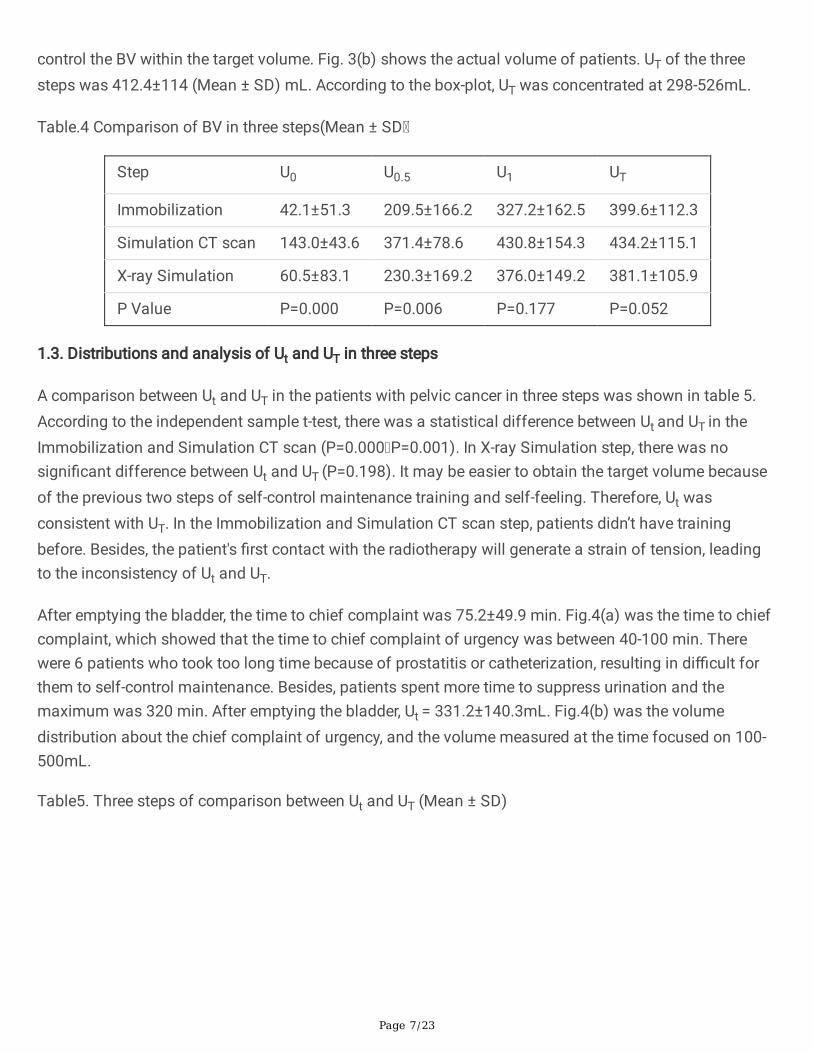
In three steps, the comparison of patient’s BV was shown in table 4. According to the independentsamples t-test, compared with the three steps, there was a statistical difference between U0 (P=0.000) andU0.5 (P=0.006). There was no statistical difference between U1 (P=0.177) and UT (P=0.052). It means thatbladder �lling had consistency between U1 and UT. The volume measured in Simulation CT scan waslarger than Immobilization and X-ray Simulation (U0=143.0±43.6mL, U0.5=371.4±78.6mL,U1=430.8±154.3mL, UT=434.2±115.1mL).
BV increased with the chief complaint of urgency time in �ve patients (P1, P2, P3, P4, P5) as shown in�g.3(a). After self-controlled maintenance for 1 hour, UP5 was > 600mL, and it didn’t conform to therequirement of bladder �lling before radiotherapy. Therefore, P5 was required to urinate properly and
Page 7/23
control the BV within the target volume. Fig. 3(b) shows the actual volume of patients. UT of the threesteps was 412.4±114 (Mean ± SD) mL. According to the box-plot, UT was concentrated at 298-526mL.
Table.4 Comparison of BV in three steps(Mean ± SD
Step U0 U0.5 U1 UT
Immobilization 42.1±51.3 209.5±166.2 327.2±162.5 399.6±112.3
Simulation CT scan 143.0±43.6 371.4±78.6 430.8±154.3 434.2±115.1
X-ray Simulation 60.5±83.1 230.3±169.2 376.0±149.2 381.1±105.9
P Value P=0.000 P=0.006 P=0.177 P=0.052
1.3. Distributions and analysis of Ut and UT in three steps
A comparison between Ut and UT in the patients with pelvic cancer in three steps was shown in table 5.According to the independent sample t-test, there was a statistical difference between Ut and UT in theImmobilization and Simulation CT scan (P=0.000 P=0.001). In X-ray Simulation step, there was nosigni�cant difference between Ut and UT (P=0.198). It may be easier to obtain the target volume becauseof the previous two steps of self-control maintenance training and self-feeling. Therefore, Ut wasconsistent with UT. In the Immobilization and Simulation CT scan step, patients didn’t have trainingbefore. Besides, the patient's �rst contact with the radiotherapy will generate a strain of tension, leadingto the inconsistency of Ut and UT.
After emptying the bladder, the time to chief complaint was 75.2±49.9 min. Fig.4(a) was the time to chiefcomplaint, which showed that the time to chief complaint of urgency was between 40-100 min. Therewere 6 patients who took too long time because of prostatitis or catheterization, resulting in di�cult forthem to self-control maintenance. Besides, patients spent more time to suppress urination and themaximum was 320 min. After emptying the bladder, Ut = 331.2±140.3mL. Fig.4(b) was the volumedistribution about the chief complaint of urgency, and the volume measured at the time focused on 100-500mL.
Table5. Three steps of comparison between Ut and UT (Mean ± SD)

Page 8/23
Step Ut UT P Value
Immobilization 318.6±142.2
(N=56
399.6±112.3
(N=82)
P=0.000
Simulation CT scan 500.0±17.5
(N=3)
434.2±115.1
(N=90)
P=0.001
X-ray Simulation 339.0±133.5
(N=26)
381.1±105.9
(N=29)
P=0.198
1.4. Analysis of frequency of and time to chief complaint of urgency in three steps
Frequency of and time to chief complaint of urgency was analyzed in three steps was shown in table 6. Inthe Immobilization and Simulation CT scan, patients had the �rst chief complaint of urgency in about 60minutes and more than half of the patient's BV could reach the target volume. But some patients also hadthe second or third time to complain of urgency, which may be related to the patient's status differences.The Simulation CT scan step was usually performed on the same day as the X-ray Simulation step.Patient already had self-controlled maintenance at the X-ray Simulation, and the volume might still be inthe range of the target volume by the time of Simulation CT scan. Therefore, the time to chief complaintof urgency would be shorter than "X-ray Simulation" and "Immobilization ". If patient had too much urine,he/she needed to urinate properly and then continued to suppress urination.
Table 6. Analysis about frequency of and time to the chief complaint in three steps

Page 9/23
Frequency of chiefcomplaint
N Immobilization
Averagetime(min)
EffectiveNumber
Effective meantime(min)
First 42 66.5±31.6 25 60.7±21.1
Second 9 108.8±66.8 3 78.3±10.4
Third 5 153±94.2 3 105±10
Frequency of chiefcomplaint
N Simulation CT scan
Averagetime(min)
EffectiveNumber
Effective meantime(min)
First 3 21.7±22.5 2 32.5±17.7
Second - - - -
Third - - - -
Frequency of chiefcomplaint
N X-ray Simulation
Averagetime(min)
EffectiveNumber
Effective meantime(min)
First 18 52.9±27.3 10 53±24.3
Second 6 97.5±52.6 4 75±27.4
Third 2 125±7 1 130
2. Analysis of the consistency of BV during radiotherapy
2.1 Comparison of UP and UT
The comparison of UP and UT in 18 patients with pelvic cancer was shown in Table 7. The differencebetween UP and UT was statistically signi�cant (P <0.05), and the difference was 17.81%.
Table 7. Comparison of UP and UT in 18 patients with pelvic cancer(mL)
Item N (Frequency) Mean SD P value
Planned volume 18 342.89 105.206 0.025
Treatment volume 450 281.82 54.221

Page 10/23
2.2 The RBV distribution during inter-fraction radiotherapy in 18 patients
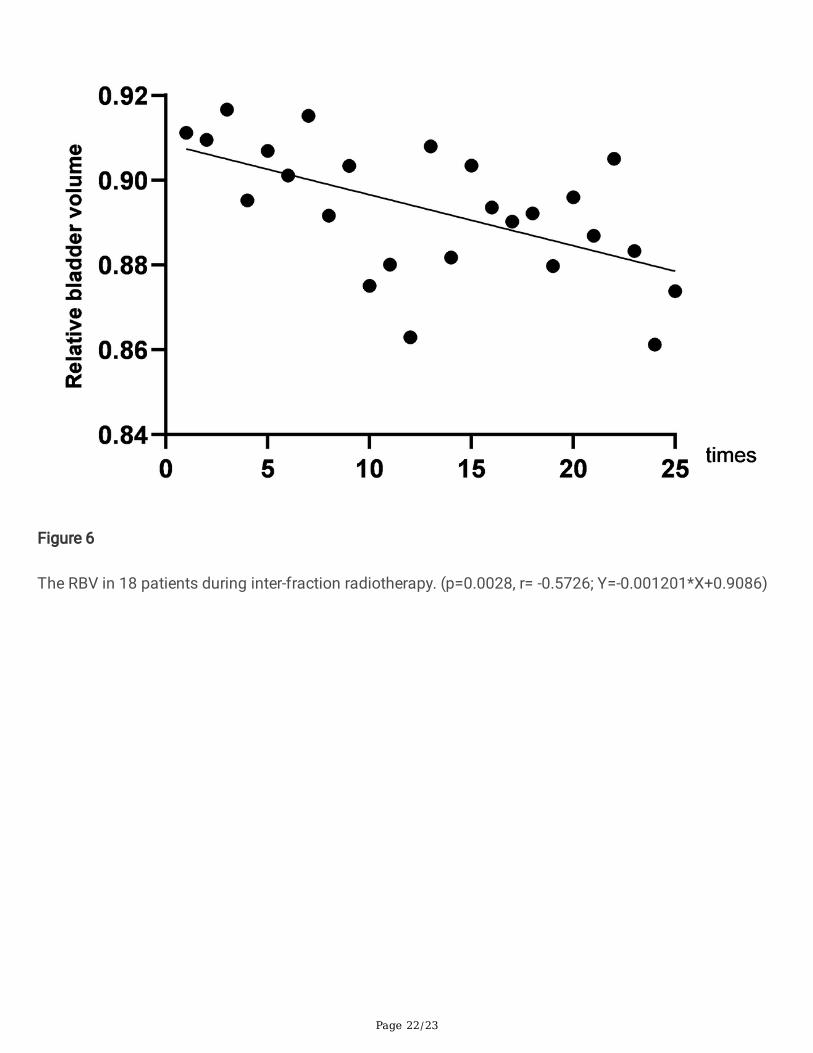
The interquartile range (IQR) indicated that the RBV was not very discrete (Fig.5). The overall RBV wasnegatively correlated with the number of radiotherapy (r=-0.5726, p=0.0028). As the inter-fractionradiotherapy continued to increase, the RBV gradually decreased, and the overall decrease was 5.53%.(Fig. 6
2.3 The consistency rate of BV of 18 patients
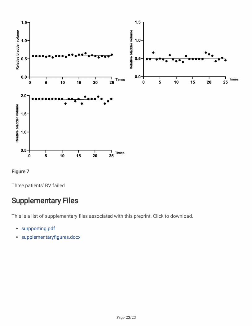
18 patients obtained a total of 450 BV (18*25), and the consistency rate was about 82.89% (373/450). 15patients (15/18) passed, and the consistency rate was 96% (360/375). Indicating that although thereduction in bladder capacity caused by radiotherapy was unavoidable, after suppress urination training,the vast majority of BV can be consistent with the planned BV (UP). 3 patients (3/18) failed, and theconsistency rate was 17.33% (13/75), as shown in Fig.7. The consistency rate was linearly independentof age (P = 0.2741).
Discussion1.The consistency of BV before radiotherapy
Radiotherapy is the main method of postoperative prevention and local recurrence treatment in the pelviccancer. Day-to-day anatomical variations complicate bladder cancer radiotherapy treatment [16]. Treatingwith a full bladder leads to unpredictability in bladder �lling, and some authors suggest that this becomesmore pronounced as treatment progresses, which could be due to poor patient compliance, disease-related anatomical changes that interfere with bladder innervation, or treatment-associated toxicity [24-28].
First of all, there were differences in BV between gender and age. U1 of patients over 60 years was277.6±134.3mL, which was much different from the target volume. Thus, it is suggested that patientsabove 60 years drank more 180mL water after self-controlled maintenance for 30 minutes, so that the BVis closer to the target volume. It can be seen that the capacity of bladder to self-controlled maintenancevaries from person to person. Chang Jee Suk et al reported that patients were asked to drink unspeci�edvolume of water because we thought there were wide variations of abilities in drinking water andsuppressing urination [32]. Besides, retaining urine was anticipated to become more di�cult over thecourse of treatment because of radiation cystitis [33].
Secondly, the comparison of BV in three steps showed that there was no signi�cant difference betweenU1 and UT. It was indicated that when patients drank 540mL water after emptying bladder and thenwaiting for 1 hour, the bladder volume before and during radiotherapy was consistent. We consider thatusing the BVI 9400 to measure the BV can better ensure that the bladder reached the �lling state duringradiotherapy. There was signi�cant difference in bladder volume between U0 and U0.5. Thus, Bladder scanwas a strategy that has been considered for increasing consistency with bladder volume. Similar to

Page 11/23
Cramp et al resulted [11]. Most patients will go to Simulation CT scan on the same day afterImmobilization. By this time, patient's BV has reached the target volume, then go to CT room to report.While waiting for the Simulation CT scan, the BV continued to increase, resulting in a larger amount of BVin the Simulation CT scan than the other two steps. Therefore, it was recommended that the radiationtherapist can allow patients to empty their bladder before Simulation CT scan, and then drank water toself-controlled maintenance.
Thirdly, there was a statistical difference between Ut and UT in the "Immobilization" and "Simulation CTscan" steps. However, there was no statistical difference between the Ut and the UT in the X-raySimulation. It was previously reported that biofeedback could improve the consistency of BV despite alack of statistical signi�cance [18]. The method (drinking 540mL water after emptying bladder and thenwaiting for 1 hour) can improve and obtain the reliable feeling about self-controlled maintenance.Because the patient was subjected to the Simulation CT scan after the Immobilization was completed.
Moreover, most patients can achieve the target volume on the �rst chief complaint of urgency. Somepatients still needed the second or the third complaint of urgency. In addition to the patient's physicalfactors, it was possible that the patient’s chief complaint of urgency was not true. Waiting for (75.2±49.9)min after emptying the bladder, patients complained of urgency. Because patients’ waiting for a long timeleads to tension, impatience and urgent completion of the treatment, they tell the radiation therapist“urgency”. But their volume doesn’t reach expected standards. In order that the patient can get the bettercooperation with treatment, the radiotherapy can be more accurate and the burden of work can bereduced, the radiation therapist should tell each patient the importance of �lling bladder and how long itwill take to wait. While patients are waiting, the radiation therapist need to appease the patient’s mood.
2.The consistency of BV during radiotherapy
There are many uncertain factors in the treatment of pelvic cancer, and the most concerned is the �llingstate of the bladder. The BV changes during the course of radiotherapy [17-22]. Bladder and rectalvolumes tend to decrease as a function of time during treatment [23]. A research reported that during the�rst week of radiotherapy treatment, 50% of patients had more than 50% change in BV. And on the �fthweek of treatment 64% of patients had more than 50% change in BV compared to the planned BV [2].Hynds et al found that 76% (828/1090), 53% (579/1090), 36% (393/1090) BV during radiotherapy were>50mL, >100mL, >150mL difference [1]. Compared with the planned volume, all men had at least one BVreduction of more than 50% during treatment. The reduction in BV was probably correlated with incidenceand severity of acute diarrhea [2].
In the result of BV measurement process during radiotherapy, �rst of all, it showed that there werestatistically signi�cant differences between UP and UT of 18 patients with pelvic cancer (P <0.05), with adifference of 17.81%. The RBV was negatively correlated with the number of radiotherapy (r=-0.5726,p=0.0028). With the inter-fraction radiotherapy, the overall RBV of 18 patients gradually decreased andthe overall decrease was 5.53%. The larger the standard deviation was, the greater the degree of
Page 12/23
dispersion would be. Stam et al believed that SD = 47.2% can be considered that the daily variation of BVwas large [18], while the overall SD = 2% of 18 patients was much less than 47.2% during radiotherapy.The change in the RBV between the inter-fractions was small.
Secondly, it showed that 18 patients obtained a total of 450 BV (18*25), and the consistency rate wasabout 82.89% (373/450). 15 patients (15/18) passed, and the consistency rate was 96% (360/375). Theconsistency rate has no linear relationship with age (P = 0.2741), similar to Mullaney [13]. It showed thatalthough the reduction of bladder capacity caused by radiotherapy was unavoidable, but patients drank540mL of water before radiotherapy and urination suppressing training; therefore, most patient’s BV canbe consistent with the planned BV.
Thirdly, it showed that 3 patients (3/18) failed, and the consistency rate was 17.33% (13/75), but their BVremained relatively consistent between inter-fractions. The reason for the failure may be that ultrasoundassessment of BV was less satisfactory in real patients than in normal volunteers. It was noted that therewas considerable variability in the shapes of different bladders and at varying volumes. These methodswere not applicable to all patients, either because the bladder outline was too indistinct or the bladderwas too large to demonstrate on a single scan [34]. Although BS provides an effective means ofassessing BV prior to treatment, studies showed that improvements in BV consistency are more di�cultto attain [1,18,35,36]. Nevertheless, there are some articles which have supported the use of the BS in aradiation therapy setting [17,35,36]. The plausible explanation is that if the BV for the �rst time ofradiotherapy cannot reach the planned BV (UP) due to the poor condition of the patient or themeasurement error of the radiation therapist. But during clinical treatment, the patient's actual BV is usedas the treatment BV and the �rst radiotherapy is used as the reference standard, so the patient's BV failsduring the entire radiotherapy process. Therefore, for a small number of special patients, attention shouldbe paid to rational determination of BV before treatment.
Shogo Hatanaka et al reported that the decrease of BV will lead to the increase of bladder dose, for boththe small and large bowel, and they found a signi�cant association between the Dmax values and BVvariation (the dose of small and large bowel less than 60Gy and 65Gy) [29]. Yaparpalvi R et al found thatthe small intestine area with 45 Gy was greater in the bladder emptying condition compared to thebladder �lling state (The average was 328.0±174.8 vs 176.0±87.5 cm) [25]. Frizzell B et al made ade�nitive treatment for prostate cancer, according to the in�uence of BV on rectal radiotherapy dose. The�lling of bladder was compared with emptied bladder, the average exposure dose of rectum decreased by27.6 Gy, and there was signi�cant difference (P=0.031) [30]. Buchali et al found that when the BVincreased, the exposure dose decreased. A full bladder led to a mean reduction in organ dose in medianfrom 94-87% calculated for 50% of the BV. For 66% of the BV the dose could be reduced in median from78-61% and for the whole bladder from 42-39% [31]. Fujioka C et al reported optimal BVs at treatmentplanning must be investigated to both maintain reproducibility of the BV and dose constraints for thebladder [8].
Page 13/23
At present, the standard of "pass" bladder volume before radiotherapy for patients with pelvic cancer isnot clear. In actual clinical practice, the radiation therapist usually judges whether the measured BV meetsthe planned BV (UP) based on experience. Patients who fail to reach the planned BV need to suppressurination for many times, which virtually increases the patient's mental tension and physical discomfort,and reduces the e�ciency of radiotherapy. However, urination suppressing training before radiotherapyand the maintenance intervention during radiotherapy showed that 82.89% (373/450) of the 18 patientsin the range of (50%, 155%) were able to keep consistency with UP and UT. This indicates that theconsistency range is reasonable. (50%,155%) can determine whether the bladder �lling of patients withpelvic cancer is consistent with the planned BV before radiotherapy, reducing the patient's mentalpressure and physical discomfort, improving the e�ciency of radiation therapy. However, urinationsuppressing training used in the present study was frequently used in clinical practice in the setting ofdose escalation to the pelvic cancer patients. Despite these limitations, there are very few data in theliterature on the optimal BV at treatment planning and during radiotherapy in pelvic cancer; therefore, wehope that the present results will serve as reference values for other institutions.
ConclusionPatients emptied the bladder and immediately drank 540 mL of water. After 1 hour of suppressingurination, patients complained of urgency and achieved the target volume (400 mL). At this time, the BVwas consistent in the Immobilization, Simulation CT scan, X-ray Simulation and during inter-fractionradiotherapy. This indicates that the consistency range is reasonable. (50%,155%) can determine whetherthe bladder �lling of patients with pelvic cancer is consistent with the planned BV before radiotherapy.
AbbreviationsBV: Bladder volume;
U0, t0: Emptied bladder volume and time;
U0.5, t0.5: 0.5 hours after drinking of bladder volume and time;
U1, t1: 1 hour after drinking of bladder volume and time;
Ut, t: Chief complaint of urgency of bladder volume and time;
UT, T: Actual bladder volume and time;
UP: Planned bladder volume;
RBV: Relative bladder volume;
CRT: Preoperative chemoradiation;

Page 14/23
IMRT: Intensity-modulated radiation therapy;
QoL: Quality of life;
IQR: Interquartile range;
Up5: Patient 5’s bladder volume.
Declarations1. Ethical Approval and Consent to participate
This project was approved by the Ethical Committee of Sun Yat-Sen University Cancer Center andinformed consent was obtained from all patients.
2. Consent for publication
All the authors listed have approved the manuscript that is enclosed.
3. Availability of supporting data
The data are fully available without restriction in the Research Data Deposit public platform (RDDNumber: RDDA2020001544,
https://www.researchdata.org.cn) and are available upon reasonable request.
4. Competing interests
None of the authors have any competing interests (�nancial and non�nancial) in the manuscript.
5. Funding
Pearl River S&T Nova Program of Guangzhou (201710010162); Natural Science Foundation of Guangdong Province (2017A030310217) Hubei Key Laboratory of Medical Information Analysis &Tumor Diagnosis and Treatment (PJS140011504); Innovation and Entrepreneurship Training Program forCollege Student Innovation and Entrepreneurship(20191390109; 201813902075; 201813902071;201713902050
�. Authors' contributions
Design of the Research: Sijuan Huang, Xin Yang,
Elaboration of the data: Yaning Li, Junyun Li, Cunxiao Li, Feng Chi, Xinping Cao,
Manuscript Preparation: Jiaying Wu, Wanjia Zheng,
Page 15/23
Writing Manuscript: Xin Feng, Qianyi Chen, Sijuan Huang, Xin Yang,
All authors have read and approved the �nal manuscript.
7. Acknowledgements
Many thanks to Ms. Yi QIN for her help on this paper.
References1. Hynds S, Mcgarry C K, Mitchell D M, et al. Assessing the daily consistency of bladder �lling using an
ultrasonic Bladderscan device in men receiving radical conformal radiotherapy for prostate cancer[J].British Journal of Radiology, 2011, 84(1005):813-818. DOI:1259/bjr/50048151
2. Sauer R, Becker H, Hohenberger W, et al. Preoperative versus Postoperative Chemoradiotherapy forRectal Cancer[J]. The New England Journal of Medicine, 2004, 351(17):1731-1740.DOI:1056/NEJMoa040694
3. Peeters K C M J, Marijnen C A M, Nagtegaal I D, et al. The TME trial after a median follow-up of 6years - Increased local control but no survival bene�t in irradiated patients with resectable rectalcarcinoma[J]. Annals of Surgery, 2007, 246(5):693-701. DOI: 1097/01.sla.0000257358.56863.ce
4. Sebag-Monte�ore D, Stephens R J, Steele R, et al. Preoperative radiotherapy versus selectivepostoperative chemoradiotherapy in patients with rectal cancer (MRC CR07 and NCIC-CTG C016): amulticentre, randomised trial[J]. The Lancet, 2009, 373(9666):811-820. DOI: 10.1016/S0140-6736(09)60484-0
5. Sauer R, Liersch T, Merkel S, et al. Preoperative Versus Postoperative Chemoradiotherapy for LocallyAdvanced Rectal Cancer: Results of the German CAO/ARO/AIO-94 Randomized Phase III Trial After aMedian Follow-Up of 11 Years[J]. Journal of Clinical Oncology, 2012, 30(16):1926-1933.DOI:1200/JCO.2011.40.1836
�. Navarro M, Dotor E, Rivera F, et al. A phase II study of preoperative radiotherapy and concomitantweekly irinotecan in combination with protracted venous infusion 5-�uorouracil, for resectable locallyadvanced rectal cancer[J]. International Journal of Radiation Oncology Biology Physics, 2006,66(1):201-205. DOI: 10.1016/j.ijrobp.2006.04.007
7. Sithamparam S, Ahmad R, et al. Bladder �lling variation during conformal radiotherapy for rectalcancer[J]. Journal of Physics: Conference Series,2017, 851(1):12-26. DOI: 10.1088/1742-6596/851/1/012026.
�. Fujioka C, Ishii K, Yamanaga T, et al. Optimal bladder volume at treatment planning for prostatecancer patients receiving volumetric modulated arc therapy[J]. Practical Radiation Oncology, 2014,90(1):688-689. DOI: 1016/j.ijrobp.2014.05.2023
9. Dearnaley D P, Hall E, Lawrence D, et al. Phase III pilot study of dose escalation using conformalradiotherapy in prostate cancer: PSA control and side effects. [J]. British Journal of Cancer, 2005,92(3):488-498. DOI: 10.1038/sj.bjc.6602301

Page 16/23
10. Nuyttens J J, Robertson J M, Yan D, et al. The in�uence of small bowel motion on intensitymodulated radiation therapy (IMRT) for rectal cancer[J]. International Journal of Radiation OncologyBiology Physics, 2000, 48(3):168. DOI: 10.1016/S0360-3016(00)80130-3
11. Cramp L, Connors V, Wood M, et al. Use of a prospective cohort study in the development of abladder scanning protocol to assist in bladder �lling consistency for prostate cancer patientsreceiving radiation therapy[J]. Journal of Medical Radiation Sciences, 2016, 63(3):179-185.DOI:1002/jmrs.162
12. Yoon H I , Chung Y , Chang J S , et al. Evaluating Variations of Bladder Volume Using an UltrasoundScanner in Rectal Cancer Patients during Chemoradiation: Is Protocol-Based Full BladderMaintenance Using a Bladder Scanner Useful to Maintain the Bladder Volume?[J]. Plos One, 2015,10(6). DOI:10.1371/journal.pone.0128791
13. Mullaney L M, O’shea E, Dunne M T, et al. A randomized trial comparing bladder volume consistencyduring fractionated prostate radiation therapy[J]. Practical radiation oncology, 2014, 4(5):203-212.DOI:1016/j.prro.2013.11.006
14. Li K, He J. Medical Statistics [M]. People's Medical Publishing House, 2013.22
15. Kirkwood B R, Sterne J A C. Medical Statistics[M]. 2003.
1�. Webster G J, Stratford J, Rodgers J, et al. Comparison of adaptive radiotherapy techniques for thetreatment of bladder cancer[J]. British Journal of Radiology, 2013, 86(1021):20120433. DOI1259/bjr.20120433
17. O’Doherty U´ M, Mcnair H A, Norman A R, et al. Variability of bladder �lling in patients receivingradical radiotherapy to the prostate[J]. Radiotherapy and Oncology, 2006, 79(3):335-340. DOI:10.1016/j.radonc.2006.05.007
1�. Stam M R, Lin E N J T V, Vight L P V D, et al. Bladder �lling variation during radiation treatment ofprostate cancer: can the use of a bladder ultrasound scanner and biofeedback optimize bladder�lling? [J]. International Journal of Radiation Oncology Biology Physics, 2006, 65(2):371-377. DOI:10.1016/j.ijrobp.2005.12.039
19. Pinkawa M, Asadpour B, Gagel B, et al. Prostate position variability and dose-volume histograms inradiotherapy for prostate cancer with full and empty bladder[J]. International Journal of RadiationOncology Biology Physics, 2006, 64(3):856-861. DOI: 10.1016/j.ijrobp.2005.08.016
20. Nakamura N, Shikama N, Takahashi O, et al. Variability in Bladder Volumes of Full Bladders inDe�nitive Radiotherapy for Cases of Localized Prostate Cancer[J]. Strahlentherapie Und Onkologie,2010, 186(11):637-642. DOI 1007/s00066-010-2105-6
21. Cambria R, Jereczek-fossa B A, Zerini D, et al. Physical and clinical implications of radiotherapytreatment of prostate cancer using a full bladder protocol[J]. Strahlentherapie Und Onkologie, 2011,187(12):799-805. DOI 1007/s00066-011-2259-x
22. Mak D, Gill S, Paul R, et al. Seminal vesicle interfraction displacement and margins in image guidedradiotherapy for prostate cancer[J]. Radiation Oncology, 2012, 7(1):1-9. DOI 1186/1748-717X-7-139
Page 17/23
23. Antolak J A, Rosen I I, Childress C H, et al. Prostate target volume variations during a course ofradiotherapy[J]. International Journal of Radiation Oncology Biology Physics, 1998, 42(3):661-672.DOI 1016/S0360-3016(98)00248-X
24. Chen V E, Gillespie E F, Manger R P, et al. The impact of daily bladder �lling on small bowel dose forintensity modulated radiation therapy for cervical cancer[J]. Medical Dosimetry, 2018, 44(2):102-106.DOI 1016/j.meddos.2018.02.010
25. Yaparpalvi R, Mehta K J, Bernstein M B, et al. Contouring and Constraining Bowel on a Full-BladderComputed Tomography Scan May Not Re�ect Treatment Bowel Position and Dose Certainty inGynecologic External Beam Radiation Therapy[J]. International Journal of Radiation OncologyBiology Physics, 2014, 90(4):802-808. DOI 1016/j.ijrobp.2014.07.016
2�. Ahmad R, Hoogeman M S, Quint S, et al. Inter-fraction bladder �lling variations and time trends forcervical cancer patients assessed with a portable 3-dimensional ultrasound bladder scanner[J].Radiotherapy and Oncology, 2008, 89(2):172-179. DOI 1016/j.radonc.2008.07.005
27. Turner S L, Swindell R, Bowl N, et al. Bladder movement during radiation therapy for bladder cancer:implications for treatment planning[J]. International Journal of Radiation Oncology Biology Physics,1997, 39(2):355-360. DOI 1016/S0360-3016(97)00070-9
2�. Jhingran A, Salehpour M, Sam M, et al. Vaginal motion and bladder and rectal volumes during pelvicintensity-modulated radiation therapy after hysterectomy[J]. International Journal of RadiationOncology Biology Physics, 2012, 82(1):256-262. DOI 1016/j.ijrobp.2010.08.024
29. Hatanaka S, Kawada Y, et al. The Impact of Variation in Bladder Volume on the Doses of Target andOrgan-at-Risk in Intensity-Modulated Radiation Therapy for Localized Prostate Cancer[J]. Journal ofCancer Therapy,2016,7(10):741-751. DOI:10.4236/jct.2016.710075.
30. Frizzell B, Lovato J, Foster J, et al. Impact of bladder volume on radiation dose to the rectum in thede�nitive treatment of prostate cancer[J]. The Journal of community and supportive oncology, 2015,13(8):288-291. DOI 12788/jcso.0156
31. Buchali A, Koswig S, Dinges S, et al. Impact of the �lling status of the bladder and rectum on theirintegral dose distribution and the movement of the uterus in the treatment planning ofgynaecological cancer[J]. Radiotherapy and Oncology, 1999, 52(1):29-34. DOI:1016/S0167-8140(99)00068-7
32. Chang J S, Yoon H I, Cha H J, et al. Bladder �lling variations during concurrent chemotherapy andpelvic radiotherapy in rectal cancer patients: early experience of bladder volume assessment usingultrasound scanner[J]. Radiation oncology journal, 2013, 31(1):41-47. DOI 3857/roj.2013.31.1.41
33. Moiseenko V, Liu M, Kristensen S, et al. Effect of bladder �lling on doses to prostate and organs atrisk: a treatment planning study[J]. Journal of Applied Clinical Medical Physics, 2007, 8(1):55-68.DOI 1120/jacmp.v8i1.2286
34. Hartnell G G, Kiely E A, Williams G, et al. Real-time ultrasound measurement of bladder volume: acomparative study of three methods[J]. British Journal of Radiology, 1987, 60(719):1063-1065. DOI1259/0007-1285-60-719-1063

Page 18/23
35. Gawthrop J, Oates R. Measured bladder volume for radiotherapy of the prostate using the hand-heldBladderScan BVI 3000[J]. Radiographer, 2012, 59(1):8-12. DOI 1002/j.2051-3909.2012.tb00167.x
3�. O’Shea E, Armstrong J, O’ Hara T, et al. Validation of an external ultrasound device for bladdervolume measurements in prostate conformal radiotherapy[J]. Radiography, 2008, 14(3):178-183.DOI 1016/j.radi.2007.06.001
Figures
Figure 1
Flow chart of measuring BV in three steps before radiotherapy.

Page 19/23
Figure 2
BV monitoring process during radiotherapy

Page 20/23
Figure 3
a). Trend of the chief complaint of urgency in 5 typical patients. b). Box-plot of actual volume (UT)distribution in 105 patients.
Figure 4
a). Time distribution about the chief complaint of urgency. b). Volume distribution of the chief complaintof urgency.

Page 21/23
Figure 5
The RBV of 25 inter-fraction radiotherapy in 18 patients.
Page 22/23
Figure 6
The RBV in 18 patients during inter-fraction radiotherapy. (p=0.0028, r= -0.5726; Y=-0.001201*X+0.9086)
Page 23/23
Figure 7
Three patients’ BV failed
Supplementary Files
This is a list of supplementary �les associated with this preprint. Click to download.
surpporting.pdf
supplementary�gures.docx