Embed Size (px)
Citation preview
Durham Research Online
Deposited in DRO:
18 June 2014
Version of attached �le:
Accepted Version
Peer-review status of attached �le:
Peer-reviewed
Citation for published item:
Dra�c��nsk�y, Martin and Hodgkinson, Paul (2014) 'E�ects of quantum nuclear delocalisation on NMRparameters from path integral molecular dynamics.', Chemistry : a European journal., 20 (8). pp. 2201-2207.
Further information on publisher's website:
http://dx.doi.org/10.1002/chem.201303496
Publisher's copyright statement:
This is the peer reviewed version of the following article: Dra�c��nsk�y, M. and Hodgkinson, P. (2014), E�ects of QuantumNuclear Delocalisation on NMR Parameters from Path Integral Molecular Dynamics. Chemistry - A European Journal,20 (8): 2201-2207, which has been published in �nal form at http://dx.doi.org/10.1002/chem.201303496. This articlemay be used for non-commercial purposes in accordance with Wiley-VCH Terms and Conditions for self-archiving.
Use policy
The full-text may be used and/or reproduced, and given to third parties in any format or medium, without prior permission or charge, forpersonal research or study, educational, or not-for-pro�t purposes provided that:
• a full bibliographic reference is made to the original source
• a link is made to the metadata record in DRO
• the full-text is not changed in any way
The full-text must not be sold in any format or medium without the formal permission of the copyright holders.
Please consult the full DRO policy for further details.
Durham University Library, Stockton Road, Durham DH1 3LY, United KingdomTel : +44 (0)191 334 3042 | Fax : +44 (0)191 334 2971
http://dro.dur.ac.uk
1
Effects of quantum nuclear delocalisation on NMR parameters from path integral
molecular dynamics
Martin Dračínský,*[a,b] Paul Hodgkinson*[a]
[a] Dr. Martin Dračínský, Dr. Paul Hodgkinson, Department of Chemistry, Durham University, South
Road, DH1 3LE, Durham, UK
Fax: (+44) 191-394-4737
E-mail: [email protected], [email protected]
[b] Institute of Organic Chemistry and Biochemistry, Flemingovo nám. 2, 16610, Prague, Czech
Republic
Key words: density functional calculations, NMR spectroscopy, nuclear delocalisation, isotope
effects, path integral molecular dynamics
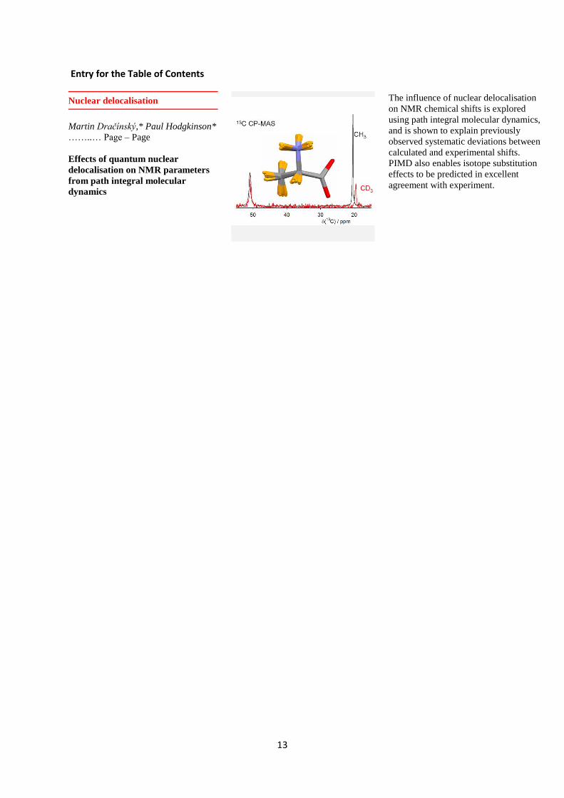
Abstract
The influence of nuclear delocalisation on NMR chemical shifts in molecular organic solids is
explored using path integral molecular dynamics (PIMD) and density functional theory calculations
of shielding tensors. Nuclear quantum effects are shown to explain previously observed systematic
deviations in correlations between calculated and experimental chemical shifts, with particularly
large PIMD-induced changes (up to 23 ppm) observed for atoms in methyl groups. The PIMD
approach also enables isotope substitution effects on chemical shifts and J couplings to be predicted
in excellent agreement with experiment for both isolated molecules and molecular crystals. An
approach based on convoluting calculated shielding or coupling surfaces with probability
distributions of selected bond distances and valence angles obtained from PIMD simulations is used
to calculate isotope effects.
Introduction
The gauge-including projector-augmented wave (GIPAW) procedure has been recently
developed for the prediction of the magnetic resonance parameters in solids,[1] and the power of
this approach for calculating NMR properties for fully periodic crystal structures has been well
documented.[2] Such quantum chemical calculations are typically performed using static structures,
i.e. at 0 K. It is well established, however, that fast molecular motions, such as vibrations,
conformational averaging, and molecular aggregation, will average NMR parameters.[2a, 3] In
particular, rovibrational averaging of shielding is observed as isotope shifts.[4] Neglecting zero-point
motion and dynamics may lead to significant discrepancies between computed and experimental
data.
Plots of calculated 13C shielding values against experimental chemical shifts generally show
slopes that deviate from the ideal value of –1. This complicates the referencing of the calculated
shielding values and typically linear regression between calculated and experimentally determined
values is used to improve agreement.[5] This makes the comparison of chemical shifts obtained in
different studies difficult because the fitted parameters vary between experimental data sets , with
the fitted slope, usually between –1 and –1.2[2b, 6], being heavily dependent on the type of molecule.
For example, trendlines with slopes of –1.05 and –1.16 respectively, have been found for aromatic
vs. carbohydrate shielding values. Such systematic deviations may limit the ability to use shielding
2
calculations for spectral assignments.[7] This behaviour has variously been attributed to errors
intrinsic to approximations of DFT,[8] but also the effects of molecular motion.[9]
Classical and ab initio molecular dynamics methods have been widely applied to study the
influence of molecular motion on NMR parameters; see for example ref.[2a, 3, 10] However, nuclear
quantum effects, such as zero-point vibrations and tunnelling, may be crucial in the case of light
nuclei, particularly hydrogen atoms. A comparison of vibrational averaging at zero temperature and
finite temperature has revealed a significant contribution of zero-point motion to calculated
chemical shifts.[4, 9, 11] The quantum vibrational phenomena might not be sufficiently characterized
even by ab initio dynamics that uses Newtonian mechanics for the nuclei (classical nucleus MD). One
route to including quantum effects on nuclear motion is provided by the formalisms based on
Feynman’s path integral[12] (PI) approach. Path integral simulations use a decomposition of nuclei
into a number of beads subject to specific harmonic nearest neighbour interactions. The strength of
the harmonic interaction depends on nuclear mass and temperature, with light nuclei at low
temperature being highly delocalised (see Figure S1 in SI for a schematic representation). The
ensemble of coordinates for a given set of beads, termed a replica, is propagated in time, forming a
path integral trajectory. Static properties of quantum objects can thus be obtained by simulating
more complicated, but still classical, objects using well-established simulation techniques such as
molecular dynamics or Monte Carlo simulations.[13] The underlying potential energy landscape may
be expressed within classical force-fields or any ab initio methods.[14]
The path integral approach has been employed in a wide variety of applications.[15] In the
context of NMR, PIMD has been used with classical force-field potentials for a protein fragment[16]
and liquid water simulation.[17] PIMD used with ab initio methods was applied in a study of isolated
deprotonated water dimer (H3O2-), and showed significant differences in the delocalisation of the
hydrogen-bonded vs. non-hydrogen-bonded protons at different temperatures.[18] Path integral
Monte Carlo simulations with ab initio methods have been used to determine the influence of
nuclear fluctuations on chemical shifts of isolated bullvalene,[19] ethylene,[20] and benzene.[21]
Changes of carbon chemical shifts of several ppm were observed. Path integrals with Car-Parrinello
molecular dynamics were used to predict 1H NMR chemical shifts in solid LiNH2 and Li2NH. Almost no
effect of nuclear quantum delocalisation was found for Li2NH whereas a broader distribution of 1H
chemical shifts in LiNH2 was found when path integration was applied.[22] We are not, however,
aware of previous systematic investigations of the effects of nuclear delocalisation on NMR spectra
of organic solids.
Isotope effects are one manifestation of the quantum nature of nuclei; zero-point
fluctuations lead to differences in vibrationally averaged properties of compounds with isotopic
substitution. Isotope effects in NMR have a multitude of practical applications, such as in the
determination of molecular structure and the verification of mechanisms of reactions. The small size
of isotope effects, however, makes them stringent tests of ab initio calculations.[23] A conventional
route to calculating isotope shifts involves calculation of intramolecular force field and shielding
surface (the dependence of isotropic shielding on nuclear coordinates). However, this procedure is
computationally demanding and is suitable only for small isolated molecules.[4] For larger molecules
and molecular crystals, many approximations (such as a drastic reduction of molecular degrees of
freedom) have to be employed.[24]
Here we use a combination of PIMD with DFT to explore the influence of nuclear quantum
effects on the NMR isotropic shielding of organic molecular solids and to predict deuterium isotope
effects on chemical shifts and coupling constants. We demonstrate that nuclear delocalisation
3
(particularly of hydrogen atoms) has a significant impact on bond distances and 13C chemical shifts.
The PIMD approach is also used for predictions of deuterium substitution effects on chemical shifts
and J couplings.
Figure 1. The studied model compounds with atom numbering.
Methods
Samples of alanine, alanine-d3, and thymine were obtained from Sigma Aldrich. High-
resolution solid-state NMR spectra were obtained using a Varian/Chemagnetics InfinityPlus
spectrometer operating at 125.7 MHz for 13C (499.7 MHz for 1H). Samples were packed into 5 mm
magic angle spinning rotors and measurements taken using a MAS rate of 10 kHz using cross
polarisation (CP). The typical CP conditions used were: recycle delay 4 s, contact time 2 ms,
acquisition time 40 ms. Spectra were referenced with respect to external neat tetramethylsilane for 13C by setting the high-frequency signal from a replacement sample of adamantane to 38.5 ppm. A
correction of +16 K was made to the set temperatures to correct for the frictional heating of the
sample under sample spinning.
Figure 1 shows the set of model compounds used for the calculations. The atomic
coordinates for glycine (GLYCIN20), alanine (LALNIN12), methyl--D-xylopyranoside (XYLOBM01),
pentaerythritol (PERYTO10), and thymine (THYMIN01) were obtained from the Cambridge
Crystallographic Database.[25] For glycine, alanine, the xylopyranoside, and pentaerythritol, neutron
diffraction structures were used. Neutron data were unavailable in the case of thymine, and X-ray
structures were used. Note that for consistency all crystal structures used were determined at room
temperature.
Born-Oppenheimer molecular dynamics (BOMD) simulations were run in the CASTEP
program,[26] which is a DFT-based code, using an NVT ensemble maintained at a constant
temperature of 300 K using a Langevin thermostat, a 0.5 fs integration time step, ultrasoft
pseudopotentials,[27] a planewave cutoff energy of 300 eV, and with integrals taken over the Brillouin
zone using a Monkhorst-Pack[28] grid of a minimum k-point sampling of 0.1 Å–1. Electron-correlation
effects were modeled using the generalized gradient approximation of Perdew, Burke, and
Ernzerhof.[29] The atomic positions were optimized at the same computational level prior to the MD
runs, while lattice parameters were fixed to the experimental values. No symmetry constraints were
applied during the runs as these are only relevant to the time-averaged structure. Simulation runs of
5 ps were performed for every compound. Dichloromethane was modeled as an isolated molecule in
4
a cubic periodic box of 11 x 11 x 11 Å3, and was pre-equilibrated by 5 ps (PI)MD simulations to
equilibrate the random initial partition of the kinetic energy into rotations, translations and
vibrations. The path integral was used on top of the DFT-MD simulations, with a Trotter
decomposition of all nuclei into P = 16 beads.
Time-averaged NMR parameters were computed from 41 snapshots from the MD and PIMD
simulations selected at 1.0, 1.1, 1.2 … 5.0 ps. The unit cells of glycine, alanine and thymine contained
four crystallographically equivalent molecules (Z = 4); Z = 2 for the xylopyranoside and
pentaerythritol unit cells; therefore, 164 or 82 values were averaged for every chemically equivalent
site. The standard deviations of the calculated shieldings for chemically equivalent sites were used
for an estimation of the error of the calculation. The real time snapshots from the PIMD simulations
contained 16 replicas and the NMR tensors calculated for the individual replicas were averaged. The
NMR calculations were performed using the GIPAW approach,[1, 30] using ‘on-the-fly’
pseudopotentials, a planewave cutoff energy of 600 eV with integrals taken over the Brillouin zone
using a Monkhorst-Pack[28] grid of a minimum k-point sampling of 0.05 Å–1.
A reasonable convergence of the calculated isotropic shieldings (changes typically less than 1
ppm for 13C) was usually achieved after the averaging of 20–30 real-time snapshots. The
convergence of the isotropic shielding of atom C3 in alanine is illustrated in Figure S2 in the SI. This
convergence is much faster than when calculating NMR parameters of compounds in solution,
where the rapid fluctuation of the solvent molecules requires the averaging of several hundreds or
thousands MD snapshots to obtain reasonably converged chemical shifts.[31]
The (PI)MD-induced change of isotropic shielding was then calculated as the difference
between the averaged NMR parameters and those calculated on a ‘’(PI)MD-averaged structure’’
structure where the positions of all atoms during the (PI)MD simulation were averaged. As discussed
previously,[10] atomic motion means that the average distance between atoms is not the same as the
distance between averaged atomic positions, and such an MD-averaged structure is more directly
comparable with the diffraction structures obtained at the same temperature. Calculating the
(PI)MD-induced change in this way, as opposed to relative to 0 K geometry-optimised structure, also
avoids any systematic errors introduced by imperfect geometry optimisation. Where required for
comparison with experimental data, NMR parameters were calculated as the values computed from
the diffraction structure plus the (PI)MD-induced change (referred to as CSD + (PI)MD data), i.e. iso
= iso(static) + iso. Alternatively, the averaged PIMD shieldings were compared directly with
experimental shifts. PIMD simulation of alanine with P = 32 decomposition gave calculated PIMD-
induced changes of chemical shifts were close to those calculated with P = 16 (maximum differences
were 0.32 and 0.08 ppm, for 13C and 1H respectively).
The following procedure was used to calculate deuterium isotope effects on alanine.
Probability distributions of the C3–H bond distance and C2–C3–H valence angle (alanine), and of the
C–H bond distance and H–C–H valence angle (dichloromethane) were extracted from the (PI)MD
simulations. The PIMD distance/angle probabilities were determined independently for all 16
replicas and then averaged. Then a dependence of isotropic shieldings in alanine on the bond
distance and the valence angle was calculated by manually adjusting the C3–H bond distance in the
range 0.9–1.4 Å (with a 0.05 Å step) and a C2–C3–H valence angle ranging from 80° to 140° (5° step).
The calculated dependence of the shielding values on the geometrical parameters was fitted to a
polynomial function (order 2 for the distance and 3 for the angle dependence). The probability
distributions and the polynomial functions were then used to calculate weighted averages of
shielding values of alanine and alanine-d3 and for the chemical shift changes induced by the isotope
5
substitution. This approach of 1D scans through the shielding surface, rather than a full, but very
computationally expensive, 2D parameterisation, is appropriate if the effects of the distance and
angle on nuclear shielding are largely additive. We checked the additivity by calculating the carbon
shielding dependence on the methyl C–H distance for three different C2–C3–H angles (100, 110, and
120°) and the calculated dependences were very similar (differences lower than 10%. A similar
procedure was applied for dichloromethane, where the dependence of carbon shielding values and 1JC-H coupling constants on the C–H distance and H–C–H angle of isolated dichloromethane was
calculated using B3LYP functional[32] and 6-31g** basis set with the Gaussian09 program.[33] The J
coupling was calculated in the range 0.7–2.0 Å (with a 0.05 Å step) and 70°–150° (5° step). The
carbon shielding dependence fitted well to a quadratic curve whereas the J coupling dependence
had a more complicated form, and so J coupling values as a function of distance were calculated by
linear extrapolation between neighbouring calculated points. The isotope effect on J-coupling is
calculated as the difference JC-H(DCM) – *JC-H(DCM-d2), where *JC-H(DCM-d2) = JC-D(DCM-d2) x H/D.
Results
Effects on isotropic shieldings
Figure 2 shows the probability distributions of the methyl C–H bond distance and of the C2–
C3–H angle in alanine and in alanine-d3 observed during MD and PIMD simulations. As expected, the
atoms, especially hydrogens, are more delocalised in the PIMD simulations. The PIMD distributions
are also shifted slightly towards longer bond lengths. The scatter of nuclear positions of hydrogen H2
in alanine during MD and PIMD simulations is shown in Figure S3 in SI.
0.9 1.0 1.1 1.2 1.3 1.4
Pro
babili
ty / %
0
2
4
6
8
10
12
14
MD
PIMD
MD-D
PIMD-D
C-H (C-D) bond distance / Å
a)
C2-C3-H angle / degrees
80 90 100 110 120 130 140
Pro
bab
ility
/ %
0
2
4
6
8
10
MD-H
PIMD-H
MD-D
PIMD-D
b)
Figure 2. Probability distribution of C–H/C–D bond distances in the methyl group (a) and of C2–C3–H
angle (b) of alanine during the MD and PIMD simulations at 300 K, sampled by 0.01 Å and 1°
respectively.
The calculated PIMD-induced changes of shielding values were negative for all atoms, i.e.
the atoms are less shielded when the nuclear motion is taken into account. This presumably reflects
the shift in the probability distribution towards longer bond lengths, reducing the shielding effect of
the electrons at the nuclei. The PIMD-induced changes for the 13C shielding were found in the range
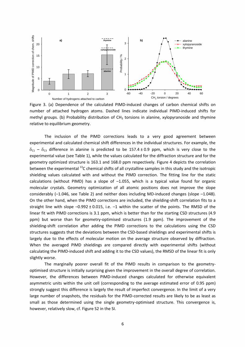
3.3–22.5 ppm (see Table S1 in SI) with the magnitude roughly correlating with the number of
attached hydrogen atoms. The changes were particularly large, and quite variable, for methyl
carbons (Figure 3): alanine 9.2 ppm, xylopyranoside 18.6 ppm, and thymine 22.5 ppm. The
corrections seem to correlate with the degree of delocalisation of the CH3 hydrogens (see Figure 3b).
6
Number of hydrogens attached to carbon
0 1 2 3
Magnitude o
f P
IMD
corr
ection o
f chem
. shifts
0
5
10
15
20
alanine
thymine
xylopyranoside
a)
CH3 torsion / degrees
-60 -40 -20 0 20 40 60
Pro
babili
ty / %
0
1
2
3alanine
xylopyranoside
thymine
b)
Figure 3. (a) Dependence of the calculated PIMD-induced changes of carbon chemical shifts on
number of attached hydrogen atoms. Dashed lines indicate individual PIMD-induced shifts for
methyl groups. (b) Probability distribution of CH3 torsions in alanine, xylopyranoside and thymine
relative to equilibrium geometry.
The inclusion of the PIMD corrections leads to a very good agreement between
experimental and calculated chemical shift differences in the individual structures. For example, the
C1 – C3 difference in alanine is predicted to be 157.4 ± 0.9 ppm, which is very close to the
experimental value (see Table 1), while the values calculated for the diffraction structure and for the
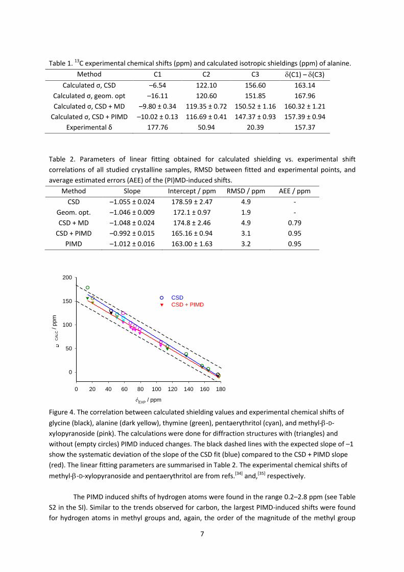
geometry optimized structure is 163.1 and 168.0 ppm respectively. Figure 4 depicts the correlation
between the experimental 13C chemical shifts of all crystalline samples in this study and the isotropic
shielding values calculated with and without the PIMD correction. The fitting line for the static
calculations (without PIMD) has a slope of –1.055, which is a typical value found for organic
molecular crystals. Geometry optimization of all atomic positions does not improve the slope
considerably (–1.046, see Table 2) and neither does including MD-induced changes (slope –1.048).
On the other hand, when the PIMD corrections are included, the shielding-shift correlation fits to a
straight line with slope –0.992 ± 0.015, i.e. –1 within the scatter of the points. The RMSD of the
linear fit with PIMD corrections is 3.1 ppm, which is better than for the starting CSD structures (4.9
ppm) but worse than for geometry-optimised structures (1.9 ppm). The improvement of the
shielding-shift correlation after adding the PIMD corrections to the calculations using the CSD
structures suggests that the deviations between the CSD-based shieldings and experimental shifts is
largely due to the effects of molecular motion on the average structure observed by diffraction.
When the averaged PIMD shieldings are compared directly with experimental shifts (without
calculating the PIMD-induced shift and adding it to the CSD values), the RMSD of the linear fit is only
slightly worse.
The marginally poorer overall fit of the PIMD results in comparison to the geometry-
optimised structure is initially surprising given the improvement in the overall degree of correlation.
However, the differences between PIMD-induced changes calculated for otherwise equivalent
asymmetric units within the unit cell (corresponding to the average estimated error of 0.95 ppm)
strongly suggest this difference is largely the result of imperfect convergence. In the limit of a very
large number of snapshots, the residuals for the PIMD-corrected results are likely to be as least as
small as those determined using the single geometry-optimised structure. This convergence is,
however, relatively slow, cf. Figure S2 in the SI.
7
Table 1. 13C experimental chemical shifts (ppm) and calculated isotropic shieldings (ppm) of alanine.
Method C1 C2 C3 (C1) – (C3)
Calculated σ, CSD –6.54 122.10 156.60 163.14
Calculated σ, geom. opt –16.11 120.60 151.85 167.96
Calculated σ, CSD + MD –9.80 ± 0.34 119.35 ± 0.72 150.52 ± 1.16 160.32 ± 1.21
Calculated σ, CSD + PIMD –10.02 ± 0.13 116.69 ± 0.41 147.37 ± 0.93 157.39 ± 0.94
Experimental δ 177.76 50.94 20.39 157.37
Table 2. Parameters of linear fitting obtained for calculated shielding vs. experimental shift
correlations of all studied crystalline samples, RMSD between fitted and experimental points, and
average estimated errors (AEE) of the (PI)MD-induced shifts.
Method Slope Intercept / ppm RMSD / ppm AEE / ppm
CSD –1.055 ± 0.024 178.59 ± 2.47 4.9 -
Geom. opt. –1.046 ± 0.009 172.1 ± 0.97 1.9 -
CSD + MD –1.048 ± 0.024 174.8 ± 2.46 4.9 0.79
CSD + PIMD –0.992 ± 0.015 165.16 ± 0.94 3.1 0.95
PIMD –1.012 ± 0.016 163.00 ± 1.63 3.2 0.95
EXP / ppm
0 20 40 60 80 100 120 140 160 180
CA
LC / p
pm
0
50
100
150
200
CSD
CSD + PIMD
Figure 4. The correlation between calculated shielding values and experimental chemical shifts of
glycine (black), alanine (dark yellow), thymine (green), pentaerythritol (cyan), and methyl--D-
xylopyranoside (pink). The calculations were done for diffraction structures with (triangles) and
without (empty circles) PIMD induced changes. The black dashed lines with the expected slope of –1
show the systematic deviation of the slope of the CSD fit (blue) compared to the CSD + PIMD slope
(red). The linear fitting parameters are summarised in Table 2. The experimental chemical shifts of
methyl--D-xylopyranoside and pentaerythritol are from refs.[34] and,[35] respectively.
The PIMD induced shifts of hydrogen atoms were found in the range 0.2–2.8 ppm (see Table
S2 in the SI). Similar to the trends observed for carbon, the largest PIMD-induced shifts were found
for hydrogen atoms in methyl groups and, again, the order of the magnitude of the methyl group
8
PIMD-induced shifts was thymine > xylopyranoside > alanine i.e. correlated to the degree of angular
delocalisation. The lack of experimental data (currently only for glycine and alanine) limits further
general conclusions.
Calculation of deuterium isotope effects
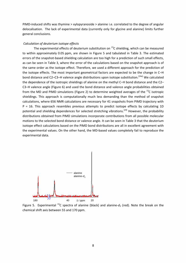
The experimental effects of deuterium substitution on 13C shielding, which can be measured
to within approximately 0.05 ppm, are shown in Figure 5 and tabulated in Table 3. The estimated
errors of the snapshot-based shielding calculation are too high for a prediction of such small effects,
as can be seen in Table 3, where the error of the calculations based on the snapshot approach is of
the same order as the isotope effect. Therefore, we used a different approach for the prediction of
the isotope effects. The most important geometrical factors are expected to be the change in C–H
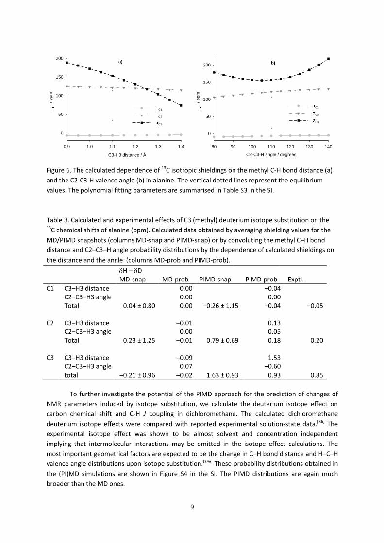
bond distance and C2–C3–H valence angle distributions upon isotope substitution.[24a] We calculated
the dependence of the isotropic shieldings of alanine on the methyl C–H bond distance and the C2–
C3–H valence angle (Figure 6) and used the bond distance and valence angle probabilities obtained
from the MD and PIMD simulations (Figure 2) to determine weighted averages of the 13C isotropic
shieldings. This approach is computationally much less demanding than the method of snapshot
calculations, where 656 NMR calculations are necessary for 41 snapshots from PIMD trajectory with
P = 16. This approach resembles previous attempts to predict isotope effects by calculating 1D
potential and shielding dependences for selected stretching vibrations.[24] However, the probability
distributions obtained from PIMD simulations incorporate contributions from all possible molecular
motions to the selected bond distance or valence angle. It can be seen in Table 3 that the deuterium
isotope effect calculations based on the PIMD bond distributions are all in excellent agreement with
the experimental values. On the other hand, the MD-based values completely fail to reproduce the
experimental data.
/ ppm 2040180
alanine
alanine-d3
Figure 5. Experimental 13C spectra of alanine (black) and alanine-d3 (red). Note the break on the
chemical shift axis between 55 and 170 ppm.
9
0.9 1.0 1.1 1.2 1.3 1.4
/ p
pm
0
50
100
150
200
C1
C2
C3
C3-H3 distance / Å
a)
C2-C3-H angle / degrees
80 90 100 110 120 130 140
/ p
pm
0
50
100
150
200
C1
C2
C3
b)
Figure 6. The calculated dependence of 13C isotropic shieldings on the methyl C-H bond distance (a)
and the C2-C3-H valence angle (b) in alanine. The vertical dotted lines represent the equilibrium
values. The polynomial fitting parameters are summarised in Table S3 in the SI.
Table 3. Calculated and experimental effects of C3 (methyl) deuterium isotope substitution on the 13C chemical shifts of alanine (ppm). Calculated data obtained by averaging shielding values for the
MD/PIMD snapshots (columns MD-snap and PIMD-snap) or by convoluting the methyl C–H bond
distance and C2–C3–H angle probability distributions by the dependence of calculated shieldings on
the distance and the angle (columns MD-prob and PIMD-prob).
H – D
MD-snap MD-prob PIMD-snap PIMD-prob Exptl.
C1 C3–H3 distance 0.00 –0.04 C2–C3–H3 angle 0.00 0.00 Total 0.04 ± 0.80 0.00 –0.26 ± 1.15 –0.04 –0.05 C2 C3–H3 distance –0.01 0.13 C2–C3–H3 angle 0.00 0.05 Total 0.23 ± 1.25 –0.01 0.79 ± 0.69 0.18 0.20 C3 C3–H3 distance –0.09 1.53 C2–C3–H3 angle 0.07 –0.60 total –0.21 ± 0.96 –0.02 1.63 ± 0.93 0.93 0.85
To further investigate the potential of the PIMD approach for the prediction of changes of
NMR parameters induced by isotope substitution, we calculate the deuterium isotope effect on
carbon chemical shift and C-H J coupling in dichloromethane. The calculated dichloromethane
deuterium isotope effects were compared with reported experimental solution-state data.[36] The
experimental isotope effect was shown to be almost solvent and concentration independent
implying that intermolecular interactions may be omitted in the isotope effect calculations. The
most important geometrical factors are expected to be the change in C–H bond distance and H–C–H
valence angle distributions upon isotope substitution.[24a] These probability distributions obtained in
the (PI)MD simulations are shown in Figure S4 in the SI. The PIMD distributions are again much
broader than the MD ones.
10
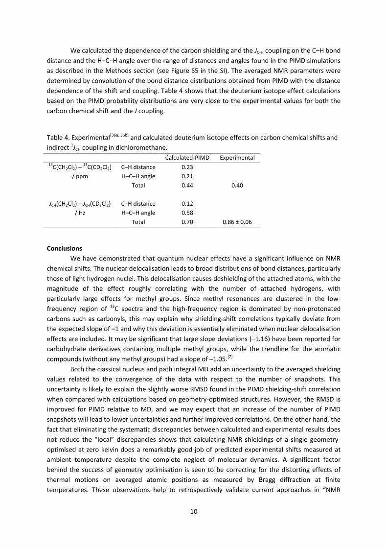
We calculated the dependence of the carbon shielding and the JC-H coupling on the C–H bond
distance and the H–C–H angle over the range of distances and angles found in the PIMD simulations
as described in the Methods section (see Figure S5 in the SI). The averaged NMR parameters were
determined by convolution of the bond distance distributions obtained from PIMD with the distance
dependence of the shift and coupling. Table 4 shows that the deuterium isotope effect calculations
based on the PIMD probability distributions are very close to the experimental values for both the
carbon chemical shift and the J coupling.
Table 4. Experimental[36a, 36b] and calculated deuterium isotope effects on carbon chemical shifts and
indirect 1JCH coupling in dichloromethane.
Calculated-PIMD Experimental 13
C(CH2Cl2) – 13
C(CD2Cl2) C–H distance 0.23
/ ppm H–C–H angle 0.21
Total 0.44 0.40
JCH(CH2Cl2) – JCH(CD2Cl2)
C–H distance 0.12
/ Hz H–C–H angle 0.58
Total 0.70 0.86 ± 0.06
Conclusions
We have demonstrated that quantum nuclear effects have a significant influence on NMR
chemical shifts. The nuclear delocalisation leads to broad distributions of bond distances, particularly
those of light hydrogen nuclei. This delocalisation causes deshielding of the attached atoms, with the
magnitude of the effect roughly correlating with the number of attached hydrogens, with
particularly large effects for methyl groups. Since methyl resonances are clustered in the low-
frequency region of 13C spectra and the high-frequency region is dominated by non-protonated
carbons such as carbonyls, this may explain why shielding-shift correlations typically deviate from
the expected slope of –1 and why this deviation is essentially eliminated when nuclear delocalisation
effects are included. It may be significant that large slope deviations (–1.16) have been reported for
carbohydrate derivatives containing multiple methyl groups, while the trendline for the aromatic
compounds (without any methyl groups) had a slope of –1.05.[7]
Both the classical nucleus and path integral MD add an uncertainty to the averaged shielding
values related to the convergence of the data with respect to the number of snapshots. This
uncertainty is likely to explain the slightly worse RMSD found in the PIMD shielding-shift correlation
when compared with calculations based on geometry-optimised structures. However, the RMSD is
improved for PIMD relative to MD, and we may expect that an increase of the number of PIMD
snapshots will lead to lower uncertainties and further improved correlations. On the other hand, the
fact that eliminating the systematic discrepancies between calculated and experimental results does
not reduce the “local” discrepancies shows that calculating NMR shieldings of a single geometry-
optimised at zero kelvin does a remarkably good job of predicted experimental shifts measured at
ambient temperature despite the complete neglect of molecular dynamics. A significant factor
behind the success of geometry optimisation is seen to be correcting for the distorting effects of
thermal motions on averaged atomic positions as measured by Bragg diffraction at finite
temperatures. These observations help to retrospectively validate current approaches in “NMR
11
crystallography” and explain their success. In contrast, similar comparison of calculated and
experimental infrared or Raman spectra of organic crystals is much less satisfactory and the use of
vibrational spectroscopy for crystal structure determination is, therefore, limited.[37]
The PIMD approach enables predictions of isotope effects in larger systems such as organic
molecular crystals in excellent agreement with experiment. Convergence errors, which would
otherwise dominate the calculated results, can be avoided by convoluting probability distributions of
selected geometrical parameters with the calculated shielding or J coupling surface scans. This
approach provides insight into the geometrical factors contributing to the isotope effect, and is
computationally much less demanding than the method of snapshot calculations.
Acknowledgement
The research leading to these results has received funding from the People Programme
(Marie Curie Actions) of the European Union's Seventh Framework Programme (FP7/2007-2013)
under REA grant agreement n° 299242.
References
[1] C. J. Pickard, F. Mauri, Phys. Rev. B 2001, 6324, 245101. [2] a) C. Bonhomme, C. Gervais, F. Babonneau, C. Coelho, F. Pourpoint, T. Azais, S. E. Ashbrook,
J. M. Griffin, J. R. Yates, F. Mauri, C. J. Pickard, Chem. Rev. 2012, 112, 5733-5779; b) R. K. Harris, P. Hodgkinson, C. J. Pickard, J. R. Yates, V. Zorin, Mag. Res. Chem. 2007, 45, S174-S186.
[3] A. C. De Dios, C. J. Jameson, in Annual Reports on NMR Spectroscopy, Vol. 77 (Ed.: G. A. Webb), Elsevier Ltd., Burlington, 2012, pp. 1-80.
[4] M. Dračínský, J. Kaminský, P. Bouř, J. Chem. Phys. 2009, 130, 094106. [5] D. A. Forsyth, A. B. Sebag, J. Am. Chem. Soc. 1997, 119, 9483-9494. [6] a) J. Czernek, T. Pawlak, M. J. Potrzebowski, Chem. Phys. Lett. 2012, 527, 31-35; b) J. M.
Griffin, J. R. Yates, A. J. Berry, S. Wimperis, S. E. Ashbrook, J. Am. Chem. Soc. 2010, 132, 15651-15660; c) L. Truflandier, M. Paris, F. Boucher, Phys. Rev. B 2007, 76; d) R. Laskowski, P. Blaha, F. Tran, Phys. Rev. B 2013, 87; e) M. Dračínský, M. Buděšínský, B. Warzajtis, U. Rychlewska, J. Phys. Chem. A 2012, 116, 680-688.
[7] J. C. Johnston, R. J. Iuliucci, J. C. Facelli, G. Fitzgerald, K. T. Mueller, J. Chem. Phys. 2009, 131, 144503.
[8] a) M. Bühl, M. Kaupp, O. L. Malkina, V. G. Malkin, J. Comput. Chem. 1999, 20, 91-105; b) P. J. Wilson, in Annual Rep. NMR Spectr., Vol. 49 (Ed.: G. A. Webb), Academic, New York, 2003, pp. 117-168.
[9] J. N. Dumez, C. J. Pickard, J. Chem. Phys. 2009, 130, 104701. [10] M. Dračínský, P. Hodgkinson, CrystEngComm. in press, DOI: 10.1039/C3CE40612A. [11] M. Dračínský, P. Bouř, J. Comput. Chem. 2012, 33, 1080-1089. [12] R. P. Feynman, A. R. Hibbs, Quantum Mechanics and Path Integrals, McGraw-Hill, New York,
1965. [13] A. Witt, S. D. Ivanov, M. Shiga, H. Forbert, D. Marx, J. Chem. Phys. 2009, 130, 194510. [14] J. Zinn-Justin, Path Integrals in Quantum Mechanics, Oxford University Press, Oxford, 2005. [15] H. Kleinert, Path integrals in quantum mechanics, statistics, and polymer physics, World
Scientific Publishing Co., Singapore, 1990. [16] S. Tang, D. A. Case, J. Biomol. NMR 2007, 38, 255-266. [17] J. Lobaugh, G. A. Voth, J. Chem. Phys. 1997, 106, 2400-2410. [18] M. Shiga, K. Suzuki, M. Tachikawa, J. Chem. Phys. 2010, 132, 114104.
12
[19] a) J. Schulte, R. Ramírez, M. C. Böhm, Chem. Phys. Lett. 2006, 432, 579-584; b) M. C. Böhm, R. Ramírez, J. Schulte, Chem. Phys. 2007, 342, 1-15.
[20] M. C. Böhm, J. Schulte, R. Ramírez, Chem. Phys. Lett. 2000, 332, 117-124. [21] a) J. Schulte, R. Ramírez, M. C. Böhm, Mol. Phys. 2001, 99, 1155-1158; b) M. C. Böhm, J.
Schulte, R. Ramírez, Int. J. Quantum Chem. 2002, 86, 280-296. [22] G. A. Luduena, M. Wegner, L. Bjålie, D. Sebastiani, ChemPhysChem 2010, 11, 2353-2360. [23] C. J. Jameson, in Encyclopedia of NMR (Ed.: R. K. Harris), John Wiley & Sons, Ltd, Chichester,
2012. [24] a) T. Dziembowska, P. E. Hansen, Z. Rozwadowski, Prog. Nucl. Magn. Reson. Spectrosc. 2004,
45, 1-29; b) J. Abildgaard, S. Bolvig, P. E. Hansen, J. Am. Chem. Soc. 1998, 120, 9063-9069. [25] F. H. Allen, Acta Cryst. B 2002, 58, 380-388. [26] S. J. Clark, M. D. Segall, C. J. Pickard, P. J. Hasnip, M. J. Probert, K. Refson, M. C. Payne, Z.
Kristallogr. 2005, 220, 567-570. [27] D. Vanderbilt, Phys. Rev. B 1990, 41, 7892-7895. [28] H. J. Monkhorst, J. D. Pack, Phys. Rev. B 1976, 13, 5188-5192. [29] J. P. Perdew, K. Burke, M. Ernzerhof, Phys. Rev. Lett. 1996, 77, 3865-3868. [30] J. R. Yates, C. J. Pickard, F. Mauri, Phys. Rev. B 2007, 76, 024401. [31] J. Kessler, M. Dračínský, P. Bouř, J. Comput. Chem. 2013, 34, 366-371. [32] a) A. D. Becke, J. Chem. Phys. 1993, 98, 5648-5652; b) C. T. Lee, W. T. Yang, R. G. Parr, Phys.
Rev. B 1988, 37, 785-789. [33] M. J. Frisch, G. W. Trucks, H. B. Schlegel, G. E. Scuseria, M. A. Robb, J. R. Cheeseman, G.
Scalmani, V. Barone, B. Mennucci, G. A. Petersson, H. Nakatsuji, X. Caricato, X. Li, H. P. Hratchian, A. F. Izmaylov, J. Bloino, G. Zheng, J. L. Sonnenberg, M. Hada, M. Ehara, K. Toyota, R. Fukuda, J. Hasegawa, M. Ishida, T. Nakajima, Y. Honda, O. Kitao, H. Nakai, T. Vreven, J. Montgomery, J. A., J. E. Peralta, F. Ogliaro, M. Bearpark, J. J. Heyd, E. Brothers, K. N. Kudin, V. N. Staroverov, R. Kobayashi, J. Normand, K. Raghavachari, A. Rendell, J. C. Burant, S. S. Iyengar, J. Tomasi, M. Cossi, N. Rega, J. M. Millam, M. Klene, J. E. Knox, J. B. Cross, V. Bakken, C. Adamo, J. Jaramillo, R. Gomperts, R. E. Stratmann, O. Yazyev, A. J. Austin, R. Cammi, C. Pomelli, J. W. Ochterski, R. L. Martin, K. Morokuma, V. G. Zakrzewski, G. A. Voth, P. Salvador, J. J. Dannenberg, S. Dapprich, A. D. Daniels, O. Farkas, J. B. Foresman, J. V. Ortiz, J. Cioslowski, D. J. Fox, Gaussian, Inc., Wallingford CT, 2009.
[34] M. G. Taylor, R. H. Marchessault, S. Perez, P. J. Stephenson, C. A. Fyfe, Can. J. Chem. 1985, 63, 270-273.
[35] F. Liu, A. M. Orendt, D. W. Alderman, D. M. Grant, J. Am. Chem. Soc. 1997, 119, 8981-8984. [36] a) N. M. Sergeyev, P. Sandor, N. D. Sergeyeva, W. T. Raynes, J. Magn. Res. A 1995, 115, 174-
182; b) N. M. Sergeyev, N. D. Sergeyeva, W. T. Raynes, J. Chem. Soc., Chem. Commun. 1994, 485-486; c) N. M. Sergeyev, N. D. Sergeyeva, W. T. Raynes, Magn. Reson. Chem. 1994, 32, 381-385.
[37] M. Dračínský, E. Procházková, J. Kessler, J. Šebestík, P. Matějka, P. Bouř, J. Phys. Chem. B 2013, 117, 7297-7307.
13
Entry for the Table of Contents
Nuclear delocalisation
The influence of nuclear delocalisation
on NMR chemical shifts is explored
using path integral molecular dynamics,
and is shown to explain previously
observed systematic deviations between
calculated and experimental shifts.
PIMD also enables isotope substitution
effects to be predicted in excellent
agreement with experiment.
Martin Dračínský,* Paul Hodgkinson*
……..… Page – Page
Effects of quantum nuclear
delocalisation on NMR parameters
from path integral molecular
dynamics
14
Effects of quantum nuclear delocalisation on NMR parameters from path integral
molecular dynamics
Martin Dračínský,a,b,* Paul Hodgkinsona,*
aDepartment of Chemistry, Durham University, South Road, DH1 3LE, Durham, UK bInstitute of Organic Chemistry and Biochemistry, Flemingovo nám. 2, 16610, Prague, Czech Republic
SUPPORTING INFORMATION
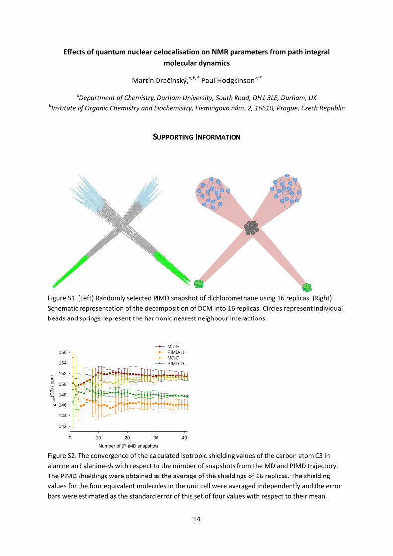
Figure S1. (Left) Randomly selected PIMD snapshot of dichloromethane using 16 replicas. (Right)
Schematic representation of the decomposition of DCM into 16 replicas. Circles represent individual
beads and springs represent the harmonic nearest neighbour interactions.
Number of (PI)MD snapshots
0 10 20 30 40
iso(C
3)
/ ppm
142
144
146
148
150
152
154
156
MD-H
PIMD-H
MD-D
PIMD-D
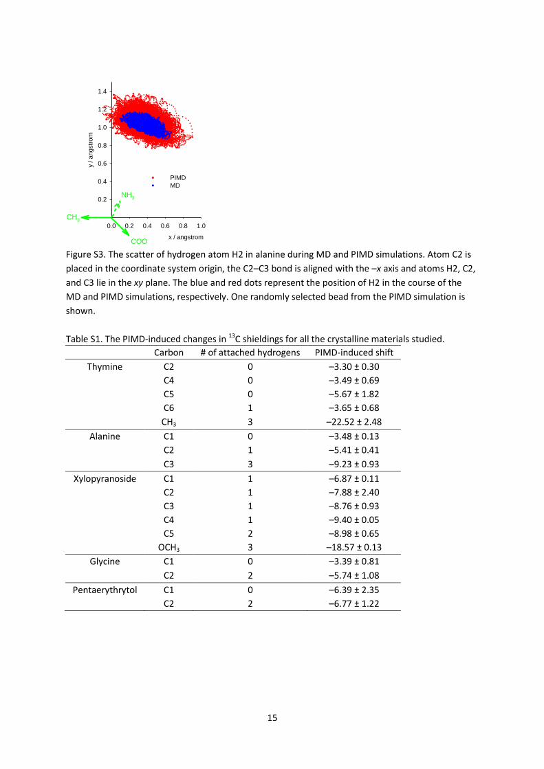
Figure S2. The convergence of the calculated isotropic shielding values of the carbon atom C3 in
alanine and alanine-d3 with respect to the number of snapshots from the MD and PIMD trajectory.
The PIMD shieldings were obtained as the average of the shieldings of 16 replicas. The shielding
values for the four equivalent molecules in the unit cell were averaged independently and the error
bars were estimated as the standard error of this set of four values with respect to their mean.
15
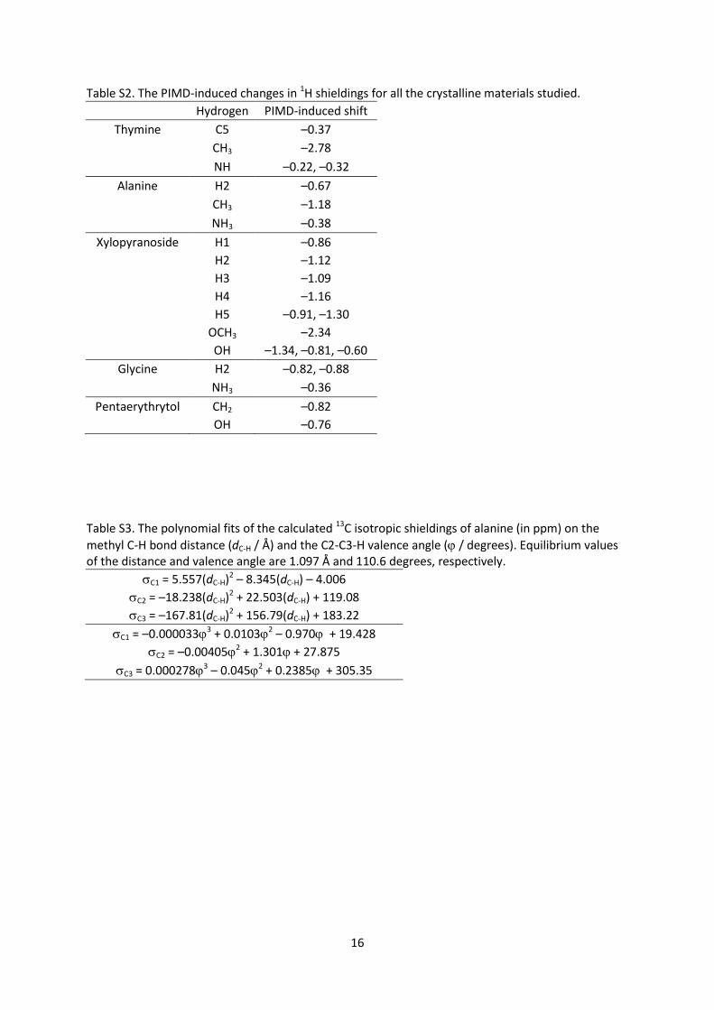
x / angstrom
0.0 0.2 0.4 0.6 0.8 1.0
y / a
ngstr
om
0.2
0.4
0.6
0.8
1.0
1.2
1.4
PIMD
MD
CH3
COO
NH3
Figure S3. The scatter of hydrogen atom H2 in alanine during MD and PIMD simulations. Atom C2 is
placed in the coordinate system origin, the C2–C3 bond is aligned with the –x axis and atoms H2, C2,
and C3 lie in the xy plane. The blue and red dots represent the position of H2 in the course of the
MD and PIMD simulations, respectively. One randomly selected bead from the PIMD simulation is
shown.
Table S1. The PIMD-induced changes in 13C shieldings for all the crystalline materials studied.
Carbon # of attached hydrogens PIMD-induced shift
Thymine C2 0 –3.30 ± 0.30
C4 0 –3.49 ± 0.69
C5 0 –5.67 ± 1.82
C6 1 –3.65 ± 0.68
CH3 3 –22.52 ± 2.48
Alanine C1 0 –3.48 ± 0.13
C2 1 –5.41 ± 0.41
C3 3 –9.23 ± 0.93
Xylopyranoside C1 1 –6.87 ± 0.11
C2 1 –7.88 ± 2.40
C3 1 –8.76 ± 0.93
C4 1 –9.40 ± 0.05
C5 2 –8.98 ± 0.65
OCH3 3 –18.57 ± 0.13
Glycine C1 0 –3.39 ± 0.81
C2 2 –5.74 ± 1.08
Pentaerythrytol C1 0 –6.39 ± 2.35
C2 2 –6.77 ± 1.22
16
Table S2. The PIMD-induced changes in 1H shieldings for all the crystalline materials studied.
Hydrogen PIMD-induced shift
Thymine C5 –0.37
CH3 –2.78
NH –0.22, –0.32
Alanine H2 –0.67
CH3 –1.18
NH3 –0.38
Xylopyranoside H1 –0.86
H2 –1.12
H3 –1.09
H4 –1.16
H5 –0.91, –1.30
OCH3 –2.34
OH –1.34, –0.81, –0.60
Glycine H2 –0.82, –0.88
NH3 –0.36
Pentaerythrytol CH2 –0.82
OH –0.76
Table S3. The polynomial fits of the calculated 13C isotropic shieldings of alanine (in ppm) on the
methyl C-H bond distance (dC-H / Å) and the C2-C3-H valence angle ( / degrees). Equilibrium values of the distance and valence angle are 1.097 Å and 110.6 degrees, respectively.
C1 = 5.557(dC-H)2 – 8.345(dC-H) – 4.006
C2 = –18.238(dC-H)2 + 22.503(dC-H) + 119.08
C3 = –167.81(dC-H)2 + 156.79(dC-H) + 183.22
C1 = –0.0000333 + 0.01032 – 0.970 + 19.428
C2 = –0.004052 + 1.301 + 27.875
C3 = 0.0002783 – 0.0452 + 0.2385 + 305.35
17
C2-C3-H angle / degree
80 100 120 140
Pro
ba
bili
ty / %
0
2
4
6
8
10
MD-H
PIMD-H
MD-D
PIMD-D
C-H (C-D) bond distance / A
0.8 0.9 1.0 1.1 1.2 1.3 1.4
Pro
ba
bili
ty / %
0
2
4
6
8
10
12
14
16
MD
PIMD
MD-D
PIMD-D
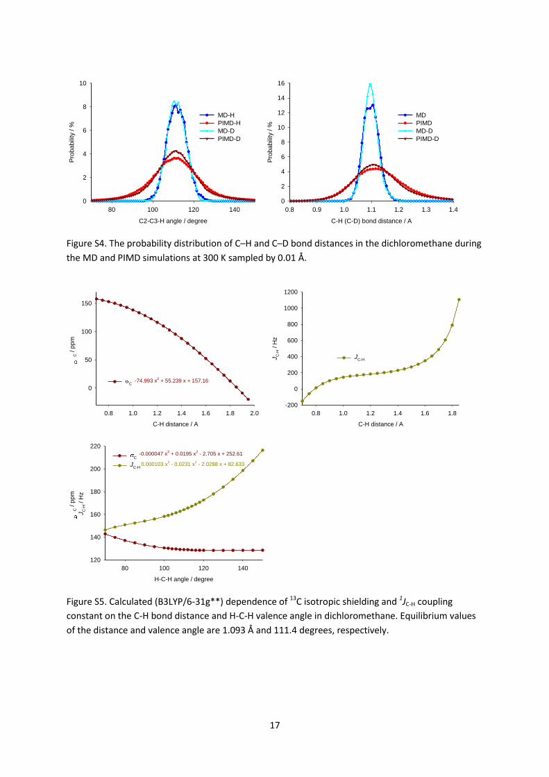
Figure S4. The probability distribution of C–H and C–D bond distances in the dichloromethane during
the MD and PIMD simulations at 300 K sampled by 0.01 Å.
C-H distance / A
0.8 1.0 1.2 1.4 1.6 1.8 2.0
C / p
pm
0
50
100
150
C-74.993 x
2 + 55.239 x + 157.16
C-H distance / A
0.8 1.0 1.2 1.4 1.6 1.8
JC
-H / H
z
-200
0
200
400
600
800
1000
1200
JC-H
H-C-H angle / degree
80 100 120 140
C / p
pm
JC
-H / H
z
120
140
160
180
200
220
C
JC-H
-0.000047 x3 + 0.0195 x
2 - 2.705 x + 252.61
0.000103 x3 - 0.0231 x
2 - 2.0288 x + 82.633
Figure S5. Calculated (B3LYP/6-31g**) dependence of 13C isotropic shielding and 1JC-H coupling
constant on the C-H bond distance and H-C-H valence angle in dichloromethane. Equilibrium values
of the distance and valence angle are 1.093 Å and 111.4 degrees, respectively.