Embed Size (px)
Citation preview

Duration and amplitude decay of acute arterial leginflow enhancement with intermittent pneumaticleg compression: An insight into the implicatedphysiologic mechanismsKonstantinos T. Delis, MS, PhD, FRCSI, EBSQvasc,a,b and Alison L. Knaggs, FFARCSI, DEAA,a
London, United Kingdom; and Rochester, Minn
Purpose: By acutely enhancing the arterial leg inflow, intermittent pneumatic leg compression (IPC) improves the walkingability, arterial hemodynamics, and quality of life of claudicants. We quantified the duration of acute leg inflow enhancementwith IPC of the foot (IPCfoot), calf (IPCcalf), or both (IPCfoot�calf) and its amplitude decay in claudicants and controls inrelation to the pulsatility index, an estimate of peripheral resistance. These findings are cross-correlated with the features of thethree implicated physiologic mechanisms: (1) an increase in the arteriovenous pressure gradient, (2) suspension of peripheralsympathetic autoregulation, and (3) enhanced release of nitric oxide with flow and shear-stress increase.Methods: Twenty-six limbs of 24 claudicants with superficial femoral artery occlusion or stenoses (>75%) and 24 limbs of20 healthy controls matched for age and sex, meeting stringent selection criteria, had their popliteal volume flow andpulsating index (peak-to-peak velocity/mean velocity) measured with duplex scanning at rest and upon delivery of IPC.Spectral waveforms were analyzed for 50 seconds after IPC delivery per 5-second segments. The three IPC modes were appliedin a true crossover design. Data analysis was performed with the Page, Friedman, Wilcoxon, Mann-Whitney and �2 tests.Results: The median duration of flow enhancement in claudicants exceeded 50 seconds with IPCfoot, IPCcalf, andIPCfoot�calf but was shorter (P < .001) in the controls (32.5 to 40 seconds). Among the three IPC modes, the durationof flow enhancement differed (P < .05) only between IPCfoot and IPCfoot�calf. After reaching its peak within 5 secondsof IPC, flow enhancement decayed at rates decreasing over time (trend, P < .05, Page test), which in both groups werehighest at 5 to 20 seconds, moderate at 20 to 35 seconds, and lowest at 35 to 50 seconds (P < .05, Friedman test).Baseline and peak flow with all IPC modes was similar between the two groups. Pulsatility index attenuation inclaudicating limbs lasted a median 32.5 seconds with IPCfoot, 37.5 seconds with IPCcalf, and 40 seconds with IPCfoot�calf;duration of pulsatility index attenuation was shorter in the control limbs with IPCfoot (30 seconds), IPCcalf (32.5seconds), or IPCfoot�calf (35 seconds), yet differences, as well as those among the 3 IPC modes, were not significant.Conclusion: Leg inflow enhancement with IPC exceeds 50 seconds in claudicants and lasts 32.5 to 40 seconds in thecontrols. Peak flow occurs concurrently with maximal pulsatility index attenuation, within 5 seconds of IPC. Irrespectiveof group or IPC mode, the decay rate (%) of flow enhancement is highest within 5 to 20 seconds of IPC, moderate at 20to 35 seconds, and lowest at 35 to 50 seconds. Since attenuation in peripheral resistance terminates with the mid timeperiod (20 to 35 seconds) of flow decay, and nitric oxide has a half-life of <7 to 10 seconds, the study’s data indicate thatall implicated physiologic mechanisms (1, 2, and 3) are likely active immediately after IPC delivery (0 to 20 sec) and allbut nitric oxide are effective in the mid time period (20 to 35 seconds). As the pulsatility index has returned to baseline,the late phase of flow enhancement (35 to 50 seconds) could be attributable to the declining arteriovenous pressure
gradient alone. (J Vasc Surg 2005;42:717-25.)Intermittent pneumatic compression (IPC) of thelower limb is a well-documented method of acute arterialleg inflow enhancement in patients with peripheral arterialdisease (PAD).1-8 Over the past 5 years, attention has beenfocused on the potential role that IPC may hold in thetreatment of arterial claudication.9-11 According to the firstclinical outcomes study released in 2000,9 patients withstable claudication (n � 25) who received IPC of the foot(IPCfoot) (�4 h/day; 4.5 months) and aspirin (75 mg/
From St Mary’s Hospital NHS Trust, Imperial College School of Medicine,a
and the Division of Vascular Surgery, Mayo Clinic.b
Competition of interest: none.Reprint requests: Konstantinos T. Delis, MD, Marco Polo Fellows (ESVS),
4 Abinger Court, 34 Gordon Road, Ealing W5 2AF, London, UK(e-mail: [email protected]).
0741-5214/$30.00Copyright © 2005 by The Society for Vascular Surgery.
doi:10.1016/j.jvs.2005.06.004day) while exercising unsupervised improved the medianinitial and absolute claudication distances by �100% andincreased the resting and postexercise ankle-brachial in-dices by 18% and 110%, respectively (all, P � .001),whereas their control counterparts, who had the sametreatment except IPC, did not improve their perfor-mance.9 Randomized controlled data confirm that IPCdelivered to the foot and calf (IPCfoot�calf) for 3 to 5months (�2.5 h/day) may indeed ameliorate claudica-tion while improving arterial hemodynamics.10,11 Inlight of the increase in the ankle-brachial indices oftreated legs, it has been suggested that the prolongedarterial flow enhancement with IPC treatment may pro-mote the development of collateral circulation.9-11
Upon IPC application, the arterial leg inflow increasesacutely twofold to threefold in claudicants and threefold to
fourfold in healthy individuals.2-7 This flow enhancement is717

JOURNAL OF VASCULAR SURGERYOctober 2005718 Delis and Knaggs
attributable to three physiologic mechanisms. As the ve-nous pressure in the dependent limb decreases with IPC,the arteriovenous pressure gradient rises inversely, causingarterial leg inflow to increase.1-8,12 At the same time,venous pressure attenuation with IPC also causes a tran-sient abolition of peripheral sympathetic autoregulation byway of the axon venoarteriolar reflex, which normally up-regulates the arterial tone and resistance to flow on depen-dency, protecting the microvasculature and interstitial tis-sues from hypertension.13-18 The acute arterial leg inflowenhancement thus generated with IPC instigates furtherrelease of nitric oxide in response to an increase in shear-stress, which in turn causes further attenuation in peripheralresistance and, thus, a reciprocal arterial flow enhance-ment.1-8,19,20 In light of the half-life of endothelium-derived vasodilatory nitric oxide being �7 to 10 seconds,21
and venous refill occurring �30 to 50 seconds in limbs withvenous valvular competence,11,22,23 determination of thedecay of the arterial leg inflow enhancement after impulsedelivery may offer an insight into the morphology of thishemodynamic phenomenon and the sequence of physio-logic mechanisms that permeate its occurrence.
The aim of this study was to determine the durationand the amplitude decay of the acute leg inflow enhance-ment with the delivery of IPC to the foot, calf, and bothcombined among patients with arterial claudication andhealthy controls in relation to the flow pulsatility index, anoninvasive measure of peripheral arterial resistance.24,25
The study findings are critically cross-correlated with thefeatures of the physiologic mechanisms implicated in thisphenomenon, with the purpose of shedding light on thesequence of events that permeate the acute arterial leginflow enhancement with IPC that remain poorly under-stood to date.
METHODS
Design. This is a cross-sectional study that was con-ducted prospectively after ethics committee approval andinformed consent of the involved subjects.
Inclusion criteria. Patients with a history of PAD andintermittent claudication due to superficial femoral artery
Table 1. Demographics, risk factors and ankle-brachial pr
Group 1 (claudicants)
Subjects 22Age, median (range) 63 (50–77)Female/male ratio 9/13Diabetes mellitus 5Smoking 4Hypertension 8r-ABI, median (IQR) 0.6 (0.42–0.84)p-eABI, median (IQR) 0.28 (0.16–0.52)Legs 26
r-ABI, Resting ankle-brachial pressure index; p-eABI, postexercise ankle-br*Mann-Whitney test.†�2 test.
occlusion or severe stenosis (�75%) were considered for
inclusion in the group of claudicants (group 1). Individualswithout a history of cardiovascular disease and claudicationwere considered for inclusion in the group of controlsubjects (group 2).
Exclusion criteria. Excluded from the study weresubjects with symptomatic chronic venous disease, CEAPclinical classes 2 to 6, hemodynamically significant (�50%diameter stenosis) aortoiliac disease on duplex sonography(�3 months), congestive cardiac failure, recent (�12months) axial deep vein thrombosis, leg edema, wounds orinfection, diabetes mellitus of �4 years duration, peripheralsensory impairment clinically, popliteal artery occlusion orsevere atherosclerosis on duplex examination, and those onvasoactive medication.
Subjects. The study comprised 26 limbs (22 patients)with intermittent claudication (group 1) and 24 controllimbs (20 subjects) with normal circulation (group 2). Thetwo groups were matched for age and sex. Summarized inTable I are the demographics of study subjects.
Leg inflow investigation. Arterial leg inflow was ex-amined in the popliteal artery with real-time gated Dopplersonography and real-time B-mode imaging, used simulta-neously (Sonos 2500, 7.5/5.5 MHz linear array probe,Hewlett Packard, Palo Alto, Calif). Spectral velocity datawere obtained at a 60o insonation angle with the samplevolume gate encompassing the entire lumen of the vessel.Doppler waveforms were enveloped automatically usingdedicated software for flow estimation. Data included thedetermination of volume flow, calculated from the meanvelocity multiplied by the cross-sectional area of the popli-teal artery (mean velocity � � � [diameter/2]2), and theflow pulsatility index (peak-to-peak velocity/mean veloc-ity), a measure of peripheral resistance to flow. Mean veloc-ity is the time average of the mean velocities of each of theDoppler spectra occurring during an interval of 5 seconds.Similarly, pulsatility index measurements represent valuesretrieved during the same (5-second) time periods. Spectralwaveforms containing aliasing or noise due to venous flowor wall motion were discarded and measurements wererepeated. The cross-sectional area of the popliteal arterywas calculated from its diameter obtained by viewing the
e indices of investigated subjects in the two study groups
Group 2 (controls) Statistical significance
20 —61 (44–78) NS*
7/13 NS†
3 NS†
5 NS†
6 NS†
�1.0 P � .0001*�1.0 P � .0001*24 —
pressure index; IQR, interquartile range.
essur
achial
artery longitudinally and by placing the tracker ball-guided

JOURNAL OF VASCULAR SURGERYVolume 42, Number 4 Delis and Knaggs 719
callipers across the intimal-luminal interphases of the nearand far walls; 10 measurements were taken, and the averagevalue was used for calculating the volume flow.
Study protocol. Three IPC modes were investigatedin the current study: IPCfoot, IPCcalf, and IPCfoot�calf.Investigation of the subjects was preceded by a restingperiod of 30 minutes. Popliteal flow was determined at restin the sitting position (three different readings). IPC wasthen applied for 5 minutes in the same position. Spectraldata were monitored with continuous duplex scanning, buttheir registration and storage in the hard-drive memory ofthe system’s computer commenced with the delivery of thelast pneumatic impulse, through a time period of 50 sec-onds. Spectral information was then analyzed by dividingthe stored Doppler signal into a sequence of 5-secondsegments.
After a 10-minute resting period, flow was obtainedagain. The second IPC mode of the three that were studiedwas applied for 5 minutes, and the spectral information offlow velocities from the delivery of the last impulse wasstored and processed as previously described. The sameprocedure was followed for the investigation of the thirdIPC mode.
All measurements were obtained by the same examinerin temperature-controlled conditions (21° to 23°C). Thereproducibility of flow velocity measurements obtainedwith the above method has been reported.4
Designation of IPC sequence. The sequence of IPCmodes in the examination protocol of the study was subjectto a true crossover design, aiming at eliminating the possi-bility of carry-over effects. Implemented evenly, the follow-ing sequences of IPC modes were examined: (1) IPCfoot-IPCcalf-IPCfoot�calf, (2) IPCfoot-IPCfoot�calf-IPCcalf, (3)IPCcalf-IPCfoot-IPCfoot�calf, (4) IPCcalf-IPCfoot�calf-IPC-calf, (5) IPCfoot�calf-IPCfoot-IPCcalf, and (6) IPCfoot�calf-IPCcalf-IPCfoot. In both groups, every new entry was allot-ted a sequence in this order.
Definitions and calculations. Duration of volumeflow enhancement in each one of the examined limbs wasdefined as the time-period that volume flow remainedhigher than baseline. This time, expressed as median andinterquartile range, enabled inter- and intragroup compar-isons. Duration of flow pulsatility index attenuation in eachone of the examined limbs was defined as the time duringwhich the pulsatility index remained lower than baseline.The percentage rate (%) of volume flow decay for each oneof the 5-second time segments after delivery of IPC, ad-justed for the amplitude of volume flow enhancement, wasdefined as ([volume flow at ttime – volume flow at ttime� 5seconds] � 100/volume flow at ttime).
IPC equipment. IPCfoot, IPCcalf and IPCfoot�calf
were delivered with the Art-Assist 1000 (ACI-Medical, SanMarcos, Calif) at the following presets: three impulses perminute, inflation pressure, 120 mm Hg; impulse rise time,0.5 to 0.6 seconds; impulse duration, 4 seconds; deflationpressure, 0 mm Hg; deflation time, 16 seconds.
Analysis and statistics. Intragroup statistical analysis
of paired volume flow and pulsatility index data was per-formed with the Wilcoxon signed ranks test. Intergroupdifferences in the volume flow and pulsatility index wereevaluated with the Mann-Whitney test. The estimated me-dian (EM), point estimate (PE), and the 95% confidenceinterval (CI) were calculated but are quoted selectively fordata presentation simplicity. The Bonferroni correction wasapplied when appropriate.26 The Page test of ordered alter-natives was used for examining the percentage rate ofvolume flow decay for “trend-over-time” after delivery ofIPC. Differences in the percentage rates of volume flowdecay after IPC delivery were evaluated with the Fried-man’s two-way analysis of variance. Differences in propor-tions were assessed with the �2 test. P � .05 was consideredsignificant. Results are expressed as median and interquar-tile range (IQR), unless otherwise stated.
Fig 1. Duration (median and interquartile range) of (A) volumeflow enhancement and (B) flow pulsatility index attenuation in thepopliteal artery of 26 limbs of claudicants (group 1, shaded bars)and 24 normal control limbs (group 2, white bars) with intermit-tent pneumatic compression (IPC) of the foot (IPCfoot), calf(IPC ) and both combined (IPC ); statistical analysis in
calf foot�calfthe Results section.
n.
JOURNAL OF VASCULAR SURGERYOctober 2005720 Delis and Knaggs
RESULTS
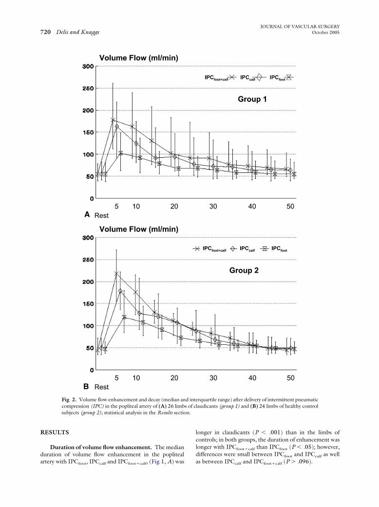
Duration of volume flow enhancement. The medianduration of volume flow enhancement in the popliteal
Fig 2. Volume flow enhancement and decay (median ancompression (IPC) in the popliteal artery of (A) 26 limbsubjects (group 2); statistical analysis in the Results sectio
artery with IPCfoot, IPCcalf and IPCfoot�calf, (Fig 1, A) was
longer in claudicants (P � .001) than in the limbs ofcontrols; in both groups, the duration of enhancement waslonger with IPCfoot�calf than IPCfoot (P � .05); however,differences were small between IPCfoot and IPCcalf as well
erquartile range) after delivery of intermittent pneumaticlaudicants (group 1) and (B) 24 limbs of healthy control
d ints of c
as between IPCcalf and IPCfoot�calf (P � .096).

JOURNAL OF VASCULAR SURGERYVolume 42, Number 4 Delis and Knaggs 721
Decay of volume flow in claudicants. Immediatelyafter the peak volume flow was reached within 5 seconds ofIPC delivery (P � .001), the volume flow declined steeply(Fig 2, A). Cumulatively, volume flow in claudicants re-mained higher than baseline in the 45- to 50-second timeperiod with IPCfoot�calf and IPCcalf (P � .009) and re-mained higher than baseline for 45 seconds with IPCfoot
(P � .035).The percentage rates of volume flow decay, determined
at nine consecutive time segments lasting 5 seconds each,from the 5th to the 50th second after impulse delivery, hada significant declining “trend-over-time” (Page test) withIPCfoot (P � .05), IPCcalf (P � .05), and IPCfoot�calf (P �.05) (Fig 3, A). Based on their magnitude, the volume flowdecay rates were stratified in three time periods: 5 to 20seconds, 20 to 35 seconds, and 35 to 50 seconds fromimpulse delivery (Table II); differences in the percentagerates (in both the median and the mean) among these timeperiods were statistically significant for IPCfoot, IPCcalf, andIPCfoot�calf (P � .01 by Friedman test). Peak volume flowwith IPCfoot�calf was significantly higher than with IPCcalf
(P � .001) and IPCfoot (P � .001). Also, peak volume flowwith IPCcalf was higher than with IPCfoot (P � .001).
Decay of volume flow in controls. Similar to the
Fig 3. Rates of decay (median and interquartile range) of volumeflow enhancement in the popliteal artery after delivery of intermit-tent pneumatic compression (IPC) of the foot (IPCfoot), calf(IPCcalf), and both (IPCfoot�calf) in (A) 26 limbs of claudicants(group 1) and (B) 24 limbs of normal control subjects (group 2)were significant for “trend-over-time” (P � .05; Page test).
decay of volume flow enhancement in claudicants, volume
flow declined rapidly after its peak was reached within 5seconds of the delivery of IPCfoot�calf, IPCcalf, and IPCfoot
(P � .001) (Fig 2, B). Volume flow remained significantlyhigher than baseline for 45 seconds with IPCfoot�calf (P �.014) and IPCcalf (P � .04), and for 40 seconds withIPCfoot (P � .022). The percentage rates of volume flowdecay, determined at nine consecutive time segments last-ing 5 seconds each, from the 5th to the 50th second afterimpulse delivery, had a significant declining “trend-over-time” (Page test) with IPCfoot (P � .05), IPCcalf (P � .05),and IPCfoot�calf (P � .05) (Fig 3, B). Based on theirmagnitude, the percentage rates of volume flow decay werestratified in three time periods: 5 to 20 seconds, 20 to 35seconds, and 35 to 50 seconds from impulse delivery (TableII). Differences in the percentage rates (in both the medianand the mean) among these time periods were statisticallysignificant for IPCfoot (P � .01), IPCcalf (P � .02), andIPCfoot�calf (P � .05) (Friedman’s test). Peak volume flowwith IPCfoot�calf was significantly higher than with IPCcalf
(P � .001) and IPCfoot (P � .001). Also, peak volume flowwith IPCcalf was higher than with IPCfoot (P � .001).
Claudicants vs controls. Baseline volume flow (onsitting) was higher in claudicants without reaching signifi-cance (P � .4; PE, 6.24 mL/min; 95%CI, 21.6 to –9.74mL/min). Peak volume flow with any IPC mode applied wasnot significantly different between the study groups (P �.2).
Duration of pulsatility index attenuation. The du-ration of pulsatility index attenuation in groups 1 and 2 isdepicted in Fig 1, B. The median duration was longer in thelimbs of claudicants with any of the three IPC modes;however, significance was not reached (all, P� .29). Simi-larly, the duration of pulsatility index attenuation was notsignificantly different among the three IPC modes in bothstudy groups (all, P � .8).
Decay of pulsatility index in claudicants. Immediatelyafter the pulsatility index reached its maximum attenuation,within 5 seconds of the delivery of IPCfoot�calf, IPCcalf, andIPCfoot (P � .001), its return to baseline was noted (Fig 4,A). The pulsatility index remained attenuated for 35 sec-onds after the delivery of IPCfoot�calf (P � .003), IPCcalf (P� .048; EMD, –.27; 95% CI, 0 to –.5) and IPCfoot (P �.04; EMD, –.24; 95% CI, 0 to –.5). Similar to previousreports, the pulsatility index with IPCfoot was less attenu-ated (P � .001) than with IPCcalf and IPCfoot�calf. Pulsa-tility index attenuation with IPCfoot�calf was similar to thatwith IPCcalf (P � .06; EMD, –.1; 95 %CI, –.2 to .005).
Decay of pulsatility index in controls. The decay ofpulsatility index attenuation in the control limbs commencedimmediately after its peak had been reached within 5 secondsof the delivery of IPCfoot�calf, IPCcalf, and IPCfoot (P � .001)(Fig 4, B). Cumulatively, the pulsatility index in the controllimbs remained significantly attenuated for 35 seconds afterdelivery of IPCfoot�calf (P � .003), for 30 seconds withIPCcalf (P � .001), and for 30 seconds with IPCfoot (P �.002). The pulsatility index with IPCfoot�calf was lowerthan with IPCcalf (P � .006) and IPCfoot (P � .001). Thepulsatility index with IPCcalf was also lower than that with
IPCfoot (P � .003).
JOURNAL OF VASCULAR SURGERYOctober 2005722 Delis and Knaggs
Claudicants vs controls. The pulsatility index waslower in the limbs of claudicants than the control, atbaseline, at 5 seconds after IPC delivery, and throughoutthe period (5 to 50 sec) of its determination with IPC (P �.0001).
DISCUSSION
The current study examined the decay of acute volumeflow enhancement with IPC of the foot, calf, and bothcombined in claudicants and determined its duration incomparison with healthy subjects. Cross-correlation of vol-ume flow with pulsatility index, an estimate of peripheralresistance,24,25 offered an insight into the sequence ofphysiologic mechanisms empowering flow enhancementwith IPC. The study data have demonstrated that themedian arterial leg inflow enhancement with IPC is a briefoccurrence that exceeds 50 seconds in claudicants and lasts30 to 40 seconds in healthy limbs, depending on the IPCmode. The difference in duration between the two groups,although small, was consistent and statistically significant.Flow enhancement in both groups was also longer withIPCfoot�calf than IPCfoot.
The decay rates of volume flow enhancement in thelimbs of both study groups, adjusted for volume flowamplitude, demonstrated a significant declining “trend-over-time” (Page test). Analysis of the decay rates based ontheir amplitude (Friedman’s test) enabled stratification inthree phases. In the early phase, commencing with peakflow at 5 seconds through 20 seconds, the decay rates ofvolume flow enhancement are highest. At the same time,the pulsatility index, having also reached its maximumattenuation within 5 seconds of IPC delivery, increasedrapidly. Volume flow continued to decline in the middlephase of flow enhancement (20 to 35 seconds), but the rateof decay was significantly lower, while the pulsatility indexcontinued to rise. In the third phase of flow enhancement(35 to 50 seconds), its decay slowed further or leveled,while the attenuation of the pulsatility index had alreadybeen exhausted.
Further, to the increase in the arteriovenous pressure
Table II. Mean and median decay rates of volume flow en50 seconds from the delivery of intermittent pneumatic co
Time afterIPC
5-20 seconds 20-35
Mean Median Mean
ClaudicantsIPCfoot 7.6 (4.8-10.7) 7.1 (4.7-11) 4.6 (1.8-5.6)IPCcalf 12 (8.5-16.6) 10.8 (7.5-16.7) 8.9 (5.2-11.2IPCfoot�calf 11.1 (7.6-15.3) 10.5 (6.9-14) 8.6 (6.1-12.5
ControlIPCfoot 12.6 (8.6-16.2) 12 (8.7-15.1) 9.9 (5.8-14.6IPCcalf 17.7 (12.3-22.2) 15.8 (11.2-21.6) 13.2 (11-16.4IPCfoot�calf 18.3 (12.9-24.7) 16.9 (13.7-27.7) 12.3 (8.7-17.5
IPC, Intermittent pneumatic compression.*Decay rates derive from the original data of depicted in Fig. 3, A and B anamong these periods are statistically significant.
gradient, the volume flow enhancement with IPC is also
the product of a sequence of compensatory re-adjustmentsin the arterial circulation resulting in the attenuation ofperipheral resistance to flow.1-10,18 The latter is supportedby the highly significant, concurrent, but mirror-imagedecay curves of volume flow and pulsatility index after IPCdelivery.1-8,18 The study’s finding that the pulsatility indexreaches maximum attenuation within 5 seconds of IPC,when peak volume flow is sustained, commencing its returnto baseline concurrently with the latter, highlights theimmediate optimal, yet short-lived, attenuation of periph-eral resistance with IPC.24,25 Pulsatility index attenuationdecays in the early (5 to 20 seconds) and mid (20 to 35seconds) phases of volume flow enhancement but is back tobaseline in the late phase (35 to 50 seconds), indicatingthat, unlike the early and mid phases, flow enhancement inthe late phase is unrelated to peripheral resistance.
The arterial leg inflow enhancement with IPC in theearly phase is twofold to threefold in claudicants and two-fold to fourfold in healthy limbs, far exceeding the levelexpected by the arteriovenous pressure gradient elevationdue to venous expulsion with IPC.1-6,18 Based on Poi-seuille’s law associating the arteriovenous pressure gradientdirectly with the arterial flow, and assuming that the arterialand venous pressures at the ankle on sitting are �130(70[diastolic] � 60 [hydrostatic]) mm Hg and 60[hydrostatic] mmHg, respectively, elevation of this gradient with IPC (sub-sequent to a reduction in venous pressure from 60 mm Hgto 10 mm Hg) could not cause the flow to increase by�70%.1,2,4,6,18 The fact that 30 seconds after IPC deliveryvenous pressure is still lower than baseline in limbs withnormal reflux 12 supports the role of arteriovenous pressuregradient in the late phase of flow enhancement. The level offlow enhancement with IPC also exceeds that (�80% to100%) expected by the transient suspension of venoarter-iolar reflex in response to IPC 13-17 underscored recently.18
The higher level of flow enhancement achieved with IPCmight be attributable to nitric oxide release with shear-stress enhancement.1-7 Human umbilical vein endothelialcells cultured in vitro in a system simulating vessel collapseconditions enhanced their nitric oxide production and up-
ement at 5 to 20 seconds, 20 to 35 seconds, and 35 tossion in groups 1 (claudicants) and 2 (control)*
ds 35-50 seconds Friedman’s test
Median Mean Median Mean Median
(1-6.1) 0.15 (0-1.6) 0 (0-3.3) P � 0.001 P � 0.001(2.8-10.7) 3.9 (0.9-7.8) 2.5 (0-6.4) P � 0.001 P � 0.01(4.6-10.7) 6.9 (3-8.7) 5.3 (2-8.3) P � 0.001 P � 0.01
(0.6-14.8) 0 (0-4.9) 0 (0-0.3) P � 0.01 P � 0.001(4.3-14.5) 2.6 (0-12.6) 0 (0-9.7) P � 0.02 P � 0.01(9.7-17.7) 7.3 (0-15.7) 0 (0-11.3) P � 0.05 P � 0.001
expressed as median and interquartile range. Differences in the decay rates
hancmpre
secon
4.3) 7.3) 5.8
) 10) 8.5) 12
d are
regulated endothelial nitric oxide synthase messenger RNA

foot�
alysis
JOURNAL OF VASCULAR SURGERYVolume 42, Number 4 Delis and Knaggs 723
expression when subjected to pulsatile flow and externalcompression with a pneumatic air pump.20
Venous pressure decrease with IPC causes the arterio-venous pressure gradient to increase and suspends the
Fig 4. Pulsatility index attenuation and its decay (mediaof IPC of the foot (IPCfoot), calf (IPCcalf) and both (IPClimbs of healthy control subjects (group 2); statistical an
venoarteriolar reflex, while acute shear stress elevation both
on the venous and arterial sides triggers the release of nitricoxide.6,11,18-20 Among the three aforementioned physio-logic mechanisms, the arteriovenous pressure gradient isthe obvious cause of flow enhancement from the delivery of
interquartile range) in the popliteal artery after deliverycalf) in (A) 26 limbs of claudicants (group 1) and (B) 24in the Results section.
n and
IPC to the restoration of leg venous pressure.6,12,18 The

JOURNAL OF VASCULAR SURGERYOctober 2005724 Delis and Knaggs
venoarteriolar reflex would be suspended while venouspressure remained below its elicitation level (�40 mmHg).17,18 Since the venous pressure on sitting is about 60mm Hg,12 the arteriovenous pressure gradient is a flow-enhancing mechanism outlasting the suspension of thevenoarteriolar reflex.18 The hemodynamic effects of IPC,being most potent in the early phase (0 to 20 seconds) afterIPC delivery both on the arterial and venous sides,27 andthe short half-life (�7 to 10 seconds) of nitric oxide,19-21
support the view that endothelium-dependent vasodilata-tion would likely hold a role (if any) in the early stage offlow enhancement.
In view of this, the study data point at a plausiblesequence of physiologic mechanisms activated with thedelivery of IPC. As volume flow enhancement and pulsatil-ity index attenuation are both highest at the early phase (0to 20 seconds) after IPC delivery, all three implicatedmechanisms would likely be effective at this time point,acting in synergy. The highest decay rate of enhancedvolume flow after the 5th second of IPC delivery, amid anincreasing pulsatility index, implicates the short half-life ofnitric oxide 21 and the immediate venous refill12 resultingin venous pressure elevation with a concomitant decrease inthe arteriovenous pressure gradient. In the middle phase,20 to 35 seconds after IPC delivery, the declining decayrate of volume flow, amid an increasing pulsatility index,implicates the diminishing arteriovenous pressure gradientand the venoarteriolar reflex,18 as the short half-life of nitricoxide would render its effect borderline at this phase.21
Finally in the late phase, 35 to 50 seconds from IPCdelivery, the slow return of volume flow to baseline amid anormalized pulsatility index excludes the roles of nitricoxide and the venoarteriolar reflex in the volume flowenhancement, implicating the declining arteriovenouspressure gradient as the sole mechanism.12,24,25 The longerduration of volume flow enhancement in limbs with clau-dication, noted with all IPC modes used in the study, couldbe attributable to an impairment of peripheral sympatheticautoregulation in PAD.6,17 Although the venoarteriolarreflex in PAD is elicited at a lower venous pressure levelthan in healthy limbs, impairment of peripheral sympa-thetic autoregulation may result in a decelerated ignition ofthe axon reflex prolonging the state of resistance recov-ery.17 The median duration of flow pulsatility index atten-uation was longer in claudicants by 2.5 seconds, both withIPCfoot and IPCcalf, and by 5 seconds with IPCfoot�calf;however, significance was not reached. The longer durationof volume flow enhancement with IPCfoot�calf than IPCfoot
could be explained by consideration of the three times asmuch venous volume expelled with the former,27 resultingin a longer venous refill time, a longer sustained arterio-venous pressure gradient elevation, and venoarteriolar re-flex suspension.
Stable claudicants randomized to receive 5 months ofIPCfoot�calf treatment (�2.5 hours/day) experienced anincrease in the median initial claudication distance by 197%,the absolute claudication distance by 212%, and the resting
and postexercise ankle-brachial indices by 17% and 64%,respectively (all, P � .001), amid a significant improvementin quality of life, when the control subjects had experiencedno significant changes.10 In light of the twofold to three-fold arterial leg inflow enhancement and the attenuation ofperipheral resistance on IPC application, the above clinicaloutcomes implicate the development of collateral circula-tion.9-11 An increase in the pressure gradient,28 volumeflow, and flow velocity around the arterial block 29 are mosteffective elements of collateralization. A single study on theduration of hyperemia with IPC is currently available.30
Peak volume flow in the common femoral artery of 19normal limbs on recumbency (head on pillows) subjectedto pneumatic calf and thigh impulses of 60 mm Hg, 10seconds per minute or 60 seconds per 2 minutes increasedby 38% and 57% (median) respectively. The duration offlow enhancement was 37 seconds and 54 seconds, respec-tively. The effects of IPC on dependency and peripheralresistance were not addressed.30
CONCLUSION
As the interest on the clinical efficacy of IPC in PADgains momentum,9-11,31-34 the current study, by quantify-ing the acute arterial leg inflow enhancement from thedelivery of IPC to the foot or calf, or both, through to itsdecay, in cross-correlation with the peripheral resistance,offers an insight into the sequence of physiologic mecha-nisms permeating this hemodynamic phenomenon thatexceeds 50 seconds in claudicants and lasts 30 to 40 sec-onds in controls. The data indicate that the arteriovenouspressure gradient, peripheral sympathetic autoregulation,and nitric oxide are likely active after IPC delivery (0 to 20seconds), and that all but nitric oxide are effective in themid phase (20 to 35 seconds) of flow enhancement. In theabsence of peripheral resistance attenuation in the late phase(35 to 50 seconds) of flow decay, the ever-diminishing arte-riovenous pressure gradient is the single effective flow-enhancing mechanism in this phase.
ACKNOWLEDGEMENTS
The authors are grateful to: i) Professor Peter Glovic-zki, MD, FACS, Mayo Clinic, Rochester, MN, USA, andEmeritus Professor Andrew N. Nicolaides, MS, FRCS,Imperial College, London, UK, for their support of thestudy; ii) Dr. Elena Kulinskaya, PhD, Imperial College,London, UK for her insight into the statistical analysis ofthe data; iii) the European Society for Vascular Surgery(ESVS) for the Marco Polo Scholarship (K.T.D.), theGrant of which partially supported the current study; iv) theCDER-Trust for a PhD scholarship, and v) ACI-Medical®,San Marcos, Calif., USA, for the loan of 5 units of Art-Assist 1000.
REFERENCES
1. Morgan RH, Psaila JV, Gardner AMN, Fox RH, Woodcock JP: Arterialflow enhancement by impulse compression. Vascular Surgery 1991;25:8-15.
2. Van Bemmelen P, Mattos M, Faught WE, Mansour MA, Barkmeier
LD, Hodgson KJ, et al. Augmentation of blood flow in limbs with
JOURNAL OF VASCULAR SURGERYVolume 42, Number 4 Delis and Knaggs 725
occlusive arterial disease by intermittent calf compression. J Vasc Surg1994;19:1052-58.
3. Eze AR, Comerota AJ, Cisek BS, Holland PL, Kerr RP, VeeramasuneniR, et al. Intermittent calf and foot compression increases lower extrem-ity blood flow. American J Surg 1996;172:130-5.
4. Delis KT, Labropoulos N, Nicolaides AN, Glenville B, Stansby G. Theeffect of intermittent pneumatic foot compression on popliteal arteryhaemodynamics. Eur J Vasc Endovasc Surg 2000;19:270-8.
5. Delis KT, Husmann MJ, Nicolaides AN, Wolfe JH, Cheshire NJ.Enhancing foot skin blood flux in peripheral vascular disease usingintermittent pneumatic compression: controlled study on claudicantsand grafted arteriopaths. World J Surg 2002;26:861-6.
6. Delis KT, Nicolaides AN, Labropoulos N, Stansby G. The acute effectof intermittent pneumatic foot vs calf vs simultaneous foot and calfcompression on popliteal artery hemodynamics: a comparative study. JVasc Surg 2000;32:284-92.
7. Delis KT, Husmann MW, Cheshire NJ, Nicolaides AN. Effects ofintermittent pneumatic compression of the calf and thigh on arterial calfinflow: a study of normals, claudicants, and grafted arteriopaths. Surgery2001;129:188-95.
8. Morris RJ, Woodcock JP. Effects of supine intermittent compression onarterial inflow to the lower limb. Arch Surg 2002;137:1269-73.
9. Delis KT, Nicolaides AN, Wolfe JHN, Stansby G. Improving walkingability and ankle brachial pressure indices in symptomatic peripheralvascular disease with intermittent pneumatic compression: a prospectivecontrolled study with one-year follow up. J Vasc Surg 2000;31:650-1.
10. Delis KT, Nicolaides AN. Effect of intermittent pneumatic compressionof foot and calf on walking distance, hemodynamics and quality of life inpatients with arterial claudication: a prospective randomized controlledstudy with 1-year follow-up. Ann Surg March 2005;241:431-41.
11. Ramaswami G, D’Ayala M, Hollier LH, Deutsch R, McElhinney AJ.Rapid foot and calf compression increases walking distance in patientswith intermittent claudication: Results of a randomized study. J VascSurg 2005;41:794-801.
12. Delis KT, Azizi ZA, Stevens RGJ, Wolfe JHN, Nicolaides AN. Deter-mining the optimum intermittent pneumatic compression stimulus forlower limb venous emptying using direct pressure measurements. Eur JVasc Endovasc Surg 2000;19:261-70.
13. Henriksen O: Orthostatic changes of blood flow in subcutaneous tissuein patients with arterial insufficiency of the legs. Scand J Clin Lab Inves1974;34:103-9.
14. Henriksen O: Local nervous mechanism in regulation of blood flow inhuman subcutaneous tissue. Acta Physiol Scand 1976;97:385-91.
15. Levick JR, Michel CC: The effects of position and skin temperature onthe capillary pressures in the fingers and toes. J Physiol 1978; 274:97-109.
16. Michel CC: Microcirculation in the limbs in venous hypertension.Medicographia 1989;11: 40-2.
17. Delis KT, Lennox AF, Nicolaides AN, Wolfe JH. Sympathetic autoreg-
ulation in peripheral vascular disease. Br J Surg 2001;88:523-8.18. Delis KT, Nicolaides AN, Wolfe JHN. Peripheral sympathetic autoreg-ulation in arterial calf inflow enhancement with intermittent pneumaticcompression. Eur J Vasc Endovasc Surg 2001;22:317-25.
19. Ranjan V, Xiao Z, Diammond SL. Constitutive NOS expression incultured endothelial cells is elevated by fluid shear stress. Am J Physiol1995;269:H550-5.
20. Dai G, Tsukurov O, Chen M, Gertler JP, Kamm RD. Endothelial nitricoxide production during in vitro simulation of external limb compres-sion. Am J Physiol Heart Circ Physiol 2002;282:H2066-75.
21. Hakim TS, Sugimori K, Camporesi EM, Anderson G. Half-life of nitricoxide in aqueous solutions with and without haemoglobin. PhysiolMeas 1996;17:267-77.
22. Gaskell P, Parrott JCV: The effect of a mechanical venous pump on thecirculation of the feet in the presence of arterial obstruction. Surg GynOb 1978;16:538-92.
23. Nicolaides AN, Zukowski AJ. The value of dynamic venous pressuremeasurements. World J Surg 1986;10:919-24.
24. Nicholls SC, Kohler TR, Martin RL, Neff R, Phillips DJ, Strandness DEJr. Diastolic flow as a predictor of arterial stenosis. J Vasc Surg 1986;3:498–501.
25. Burns P. Principles of deep Doppler ultrasonography. In: Bernstein EF,editor. Vascular diagnosis. St Louis: Mosby, 1993; p. 249-67.
26. Altman DG. Practical statistics for medical research. Boca Raton (FL):Chapman and Hall/CRC; 1999.
27. Delis KT, Slimani G, Hafez HM, Nicolaides AN. Enhancing venousoutflow in the lower limb with intermittent pneumatic compression. Acomparative haemodynamic analysis on the effect of foot vs. calf vs. footand calf compression. Eur J Vasc Endovasc Surg 2000;19:250-60.
28. Lewis T. The adjustment of the blood flow to the affected limb inarteriovenous fistula. Clin Sci 1940;4:277-83.
29. Holman E. Problems in the dynamics of blood flow. Conditions con-trolling collateral circulation in the presence of an arteriovenous fistula,following the ligation of the artery. Surgery 1949;26:889-94.
30. Morris RJ, Woodcock JP. Intermittent venous compression, and theduration of hyperaemia in the common femoral artery. Clin PhysiolFunct Imaging 2004;24:237-42.
31. Porter JM. Pneumatic limb compression: a free lunch? J Vasc Surg2000;31:821-2.
32. Montori VM, Kavros SJ, Walsh EE, Rooke TW. Intermittent compres-sion pump for nonhealing wounds in patients with limb ischemia. TheMayo Clinic experience (1998-2000). Int Angiol 2002;21:360-6.
33. Delis KT. The case for intermittent pneumatic compression of the lowerextremity as a novel treatment in arterial claudication. Perspect VascSurg Endovasc Ther 2005 March; 17(1):29-42.
34. Kakkos SK, Geroulakos G, Nicolaides AN. Improvement of the walkingability in intermittent claudication due to superficial femoral arteryocclusion with supervised exercise and pneumatic foot and calf com-pression: a randomized controlled trial. Eur J Vasc Endovasc Surg 2005Aug; 30(2):164-75.
Submitted Mar 7, 2005; accepted Jun 5, 2005.
![Distribution From A Physiologic Perspective Problems / Questions Related to Introduction Distribution From A Physiologic Perspective] Five “Distribution](https://img.pdfslide.us/doc/110x75/56649ea35503460f94ba7de8/distribution-from-a-physiologic-perspective-problems-questions-related-to.jpg)