Embed Size (px)
Citation preview
Small Molecule Therapeutics
Dual Targeting of Bromodomain andExtraterminalDomainProteins, andWNTorMAPKSignaling, Inhibits c-MYC Expression andProliferation of Colorectal Cancer CellsLars T€ogel1,2, Rebecca Nightingale1,2, Anderly C. Chueh2, Aparna Jayachandran2,Hoanh Tran1,2, Toby Phesse1, Rui Wu2, Oliver M. Sieber3, Diego Arango4,Amardeep S. Dhillon5, Mark A. Dawson5, Beatriz Diez-Dacal6, Timothy C. Gahman7,Panagis Filippakopoulos6, Andrew K. Shiau7, and John M. Mariadason1,2
Abstract
Inhibitors of the bromodomain and extraterminal domain(BET) protein family attenuate the proliferation of several tumorcell lines. These effects are mediated, at least in part, throughrepression of c-MYC. In colorectal cancer, overexpression ofc-MYC due to hyperactive WNT/b-catenin/TCF signaling is a keydriver of tumor progression; however, effective strategies to targetthis oncogene remain elusive. Here, we investigated the effect ofBET inhibitors (BETi) on colorectal cancer cell proliferation andc-MYC expression. Treatment of 20 colorectal cancer cell lineswith the BETi JQ1 identified a subset of highly sensitive lines. JQ1sensitivity was higher in cell lines with microsatellite instabilitybut was not associated with the CpG island methylator pheno-type, c-MYC expression or amplification status, BET proteinexpression, or mutation status of TP53, KRAS/BRAF, orPIK3CA/PTEN. Conversely, JQ1 sensitivity correlated significantly
with the magnitude of c-MYC mRNA and protein repression.JQ1-mediated c-MYC repression was not due to generalizedattenuation of b-catenin/TCF-mediated transcription, as JQ1 hadminimal effects on other b-catenin/TCF target genes or b-catenin/TCF reporter activity. BETi preferentially target super-enhancer–regulated genes, and a super-enhancer in c-MYC was recentlyidentified in HCT116 cells to which BRD4 and effector transcrip-tion factors of the WNT/b�catenin/TCF and MEK/ERK pathwaysare recruited. Combined targeting of c-MYC with JQ1 and inhi-bitors of these pathways additively repressed c-MYC and prolif-eration of HCT116 cells. These findings demonstrate that BETidownregulate c-MYC expression and inhibit colorectal cancer cellproliferation and identify strategies for enhancing the effects ofBETi on c-MYC repression by combinatorial targeting the c-MYCsuper-enhancer. Mol Cancer Ther; 15(6); 1–10. �2016 AACR.
IntroductionThe bromodomain and extraterminal domain (BET) family of
epigenetic readers, consists of four members (BRD2, BRD3, BRD4,and BRDT), that regulate RNA polymerase II (RNA Pol II)-depen-dent transcription. The BET family is characterized by two tandembromodomains that recognize acetylated lysine residues and anextraterminal (ET) domain that associates with histone modifiersand chromatin remodeling factors (1). The bromodomain com-
prises a left-handed bundle of four a-helices linked by variableloop regions that recognize and bind to acetylated lysine residueson histones and other non-histone proteins (2). BET inhibitors(BETi) such as JQ1 and I-BET151 bind competitively to thebromodomain, inhibiting its interaction with acetylated histonesand displacing the BET protein from chromatin (3, 4). BETi havedemonstrated robust growth inhibition in hematologic and somesolid tumor cell lines (5–10).
Among the BET family, the function of BRD4 has been themost extensively investigated. BRD4 regulates transcription atmultiple levels including the initiation and elongation oftranscription through its interaction with the Mediator com-plex, and the positive elongation factor B (P-TEFb), respectively(11–13). To induce transcriptional elongation, BRD4 recruitsP-TEFb which phosphorylates the negative elongation factor(NELF) complex, as well as serine 2 in the C-terminal domainof RNA Pol II. This results in NELF dissociation from RNA Pol IIpaused at proximal promoter regions and in transcriptionalelongation (14). A number of genes are regulated at the level oftranscriptional elongation, in particular primary response genessuch as FOS, JUNB, and c-MYC (15).
Consistent with the important role BET proteins play in tran-scriptional regulation, BETi can alter transcription of a number ofgenes (5, 6). In particular, genes regulated by super-enhancers arehighly sensitive to BETi (16). Super-enhancers are chromatin
1Olivia Newton-John Cancer Research Institute and La Trobe UniversitySchool of Cancer Medicine, Melbourne,Victoria, Australia. 2Ludwig Insti-tute forCancer Research,Melbourne,Victoria, Australia. 3Walter andElizaHall InstituteofMedicalResearch,Melbourne,Victoria,Australia. 4CIBBIM-Nanomedicine,Vall d'Hebron University Hospital Research Institute, Uni-versitat Aut�onoma de Barcelona, Barcelona, Spain. 5Peter MacCallumCancer Institute, Melbourne, Victoria, Australia. 6Ludwig Institute forCancer Research and UK and Structural Genomics Consortium, Oxford,UnitedKingdom.7SmallMoleculeDiscoveryProgram,LudwigInstitute forCancer Research, La Jolla, California.
Note: Supplementary data for this article are available at Molecular CancerTherapeutics Online (http://mct.aacrjournals.org/).
Corresponding Author: John M. Mariadason, Olivia Newton-John CancerResearch Institute, Austin Health, 145 Studley Road, Heidelberg, Vic 3084,Australia. Phone: þ61 3 94963068; E: [email protected]
doi: 10.1158/1535-7163.MCT-15-0724
�2016 American Association for Cancer Research.
MolecularCancerTherapeutics
www.aacrjournals.org OF1
on May 24, 2019. © 2016 American Association for Cancer Research. mct.aacrjournals.org Downloaded from
Published OnlineFirst March 16, 2016; DOI: 10.1158/1535-7163.MCT-15-0724
regions characterized by elevated levels of mediator and BRD4occupancy, are typically organized in clusters bound by tissue-specific transcription factors, and can span up to several kb insize (16, 17).
One gene that has been a strong focus of regulation by BETi isc-MYC (6, 9, 10). c-MYC regulates the transcription of up to15% of all genes (18, 19) and is among the most frequentlyoverexpressed oncogenes in human cancers (20). Overexpres-sion of c-MYC occurs through multiple mechanisms includinggene locus amplification (8q24.21), translocation (21, 22),mutations that enhance protein stability (23), SNPs in regula-tory sequences (24), and transcriptional activation throughconstitutively activated signaling pathways such as WNT/b-catenin/TCF (25).
In colon cancers, signaling through the WNT/b-catenin/TCFpathway is constitutively activated in more than 90% of cases,mostly due to inactivating mutations in the APC gene or in somecases activating mutations in CTNNB1 (b-catenin). b-Catenin/TCF complexes bind to consensus sites in the c-MYC promoterto drive its expression (25). c-MYC gene amplification has alsobeen reported in approximately 10%of colon cancers, collectivelyresulting in c-MYC overexpression in more than 70% of cases, afeature associated with poorer outcome (26).
The importance of c-MYC in the initiation and progression ofcolorectal cancer was demonstrated by in vitro studies whereknockdown of c-MYC inhibits the growth of colon cancer celllines (27, 28) and confirmed by studies of the mouse intestine, inwhich the pro-proliferative phenotype induced by APC inactiva-tion was rescued by parallel inactivation of c-MYC (29–31).
Given the ability of BET inhibitors to repress c-MYC expres-sion in a range of tumor types (7, 9, 32, 33) and the importanceof c-MYC in promoting the initiation and progression of coloncancer, we sought to determine the effect of BET inhibitorson the growth of colorectal cancer cells and the role of c-MYCin mediating these effects. By analyzing a panel of 20 coloncancer cell lines, we found that sensitivity to JQ1 was signif-icantly associated with the magnitude of repression of c-MYC.We also identified novel combinatorial strategies to enhancethe efficacy of BETi through targeting of the c-MYC super-enhancer, which additively inhibited c-MYC expression andproliferation of colon cancer cells.
Materials and MethodsChemicals and reagents
All chemicals were obtained from Sigma-Aldrich unlessstated otherwise. The BET inhibitors (þ)JQ1 (3) and I-BET151(GSK1210151A; ref. 5) were obtained fromHauyuan ChemexpressLtd. and ChemieTek, respectively. Trametinib (GSK1120212)was obtained from Selleck Chemicals.
Cell lines and cell cultureColon cell lines used in this studywere obtained from theATCC
or other investigators as previously described (34) and weremaintained in DMEM (Invitrogen), supplemented with 10% FCSand 1% GlutaMAX (Invitrogen). The unique identity of each cellline was authenticated by short tandem repeat (STR) profiling.MV4;11 and MOLM13 were kindly provided by Mark Dawson.The microsatellite instability (MSI), CpG island methylator phe-notype (CIMP), and mutation status of the cell lines have beenpreviously described by us and others (34, 35).
Gene expression analysis by qPCRTotal RNA was purified employing the High Pure RNA iso-
lation kit (Roche) and reverse transcribed using the TranscriptorHigh Fidelity cDNA Synthesis Kit (Roche). Gene expressionlevels were determined by qPCR in technical triplicates usingPowerSYBR green (Applied Biosystems) on a ViiA 7 Real-Timesystem (Life Technologies). Primers used are listed in Supple-mentary Table S1.
ImmunohistochemistryIHC was performed on formalin-fixed, paraffin-embedded sec-
tions of primary colon cancers collected under an IRB-approvedprotocol. Sections were incubated with rabbit anti-BRD2 (5848,Cell SignallingTechnology, 1:100), rabbit anti-BRD3(A302-368A,Bethyl Laboratories, 1:100), and rabbit anti-BRD4 (ab128874,Abcam 1:100), overnight at 4�C, and a horseradish peroxidase(HRP)-conjugated anti-rabbit secondary antibody (DAKO Envi-sion þ Labeled Polymer HRP, K4011, Agilent Technologies) for30 minutes.
Western blot analysisFor assessment of BRD protein expression, cells were lysed in 50
mmol/LHEPES, pH 8.0, 100mmol/L KCl, 2mmol/L EDTA, 0.1%NP-40, 10% glycerol, 1 mmol/L dithiothreitol (DTT; Roche),1mmol/L PMSF (Sigma-Aldrich), protease inhibitor cocktail(Sigma-Aldrich). For assessment of all other proteins, cells werelysed in RIPA buffer (Sigma-Aldrich). Antibodies used for immu-noblotting were: anti-BRD2 (HPA042816, Sigma, 1:250), antiBRD3 (ab50818, Abcam, 1:50), anti-BRD4 (ab128874, Abcam,1:1,000), anti-b-actin (sc-47778, Santa Cruz, 1:2,000 or A5316,Sigma, 1:5,000), anti-CTNNB1 (610154, BD Transduction Labo-ratories, 1:1,000), anti-FOSL1(R20, SantaCruz, 1:2,000), andanti-c-MYC(N262, SantaCruz, 1:200). Secondary antibodies usedwerefluorescent-labeled goat anti-mouse (IRdye680CW, Li-Cor,1:15,000) and goat anti-rabbit (IRdye800CW; Li-Cor, 1:15,000).
Assessment of cell proliferation and cell-cycle kineticsCell proliferation was determined by MTS [3-(4,5-dimethyl-2-
yl)-5-(3-carboxymethoxyphenyl)-2-(4-sulfophenyl)-2H-tetrazo-lium, inner salt] assay using theCellTiter 96AQueousOne SolutionAssay Kit (Promega). Cell-cycle distribution was assessed bypropidium iodide (PI) staining and FACS analysis as previouslydescribed (36).
Clonogenic cell growth assayCells (400–500) were seeded in 96-well plates as single-cell
suspensions in 1� DMEM (Life Technologies) supplementedwith 10% FCS and 0.45% lowmelting agarose (SeaPrep Agarose,Lonza) on top of a layer consisting of 1� DMEM (Life Technol-ogies) supplemented with 10% FCS and 0.7% low meltingagarose (SeaPrep Agarose, Lonza). Cells were treated with JQ1for 14 days and colony area determined using ImageJ (37).
Animal studiesAnimal studies were performed with the approval of the Austin
Health Animal Ethics Committee. Eight-week-old female Balb/cnu/nu mice were obtained from the Australian Resources Centre,(ARC, Perth, Australia). HCT116 cells (2� 106 cells) were injectedsubcutaneously into the right and left flank of each animal in a150 mL suspension consisting of a 1:1 mixture of DMEM (Invitro-gen) and BD Matrigel Basement Matrix (BD Biosciences). Oncepalpable tumors developed, mice were randomized to receive an
T€ogel et al.
Mol Cancer Ther; 15(6) June 2016 Molecular Cancer TherapeuticsOF2
on May 24, 2019. © 2016 American Association for Cancer Research. mct.aacrjournals.org Downloaded from
Published OnlineFirst March 16, 2016; DOI: 10.1158/1535-7163.MCT-15-0724
intraperitoneal daily dose of 50 mg/kg JQ1, or vehicle (20%hydroxypropyl-b-cyclodextrin, 5% DMSO, 0.2% Tween-80 insaline), for 18 days. Animals in both groups were not treatedon days 12 to 14 due to weight loss in the JQ1 group. Tumorgrowth was monitored every second day by caliper measurementuntil the end of the experimental period or when tumors reached 1cm3 in size. At this point, tumors were extracted and weighed.
ResultsBRD2, BRD3, and BRD4 are expressed in colon cancer cells
To confirm that members of the BET family are expressed incolon cancer cells, we examined BRD2, BRD3, and BRD4 proteinexpression in a panel of 20 colon cancer cell lines by Westernblotting. Variable expression of BRD2, BRD3, and BRD4 expres-sion was observed across the cell lines, with each cell line dis-playing expression of at least one of the three BRD proteins(Supplementary Fig. S1). We also examined the expression ofBRD2, BRD3, and BRD4 in primary colon cancers by IHC. Robustnuclear staining of BRD4 was evident in the majority of tumorsexamined. Staining of BRD2 and BRD3 was weaker overall butevident in a subset of cases (Supplementary Fig. S2). Collectivelythesefindings establish that BRD2, BRD3, andBRD4are expressedin colon cancer cells.
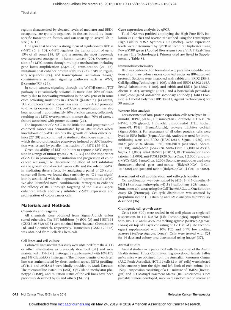
JQ1 inhibits proliferation of a subset of colon cancer cell linesTo determine whether the BET family represents a potential
therapeutic target in colon cancer, we determined the effect of theBETi JQ1 on the proliferation of a panel of 20 colon cancer celllines over 72hours. A continuumof response to JQ1was observedwith GI50 values ranging from 19.8 nmol/L (GP5D) through to1367.7 nmol/L (HuTu80; Fig. 1A). In the three most sensitive celllines (GP5D, HT29, and LIM1215), JQ1 (500 nmol/L) inhibitedcell growth by 79.4% � 7.6% (mean � SD) relative to controlcompared with 12.2% � 8.9% in the three most resistant lines(HuTu80, SW480, KM12; Fig. 1B). The acute myeloid leukemia(AML) cell lines MOLM13 and MV4;11 have previously beenreported to be highly sensitive to JQ1 (5, 10, 33) and thereforewere used to compared the sensitivity of colon lines to that of AMLlines. The GI50 values of the three most sensitive colorectal cancerlines GP5D, HT29, and LIM1215 (19.8, 22.1, and 92.7 nmol/L
respectively) were comparable to that of MV4;11 (41.3 nmol/L)and MOLM13 (158.5 nmol/L; Fig. 1B).
Association between JQ1 response and molecular subgroupsof colon cancer
To identify potential biomarkers of JQ1 response, cell lineswere separated according to established colon cancer subgroupsor mutation status of commonly altered oncogenes and tumorsuppressor genes and sensitivity compared. The mutation statusof the cell lines is listed in Supplementary Table S2. Cell lineswith microsatellite instability (MSI) were significantly moresensitive to JQ1 than microsatellite-stable (MSS) lines (P ¼0.038, Mann–Whitney unpaired t test). Conversely, no associ-ation between JQ1 sensitivity and CIMP status was observed(Supplementary Fig. S3). As KRAS and BRAF mutations, andsimilarly PIK3CA and PTEN mutations occur in a mutuallyexclusive manner in colon cancer, cell lines were classified asWT or mutant based on the collective mutation status of thesegenes. No association between JQ1 response and cell lines wild-type or mutant for RAS/BRAF, PIK3CA/PTEN, or TP53 wasobserved, nor was there a correlation between JQ1 responseand basal expression of BRD2, BRD3, and BRD4 expression(Supplementary Fig. S3). As MYCN-amplified neuroblastomaand medulloblastoma cell lines have been shown to be par-ticularly sensitive to JQ1 (9, 38), we examined JQ1 response in17 of the 20 colon cancer cell lines for which we had c-MYCamplification status information (34). c-MYC copy numbervaried from 2 (n ¼ 13) to >6 (n ¼ 4, Colo320, HT29, SKCO1,SW480) across the cell line panel. In contrast to neuroblasto-ma, no significant difference in JQ1 sensitivity was observedbetween c-MYC–amplified and nonamplified lines, with if atall, c-MYC amplified lines tending to be more resistant to JQ1(Supplementary Fig. S3). Similarly, no correlation betweenbasal c-MYC mRNA and protein expression and JQ1 responsewas observed (Supplementary Fig. S4).
JQ1 is largely cytostatic in colon cancer cellsTo determine the effect of JQ1 on cell cycle, we treated the three
most sensitive and resistant cell lines with JQ1 for 24 hours andanalyzed cell-cycle changes by PI staining and FACS analysis. Insensitive cells, JQ1 induced a 49% � 9% decrease in the
Figure 1.Effect of JQ1 on colorectal cancer cell proliferation. A, panel of 20 colorectal cancer cell lines ranked, ordered by increasing resistance to JQ1. Response to JQ1was determined in MTS assays by computation of GI50. B, dose–response analysis of the effect of JQ1 on cell growth inhibition in the 3 most sensitive (GP5D,HT29, LIM1215) and resistant (KM12, SW480, HuTu80) colorectal cancer lines and 2 BETi-sensitive AML cell lines (MOLM13, MV4;11). Growth inhibition wasdetermined by MTS after 72 hours.
Targeting the BET Family in Colorectal Cancer
www.aacrjournals.org Mol Cancer Ther; 15(6) June 2016 OF3
on May 24, 2019. © 2016 American Association for Cancer Research. mct.aacrjournals.org Downloaded from
Published OnlineFirst March 16, 2016; DOI: 10.1158/1535-7163.MCT-15-0724

proportionof cells in S-phase, significantly greater than the26%�11%decrease in resistant cells (mean� SD, n¼ 3, P < 0.05, Fig. 2Aand C). In parallel, JQ1 increased the percentage of cells in G1 by40% � 11%, significantly greater than the magnitude of induc-tion in resistant cells (15%� 8%, n¼ 3, P < 0.05, Fig. 2B and C).These results indicate that JQ1 sensitivity in colon cancer cells isassociated with a block in the transition from G1 to S-phase.Previous reports in other tumor types have suggested that JQ1induces apoptosis (7, 10). To address this in colon cancer cells,we investigated the effect of JQ1 on apoptosis induction in thethree most sensitive lines. In LIM1215 cells, JQ1 modestlyincreased the percentage of apoptotic cells from 0.5% � 0.5%to 4.4% � 1.0% (P < 0.005) but had no effect on apoptosisin GP5D (2.4%� 1.4% vs. 2.6%� 1.0%) or HT29 (0.6%� 0.6%vs. 0.6% � 0.7%) cells. In comparison, the topoisomerase Iinhibitor and known cytotoxic agent irinotecan increased thepercentage of apoptotic cells in GP5D cells to 13.7% � 3.8%, inHT29 cells to 37.4% � 6.0%, and in LIM1215 to 8.3% � 3.6%(Fig. 2D). These findings indicate that the effect of JQ1 on coloncancers cell lines is primarily cytostatic.
To extend these findings, we also examined the effect of JQ1 onanchorage-independent growth.We tested the three JQ1-sensitive
cell lines, GP5D, HT29, and LIM1215, in this assay. JQ1 signif-icantly reduced colony size in all three sensitive cell lines but hadminimal effect on colony number (data not shown), consistentwith its largely cytostatic effect in colon cancer cell lines (Fig. 2E).
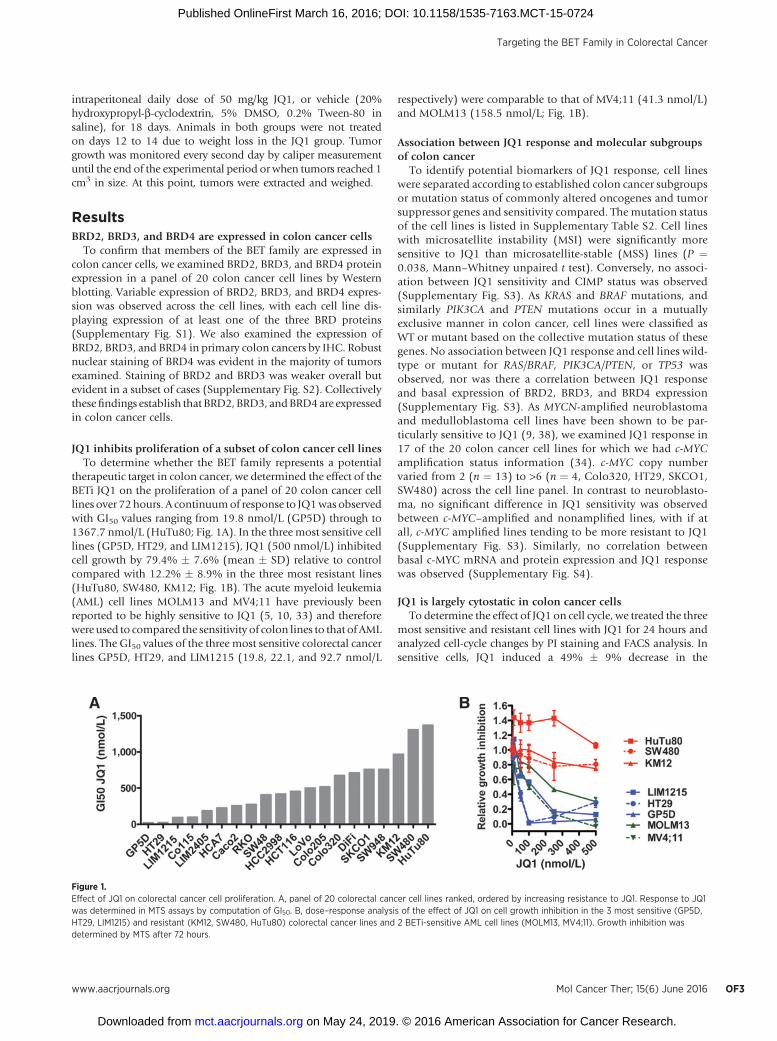
JQ1 inhibits the growth of colorectal cancer xenografts in vivoWe next determined the effect of JQ1 on the growth of
colorectal cancer xenografts in vivo. To test this, we utilized theHCT116 cell line which exhibits intermediate sensitivity to JQ1in vitro, and which rapidly and reproducibly develops tumorswhen grown as xenografts in vivo. HCT116 cells were engraftedinto the right and left flank of nude mice and treatment com-menced after 4 days when palpable tumors had formed. Animalswere treated daily, except for days 12 to 14, for 18 days. JQ1treatment induced a modest but statistically significant inhibi-tion of tumor growth when assessed by caliper measurements atdays 10, 12, and 18 (Fig. 3B) and when assessed by tumor weightat the completion of the experiment on day 18 (Fig. 3A and C).Notably, a decrease in body weight began to develop in the JQ1treatment group after 12 days of treatment. We therefore intro-duced a treatment holiday between days 12 and 14 to enable themice to recover from body weight loss (Fig. 3D).
Figure 2.Effect of JQ1 on cell-cycle distribution, apoptosis, and clonogenic growth. The 3 most sensitive (GP5D, HT29, LIM1215) and resistant (KM12, SW480,HuTu80) colorectal cancer lines were treated with JQ1 (500 nmol/L) or vehicle (0.1% DMSO) for 24 hours and changes in S-phases (A) and G0–G1 (B)determined by FACS analysis. Values shown are mean � SEM of 3 independent experiments. � , P < 0.05, Student t test. C, representative FACS plotsof JQ1-sensitive LIM1215 and JQ1-resistant KM12 cells treated for 24 hours with DMSO or JQ1. D, effect of JQ1 on apoptosis. The 3 most sensitive celllines were treated with DMSO (0.1%) or JQ1 (500 nmol/L) for 72 hours and apoptosis determined by FACS analysis. Cells were also treated with theknown apoptosis-inducing agent irinotecan (25 mmol/L). Values shown are the mean � SEM of n ¼ 3 independent experiments performed in triplicate.� , P < 0.05; ���� , P < 0.0001, Student t test. E, JQ1 reduces anchorage-independent growth. Cells were grown in soft agar in the presence or absence ofJQ1 (500 nmol/L) for 14 days and stained with crystal violet. Values shown are mean � SD of a representative experiment performed in triplicate (�� , P < 0.01;��� , P < 0.001; ���� , P < 0.0001, Student t test).
T€ogel et al.
Mol Cancer Ther; 15(6) June 2016 Molecular Cancer TherapeuticsOF4
on May 24, 2019. © 2016 American Association for Cancer Research. mct.aacrjournals.org Downloaded from
Published OnlineFirst March 16, 2016; DOI: 10.1158/1535-7163.MCT-15-0724
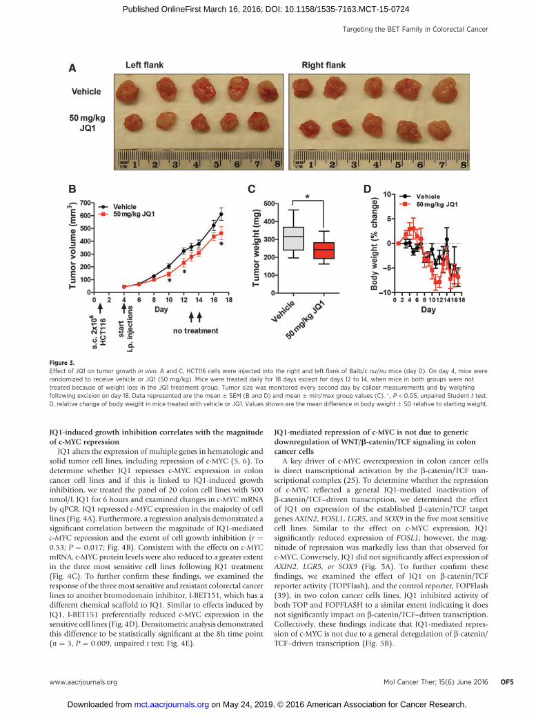
JQ1-induced growth inhibition correlates with the magnitudeof c-MYC repression
JQ1 alters the expression of multiple genes in hematologic andsolid tumor cell lines, including repression of c-MYC (5, 6). Todetermine whether JQ1 represses c-MYC expression in coloncancer cell lines and if this is linked to JQ1-induced growthinhibition, we treated the panel of 20 colon cell lines with 500nmol/L JQ1 for 6 hours and examined changes in c-MYC mRNAby qPCR. JQ1 repressed c-MYC expression in the majority of celllines (Fig. 4A). Furthermore, a regression analysis demonstrated asignificant correlation between the magnitude of JQ1-mediatedc-MYC repression and the extent of cell growth inhibition (r ¼0.53; P ¼ 0.017; Fig. 4B). Consistent with the effects on c-MYCmRNA, c-MYC protein levels were also reduced to a greater extentin the three most sensitive cell lines following JQ1 treatment(Fig. 4C). To further confirm these findings, we examined theresponse of the threemost sensitive and resistant colorectal cancerlines to another bromodomain inhibitor, I-BET151, which has adifferent chemical scaffold to JQ1. Similar to effects induced byJQ1, I-BET151 preferentially reduced c-MYC expression in thesensitive cell lines (Fig. 4D).Densitometric analysis demonstratedthis difference to be statistically significant at the 8h time point(n ¼ 3, P ¼ 0.009, unpaired t test; Fig. 4E).
JQ1-mediated repression of c-MYC is not due to genericdownregulation of WNT/b-catenin/TCF signaling in coloncancer cells
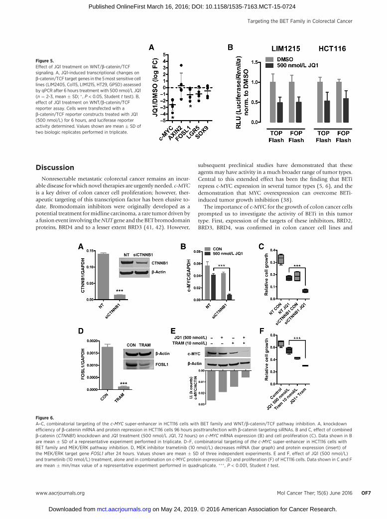
A key driver of c-MYC overexpression in colon cancer cellsis direct transcriptional activation by the b-catenin/TCF tran-scriptional complex (25). To determine whether the repressionof c-MYC reflected a general JQ1-mediated inactivation ofb-catenin/TCF–driven transcription, we determined the effectof JQ1 on expression of the established b-catenin/TCF targetgenes AXIN2, FOSL1, LGR5, and SOX9 in the five most sensitivecell lines. Similar to the effect on c-MYC expression, JQ1significantly reduced expression of FOSL1; however, the mag-nitude of repression was markedly less than that observed forc-MYC. Conversely, JQ1 did not significantly affect expression ofAXIN2, LGR5, or SOX9 (Fig. 5A). To further confirm thesefindings, we examined the effect of JQ1 on b-catenin/TCFreporter activity (TOPFlash), and the control reporter, FOPFlash(39), in two colon cancer cells lines. JQ1 inhibited activity ofboth TOP and FOPFLASH to a similar extent indicating it doesnot significantly impact on b-catenin/TCF–driven transcription.Collectively, these findings indicate that JQ1-mediated repres-sion of c-MYC is not due to a general deregulation of b-catenin/TCF–driven transcription (Fig. 5B).
Figure 3.Effect of JQ1 on tumor growth in vivo. A and C, HCT116 cells were injected into the right and left flank of Balb/c nu/nu mice (day 0). On day 4, mice wererandomized to receive vehicle or JQ1 (50 mg/kg). Mice were treated daily for 18 days except for days 12 to 14, when mice in both groups were nottreated because of weight loss in the JQ1 treatment group. Tumor size was monitored every second day by caliper measurements and by weighingfollowing excision on day 18. Data represented are the mean � SEM (B and D) and mean � min/max group values (C). � , P < 0.05, unpaired Student t test.D, relative change of body weight in mice treated with vehicle or JQ1. Values shown are the mean difference in body weight � SD relative to starting weight.
Targeting the BET Family in Colorectal Cancer
www.aacrjournals.org Mol Cancer Ther; 15(6) June 2016 OF5
on May 24, 2019. © 2016 American Association for Cancer Research. mct.aacrjournals.org Downloaded from
Published OnlineFirst March 16, 2016; DOI: 10.1158/1535-7163.MCT-15-0724
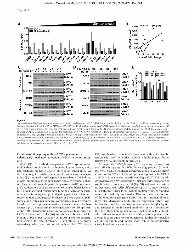
Combinatorial targeting of the c-MYC super-enhancerenhances JQ1-mediated repression of c-MYC in colon cancercells
While JQ1 effectively downregulated c-MYC expression andinhibited cell proliferation in a subset of colon cancer cells, it alsohad relatively modest effects in other colon cancer lines. Wetherefore sought to establish strategies for enhancing the magni-tude of JQ1-induced c-MYC repression, to enhance JQ1-inducedgrowth inhibition of these lines. Genes regulated by super-enhan-cers have been shown to be particularly sensitive to JQ1 treatment(16). Furthermore, as super-enhancers contain both high levels ofBRD4 occupancy and concentrated binding of effector transcrip-tion factors from key oncogenic signaling pathways, it has beensuggested that combinatorial blockade of these signaling path-ways, along with super-enhancer components, may be requiredfor efficient transcriptional repression of genes regulated by theseelements (40). A super-enhancer located about 500 kb upstreamof the c-MYC transcription start site was previously identified inHCT116 colon cancer cells (40) and shown to be enriched forbinding of TCF4 (TCF7L2) and FRA1 (FOSL1), effector transcrip-tion factors of the WNT/b-catenin/TCF and MEK/ERK pathways,respectively, which are constitutively activated in HCT116 cells
(34). We therefore reasoned that treatment with JQ1 in combi-nation with WNT or MAPK pathway inhibition may furtherrepress c-MYC expression in these cells.
To target the WNT/b-catenin/TCF signaling pathway, weused siRNAs against the TCF4 interacting partner, b-catenin(CTNNB1), which resulted in downregulation of b-cateninmRNAexpression by 90% � 1.0% and protein expression by 78% �2.8% (n¼ 2 independent experiments; Fig. 6A). CTNNB1 knock-downdecreased c-MYCexpressionwhichwas further enhancedbycombination treatment with JQ1 (Fig. 6B) and associated with afurther reduction in cell proliferation (Fig. 6C). To target theMEK/ERK pathway, we used theMEK inhibitor trametinib. As expected,trametinib markedly decreased mRNA and protein expressionof the MEK/ERK target gene, FOSL1 (Fig. 6D). Trametinib treat-ment also decreased c-MYC protein expression, which wasfurther enhanced by combination treatment with JQ1 (Fig. 6E)and reflected in an increased inhibition of cell proliferation(Fig. 6F). These findings demonstrate that combinatorial block-ade of effector transcription factors of the c-MYC super-enhanceralongside super-enhancer components can further downregulatec-MYC expression and induce more robust antiproliferativeeffects in colorectal cancer cells.
Figure 4.JQ1-mediated c-MYC repression correlates with growth inhibition. A, c-MYC mRNA expression is inhibited by JQ1. Each of the 20 colon colorectal cancerlines were treated with vehicle (0.1% DMSO) or JQ1 (500 nmol/L) for 6 hours and c-MYCmRNA expression determined by qPCR. Values shown are mean � SDof n ¼ 2 to 3 experiments. Cell lines are rank ordered from most to least sensitive to JQ1-induced growth inhibition as per Fig. 1A. B, linear regressionanalysis of JQ1 GI50 values (y-axis) versus the magnitude of c-MYC mRNA repression following JQ1 treatment JQ1 (x-axis; r ¼ 0.567, P ¼ 0.017, Spearmancorrelation). C and D, BETi preferentially inhibit c-MYC protein expression in sensitive cell lines. JQ1-sensitive GP5D, HT29, and LIM1215 cells and JQ1-resistantKM12, SW480, and HuTu80 cells were treated with JQ1 (500 nmol/L; C) or I-BET151 (500 nmol/L; D) for 2 to 24 hours and c-MYC protein expressiondetermined by Western blotting. E, pooled densitometric analysis of c-MYC protein expression pre- and post-iBET treatment in the 3 sensitive and resistantcell lines. Values shown are mean � SEM, n ¼ 3; � , P < 0.05.
T€ogel et al.
Mol Cancer Ther; 15(6) June 2016 Molecular Cancer TherapeuticsOF6
on May 24, 2019. © 2016 American Association for Cancer Research. mct.aacrjournals.org Downloaded from
Published OnlineFirst March 16, 2016; DOI: 10.1158/1535-7163.MCT-15-0724
Discussion
Nonresectable metastatic colorectal cancer remains an incur-able disease for which novel therapies are urgently needed. c-MYCis a key driver of colon cancer cell proliferation; however, ther-apeutic targeting of this transcription factor has been elusive to-date. Bromodomain inhibitors were originally developed as apotential treatment formidline carcinoma, a rare tumor driven bya fusion event involving theNUT gene and the BETbromodomainproteins, BRD4 and to a lesser extent BRD3 (41, 42). However,
subsequent preclinical studies have demonstrated that theseagents may have activity in a much broader range of tumor types.Central to this extended effect has been the finding that BETirepress c-MYC expression in several tumor types (5, 6), and thedemonstration that MYC overexpression can overcome BETi-induced tumor growth inhibition (38).
The importance of c-MYC for the growth of colon cancer cellsprompted us to investigate the activity of BETi in this tumortype. First, expression of the targets of these inhibitors, BRD2,BRD3, BRD4, was confirmed in colon cancer cell lines and
Figure 6.A–C, combinatorial targeting of the c-MYC super-enhancer in HCT116 cells with BET family and WNT/b-catenin/TCF pathway inhibition. A, knockdownefficiency of b-catenin mRNA and protein repression in HCT116 cells 96 hours posttransfection with b-catenin targeting siRNAs. B and C, effect of combinedb-catenin (CTNNB1) knockdown and JQ1 treatment (500 nmol/L JQ1, 72 hours) on c-MYC mRNA expression (B) and cell proliferation (C). Data shown in Bare mean � SD of a representative experiment performed in triplicate. D–F, combinatorial targeting of the c-MYC super-enhancer in HCT116 cells withBET family and MEK/ERK pathway inhibition. D, MEK inhibitor trametinib (10 nmol/L) decreases mRNA (bar graph) and protein expression (insert) ofthe MEK/ERK target gene FOSL1 after 24 hours. Values shown are mean � SD of three independent experiments. E and F, effect of JQ1 (500 nmol/L)and trametinib (10 nmol/L) treatment, alone and in combination on c-MYC protein expression (E) and proliferation (F) of HCT116 cells. Data shown in C and Fare mean � min/max value of a representative experiment performed in quadruplicate. ��� , P < 0.001, Student t test.
Figure 5.Effect of JQ1 treatment on WNT/b-catenin/TCFsignaling. A, JQ1-induced transcriptional changes onb-catenin/TCF target genes in the 5most sensitive celllines (LIM2405, Co115, LIM1215, HT29, GP5D) assessedby qPCR after 6 hours treatment with 500 nmol/L JQ1(n ¼ 2-3, mean � SD; � , P < 0.05, Student t test). B,effect of JQ1 treatment on WNT/b-catenin/TCFreporter assay. Cells were transfected with ab-catenin/TCF reporter constructs treated with JQ1(500 nmol/L) for 6 hours, and luciferase reporteractivity determined. Values shown are mean � SD oftwo biologic replicates performed in triplicate.
Targeting the BET Family in Colorectal Cancer
www.aacrjournals.org Mol Cancer Ther; 15(6) June 2016 OF7
on May 24, 2019. © 2016 American Association for Cancer Research. mct.aacrjournals.org Downloaded from
Published OnlineFirst March 16, 2016; DOI: 10.1158/1535-7163.MCT-15-0724
primary tumors. Subsequently, screening of a panel of 20colorectal cancer cell lines with the BETi JQ1, identified a subsetof cell lines particularly sensitive to this agent with GI50 valuesin the low nanomolar range. A previous study reported JQ1IC50 values in more than 650 cancer cell lines, including 35 celllines derived from the intestine, of which 11 were also analyzedin our screen (9). A comparison of both datasets revealed astrong overall concordance in the relative sensitivity of thesecell lines. Furthermore, cell lines derived from hematologiccancers, including the AML cell lines MOLM13 and MV4;11,have been shown to be particularly sensitive to bromodomaininhibitors both in vitro and in vivo (5–7, 10, 33). Notably, wefound that the in vitro sensitivity of these two AML cell lines wascomparable to that of the most sensitive colorectal cancer lines.
Separation of the cell lines according to the major molecularsubtypes of colon cancer revealed increased sensitivity of MSIcell lines to JQ1. The mechanistic basis for this difference isunknown; however, MSI tumors tend to be diploid and harborfewer chromosomal alterations compared with MSS tumors.Whether these global genomic differences impact on the higherorder organization of the c-MYC promoter and enhancer, andits subsequent regulation in MSS versus MSI tumors, is a pointworthy of investigation. Conversely, we did not observe anysignificant associations between JQ1 response and CIMP statusof the cell lines, or of mutation status of TP53, KRAS/BRAF orPIK3CA/PTEN, or of BRD2, BRD3, or BRD4 protein expression.
Recently, MYCN-amplified neuroblastoma and medulloblas-toma cell lines were identified as being exquisitely sensitive tobromodomain inhibitors from a screen of multiple cancer celllines (9, 38). In contrast, we observed no significant correlationbetween c-MYC amplification status and JQ1 response in colorec-tal cancer cells. Similarly, basal c-MYC mRNA or protein expres-sion levels were not predictive of JQ1 response. The lack ofcorrelation between basal levels of c-MYC expression and JQ1response is possibly a reflection of the multiple mechanisms ofc-MYC regulation in colon cancer cells, including gene amplifi-cation (26), transcriptional (25), and posttranslational mechan-isms (43), and the ability of JQ1 to impact primarily on only oneof these processes—c-MYC transcription (5, 6).
Consistent with this mechanism, a key finding of this study isthe observation that JQ1 treatment robustly represses c-MYCmRNA and protein expression in colon cancer cells and that themagnitude of c-MYC mRNA and protein downregulation corre-lates significantly with themagnitude of JQ1-induced cell growth.These findings are also consistent with previous reports in mul-tiple myeloma (6, 44), acute lymphoblastic lymphoma (7), andmedulloblastoma (38) cell lines where a correlation betweenMYC repression and growth inhibition was observed.
Colon cancer cell lines with intermediate sensitivity to JQ1, orwhich were JQ1-refractory, showed less robust downregulationof c-MYC following drug treatment. We therefore exploredapproaches for enhancing the efficacy of JQ1 on c-MYC repres-sion and subsequent cell proliferation. Specifically, as genesregulated by super-enhancers have been shown to be particularlysensitive to BET inhibition (16), and as the c-MYC super-enhancerin HCT116 cells is enriched for binding of terminal transcriptionfactors from the WNT/b-catenin/TCF and MAPK/ERK signalingpathways, we explored combinatorial targeting of these path-ways with JQ1. This strategy significantly increased the magni-tude of c-MYC downregulation with a parallel enhancement ofHCT116 cell growth inhibition.
The findings in HCT116 cells suggest that the c-MYC super-enhancer is a key point of integration of the WNT and MAPKsignaling pathways. Given that approximately 50% of colorectalcancers harbor concurrent mutations which simultaneouslyhyperactivate these pathways, this combination strategymayhavebroad applicability in this tumor type. We also note that inaddition to the role of MEK/ERK signaling in driving c-MYCtranscription, ERK also stabilizes c-MYC protein by phosphory-lating serine 62 (45). The effects of JQ1 and trametinib combi-nation treatment on downregulating c-MYC protein levels maytherefore be mediated by both transcriptional and posttransla-tional mechanisms.
While our findings establish a strong link between the magni-tude of c-MYC repression and JQ1-induced growth inhibition, itmay not be the only mechanism through which JQ1 induces itseffects. Notably, Hu and colleagues, recently reported sensitivityof colon cancer cell lines to the BET inhibitorMS417,which sharesthe same thieno-1,2,4-tria-zolo-1,4-diazepine scaffold as JQ1(46). This study demonstrated that in addition to inhibitingtumor cell growth,MS417 inhibited themigration andmetastasisof colon cancer cells both in vitro and in vivo. While the role ofc-MYC was not investigated, the authors demonstrated increasedexpression of E-cadherin (CDH1) following BET inhibition,which may explain the antimigratory and metastatic effectsobserved. In addition, increased E-cadherin has recently beenlinked to perturbation of the WNT/b-catenin pathway whichcould also result in reduced tumor cell growth (47). Whether theeffects of these inhibitors on CDH1 induction are directly medi-ated or secondary to inhibition of c-MYC would be worthy ofinvestigation.
In summary, these findings demonstrate that pharmacologicinhibition of the BET family abrogates the growth of colon cancercell lines in vitro and in vivo, identifying this family of proteins aspotential treatment targets in colorectal cancer. Mechanistically,we demonstrate that response to these agents correlates signifi-cantly with the magnitude of inhibition of c-MYC, an establisheddriver of colorectal cancer. In addition, we identify two rationalcombination strategies to enhance BETi-mediated repressionof c-MYC through combinatorial targeting of the c-MYC super-enhancer in colorectal cancer cells.
Disclosure of Potential Conflicts of InterestNo potential conflicts of interest were disclosed.
Authors' ContributionsConception and design: L. T€ogel; A.C. Chueh, A.K. Shiau, J.M. MariadasonDevelopment of methodology: L. T€ogel; A.C. Chueh, B. Diez-Dacal,T.C. Gahman, J.M. MariadasonAcquisition of data (provided animals, acquired and managed patients,provided facilities, etc.): L. T€ogel; R. Nightingale, A.C. Chueh, A. Jayachandran,T. Phesse, R. Wu, O.M. Sieber, J.M. MariadasonAnalysis and interpretation of data (e.g., statistical analysis, biostatistics,computational analysis): L. T€ogel; R. Nightingale, A.C. Chueh, A. Jayachan-dran, H. Tran, T. Phesse, D. Arango, A.S. Dhillon, T.C. Gahman, P. Filippako-poulos, A.K. Shiau, J.M. MariadasonWriting, review, and/or revision of the manuscript: L. T€ogel; A.C. Chueh,A. Jayachandran, H. Tran, T. Phesse, O.M. Sieber, D. Arango, A.S. Dhillon,M.A. Dawson, T.C. Gahman, P. Filippakopoulos, A.K. Shiau, J.M. MariadasonAdministrative, technical, or material support (i.e., reporting or orga-nizing data, constructing databases): L. T€ogel; R. Nightingale, A.K. Shiau,J.M. MariadasonStudy supervision: P. Filippakopoulos, J.M. Mariadason
T€ogel et al.
Mol Cancer Ther; 15(6) June 2016 Molecular Cancer TherapeuticsOF8
on May 24, 2019. © 2016 American Association for Cancer Research. mct.aacrjournals.org Downloaded from
Published OnlineFirst March 16, 2016; DOI: 10.1158/1535-7163.MCT-15-0724
Grant SupportThis work was supported, in part, by grants from the CASS Foundation
(L. T€ogel), Ludwig Cancer Research, NHMRC SRF 1046092 (J.M. Mariadason),NHMRC project grant 1026555 and the Operational Infrastructure SupportProgram, VictorianGovernment, Australia. P. Filippakopoulos is supported by aWellcome Trust Career-Development Fellowship (095751/Z/11/Z).
The costs of publication of this article were defrayed in part by the paymentof page charges. This article must therefore be hereby marked advertisementin accordance with 18 U.S.C. Section 1734 solely to indicate this fact.
ReceivedAugust 31, 2015; revised February 19, 2016; acceptedMarch 6, 2016;published OnlineFirst March 16, 2016.
References1. Shi J, Vakoc CR. The mechanisms behind the therapeutic activity of BET
bromodomain inhibition. Mol Cell 2014;54:728–36.2. Filippakopoulos P, Picaud S, Mangos M, Keates T, Lambert JP, Barsyte-
Lovejoy D, et al. Histone recognition and large-scale structural analysis ofthe human bromodomain family. Cell 2012;149:214–31.
3. Filippakopoulos P, Qi J, Picaud S, Shen Y, Smith WB, Fedorov O,et al. Selective inhibition of BET bromodomains. Nature 2010;468:1067–73.
4. Nicodeme E, Jeffrey KL, Schaefer U, Beinke S, Dewell S, Chung CW, et al.Suppression of inflammation by a synthetic histone mimic. Nature2010;468:1119–23.
5. DawsonMA, Prinjha RK, Dittmann A, Giotopoulos G, Bantscheff M, ChanWI, et al. Inhibition of BET recruitment to chromatin as an effectivetreatment for MLL-fusion leukaemia. Nature 2011;478:529–33.
6. Delmore JE, Issa GC, Lemieux ME, Rahl PB, Shi J, Jacobs HM, et al. BETbromodomain inhibition as a therapeutic strategy to target c-Myc. Cell2011;146:904–17.
7. Ott CJ, Kopp N, Bird L, Paranal RM, Qi J, Bowman T, et al. BET bromo-domain inhibition targets both c-Myc and IL7R in high-risk acute lym-phoblastic leukemia. Blood 2012;120:2843–52.
8. Banerjee C, Archin N, Michaels D, Belkina AC, Denis GV, Bradner J, et al.BET bromodomain inhibition as a novel strategy for reactivation of HIV-1.J Leukoc Biol 2012;92:1147–54.
9. Puissant A, Frumm SM, Alexe G, Bassil CF, Qi J, Chanthery YH, et al.Targeting MYCN in neuroblastoma by BET bromodomain inhibition.Cancer Discov 2013;3:308–23.
10. Mertz JA, Conery AR, Bryant BM, Sandy P, Balasubramanian S, Mele DA,et al. Targeting MYC dependence in cancer by inhibiting BET bromodo-mains. Proc Natl Acad Sci U S A 2011;108:16669–74.
11. Jiang YW, Veschambre P, Erdjument-Bromage H, Tempst P, Conaway JW,Conaway RC, et al.Mammalianmediator of transcriptional regulation andits possible role as an end-point of signal transduction pathways. Proc NatlAcad Sci U S A 1998;95:8538–43.
12. Jang MK, Mochizuki K, Zhou M, Jeong HS, Brady JN, Ozato K. Thebromodomain protein Brd4 is a positive regulatory component of P-TEFband stimulates RNA polymerase II-dependent transcription. Mol Cell2005;19:523–34.
13. Yang Z, Yik JH, Chen R, He N, Jang MK, Ozato K, et al. Recruitment ofP-TEFb for stimulation of transcriptional elongation by the bromodomainprotein Brd4. Mol Cell 2005;19:535–45.
14. Peterlin BM, Price DH. Controlling the elongation phase of transcriptionwith P-TEFb. Mol Cell 2006;23:297–305.
15. Adelman K, Lis JT. Promoter-proximal pausing of RNA polymerase II:emerging roles in metazoans. Nat Rev Genet 2012;13:720–31.
16. Loven J, Hoke HA, Lin CY, Lau A, Orlando DA, Vakoc CR, et al. Selectiveinhibition of tumor oncogenes by disruption of super-enhancers. Cell2013;153:320–34.
17. Whyte WA, Orlando DA, Hnisz D, Abraham BJ, Lin CY, Kagey MH, et al.Master transcription factors andmediator establish super-enhancers at keycell identity genes. Cell 2013;153:307–19.
18. Fernandez PC, Frank SR, Wang L, Schroeder M, Liu S, Greene J, et al.Genomic targets of the human c-Myc protein. Genes Dev 2003;17:1115–29.
19. Li Z, Van Calcar S, Qu C, Cavenee WK, Zhang MQ, Ren B. A globaltranscriptional regulatory role for c-Myc in Burkitt's lymphoma cells. ProcNatl Acad Sci U S A 2003;100:8164–9.
20. Beroukhim R, Mermel CH, Porter D, Wei G, Raychaudhuri S, Donovan J,et al. The landscape of somatic copy-number alteration across humancancers. Nature 2010;463:899–905.
21. Shou Y, Martelli ML, Gabrea A, Qi Y, Brents LA, Roschke A, et al. Diversekaryotypic abnormalities of the c-myc locus associated with c-myc dysre-
gulation and tumor progression in multiple myeloma. Proc Natl Acad SciU S A 2000;97:228–33.
22. TaubR,Kirsch I,MortonC, LenoirG, SwanD, Tronick S, et al. Translocationof the c-myc gene into the immunoglobulin heavy chain locus in humanBurkitt lymphoma and murine plasmacytoma cells. Proc Natl Acad SciU S A 1982;79:7837–41.
23. Yeh E, Cunningham M, Arnold H, Chasse D, Monteith T, Ivaldi G, et al. Asignalling pathway controlling c-Myc degradation that impacts oncogenictransformation of human cells. Nature Cell Biol 2004;6:308–18.
24. Wright JB, Brown SJ, Cole MD. Upregulation of c-MYC in cis through alarge chromatin loop linked to a cancer risk-associated single-nucleo-tide polymorphism in colorectal cancer cells. Mol Cell Biol 2010;30:1411–20.
25. He TC, Sparks AB, Rago C, Hermeking H, Zawel L, da Costa LT, et al.Identification of c-MYC as a target of the APC pathway. Science 1998;281:1509–12.
26. Kozma L, Kiss I, Szakall S, Ember I. Investigation of c-myc oncogeneamplification in colorectal cancer. Cancer Lett 1994;81:165–9.
27. Hongxing Z, Nancai Y, Wen S, Guofu H, Yanxia W, Hanju H, et al.Depletion of c-Myc inhibits human colon cancer colo 320 cells' growth.Cancer Biother Radiopharm 2008;23:229–37.
28. Zhang X, Ge YL, Tian RH. The knockdown of c-myc expression by RNAiinhibits cell proliferation in human colon cancerHT-29 cells in vitro and invivo. Cell Mol Biol Lett 2009;14:305–18.
29. SansomOJ,Meniel VS,Muncan V, Phesse TJ,Wilkins JA, ReedKR, et al.Mycdeletion rescues Apc deficiency in the small intestine. Nature 2007;446:676–9.
30. Yekkala K, Baudino TA. Inhibition of intestinal polyposis with reducedangiogenesis in ApcMin/þmice due to decreases in c-Myc expression. MolCancer Res 2007;5:1296–303.
31. Ignatenko NA, Holubec H, Besselsen DG, Blohm-Mangone KA, Padilla-Torres JL, Nagle RB, et al. Role of c-Myc in intestinal tumorigenesis of theApcMin/þ mouse. Cancer Biol Ther 2006;5:1658–64.
32. ChoH,Herzka T, ZhengW,Qi J,Wilkinson JE, Bradner JE, et al. RapidCaP, anovel GEM model for metastatic prostate cancer analysis and therapy,reveals myc as a driver of Pten-mutant metastasis. Cancer Discov 2014;4:318–33.
33. Zuber J, Shi J, Wang E, Rappaport AR, Herrmann H, Sison EA, et al. RNAiscreen identifies Brd4 as a therapeutic target in acute myeloid leukaemia.Nature 2011;478:524–8.
34. Mouradov D, Sloggett C, Jorissen RN, Love CG, Li S, Burgess AW, et al.Colorectal cancer cell lines are representativemodels of themainmolecularsubtypes of primary cancer. Cancer Res 2014;74:3238–47.
35. Medico E, Russo M, Picco G, Cancelliere C, Valtorta E, Corti G, et al. Themolecular landscape of colorectal cancer cell lines unveils clinically action-able kinase targets. Nature Commun 2015;6:7002.
36. Mariadason JM, Rickard KL, Barkla DH, Augenlicht LH, Gibson PR.Divergent phenotypic patterns and commitment to apoptosis of Caco-2cells during spontaneous and butyrate-induced differentiation. J CellPhysiol 2000;183:347–54.
37. Schneider CA, Rasband WS, Eliceiri KW. NIH Image to ImageJ: 25 years ofimage analysis. Nature Methods 2012;9:671–5.
38. Bandopadhayay P, Bergthold G, Nguyen B, Schubert S, Gholamin S, TangY, et al. BET bromodomain inhibition of MYC-amplified medulloblasto-ma. Clin Cancer Res 2014;20:912–25.
39. Korinek V, Barker N,Morin PJ, vanWichenD, deWeger R, Kinzler KW, et al.Constitutive transcriptional activation by a b-catenin-Tcf complex inAPC�/� colon carcinoma. Science 1997;275:1784–87.
40. Hnisz D, Schuijers J, Lin CY, Weintraub AS, Abraham BJ, Lee TI, et al.Convergence of developmental and oncogenic signaling pathways attranscriptional super-enhancers. Mol Cell 2015;58:362–70.
Targeting the BET Family in Colorectal Cancer
www.aacrjournals.org Mol Cancer Ther; 15(6) June 2016 OF9
on May 24, 2019. © 2016 American Association for Cancer Research. mct.aacrjournals.org Downloaded from
Published OnlineFirst March 16, 2016; DOI: 10.1158/1535-7163.MCT-15-0724

41. French CA, Ramirez CL, Kolmakova J, Hickman TT, Cameron MJ, ThyneME, et al. BRD-NUT oncoproteins: a family of closely related nuclearproteins that block epithelial differentiation and maintain the growth ofcarcinoma cells. Oncogene 2008;27:2237–42.
42. Stelow EB.A review of NUT midline carcinoma. Head Neck Pathol 2011;5:31–5.
43. RajagopalanH, Jallepalli PV, RagoC,VelculescuVE, Kinzler KW,VogelsteinB, et al. Inactivation of hCDC4 can cause chromosomal instability. Nature2004;428:77–81.
44. Baratta MG, Schinzel AC, Zwang Y, Bandopadhayay P, Bowman-Colin C,Kutt J, et al. An in-tumor genetic screen reveals that the BET bromodomain
protein, BRD4, is a potential therapeutic target in ovarian carcinoma.Proc Natl Acad Sci U S A 2015;112:232–7.
45. Sears R, Nuckolls F, Haura E, Taya Y, Tamai K, Nevins JR. Multiple Ras-dependent phosphorylation pathways regulate Myc protein stability.Genes Dev 2000;14:2501–14.
46. Hu Y, Zhou J, Ye F, Xiong H, Peng L, Zheng Z, et al. BRD4 inhibitorinhibits colorectal cancer growth and metastasis. Int J Mol Sci 2015;16:1928–48.
47. Huels DJ, Ridgway RA, Radulescu S, Leushacke M, Campbell AD, Biswas S,et al. E-cadherin can limit the transforming properties of activating beta-catenin mutations. EMBO J 2015;34:2321–33.
Mol Cancer Ther; 15(6) June 2016 Molecular Cancer TherapeuticsOF10
T€ogel et al.
on May 24, 2019. © 2016 American Association for Cancer Research. mct.aacrjournals.org Downloaded from
Published OnlineFirst March 16, 2016; DOI: 10.1158/1535-7163.MCT-15-0724
Published OnlineFirst March 16, 2016.Mol Cancer Ther Lars Tögel, Rebecca Nightingale, Anderly C. Chueh, et al. Expression and Proliferation of Colorectal Cancer CellsProteins, and WNT or MAPK Signaling, Inhibits c-MYC Dual Targeting of Bromodomain and Extraterminal Domain
Updated version
10.1158/1535-7163.MCT-15-0724doi:
Access the most recent version of this article at:
Material
Supplementary
http://mct.aacrjournals.org/content/suppl/2016/03/15/1535-7163.MCT-15-0724.DC1
Access the most recent supplemental material at:
E-mail alerts related to this article or journal.Sign up to receive free email-alerts
Subscriptions
Reprints and
To order reprints of this article or to subscribe to the journal, contact the AACR Publications
Permissions
Rightslink site. (CCC)Click on "Request Permissions" which will take you to the Copyright Clearance Center's
.http://mct.aacrjournals.org/content/early/2016/05/20/1535-7163.MCT-15-0724To request permission to re-use all or part of this article, use this link
on May 24, 2019. © 2016 American Association for Cancer Research. mct.aacrjournals.org Downloaded from
Published OnlineFirst March 16, 2016; DOI: 10.1158/1535-7163.MCT-15-0724


![The Arabidopsis BET Bromodomain Factor GTE4 Is Involved ......The Arabidopsis BET Bromodomain Factor GTE4 Is Involved in Maintenance of the Mitotic Cell Cycle during Plant Development1[C][W][OA]](https://img.pdfslide.us/doc/110x75/61234df471105c695c6f7968/the-arabidopsis-bet-bromodomain-factor-gte4-is-involved-the-arabidopsis.jpg)