Embed Size (px)
Citation preview

1
Dual roles for CXCL4 chemokines and CXCR3 in angiogenesis and invasion of pancreatic cancer Cathy Quemener1,,2, Jessica Baud1,2, Kevin Boyé1,2, Alexandre Dubrac1,2,3, Clotilde Billottet 1,2, Fabienne Soulet1,2, Florence Darlot1,2, Laurent Dumartin1,2, Marie Sire1,2, Renaud
Grepin1,2, Thomas Daubon1,2, Fabienne Rayne2,7, Harald Wodrich2,7, Anne Couvelard4,
Raphael Pineau1,2, Martin Schilling6, Vincent Castronovo5, Shih-Che Sue8, Kim Clark9,
Abderrahim Lomri 1,2, Abdel-Majid Khatib1,2, Martin Hagedorn 1,2,, Hervé Prats3, Andreas
Bikfalvi1,2 1INSERM U1029, Pessac, France; 2Université Bordeaux, Pessac, France; 3INSERM U1037 Toulouse, 4Department of Pathology, Hôpital Bichat, Paris, France,5Department of Pathology, University of Liège, Belgium; 6Department of visceral surgery, University Homburg/Saar, Germany; 7CNRS UMR 5234, Pessac, France; 8Department of Life Science NTHU Taiwan; 9Department of Integrative Biology, University Liverpool . Correspondance to: Andreas Bikfalvi MD PhD INSERM U1029 University Bordeaux 33 560 Pessac Phone : (33) 5 40 00 87 03 Fax : (33) 5 40 00 87 05 E-mail : [email protected] Financial support : grants from “Agence Nationale de la Recherche “, the “Association de la Recherche sur le Cancer” and the “Ligue Nationale contre Cancer” to A. Bikfalvi, from the “Institut National du Cancer” (INCA) to A.Bikfalvi and H. Prats and from ANR to M. Hagedorn. PhD fellowship from INCA to A. Dubrac and from the “Ministère de la Recherche” and the “Fondation pour la Recherche Médicale” to L. Dumartin. Disclosures: No conflict of interest to declare. "The funders had no role in study design, data collection and analysis, decision to publish, or preparation of the manuscript." Keywords: Angiogenesis, angiogenesis inhibition, tumor invasion, chemokines Abbreviations: 5’Aza, 5 azacytidine, α-SMA,α-smooth muscle actin; CAM, chick chorioallantoic membrane; FGF, fibroblast growth factor; HUVEC, human umbilical vein endothelial cells, IF, immunofluorescence, IHC, immunohistochemistry; IL1, interleukine-1; Luc, Luciferase; MF, myofibroblasts; MabL1, anti-CXCL4L1 monoclonal antibody; PECAM1, Platelet endothelial cell adhesion molecule 1; PDAC, pancreatic ducal carcinoma; TSA, Trichostatine A; VAMP2, vesicle-associated membrane protein 2.
on March 25, 2020. © 2016 American Association for Cancer Research.cancerres.aacrjournals.org Downloaded from
Author manuscripts have been peer reviewed and accepted for publication but have not yet been edited. Author Manuscript Published OnlineFirst on September 9, 2016; DOI: 10.1158/0008-5472.CAN-15-2864

2
ABSTRACT
The CXCL4 paralog CXCL4L1 is a little studied chemokine that has been suggested to exert
an anti-angiogenic function. However, CXCL4L1 is also expressed in patient tumors, tumor
cell lines and murine xenografts, prompting a more detailed analysis of its role in cancer
pathogenesis. We used genetic and antibody-based approaches to attenuate CXCL4L1 in
models of pancreatic adenocarcinoma (PDAC). Mechanisms of expression were assessed in
cell co-culture experiments, murine and avian xenotransplants, including through an
evaluation of CpG methylation and mutation of critical CpG residues. CXCL4L1 gene
expression was increased greatly in primary and metastatic PDAC. We found that
myofibroblasts triggered cues in the tumor microenvironment which led to induction of
CXCL4L1 in tumor cells. CXCL4L1 expression was also controlled by epigenetic
modifications at critical CpG islands which were mapped. CXCL4L1 inhibited vasculogenesis
but also affected tumor development more directly depending on the tumor cell type. In vivo
administration of a monoclonal antibody against CXCL4L1, we demonstrated a blockade in
the growth of tumors positive for CXCR3, a critical receptor for CXCL4 ligands. Our findings
define a pro-tumorigenic role in PDAC development for endogenous CXCL4L1, which is
independent of its anti-angiogenic function.
on March 25, 2020. © 2016 American Association for Cancer Research.cancerres.aacrjournals.org Downloaded from
Author manuscripts have been peer reviewed and accepted for publication but have not yet been edited. Author Manuscript Published OnlineFirst on September 9, 2016; DOI: 10.1158/0008-5472.CAN-15-2864

3
INTRODUCTION
A paralog of CXCL4, named platelet factor variant-1 (PF4v1) or CXCL4L1, was
identified in 1989, but its role in cancer progression has not yet been investigated (1, 2).
CXCL4L1 and CXCL4 exhibit only 34 % differences in the amino-terminus encoding the
signal sequence and 4.3 % difference in the mature protein. CXCL4L1 has been shown to be
more potent than CXCL4 at inhibiting cell proliferation and migration (2) (3). Furthermore,
inhibition of tumor development was observed in tumors derived from implantation of A549,
LLC and B16 cells in mice, treated with recombinant CXCL4L1 (1). In addition, the secretion
and processing mechanisms of CXCL4L1 and CXCL4 are different (4). However, both
chemokines are secreted with similar efficiency (3). CXCL4L1 has much lower glycan
binding affinity, better diffusibility in vitro and in vivo (3). These differences are caused by
single amino-acid changes, which confer a unique structure to the molecule (3) (5). CXCL4L1
is not only expressed in the platelet-megacaryocytic lineage but also in immune cells and in
other cell types as well such as smooth muscle cells and endothelial cells(4). It has been
reported that CXCL4L1 interacts with CXCR3 and that its blockade in vivo using specific
anti-CXCR3 monoclonal antibodies abrogates the inhibitory effect of CXCL4L1 on corneal
angiogenesis in the mouse (6) (7, 8). CXCR3 is also expressed in a variety of cell types
including immune cells, vascular cells and tumor cells. CXCR3 exists as different isoforms
including CXCR3A, CXCRB and CXCR3 alt (7). CXCR3A has been implicated in
chemotactic activity and is highly expressed in Th1-type CD4+ T cells, effector CD8+ T cells
and natural killer (NK) cells and exhibits tumor promoting abilities (9). CXCR3B, which
differ from CXCR3A by an amino-terminal extension, is believed to mediate angiostatic
activities (7). These findings suggest that CXCL4L1 has regulatory functions in tumor
angiogenesis.
on March 25, 2020. © 2016 American Association for Cancer Research.cancerres.aacrjournals.org Downloaded from
Author manuscripts have been peer reviewed and accepted for publication but have not yet been edited. Author Manuscript Published OnlineFirst on September 9, 2016; DOI: 10.1158/0008-5472.CAN-15-2864

4
While some data have been published on the effect of exogenous full length
CXCL4L1 or of a CXCL4L1 C-terminal fragment in subcutaneous tumor models(1, 6, 10-12),
nothing is known about its precise role in controlling tumor growth and invasion. We provide
herein the first study, to our knowledge, of the regulation of CXCL4L1 expression and the
role of endogenous CXCL4L1 in tumor development using PDAC as a tumor model.
Furthermore, the results described in this article are also of clinically relevance.
on March 25, 2020. © 2016 American Association for Cancer Research.cancerres.aacrjournals.org Downloaded from
Author manuscripts have been peer reviewed and accepted for publication but have not yet been edited. Author Manuscript Published OnlineFirst on September 9, 2016; DOI: 10.1158/0008-5472.CAN-15-2864

5
MATERIALS AND METHODS Cells Pancreatic tumor cell lines (BxPC3 ATTC CRL-1687TM), Panc-1 ATCC CRL-1469TM, MIA
PaCa-2 ATTC CRL-1420TM), human osteosarcoma MG63 cells (ATCC CRL-1427 TM), U87
cell line (ATCC-HTB-14TM), human umbilical vein endothelial cells (HUVEC, Lonza
Levallois Perret, France), human coronary artery smooth muscle cells (SMC, Promocell,
Heidelberg, GE), NIH3T3 cells (ATCC CRL-1658 TM), Hek cells (ATCC CRL-1573 TM),
human hepatic myofibroblasts (MF, kindly donated by J Rosenbaum, INSERM U 1034,
University Bordeaux) were grown as described in Supplemental Materials and Methods.
The last cell authentification was carried out by PCR-single-locus-technology (Eurofins, GE,
date of report: 10/14/2015).
BxPC3 Panc-1 and MIA PaCa-2 tumor cells were transduced with recombinant
lentiviral vectors expressing firefly luciferase. Tumor cell implantation in fertilized chicken
eggs (Gallus gallus) was handled as previously described (13) (14). Tumor nodule isolation,
processing for microarrays and subsequent analysis are described in (12) and in
Supplemental Materials and Methods.
Co-culture experiments
BxPC3 cells or PANC-1 cells were co-cultured with human liver MF, HUVEC, or SMC.
BxPC3 cells were co-cultured for 24-96 h with MF on PEN-membrane. Laser Capture
Microdissection Analyses (LCM) was performed by using a PALM MicroBeam
microdissection system version 4.0–1206 equipped with a P.A.L.M. RoboSoftware (P.A.L.M.
Microlaser Technologies, Zeiss, Germany). Furthermore, co-culture experiments using
transwell system (Greiner bio-one< 0.45 μm) with BxPC3 cells and MF were used. These
on March 25, 2020. © 2016 American Association for Cancer Research.cancerres.aacrjournals.org Downloaded from
Author manuscripts have been peer reviewed and accepted for publication but have not yet been edited. Author Manuscript Published OnlineFirst on September 9, 2016; DOI: 10.1158/0008-5472.CAN-15-2864

6
experiments were conducted in two ways. Tumor cells (2 x 10⁵) were place in the upper side
of the membrane and MF at the lower side in DMEM containing 20% FBS. Alternatively, MF
are removed after 24h and RNA extracted after additional 24 h and CXCL4L1 expression
measured. For details see Supplemental Materials and Methods.
Isolation of microparticles
Microparticles were isolated from supernatants of MF grown to confluency (T75 at
confluency). Centrifugation at 2500 RPM for the elimination of cellular fragments (10 min).
The supernatant was centrifugated at 100 000 g for 1 h and the pellet is collected and
resuspended in medium before use.
In vivo mouse models
To evidence tumor targeting of MabL1, the antibody was labeled with IRdye and 25µg of
labeled antibodies was injected into the tail vein of mice with subcutaneous BxPC3 tumors or
with metastasis after spreading of BxPC3 cells through the portal vein following intra-splenic
injection according to standard procedures (15). At different times post-injection, animals
were imaged with the Odyssey Imaging System (LI-COR®).
For xenograft models, eight weeks old mice were anaesthetized with intraperitoneal
injection of ketamine (150mg/kg) and xylazine (15mg/kg) and xenografted with 3x10⁶, 1x10⁶
and 3.75x10⁵ BxPC3, Panc-1 or MIA PaCa-2 cells stably transfected with a luciferase reporter
gene in 100µl serum free medium by subcutaneous, intra-pancreatic and intra-splenic
injection respectively. Treatment with MabL1 antibody was carried according to standard
procedures (15). Tumor volumes were measured by Caliper or bioluminescence (Photon
Imager, Biospace Lab, Paris, France).
For more details see Supplemental Materials and Methods.
on March 25, 2020. © 2016 American Association for Cancer Research.cancerres.aacrjournals.org Downloaded from
Author manuscripts have been peer reviewed and accepted for publication but have not yet been edited. Author Manuscript Published OnlineFirst on September 9, 2016; DOI: 10.1158/0008-5472.CAN-15-2864

7
All other methods are carried out according to standard procedures (references below) and are
detailed in the Supplemental Materials and Methods. These include: Reagents; Small
interfering RNA transfection with lipofectamine (in Vitrogen, manufacture’s instructions);
Cell proliferation and invasion assay (16); Western blotting and Immunoprecipitation (17,
18); RNA extraction and semi-quantitative and quantitative RT-PCR (18); Histology,
immunohistochemistry and tissue microarray (18); In-Situ Hybridization (Roche RNA
Labeling Kit SP6/T7, manufacturers instructions), Labeling of MabL1 (IRDye 800CW,
Protein Labeling Kit–HighMW#928-38040, LI-COR®); Construction of luciferase reporter
vectors Luciferase reporter assay (Promega, manufacture’s instruction’s); In-vitro methylation
of plasmid DNA and Bisulfite sequencing (New England Biolabs, manufacturer’s
instructions); Clearance kinetic of MabL1 conjugated to IRDye or Biotin.
In silico and statistical analysis
In silico online research was done on Oncomine (https://www.oncomine.org) and TCGA
(cancergenome.nih.gov) data sets.
Experimental data: Results are presented as mean ± the standard error of the mean. Statistical
significance was determined by a one-tailed, unpaired Student’s t test using Prism 5.03
GraphPad Software. For the tissue microarrays, the statistical analysis was performed by
using a Kruskal-Wallis test, followed by Dunn’s multiple comparison procedure. P-values
<0.05 were considered significant (***, <0.001; **, <0.01; *, <0.05).
Ethical issues
Male RAG-γ/c mice were housed and treated in the animal facility of Bordeaux University
(“Animalerie Mutualisée Bordeaux ”). All animal procedures have been done according to the
institutional guidelines and approved by the local ethics committee.
on March 25, 2020. © 2016 American Association for Cancer Research.cancerres.aacrjournals.org Downloaded from
Author manuscripts have been peer reviewed and accepted for publication but have not yet been edited. Author Manuscript Published OnlineFirst on September 9, 2016; DOI: 10.1158/0008-5472.CAN-15-2864

8
Fresh human adenocarcinoma samples were provided by Prof Martin Schilling (Klinik
für Allgemeine Chirurgie, Viszeral-, Gefäß- und Kinderchirurgie, Homburg, Germany). Fresh
tumor tissues were obtained during surgery and directly snap-frozen in liquid nitrogen. Tissue
microarrays were provided by Prof Anne Couvelard (Hôpital Bichat, Paris). Patients gave
their consent prior tissue analysis according to the clinical guidelines.
on March 25, 2020. © 2016 American Association for Cancer Research.cancerres.aacrjournals.org Downloaded from
Author manuscripts have been peer reviewed and accepted for publication but have not yet been edited. Author Manuscript Published OnlineFirst on September 9, 2016; DOI: 10.1158/0008-5472.CAN-15-2864

9
RESULTS
Expression of CXCL4L1 or CXCL4 in pancreatic adenocarcinoma
Transcriptomic profiling identifies CXCL4L1 but not CXCL4 in the PDAC-CAM
model
Dual transcriptomic analysis using human Affymetrix or chicken Affymetrix
microarrays was performed between tumor day 1 (T1) and tumor day 6 (T6) of BxPC3 cells
implanted onto the chick chorioallantoic membrane (E11 and E16 of embryonic
development)(14). CXCL4L1 was upregulated at T6 compared to T1 (a 14-fold increase) in
human affymetrix arrays.
Real-time PCR (Sybr green) revealed significant CXCL4L1 expression in T6 tumors
when compared to T1 CAM tumors (9.7-fold). No expression was seen in BxPC3 cells in
vitro (Fig.1A). When TaqMan for RT-PCR was used, a very small amount of CXCL4 was
detected in BxPC3 cells but only at 37 cycles (CT). CXCL4L1 was never detected in BxPC3
cells cultured under these conditions.
A monoclonal antibody (MabL1) specific for CXCL4L1, revealed immunoreactivity
in T6 CAM tumors (Fig. 1B), and CXCL4L1 protein was also detected by Western blotting
(Fig.1C). In situ hybridization demonstrated CXCL4L1 but not CXCL4 expression in T6
CAM tumors (Fig.1D).
Expression analysis in mouse models of pancreatic carcinoma
CXCL4L1 is expressed in BxPC3 tumors grown in mice (Fig.1E, left panel) in the
primary tumor and lung metastasis (Fig.1F upper panels). In contrast to BxPC3, some
CXCL4L1 expression was already detected in Panc-1 cells in culture. However, similar to
BxPC3, expression was further increased (4.49-fold) when cells were implanted into mice
on March 25, 2020. © 2016 American Association for Cancer Research.cancerres.aacrjournals.org Downloaded from
Author manuscripts have been peer reviewed and accepted for publication but have not yet been edited. Author Manuscript Published OnlineFirst on September 9, 2016; DOI: 10.1158/0008-5472.CAN-15-2864

10
(Fig.1E, middle panel; Fig.1F lower middle panel). The effect of the microenvironment is
specific to CXCL4L1, and does not occur for CXCL4. Furthermore, BxPC3 grafts form lung
micrometastases (average size: 0.024 mm2, n=10), whereas Panc-1 grafts form larger
metastases (average size: 0.15 mm2 n= 8).
The situation was different for MIA PaCa-2 another PDAC cell line (Fig.1 E and F
right panel). In this case, cells merely express CXCL4, but not CXCL4L1 at significant levels
in vitro. In xenografted tumors, CXCL4 was further increased and CXCL4L1 became
detectable.
Thus, the expression profile in vitro and in vivo is heterogeneous in these three PDAC
cell line.
Expression analysis of CXCL4L1/CXCL4 in human tumor samples
CXCL4L1 is merely expressed in fetal liver, colon, and to some extent in spleen,
CXCL4 is highly expressed in spleen (Fig.S1A). Normal human pancreas does comparatively
not express elevated levels of CXCL4L1 or CXCL4 mRNA.
CXCL4L1 mRNA was overexpressed in 33/33 samples from human PDAC patients
(median log2 of 2.55 corresponding to a median of 5.85) (Fig.S1B). CXCL4L1 protein was
detected in primary pancreatic tumors and metastasis (Fig. S1C-E). CXCL4 was expressed in
patient samples, but at lower expression levels than CXCL4L1 (Fig.S1B right panel,
Fig.S1F).
In the tissue microarray (TMA), increased CXCL4L1 immunoreactivity was
significantly associated with pancreatic adenocarcinoma (P<0.001) (Fig.S1G-O). A
statistically significant increase was also observed in adenosquamous tumors and chronic
pancreatitis (P< 0.05).
on March 25, 2020. © 2016 American Association for Cancer Research.cancerres.aacrjournals.org Downloaded from
Author manuscripts have been peer reviewed and accepted for publication but have not yet been edited. Author Manuscript Published OnlineFirst on September 9, 2016; DOI: 10.1158/0008-5472.CAN-15-2864

11
CXCL4L1 expression is significantly higher in PDAC in comparison to control in the
Segara Pancreatic statistics (19). In the Stratford&Yeh data set (GSE21501), survival was
significantly linked to CXCL4L1 expression (p value of 0.023). In the TCGA data set
(cancergenome.nih.gov), statistical significance was > 0.05 probably due to the insufficient
amount of patients included in the analysis (Fig. S2).
The Ishikawa Pancreas statistics (20)(Left panel) was used to compare CXCL4 gene
expression in pancreas or in pancreatic ductal adenocarcinoma. Survival analysis (right panel)
did not show a significant link to survival in the TCGA pancreatic cancer cohort (p = 0.096)
(Fig. S2).
Thus, these data suggest that tumor-derived CXCL4L1 but not CXCL4 is a possible
marker for the clinical evolution of PDAC.
Cellular and molecular analysis of CXCL4L1 expression
We focused all of our subsequent analyses on CXCL4L1.
We reasoned that some micro-environmental cues could induce CXCL4L1 expression.
We therefore co-cultured BxPC3 cells with endothelial cells (HUVECs, HUACs), human
SMCs, NIH 3T3 cells or myofibroblasts (MF). None of the cell types was able to induce
CXCL4L1 expression in BxPC3 cells except MF (Fig.2A,B). For VAMP2, co-localization
was observed but to a lesser extent (Pearson coefficient 0.67) (Fig.2C).
Co-culturing BxPC3 cells with MF in the transwell system revealed significant
induction of CXCL4L1 in tumor cells. Microparticles from the MF conditioned medium (CM)
were able to increase CXCL4L1 expression to some extent, albeit much lower than the
complete CM (Fig.2D).
on March 25, 2020. © 2016 American Association for Cancer Research.cancerres.aacrjournals.org Downloaded from
Author manuscripts have been peer reviewed and accepted for publication but have not yet been edited. Author Manuscript Published OnlineFirst on September 9, 2016; DOI: 10.1158/0008-5472.CAN-15-2864

12
These results indicate that a paracrine factor derived from MF is able to increase
CXCL4L1 expression in tumor cells. The majority of this factor is soluble and a fraction is
also contained in microparticles.
We also demonstrated that expression of CXCL4L1 is reversible in BxPC3 cells after
removal of MF from the transwell chamber (Fig 2E). However, co-culturing MF with PANC-
1 in the transwell system could not further increase CXCL4L1 expression (Fig S3A). Thus,
MF may provide the initial trigger for CXCL4L1 expression but additional factors may be
required for full stimulation.
Myofibroblasts express, by their own, high levels of vimentin and FAP, low levels of
FSP1 and both chemokines (Fig. S3B).
We next focused on epigenetic modifications that might be involved in CXCL4L1
expression. CXCL4L1 gene is located within a cluster of chemokines encoded genes
(Fig.3A). To determine the expression of these genes and to examine the putative influence of
CpG islands methylation, BxPC3 cells were stimulated with 5’-Aza, a potent DNA
methyltransferase inhibitor, and with or without the histone deacetylase inhibitor,
Trichostatine A (TSA) (Fig.3B). Treatment with 5’Aza or TSA increased the level of
CXCL4L1 mRNA in BxPC3 cells (Fig.3B, left panel). In opposite, no changes were observed
in CXCL1 and CXCL6 expression with this treatment. This indicates that DNA methylation is
involved in the silencing of CXCL4L1 gene in BxPC3 cells.
Using the Methprimer website, one CpG island was found to be located within the first
intron (Fig.3C). Bisulfite sequencing indicated that two out of 8 sites (+150, +193, +210,
+212, + 214, +227, +250 and +259) were demethylated in the presence of 5`Aza (CpG 6
(+227) and CpG 7 (+250)), the CpG site number referring to the transcription start site
(Fig.3D). All 7 sequences from the most hypomethylated lung metastasis showed
on March 25, 2020. © 2016 American Association for Cancer Research.cancerres.aacrjournals.org Downloaded from
Author manuscripts have been peer reviewed and accepted for publication but have not yet been edited. Author Manuscript Published OnlineFirst on September 9, 2016; DOI: 10.1158/0008-5472.CAN-15-2864
13
hypomethylation of the two CpG sites. Globally, hypomethylation at CpG 6 and 7 was of
58.75 % +/-13.38 SD (n=5 metastasis). In the most hypomethylated subcutaneous primary
tumors, the majority of sequences (4 out of 7) showed hypomethylation. However, this was
not uniform for all tumors examined and some variability was observed. Globally,
hypomethylation was of 25.09 % +/- 4.45 SD (n=8 s.c. tumors). This correlates with the
induction of expression of CXCL4L1 (Fig.3D, right panel).
In patients (n=10), for all 8 methylation sites, 69.38 %+/- 4.9 SD hypomethylation was
observed (Fig.3D). For CpG 6 (+227) and CpG 7 (+250), this percentage was of 74.60 % +/-
7.3 SD. Thus, the majority of sequences demonstrate hypomethylation at site CpG 6 (+227)
and CpG 7 (+250), which is in agreement with results observed with the cell line. It is to
emphasize that DNA from the entire tumor tissue was analyzed which also includes tumor
stroma and some normal pancreatic tissue.
To determine whether CpG 6 (+227) and CpG 7 (+250), are involved in the induction of
CXCL4L1, CpG 6 (+227) and CpG 7 (+250), were mutated (C->T). After in vitro methylation
followed by transfection, a Luc reporter assays was performed. The ratio of LucF/LucR was
highly increased when CpG 6 (+227) and CpG 7 (+250), were mutated when compared to the
non-mutated wild type (Fig.3E).
Short-term stimulation with IL1-β was unable to induce CXCL4L1 expression in BxPC3
cells, in contrast to what was observed in MG63 osteosarcoma cells (see Fig.3B right
panel)(10). Long-term stimulation with IL1-β was reported to trigger demethylation (21) but
was unable to increase CXCL4L1 in PDAC.
on March 25, 2020. © 2016 American Association for Cancer Research.cancerres.aacrjournals.org Downloaded from
Author manuscripts have been peer reviewed and accepted for publication but have not yet been edited. Author Manuscript Published OnlineFirst on September 9, 2016; DOI: 10.1158/0008-5472.CAN-15-2864

14
Expression of CXCR3 in pancreatic adenocarcinoma
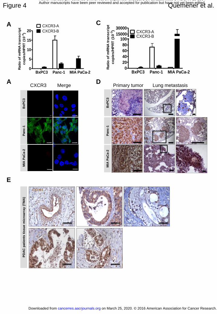
High expression of CXCR3-A mRNA was seen in Panc-1 cells (Fig.4A and B) but no
expression in BXPC3 or MIA PaCa-2 cells. Some CXCR3-B mRNA was detected in Panc-1
cells and low mRNA expression was also seen in BxPC3 and MIA PaCa-2 cells. However, no
CXCR3 protein expression was seen in BxPC3 and MIA PaCa-2 cells in vitro in contrast to
Panc-1 (Fig. 4B).
When Panc-1 cells were implanted subcutaneously or orthotopically into mice, a
strong increase of mRNA expression was observed for CXCR3-A and CXCR-B (50-fold and
35-fold increase respectively, Fig.4A and C). Positive immunoreactivity for CXCR3 was also
evidenced in these tumors (Fig.4D). For MIA PaCa-2 cells, after in vivo implantation,
CXCR3-B was highly expressed (Fig. 4C and D) and this much higher than in vitro (copy
number ratio CXCR3/HPRT in vivo : 14, 687 +/- 0.011 versus 5.39x10⁻⁴ +/-1.1x10-4 in vitro).
Metastatic foci were also positive for CXCR3. On the contrary, BxPC3 cells do not
significantly express CXCR3-A and B in vivo.
Seventy PDAC samples included in tissue microarrays were then analyzed. The
cytoplasmic and nuclear scores ranged from 20 to 300 (median: 175) and from 0 to 300
(median: 175) respectively (Fig.4E). Twenty-two (31%) of tumors presented a membranous
staining pattern, some of which had cytoplasmic staining (Fig. 4E). CXCR3 nuclear and
membranous expression correlated positively with tumor size (p=0.02 and p=0.06
respectively). CXCR3 membranous expression correlated with presence of perineural
invasion (p=0.04).
We furthermore performed an in silico analysis of CXCR3 expression using the
Ramaswamy multi-cancer set (22) and the Bucholz pancreas dataset (23) (Fig S4). In the
Ramaswamy dataset, high expression of CXCR3 could be evidenced in PDAC, a finding that
on March 25, 2020. © 2016 American Association for Cancer Research.cancerres.aacrjournals.org Downloaded from
Author manuscripts have been peer reviewed and accepted for publication but have not yet been edited. Author Manuscript Published OnlineFirst on September 9, 2016; DOI: 10.1158/0008-5472.CAN-15-2864

15
is further confirmed in the Bucholz data set where expression is also significantly increased in
PDAC compared to normal pancreas.
In vitro and in vivo functional studies
In vitro effects of CXCL4L1
CXCL4L1 inhibited endothelial cell but not BXPC3 cell proliferation (Fig. 5A and B,
left panels)(3). Invasion of endothelial cells, but not that of BxPC3 cells, was significantly
inhibited by CXCL4L1 (Fig. 5A middle panel; Fig.5B, right panels). No effect on apoptosis
on either tumor or endothelial cells was seen (Fig.S5).
We verified, in addition, CXCR3 expression in HUVECs, which is indeed
significantly expressed (Fig 5A right panel).
We next compared the role of CXCL4L1 in another pancreatic cancer cell line, Panc-1 cells,
which, in contrast to BxPC3 cells, do express CXCR3 protein (Fig.4C-D). This cell line
already expresses CXCL4L1 in cell culture (Fig.1E). Blocking by either CXCL4L1
monoclonal antibody or siRNA against CXCL4L1 as well as CXCR3 led to inhibition of cell
proliferation (Fig.5C). Combining blockade by MabL1 and CXCR3 siRNA further decreased
cell proliferation. This indicates that CXCL4L1 is a positive regulator of Panc-1 growth and
that CXCR3 is also involved in the growth promoting activity.
Since Panc-1 cells express CXCR3, we wanted to investigate whether signaling induced by
CXCL4L1 is modulated (Fig.5D). Indeed, ERK phosphorylation was increased after
CXCL4L1 treatment. This increase could be blocked by U1026, a specific inhibitor of MAP
kinases.
on March 25, 2020. © 2016 American Association for Cancer Research.cancerres.aacrjournals.org Downloaded from
Author manuscripts have been peer reviewed and accepted for publication but have not yet been edited. Author Manuscript Published OnlineFirst on September 9, 2016; DOI: 10.1158/0008-5472.CAN-15-2864

16
Effect of endogenous CXCL4L1 on tumor development
RAG-γ/c mice were inoculated subcutaneously with BxPC3 or Panc-1 cells and then
treated with MabL1 antibody. Prior to the tumor experiments, we characterize the half-life,
clearance and tumor targeting of this antibody in mice (Fig.S6A-E; supplemental Materials
and Methods).
We next investigated whether MabL1 was able to target tumor lesions in mice (Fig.6
A-F). BxPC3 cells were injected in the spleen (1x10⁶ cells) in mice. Tumors usually occurred
at 2 months post-injection. Infrared-dye labeled MabL1 antibody was administered by i.v.
injection. Imaging of organs revealed targeting of the antibody to metastatic lesions in the
lung and kidney.
In BxPC3 tumor xenografts, injection of MabL1 in mice yielded to an increase in
tumor growth (Fig.7A). MabL1 (25µg) was injected 2-times a week and tumor growth was
monitored during a 2-month period. Forty-eight days after implantation tumor size increased
by 2-fold in MabL1 treated animals when compared to non-treated controls (2612 mm3 in
MabL1-treated mice versus 1184 mm3 in control mice). Tumor weight was also increased
after antibody treatment (Fig. 7B). The kinetics of tumor growth in MabL1 treated mice was
81.5 mm3/day in comparison to 34.9 mm3/day in untreated controls. An increase in the density
of small vessels was observed in treated tumors compatible with an angiogenesis-related
effect (Fig.7C,D). The vessel increase in MabL1 treated tumor was not accompanied by
variation in NG2+ cell coverage (Fig.7E).
In Panc-1 tumor xenografts, the administration of CXCL4L1 antibody had an opposite
effect (Fig.7F,G). Using the same treatment protocol as for BxPC3 cells, administration of the
MabL1 antibody led to a reduction of tumor size in both subcutaneously and orthotopically
implanted tumors (s.c.implantation: 1140 mm3 in MabL1-treated mice versus 1845 mm3 in
control mice; orthotopic implantation: 2.8e+06 cpm in MabL1-treated mice versus 1.25e+07
on March 25, 2020. © 2016 American Association for Cancer Research.cancerres.aacrjournals.org Downloaded from
Author manuscripts have been peer reviewed and accepted for publication but have not yet been edited. Author Manuscript Published OnlineFirst on September 9, 2016; DOI: 10.1158/0008-5472.CAN-15-2864

17
cpm in control mice). Tumor weight was reduced in both orthotopic and subcutaneous tumors
in the presence of MabL1 (Fig.7H). Lung metastasis as measured by the presence of hS16
mRNA was significantly reduced in orthotopic tumors (Fig.7I). Furthermore, proliferation
measured by hMIB1 expression and Ki67 staining was decreased in orthotopic tumors after
MabL1 injection (Fig.7J and L). However, no effect on vessel quantity as measured by
PCAM1 expression was seen (Fig.7K and L).
Taken together, these results point to an opposite effect of CXCL4L1 in these two pancreatic
tumor cell lines, which correlates with the presence of CXCR3 on tumor cells.
We next performed qPCR analysis for PECAM1 in Panc-1 and MIA PaCa-2 tumor samples
(Fig. S7A). Panc-1 cells express CXCL4L1 contrary to MIA PaCa-2 cells. Fig S7A clearly
shows that PECAM1 levels were much reduced in Panc1 in comparison to MIA PaCa-2 cells
(6.16 fold) and this was inversely correlated with CXCL4L1 expression. Furthermore, we
analyzed mouse CXCR3 expression in both tumor types (Fig. S7B). Primers are only
detecting stroma-derived CXCR3 but no tumor-derived CXCR3. As seen in the figure, this
correlated well with PECAM1 expression and inversely with CXCR3 expression (6.16 fold in
both cases). In addition, the figure shows that CXCL4L1 is 11 times more expressed in Panc-
1 tumors when compared to healthy control pancreas (Fig S7B).
Modulation of CXCR3 in PDAC cells
To study the impact of CXCR3-A, we focused on MIA PaCa-2 cells, since Panc-1 cell knock-
down or overexpression was unsuccessful. CXCR3-A was cloned into pGFPN2 vector and
stable clones were isolated. Very high expression levels were observed (Copy number 574
943 for CXCR3-A / HPRT) (Fig. S8A). Cells were also positive by immunodetection (Fig.
S8B).
on March 25, 2020. © 2016 American Association for Cancer Research.cancerres.aacrjournals.org Downloaded from
Author manuscripts have been peer reviewed and accepted for publication but have not yet been edited. Author Manuscript Published OnlineFirst on September 9, 2016; DOI: 10.1158/0008-5472.CAN-15-2864
18
We next investigated the functionality of CXCR3 in the condition of chronic stimulation.
(Fig.S8C, Fig.S9A-C). Only, CXCL4L1 produced in Hek cells (Hek-CXCL4L1) stimulated
proliferation of MIA PaCa-2-CXCR3 cells. We verified that this effect is dependent on
CXCL4L1 and CXCR3 by using respectively MabL1 and SCH546738 a specific high affinity
antagonist of CXCR3, which demonstrated that this is indeed the case (Fig.S9B). We also
verified the signaling ability and demonstrated that chemokine stimulation induced ERK
phosphorylation (Fig.S9C).
Orthotopic implantation of MIA PaCa-2-CXCR3 cells did not show a difference in tumor
weight when compared to control vector-transfected MIA PaCa-2 cells at sacrifice (day 34
after implantation) (Fig.S9D). However, the number of lung metastasis was increased (human
HPRT-1 / mouse HPRT-1 by qPCR; 3.5+-0.86 (controls) versus 7.3+-1.46 (CXCR3-A
overexpressors) (Fig.S9E). Since MIA PaCa-2 cells did not show significant elevation of
CXCL4L1 expression when compared to BxPC3 or Panc-1 cells, we did not perform
inhibition studies using the blocking antibody (MabL1). Despite the fact that tumor weight
was not modified, changes in hypoxia signature and MMP9 were observed (Fig.S9F) in cells
that overexpress CXCR3A. This indicates that CXCR3 has in vivo intrinsically, in the
absence of CXCL4L1, pro-metastatic abilities in pancreatic tumor cells.
on March 25, 2020. © 2016 American Association for Cancer Research.cancerres.aacrjournals.org Downloaded from
Author manuscripts have been peer reviewed and accepted for publication but have not yet been edited. Author Manuscript Published OnlineFirst on September 9, 2016; DOI: 10.1158/0008-5472.CAN-15-2864

19
DISCUSSION
Our data show that tumor cells up-regulate CXCL4L1 expression when placed in a
suitable microenvironment. Myofibroblasts (MF) mimic the in vivo microenvironment and
were able to induce expression of CXCL4L1. Among potential factors, IL-1β was reported to
induce promoter demethylation after long-term exposure(21). However, IL-1β did not induce
CXCL4L1 expression in PDAC cells after short or long-term exposure. Microparticles from
MF conditioned medium induced CXCL4L1 expression tumor cells. However, the effect of
the microparticle preparation was much less when compared to chronic exposure in the
coculture system. This indicates that a yet non-identified soluble factor is involved in the
induction of CXCL4L1 expression in PDAC cells.
We demonstrated that epigenetic modifications through methylation play a critical role
in the induction of CXCL4L1 in tumor cells similar to what has been reported for some other
angiogenic factors, receptors or chemokines such as Maspin (24), intercellular adhesion
molecule-1 (ICAM-1) (25) or CXCL12 (SDF1) (26, 27). We found that human PDAC tumors
are usually hypomethylated on all 8 methylation sites of the CpG island identified, of which
two are involved in the induction of CXCL4L1 expression. This is in contrast to many genes
where methylation follows tumor progression. However, hypomethylation during tumor
progression has also been reported for other genes including urokinase-type plasminogen
activator (28), maspin (29) or matrix metalloproteinases (30), which is in line with our
findings.
It is to emphasize that CXCL4L1 or CXCL4 expression is not uniform for all
pancreatic carcinoma cell lines tested. When CXCL4L1 or CXCL4 were analyzed in
pancreatic carcinoma cells in vitro or after tumor implantation, two of the cell lines (BxPC3,
on March 25, 2020. © 2016 American Association for Cancer Research.cancerres.aacrjournals.org Downloaded from
Author manuscripts have been peer reviewed and accepted for publication but have not yet been edited. Author Manuscript Published OnlineFirst on September 9, 2016; DOI: 10.1158/0008-5472.CAN-15-2864

20
Panc-1) expressed increased levels of CXCL4L1 but not of CXCL4 in vivo. On the contrary,
another cell line (Mia PaCa-2) overexpressed already in vitro CXCL4 but not CXCL4L1 and
this was further increased in tumors generated after implantation in mice. Thus, CXCL4 may
also have a role in PDAC physiopathology.
Expression of CXCL4L1 was also evidenced in other tumor types such as colon
tumors or esophageal cancer (31). Endometriosis-associated ovarian cancer seem to express
decreased levels of CXCL4L1 (and CXCL4) in comparison to normal endometrium and
endometriosis lesion, but only in tumor-associated macrophages (32).
Anti-CXCL4L1 antibodies were able to target metastatic lesions. This suggests that
CXCL4L1 could be used for tumor targeting. However, mice do not express CXCL4L1 and,
in humans, targeting of CXCL4L1 may also affect normal cells, which would not be
appropriate for tumor targeting applications. CXCL4L1 diffuses much better than CXCL4
and, therefore, may not stay within the tumor tissue. Thus, further experiments are needed to
validate CXCL4L1 for tumor targeting approaches.
Functional data indicate that CXCL4L1 has no effect on proliferation, migration or
survival when tumor cells do not express CXCR3 protein, as it is the case for BxPC3.
Furthermore, significant effects on endothelial cell proliferation, migration, survival or tube
formation and cell signaling were observed after CXCL4L1 stimulation, which is in
agreement with previous studies. Endothelial cell-expressed CXCR3 has been proposed to
mediate the anti-angiogenic effects of CXCL4 and CXCL4L1 (6, 33). Alternatively,
CXCL4L1 may bind to an, as yet, unknown receptor.
on March 25, 2020. © 2016 American Association for Cancer Research.cancerres.aacrjournals.org Downloaded from
Author manuscripts have been peer reviewed and accepted for publication but have not yet been edited. Author Manuscript Published OnlineFirst on September 9, 2016; DOI: 10.1158/0008-5472.CAN-15-2864

21
We next investigated the role of endogenous CXCL4L1 in PDAC development.
Transgenic mice models using conditional expression of mutant K-ras and p53 allele or TGF-
β receptor seem to better recapitulate the multistage PDAC developmental process(34, 35)
than implant models. However, CXCL4L1 is not present in mice and, thus, experiments
aimed at blocking endogenous CXCL4L1 cannot be performed in these models.
In BxPC3 tumor xenografts, which do not express CXCR3 protein, blockade of
endogenous CXCL4L1 resulted in an increase in tumor development. This is in agreement
with an effect on the tumor stroma. In contrast, in Panc-1 tumor xenografts, which express
high levels of CXCR3, the blockade of endogenous CXCL4L1 led to a decreased tumor
development. Furthermore, we observed that CXCR3-A is mainly up-regulated in Panc-1
cells in vivo. This is reinforced by functional studies that show that CXCR3 knock-down
diminishes Panc-1 cell proliferation. This is in line with the observation that tumors cells,
which express CXCR3-A, exhibit an increase in tumor cell survival and proliferation and with
our clinical data (36). Furthermore, CXCR3 has intrinsically pro-metastatic activity.
All in all, our work establishes CXCL4L1 as a novel regulator of pancreatic tumor
development that not only acts on the tumor microenvironment but also directly on tumor
cells themselves. Expression of the CXCL4L1 in tumor cells is controlled by stroma-tumor
cell interactions and by epigenetic regulations. Further work is required to fully understand
the specific micro-environmental and epigenetic mechanisms that regulate CXCL4L1
expression in tumor cells.
on March 25, 2020. © 2016 American Association for Cancer Research.cancerres.aacrjournals.org Downloaded from
Author manuscripts have been peer reviewed and accepted for publication but have not yet been edited. Author Manuscript Published OnlineFirst on September 9, 2016; DOI: 10.1158/0008-5472.CAN-15-2864

22
REFERENCES
1. Struyf S, Burdick MD, Peeters E, Van den Broeck K, Dillen C, Proost P, et al. Platelet factor-4 variant chemokine CXCL4L1 inhibits melanoma and lung carcinoma growth and metastasis by preventing angiogenesis. Cancer research. 2007;67:5940-8. 2. Struyf S, Burdick MD, Proost P, Van Damme J, Strieter RM. Platelets release CXCL4L1, a nonallelic variant of the chemokine platelet factor-4/CXCL4 and potent inhibitor of angiogenesis. Circ Res. 2004;95:855-7. 3. Dubrac A, Quemener C, Lacazette E, Lopez F, Zanibellato C, Wu WG, et al. Functional divergence between 2 chemokines is conferred by single amino acid change. Blood. 2010;116:4703-11. 4. Lasagni L, Grepin R, Mazzinghi B, Lazzeri E, Meini C, Sagrinati C, et al. PF-4/CXCL4 and CXCL4L1 exhibit distinct subcellular localization and a differentially regulated mechanism of secretion. Blood. 2007;109:4127-34. 5. Kuo JH, Chen YP, Liu JS, Dubrac A, Quemener C, Prats H, et al. Alternative C-Terminal Helix Orientation Alters Chemokine Function: Structure of the Anti-angiogenic Chemokine, CXCL4L1. The Journal of biological chemistry. 2013. 6. Struyf S, Salogni L, Burdick MD, Vandercappellen J, Gouwy M, Noppen S, et al. Angiostatic and chemotactic activities of the CXC chemokine CXCL4L1 (platelet factor-4 variant) are mediated by CXCR3. Blood. 2011;117:480-8. 7. Billottet C, Quemener C, Bikfalvi A. CXCR3, a double-edged sword in tumor progression and angiogenesis. Biochimica et biophysica acta. 2013;1836:287-95. 8. Van Raemdonck K, Van den Steen PE, Liekens S, Van Damme J, Struyf S. CXCR3 ligands in disease and therapy. Cytokine & growth factor reviews. 2015;26:311-27. 9. Karin N, Wildbaum G, Thelen M. Biased signaling pathways via CXCR3 control the development and function of CD4+ T cell subsets. Journal of leukocyte biology. 2015. 10. Vandercappellen J, Noppen S, Verbeke H, Put W, Conings R, Gouwy M, et al. Stimulation of angiostatic platelet factor-4 variant (CXCL4L1/PF-4var) versus inhibition of angiogenic granulocyte chemotactic protein-2 (CXCL6/GCP-2) in normal and tumoral mesenchymal cells. Journal of leukocyte biology. 2007;82:1519-30. 11. Vandercappellen J, Liekens S, Bronckaers A, Noppen S, Ronsse I, Dillen C, et al. The COOH-terminal peptide of platelet factor-4 variant (CXCL4L1/PF-4var47-70) strongly inhibits angiogenesis and suppresses B16 melanoma growth in vivo. Molecular cancer research : MCR. 2010;8:322-34. 12. Van Raemdonck K, Berghmans N, Vanheule V, Bugatti A, Proost P, Opdenakker G, et al. Angiostatic, tumor inflammatory and anti-tumor effects of CXCL4(47-70) and CXCL4L1(47-70) in an EGF-dependent breast cancer model. Oncotarget. 2014;5:10916-33. 13. Hagedorn M, Javerzat S, Gilges D, Meyre A, de Lafarge B, Eichmann A, et al. Accessing key steps of human tumor progression in vivo by using an avian embryo model. Proceedings of the National Academy of Sciences of the United States of America. 2005;102:1643-8. 14. Dumartin L, Quemener C, Laklai H, Herbert J, Bicknell R, Bousquet C, et al. Netrin-1 mediates early events in pancreatic adenocarcinoma progression, acting on tumor and endothelial cells. Gastroenterology. 2010;138:1595-606, 606 e1-8. 15. Hendrich H. The Laboratory Mouse. 2d Edition ed: Elsevier; 2012. 16. Platonova N, Miquel G, Regenfuss B, Taouji S, Cursiefen C, Chevet E, et al. Evidence for the interaction of fibroblast growth factor-2 with the lymphatic endothelial cell marker LYVE-1. Blood. 2013;121:1229-37.
on March 25, 2020. © 2016 American Association for Cancer Research.cancerres.aacrjournals.org Downloaded from
Author manuscripts have been peer reviewed and accepted for publication but have not yet been edited. Author Manuscript Published OnlineFirst on September 9, 2016; DOI: 10.1158/0008-5472.CAN-15-2864

23
17. Liu ZQ, Mahmood T, Yang PC. Western blot: technique, theory and trouble shooting. North American journal of medical sciences. 2014;6:160. 18. Clarke K, Daubon T, Turan N, Soulet F, Mohd Zahari M, Ryan KR, et al. Inference of Low and High-Grade Glioma Gene Regulatory Networks Delineates the Role of Rnd3 in Establishing Multiple Hallmarks of Cancer. PLoS genetics. 2015;11:e1005325. 19. Segara D, Biankin AV, Kench JG, Langusch CC, Dawson AC, Skalicky DA, et al. Expression of HOXB2, a retinoic acid signaling target in pancreatic cancer and pancreatic intraepithelial neoplasia. Clinical cancer research : an official journal of the American Association for Cancer Research. 2005;11:3587-96. 20. Ishikawa M, Yoshida K, Yamashita Y, Ota J, Takada S, Kisanuki H, et al. Experimental trial for diagnosis of pancreatic ductal carcinoma based on gene expression profiles of pancreatic ductal cells. Cancer science. 2005;96:387-93. 21. Hashimoto K, Oreffo RO, Gibson MB, Goldring MB, Roach HI. DNA demethylation at specific CpG sites in the IL1B promoter in response to inflammatory cytokines in human articular chondrocytes. Arthritis and rheumatism. 2009;60:3303-13. 22. Ramaswamy S, Tamayo P, Rifkin R, Mukherjee S, Yeang CH, Angelo M, et al. Multiclass cancer diagnosis using tumor gene expression signatures. Proceedings of the National Academy of Sciences of the United States of America. 2001;98:15149-54. 23. Buchholz M, Braun M, Heidenblut A, Kestler HA, Kloppel G, Schmiegel W, et al. Transcriptome analysis of microdissected pancreatic intraepithelial neoplastic lesions. Oncogene. 2005;24:6626-36. 24. Beltran AS, Sun X, Lizardi PM, Blancafort P. Reprogramming epigenetic silencing: artificial transcription factors synergize with chromatin remodeling drugs to reactivate the tumor suppressor mammary serine protease inhibitor. Molecular cancer therapeutics. 2008;7:1080-90. 25. Hellebrekers DM, Castermans K, Vire E, Dings RP, Hoebers NT, Mayo KH, et al. Epigenetic regulation of tumor endothelial cell anergy: silencing of intercellular adhesion molecule-1 by histone modifications. Cancer research. 2006;66:10770-7. 26. Seo J, Kim YO, Jo I. Differential expression of stromal cell-derived factor 1 in human brain microvascular endothelial cells and pericytes involves histone modifications. Biochemical and biophysical research communications. 2009;382:519-24. 27. Sowinska A, Jagodzinski PP. RNA interference-mediated knockdown of DNMT1 and DNMT3B induces CXCL12 expression in MCF-7 breast cancer and AsPC1 pancreatic carcinoma cell lines. Cancer letters. 2007;255:153-9. 28. Pakneshan P, Szyf M, Rabbani SA. Methylation and inhibition of expression of uPA by the RAS oncogene: divergence of growth control and invasion in breast cancer cells. Carcinogenesis. 2005;26:557-64. 29. Sato N, Fukushima N, Matsubayashi H, Goggins M. Identification of maspin and S100P as novel hypomethylation targets in pancreatic cancer using global gene expression profiling. Oncogene. 2004;23:1531-8. 30. Sato N, Maehara N, Su GH, Goggins M. Effects of 5-aza-2'-deoxycytidine on matrix metalloproteinase expression and pancreatic cancer cell invasiveness. Journal of the National Cancer Institute. 2003;95:327-30. 31. Verbeke H, De Hertogh G, Li S, Vandercappellen J, Noppen S, Schutyser E, et al. Expression of angiostatic platelet factor-4var/CXCL4L1 counterbalances angiogenic impulses of vascular endothelial growth factor, interleukin-8/CXCL8, and stromal cell-derived factor 1/CXCL12 in esophageal and colorectal cancer. Human pathology. 2010;41:990-1001. 32. Furuya M, Tanaka R, Miyagi E, Kami D, Nagahama K, Miyagi Y, et al. Impaired CXCL4 expression in tumor-associated macrophages (TAMs) of ovarian cancers arising in endometriosis. Cancer biology & therapy. 2012;13:671-80.
on March 25, 2020. © 2016 American Association for Cancer Research.cancerres.aacrjournals.org Downloaded from
Author manuscripts have been peer reviewed and accepted for publication but have not yet been edited. Author Manuscript Published OnlineFirst on September 9, 2016; DOI: 10.1158/0008-5472.CAN-15-2864

24
33. Lasagni L, Francalanci M, Annunziato F, Lazzeri E, Giannini S, Cosmi L, et al. An alternatively spliced variant of CXCR3 mediates the inhibition of endothelial cell growth induced by IP-10, Mig, and I-TAC, and acts as functional receptor for platelet factor 4. J Exp Med. 2003;197:1537-49. 34. Guerra C, Schuhmacher AJ, Canamero M, Grippo PJ, Verdaguer L, Perez-Gallego L, et al. Chronic pancreatitis is essential for induction of pancreatic ductal adenocarcinoma by K-Ras oncogenes in adult mice. Cancer cell. 2007;11:291-302. 35. Cook N, Olive KP, Frese K, Tuveson DA. K-Ras-driven pancreatic cancer mouse model for anticancer inhibitor analyses. Methods Enzymol. 2008;439:73-85. 36. Giuliani N, Bonomini S, Romagnani P, Lazzaretti M, Morandi F, Colla S, et al. CXCR3 and its binding chemokines in myeloma cells: expression of isoforms and potential relationships with myeloma cell proliferation and survival. Haematologica. 2006;91:1489-97.
on March 25, 2020. © 2016 American Association for Cancer Research.cancerres.aacrjournals.org Downloaded from
Author manuscripts have been peer reviewed and accepted for publication but have not yet been edited. Author Manuscript Published OnlineFirst on September 9, 2016; DOI: 10.1158/0008-5472.CAN-15-2864

25
FIGURE LEGENDS
Figure 1: CXCL4L1 expression in the PDAC-CAM and mouse xenograft model
(A) CXCL4L1 expression in BxPC3 cells in vitro and in the PDAC-CAM model at day 1 (T1)
and day 6 (T6), normalized to S16 expression. CXCL4L1 mRNA at T1 with arbitrary value 1.
(B) Immunolabelling on T6 CAM with MabL1. (C) Western blotting for CXCL4L1 from
pooled T6 CAM lysates (n=3) or BxPC3 cells lysates with MabL1. The gel is representative
of two independent experiments. (D) In situ hybridization of T6 PDAC-CAM samples with
CXCL4L1 riboprobes. Tumor nodes (dotted lines), CAM surface (full lines). (E) qRT-PCR
from mRNA of BXPC3, Panc-1, MIA PaCa-2 cells in culture (n = 4) or of tumors derived
from subcutaneously injected cells (n = 8 tumors; **, P<0.01). (F) CXCL4L1
immunolabeling of BxPC3 (subcutaneous), Panc-1 and MIA PaCa-2 tumors (orthotopic) and
lung metastasis. Global view (Insert). Scale bars: (primary tumors) 50µm; (lung metastasis)
100µm. Error bars indicate SEM. Asterisk*: positive node.
Figure 2: Coculture of tumor cells with myofibroblasts
(A) Immunofluorescence (IF) staining for CXCL4L1 on BxPC3 cells alone (left) or
co-cultured for 4 days with myofibroblasts (MF) (right) DAPI-labeled nuclei are in blue. (B)
qRT-PCR from laser micro-dissected BxPC3 cell mRNA. Data as CT value as means ± SD of
5 micro-dissected samples. HPRT1 as a housekeeping gene and α-SMA as a control for
contamination by MF. (C) IF staining on co-cultured BxPC3 for CXCL4L1 and VAMP-2.
DAPI-labeled nuclei in blue. Merged staining (yellow) revealed areas of co-localization and
scatter 2D plots of intensities in red and green immunofluorescence channels (NIS software).
(D) BxPC3 cells (2 x 10⁵) were co-cultured using transwell system with MF (2x10⁵) for 24 h
or with microparticles. mRNA was extracted and analyzed by qPCR for CXCL4L1
on March 25, 2020. © 2016 American Association for Cancer Research.cancerres.aacrjournals.org Downloaded from
Author manuscripts have been peer reviewed and accepted for publication but have not yet been edited. Author Manuscript Published OnlineFirst on September 9, 2016; DOI: 10.1158/0008-5472.CAN-15-2864

26
expression. Data as copy number / HPRT. Scale bar: (A) 50 µm (C) 10µm. (E) BxPC3 cells
(2 x 10⁵) were co-cultured using a transwell system with MF (2x10⁵) for 24h. Myofibroblasts
were then removed from the transwell chamber for 24h. mRNA was then extracted and
analyzed by qPCR for CXCL4L1 expression. Data as copy number / HPRT.
Figure 3: Modulation of CXCL4L1 expression by DNA methylation
(A) CXCL4L1 and CXCL4 genomic organization. (B) RT–PCR analyses of CXCL4,
CXCL4L1, CXCL1 and CXCL5 mRNA from BxPC3 cells treated with 5’-Aza and/or TSA
(left panel). RT–PCR analyses of CXCL4 and CXCL4L1 mRNA from platelets, BxPC3 and
MG63 cells treated with IL-1β and 5’-Aza/ TSA (right panel). GAPDH as an internal control.
(C) Location of CpG sites (vertical bars), bisulfite sequencing primers in the first intron with
the Methprimer website. (D) Methylation of CXCL4L1 in BxPC3 cells treated or not with 5’-
Aza, in BxPC3 sub-cutaneous primary tumors and in lung metastasis from mice injected with
BxPC3 into the spleen (one representative tumor and metastasis, n=10 tumors/group) (left
panel). Filled and open circles represent methylated and unmethylated CpG sites respectively.
qRT-PCR of CXCL4L11 mRNA from 5’-Aza-treated or untreated BxPC3 cells and from
subcutaneous tumor and lung metastasis (right panel). HPRT1, internal control. (n = 6
samples/group). (E) Characterization of CXCL4L1 expression. The 1-kb upstream promoter,
exon 1 and intron 1 regions of human CXCL4L1 cloned in front of the Luciferase Firefly
gene. Wild-type sequence (WT) or mutated sequence in the two CpG sites regulated by the
5’Aza treatment. After in vitro methylation and transfection, and the ratio of
LucFirefly/LucRenilla was determined (n = 3; **, P<0.01). Error bars, SEM.
on March 25, 2020. © 2016 American Association for Cancer Research.cancerres.aacrjournals.org Downloaded from
Author manuscripts have been peer reviewed and accepted for publication but have not yet been edited. Author Manuscript Published OnlineFirst on September 9, 2016; DOI: 10.1158/0008-5472.CAN-15-2864

27
Figure 4: Expression of CXCR3 in pancreatic adenocarcinoma
(A) qRT-PCR for CXCR3-A and CXCR3-B. (B) qRT-PCR for CXCR3-A and B in primary
tumors from BxPC3, PANC-1 and MIA PaCa-2 cells implanted mice (n=12 and 10
tumors/group). (C) Immunofluorescence staining in vitro. (D) Immunostaining for human
CXCR3 of subcutaneous BxPC3 and orthotopic PANC-1 and MIA PaCa-2 tumors (left
panels) and lung metastasis (middle and right panels). (E) Immunohistochemistry for CXCR3
(brown) and hematoxylin counterstaining (blue) of PDAC patients tissue microarray (TMA)
showing apical (left upper panel), cytoplasmic (upper middle panel), membrane (upper right
panel) and nuclear staining (lower panels). Scale bars: (B) 20 µm, (D, left panels and right
lower panel) 100µm, (D, middle panels) 500µm, (D, upper right panel, E) 50µm. Error bars
indicate SEM.
Figure 5: Functional studies of CXCL4L1 in pancreatic adenocarcinoma and endothelial
cells
(A) Activity of recombinant GST-CXCL4L1 and cleaved CXCL4L1 on HUVEC proliferation
after 48 h(left panel). Endothelial cells cultured with FGF2 (20ng/mL) and 50ng/mL of
recombinant proteins in presence of MabL1 (10 μg/mL) or of an IgG control antibody. Effect
of recombinant CXCL4L1 (GST-CXCL4L1) on FGF2-stimulated (10 ng/ml) proliferation or
invasion of HUVECs (proliferation: left panel, invasion: middle panel). CXCR3 protein
expression assessed by Western-blot in HUVEC cells (right panel). Vinculin as loading
control. (B) BXPC3 cells (proliferation: left panel; invasion: right panel) in the presence of
CXCL4L1. (C) Effect of CXCL4L1 and CXCR3 knock-down or antibody treatment on the
proliferation of Panc-1 cells. Cells transfected with the specific siRNAs or control siRNAs,
grown in complete medium and treated or not with the monoclonal anti-CXCL4L1 antibody
over 120 h. Results as average ± SEM of three independent experiments done in triplicates;
on March 25, 2020. © 2016 American Association for Cancer Research.cancerres.aacrjournals.org Downloaded from
Author manuscripts have been peer reviewed and accepted for publication but have not yet been edited. Author Manuscript Published OnlineFirst on September 9, 2016; DOI: 10.1158/0008-5472.CAN-15-2864

28
** <0.01, * P<0.05. (D) Effect of CXCL4L1 on signaling in Panc-1 cells. Cells stimulated
with minimal medium from CXCL4L1 infected cells (MIA PaCa-2) containing > 300 ng/ml
CXCL4L1 and ERK phosphorylation measured by Western Blotting with and without a
specific inhibitor of MAP-kinases (U1026, 50µM). 0%M : Serum-free medium. CM :
complete medium.
Figure 6: Targeting of monoclonal MabL1 to tumor lesion in mice
Antibodies labeled with IRdye (MabL1-IRdye) and injected into the tail vein in tumor
bearing mice. 6 days after Mabl1-IRdye injection, tissues were removed after sacrifice and
imaged on the Odyssey Imaging System, frozen, cut into 40 μm sections and imaged again.
Lesions targeted by MabL1 are shown in the lung (Figure S7A-C) and kidney (Figure S7D-F)
in green. Figure S7C and 7F depict images of frozen sections.
Figure 7: Effect of CXCL4L1 blockade on in vivo pancreatic tumor development (A) Tumor development in subcutaneously implanted BxPC3 cells (n=12/group) and followed
by Caliper measurements in MabL1 or control antibody treated animals. Treated tumors were
significantly larger (**, P<0.01). (B) On the day of sacrifice, tumors were excised and
weighted (**, P<0.01). (C) Immunofluorescence staining for CD31 and NG2 on subcutaneous
BxPC3 tumor section from control and MabL1 treated mice. (D) Quantification of vessel
density grouped in small (<10 µm2), medium (10-100 µm2) or large CD31-positive vessels
(>100 µm2) (n=5 tumors/group, 10 pictures/tumor). MabL1-treated mice have significantly
more small-diameter vessels in tumors compared to untreated controls (*, P<0.05). (E)
Quantification of smooth muscle cell (SMC) coverage.
(F) Panc-1 cells implanted subcutaneously or orthotopically by direct intra-pancreatic
injection in Rag2 γ/c mice and treated with MabL1 or with the control antibody. MabL1
on March 25, 2020. © 2016 American Association for Cancer Research.cancerres.aacrjournals.org Downloaded from
Author manuscripts have been peer reviewed and accepted for publication but have not yet been edited. Author Manuscript Published OnlineFirst on September 9, 2016; DOI: 10.1158/0008-5472.CAN-15-2864

29
treated mice with subcutaneously implanted cells had significantly smaller tumors (**,
P<0,01). (G) Bioluminescent imaging on orthotopic PANC-1-LucFirefly tumors using the
Photon Imager (Biospace). Analysis by M3Vision software and represented as total flux
measurements in count per minute. (H) Weights recorded from the excised tumors at sacrifice
day 75 and 92 for orthotopic and subcutaneous tumors respectively (**, P<0,01; *, P<0.05).
(I) Quantification of lung metastasis in subcutaneously and orthotopically implanted mice by
qRT-PCR for the human S16 gene (n = 10 lungs/group) (*, P<0,05). mHPRT1, housekeeping
gene. (J) qRT-PCR analyses for human MIB1 and (K) mouse PECAM1 in tumors (*, P<0.05).
(L) Immunofluorescence staining for mouse CD31 and human Ki67 on orthotopic tumor
sections. Scale bar: 50µm. Error bars are indicated as SEM.
on March 25, 2020. © 2016 American Association for Cancer Research.cancerres.aacrjournals.org Downloaded from
Author manuscripts have been peer reviewed and accepted for publication but have not yet been edited. Author Manuscript Published OnlineFirst on September 9, 2016; DOI: 10.1158/0008-5472.CAN-15-2864

Figure 1
Lu
ng m
eta
sta
sis
D
* *
B C
In vitro CAM T6
A
Quemener et al.
sense anti-sense
*
Panc-1
Prim
ary
tu
mo
r
BxPC3
PD
AC
CA
M m
od
el
PD
AC
mo
us
e m
od
el
E
** **
F MIA PaCa-2
***
*
MIA PaCa-2
on March 25, 2020. © 2016 American Association for Cancer Research.cancerres.aacrjournals.org Downloaded from
Author manuscripts have been peer reviewed and accepted for publication but have not yet been edited. Author Manuscript Published OnlineFirst on September 9, 2016; DOI: 10.1158/0008-5472.CAN-15-2864

Figure 2
Quemener et al.
CXCL4L1 merged VAMP2
A
Intensity scatterplot
HPRT1 CXCL4L1 HPRT1 CXCL4L1 SMA
26.3 ± 1.02 No CT 19.41 ± 3.23 25.85 ± 1.48 No CT
11.4 ± 0.56 No CT 21.74 ± 5.58 31.63 ± 5.08 No CT
17.57 ± 2.24 No CT 23.05 ± 3.84 30.25 ± 3.54 No CT
BxPC3 cocultured BxPC3
B
C
CXC
L4L1
BxPC3 BxPC3 + MF
D
CTRL co-culture microparticule0
10
20
30CTRL
co-culture
microparticule
Rati
o o
f m
RN
A C
XC
L4L
1 t
ran
scri
pt
co
pie
s/H
PR
T1 (
10
-5)
CTRL co-culture microparticule0
10
20
30CTRL
co-culture
microparticule
Rati
o o
f m
RN
A C
XC
L4L
1 t
ran
scri
pt
co
pie
s/H
PR
T1 (
10
-5)
CTRL co-culture microparticles0
10
20
30CTRL
co-culture
microparticles
Rati
o o
f m
RN
A C
XC
L4L
1 t
ran
scrip
t co
pie
s/H
PR
T1 (
10
-5)
Rati
o o
f m
RN
A C
XC
L4L
1 t
ran
scri
pt
co
pie
s/H
PR
T1 (
10
-2)
BxPC3 BxPC3
MF
BxPC3
MF
BxPC3
24h 24h
24h 24h
1
.
2
.
3
.
1 2 3
***
E
Microparticles Co-culture CTRL
on March 25, 2020. © 2016 American Association for Cancer Research.cancerres.aacrjournals.org Downloaded from
Author manuscripts have been peer reviewed and accepted for publication but have not yet been edited. Author Manuscript Published OnlineFirst on September 9, 2016; DOI: 10.1158/0008-5472.CAN-15-2864

A B
+1250
+1 CpG island
E1 I1
-1000
E2 I2 E3
E
D BxPC3 BxPC3 + 5’ Aza
Subcutaneous
Tumor
Lung Metastasis
-1000 firefly ORF
+1
firefly ORF
+1
-1000
Figure 3 Quemener et al.
WT Mut1,20
5
10
15
20 **
Ra
tio
Lu
c F
/Lu
c R
Chr.4q12-q21 109.84Kb
CXCL1 CXCL6 CXCL4 CXCL4L1
CXCL4L1
CXCL4
CXCL1
CXCL6
GAPDH
CXCL4
CXCL4L1
GAPDH
C
Human tumor
BxPC3
BxPC3 5' aza
Subcutaneous tumor
Lung Metastasis
Human tumor
02468
6080
100120140
Ra
tio
of
CX
CL
4L
1 m
RN
A
tra
ns
cri
pt
co
pie
s/H
PR
T (
10
-4)
pGL3-Mut1,2
pGL3-WT
on March 25, 2020. © 2016 American Association for Cancer Research.cancerres.aacrjournals.org Downloaded from
Author manuscripts have been peer reviewed and accepted for publication but have not yet been edited. Author Manuscript Published OnlineFirst on September 9, 2016; DOI: 10.1158/0008-5472.CAN-15-2864
Quemener et al. Figure 4
D
E
Primary tumor Lung metastasis
Pan
c-1
B
xP
C3
M
IA P
aC
a-2
I
J
CXCR3 Merge
A C
BxPC3 Panc-1 MIA PaCa-20
20
40
60
80
10015000
30000CXCR3-ACXCR3-B
Ra
tio
of
mR
NA
tra
ns
cri
pt
co
pie
s/H
PR
T (
10
-3)
BxPC3 Panc-1 MIA PaCa-20
5
10
15
20 CXCR3-B
CXCR3-A
Ra
tio
of
mR
NA
tra
ns
cri
pt
co
pie
s/H
PR
T (
10
-4)
Pan
c-1
B
xP
C3
M
IA P
aC
a-2
P
DA
C p
ati
en
ts t
issu
e m
icro
arr
ay (
TM
A)
CXCR3
A
on March 25, 2020. © 2016 American Association for Cancer Research.cancerres.aacrjournals.org Downloaded from
Author manuscripts have been peer reviewed and accepted for publication but have not yet been edited. Author Manuscript Published OnlineFirst on September 9, 2016; DOI: 10.1158/0008-5472.CAN-15-2864

Figure 5 Quemener et al.
A
0 12 24 36 48 60 72 84 96 108 1200
20
40
60
80
100
SiCXCL4L1Si Ctrl + MabL1
Si Ctrl
SiCXCR3SiCXCR3 + MabL1
Time (h)
Co
nflu
en
ce (
%)
Si C
trl
SiC
XCL4
L1
SiC
XCR3
Si C
trl +
Mab
L1
Mab
L1 +
SiC
XCR3
0
20
40
60
80
100
**
****
Co
nflu
en
ce (
%)
HUVEC
B
BxPC3 Panc-1
C
D Panc-1
P-ERK
Vinculin
0%M CM
CXCL4L1 Control U1026
55 kDa
130 kDa
+ - + + + + - + - - - - - - + - + - - - - + - - - - - - + +
Total ERK
55 kDa
Vinculin
CXCR3
CTRL- HUVEC
on March 25, 2020. © 2016 American Association for Cancer Research.cancerres.aacrjournals.org Downloaded from
Author manuscripts have been peer reviewed and accepted for publication but have not yet been edited. Author Manuscript Published OnlineFirst on September 9, 2016; DOI: 10.1158/0008-5472.CAN-15-2864

Figure 6 Quemener et al.
on March 25, 2020. © 2016 American Association for Cancer Research.cancerres.aacrjournals.org Downloaded from
Author manuscripts have been peer reviewed and accepted for publication but have not yet been edited. Author Manuscript Published OnlineFirst on September 9, 2016; DOI: 10.1158/0008-5472.CAN-15-2864

control MabL10
500
1000
1500
2000
2500 **
Tu
mo
r w
eig
ht
(mg
)
BxP
C3
B
C D
E
20 40 60 80 1000
1000
2000
Control
MabL1**
Days after injection
Tu
mo
r v
olu
me
(m
m3)
30 40 50 60 700
5.0106
1.0107
1.5107
2.0107control
MabL1
*
**
Days post-injection
Lig
ht
inte
ns
ity
(c
pm
)
cp
m
co
ntr
ol
Ma
bL1
Control MabL1 Control MabL10
500
1000
SubcutaneousOrthotopic
***
Tu
mo
r w
eig
ht
(mg
)
Control MabL1 Control MabL1
0.0
0.5
1.0
1.5
2.0
2.5 NS
SubcutaneousOrthotopic
Ra
tio
n o
f m
PE
CA
M1
Tra
ns
cri
pt
co
pie
s/H
PR
T
Control MabL1 Control MabL10
2
4
6
8
SubcutaneousOrthotopic
NS
*
Ra
tio
n o
f h
MIB
1 T
ran
sc
rip
t
co
pie
s/H
PR
T
Control MabL1 Control MabL10
20
40
60
SubcutaneousOrthotopic
NS
*
Re
lati
ve
hS
16
mR
NA
le
ve
l
Co
ntr
ol
Ma
bL1
Pan
c-1
F G
H
K J
I L
Figure 7
A
Ma
bL1
NG2 CD31 DAPI
Co
ntr
ol
Quemener et al.
*
CD31 Ki67 Merged+ dapi
on March 25, 2020. © 2016 American Association for Cancer Research.cancerres.aacrjournals.org Downloaded from
Author manuscripts have been peer reviewed and accepted for publication but have not yet been edited. Author Manuscript Published OnlineFirst on September 9, 2016; DOI: 10.1158/0008-5472.CAN-15-2864

Published OnlineFirst September 9, 2016.Cancer Res Cathy Quemener, Jessica Baud, Kevin Boye, et al. and invasion of pancreatic cancerDual roles for CXCL4 chemokines and CXCR3 in angiogenesis
Updated version
10.1158/0008-5472.CAN-15-2864doi:
Access the most recent version of this article at:
Material
Supplementary
http://cancerres.aacrjournals.org/content/suppl/2016/09/09/0008-5472.CAN-15-2864.DC1
Access the most recent supplemental material at:
Manuscript
Authoredited. Author manuscripts have been peer reviewed and accepted for publication but have not yet been
E-mail alerts related to this article or journal.Sign up to receive free email-alerts
Subscriptions
Reprints and
To order reprints of this article or to subscribe to the journal, contact the AACR Publications
Permissions
Rightslink site. Click on "Request Permissions" which will take you to the Copyright Clearance Center's (CCC)
.http://cancerres.aacrjournals.org/content/early/2016/09/09/0008-5472.CAN-15-2864To request permission to re-use all or part of this article, use this link
on March 25, 2020. © 2016 American Association for Cancer Research.cancerres.aacrjournals.org Downloaded from
Author manuscripts have been peer reviewed and accepted for publication but have not yet been edited. Author Manuscript Published OnlineFirst on September 9, 2016; DOI: 10.1158/0008-5472.CAN-15-2864


![CENTERITY SERVICE PACK FOR CLOUDERA€¦ · OOZIE [roles status] • CLOUDERA ROLES SOLR [roles status] • CLOUDERA ROLES SPARK [roles status] • CLOUDERA ROLES SQOOP [roles status]](https://img.pdfslide.us/doc/110x75/5fc0df6d43307a59a12ae0a7/centerity-service-pack-for-cloudera-oozie-roles-status-a-cloudera-roles-solr.jpg)