Embed Size (px)
Citation preview
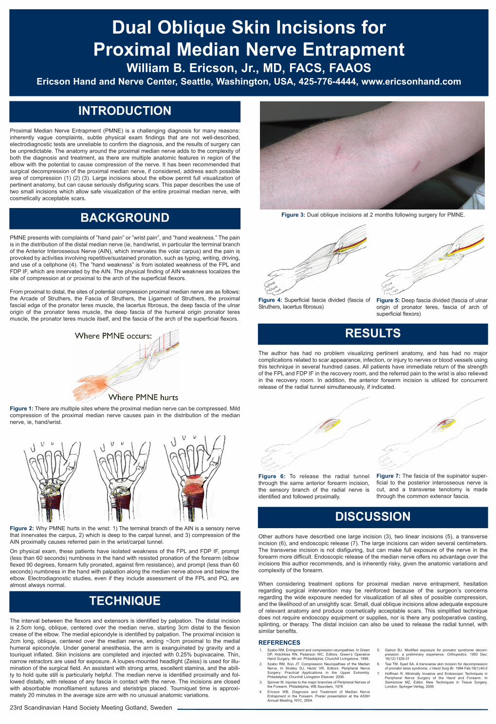
Dual Oblique Skin Incisions for
Proximal Median Nerve EntrapmentWilliam B. Ericson, Jr., MD, FACS, FAAOS
Ericson Hand and Nerve Center, Seattle, Washington, USA, 425-776-4444, www.ericsonhand.com
Proximal Median Nerve Entrapment (PMNE) is a challenging diagnosis for many reasons:
inherently vague complaints, subtle physical exam findings that are not well-described,
electrodiagnostic tests are unreliable to confirm the diagnosis, and the results of surgery can
be unpredictable. The anatomy around the proximal median nerve adds to the complexity of
both the diagnosis and treatment, as there are multiple anatomic features in region of the
elbow with the potential to cause compression of the nerve. It has been recommended that
surgical decompression of the proximal median nerve, if considered, address each possible
area of compression (1) (2) (3). Large incisions about the elbow permit full visualization of
pertinent anatomy, but can cause seriously disfiguring scars. This paper describes the use of
two small incisions which allow safe visualization of the entire proximal median nerve, with
cosmetically acceptable scars.
INTRODUCTION
The author has had no problem visualizing pertinent anatomy, and has had no major
complications related to scar appearance, infection, or injury to nerves or blood vessels using
this technique in several hundred cases. All patients have immediate return of the strength
of the FPL and FDP IF in the recovery room, and the referred pain to the wrist is also relieved
in the recovery room. In addition, the anterior forearm incision is utilized for concurrent
release of the radial tunnel simultaneously, if indicated.
RESULTS
Other authors have described one large incision (3), two linear incisions (5), a transverse
incision (6), and endoscopic release (7). The large incisions can widen several centimeters.
The transverse incision is not disfiguring, but can make full exposure of the nerve in the
forearm more difficult. Endoscopic release of the median nerve offers no advantage over the
incisions this author recommends, and is inherently risky, given the anatomic variations and
complexity of the forearm.
When considering treatment options for proximal median nerve entrapment, hesitation
regarding surgical intervention may be reinforced because of the surgeon’s concerns
regarding the wide exposure needed for visualization of all sites of possible compression,
and the likelihood of an unsightly scar. Small, dual oblique incisions allow adequate exposure
of relevant anatomy and produce cosmetically acceptable scars. This simplified technique
does not require endoscopy equipment or supplies, nor is there any postoperative casting,
splinting, or therapy. The distal incision can also be used to release the radial tunnel, with
similar benefits.
DISCUSSION
PMNE presents with complaints of ”hand pain” or ”wrist pain”, and ”hand weakness.” The pain
is in the distribution of the distal median nerve (ie, hand/wrist, in particular the terminal branch
of the Anterior Interosseous Nerve (AIN), which innervates the volar carpus) and the pain is
provoked by activities involving repetitive/sustained pronation, such as typing, writing, driving,
and use of a cellphone (4). The ”hand weakness” is from isolated weakness of the FPL and
FDP IF, which are innervated by the AIN. The physical finding of AIN weakness localizes the
site of compression at or proximal to the arch of the superficial flexors.
From proximal to distal, the sites of potential compression proximal median nerve are as follows:
the Arcade of Struthers, the Fascia of Struthers, the Ligament of Struthers, the proximal
fascial edge of the pronator teres muscle, the lacertus fibrosus, the deep fascia of the ulnar
origin of the pronator teres muscle, the deep fascia of the humeral origin pronator teres
muscle, the pronator teres muscle itself, and the fascia of the arch of the superficial flexors.
BACKGROUND
The interval between the flexors and extensors is identified by palpation. The distal incision
is 2.5cm long, oblique, centered over the median nerve, starting 3cm distal to the flexion
crease of the elbow. The medial epicondyle is identified by palpation. The proximal incision is
2cm long, oblique, centered over the median nerve, ending ~3cm proximal to the medial
humeral epicondyle. Under general anesthesia, the arm is exanguinated by gravity and a
touriquet inflated. Skin incisions are completed and injected with 0.25% bupivacaine. Thin,
narrow retractors are used for exposure. A loupes-mounted headlight (Zeiss) is used for illu-
mination of the surgical field. An assistant with strong arms, excellent stamina, and the abili-
ty to hold quite still is particularly helpful. The median nerve is identified proximally and fol-
lowed distally, with release of any fascia in contact with the nerve. The incisions are closed
with absorbable monofilament sutures and steristrips placed. Tourniquet time is approxi-
mately 20 minutes in the average size arm with no unusual anatomic variations.
TECHNIQUE
23rd Scandinavian Hand Society Meeting Gotland, Sweden
1. Szabo RM. Entrapment and compression neuropathies. In Green
DP, Hotchkiss RN, Pederson WC, Editors. Green’s Operative
Hand Surgery. 4th ed. Philadelphia: Churchill Livingstone, 1999.
2. Szabo RM, Koo JT. Compression Neuropathies of the Median
Nerve. In Slutsky DJ, Hentz VR, Editors. Peripheral Nerve
Surgery: Practical Applications in the Upper Extremity.
Philadelphia: Churchill Livingston Elsevier. 2006.
3. Spinner M. Injuries to the major branches of Peripheral Nerves of
the Forearm. Philadelphia, WB Saunders, 1978
4. Ericson WB. Diagnosis and Treatment of Median Nerve
Entrapment in the Forearm. Poster presentation at the ASSH
Annual Meeting, NYC, 2004.
5. Gainor BJ. Modified exposure for pronator syndrome decom-
pression: a preliminary experience. Orthopedics. 1993 Dec;
16(12):1329-31
6. Tsai TM, Syed SA. A transverse skin incision for decompression
of pronator teres syndrome. J Hand Surg Br. 1994 Feb;19(1):40-2
7. Hoffman R. Minimally Invasive and Endoscopic Techniques in
Peripheral Nerve Surgery of the Hand and Forearm. In
Siemionow MZ, Editor. New Techniques in Tissue Surgery.
London: Springer-Verlag, 2006
Figure 1: There are multiple sites where the proximal median nerve can be compressed. Mild
compression of the proximal median nerve causes pain in the distribution of the median
nerve, ie, hand/wrist.
Figure 5: Deep fascia divided (fascia of ulnar
origin of pronator teres, fascia of arch of
superficial flexors)
Figure 3: Dual oblique incisions at 2 months following surgery for PMNE.
Figure 6: To release the radial tunnel
through the same anterior forearm incision,
the sensory branch of the radial nerve is
identified and followed proximally.
Figure 7: The fascia of the supinator super-
ficial to the posterior interosseous nerve is
cut, and a transverse tenotomy is made
through the common extensor fascia.
Figure 4: Superficial fascia divided (fascia of
Struthers, lacertus fibrosus)
Figure 2: Why PMNE hurts in the wrist: 1) The terminal branch of the AIN is a sensory nerve
that innervates the carpus, 2) which is deep to the carpal tunnel, and 3) compression of the
AIN proximally causes referred pain in the wrist/carpal tunnel.
On physical exam, these patients have isolated weakness of the FPL and FDP IF, prompt
(less than 60 seconds) numbness in the hand with resisted pronation of the forearm (elbow
flexed 90 degrees, forearm fully pronated, against firm resistance), and prompt (less than 60
seconds) numbness in the hand with palpation along the median nerve above and below the
elbow. Electrodiagnostic studies, even if they include assessment of the FPL and PQ, are
almost always normal.
REFERENCES