Embed Size (px)
Citation preview
TrendsA dual-network model for the evolutionof language is proposed that consistsof two interacting brain networks.
A phylogenetically conserved PVMNproduces genetically predeterminedvocalizations in nonhuman primatesand non-verbal vocalizations inhumans.
During the course of primate evolution,an additional VAMN in the lateral frontallobe (including Broca's area) emerges
Feature ReviewDual Neural Network Modelfor the Evolution of Speechand LanguageSteffen R. Hage1,* and Andreas Nieder2,*
Explaining the evolution of speech and language poses one of the biggestchallenges in biology. We propose a dual network model that posits a volitionalarticulatory motor network (VAMN) originating in the prefrontal cortex (PFC;including Broca's area) that cognitively controls vocal output of a phylogeneti-cally conserved primary vocal motor network (PVMN) situated in subcorticalstructures. By comparing the connections between these two systems in humanand nonhuman primate brains, we identify crucial biological preadaptations inmonkeys for the emergence of a language system in humans. This model oflanguage evolution explains the exclusiveness of non-verbal communicationsounds (e.g., cries) in infants with an immature PFC, as well as the observedemergence of non-linguistic vocalizations in adults after frontal lobe pathologies.
that cognitively controls vocal output.
In humans, the VAMN gains controlover articulation by modulating the out-put of the PVMN.
The new VAMN also plays a vital role inestablishing semantics and syntax, twohallmark characteristics of symbol sys-tems in humans.
1Neurobiology of VocalCommunication, Werner ReichardtCentre for Integrative Neuroscience,University of Tübingen, Otfried-Müller-Strasse 25, 72076 Tübingen, Germany2Animal Physiology Unit, Institute ofNeurobiology, University of Tübingen,Auf der Morgenstelle 28, 72076Tübingen, Germany
*Correspondence:[email protected](S.R. Hage) [email protected](A. Nieder).
The Apparent DiscrepancyFew questions in biology are as difficult and controversial as the evolution of human speech andlanguage, and the emergence of essential speech and language brain structures such asBroca's area in the lateral frontal lobe. This is because human language vastly outperforms anyprimate communication system in scope and flexibility [1–3], with seemingly no counterpart inthe animal kingdom, even among hominids. The vocalizations of nonhuman primates are largelyinnate, stereotypic, and were thought to be almost exclusively uttered affectively (Box 1) [4,5].Humans, on the other hand, learn speech sounds, use them flexibly in combinatorial symbolsystems, and can volitionally control their utterances.
This apparent discrepancy between monkey vocalization and human language is also reflectedin traditionally disparate research agendas. On the one hand, neurobiologists have meticulouslydeciphered the neural pathways causing nonverbal vocal output in monkeys. This body of workhas identified different numbers of primarily subcortical pathways that allow nonhuman primatesto produce innate vocalizations. However, cortical association areas that are of paramountimportance to human language production, such as Broca's area in the inferior frontal lobe, havenot been assumed to play a part in it. Cognitive neuroscientists, on the other hand, usually focuson the linguistic articulation network and strive to understand how Broca's area allows humansto structure semantic verbal expressions. How the language executive associated with Broca'sarea becomes coupled to the necessary vocal machinery in the brainstem and how it canemerge throughout evolution has largely been neglected. Occasionally, links between Broca'sarea and the primate vocal pathways have been suspected but remained speculative as aconsequence of lacunae in neurobiological data [5–8].
This review tries to bridge this gap by integrating novel insights about vocalizations in monkeysas well as verbal and nonverbal output in humans. We review recent behavioral, anatomical, and
Trends in Neurosciences, December 2016, Vol. 39, No. 12 http://dx.doi.org/10.1016/j.tins.2016.10.006 813© 2016 Elsevier Ltd. All rights reserved.
GlossaryArticulation: complex movements ofthe cranial muscles (facial, lip,tongue, jaw) to alter a basic tone,which is produced by the vocal folds,into decodable sounds.Grammar: rules that govern howwords can be combined to formsentences.Language: a generative symbol-system to generate infinite meanings(semantics) based on a finite set ofrules (syntax) (and usually is used tocommunicate).Limbic network: complex networkof brain structures including ACC,hypothalamus, amygdala and severalother structures that seem to beprimarily responsible for emotion andthat participate in memory formationand learning.Motor/central pattern generator: aneuronal circuit that produces motorcommands such as vocalizations.Pattern generators produce distinctoutputs even in isolation from motorand sensory feedback.Pyramidal tract: direct projectionsfrom neurons in the primary motorcortex to interneurons andmotoneurons in the brainstem(corticobulbar tract) or the spinal cord(corticospinal tract).Semantic: meaning of linguisticexpressions.Speech: human speech is the abilityto cognitively control and produce aseries of arbitrary complexarticulatory movements.Volitional vocal initiation: ability tovocalize in response to abstract,learned sensory stimuli in a goal-directed manner.
Box 1. Monkey Vocalization
While human speech is above all a learned vocal pattern, the vocal motor system of non-human primates consists mainlyof stereotyped and largely innate calls that are almost exclusively uttered affectively [2,5]. These assumptions are mainlybolstered by so-called ‘Kaspar Hauser’ experiments as well as by deafening and lesion studies. In these experiments,squirrel monkeys were raised without auditory feedback from conspecifics, by either hand-raising them or by muting themother. These monkeys still produced the complete species-specific vocal repertoire and used it in the appropriatemotivational and social contexts [177,178]. Deaf-born or deafened monkeys showed similar call patterns as their normal-hearing conspecifics, with only minor changes in call frequency and amplitude [178,179]. Finally, several studies in OldWorld and New World monkeys observed no differences in vocal pattern production after lesioning brain structures thatare homologs to the brain regions that are crucial for speech production in humans [18,35–37].
However, several behavioral studies report that monkeys are able to volitionally initiate vocal output and instru-mentalize their calls in a goal-directed (adaptive) way. Non-human primates are able to produce a vocalization or remainsilent when submitted to operant conditioning tasks [65,76,84,180–183]. These studies support field studies showingthat non-human primates vocalize in different ways when addressing different individuals [184], and produce or withholdalarm calls depending on the social context [185]. Therefore, calls might encode the vocalizing individual's informationabout the presence of a predator [185], the behavior of other individuals [186], or specific external events [187]. In a recentstudy we demonstrated that rhesus monkeys are able to selectively emit different call types in response to distinct visualcues [86]. Furthermore, several studies observed volitional changes of vocal parameters such as vocal duration,amplitude, and frequency within the natural constraints [33,84–86]. These results indicate that monkeys have rudimen-tary control over specific acoustic call parameters. In addition, recent work on highly-vocal marmoset monkeys hasrevealed dynamics in auditory perception of vocalizations [188], vocal flexibility [189], and some evidence of auditorycomprehension learning [190,191]. Overall, these results indicate that monkeys are capable of some types of vocallearning, in other words they are able to cognitively control the onset of their vocal output and to modify their vocalpatterns within the range of their natural repertoire.
physiological findings suggesting that monkeys possess the rudiments to cognitively controltheir vocal output so as to develop a neurobiological scaffold for language evolution in primates.Cognitive or executive control – the ability to orchestrate thought and action in accordance withinternal goals [9] – is an obligatory precursor for speech and language production. Over thecourse of primate evolution, executive control structures residing in the PFC gradually becamecoupled to ancient vocal pattern-generating and limbic networks. By comparing and con-trasting the corticocortical and corticosubcortical connections relevant for human speech andlanguage production with those present in nonhuman primates for vocalizations, we identifycrucial biological preadaptations for the emergence of a full-blown language system. Based onthese findings, we suggest a dual-network model of speech and language evolution thataccounts for the observation that the humble beginnings of the structural and functionalevolution of a cortical speech and language system can already be observed in nonhumanprimates. This model integrates seemingly disparate neurobiological findings in human andnonhuman primates and attempts to provide a conceptual framework of how speech andlanguage might have arisen during primate evolution.
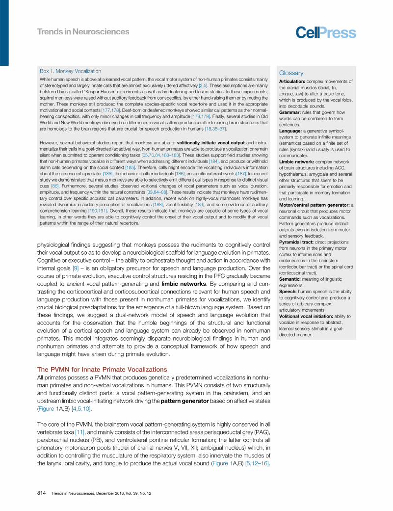
The PVMN for Innate Primate VocalizationsAll primates possess a PVMN that produces genetically predetermined vocalizations in nonhu-man primates and non-verbal vocalizations in humans. This PVMN consists of two structurallyand functionally distinct parts: a vocal pattern-generating system in the brainstem, and anupstream limbic vocal-initiating network driving the pattern generator based on affective states(Figure 1A,B) [4,5,10].
The core of the PVMN, the brainstem vocal pattern-generating system is highly conserved in allvertebrate taxa [11], and mainly consists of the interconnected areas periaqueductal grey (PAG),parabrachial nucleus (PB), and ventrolateral pontine reticular formation; the latter controls allphonatory motoneuron pools (nuclei of cranial nerves V, VII, XII; ambigual nucleus) which, inaddition to controlling the musculature of the respiratory system, also innervate the muscles ofthe larynx, oral cavity, and tongue to produce the actual vocal sound (Figure 1A,B) [5,12–16].
814 Trends in Neurosciences, December 2016, Vol. 39, No. 12
Monkey Human
VAMN
vIPFC/Broca
vIPFC/Broca
ACC Hypothalamusamygdala
PMv M1
ACC Hypothalamusamygdala
Motoneuron pools
Ar�cula�ontongue
facialjaw
Respira�onabdominalintercostal
diaphragmalLaryngeal
externalinternal
V
VII
XII
VH
NA
CS
ASPS
45Broca 44 PMv
Broca
CS
VVII
CranialLarynx
TongueExpira�on
Anterior
Muscles
Muscles
XII
NAVH
M1
M1
PMv
V XIIVII
NACranialLarynx
TongueExpira�on
VH
4445
ACC
ACC
AmHy PAG
LRF
PB
PB
AmHy
PAG
LRF
Motoneuron pools
Ar�cula�ontongue
facialjaw
Respira�onabdominalintercostal
diaphragmalLaryngeal
externalinternal
V
VII
XII
VH
NA
Cor�cobulbar/cor�cospinal tract
Cor�cobulbar/cor�cospinal tract
PAGand
adjacent lateraltegmentum
PB
PVMN
(B)
(A)
PVMN
Lateralret.form.
withNRA
PAGand
adjacent lateraltegmentum
PB
Lateralret.form.
withNRA
PMv M1
VAMN
Figure 1. Dual-Network Model. (A) Simplified circuit diagram summarizing the most relevant structures for vocal production in monkeys and speech in humans.Arrows indicate anatomically verified and relevant direct connections. The VAMN (indicated in green) is capable of initiating and modulating vocal production in monkeysduring cognitive control of vocal onset, or the modulation of vocal patterns within natural constraints. These connections are enhanced during speech evolution in theprimate lineage. (B) Anatomical locations and connections of the structures comprising the dual-network in monkeys (left) and humans (right). Lateral (front) and medial(back) views of the endbrain hemispheres are shown. Regions and arrows shaded in blue indicate the structures of the PVMN. Regions and arrows shaded in greendepict cortical areas involved in the VAMN. Red arrow indicates the direct connection between the larynx area of the primary motor cortex with the ambigual nucleus as anew development in the human lineage (adapted from [4,67,200]). Abbreviations: Am, amygdala; ACC, anterior cingulate cortex; AS, arcuate sulcus; CS, central sulcus;Hy, hypothalamus; LRF, lateral reticular formation; M1, ventral primary motor cortex; NA, ambigual nucleus; NRA, retroambigual nucleus; PAG, periaqueductal grey; PB,parabrachial nucleus; PMv, ventral premotor cortex; PS, principal sulcus; PVMN, primary vocal motor network; VH, respiratory motorneuron pools in the ventral horn ofthe spinal tract; V, motor trigeminal nucleus; VAMN, volitional articulatory motor network; VI,I facial nucleus; XII, hypoglossal nucleus.
The pivotal role of these brainstem structures has been deciphered based on electrophysiologi-cal recording, stimulation, and lesion studies in monkeys. In all of these brainstem areas, neuronsshow vocalization-related neuronal activity [13,15,17–19]. Electrical and chemical stimulation ofthe PAG reliably elicits distinct calls of the complete species-specific vocal repertoire in monkeys[20,21], whereas lesioning PAG results in mutism [22,23]. In contrast to the more general (orglobal) function of the PAG in producing calls, the PB is involved in the gating of vocal onset on
Trends in Neurosciences, December 2016, Vol. 39, No. 12 815
the basis of the momentary respiratory status [5] because electrical stimulation of the PBproduces only simple vocalizations [20,24], and PB lesions uncoupled respiratory rhythms fromlaryngeal activity during vocalizations elicited via PAG stimulation [23].
This brainstem vocal pattern-generating system is controlled by a limbic vocal-initiating networkthat projects to the PAG and the entire PVMN to elicit vocalizations representing affective states.Its most important structures comprise a vocalization region in the anterior cingulate cortex(ACC) around the rostrum of the corpus callosum (including parts of areas 24, 25, and 32), thehypothalamus, other limbic diencephalic structures (such as the septum and the subcallosalgyrus), and the amygdala [23,25,26]. Electrical stimulation in all of these limbic structuresincluding ACC elicits vocal utterances [20,27–29]. However, vocal latencies relative to stimula-tion onset were much longer (>1 s) compared to response latencies in PAG (<1 s) [20],suggesting that ACC and other limbic structures are coupled to, but are not part of, the patterngenerator. In contrast to PAG stimulation, stimulation of most limbic structures could only elicit asubset of call types of the species-specific call repertoire. For example, vocalization withhedonistic quality can be elicited in the septum, while stimulation sites in the amygdala elicitedmore-aversive vocal utterances [20,30,31].
Bilateral ablations of the cingulate vocalization region have either no consistent effect onspontaneous vocal behavior [32–34], and lead to a decrease in spontaneous vocalization rate[35] or to calls weakened in amplitude and duration [36]. In all cases, however, animals were stillable to spontaneously phonate after ACC lesions. In contrast to spontaneous calls, discrim-inatively conditioned vocal behavior was disrupted in monkeys with such bilateral lesions [35,36].Ablations of other limbic structure such as amygdala and hypothalamus suppress distinctspontaneously uttered vocalizations, but do not abolish calls elicited by stimulating PAG [23,37].
These findings strongly suggest that the limbic vocal-initiating network is not involved in theproduction of vocal patterns themselves, but governs the affective (emotional or motivational)initiation of the vocal output.
The Role of the PVMN in Human Non-Verbal Vocalizations and SpeechIt is important to realize that the phylogenetically conserved PVMN is still involved in vocalizationin humans. One of its functions is to produce non-verbal vocal utterances such as crying,laughing, or moaning, all of which are innately predetermined and affective vocalizationsconsidered to be directly homologous to monkey vocalizations [4,5]. Although brainstem lesionsare often fatal, a clinical study revealed pathological laughter and crying due to a tumor beneaththe brainstem that most likely deteriorated networks within the brainstem [38].
Several clinical studies also implicate the ACC in the initiation of non-verbal vocal utterances. In atype of frontal lobe epilepsy characterized by involuntary and stereotyped bursts of laughter(’gelastic seizures’ [39]), the cingulate gyrus appears to be the most commonly disrupted site[40]. In agreement with this idea, electrical stimulation of the rostral ACC (and the hypothalamus)elicited uncontrollable, but natural-sounding laughter [39,41,42]. At the same time, ACC isimportant for speech. In humans, bilateral infarction of the ACC near the rostrum of the corpuscallosum results in akinetic mutism [43,44], but with the potential for restoration of speechcharacterized by monotonous intonation, suggesting that the ACC is involved in the emotionalintonation of human speech [37,45]. This idea is supported by recent imaging studies showingACC activation during anger-expression of human speech [46]. Another important function ofthe PVMN is vocal output during speech production. Midbrain areas such as the PAG play asignificant role in vocal production in humans, and its lesions can cause akinetic mutism [47,48].In a case report, lesions in the PAG resulted in mutism, in other words it caused not only theabsence of all non-verbal vocal utterances, but also the absence of speech [49]. Interestingly,
816 Trends in Neurosciences, December 2016, Vol. 39, No. 12
this patient was still able to produce the articulatory movements that are accompanied by aspecific speech pattern, but the tonal component was no longer elicited [49]. However, PAG isactive during the expression of non-verbal vocal utterances such as laughter in humans [50].Moreover, a PET study showed that PAG is functionally coupled to a wide array of regions duringvoiced speech, but not during whispered speech [51].
Collectively, patient studies indicate that the PVMN is responsible for eliciting non-verbal affectivevocalization but, in addition, plays a major role in speech production at the expense of affectivevocalizations. Because the PVMN cannot subserve both affective preprogrammed vocalizationsand volitional speech articulation at the same time, the evolution of speech and language mayhave required the taking-over and dissolution of the majority of preprogrammed vocalizationpatterns [52]. Potentially recapitulating such an evolutionary scenario during ontogeny, bothlaughter and crying constitute extremely important forms of communication in human infancy,bridging the gap between a prelinguistic stage to the later stage of speech and languageacquisition. The advent of speech and language in Hominini therefore predicts the emergence ofa new neocortical cognitive control network that occupies the ancient brainstem vocalizationcircuits through a VAMN. The neurobiological foundations for this radical reorganization arevisible in nonhuman primates.
Volitional Articulatory Motor NetworkHumans possess an additional VAMN consisting of cortical structures crucial for human speechcontrol, and which is already present, although being anatomically and functionally underdevel-oped, in the monkey brain. The VAMN comprises the inferior frontal gyrus (IFG), the caudallybordering ventral premotor cortex (area 6, PMv), and ventrolateral primary motor cortex (area 4,M1) including the facial and laryngeal motor cortex (Figure 1A,B). The central executive of thisnetwork is Broca's area, located in the IFG of the granular ventrolateral PFC (vlPFC). The granularPFC was added newly to the anterior pole of the frontal lobe during the evolution of primates[53,54].
Broca's area classically comprises cytoarchitectonic areas 44 (pars opercularis) and 45 (parstriangularis) in the left hemispheres, complemented by some authors by area 47 (pars orbitalis)[55]. Broca's pioneering work on brains of aphasics [56] revealed that areas 44 and 45 on the leftside of the brain are instrumental for the production, or articulation, of speech and language.Studies on the cytoarchitectonics of macaque brains identified homologs of area 45 on theposterior convexity of the vlPFC, and of area 44 in the fundus of the inferior limb of the arcuatesulcus, a landmark that separates the PFC from the PMv (Figure 1B) [57,58]. No directconnections exist between the vlPFC and the primary motor cortex, but there are extensiveprojections to adjacent PMv that, in turn, send projections to primary motor cortex and the spinalcord. Specifically, area 44 is connected with the anterior PMv at the convexity of the inferiorarcuate sulcus. The anterior PMv integrates sensorimotor signals of the posterior parietal cortexwith cognitive information originating from vlPFC, pre-SMA, and cingulate area 24, the latterbeing in turn connected with the vlPFC [59]. Information in anterior PMv is then broadcast to theadjacent areas of the PMv and finally to ventrolateral M1, which also integrates laryngealsensorimotor information [60] for the generation and control of face/mouth movements [61].Neurons in both the PMv and M1 have direct access to the spinal cord, and thus can influencethe generation and control of speech-related movements [62].
While the motor cortex of non-primate mammals is connected with the phonatory motornuclei via interneurons in the reticular formation, only primates show direct connections viathe corticobulbar and corticospinal tracts, respectively [63]. These connections give bothNew World and Old World monkeys the capability to volitionally control their articulatorymuscles to respond with licking at a feeding tube in an operant conditioning task [64], and to
Trends in Neurosciences, December 2016, Vol. 39, No. 12 817
mimic articulatory vocal movements in a detection task [65], respectively. In humans only,additional direct connections of the motor cortex with the ambigual nucleus seem to exist[66] (Figure 1A,B), and humans also possess direct projections into the anterior horn of thethoracic and upper lumbar cord (reviewed in [5]). The motor cortex therefore plays a crucialrole within the VAMN in humans [67]. Lesions in the human ventral motor cortex, whichcontrols cranial and laryngeal muscles via the corticobulbar tract, lead to severe speechimpairments, while the production of innate non-verbal vocal utterances such as laughingand crying remains intact [68].
Despite the crucial role of Broca's area in human speech and language production, neitherelectrical stimulation nor lesions in homologous areas of monkeys have significant effects onspontaneous vocalizations [5]. For example, bilateral destruction of the ‘cortical face area’,including the premotor, motor, and sensory representation of the jaw muscles, lips, tongue,velum, and larynx, does not affect the acoustic structure of vocalizations in either squirrel ormacaque monkeys [18,32,69]. Similarly, ablation of lateral frontal areas in macaques does notaffect vocalizations [35,36]. Moreover, electrical stimulation in the motor cortex of monkeys doesnot elicit vocal output [70,71], and vocal utterances cannot reproducibly be elicited by stimulat-ing the premotor and motor cortex of anesthetized chimpanzees [72–74]. These findings weretaken as evidence that the lateral frontal lobe areas do not play any role in monkey vocalizations.If true, this would constitute an apparent discontinuity between the human speech system andthe monkey vocalization network.
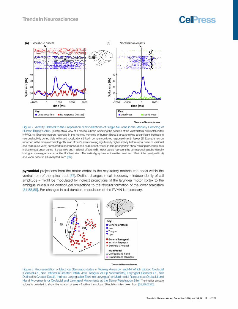
Recent experiments in nonhuman primates, however, showed that the monkey homolog ofBroca's area, as well as the premotor and/or primary motor cortices, are all involved in theinitiation of volitional calls that have been uttered in response to visual or auditory stimuli,respectively [65,75–77]. Vocalization-related activity that specifically predicts the preparationof instructed vocalizations in response to the detection of arbitrary visual stimuli wasrecorded in monkey vlPFC (areas 44 and 45) [76] and PMv [65,76] (Figure 2). Moreover,the activity of many call-related neurons before vocal output correlated with call parametersof instructed vocalizations. Furthermore, neuronal responses during conditioned vocaliza-tions were higher than during spontaneous vocalizations, suggesting a specific involvementof the monkey homolog of Broca's area during volitional monkey vocalizations (Figure 2). Atfirst sight, these results seem to be contradictory to earlier studies showing that bilateralablation of ventrolateral aspects of the frontal lobe have no significant impact on discrim-inatively conditioned vocal behavior [36]. However, the exact positions of areas 44 and 45were anatomically not verified until recently [57,78]. Therefore, the most likely explanation forthis discrepancy is that experimental lesions in the previous studies have not included the fullextent or even the bulk of the monkey vlPFC, PMv, and M1. In all lesioned monkeys,ablations left areas 6vr, 44, 45, and M1 at least partially intact in one hemisphere (seeFigure 1 in [36]).
Based on recent results and neuroanatomical projections, the vlPFC might take control over thevocal motor network via ACC or alternatively via the premotor cortex that has direct input tomotor cortex [79,80]. Because electrical microstimulation in area 44 and the PMv elicitedorofacial and laryngeal responses (Figure 3) [65,79,81–83], the pathway leading from vlPFCvia PMv to M1 to the corticobulbar and corticospinal tract to control phonatory motor neuronsseems more likely.
An influence via the VAMN could explain the rudimentary capability of monkeys to change vocalparameters such as vocal duration, amplitude, and frequency during cognitive vocal behavior[84–86]. Amplitude and frequency modulations can be generated by increasing the expiratory airflow passing the vocal folds in the larynx. This modulation might be solely controlled via direct
818 Trends in Neurosciences, December 2016, Vol. 39, No. 12
Vocal cue onsets Vocaliza�on onsets
40
(A) (B)
Cued vocs (hits) No response (misses)
30
20
10
0–1000 0 1000
Time (ms)
Key:
Time (ms)
Spik
e ra
te (H
z)
40
30
20
10
0
Spik
e ra
te (H
z)
2000 3000 –2000 –1000 0 1000
Cued vocs Spont. vocsKey:
Figure 2. Activity Related to the Preparation of Vocalizations of Single Neurons in the Monkey Homolog ofHuman Broca's Area. (Inset) Lateral view of a macaque brain indicating the position of the ventrolateral prefrontal cortex(vlPFC). (A) Example neuron recorded in the monkey homolog of human Broca's area showing a significant increase inneuronal activity during trials with cued vocalizations (hits) in comparison to no response trials (misses). (B) Example neuronrecorded in the monkey homolog of human Broca's area showing significantly higher activity before vocal onset of volitionalcoo calls (cued vocs) compared to spontaneous coo calls (spont. vocs). (A,B) Upper panels show raster plots, black dotsindicate vocal onset during hit trials in (A) and mark call offsets in (B); lower panels represent the corresponding spike-densityhistograms averaged and smoothed for illustration. The vertical grey lines indicate the onset and offset of the go-signal in (A)and vocal onset in (B) (adapted from [76]).
pyramidal projections from the motor cortex to the respiratory motoneuron pools within theventral horn of the spinal tract [87]. Distinct changes in call frequency – independently of callamplitude – might be modulated by indirect projections of the laryngeal motor cortex to theambigual nucleus via corticofugal projections to the reticular formation of the lower brainstem[81,88,89]. For changes in call duration, modulation of the PVMN is necessary.
45
446vr
General orofacialKey:
General larnygeal
Mul�modal
Jaw
Intrinsic laryngeal
Orofacial and handOrofacial and laryngeal
Extrinsic laryngeal
TongueLips
Figure 3. Representation of Electrical Stimulation Sites in Monkey Areas 6vr and 44 Which Elicited Orofacial[General (i.e., Not Defined in Greater Detail), Jaw, Tongue, or Lip Movements], Laryngeal [General (i.e., NotDefined in Greater Detail), Intrinsic Laryngeal or Extrinsic Laryngeal] or Multimodal Responses (Orofacial andHand Movements or Orofacial and Laryngeal Movements at the Same Penetration Site). The inferior arcuatesulcus is unfolded to show the location of area 44 within the sulcus. Stimulation sites taken from [65,79,82,83].
Trends in Neurosciences, December 2016, Vol. 39, No. 12 819
Brain imaging evidence suggests that the mechanism of how the lateral frontal lobe gains controlover articulation is not a direct excitation of the phonatory motor neurons in the brainstem, but isinstead a disinhibition of articulatory muscle activity briefly before vocal output [90]. This line ofargumentation would predict that non-verbal, emotional vocalizations might emerge once themodulatory (and/or inhibitory) influence of the voluntary articulation network vanishes [91].Indeed, non-verbal vocal utterances remain intact despite devastating impairments in speechand language production (Broca's aphasia) after damage to the posterior IFG [56,68,92,93].Moreover, patients with a clinical diagnosis of primary progressive aphasia develop abnormallaughter-like vocalizations that increasingly replace speech in the context of progressive speech-output impairment leading to mutism, until ultimately laughter-like vocalizations are the onlyextended utterance produced by these patients [94]. Interestingly, some non-verbal vocalutterances occur more often during the conversation of aphasic than of nonaphasic adults[95], suggesting a competitive modulatory influence of the VAMN over the PVMN.
In agreement with such a modulatory, possibly inhibitory, effect of the VAMN is the classic findingthat electrical stimulation of the Broca's area leads to speech arrest rather than to speechproduction [96–98]. Direct cortical recordings revealed that Broca's area is predominantlyactivated before the utterance of a speech signal, but is silent during the correspondingarticulation [99], again indicating that Broca's area is indirectly involved in coordinating speechinitiation rather than in producing speech output directly [93,100]. These findings are supportedby recent findings in awake neurosurgical patients in which the activity of distinct cortical speechsites was focally decreased via cooling [101]. Cooling of Broca's area predominantly alteredspeech timing, again indicating an involvement of Broca's area in speech coordination ratherthan in direct speech production. By contrast, focal lowering of temperature in speech motorcortex led to modulation of articulation, confirming the direct role of the speech motor cortex inarticulatory motor control. The recent cognitive control signals found in the monkey lateral frontallobe, in combination with the modulatory function of the VAMN and the emergence of non-linguistic vocalizations after its damage in humans, suggest that the humble beginnings ofspeech control can already be witnessed in nonhuman primates. From there, they seemed tohave evolved into a full-blown language-production system in humans after expansion of PFCcircuitry (Box 2).
The PFC Receives and Classifies Communicative SignalsFor a cortical network to control vocal output during reciprocal communication acts, sensoryinput from a sender is required. The vlPFC as the core of the VAMN receives highly processedinformation from higher-order sensory areas of all modalities (Figure 4). Neurons in the vlPFCcategorize and maintain communicative signals in working memory to guide goal-directedoutput.
Auditory information reaches the lateral PFC via two largely anatomically and functionallysegregated cortical streams: the anteroventral and the posterodorsal stream originating fromthe primary auditory cortex of the temporal lobe [102]. In the posterodorsal stream, the posteriorauditory belt and parabelt areas project directly to the dorsolateral PFC (dlPFC; areas 8, 46, and9) [102,103] (Figure 4). In addition, the posterior belt, parabelt, and superior temporal regionsproject indirectly to the posterior parietal association cortex before projecting to the dlPFC andprincipal sulcus (BA 46). This stream is thought to primarily encode auditory space [102] andquantity [104]. In the anteroventral stream, on the other hand, both the anterior belt and anteriorparabelt regions project directly and reciprocally to vlPFC (areas 12/47, 45, 44, and 12 orbital)[103,105]. In addition to this direct projection, there is also an indirect projection to the PFC viathe temporal association cortices. The anterior belt and parabelt regions are connected with therostral (TS1, TS2) superior temporal gyrus (STG) that contains a ‘voice region’ in which neuronsrespond preferentially to monkey calls [106–108]. The STG in turn projects to relatively restricted
820 Trends in Neurosciences, December 2016, Vol. 39, No. 12
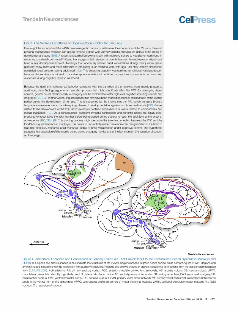
Box 2. The Neoteny Hypothesis of Cognitive Vocal Control for Language
How might the expansion of the VAMN have emerged in human primates over the course of evolution? One of the mostpowerful mechanisms evolution can use to remodel organs with very few genetic changes are delays in the timing ofdevelopmental stages [192]. A recent longitudinal behavioral study with monkeys trained to vocalize on command inresponse to a visual cue in a self-initiated trial suggests that retention of juvenile features, termed neoteny, might havebeen a key developmental event. Monkeys that laboriously master cued vocalizations during their juvenile phasegradually show more and more difficulties in producing such volitional calls with age, until they entirely discontinuecontrolled vocal behavior during adulthood [193]. This emerging disability was confined to volitional vocal productionbecause the monkeys continued to vocalize spontaneously and continued to use hand movements as instructedresponses during cognitive tasks in adulthood.
Because the decline in volitional call behavior correlated with the transition of the monkeys from juvenile phases toadulthood, these findings argue for a maturation process that might specifically affect the PFC. By prolonging devel-opment, greater neural plasticity early in ontogeny can be exploited to foster high-level cognition including speech andlanguage [194,195]. In other words, linguistic capabilities may have been enabled because of an expansion of the juvenileperiod during the development of humans. This is supported by the finding that the PFC which contains Broca'slanguage area experiences extraordinary long phases of developmental reorganization of neuronal circuits [196]. Genesrelated to the development of the PFC show excessive neotenic expression in humans relative to chimpanzees andrhesus macaques [197]. As a consequence, excessive synaptic connections and dendritic spines are initially over-produced to about twice the adult number before being pruned during puberty to reach the adult level at the onset ofadolescence [196,198,199]. This pruning process might decouple the juvenile connection between the PFC and thePVMN during adolescence in monkeys. This points to non-activity-related developmental reorganization in the brain ofmaturing monkeys, rendering adult monkeys unable to bring vocalizations under cognitive control. This hypothesissuggests that expansion of the juvenile period during ontogeny may be one of the key events in the evolution of speechand language.
M1
M1
A1
A1
V1
V1
dlPFC
dlPFC
vlPFC
vlPFC
Am
Am
Hy
Hy
PAG
PAG
LRF
LRF
PB
PB
ACC
ACC
45
45
44
44
Cranial
Cranial
Larynx
Larynx
Tongue
Tongue
Expira�on
Expira�on
Anterior
Muscles
Muscles
PMv
PMvV
V
VII
VII
XII
XII
NA
NA
VH
VH
Broca
Broca
CS
CS
AS
PS
Figure 4. Anatomical Locations and Connections of Sensory Structures That Provide Input to the Vocalization/Speech Systems in Monkeys andHumans. Regions and arrows shaded in blue indicate the structures of the PVMN. Regions shaded in green depict cortical areas comprising the VAMN. Regions andarrows shaded in purple show the interaction with auditory structures. Regions and arrows shaded in orange indicate the connections from the visual system (adaptedfrom [4,67,102,200]). Abbreviations: A1, primary auditory cortex; ACC, anterior cingulate cortex; Am, amygdala; AS, arcuate sulcus; CS, central sulcus; dlPFC,dorsolateral prefrontal cortex; Hy, hypothalamus; LRF, lateral reticular formation; M1, ventral primary motor cortex; NA, ambigual nucleus; PAG, periaqueductal grey; PB,parabrachial nucleus; PMv, ventral premotor cortex; PS, principal sulcus; PVMN, primary vocal motor network; V1, primary visual cortex; VH, respiratory motorneuronpools in the ventral horn of the spinal tract; vlPFC, ventrolateral prefrontal cortex; V, motor trigeminal nucleus; VAMN, volitional articulatory motor network; VII, facialnucleus; XII, hypoglossal nucleus.
Trends in Neurosciences, December 2016, Vol. 39, No. 12 821

Spik
e ac
�vi
ty (H
z)
(A) Vocal and auditorys�mula�on onsets Vocal offsets
Time (ms) Time (ms)
20
10
0
20
10
0–500 –250 0 500 10000 500 1000
(B)
Spik
e ac
�vi
ty (H
z)
Auditory only
Visual only
Audiovisual
Cued vocsKey:
Auditorys�mula�on
Figure 5. Audio–Vocal and Multisensory Activity in Single Neurons of the Monkey Ventrolateral PrefrontalCortex (vlPFC). (Inset) Lateral view of a macaque brain indicating the position of the vlPFC. (A) Example neuron recorded inthe monkey homolog of human Broca's area showing a phasic response during auditory stimulation and increased neuronalactivity before volitional calls (cued vocs) (from [111]). (B) Example neuron showing nonlinear activity in response to auditory(vocalization), visual (silent movie), and audiovisual stimuli (congruent face-vocalization movie) demonstrating a significantinteraction between auditory and visual stimuli, and exhibiting multisensory enhancement (adapted from [126]), Upperpanels show raster plots (A,B); black dots indicate vocal onset during hit trials in (A), and lower panels represent thecorresponding spike-density histograms averaged and smoothed for illustration. The vertical grey lines indicate the onset ofthe go-signal in (A), and the onset and offset of the vocalization, the silent movie, and the face-vocalization movies (B).
clusters of the vlPFC. The anteroventral stream is thought to encode auditory identity. Patches ofsingle neurons in vlPFC respond robustly to complex sounds such as species-specific vocal-izations or human vocalizations [109–111] (Figure 5A), and to some categories of vocalizationcalls [112]. In addition, dense auditory projections also exist from the rostral STG to the medialPFC, specifically to areas 32 and 25 of the ACC [113].
These separate projection streams in the auditory cortex are reminiscent of the temporal ‘what’and dorsal ‘where’ visual pathways [114], or ‘perception–action’ pathways, respectively [115]. Inthe temporal visual pathway, higher-order representations of objects are found in the terminationzone of the inferior temporal cortex where neurons encode specific object categories such asfaces [116,117]. The downstream projections from IT cortex give rise to patches of face-selective neurons in the lPFC [118,119].
The largely segregated visual and auditory pathways converge in the vlPFC to give rise toneurons that represent higher-order multisensory and categorical representations of communi-cative signals. In line with evidence that vlPFC neurons represent perceptual and abstractcategories [120,121], neuronal responses in this region correlate with monkeys’ choices in anauditory same–different task [122] and during categorization of human speech sounds [123].Supporting the integration of modal communication channels, neurons in vlPFC also respond toparticular face–voice combinations [124–126] (Figure 5B). In addition, recent recording andinactivation studies in monkeys showed that the vlPFC is particularly important for processingbehaviorally-relevant stimuli during auditory and audiovisual working memory [127,128]. Suchmultisensory PFC activity might enable human and nonhuman primates to recognize thecorrespondence between vocalizations and the facial postures associated with them [126–129].
Basic auditory tasks also activate the human vlPFC [130–132]. More-dorsal PFC regions (area46/9) are utilized when verbal working memory is required, whereas vlPFC regions (47/12; 45)are recruited during active retrieval of verbal and nonverbal auditory information [133,134].During the selective retrieval of information from auditory memory, the vlPFC interacts both with
822 Trends in Neurosciences, December 2016, Vol. 39, No. 12
the auditory temporal region (object information) and the inferior parietal lobule (spatial informa-tion) [134]. The broad involvement of the vlPFC in both verbal and nonverbal auditory taskssuggests that this area may be an interface in the auditory–vocal cycle. Collectively, these datasuggest that vlPFC is specialized for processing and integrating social communication informa-tion in monkeys [135], in the same way as the human IFG is specialized for processing andintegrating speech and gestures [136,137].
PFC Precursors of Semantics in Primate Referential SystemsThe VAMN is much more than merely a high-order motor network. It also plays a vital role inestablishing semantics and syntax, two hallmark characteristics of the two symbol systems –
language and number theory. In symbolic reference, relations are established between spoken/written words or numbers, respectively, on the basis of compositional rules (i.e., syntax)[138,139]. However, simpler and both phylogenetically and ontogenetically earlier referentialassociations are ‘indices’ – signs that are characterized by spatial or temporal associationbetween sign and object (reference based on contiguity or correlation) [140]. Animal communi-cation typically is indexical, for example vervet monkey alarm calls indicate the presence ofspecific predator categories that have to be learned by juvenile monkeys [141]. Moreover,conditioned sign–object associations established in animals by reward contingencies aretypically indexical, and can be investigated as a precursor for human symbolic reference [142].
Neurons in the vlPFC establish semantic associations: after training monkeys to associate thenumber of items in a set with arbitrary visual signs, many of the same lPFC neurons represent theabstract numerical meaning associated with such signs [143]. Similarly, fMRI studies show thatthe PFC is much more active in children learning semantic associations compared to proficientadults [144,145]. Damage to the human lateral frontal cortex results in severe impairment intasks that require learning of arbitrary associations [146]. Networks within the lateral frontalcortex may thus fulfill the requirements for high-order associations between signs, ultimatelygiving rise to the cultural invention of linguistic and number symbols. Symbolic reference maythus emerge as a function of a largely expanded lPFC in humans [140].
Semantic associations require interactions of the PFC with the medial temporal lobe, whichtraditionally has been linked to declarative long-term memories [147]. Recordings in behavingmonkeys show that, during learning and retrieval of long-term associations, the temporal lobeinteracts with the PFC to store memories or reactivate information about past experiences[148,149]. With its link to the medial temporal lobe structures, the PFC is ideally suited tocognitively control memories, permitting an organism to establish sign reference and to strate-gically plan communicative acts in a flexible and goal-directed way.
PFC Precursors of Grammar in Primate Referential SystemsTo establish a fully fledged symbol/language system, meaningful associations (semantics) are notsufficient. Sign sequences must be hierarchically structured according to action plans or rulesguiding the structuring of signs – ‘syntax’. Syntax refers to the rules governing structure in naturallanguage sentences or mathematical systems. Syntax establishes relations between signs thatdetermine the meaning of an expression. Therefore, syntax and semantics are inextricably linkedin symbolic systems.
Simple syntactical rules can be mastered by monkeys [150]. As a putative correlate for simplesyntactical processing, single neurons in the primate lPFC encode sequence plans [151], thestart and end states of behavioral sequences [152], and changing abstract rules [153,154].Moreover, precursors of a code for abstract temporal structures of sounds and hand move-ments have been revealed by single-unit and fMRI studies in monkey PFC [155,156]. In humans,fMRI data suggest that Broca's area processes the start- and end-points of higher-order motor
Trends in Neurosciences, December 2016, Vol. 39, No. 12 823
Outstanding QuestionsHow does activation or inactivation ofdistinct areas of the monkey homologof the human Broca's area deterioratevolitional vocal output?
Do preSMA, SMA, and the insula play asignificant role in cognitive control ofvocal output in non-human primates?
How is the VAMN interconnected, andhow is it linked to the PVMN, duringdevelopment from prelinguistic infantsto linguistic children?
What role do auditory feedback mech-anisms play in shaping vocal patterns innon-human primates? How does thePFC participate in potential vocal learn-ing mechanisms?
segments and controls the nesting of functional segments, thereby forming the hierarchicalstructure of action plans [157]. By contrast, other speech-related areas, such as the SMAcomplex and the insula, are more specifically involved in the precise timing of ongoing motor actsunderlying the execution of motor sequences needed for articulation [157–159].
Although neuronal circuits representing abstract sequences and rules are present in the monkeyvlPFC, a grasp of high-level recursive tree structures seems to be unique to human language andmathematics [160–162]. Complex syntactical representations may have impinged on inferiorfrontal cortex networks [163]: while simple, non-recursive grammar (finite-state grammar)activates the phylogenetically-older frontal operculum (i.e., premotor cortex), the computationof recursive hierarchical sequences (phrase/structure grammar) that is characteristic of humanlanguage additionally recruits the phylogenetically-younger Broca's area (areas 44 and 45).Human fMRI studies suggest purely syntax-related activation in Broca's area, either in BA 45 and47 [164], in BA 45 [165], or in BA 44 [166]. Using meta-analytic connectivity-based parcellation,Clos et al. [167] identified five functionally distinct clusters within left area 44 (associated withaction processing, sequencing, linguistic working memory, meaning, and task-switching/cog-nitive control). While these functions are highly relevant in the context of language production andcomprehension, they will obviously also be recruited by other domains including action andsocial cognition, therefore pointing to putative pre-adaptive functions in nonhuman primates.
Conclusions and Future DirectionsIn this review we suggest that the incipient linking of the prefrontal central executive of the brainwith the vocalization system is a key neurobiological event and pre-adaptation for the evolutionof speech and language. Crucial evidence for this hypothesis stems from comparative inves-tigations of the cytoarchitecture of the human and the monkey vlPFC. These studies demon-strated that the basic architectonic plans are similar in these two primate brains, despiteconsiderable development of the vlPFC areas in the human brain [57,78,168]. In addition toanatomical similarities, recent neurophysiological experiments in nonhuman primates providethe necessary functional evidence. Studies in behaving macaque monkeys showed that neuro-nal activity in vlPFC is correlated with volitional call initiation [76]. Although these correlativemeasures have led to novel insights concerning the role of inferior frontal lobe structures incontrolling vocal output, the causal efficacy of vocalization-related activity needs to beaddressed in the future. This could be achieved by probing the direct impact of experimentalneural perturbations (stimulation or inhibition) on vocal behavior [169]. If physiologically charac-terized vocalization-related neurons are causally involved in eliciting cued vocalizations, lowervocalization thresholds (i.e., higher vocalization rates) can be expected while electrically stimu-lating such neurons [170], whereas their transient chemical inactivation (i.e., by increasingsynaptic inhibition) is predicted to temporarily impair volitional call initiation. Such coarsehypotheses are of course subject to adjustments and refinements with increasing knowledgeabout the cortical vocal network.
As indicated by the (simplified) connections in our model, the vlPFC is the central executive of theVAMN. At the same time, however, it likely interacts with other areas of a larger frontal lobenetwork to encode cognitively controlled vocalizations. Medial frontal lobe areas, such as theanterior cingulate cortex (ACC) and the pre-supplementary motor area (pre-SMA), might playadditional roles not only in affective vocal output but also in controlling volitional vocalizations[5,29,171,172]. Data from the ACC and pre-SMA, among other areas, will be necessary tocomplete the emerging picture of a frontal vocal network.
A third important aspect is to clarify the level of specialization of frontal lobe circuitry in initiatinggoal-directed vocalizations. Instead of encoding any volitional motor act, neurons of the VAMNare expected to show some specificity towards controlling vocal output. To decipher the
824 Trends in Neurosciences, December 2016, Vol. 39, No. 12
vocalization-specific aspects of the VAMN, neurophysiological investigations in monkeys trainedto perform volitional acts using not only the vocal apparatus but also other effector organs (e.g.,hand movements) would be appropriate. This would allow the dedicated role of the VAMN vis-à-vis other volitional behaviors to be investigated. However, that neurons might respond exclu-sively to volitional vocal output does not appear to be realistic. After all, the primate vlPFCoperates at the apex of the cortical hierarchy and is involved in a variety of executive functions indifferent domains [9,173–175]. Despite its reputation as classical language area, even Broca'sarea in humans is part of a larger cognitive control network and plays important roles instructuring a variety of hierarchically organized behaviors [157,176]. Neurophysiological studiesin nonhuman primates will remain indispensable for identifying prefrontal cognitive controlfunctions as well as specific aspects of human speech and language functions.
AcknowledgementsWe thank Barbara Peysakhovich for proofreading. This work was supported by the Werner Reichardt Centre for Integrative
Neuroscience (CIN) at the Eberhard Karls University of Tübingen (CIN is an Excellence Cluster funded by the Deutsche
Forschungsgemeinschaft within the frame-work of the Excellence Initiative EXC 307).
References
1. Balter, M. (2010) Animal communication helps reveal roots oflanguage. Science 328, 969–971
2. Hammerschmidt, K. and Fischer, J. (2008) Constraints in primatevocal production. In Evolution of Communicative Flexibility: Com-plexity, Creativity, and Adaptability in Human and Animal Com-munication (Oller, D.K. and Griebel, U., eds), pp. 93–121, MITPress
3. Ghazanfar, A.A. (2008) Language evolution: neural differencesthat make the difference. Nat. Neurosci. 11, 382–384
4. Ackermann, H. et al. (2014) Brain mechanisms of acoustic com-munication in humans and nonhuman primates: an evolutionaryperspective. Behav. Brain Sci. 37, 529–546
5. Jürgens, U. (2002) Neural pathways underlying vocal control.Neurosci. Biobehav. Rev. 26, 235–258
6. Deacon, T.W. (1992) The neural circuitry underlying primate callsand human language. In Language Origin: A MultidisciplinaryApproach (Wind, J. et al., eds), pp. 121–162, Springer
7. Owren, M.J. et al. (2011) Two organizing principles of vocalproduction: implications for nonhuman and human primates.Am. J. Primatol. 73, 530–544
8. Holstege, G. and Subramanian, H.H. (2016) Two different motorsystems are needed to generate human speech. J. Comp.Neurol. 524, 1558–1577
9. Miller, E.K. and Cohen, J.D. (2001) An integrative theory ofprefrontal cortex function. Annu. Rev. Neurosci. 24, 167–202
10. Jürgens, U. (2009) The neural control of vocalization in mammals:a review. J. Voice 23, 1–10
11. Bass, A.H. and Chagnaud, B.P. (2012) Shared developmentaland evolutionary origins for neural basis of vocal–acoustic andpectoral–gestural signaling. Proc. Natl. Acad. Sci. U. S. A. 109(Suppl. 1), 10677–10684
12. Jürgens, U. (2000) Localization of a pontine vocalization-control-ling area. J. Acoust. Soc. Am. 108, 1393–1396
13. Lüthe, L. et al. (2000) Neuronal activity in the medulla oblongataduring vocalization. A single-unit recording study in the squirrelmonkey. Behav. Brain Res. 116, 197–210
14. Hannig, S. and Jürgens, U. (2006) Projections of the ventrolateralpontine vocalization area in the squirrel monkey. Exp. Brain Res.169, 92–105
15. Hage, S.R. and Jürgens, U. (2006) On the role of the pontinebrainstem in vocal pattern generation. A telemetric single-unitrecording study in the squirrel monkey. J. Neurosci. 26, 7105–7115
16. Hage, S.R. and Jürgens, U. (2006) Localization of a vocal patterngenerator in the pontine brainstem of the squirrel monkey. Eur. J.Neurosci. 23, 840–844
17. Larson, C.R. and Kistler, M.K. (1984) Periaqueductal gray neu-ronal activity associated with laryngeal EMG and vocalization inthe awake monkey. Neurosci. Lett. 46, 261–266
18. Kirzinger, A. and Jürgens, U. (1991) Vocalization-correlated sin-gle-unit activity in the brain stem of the squirrel monkey. Exp.Brain Res. 84, 545–560
19. Düsterhöft, F. et al. (2004) Neuronal activity in the periaqueductalgray and bordering structures during vocal communication in thesquirrel monkey. Neuroscience 123, 53–60
20. Jürgens, U. and Ploog, D. (1970) Cerebral representation ofvocalization in the squirrel monkey. Exp. Brain Res. 10, 532–554
21. Lu, C.L. and Jürgens, U. (1993) Effects of chemical stimulation inthe periaqueductal gray on vocalization in the squirrel monkey.Brain Res. Bull. 32, 143–151
22. Adametz, J. and O’Leary, J.L. (1959) Experimental mutismresulting from periaqueductal lesions in cats. Neurology 9,636–642
23. Jürgens, U. and Pratt, R. (1979) Role of the periaqueductal greyin vocal expression of emotion. Brain Res. 167, 367–378
24. Jürgens, U. and Müller-Preuss, P. (1977) Convergent projectionsof different limbic vocalization areas in the squirrel monkey. BrainRes. 29, 75–83
25. Dujardin, E. and Jürgens, U. (2005) Afferents of vocalization-controlling periaqueductal regions in the squirrel monkey. BrainRes. 1034, 114–131
26. Dujardin, E. and Jürgens, U. (2006) Call type-specific differencesin vocalization-related afferents to the periaqueductal gray ofsquirrel monkeys (Saimiri sciureus). Behav. Brain Res. 168,23–36
27. Smith, W.K. (1945) The functional significance of the rostralcingulate cortex as revealed by its responses to electrical excita-tion. J. Neurophysiol. 8, 241–255
28. Apfelbach, R. (1972) Electrically elicited vocalizations in the gib-bon Hylobates lar (Hylobatidae), and their behavioral significance.Z. Tierpsychol. 30, 420–430
29. Vogt, B.A. and Barbas, H. (1988) Structure and connections ofthe cingulate vocalization region in the rhesus monkey. In ThePhysiological Control of Mammalian Vocalization (Newman, J.D.,ed.), pp. 203–225, Plenum Press
30. Jürgens, U. (1979) Vocalization as an emotional indicator. Aneuroethological study in the squirrel monkey. Behaviour 69,88–117
31. Jürgens, U. (1982) Amygdalar vocalization pathways in the squir-rel monkey. Brain Res. 241, 189–196
32. Kirzinger, A. and Jürgens, U. (1982) Cortical lesion effects andvocalization in the squirrel monkey. Brain Res. 233, 299–315
Trends in Neurosciences, December 2016, Vol. 39, No. 12 825
33. Trachy, R.E. et al. (1981) Primate phonation: anterior cingulatelesion effects on response rate and acoustical structure. Am. J.Primatol. 1, 43–55
34. Franzen, E.A. and Myers, R.E. (1973) Neural control of socialbehavior: prefrontal and anterior temporal cortex. Neuropsychol-ogy 11, 141–157
35. Aitken, P.G. (1981) Cortical control of conditioned and sponta-neous vocal behaviour in rhesus monkeys. Brain Lang. 13, 171–184
36. Sutton, D. et al. (1974) Neocortical and limbic leasion effects onprimate phonation. Brain Res. 71, 61–75
37. Jürgens, U. et al. (1982) The effects of deep-reaching lesions inthe cortical face area on phonation: a combined case report andexperimental monkey study. Cortex 18, 125–139
38. Cantu, R.C. (1966) Importance of pathological laughing and/orcrying as a sign of occurrence or recurrence of a tumor lyingbeneath the brainstem. J. Nerv. Ment. Dis. 143, 508–512
39. Wild, B. et al. (2003) Neural correlates of laughter and humour.Brain 126, 2121–2138
40. Kovac, S. et al. (2009) Gelastic seizures: a case of lateral frontallobe epilepsy and review of the literature. Epilepsy Behav. 15,249–253
41. Kuzniecky, R. et al. (1997) Intrinsic epileptogenesis of hypotha-lamic hamartomas in gelastic epilepsy. Ann. Neurol. 42, 60–67
42. Sperli, F. et al. (2006) Contralateral smile and laughter, but nomirth, induced by electrical stimulation of the cingulate cortex.Epilepsia 47, 440–443
43. Nielsen, J.M. and Jacobs, L.L. (1951) Bilateral lesions of theanterior cingulate gyri; report of case. Bull. Los Angel. Neuro.Soc. 16, 231–234
44. Barris, R.W. and Schuman, H.R. (1953) Bilateral anterior cingu-late gyrus lesions; syndrome of the anterior cingulate gyri. Neu-rology 3, 44–52
45. Paus, T. (2001) Primate anterior cingulate cortex: where motorcontrol, drive and cognition interface. Nat. Rev. Neurosci. 2, 417–424
46. Frühholz, S. et al. (2015) Talking in fury: the cortico-subcorticalnetwork underlying angry vocalizations. Cereb. Cortex 25, 2752–2762
47. Nagaratnam, N. et al. (1999) Akinetic mutism and mixed trans-cortical aphasia following left thalamo-mesencephalic infarction.J. Neurol. Sci. 163, 70–73
48. Nagaratnam, N. et al. (2004) Akinetic mutism following stroke. J.Clin. Neurosci 11, 25–30
49. Esposito, A. et al. (1999) Complete mutism after midbrain peri-aqueductal gray lesion. Neuroreport 10, 681–685
50. Wattendorf, E. et al. (2013) Exploration of the neural correlates ofticklish laughter by functional magnetic resonance imaging.Cereb. Cortex 23, 1280–1289
51. Schulz, G.M. et al. (2005) Functional neuroanatomy of humanvocalization: an H2
15O PET study. Cereb. Cortex 15, 1835–1847
52. Deacon, T.W. (1992) The neural circuitry underlying primate callsand human language. In Language Origin: A MultidisciplinaryApproach (Wind, J. et al., eds), pp. 121–162, Kluwer AcademicPublishers
53. Preuss, T.M. (2006) Evolutionary specializations of primate brainsystems. In Primate Origins and Adaptations (Ravoso, M.J. andDagosto, M., eds), pp. 625–675, Kluwer Academic/PlenumPress
54. Wise, S.P. (2008) Forward frontal fields: phylogeny and funda-mental function. Trends Neurosci. 31, 599–608
55. Brodmann, K. ed. (1909) Vergleichende Lokalisationslehre derGrosshirnrinde in ihren Prinzipien dargestellt auf Grund des Zel-lenbaues, Barth JA
56. Broca, P. (1861) Remarques sur le siege de la faculté du langagearticulé, suivies d’une observation d’aphémie (perte de la parole)[Remarks on the seat of the faculty of articulated language,following an observation of aphemia (loss of speech)]. Bull.Mem. Soc. Anat. Paris 36, 330–357
57. Petrides, M. and Pandya, D.N. (2002) Comparative cytoarchi-tectonic analysis of the human and the macaque ventrolateral
826 Trends in Neurosciences, December 2016, Vol. 39, No. 12
prefrontal cortex and corticocortical connection patterns in themonkey. Eur J. Neurosci. 16, 291–310
58. Preuss, T.M. (2000) What's human about the human brain? InThe New Cognitive Neurosciences (Gazzaniga, M.S., ed.), pp.1219–1234, MIT Press
59. Gerbella, M. et al. (2011) Cortical connections of the anterior (F5a)subdivision of the macaque ventral premotor area F5. BrainStruct. Funct. 216, 43–65
60. Kumar, V. et al. (2016) Structural organization of the laryngealmotor cortical network and its implication for evolution of speechand language. J. Neurosci. 36, 4170–4181
61. Rizzolatti, G. et al. (1981) Response properties and behaviouralmodulation of ‘mouth’ neurons of the postarcuate. Brain Res.255, 421–424
62. Dum, R.P. and Strick, P.L. (1991) The origin of corticospinalprojections from the premotor areas in the frontal lobe. J. Neuro-sci. 11, 667–689
63. Simonyan, K. and Horwitz, B. (2011) Laryngeal motor cortex andcontrol of speech in humans. Neuroscientist 17, 197–208
64. Song, X. et al. (2016) Complex pitch perception mechanisms areshared by humans and a New World monkey. Proc. Natl. Acad.Sci. U. S. A. 113, 781–786
65. Coude, G. et al. (2011) Neurons controlling voluntary vocalizationin the macaque ventral premotor cortex. PLoS One 6, e26822
66. Simonyan, K. (2014) The laryngeal motor cortex: its organizationand connectivity. Curr. Opin. Neurobiol. 28, 15–21
67. Fuertinger, S. et al. (2015) The functional connectome of speechcontrol. PLoS Biol. e1002209
68. Groswasser, Z. et al. (1988) Mutism associated with buccofacialapraxia and bihemispheric lesions. Brain Lang. 34, 157–168
69. Green, H.D. and Walker, A.E. (1938) The effects of ablation of thecortical motor face area in monkeys. J. Neurophysiol. 1, 262–280
70. Kaada, B.R. (1951) Somato-motor, autonomic and electro-cor-ticographic responses to electrical stimulation of ‘rhinencephalic’and other structures in primates, cat and dog. Acta Physiol.Scand. 24 (Suppl. 83), 1–285
71. Robinson, B.W. (1967) Vocalization evoked from forebrain inMacaca mulatta. Physiol Behav 2, 345–354
72. Leyton, A.S.F. and Sherrington, C.S. (1917) Observations on theexcitable cortex of the chimpanzee, orangutan, and gorilla.Quart. J. Exp. Biol. 11, 135–222
73. Dusser de Barenne, J. et al. (1941) The ‘motor’ cortex of thechimpanzee. J. Neurophysiol. 4, 287–303
74. Hines, M. (1940) Movements elicited from precentral gyrus ofadult chimpanzees by stimulation with sine wave currents. J.Neurophysiol. 3, 442–466
75. Gemba, H. et al. (1999) Cortical field potentials associated withaudio-initiated vocalization in monkeys. Neurosci. Lett. 272, 49–52
76. Hage, S.R. and Nieder, A. (2013) Single neurons in monkeyprefrontal cortex encode volitional initiation of vocalizations.Nat. Commun. 4, 2409
77. Fukushima, M. et al. (2014) Modeling vocalization with ECoGcortical activity recorded during vocal production in the macaquemonkey. Conf. Proc. IEEE Eng. Med. Biol. Soc. 2014, 6794–6797
78. Petrides, M. and Pandya, D.N. (1999) Dorsolateral prefrontalcortex: comparative cytoarchitectonic analysis in the humanand the macaque brain and corticocortical connection patterns.Eur. J. Neurosci. 11, 1011–1036
79. Simonyan, K. and Jürgens, U. (2002) Cortico-cortical projectionsof the motorcortical larynx area in the rhesus monkey. Brain Res.949, 23–31
80. Yeterian, E.H. et al. (2012) The cortical connectivity of the pre-frontal cortex in the monkey brain. Cortex 48, 58–81
81. Simonyan, K. and Jürgens, U. (2003) Efferent subcortical pro-jections of the laryngeal motorcortex in the rhesus monkey. BrainRes. 974, 43–59
82. Petrides, M. et al. (2005) Orofacial somatomotor responses in themacaque monkey homologue of Broca's area. Nature 435,1235–1238
83. Hast, M.H. et al. (1974) Cortical motor representation of thelaryngeal muscles in Macaca mulatta. Brain Res. 73, 229–240
84. Sutton, D. et al. (1973) Vocalization in rhesus monkeys: con-ditionability. Brain Res. 52, 225–231
85. Larson, C.R. et al. (1973) Sound spectral properties of condi-tioned vocalization in monkeys. Phonetica 27, 100–110
86. Hage, S.R. et al. (2013) Cognitive control of distinct vocalizationsin rhesus monkeys. J. Cogn. Neurosci. 25, 1692–1701
87. Lemon, R.N. (2008) Descending pathways in motor control. Ann.Rev. Neurosci. 31, 195–218
88. Jürgens, U. and Zwirner, P. (1996) The role of the periaqueductalgrey in limbic and neocortical vocal fold control. Neuroreport 7,2921–2923
89. Jürgens, U. and Ehrenreich, L. (2007) The descending motor-cortical pathway to the laryngeal motoneurons in the squirrelmonkey. Brain Res. 1148, 90–95
90. McCairn, K.W. et al. (2016) A primary role for nucleus accumbensand related limbic network in vocal tics. Neuron 89, 300–307
91. Lauterbach, E.C. et al. (2013) Toward a more precise, clinically-informed pathophysiology of pathological laughing and crying.Neurosci. Biobehav. Rev. 37, 1893–1916
92. Caplan, D. et al. (1996) Location of lesions in stroke patients withdeficits in syntactic processing in sentence comprehension.Brain 119, 933–994
93. Lazar, R.M. and Mohr, J.P. (2011) Revisiting the contributions ofPaul Broca to the study of aphasia. Neuropsychol. Rev. 21, 236–239
94. Rohrer, J.D. et al. (2009) Abnormal laughter-like vocalisationsreplacing speech in primary progressive aphasia. J. Neurol. Sci.284, 120–123
95. Norris, M.R. and Drummond, S.S. (1998) Communicative func-tions of laughter in aphasia. J. Neuroling. 11, 391–402
96. Penfield, W. and Roberts, L. (1959) Speech and Brain Mecha-nisms, Princeton University Press
97. Epstein, C.M. et al. (1999) Localization and characterization ofspeech arrest during transcranial magnetic stimulation. Clin.Neurophysiol. 110, 1073–1079
98. Axelson, H.W. et al. (2009) Successful localization of the Brocaarea with short-train pulses instead of ‘Penfield’ stimulation.Seizure 18, 374–375
99. Flinker, A. et al. (2015) Redefining the role of Broca's area inspeech. Proc. Natl. Acad. Sci. U. S. A. 112, 2871–2875
100. Dronkers, N.F. and Baldo, J.V. (2010) Broca's area. In TheCambridge Encyclopedia of the Language Sciences (Hogan,P.C., ed.), pp. 139–142, Cambridge University Press
101. Long, M.A. et al. (2016) Functional segregation of cortical regionsunderlying speech timing and articulation. Neuron 89, 1187–1193
102. Rauschecker, J.P. and Scott, S.K. (2009) Maps and streams inthe auditory cortex: nonhuman primates illuminate humanspeech processing. Nat. Neurosci. 12, 718–724
103. Romanski, L.M. et al. (1999) Dual streams of auditory afferentstarget multiple domains in the primate prefrontal cortex. Nat.Neurosci. 2, 1131–1136
104. Nieder, A. (2012) Supramodal numerosity selectivity of neurons inprimate prefrontal and posterior parietal cortices. Proc. Natl.Acad. Sci. U. S. A. 109, 11860–11865
105. Hackett, T.A. et al. (1998) Thalamocortical connections of theparabelt auditory cortex in macaque monkeys. J. Comp. Neurol.400, 271–286
106. Poremba, A. et al. (2004) Species-specific calls evoke asym-metric activity in the monkey's temporal poles. Nature 427,448–451
107. Gil-da-Costa, R. et al. (2006) Species-specific calls activatehomologs of Broca's and Wernicke's areas in the macaque.Nat. Neurosci. 9, 1064–1070
108. Perrodin, C. et al. (2011) Voice cells in the primate temporal lobe.Curr. Biol. 21, 1408–1415
109. Romanski, L.M. and Goldman-Rakic, P.S. (2002) An auditorydomain in primate prefrontal cortex. Nat. Neurosci. 5, 15–16
110. Romanski, L.M. et al. (2005) Neural representation of vocaliza-tions in the primate ventrolateral prefrontal cortex. J. Neurophy-siol. 93, 734–747
111. Hage, S.R. and Nieder, A. (2015) Audio-vocal interaction in singleneurons of the monkey ventrolateral prefrontal cortex. J. Neuro-sci. 35, 7030–7040
112. Averbeck, B.B. and Romanski, L.M. (2006) Probabilistic encod-ing of vocalizations in macaque ventral lateral prefrontal cortex. J.Neurosci. 26, 11023–11033
113. Medalla, M. and Barbas, H. (2014) Specialized prefrontal ‘audi-tory fields’: organization of primate prefrontal–temporal path-ways. Front. Neurosci. 8, 77
114. Mishkin, M. et al. (1983) Object vision and spatial vision: twocortical pathways. Trends Neurosci. 6, 414–417
115. Goodale, M.A. and Milner, A.D. (1992) Separate visual pathwaysfor perception and action. Trends Neurosci. 15, 20–25
116. Gross, C.G. et al. (1972) Visual properties of neurons in infero-temporal cortex of the macaque. J. Neurophysiol. 35, 96–111
117. Desimone, R. et al. (1984) Stimulus-selective properties of inferiortemporal neurons in the macaque. J. Neurosci. 4, 2051–2062
118. O'Scalaidhe, S.P. et al. (1997) Areal segregation of face-proc-essing neurons in prefrontal cortex. Science 278, 1135–1138
119. Tsao, D.Y. et al. (2008) Patches of face-selective cortex in themacaque frontal lobe. Nat. Neurosci. 11, 877–879
120. Freedman, D.J. et al. (2001) Categorical representation of visualstimuli in the primate prefrontal cortex. Science 291, 312–316
121. Nieder, A. (2016) The neuronal code for number. Nat. Rev.Neurosci. 17, 366–382
122. Russ, B.E. et al. (2008) Prefrontal neurons predict choices duringan auditory samedifferent task. Curr. Biol. 18, 1483–1488
123. Lee, J.H. et al. (2009) Prefrontal activity predicts monkeys’ decisionsduring an auditory category task. Front. Integr. Neurosci. 3, 16
124. Sugihara, T. et al. (2006) Integration of auditory and visual com-munication information in the primate ventrolateral prefrontalcortex. J. Neurosci. 26, 11138–11147
125. Romanski, L.M. and Hwang, J. (2012) Timing of audiovisualinputs to the prefrontal cortex and multisensory integration.Neuroscience 214, 36–48
126. Diehl, M.M. and Romanski, L.M. (2014) Responses of prefrontalmultisensory neurons to mismatching faces and vocalizations. J.Neurosci. 34, 11233–11243
127. Hwang, J. and Romanski, L.M. (2015) Prefrontal neuronalresponses during audiovisual mnemonic processing. J. Neuro-sci. 35, 960–971
128. Plakke, B. et al. (2015) Inactivation of primate prefrontal corteximpairs auditory and audiovisual working memory. J. Neurosci.35, 9666–9675
129. Ghazanfar, A.A. and Takahashi, D.Y. (2014) The evolution ofspeech: vision, thythm, cooperation. Trends Cogn. Sci. 18,543–553
130. Zatorre, R.J. et al. (1994) Neural mechanisms underlying melodicperception and memory for pitch. J. Neurosci. 14, 1908–1919
131. Grady, C.L. et al. (2008) Age-related differences in brain activityunderlying working memory for spatial and nonspatial auditoryinformation. Cereb. Cortex 18, 189–199
132. Protzner, A.B. and McIntosh, A.R. (2009) Modulation of ventralprefrontal cortex functional connections reflects the inteplay ofcognitive processes and stimulus characteristics. Cereb. Cortex19, 1042–1054
133. Petrides, M. (1996) Specialized systems for the processing ofmnemonic information within the primate frontal cortex. Philos.Trans. R. Soc. Lond. B Biol. Sci. 351, 1455–1461
134. Kostopoulos, P. and Petrides, M. (2016) Selective memoryretrieval of auditory what and auditory where involves the ven-trolateral prefrontal cortex. Proc. Natl. Acad. Sci. U. S. A. 113,1919–1924
135. Romanski, L.M. (2012) Integration of faces and vocalizations inventral prefrontal cortex: implications for the evolution of audio-visual speech. Proc. Natl. Acad. Sci. U. S. A. 109 (Suppl. 1),10717–10724
Trends in Neurosciences, December 2016, Vol. 39, No. 12 827
136. Homae, F. et al. (2002) From perception to sentence compre-hension: the convergence of auditory and visual information oflanguage in the left inferior frontal cortex. Neuroimage 16, 883–900
137. Xu, J. et al. (2009) Symbolic gestures and spoken language areprocessed by a common neural system. Proc. Natl. Acad. Sci. U.S. A. 106, 20664–20669
138. Deacon, T.W. (1996) Prefrontal cortex and symbol learning: whya brain capable of language evolved only once. In Communicat-ing Meaning. The Evolution and Development of Language(Velichkovsky, B.M. and Rumbaugh, D.M., eds), pp. 103–138,Lawrence Earlbaum Associates
139. Wiese, H. (2003) Numbers, Language, and the Human Mind,Cambridge University Press
140. Deacon, T.W. (1997) The Symbolic Species: The Co-evolution ofLanguage and the Human Brain, W. W. Norton & Company
141. Cheney, D.L. and Seyfarth, R.M. (1992) How Monkeys See TheWorld: Inside the Mind of Another Species, University of ChicagoPress
142. Nieder, A. (2009) Prefrontal cortex and the evolution of symbolicreference. Curr. Opin. Neurobiol. 19, 99–108
143. Diester, I. and Nieder, A. (2007) Semantic associations betweensigns and numerical categories in the prefrontal cortex. PLoSBiol. 5, e294
144. Ansari, D. et al. (2005) Neural correlates of symbolic numberprocessing in children and adults. Neuroreport 16, 1769–1773
145. Cantlon, J.F. et al. (2009) The neural development of an abstractconcept of number. J. Cogn. Neurosci. 21, 2217–2229
146. Petrides, M. (1985) Deficits on conditional associative-learningtasks after frontal- and temporal-lobe lesions in man. Neuro-psychologia 23, 601–614
147. Simons, J.S. and Spiers, H.J. (2003) Prefrontal and medialtemporal lobe interactions in long-term memory. Nat. Rev. Neu-rosci. 4, 637–648
148. Tomita, H. et al. (1999) Top-down signal from prefrontal cortex inexecutive control of memory retrieval. Nature 401, 699–703
149. Brincat, S.L. and Miller, E.K. (2015) Frequency-specific hippo-campal–prefrontal interactions during associative learning. Nat.Neurosci. 18, 576–581
150. Wilson, B. et al. (2013) Auditory artificial grammar learning inmacaque and marmoset monkeys. J. Neurosci. 33, 18825–18835
151. Mushiake, H. et al. (2006) Activity in the lateral prefrontal cortexreflects multiple steps of future events in action plans. Neuron 50,631–641
152. Fujii, N. and Graybiel, A.M. (2003) Representation of actionsequence boundaries by macaque prefrontal cortical neurons.Science 301, 1246–1249
153. Wallis, J.D. et al. (2001) Single neurons in prefrontal cortexencode abstract rules. Nature 411, 953–956
154. Bongard, S. and Nieder, A. (2010) Basic mathematical rules areencoded by primate prefrontal cortex neurons. Proc. Natl. Acad.Sci. U. S. A. 107, 2277–2282
155. Shima, K. et al. (2007) Categorization of behavioural sequencesin the prefrontal cortex. Nature 445, 315–318
156. Wang, L. et al. (2015) Representation of numerical and sequentialpatterns in macaque and human brains. Curr. Biol. 25, 1966–1974
157. Koechlin, E. and Jubault, T. (2006) Broca's area and the hierar-chical organization of human behavior. Neuron 50, 963–974
158. Dronkers, N.F. (1996) A new brain region for coordinating speecharticulation. Nature 384, 159–161
159. Ackermann, H. and Riecker, A. (2004) The contribution of theinsula to motor aspects of speech production: a review and ahypothesis. Brain Lang. 89, 320–328
160. Chomsky, N. (1956) Three models for the description of lan-guage. IEEE Trans. Inf. Theory 2, 113–124
161. Penn, D.C. et al. (2008) Darwin's mistake: explaining the discon-tinuity between human and nonhuman minds. Behav. Brain Sci.31, 109–130
828 Trends in Neurosciences, December 2016, Vol. 39, No. 12
162. Dehaene, S. et al. (2015) The neural representation of sequen-ces: from transition probabilities to algebraic patterns and lin-guistic trees. Neuron 88, 2–19
163. Friederici, A.D. et al. (2006) The brain differentiates human andnon-human grammars: functional localization and structural con-nectivity. Proc. Natl. Acad. Sci. U. S. A. 103, 2458–2463
164. Pallier, C. et al. (2011) Cortical representation of the constituentstructure of sentences. Proc. Natl. Acad. Sci. U. S. A. 108, 2522–2527
165. Shetreet, E. et al. (2009) An fMRI study of syntactic layers:sentential and lexical aspects of embedding. Neuroimage 48,707–716
166. Goucha, T. and Friederici, A.D. (2015) The language skeletonafter dissecting meaning: A functional segregation within Broca'sArea. Neuroimage 114, 294–302
167. Clos, M. et al. (2013) Tackling the multifunctional nature ofBroca's region meta-analytically: co-activationbased parcellationof area 44. Neuroimage 83, 174–188
168. Petrides, M. and Pandya, D.N. (2009) Distinct parietal and tem-poral pathways to the homologues of Broca's area in the mon-key. PLoS Biol. 7, e1000170
169. Wurtz, R.H. (2015) Using perturbations to identify the braincircuits underlying active vision. Philos. Trans. R. Soc. Lond.B. Biol. Sci. 370, 20140205
170. Cohen, M.R. and Newsome, W.T. (2004) What electrical micro-stimulation has revealed about the neural basis of cognition. Curr.Opin. Neurobiol. 14, 169–177
171. West, R.A. and Larson, C.R. (1995) Neurons of the anteriormesial cortex related to faciovocal activity in the awake monkey.J. Neurophysiol. 74, 1856–1869
172. Alario, F.X. et al. (2006) The role of the supplementary motor area(SMA) in word production. Brain Res. 1076, 129–143
173. Merten, K. and Nieder, A. (2012) Active encoding of decisionsabout stimulus absence in primate prefrontal cortex neurons.Proc. Natl. Acad. Sci. U. S. A. 109, 6289–6294
174. Vallentin, D. et al. (2012) Numerical rule coding in the prefrontal,premotor, and posterior parietal cortices of macaques. J. Neuro-sci. 32, 6621–6630
175. Jacob, S.N. and Nieder, A. (2014) Complementary roles forprimate frontal and parietal cortex in guarding working memoryfrom distractor stimuli. Neuron 83, 226–237
176. Fazio, P. et al. (2009) Encoding of human action in Broca's area.Brain 132, 1980–1988
177. Winter, P. et al. (1973) Ontogeny of squirrel monkey calls undernormal conditions and under acoustic isolation. Behaviour 47,230–239
178. Hammerschmidt, K. et al. (2001) Vocal development in squirrelmonkeys. Behaviour 138, 1179–1204
179. Talmage-Riggs, G. et al. (1972) Effect of deafening on the vocalbehavior of the squirrel monkey (Saimiri sciureus). Folia Primatol.17, 404–420
180. Aitken, P.G. and Wilson, D.A. (1979) Discriminative vocal condi-tioning in Rhesus monkeys: evidence for volitional control? BrainLang. 8, 227–240
181. Hihara, S. et al. (2003) Spontaneous vocal differentiation of coo-calls for tools and food in Japanese monkeys. Neurosci. Res. 45,383–389
182. Koda, H. et al. (2007) Experimental evidence for the volitionalcontrol of vocal production in an immature gibbon. Behaviour144, 681–692
183. Sutton, D. et al. (1985) Discriminative phonation in macaques:effects of anterior mesial cortex damage. Exp. Brain Res. 59,410–413
184. Cheney, D.L. and Seyfarth, R.M., eds (2007) Baboon Metaphys-ics: The Evolution of a Social Mind, University of Chicago Press
185. Seyfarth, R.M. et al. (1980) Monkey responses to three differentalarm calls: evidence for predator classification and semanticcommunication. Science 210, 801–803
186. Wich, S.A. and de Vries, H. (2006) Male monkeys rememberwhich group members have given alarm calls. Proc. Biol. Sci.273, 735–740
187. Ouattara, K. et al. (2009) Campbell's monkeys concatenatevocalizations into context-specific call sequences. Proc. Natl.Acad. Sci. U. S. A. 106, 22026–22031
188. Eliades, S.J. and Wang, X. (2005) Dynamics of auditory–vocalinteraction in monkey auditory cortex. Cereb. Cortex 15,1510–1523
189. Choi, J.Y. et al. (2015) Cooperative vocal control in marmosetmonkeys via vocal feedback. J. Neurophysiol. 114, 274–283
190. Takahashi, D.Y. et al. (2015) The developmental dynamics ofmarmoset monkey vocal production. Science 349, 734–738
191. Takahashi, D.Y. et al. (2016) Early development of turn-takingwith parents shapes vocal acoustics in infant marmoset mon-keys. Phil. Trans. R. Soc. B 371, 20150370
192. Gould, S.J. ed. (1977) Ontogeny and Phylogeny, Harvard Uni-versity Press
193. Hage, S.R. et al. (2016) Developmental changes of cognitivevocal control in monkeys. J. Exp. Biol. 219, 1744–1749
194. Carroll, S.B. (2003) Genetics and the making of Homo sapiens.Nature 422, 849–857
195. Oller, D.K. ed. (2000) The Emergence of the Speech Capacity,Psychology Press
196. Petanjek, Z. et al. (2011) Extraordinary neoteny of synaptic spinesin the human prefrontal cortex. Proc. Natl. Acad. Sci. U. S. A.108, 13281–13286
197. Somel, M. et al. (2013) Human brain evolution: transcripts, metab-olites and their regulators. Nat. Rev. Neurosci. 14, 112–127
198. Bourgeois, J.P. et al. (1994) Synaptogenesis in the prefrontalcortex of rhesus monkeys. Cereb. Cortex 4, 78–96
199. Huttenlocher, P.R. and Dabholkar, A.S. (1997) Regional differ-ences in synaptogenesis in human cerebral cortex. J. Comp.Neurol. 387, 167–178
200. Hickok, G. and Poeppel, D. (2007) The cortical organization ofspeech processing. Nat. Rev. Neurosci. 8, 393–402
Trends in Neurosciences, December 2016, Vol. 39, No. 12 829