Embed Size (px)
Citation preview
Cellular Oncology 31 (2009) 393–405 393DOI 10.3233/CLO-2009-0484IOS Press
Dual function microtubule- andmitochondria-associated proteins mediatemitotic cell death
Leyuan Liu, Rui Xie, Chaofeng Yang and Wallace L. McKeehan ∗
Center for Cancer and Stem Cell Biology, Institute of Biosciences and Technology, Texas A&M Health ScienceCenter, Houston, TX, USA
Abstract. Background: Survival and evolution of aneuploid cells after an asymmetric segregation of chromosomes at mitosismay be the common initiating event and underlying cause of the genetic diversity and adaptability of cancers. We hypothesizethat mechanisms exist to detect impending aneuploidy and prevent it before completion of an aberrant mitosis.
Methods: The distribution of isoforms of C19ORF5, an interactive partner with mitochondria-associated LRPPRC and tumorsuppressor RASSF1A, state of spindle microtubules and mitochondrial aggregation was analyzed in synchronized mitotic cellsand cells stalled in mitosis after treatment with paclitaxel.
Results: C19ORF5 distributed broadly across the mitotic spindle and reversibly accumulated during reversible mitotic arrest.Prolonged stabilization of microtubules caused an accumulation of a C19ORF5 product with dual MAP and MtAP propertiesthat caused irreversible aggregation of mitochondria and death of mitotic cells.
Conclusion: Dual function microtubule-associated (MAP) and mitochondria-associated (MtAP) proteins generated by pro-longed mitotic arrest trigger mitochondrial-induced mitotic cell death. This is a potential mechanism to prevent minimal surviv-able aneuploidy resulting from an aberrant cell division and cancers in general at their earliest common origin.
Keywords: Aneuploidy, C19ORF5, genetic instability, LRPPRC, microtubule dynamics, mitochondrial dynamics, RASSF1A,paclitaxel, tumor suppression, mitochondria aggregation
1. Introduction
Abnormal chromosome number (aneuploidy) result-ing from an asymmetric chromosome segregation isthe single genetic abnormality that is common to alltumor cells [1–4]. The symmetry of chromosome seg-regation at mitosis is dependent on mitotic spindlefunction and alignment of all chromosome pairs atthe metaphase plate prior to onset of anaphase [5,6].Capture and alignment of chromosomes by micro-tubules is a stochastic and asynchronous process. Elab-orate short range, non-diffusible signals communicatethe status of microtubule-chromosomal junctions tothe anaphase-promoting complex/cyclosome (APC/C).The APC/C complex controls degradation of proteins
*Corresponding author: Dr. Wallace L. McKeehan, Center forCancer and Stem Cell Biology, Institute of Biosciences and Tech-nology, Texas A&M Health Science Center, Houston, TX 77030-3303, USA. Tel.: +1 713 677 7510; Fax: +1 713 677 7512; E-mail:[email protected].
that hinder chromosome separation and completion ofmitosis. Mechanisms that monitor chromosome align-ment are sufficiently sensitive to delay the onset ofanaphase because of a single unaligned chromosome.Delays known as mitotic checkpoints are necessar-ily of limited duration so that already aligned chro-mosomes with bipolar attachments to relatively stableK-fiber spindle microtubule bundles hold in position atthe plate while more dynamic microtubules continueto search for, capture and bring outliers into alignment[7]. If not for this relative stability of K-fiber micro-tubules, alignment of chromosomes would be an end-less process. It is during the final stages of the align-ment process that the threat of generation of a mini-mal non-lethal aneuploidy is the greatest should a celldivision proceed.
Mitochondria transition between punctate foci andtubular structures through fusion and fission dynam-ics [8]. Few studies have addressed in detail the rela-tionships between mammalian mitochondrial dynam-ics and mitochondrial spindle microtubule dynamics
1570-5870/09/$17.00 © 2009 – IOS Press and the authors. All rights reserved
394 L. Liu et al. / Dual function microtubule- and mitochondria-associated proteins mediate mitotic cell death
during mitosis [9]. In previous reports, we describeda novel complex of dual function MtAPs and MAP’sinvolved in communication between the microtubularcytoskeleton and mitochondria during chromosome re-modeling and mitosis [10]. Mitochondria-associatedLRPPRC (LRP130) is an mtAP that affects mitochon-drial gene expression and metabolism [11]. LRPPRCinteracts directly or indirectly with three dual functionMtAPs and MAPs. It interacts directly with dual func-tion MAP and MtAP UXT [12,13] and C19ORF5 (alsocalled MAP1S and RABP1) [10,14–16] and indirectlythrough C19ORF5 with the tumor suppressor RASSF1[10,17]. When RASSF1 that normally resides on mi-tochondria appears on microtubules, its buildup causesmicrotubule stabilization and bundling similar to thedrug paclitaxel [17]. C19ORF5 is distributed in the cy-tosol, but exists in multiple isoforms that conditionallyappear on both microtubules and mitochondria [14]. Atsufficient levels, one or more isoforms of C19ORF5concentrate on mitochondria and cause progressive ag-gregation of mitochondria resulting in DNA degrada-tion and cell death [14].
Isoforms of broadly expressed C19ORF5 that span115 to 20 kDa in cells and tissues exhibit homology toand features of neuronal tissue-associated MAP1 pro-teins, MAP1A and MAP1B [14,18]. Similar to MAP1proteins [19], C19ORF5 is expressed as the full lengthproduct deduced from cDNA and gives rise to di-verse posttranslational products. In this study we ex-amined the isoforms of C19ORF5 and their redistrib-ution among cytosol, microtubules and mitochondriain interphase and mitosis. Our results suggest a mech-anism for prevention of aneuploidy by mitochondria-induced death of mitotic cells prior to completion of anaberrant cell division. This is mediated by dual func-tion MAPs and MtAPs that bridge spindle microtubuleand mitochondrial dynamics.
2. Materials and methods
2.1. Immunoreagents
Antibodies for p-Histone H3 and β-actin were fromSanta Cruz Biotechnology and Sigma Chemicals, re-spectively. Anti-C19ORF5 monoclonal antibody 4G1was developed from purified GST-C19ORF5SC andrecognized an epitope between C19ORF5 sequenceD667 to S767 [14]. Anti-C19ORF5 polyclonal anti-bodies Magd and Mt were developed in rabbits witha synthetic peptide corresponding to the 25-residue
C19ORF5 MAGD domain (F967-A991) and a puri-fied C19ORF5 recombinant protein containing the mi-crotubule association domain (MT) (Fig. 1A) [14].The recombinant antigen was made from a con-struct of C19ORF5 sequence A867-E966 N-terminallytagged with GST that was expressed in E. coli, pu-rified with GSH affinity chromatography, the GSTwas removed with thrombin and the released pro-tein further purified by DNA affinity chromatogra-phy [14]. Specificity, titer and epitope for antibodieswas tested by immunoblot screens of lysates and im-munohistochemical analysis of cells transiently trans-fected with recombinant C19ORF5SC and productsof shorter constructs tagged at the N-terminus withGFP [14]. The Magd and Mt antibodies were use-ful for immunoblotting and immunostaining, respec-tively.
2.2. C19ORF5 recombinant construct
All recombinant constructs used in transfection ex-periments contained GFP at the N-terminus. The con-struct coding for the counterpart of the C19ORF5 shortchain (C19ORF5SC) band observed in cell extractswas approximated with the C-terminal 393 aminoacid residues of C19ORF5 as described [14]. TheGFP-tagged full length, GFP-C19ORF5FL, was con-structed by replacement of the N-terminal sequenceof the GFP-C19ORF5SC construct with an N-terminal2.6-kb fragment from the full length cDNA of hu-man C19ORF5 in the pSPORT1 vector (RZPD Ger-man Resource Center for Genome Research, Berlin,German). The GFP-C19ORF5SC DNA was linearizedby a HindIII digestion at the downstream of the GFPtag, end-filled with Klenow fragment and then cutwith BstXI which recognizes a site in the coding se-quence for the C-terminal region of C19ORF5 to re-move the N-terminus of C19ORF5SC. The 2.6-kbDNA sequence for the N-terminal portion of C19ORF5was generated by SalI cut in the sequence immedi-ately upstream of the translation initiation codon offull length C19ORF5 and then end-filled followedby re-digestion with BstXI. The 2.6-kb fragment wasligated into the linearized GFP-C19ORF5SC plasmidto form the GFP-C19ORF5FL through a blunt- andcohesive-end ligation. A GFP-labeled construct cor-responding to the C19ORF5HC band in cell extractscoding for amino acid sequence M1-P799 was cre-ated directly by removal of the sequence between twoBamHI sites in the GFP-C19ORF5FL plasmid fol-
L. Liu et al. / Dual function microtubule- and mitochondria-associated proteins mediate mitotic cell death 395
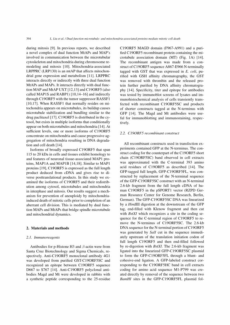
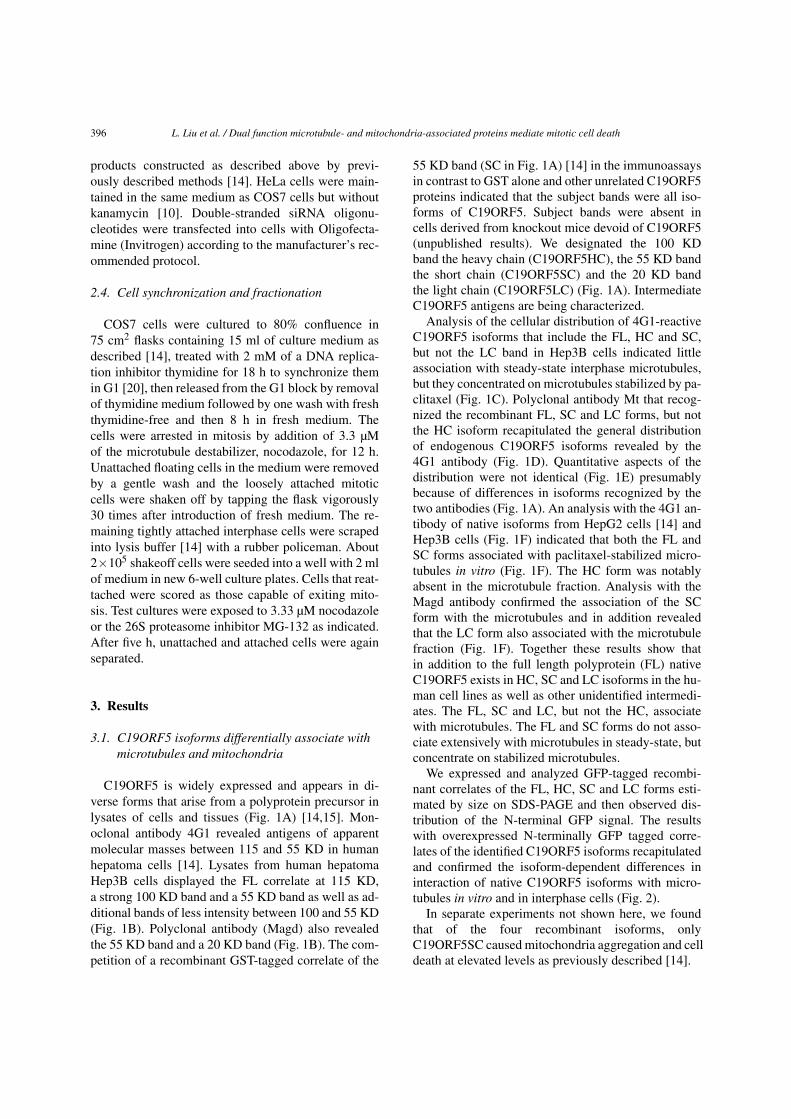
Fig. 1. Differential association of C19ORF5 isoforms with microtubules. (A) Human C19ORF5 isoforms estimated from immunoblots. Fulllength C19ORF5 was predicted from cDNA with amino acid sequence residues numbered from the initiation site. Percent identity of sequencebetween the indicated residues among Homo sapiens, Macaca mulatta, Mus musculus and Rattus norvegicus C19ORF5 is indicated. Epitopeof the 4G1 monoclonal antibody has been mapped to the indicated region. The microtubule-association (MT) and MAGD domains againstwhich Mt and Magd antibodies were directed [14] are also shown. FL, full length; HC, heavy chain; SC, short chain; and LC, light chain.(B) Profile of C19ORF5 isoforms in lysates of Hep3B cells revealed by 4G1 and Magd antibodies. Reaction mixtures were performed in thepresence of 2 mg/ml purified GST or GST-C19ORF5SC (GST-SC) [14] as indicated. (C–E) Distribution of C19ORF5 in steady state and afterpaclitaxel-induced stabilization of microtubules. Hep3B cells were treated with 10 µM of paclitaxel for 12 h and stained with the 4G1, Mtand/or anti-β-tubulin antibody and visualized with FITC (green) or Rhodamine (red)-conjugated second antibody. Images were captured withan UltraPix cold CCD camera system through an Olympus IX70 inverted microscope using a 40×/1.35 oil objective. Yellow indicates intimateoverlap of the two in the merge panels. Bar = 10 µm (all panels at the same magnification). (F) Differential association of C19ORF5 isoformswith microtubules in vitro. In vitro microtubule assembly and association of C19ORF5 isoforms from Hep3B cell lysates was performed bySDS-PAGE followed by immunoblot using the antibodies indicated in the text and visualized by secondary antibody conjugated with alkalinephosphatase as described [14,17]. P and S represent the respective pellet containing the microtubules and supernatant, respectively. Asterisksindicate unidentified C19ORF5 antigens. (The colors are visible in the online version of the article.)
lowed by re-ligation. The GFP-labeled recombinantcorrelate of the C19ORF5LC band in cell extracts cod-ing for amino acids M842-F1059 was created by ligat-ing the SmaI-digested pEGFP-C3 vector with an end-filled PCR product. The fidelity of constructs was con-firmed by DNA sequence.
2.3. Cell transfection and siRNA
Except where indicated cells were grown on cov-erslips, processed, fixed and stained as previously de-scribed [14]. Monkey kidney COS7 cells were tran-siently transfected with GFP tagged C19ORF5 fusion
396 L. Liu et al. / Dual function microtubule- and mitochondria-associated proteins mediate mitotic cell death
products constructed as described above by previ-ously described methods [14]. HeLa cells were main-tained in the same medium as COS7 cells but withoutkanamycin [10]. Double-stranded siRNA oligonu-cleotides were transfected into cells with Oligofecta-mine (Invitrogen) according to the manufacturer’s rec-ommended protocol.
2.4. Cell synchronization and fractionation
COS7 cells were cultured to 80% confluence in75 cm2 flasks containing 15 ml of culture medium asdescribed [14], treated with 2 mM of a DNA replica-tion inhibitor thymidine for 18 h to synchronize themin G1 [20], then released from the G1 block by removalof thymidine medium followed by one wash with freshthymidine-free and then 8 h in fresh medium. Thecells were arrested in mitosis by addition of 3.3 µMof the microtubule destabilizer, nocodazole, for 12 h.Unattached floating cells in the medium were removedby a gentle wash and the loosely attached mitoticcells were shaken off by tapping the flask vigorously30 times after introduction of fresh medium. The re-maining tightly attached interphase cells were scrapedinto lysis buffer [14] with a rubber policeman. About2×105 shakeoff cells were seeded into a well with 2 mlof medium in new 6-well culture plates. Cells that reat-tached were scored as those capable of exiting mito-sis. Test cultures were exposed to 3.33 µM nocodazoleor the 26S proteasome inhibitor MG-132 as indicated.After five h, unattached and attached cells were againseparated.
3. Results
3.1. C19ORF5 isoforms differentially associate withmicrotubules and mitochondria
C19ORF5 is widely expressed and appears in di-verse forms that arise from a polyprotein precursor inlysates of cells and tissues (Fig. 1A) [14,15]. Mon-oclonal antibody 4G1 revealed antigens of apparentmolecular masses between 115 and 55 KD in humanhepatoma cells [14]. Lysates from human hepatomaHep3B cells displayed the FL correlate at 115 KD,a strong 100 KD band and a 55 KD band as well as ad-ditional bands of less intensity between 100 and 55 KD(Fig. 1B). Polyclonal antibody (Magd) also revealedthe 55 KD band and a 20 KD band (Fig. 1B). The com-petition of a recombinant GST-tagged correlate of the
55 KD band (SC in Fig. 1A) [14] in the immunoassaysin contrast to GST alone and other unrelated C19ORF5proteins indicated that the subject bands were all iso-forms of C19ORF5. Subject bands were absent incells derived from knockout mice devoid of C19ORF5(unpublished results). We designated the 100 KDband the heavy chain (C19ORF5HC), the 55 KD bandthe short chain (C19ORF5SC) and the 20 KD bandthe light chain (C19ORF5LC) (Fig. 1A). IntermediateC19ORF5 antigens are being characterized.
Analysis of the cellular distribution of 4G1-reactiveC19ORF5 isoforms that include the FL, HC and SC,but not the LC band in Hep3B cells indicated littleassociation with steady-state interphase microtubules,but they concentrated on microtubules stabilized by pa-clitaxel (Fig. 1C). Polyclonal antibody Mt that recog-nized the recombinant FL, SC and LC forms, but notthe HC isoform recapitulated the general distributionof endogenous C19ORF5 isoforms revealed by the4G1 antibody (Fig. 1D). Quantitative aspects of thedistribution were not identical (Fig. 1E) presumablybecause of differences in isoforms recognized by thetwo antibodies (Fig. 1A). An analysis with the 4G1 an-tibody of native isoforms from HepG2 cells [14] andHep3B cells (Fig. 1F) indicated that both the FL andSC forms associated with paclitaxel-stabilized micro-tubules in vitro (Fig. 1F). The HC form was notablyabsent in the microtubule fraction. Analysis with theMagd antibody confirmed the association of the SCform with the microtubules and in addition revealedthat the LC form also associated with the microtubulefraction (Fig. 1F). Together these results show thatin addition to the full length polyprotein (FL) nativeC19ORF5 exists in HC, SC and LC isoforms in the hu-man cell lines as well as other unidentified intermedi-ates. The FL, SC and LC, but not the HC, associatewith microtubules. The FL and SC forms do not asso-ciate extensively with microtubules in steady-state, butconcentrate on stabilized microtubules.
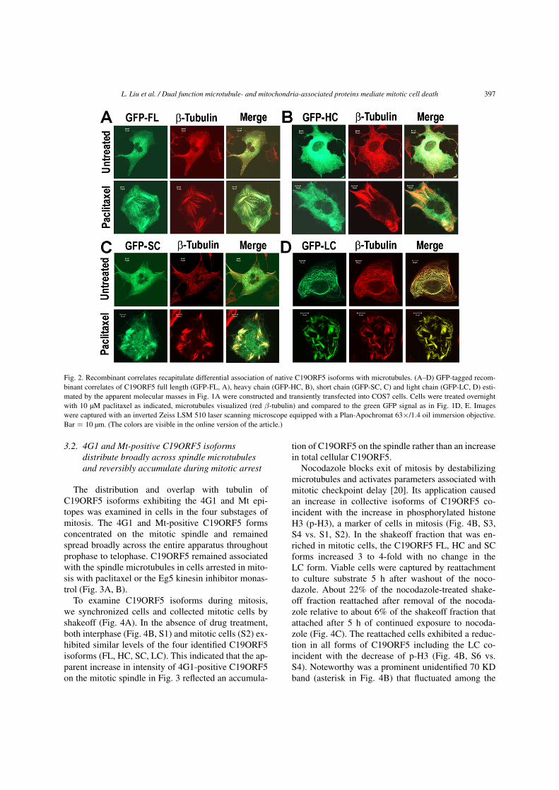
We expressed and analyzed GFP-tagged recombi-nant correlates of the FL, HC, SC and LC forms esti-mated by size on SDS-PAGE and then observed dis-tribution of the N-terminal GFP signal. The resultswith overexpressed N-terminally GFP tagged corre-lates of the identified C19ORF5 isoforms recapitulatedand confirmed the isoform-dependent differences ininteraction of native C19ORF5 isoforms with micro-tubules in vitro and in interphase cells (Fig. 2).
In separate experiments not shown here, we foundthat of the four recombinant isoforms, onlyC19ORF5SC caused mitochondria aggregation and celldeath at elevated levels as previously described [14].
L. Liu et al. / Dual function microtubule- and mitochondria-associated proteins mediate mitotic cell death 397
Fig. 2. Recombinant correlates recapitulate differential association of native C19ORF5 isoforms with microtubules. (A–D) GFP-tagged recom-binant correlates of C19ORF5 full length (GFP-FL, A), heavy chain (GFP-HC, B), short chain (GFP-SC, C) and light chain (GFP-LC, D) esti-mated by the apparent molecular masses in Fig. 1A were constructed and transiently transfected into COS7 cells. Cells were treated overnightwith 10 µM paclitaxel as indicated, microtubules visualized (red β-tubulin) and compared to the green GFP signal as in Fig. 1D, E. Imageswere captured with an inverted Zeiss LSM 510 laser scanning microscope equipped with a Plan-Apochromat 63×/1.4 oil immersion objective.Bar = 10 µm. (The colors are visible in the online version of the article.)
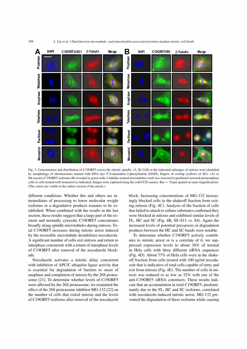
3.2. 4G1 and Mt-positive C19ORF5 isoformsdistribute broadly across spindle microtubulesand reversibly accumulate during mitotic arrest
The distribution and overlap with tubulin ofC19ORF5 isoforms exhibiting the 4G1 and Mt epi-topes was examined in cells in the four substages ofmitosis. The 4G1 and Mt-positive C19ORF5 formsconcentrated on the mitotic spindle and remainedspread broadly across the entire apparatus throughoutprophase to telophase. C19ORF5 remained associatedwith the spindle microtubules in cells arrested in mito-sis with paclitaxel or the Eg5 kinesin inhibitor monas-trol (Fig. 3A, B).
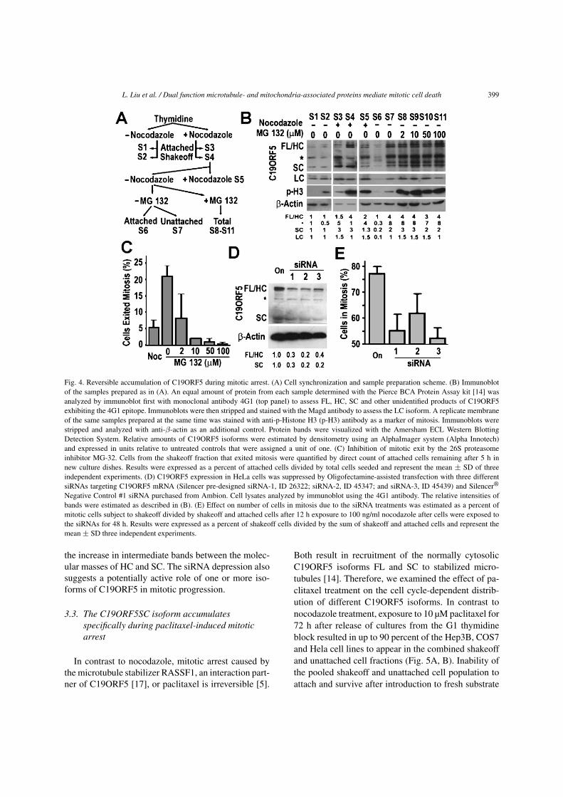
To examine C19ORF5 isoforms during mitosis,we synchronized cells and collected mitotic cells byshakeoff (Fig. 4A). In the absence of drug treatment,both interphase (Fig. 4B, S1) and mitotic cells (S2) ex-hibited similar levels of the four identified C19ORF5isoforms (FL, HC, SC, LC). This indicated that the ap-parent increase in intensity of 4G1-positive C19ORF5on the mitotic spindle in Fig. 3 reflected an accumula-
tion of C19ORF5 on the spindle rather than an increasein total cellular C19ORF5.
Nocodazole blocks exit of mitosis by destabilizingmicrotubules and activates parameters associated withmitotic checkpoint delay [20]. Its application causedan increase in collective isoforms of C19ORF5 co-incident with the increase in phosphorylated histoneH3 (p-H3), a marker of cells in mitosis (Fig. 4B, S3,S4 vs. S1, S2). In the shakeoff fraction that was en-riched in mitotic cells, the C19ORF5 FL, HC and SCforms increased 3 to 4-fold with no change in theLC form. Viable cells were captured by reattachmentto culture substrate 5 h after washout of the noco-dazole. About 22% of the nocodazole-treated shake-off fraction reattached after removal of the nocoda-zole relative to about 6% of the shakeoff fraction thatattached after 5 h of continued exposure to nocoda-zole (Fig. 4C). The reattached cells exhibited a reduc-tion in all forms of C19ORF5 including the LC co-incident with the decrease of p-H3 (Fig. 4B, S6 vs.S4). Noteworthy was a prominent unidentified 70 KDband (asterisk in Fig. 4B) that fluctuated among the
398 L. Liu et al. / Dual function microtubule- and mitochondria-associated proteins mediate mitotic cell death
Fig. 3. Concentration and distribution of C19ORF5 across the mitotic spindle. (A, B) Cells at the indicated substages of mitosis were identifiedby morphology of chromosomes stained with DNA dye 4′ ,6-diamidino-2-phenylindole (DAPI). Degree of overlap (yellow) of 4G1- (A) orMt-reactive C19ORF5 isoforms (B) revealed in green with β-tubulin-stained microtubules (red) was assessed in paclitaxel-arrested premetaphasecells or cells treated with monastrol as indicated. Images were captured using the cold CCD camera. Bar = 10 µm (panels at same magnification).(The colors are visible in the online version of the article.)
different conditions. Whether this and others are in-termediates of processing to lower molecular weightisoforms or a degradative products remains to be es-tablished. When combined with the results in the lastsection, these results suggest that a large part of the ex-istent and normally cytosolic C19ORF5 concentratesbroadly along spindle microtubules during mitosis. To-tal C19ORF5 increases during mitotic arrest inducedby the reversible microtubule destabilizer nocodazole.A significant number of cells exit mitosis and return tointerphase concurrent with a return of interphase levelsof C19ORF5 after removal of the nocodazole block-ade.
Nocodazole activates a mitotic delay concurrentwith inhibition of APC/C ubiquitin ligase activity thatis essential for degradation of barriers to onset ofanaphase and completion of mitosis by the 26S protea-some [21]. To determine whether levels of C19ORF5were affected by the 26S proteasome, we examined theeffect of the 26S proteasome inhibitor MG-132 [22] onthe number of cells that exited mitosis and the levelsof C19ORF5 isoforms after removal of the nocodazole
block. Increasing concentrations of MG-132 increas-ingly blocked cells in the shakeoff fraction from exit-ing mitosis (Fig. 4C). Analysis of the fraction of cellsthat failed to attach to culture substrates confirmed theywere blocked in mitosis and exhibited similar levels ofFL, HC and SC (Fig. 4B, S8–S11 vs. S4). Again theincreased levels of potential precursors or degradationproducts between the HC and SC bands were notable.
To determine whether C19ORF5 actively contrib-utes to mitotic arrest or is a correlate of it, we sup-pressed expression levels to about 30% of normalin Hela cells with three different siRNA sequences(Fig. 4D). About 77% of Hela cells were in the shake-off fraction from cells treated with 100 ng/ml nocoda-zole that is indicative of total cells capable of entry andexit from mitosis (Fig. 4E). The number of cells in mi-tosis was reduced to as low as 52% with one of theanti-C19ORF5 siRNA constructs. These results indi-cate that an accumulation in total C19ORF5, predomi-nantly due to the FL, HC and SC isoforms, correlatedwith nocodazole-induced mitotic arrest. MG-132 pre-vented the degradation of these isoforms while causing
L. Liu et al. / Dual function microtubule- and mitochondria-associated proteins mediate mitotic cell death 399
Fig. 4. Reversible accumulation of C19ORF5 during mitotic arrest. (A) Cell synchronization and sample preparation scheme. (B) Immunoblotof the samples prepared as in (A). An equal amount of protein from each sample determined with the Pierce BCA Protein Assay kit [14] wasanalyzed by immunoblot first with monoclonal antibody 4G1 (top panel) to assess FL, HC, SC and other unidentified products of C19ORF5exhibiting the 4G1 epitope. Immunoblots were then stripped and stained with the Magd antibody to assess the LC isoform. A replicate membraneof the same samples prepared at the same time was stained with anti-p-Histone H3 (p-H3) antibody as a marker of mitosis. Immunoblots werestripped and analyzed with anti-β-actin as an additional control. Protein bands were visualized with the Amersham ECL Western BlottingDetection System. Relative amounts of C19ORF5 isoforms were estimated by densitometry using an AlphaImager system (Alpha Innotech)and expressed in units relative to untreated controls that were assigned a unit of one. (C) Inhibition of mitotic exit by the 26S proteasomeinhibitor MG-32. Cells from the shakeoff fraction that exited mitosis were quantified by direct count of attached cells remaining after 5 h innew culture dishes. Results were expressed as a percent of attached cells divided by total cells seeded and represent the mean ± SD of threeindependent experiments. (D) C19ORF5 expression in HeLa cells was suppressed by Oligofectamine-assisted transfection with three differentsiRNAs targeting C19ORF5 mRNA (Silencer pre-designed siRNA-1, ID 26322; siRNA-2, ID 45347; and siRNA-3, ID 45439) and Silencer®
Negative Control #1 siRNA purchased from Ambion. Cell lysates analyzed by immunoblot using the 4G1 antibody. The relative intensities ofbands were estimated as described in (B). (E) Effect on number of cells in mitosis due to the siRNA treatments was estimated as a percent ofmitotic cells subject to shakeoff divided by shakeoff and attached cells after 12 h exposure to 100 ng/ml nocodazole after cells were exposed tothe siRNAs for 48 h. Results were expressed as a percent of shakeoff cells divided by the sum of shakeoff and attached cells and represent themean ± SD three independent experiments.
the increase in intermediate bands between the molec-ular masses of HC and SC. The siRNA depression alsosuggests a potentially active role of one or more iso-forms of C19ORF5 in mitotic progression.
3.3. The C19ORF5SC isoform accumulatesspecifically during paclitaxel-induced mitoticarrest
In contrast to nocodazole, mitotic arrest caused bythe microtubule stabilizer RASSF1, an interaction part-ner of C19ORF5 [17], or paclitaxel is irreversible [5].
Both result in recruitment of the normally cytosolicC19ORF5 isoforms FL and SC to stabilized micro-tubules [14]. Therefore, we examined the effect of pa-clitaxel treatment on the cell cycle-dependent distrib-ution of different C19ORF5 isoforms. In contrast tonocodazole treatment, exposure to 10 µM paclitaxel for72 h after release of cultures from the G1 thymidineblock resulted in up to 90 percent of the Hep3B, COS7and Hela cell lines to appear in the combined shakeoffand unattached cell fractions (Fig. 5A, B). Inability ofthe pooled shakeoff and unattached cell population toattach and survive after introduction to fresh substrate
400 L. Liu et al. / Dual function microtubule- and mitochondria-associated proteins mediate mitotic cell death
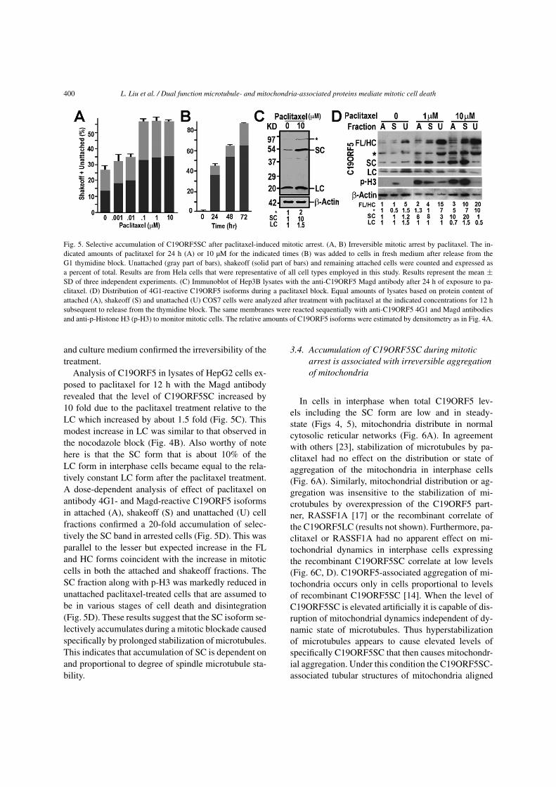
Fig. 5. Selective accumulation of C19ORF5SC after paclitaxel-induced mitotic arrest. (A, B) Irreversible mitotic arrest by paclitaxel. The in-dicated amounts of paclitaxel for 24 h (A) or 10 µM for the indicated times (B) was added to cells in fresh medium after release from theG1 thymidine block. Unattached (gray part of bars), shakeoff (solid part of bars) and remaining attached cells were counted and expressed asa percent of total. Results are from Hela cells that were representative of all cell types employed in this study. Results represent the mean ±SD of three independent experiments. (C) Immunoblot of Hep3B lysates with the anti-C19ORF5 Magd antibody after 24 h of exposure to pa-clitaxel. (D) Distribution of 4G1-reactive C19ORF5 isoforms during a paclitaxel block. Equal amounts of lysates based on protein content ofattached (A), shakeoff (S) and unattached (U) COS7 cells were analyzed after treatment with paclitaxel at the indicated concentrations for 12 hsubsequent to release from the thymidine block. The same membranes were reacted sequentially with anti-C19ORF5 4G1 and Magd antibodiesand anti-p-Histone H3 (p-H3) to monitor mitotic cells. The relative amounts of C19ORF5 isoforms were estimated by densitometry as in Fig. 4A.
and culture medium confirmed the irreversibility of thetreatment.
Analysis of C19ORF5 in lysates of HepG2 cells ex-posed to paclitaxel for 12 h with the Magd antibodyrevealed that the level of C19ORF5SC increased by10 fold due to the paclitaxel treatment relative to theLC which increased by about 1.5 fold (Fig. 5C). Thismodest increase in LC was similar to that observed inthe nocodazole block (Fig. 4B). Also worthy of notehere is that the SC form that is about 10% of theLC form in interphase cells became equal to the rela-tively constant LC form after the paclitaxel treatment.A dose-dependent analysis of effect of paclitaxel onantibody 4G1- and Magd-reactive C19ORF5 isoformsin attached (A), shakeoff (S) and unattached (U) cellfractions confirmed a 20-fold accumulation of selec-tively the SC band in arrested cells (Fig. 5D). This wasparallel to the lesser but expected increase in the FLand HC forms coincident with the increase in mitoticcells in both the attached and shakeoff fractions. TheSC fraction along with p-H3 was markedly reduced inunattached paclitaxel-treated cells that are assumed tobe in various stages of cell death and disintegration(Fig. 5D). These results suggest that the SC isoform se-lectively accumulates during a mitotic blockade causedspecifically by prolonged stabilization of microtubules.This indicates that accumulation of SC is dependent onand proportional to degree of spindle microtubule sta-bility.
3.4. Accumulation of C19ORF5SC during mitoticarrest is associated with irreversible aggregationof mitochondria
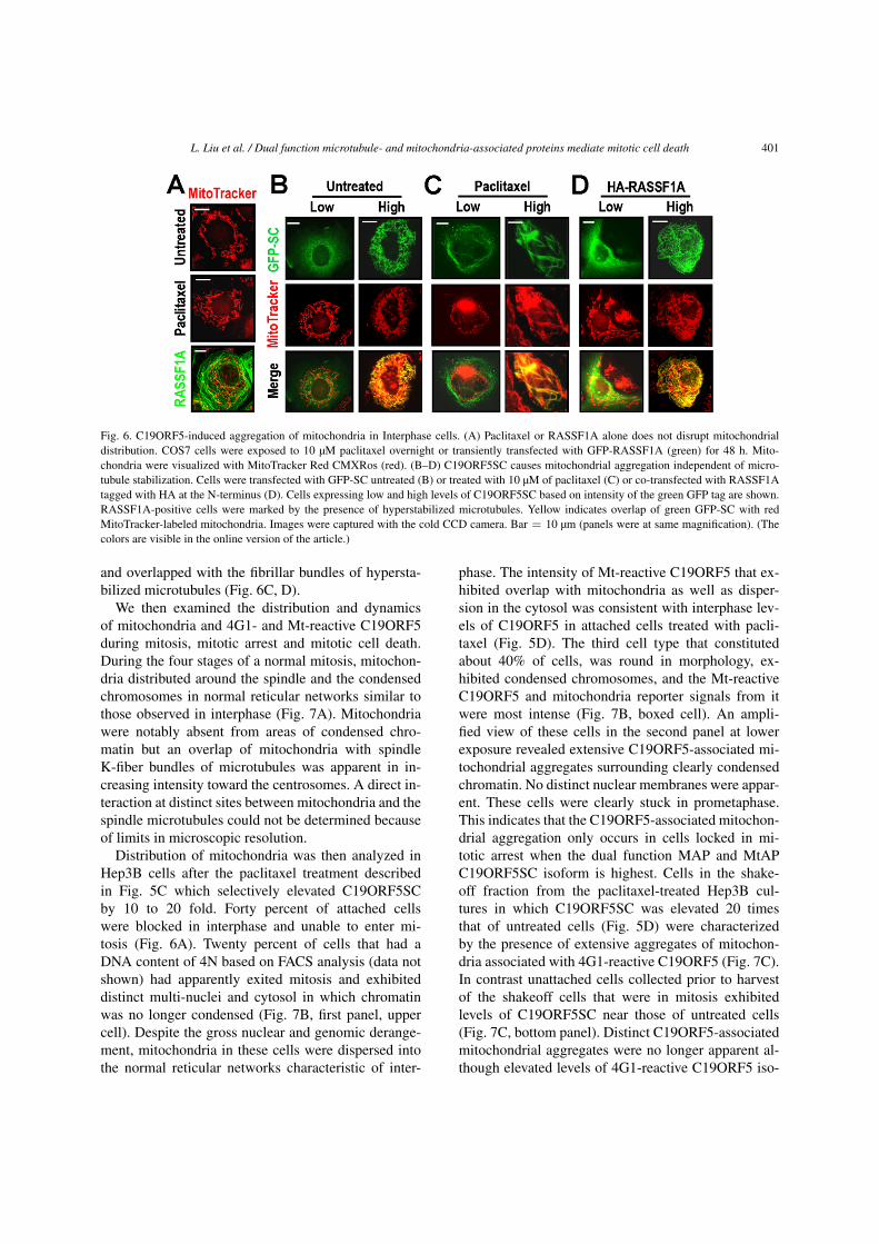
In cells in interphase when total C19ORF5 lev-els including the SC form are low and in steady-state (Figs 4, 5), mitochondria distribute in normalcytosolic reticular networks (Fig. 6A). In agreementwith others [23], stabilization of microtubules by pa-clitaxel had no effect on the distribution or state ofaggregation of the mitochondria in interphase cells(Fig. 6A). Similarly, mitochondrial distribution or ag-gregation was insensitive to the stabilization of mi-crotubules by overexpression of the C19ORF5 part-ner, RASSF1A [17] or the recombinant correlate ofthe C19ORF5LC (results not shown). Furthermore, pa-clitaxel or RASSF1A had no apparent effect on mi-tochondrial dynamics in interphase cells expressingthe recombinant C19ORF5SC correlate at low levels(Fig. 6C, D). C19ORF5-associated aggregation of mi-tochondria occurs only in cells proportional to levelsof recombinant C19ORF5SC [14]. When the level ofC19ORF5SC is elevated artificially it is capable of dis-ruption of mitochondrial dynamics independent of dy-namic state of microtubules. Thus hyperstabilizationof microtubules appears to cause elevated levels ofspecifically C19ORF5SC that then causes mitochondr-ial aggregation. Under this condition the C19ORF5SC-associated tubular structures of mitochondria aligned
L. Liu et al. / Dual function microtubule- and mitochondria-associated proteins mediate mitotic cell death 401
Fig. 6. C19ORF5-induced aggregation of mitochondria in Interphase cells. (A) Paclitaxel or RASSF1A alone does not disrupt mitochondrialdistribution. COS7 cells were exposed to 10 µM paclitaxel overnight or transiently transfected with GFP-RASSF1A (green) for 48 h. Mito-chondria were visualized with MitoTracker Red CMXRos (red). (B–D) C19ORF5SC causes mitochondrial aggregation independent of micro-tubule stabilization. Cells were transfected with GFP-SC untreated (B) or treated with 10 µM of paclitaxel (C) or co-transfected with RASSF1Atagged with HA at the N-terminus (D). Cells expressing low and high levels of C19ORF5SC based on intensity of the green GFP tag are shown.RASSF1A-positive cells were marked by the presence of hyperstabilized microtubules. Yellow indicates overlap of green GFP-SC with redMitoTracker-labeled mitochondria. Images were captured with the cold CCD camera. Bar = 10 µm (panels were at same magnification). (Thecolors are visible in the online version of the article.)
and overlapped with the fibrillar bundles of hypersta-bilized microtubules (Fig. 6C, D).
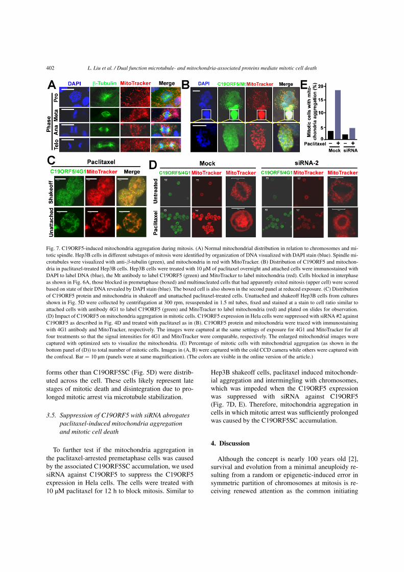
We then examined the distribution and dynamicsof mitochondria and 4G1- and Mt-reactive C19ORF5during mitosis, mitotic arrest and mitotic cell death.During the four stages of a normal mitosis, mitochon-dria distributed around the spindle and the condensedchromosomes in normal reticular networks similar tothose observed in interphase (Fig. 7A). Mitochondriawere notably absent from areas of condensed chro-matin but an overlap of mitochondria with spindleK-fiber bundles of microtubules was apparent in in-creasing intensity toward the centrosomes. A direct in-teraction at distinct sites between mitochondria and thespindle microtubules could not be determined becauseof limits in microscopic resolution.
Distribution of mitochondria was then analyzed inHep3B cells after the paclitaxel treatment describedin Fig. 5C which selectively elevated C19ORF5SCby 10 to 20 fold. Forty percent of attached cellswere blocked in interphase and unable to enter mi-tosis (Fig. 6A). Twenty percent of cells that had aDNA content of 4N based on FACS analysis (data notshown) had apparently exited mitosis and exhibiteddistinct multi-nuclei and cytosol in which chromatinwas no longer condensed (Fig. 7B, first panel, uppercell). Despite the gross nuclear and genomic derange-ment, mitochondria in these cells were dispersed intothe normal reticular networks characteristic of inter-
phase. The intensity of Mt-reactive C19ORF5 that ex-hibited overlap with mitochondria as well as disper-sion in the cytosol was consistent with interphase lev-els of C19ORF5 in attached cells treated with pacli-taxel (Fig. 5D). The third cell type that constitutedabout 40% of cells, was round in morphology, ex-hibited condensed chromosomes, and the Mt-reactiveC19ORF5 and mitochondria reporter signals from itwere most intense (Fig. 7B, boxed cell). An ampli-fied view of these cells in the second panel at lowerexposure revealed extensive C19ORF5-associated mi-tochondrial aggregates surrounding clearly condensedchromatin. No distinct nuclear membranes were appar-ent. These cells were clearly stuck in prometaphase.This indicates that the C19ORF5-associated mitochon-drial aggregation only occurs in cells locked in mi-totic arrest when the dual function MAP and MtAPC19ORF5SC isoform is highest. Cells in the shake-off fraction from the paclitaxel-treated Hep3B cul-tures in which C19ORF5SC was elevated 20 timesthat of untreated cells (Fig. 5D) were characterizedby the presence of extensive aggregates of mitochon-dria associated with 4G1-reactive C19ORF5 (Fig. 7C).In contrast unattached cells collected prior to harvestof the shakeoff cells that were in mitosis exhibitedlevels of C19ORF5SC near those of untreated cells(Fig. 7C, bottom panel). Distinct C19ORF5-associatedmitochondrial aggregates were no longer apparent al-though elevated levels of 4G1-reactive C19ORF5 iso-
402 L. Liu et al. / Dual function microtubule- and mitochondria-associated proteins mediate mitotic cell death
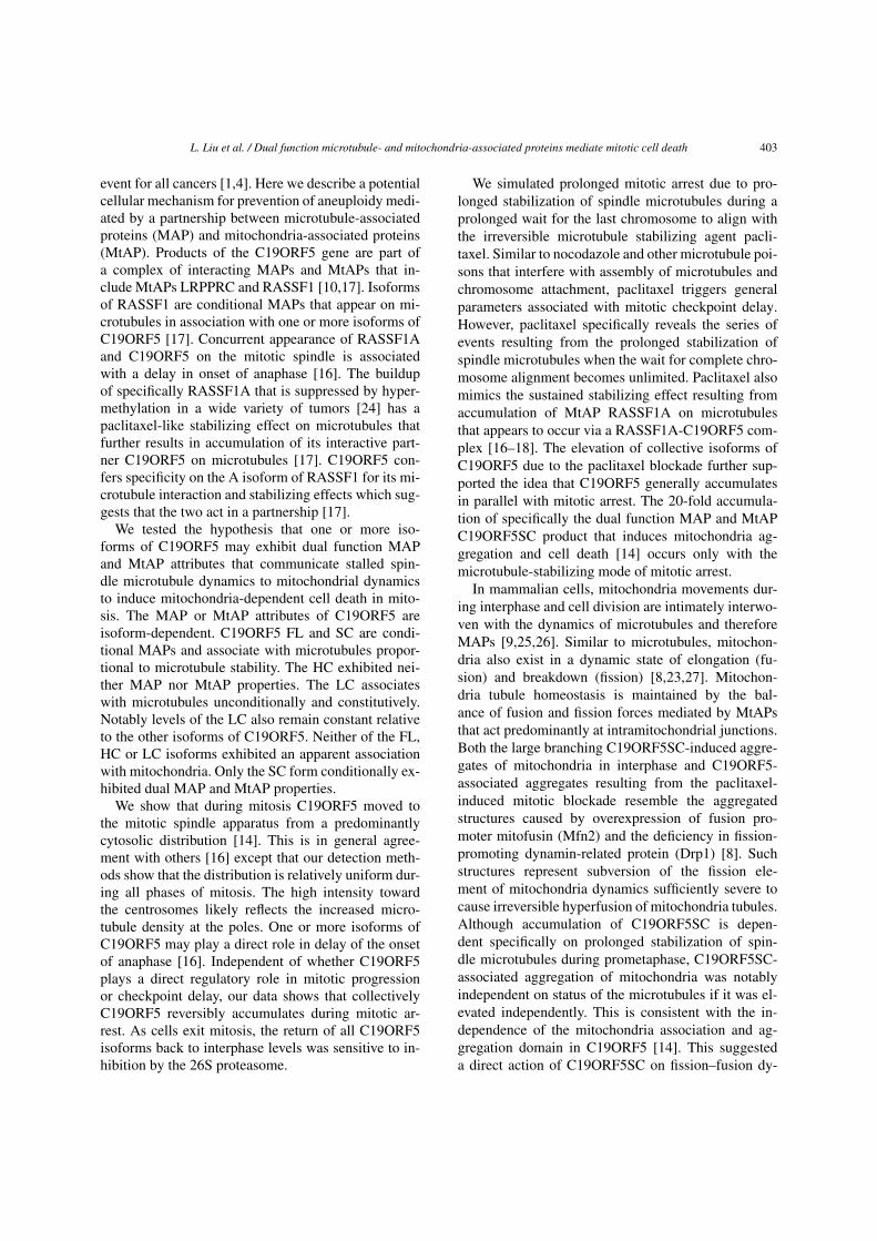
Fig. 7. C19ORF5-induced mitochondria aggregation during mitosis. (A) Normal mitochondrial distribution in relation to chromosomes and mi-totic spindle. Hep3B cells in different substages of mitosis were identified by organization of DNA visualized with DAPI stain (blue). Spindle mi-crotubules were visualized with anti-β-tubulin (green), and mitochondria in red with MitoTracker. (B) Distribution of C19ORF5 and mitochon-dria in paclitaxel-treated Hep3B cells. Hep3B cells were treated with 10 µM of paclitaxel overnight and attached cells were immunostained withDAPI to label DNA (blue), the Mt antibody to label C19ORF5 (green) and MitoTracker to label mitochondria (red). Cells blocked in interphaseas shown in Fig. 6A, those blocked in premetaphase (boxed) and multinucleated cells that had apparently exited mitosis (upper cell) were scoredbased on state of their DNA revealed by DAPI stain (blue). The boxed cell is also shown in the second panel at reduced exposure. (C) Distributionof C19ORF5 protein and mitochondria in shakeoff and unattached paclitaxel-treated cells. Unattached and shakeoff Hep3B cells from culturesshown in Fig. 5D were collected by centrifugation at 300 rpm, resuspended in 1.5 ml tubes, fixed and stained at a stain to cell ratio similar toattached cells with antibody 4G1 to label C19ORF5 (green) and MitoTracker to label mitochondria (red) and plated on slides for observation.(D) Impact of C19ORF5 on mitochondria aggregation in mitotic cells. C19ORF5 expression in Hela cells were suppressed with siRNA #2 againstC19ORF5 as described in Fig. 4D and treated with paclitaxel as in (B). C19ORF5 protein and mitochondria were traced with immunostainingwith 4G1 antibody and MitoTracker, respectively. The images were captured at the same settings of exposure for 4G1 and MitoTracker for allfour treatments so that the signal intensities for 4G1 and MitoTracker were comparable, respectively. The enlarged mitochondrial images werecaptured with optimized sets to visualize the mitochondria. (E) Percentage of mitotic cells with mitochondrial aggregation (as shown in thebottom panel of (D)) to total number of mitotic cells. Images in (A, B) were captured with the cold CCD camera while others were captured withthe confocal. Bar = 10 µm (panels were at same magnification). (The colors are visible in the online version of the article.)
forms other than C19ORF5SC (Fig. 5D) were distrib-uted across the cell. These cells likely represent latestages of mitotic death and disintegration due to pro-longed mitotic arrest via microtubule stabilization.
3.5. Suppression of C19ORF5 with siRNA abrogatespaclitaxel-induced mitochondria aggregationand mitotic cell death
To further test if the mitochondria aggregation inthe paclitaxel-arrested premetaphase cells was causedby the associated C19ORF5SC accumulation, we usedsiRNA against C19ORF5 to suppress the C19ORF5expression in Hela cells. The cells were treated with10 µM paclitaxel for 12 h to block mitosis. Similar to
Hep3B shakeoff cells, paclitaxel induced mitochondr-ial aggregation and intermingling with chromosomes,which was impeded when the C19ORF5 expressionwas suppressed with siRNA against C19ORF5(Fig. 7D, E). Therefore, mitochondria aggregation incells in which mitotic arrest was sufficiently prolongedwas caused by the C19ORF5SC accumulation.
4. Discussion
Although the concept is nearly 100 years old [2],survival and evolution from a minimal aneuploidy re-sulting from a random or epigenetic-induced error insymmetric partition of chromosomes at mitosis is re-ceiving renewed attention as the common initiating
L. Liu et al. / Dual function microtubule- and mitochondria-associated proteins mediate mitotic cell death 403
event for all cancers [1,4]. Here we describe a potentialcellular mechanism for prevention of aneuploidy medi-ated by a partnership between microtubule-associatedproteins (MAP) and mitochondria-associated proteins(MtAP). Products of the C19ORF5 gene are part ofa complex of interacting MAPs and MtAPs that in-clude MtAPs LRPPRC and RASSF1 [10,17]. Isoformsof RASSF1 are conditional MAPs that appear on mi-crotubules in association with one or more isoforms ofC19ORF5 [17]. Concurrent appearance of RASSF1Aand C19ORF5 on the mitotic spindle is associatedwith a delay in onset of anaphase [16]. The buildupof specifically RASSF1A that is suppressed by hyper-methylation in a wide variety of tumors [24] has apaclitaxel-like stabilizing effect on microtubules thatfurther results in accumulation of its interactive part-ner C19ORF5 on microtubules [17]. C19ORF5 con-fers specificity on the A isoform of RASSF1 for its mi-crotubule interaction and stabilizing effects which sug-gests that the two act in a partnership [17].
We tested the hypothesis that one or more iso-forms of C19ORF5 may exhibit dual function MAPand MtAP attributes that communicate stalled spin-dle microtubule dynamics to mitochondrial dynamicsto induce mitochondria-dependent cell death in mito-sis. The MAP or MtAP attributes of C19ORF5 areisoform-dependent. C19ORF5 FL and SC are condi-tional MAPs and associate with microtubules propor-tional to microtubule stability. The HC exhibited nei-ther MAP nor MtAP properties. The LC associateswith microtubules unconditionally and constitutively.Notably levels of the LC also remain constant relativeto the other isoforms of C19ORF5. Neither of the FL,HC or LC isoforms exhibited an apparent associationwith mitochondria. Only the SC form conditionally ex-hibited dual MAP and MtAP properties.
We show that during mitosis C19ORF5 moved tothe mitotic spindle apparatus from a predominantlycytosolic distribution [14]. This is in general agree-ment with others [16] except that our detection meth-ods show that the distribution is relatively uniform dur-ing all phases of mitosis. The high intensity towardthe centrosomes likely reflects the increased micro-tubule density at the poles. One or more isoforms ofC19ORF5 may play a direct role in delay of the onsetof anaphase [16]. Independent of whether C19ORF5plays a direct regulatory role in mitotic progressionor checkpoint delay, our data shows that collectivelyC19ORF5 reversibly accumulates during mitotic ar-rest. As cells exit mitosis, the return of all C19ORF5isoforms back to interphase levels was sensitive to in-hibition by the 26S proteasome.
We simulated prolonged mitotic arrest due to pro-longed stabilization of spindle microtubules during aprolonged wait for the last chromosome to align withthe irreversible microtubule stabilizing agent pacli-taxel. Similar to nocodazole and other microtubule poi-sons that interfere with assembly of microtubules andchromosome attachment, paclitaxel triggers generalparameters associated with mitotic checkpoint delay.However, paclitaxel specifically reveals the series ofevents resulting from the prolonged stabilization ofspindle microtubules when the wait for complete chro-mosome alignment becomes unlimited. Paclitaxel alsomimics the sustained stabilizing effect resulting fromaccumulation of MtAP RASSF1A on microtubulesthat appears to occur via a RASSF1A-C19ORF5 com-plex [16–18]. The elevation of collective isoforms ofC19ORF5 due to the paclitaxel blockade further sup-ported the idea that C19ORF5 generally accumulatesin parallel with mitotic arrest. The 20-fold accumula-tion of specifically the dual function MAP and MtAPC19ORF5SC product that induces mitochondria ag-gregation and cell death [14] occurs only with themicrotubule-stabilizing mode of mitotic arrest.
In mammalian cells, mitochondria movements dur-ing interphase and cell division are intimately interwo-ven with the dynamics of microtubules and thereforeMAPs [9,25,26]. Similar to microtubules, mitochon-dria also exist in a dynamic state of elongation (fu-sion) and breakdown (fission) [8,23,27]. Mitochon-dria tubule homeostasis is maintained by the bal-ance of fusion and fission forces mediated by MtAPsthat act predominantly at intramitochondrial junctions.Both the large branching C19ORF5SC-induced aggre-gates of mitochondria in interphase and C19ORF5-associated aggregates resulting from the paclitaxel-induced mitotic blockade resemble the aggregatedstructures caused by overexpression of fusion pro-moter mitofusin (Mfn2) and the deficiency in fission-promoting dynamin-related protein (Drp1) [8]. Suchstructures represent subversion of the fission ele-ment of mitochondria dynamics sufficiently severe tocause irreversible hyperfusion of mitochondria tubules.Although accumulation of C19ORF5SC is depen-dent specifically on prolonged stabilization of spin-dle microtubules during prometaphase, C19ORF5SC-associated aggregation of mitochondria was notablyindependent on status of the microtubules if it was el-evated independently. This is consistent with the in-dependence of the mitochondria association and ag-gregation domain in C19ORF5 [14]. This suggesteda direct action of C19ORF5SC on fission–fusion dy-
404 L. Liu et al. / Dual function microtubule- and mitochondria-associated proteins mediate mitotic cell death
namics of mitochondria independent of direct as-sociation with microtubules. However, in the pres-ence of paclitaxel or elevated levels of RASSF1A theC19ORF5-associated hyperfused tubules of mitochon-dria aligned with fibrillar bundles of stabilized micro-tubules. This may indicate a concurrent association ofC19ORF5SC with mitochondria and microtubules de-spite the fact that mitochondria and microtubule as-sociation domains can be functionally dissected [14].Whether C19ORF5-associated intermitochondrial andmitochondrial-microtubule junction points are overlap-ping together with factors as Mfn2 and Drp1 at mi-tochondrial tubule scission sites is under investiga-tion.
In sum, we have described a mechanism wherebya survivable minimal aneuploidy can be prevented byinduction of mitotic cell death before it happens asa result of completion of an aberrant cell division.This is mediated by cooperativity among dual func-tion MAPs and MtAPs as RASSF1A and C19ORF5.A normally short-lived specific product of C19ORF5accumulates due to prolonged stabilization of predom-inantly K-fiber bundles when cells are stuck in mito-sis due to failure of alignment of the last chromosome.Accumulation of RASSF1A-C19ORF5 on the spindlepast a certain threshold causes irreversible hypersta-bilization of spindle microtubules. Resultant accumu-lation of C19ORF5SC causes hyper and irreversiblefusion of mitochondria that causes mitotic cell death.Gross aneuploidy arising via an aberrant cell divi-sion is on average intrinsically lethal. However, our re-sults suggest a natural mechanism that may limit thefrequency of cancers at their earliest origin throughprevention of a minimal survivable aneuploidy. Thismechanism may also underlie the effectiveness of pa-clitaxel as a chemotherapeutic agent even in tumorcells that have become tolerant to high levels of aneu-ploidy.
Note added in proof
While this paper was in review, Liu et al., demon-strated that autophagy/mitophagy is robust during mi-tosis [28]. Histochemical marker analysis indicates themitochondrial aggregates described in this study arelargely cytochrome c-deficient mitochondria that accu-mulate due to blocked mitophagy.
Acknowledgements
We thank Dr. Le Sun and Joe Corvera (A&G Phar-maceuticals, Inc., Columbia, MD) for anti-C19ORF5
mouse monoclonal antibody 4G1. This work was sup-ported by Public Health Service Grants CA59971and DK35310 (WLM), DOD New Investigator AwardW81XWH-08-1-0475 (LL), and aid from the John S.Dunn Research Foundation (WLM).
References
[1] P. Duesberg, Chromosomal chaos and cancer, Sci. Am. 296(2007), 52–59.
[2] T. Boveri, Zur Frage der Entstehung maligner Tumoren, Gus-tav Fischer Verlag, Jena, Germany, 1914.
[3] P. Duesberg and D. Rasnick, Aneuploidy, the somatic mutationthat makes cancer a species of its own, Cell Motil. Cytoskel. 47(2000), 81–107.
[4] B.A. Weaver and D.W. Cleveland, Does aneuploidy cause can-cer?, Curr. Opin. Cell Biol. 18 (2006), 658–667.
[5] P.K. Sorger, M. Dobles, R. Tournebize and A.A. Hyman, Cou-pling cell division and cell death to microtubule dynamics,Curr. Opin. Cell Biol. 9 (1997), 807–814.
[6] G.A. Pihan and S.J. Doxsey, The mitotic machinery as a sourceof genetic instability in cancer, Semin. Cancer Biol. 9 (1999),289–302.
[7] S.L. Kline-Smith and C.E. Walczak, Mitotic spindle assemblyand chromosome segregation: refocusing on microtubule dy-namics, Mol. Cell 15 (2004), 317–327.
[8] A.E. Frazier, C. Kiu, D. Stojanovski, N.J. Hoogenraad andM.T. Ryan, Mitochondrial morphology and distribution inmammalian cells, Biol. Chem. 387 (2006), 1551–1558.
[9] M.P. Yaffe, N. Stuurman and R.D. Vale, Mitochondrial posi-tioning in fission yeast is driven by association with dynamicmicrotubules and mitotic spindle poles, Proc. Natl. Acad. Sci.USA 100 (2003), 11424–11428.
[10] L. Liu, V. Amy, G. Liu and W.L. McKeehan, Novel complex in-tegrating mitochondria and the microtubular cytoskeleton withchromosome remodeling and tumor suppressor RASSF1 de-duced by in silico homology analysis, interaction cloning inyeast, and colocalization in cultured cells, In Vitro Cell. Dev.Biol. Anim. 38 (2002), 582–594.
[11] M.P. Cooper, L. Qu, L.M. Rohas, J. Lin, W. Yang,H. Erdjument-Bromage, P. Tempst and B.M. Spiegelman, De-fects in energy homeostasis in Leigh syndrome French Cana-dian variant through PGC-1alpha/LRP130 complex, GenesDev. 20 (2006), 2996–3009.
[12] H. Zhao, Q. Wang, H. Zhang, Q. Liu, X. Du, M. Richter andM.I. Greene, UXT is a novel centrosomal protein essential forcell viability, Mol. Biol. Cell 16 (2005), 5857–5865.
[13] T.N. Moss, A. Vo, W.L. McKeehan and L. Liu, UXT (Ubiq-uitously Expressed Transcript) causes mitochondrial aggrega-tion, In Vitro Cell. Dev. Biol. Anim. 43 (2007), 139–146.
[14] L. Liu, A. Vo, G. Liu and W.L. McKeehan, Distinct structuraldomains within C19ORF5 support association with stabilizedmicrotubules and mitochondrial aggregation and genome de-struction, Cancer Res. 65 (2005), 4191–4201.
[15] Z. Orban-Nemeth, H. Simader, S. Badurek, A. Trancikova andF. Propst, Microtubule-associated protein 1S, a short and ubiq-
L. Liu et al. / Dual function microtubule- and mitochondria-associated proteins mediate mitotic cell death 405
uitously expressed member of the microtubule-associated pro-tein 1 family, J. Biol. Chem. 280 (2005), 2257–2265.
[16] M.S. Song, J.S. Chang, S.J. Song, T.H. Yang, H. Lee and D.S.Lim, The centrosomal protein RAS association domain fam-ily protein 1A (RASSF1A)-binding protein 1 regulates mitoticprogression by recruiting RASSF1A to spindle poles, J. Biol.Chem. 280 (2005), 3920–3927.
[17] L. Liu, A. Vo and W.L. McKeehan, Specificity of themethylation-suppressed A isoform of candidate tumor suppres-sor RASSF1 for microtubule hyperstabilization is determinedby cell death inducer C19ORF5, Cancer Res. 65 (2005), 1830–1838.
[18] A. Dallol, A. Agathanggelou, S.L. Fenton, J. Ahmed-Choudhury, L. Hesson, M.D. Vos, G.J. Clark, J. Downward,E.R. Maher and F. Latif, RASSF1A interacts with microtubule-associated proteins and modulates microtubule dynamics, Can-cer Res. 64 (2004), 4112–4116.
[19] T.A. Schoenfeld, L. McKerracher, R. Obar and R.B. Vallee,MAP 1A and MAP 1B are structurally related microtubule as-sociated proteins with distinct developmental patterns in theCNS, J. Neurosci. 9 (1989), 1712–1730.
[20] G. Fang, H. Yu and M.W. Kirschner, Direct binding of CDC20protein family members activates the anaphase-promotingcomplex in mitosis and G1, Mol. Cell 2 (1998), 163–171.
[21] J.M. Peters, The anaphase promoting complex/cyclosome:a machine designed to destroy, Nat. Rev. Mol. Cell Biol. 7(2006), 644–656.
[22] J. Zhao, T. Tenev, L.M. Martins, J. Downward and N.R.Lemoine, The ubiquitin-proteasome pathway regulates sur-vivin degradation in a cell cycle-dependent manner, J. Cell Sci.113(23) (2000), 4363–4371.
[23] M. Muller, S.L. Mironov, M.V. Ivannikov, J. Schmidt and D.W.Richter, Mitochondrial organization and motility probed bytwo-photon microscopy in cultured mouse brainstem neurons,Exp. Cell Res. 303 (2005), 114–127.
[24] A. Agathanggelou, W.N. Cooper and F. Latif, Role of the Ras-association domain family 1 tumor suppressor gene in humancancers, Cancer Res. 65 (2005), 3497–3508.
[25] R.D. Vale, Intracellular transport using microtubule-based mo-tors, Annu. Rev. Cell Biol. 3 (1987), 347–378.
[26] M.P. Yaffe, The machinery of mitochondrial inheritance andbehavior, Science 283 (1999), 1493–1497.
[27] V.P. Skulachev, Mitochondrial filaments and clusters as intra-cellular power-transmitting cables, Trends Biochem. Sci. 26(2001), 23–29.
[28] Liu et al., Cell Cycle 8(10) (2009), in press.

Submit your manuscripts athttp://www.hindawi.com
Stem CellsInternational
Hindawi Publishing Corporationhttp://www.hindawi.com Volume 2014
Hindawi Publishing Corporationhttp://www.hindawi.com Volume 2014
MEDIATORSINFLAMMATION
of
Hindawi Publishing Corporationhttp://www.hindawi.com Volume 2014
Behavioural Neurology
EndocrinologyInternational Journal of
Hindawi Publishing Corporationhttp://www.hindawi.com Volume 2014
Hindawi Publishing Corporationhttp://www.hindawi.com Volume 2014
Disease Markers
Hindawi Publishing Corporationhttp://www.hindawi.com Volume 2014
BioMed Research International
OncologyJournal of
Hindawi Publishing Corporationhttp://www.hindawi.com Volume 2014
Hindawi Publishing Corporationhttp://www.hindawi.com Volume 2014
Oxidative Medicine and Cellular Longevity
Hindawi Publishing Corporationhttp://www.hindawi.com Volume 2014
PPAR Research
The Scientific World JournalHindawi Publishing Corporation http://www.hindawi.com Volume 2014
Immunology ResearchHindawi Publishing Corporationhttp://www.hindawi.com Volume 2014
Journal of
ObesityJournal of
Hindawi Publishing Corporationhttp://www.hindawi.com Volume 2014
Hindawi Publishing Corporationhttp://www.hindawi.com Volume 2014
Computational and Mathematical Methods in Medicine
OphthalmologyJournal of
Hindawi Publishing Corporationhttp://www.hindawi.com Volume 2014
Diabetes ResearchJournal of
Hindawi Publishing Corporationhttp://www.hindawi.com Volume 2014
Hindawi Publishing Corporationhttp://www.hindawi.com Volume 2014
Research and TreatmentAIDS
Hindawi Publishing Corporationhttp://www.hindawi.com Volume 2014
Gastroenterology Research and Practice
Hindawi Publishing Corporationhttp://www.hindawi.com Volume 2014
Parkinson’s Disease
Evidence-Based Complementary and Alternative Medicine
Volume 2014Hindawi Publishing Corporationhttp://www.hindawi.com