Embed Size (px)
DESCRIPTION
eye
Citation preview
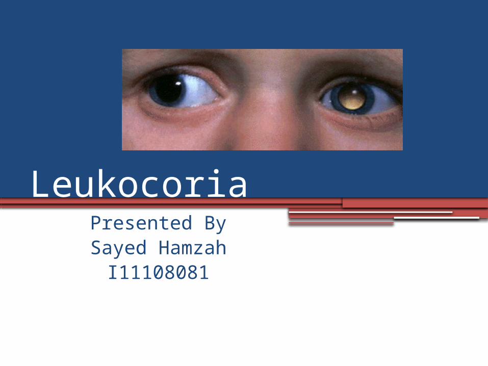
LeukocoriaPresented By
Sayed HamzahI11108081
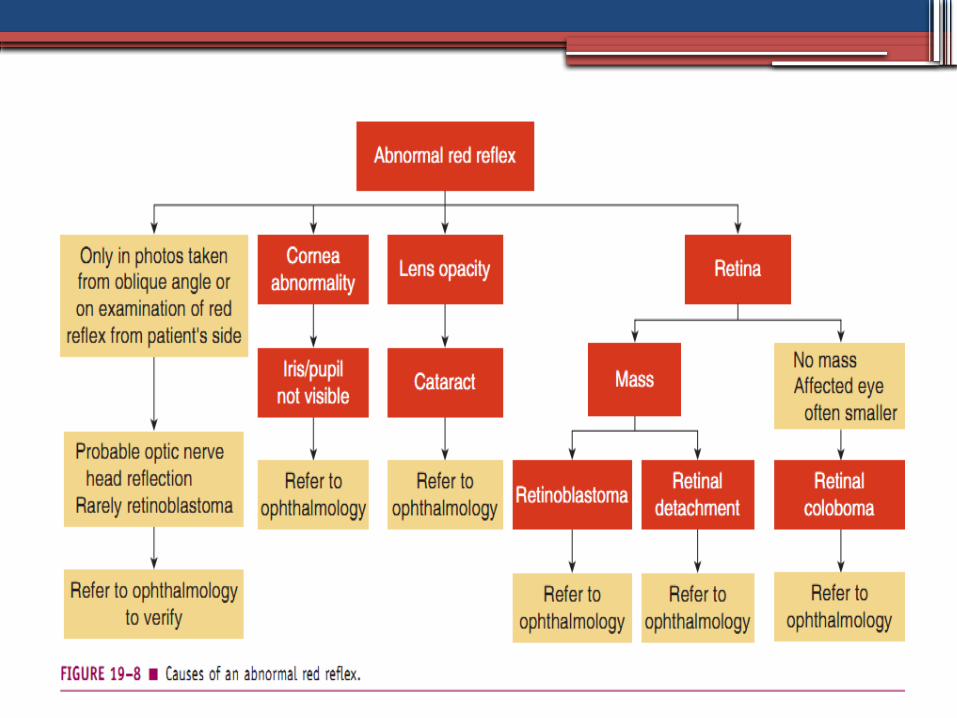
Definition(Leukos: white, Kore: pupil)Leukokoria is an abnormal pupillary light
reflection that usually results from an intraocular abnormality and is seen most often in children.
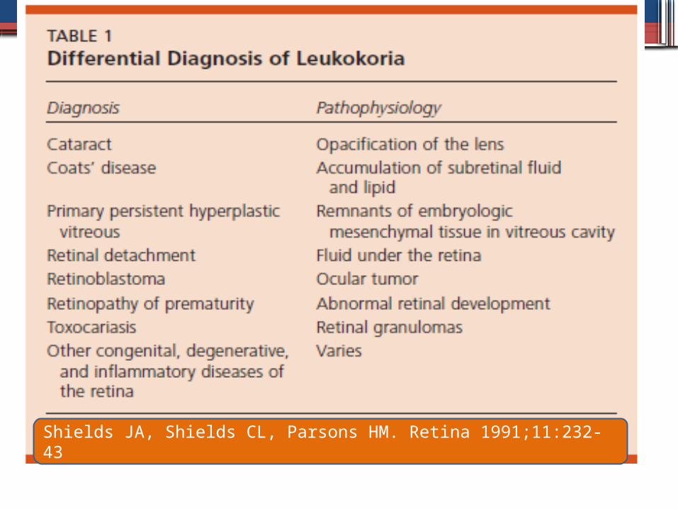
Shields JA, Shields CL, Parsons HM. Retina 1991;11:232-43
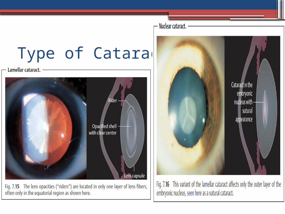
Congenital Cataract
•Opacity of the lens at birth, either hereditary or acquired throughout the placenta
Signs :LeukocoriaNystagmusStrabismusBlunted red reflex
etiology•Idiopathic (most common).•Familial•Galactosemia•PHPV•Rubella•Lowe syndrome (oculocerebrorenal
syndrome•Others
Type of Cataract
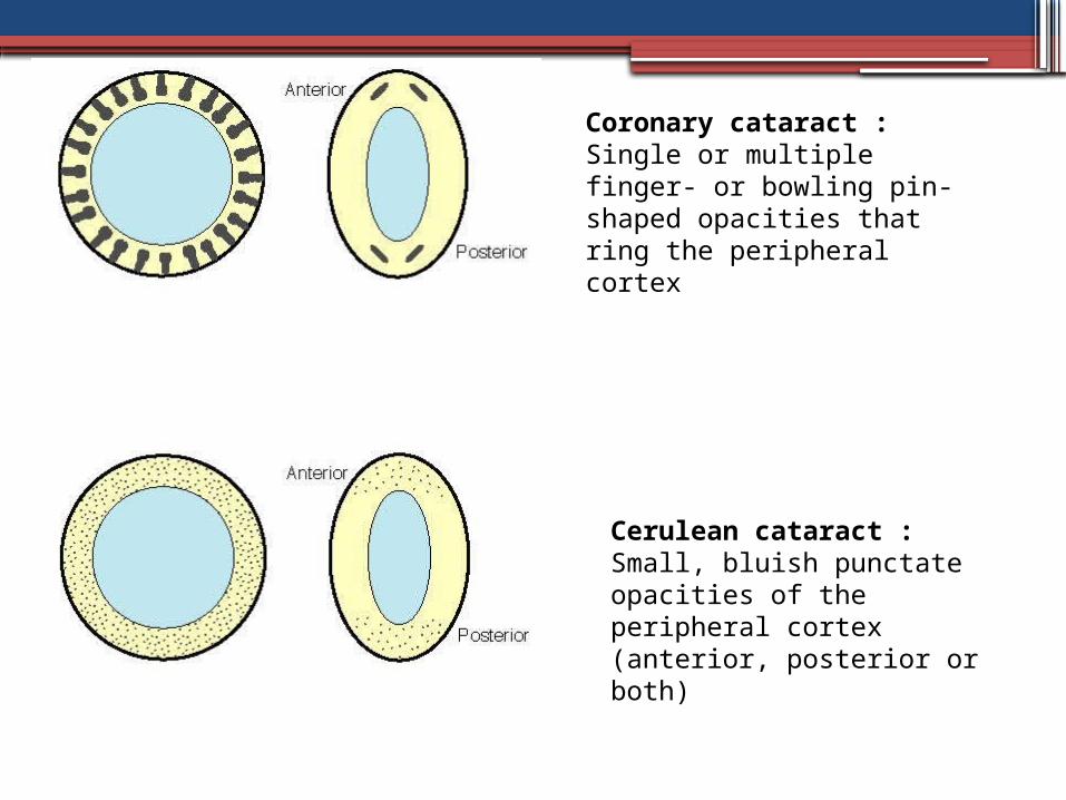
Coronary cataract : Single or multiple finger- or bowling pin-shaped opacities that ring the peripheral cortex
Cerulean cataract : Small, bluish punctate opacities of the peripheral cortex (anterior, posterior or both)
Treatment• Referral to a pediatrician to treat any underlying
disorder.• Treat associated ocular diseases.• Cataract extraction, • Treat amblyopia in children younger than 9 to 11
years• A dilating agent (e.g., phenylephrine 2.5%, t.i.d.,
homatropine 2% t.i.d., or scopolamine 0.25% q.d.) may be used as a temporizing measure, allowing peripheral light rays to pass around the lens opacity and reach the retina. This rarely is successful long-term.
Retinoblastoma
Definition•A retinoblastoma is a malignant tumor of
early childhood that develops from immature retinal cells.
Epidemiology: • the most common malignant ocular tumor
in children, occurring in approximately one of 20000 births.
• In 30% of all cases, it is bilateral.
Pathogenesis: • A somatic mutation is detected in about 95% of all
patients. • it is inherited as an autosomal dominant trait.• Changes on chromosome 13q
Symptoms: • manifests itself before the age of three in 90% of
affected children. • Parents observe leukocoria (a whitish yellow pupil)
in 60% of these children, strabismus in 20%, and a reddened eye in 10%.
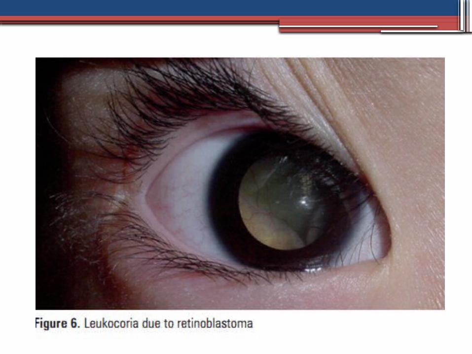
Clinical presentations• leukocoria (from 60 to 80% of cases)• Strabismus• red eyes• Excessive tearing• enlarged ocular globe due to increased
intraocular pressure• loss of vision• Less common forms are : aseptic orbit
cellulitis due to tumor necrosis, altered color of the iris (heterochromia), intraocular bleeding and pseudouveitis.
Diagnosis
•Family history•Ophthalmoscopy A grayish white, vascularized retinal
tumor•CT –scan detecting calcium within the
tumor•Ultrasoundthe size of the lesions and may
find calcification within the tumor •MRI extraocular involvement and invasion
of the optic nerve
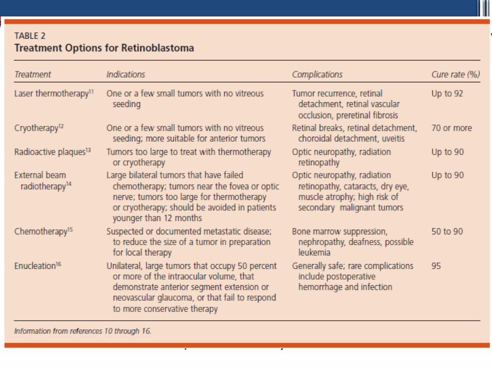
Treatment• Intravenou chemotherapy • Enucleation • Radiation therapy • External beam radiation therapy • Plaque radiotherapy • Laser therapy • Photocoagulation • Transpupillary thermotherapy (TTT) • Cryotherapy • Observation (for spontaneously arrested
retinoblastoma, retinoma)
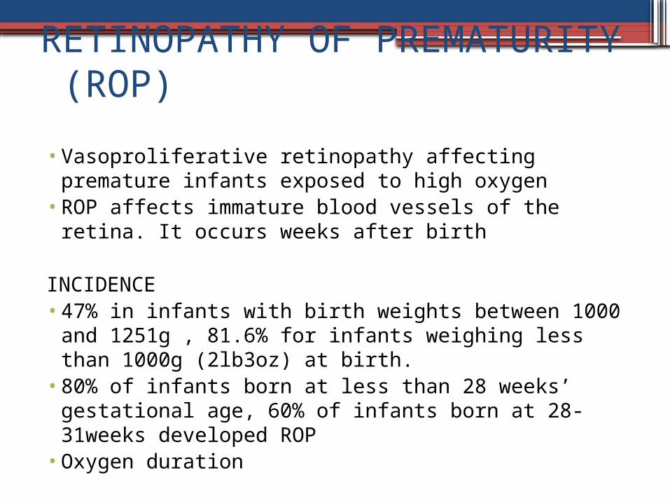
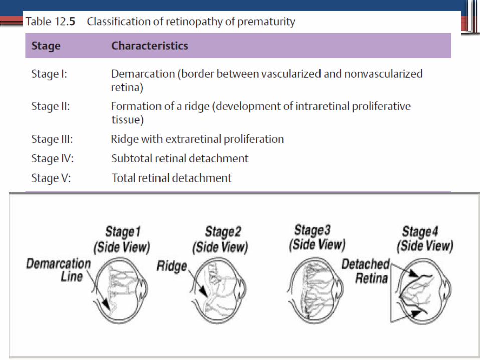
RETINOPATHY OF PREMATURITY (ROP)
• Vasoproliferative retinopathy affecting premature infants exposed to high oxygen
• ROP affects immature blood vessels of the retina. It occurs weeks after birth
INCIDENCE • 47% in infants with birth weights between 1000
and 1251g , 81.6% for infants weighing less than 1000g (2lb3oz) at birth.
• 80% of infants born at less than 28 weeks’ gestational age, 60% of infants born at 28-31weeks developed ROP
• Oxygen duration
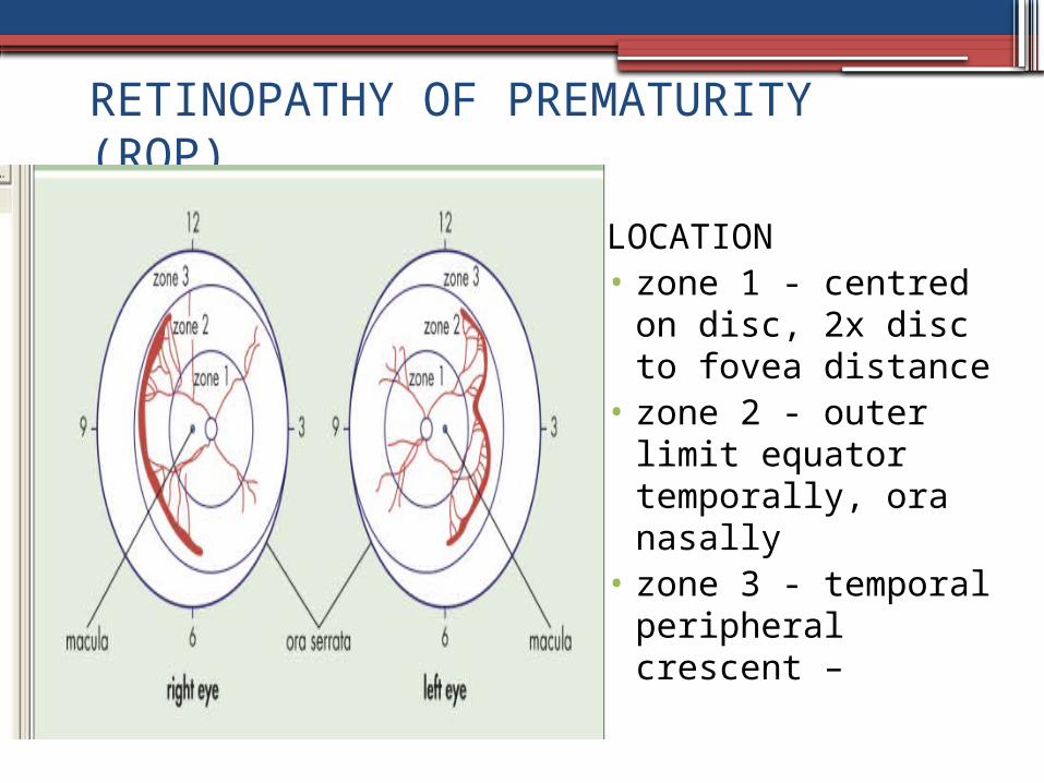
RETINOPATHY OF PREMATURITY (ROP)
LOCATION• zone 1 - centred on
disc, 2x disc to fovea distance
• zone 2 - outer limit equator temporally, ora nasally
• zone 3 - temporal peripheral crescent –
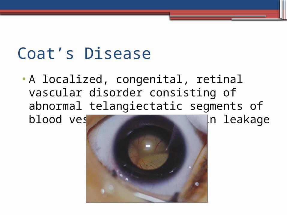
Coat’s Disease
•A localized, congenital, retinal vascular disorder consisting of abnormal telangiectatic segments of blood vessels that result in leakage
Characteristic
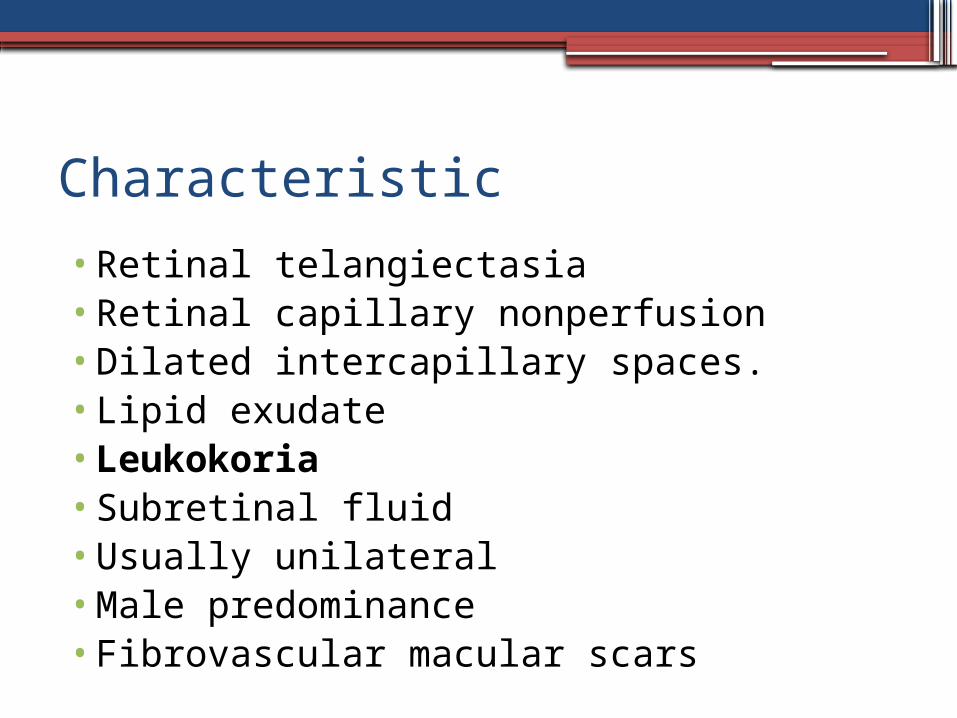
•Retinal telangiectasia•Retinal capillary nonperfusion•Dilated intercapillary spaces.•Lipid exudate•Leukokoria•Subretinal fluid•Usually unilateral•Male predominance•Fibrovascular macular scars
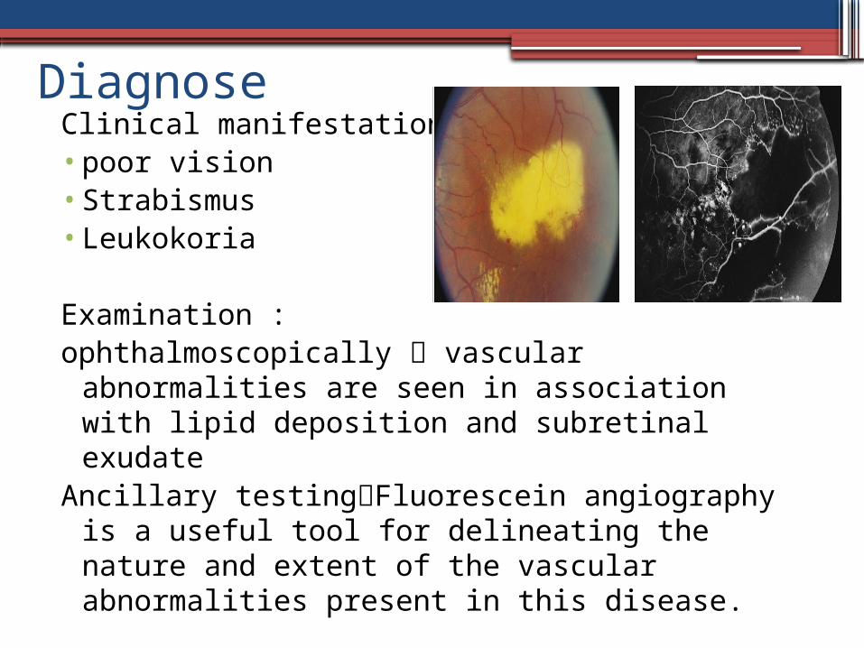
DiagnoseClinical manifestation :•poor vision•Strabismus•Leukokoria
Examination :ophthalmoscopically vascular
abnormalities are seen in association with lipid deposition and subretinal exudate
Ancillary testingFluorescein angiography is a useful tool for delineating the nature and extent of the vascular abnormalities present in this disease.
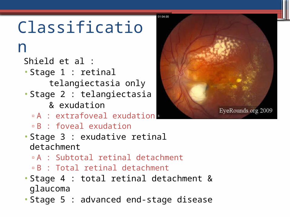
Classification Shield et al :• Stage 1 : retinal telangiectasia only• Stage 2 : telangiectasia & exudation
▫A : extrafoveal exudation▫B : foveal exudation
• Stage 3 : exudative retinal detachment▫A : Subtotal retinal detachment▫B : Total retinal detachment
• Stage 4 : total retinal detachment & glaucoma
• Stage 5 : advanced end-stage disease
Treatment
•The main goal : to obliterate the telangiectasiae to facilitate the reabsorption of exudates and maintain as much visual acuity as possible
•Laser photocoagulation or cryotherapy to destroy anomalous vasculature.
• intravitreal proliferation and traction detachment vitreous surgery may improve the clinical course
PHPV (PRIMER HIPERPLASTIK PERSISTEN VITREUS)• developmental malformation of the eye where the
embryological primary vitreous and hyaloid vasculature fail to regress.
• Persistent hyperplastic primary vitreous (PHPV) is a rare,non-hereditary,
• It presents unilaterally in 90% of cases and can be associated with microphthalmia and cataract formation.
• It is usually not associated with other congenital defects.
• In the majority of cases the fibrovascular membrane, fed by the persistent hyaloid artery, adheres to the posterior capsule of the crystalline lens.
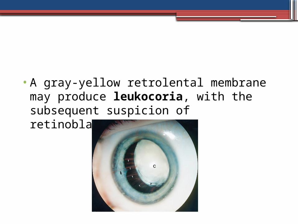
•A gray-yellow retrolental membrane may produce leukocoria, with the subsequent suspicion of retinoblastoma.
PHPVDiagnostic Consideration•Based on the characteristic clinical
picture (symptoms and findings) and additional ultrasound studies (when the posterior segment is obscured by lens opacities)
TreatmentTreatment options include cataract
surgery and removal of the fibrovascular membrane.
Ocular Toxocariasis
Ocular toxocariasis is a unilateral disorder that presents as strabismus, leukocoria or decreased vision.
Can be associated with systemic oxocariasis
INCIDENCEToxocariasis is the most common nematode
infection affecting the eye in the United States; however, the exact incidence of ocular toxocariasis in unknown.
ETIOLOGYMost cases of human toxocariasis are caused
by infection with the dog intestinal roundworm Toxocara canis or, rarely, the cat Toxocara catis.Ocular toxocariasis results from invasion of the eye by the second- or third-stage larva of the
nematode.
Clinical Features•80% less than 16 years.•Another common manifestation is chronic
endophthalmitis.•often present anterior uveitis, hypopyn,
posterior synechiae, cyclitic membrane, vitritis, and retinal detachment.
•Leukocoria because of the severe inflammatory reaction
•decreased visual Acuity•amblyopia,•strabismus as a result of damage to the
macula.
Systemic manifestation
•known as visceral larval migrans (VLM). •occurs most commonly in children less than
6 years •eosinophilia,• fever•hepatosplenomegaly•If ocular disease and VLM are both
present, flu-like symptoms•Less common manifestations pulmonary
symptoms, splenomegaly, and seizures.
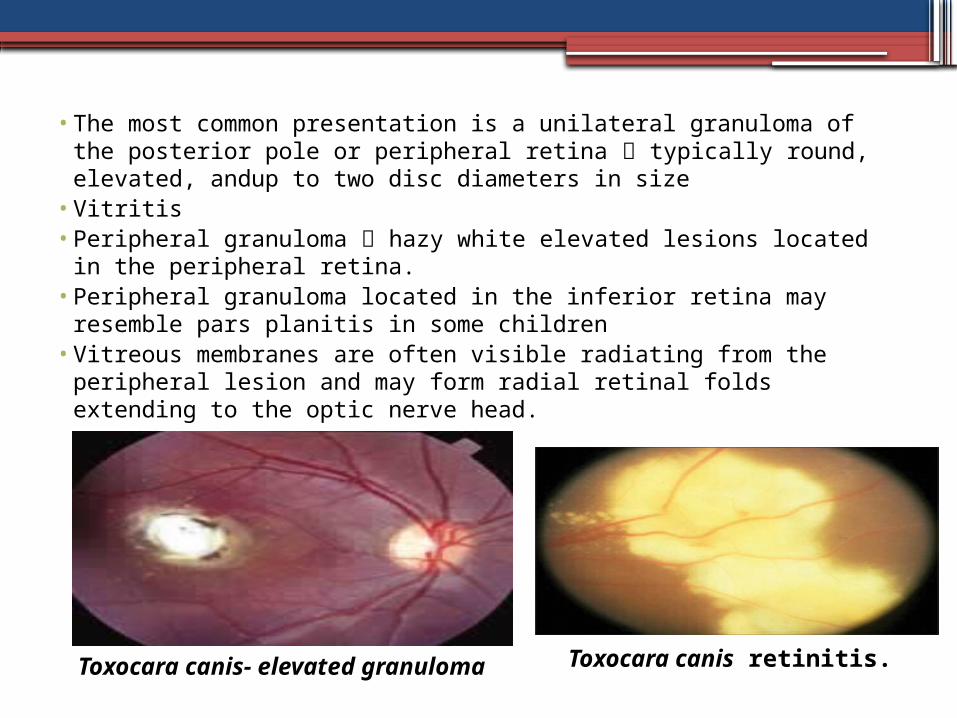
• The most common presentation is a unilateral granuloma of the posterior pole or peripheral retina typically round, elevated, andup to two disc diameters in size
• Vitritis • Peripheral granuloma hazy white elevated lesions located in the
peripheral retina.• Peripheral granuloma located in the inferior retina may resemble
pars planitis in some children• Vitreous membranes are often visible radiating from the
peripheral lesion and may form radial retinal folds extending to the optic nerve head.
Toxocara canis retinitis.Toxocara canis- elevated granuloma
Diagnosis
•Diagnosis is based upon clinical features observed in a young patient and should be confirmed at least by the presence of specific IgG in the serum (ELISA test, 90% specificity and 91% sensibility).
Treatment
•based on : severity of inflammation, macular involvement, and the visual potential of the eye.
•periocular or systemic corticosteroids active vitritis
• topical corticosteroids and cycloplegic agents anterior uveitis, Thiabendazole at 50mg/kg/day for 7 days may be considered in children who fail to respond to systemic corticosteroid therapy
•antihelminthic therapy potential toxicity•Surgical procedures refractory vitritis,
vitreous membranes, epiretinal membrane, and traction or rhegmatogenous retinal detachment.
Retinal detachment
•Retinal detachment is a disorder of the eye in which the retina peels away from its underlying layer of support tissue
•incidence : 1:10 000 •Variety of ocular and systemic disorders
are associated with pathological vitreous liquefaction, premature vitreous detachment, and extensive sites of vitreoretinal adhesion
Pathophysiology• extensive liquefaction within the vitreous
cavity reduction in both the shock-absorbing capabilities and the stability of the gel associated by aging
Accelerated vitreous liquefaction :• significant myopia• post cataract surgery (40%)• severe ocular trauma (10-15 %)• intraocular inflammation • a variety of other congenital, inherited, or
acquired ocular disorders.
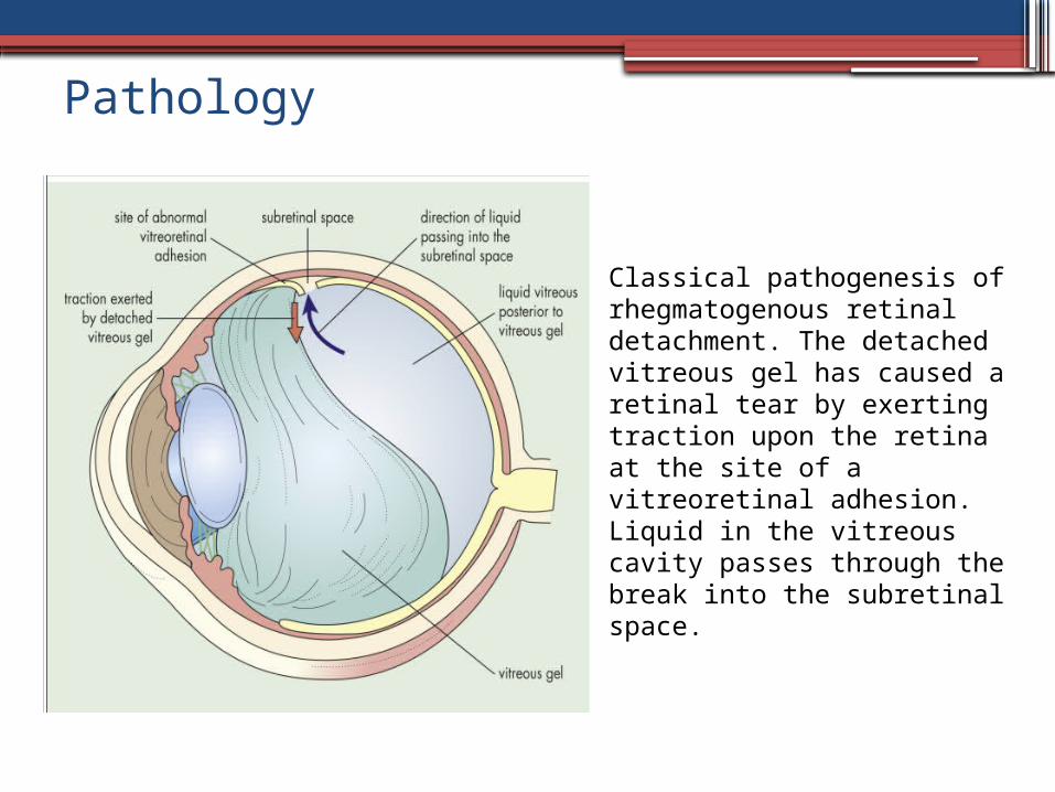
Pathology
Classical pathogenesis of rhegmatogenous retinal detachment. The detached vitreous gel has caused a retinal tear by exerting traction upon the retina at the site of a vitreoretinal adhesion. Liquid in the vitreous cavity passes through the break into the subretinal space.
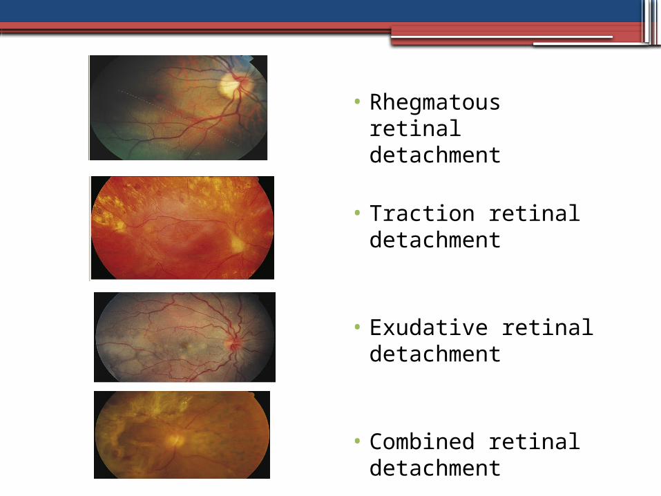
• Rhegmatous retinal detachment
• Traction retinal detachment
• Exudative retinal detachment
• Combined retinal detachment
Treatment
•scleral buckling techniques and the creation of a chorioretinal adhesion around each break to eliminate and counteract vitreoretinal traction
•Vitrectomy techniques•Pneumatic retinopexy
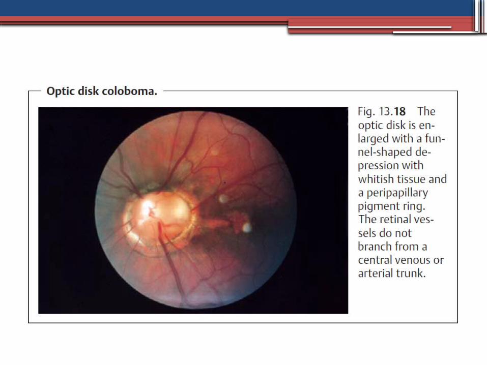
Optic Disk Coloboma•An optic disk coloboma is the result of
incomplete closure of the embryonic optic cup.• The optic disk is enlarged with a funnel-
shaped depression with whitish tissue and a peripapillary pigment ring.
•The retinal vessels extend outward across the margin of the disk in a radial pattern without a central trunk vessel.
•Patients with optic disk coloboma often have decreased visual acuity and visual field defects.
•retinochoroidal colobomas that has received particular attention CHARGE syndrome (coloboma, heart disease, atresia choanae, retarded growth, genital hypoplasia, ear anomalies, with or without deafness
Norrie disease•Norrie disease, or the progressive
oculoacousticocerebral degeneration of Norrie, is a rare, X-linked recessive heritable disorder characterized by bilateral leukocoria caused by retinal detachment.
•Affected boys classically have a triad of blindness, deafness, and mental retardation.
• Apparent at birth or in early infancy, the ocular findings usually progress to phthisis bulbi.

END
THANK YOU