Embed Size (px)
Citation preview

3
LaboratoryGeneral information on TB laboratory work . . . . . . . . . . . . . . . . . . . . 32
Communication between clinician and laboratory
How should specimens be collected for smear and culture?
Microscopy, culture identification, and growth-based testing . . . . . . . . . . . . 36
AFB smear
Culture identification
Conventional growth-based drug susceptibility testing (DST)
Critical concentration and minimum inhibitory concentration (MIC) . . . . . . . . 41
Molecular methods for detection of M. tuberculosis complex and drug resistance . . . . . . . . . . . . . . . . . . . . . . . . . 44
Molecular detection of M. tuberculosis complex
Genes associated with drug resistance
Molecular tests for drug resistance
Probe-based tests (Molecular beacon assay: Xpert MTB/RIF; Line-probe assays)
Sequence-based tests
Choice of molecular tests
Difficulties interpreting results from molecular tests
Molecular tests on extrapulmonary specimens
Molecular tests on formalin-fixed specimens
Therapeutic drug monitoring (TDM) . . . . 56
National TB genotyping service . . . . . . . 59
References . . . . . . . . . . . . . . . . . . . . . . . . 61
3rd edition contributors: PENNAN M. BARRY, MD, MPH & SHOU-YEAN GRACE LIN, MS
DRUG-RESISTANT TUBERCULOSIS : 31 : A SURVIVAL GUIDE FOR CLINICIANS : 3RD EDITION
Definitive diagnosis of drug-resistant tuberculosis (TB) requires that Mycobacterium (M.)
tuberculosis be isolated and drug susceptibility results be completed and conveyed to the
clinician. Prompt turnaround time for laboratory results is of paramount importance in
rapid diagnosis and appropriate treatment, infection control, and public health manage-
ment of drug-resistant TB.
Molecular technology is enabling much more rapid diagnosis of drug resistance. It is
important to note that new technologies generate new questions, and the best way to
interpret molecular resistance results is still evolving. Despite the expanding knowledge
and experience with molecular methods, conventional growth-based drug-suscepti-bility testing (DST) remains the gold standard. However, growth-based DST is com-
plex and various methods are used. Discrepant results may be generated due to differ-
ences in methodology, critical concentrations, and inoculum preparation and render the
interpretation of growth-based DST results very challenging. These challenging laboratory
results can have significant implications for treatment and often necessitate expert con-
sultation.
General information on TB laboratory workSeveral types of laboratories perform diagnostic mycobacteriology testing, including hos-
pital-based laboratories, local and state public health laboratories, and commercial labo-
ratories. Laboratories may choose to provide different levels of services and different
methods for the services they offer. Refer to Table 1 for a list of mycobacteriology labora-
tory services. Services and protocols may vary based on the setting where the specimen
is collected (e.g., outpatient vs. hospital), type of specimen (e.g., sputum vs. cerebrospi-
nal fluid [CSF]), and third-party payer source. A single specimen can pass through several
different laboratories in order to complete testing.
Case managers and treating physicians should have an understanding of the laboratory
practices of the facilities processing their patients’ specimens.
The role of the laboratory is critical in the diagnosis of TB, and even more so for drug-resistant TB.
LA
BO
RA
TO
RY
DRUG-RESISTANT TUBERCULOSIS : 32 : A SURVIVAL GUIDE FOR CLINICIANS : 3RD EDITION

TABLE 1.
Mycobacteriology laboratory services
Test Expected turnaround time (from specimen receipt at laboratory)
Comments
AFB smear 1 day Fluorochrome staining is more sensitive than carbol-fuchsin acid fast staining (Ziehl-Neelsen or Kinyoun methods).
Nucleic acid amplification testing (NAAT)
For identification of M. tb complex
1-2 days Commercial, FDA-cleared tests and laboratory developed tests available.
Excellent sensitivity and specificity for testing smear-positive sediments.
Testing smear-negative sediments usually has reduced sensitivity and specificity.
Molecular detection of drug resistance
(may also include identification of M. tb complex)
1-3 days Becoming more widely available, particularly for rifampin testing.
See Table 5 for more information.
New technologies are emerging.
Mycobacterial culture and identification
Positive cultures: average of 2-3 weeks incubation.
Smear-negative specimens may take >4 weeks to turn positive.
6–8 weeks to report negative.
When a culture takes 5-6 weeks to turn positive, consider investigation for possible cross-contamination.
Identification of positive cultures
1 day to 1 week for identification of M. tb complex, MAC, M. kansasii, and M. gordonae by DNA probes.
Identification of other non-TB mycobacteria may take days or months depending on method used.
Laboratories may batch tests; testing time by DNA probes or MALDI-TOF is less than 2 hours.
Growth-based DST Liquid broth systems: 1-2 weeks after setting up DST. (4 weeks or longer from specimen receipt at laboratory.)
Solid media (agar proportion method): 3-4 weeks.
DST cannot be performed on mixed or contaminated cultures.
Laboratories usually perform DST in batches.
Genotyping MIRU: 2 weeks
Spoligotype: 1 month
MIRU is performed at the Michigan TB laboratory.
Spoligotyping is performed at CDC.
Expedited genotyping may be requested for investigation of outbreaks or cross-contamination.
Interferon gamma release assays (IGRA)
1-2 days (longer if batched) Usually performed by clinical laboratory (not mycobacteriology laboratory).
LA
BO
RA
TO
RY
DRUG-RESISTANT TUBERCULOSIS : 33 : A SURVIVAL GUIDE FOR CLINICIANS : 3RD EDITION

Communication between clinician and laboratoryThe optimal laboratory diagnosis of TB begins with a close relationship and open dialogue
between the healthcare provider, TB control, and the TB laboratory.
Include the following information with the laboratory request in order to maximize the
laboratory’s contribution:
• Diagnostic versus follow-up specimen
• Date when anti-TB treatment was started and drug regimen
• Is drug resistance suspected?
The laboratory should inform submitting providers about test availability and requirements
for optimum testing, such as sample volume requirements, transit conditions, and test
performance and limitations. Such information promotes proper utilization of the test by
clinicians, and laboratories benefit from having optimal samples to test for better testing
outcomes. As laboratory technologies advance, laboratories may need to inform clini-
cians about new tests that are available for implementation. As clinical practices evolve,
clinicians may need to inform laboratories about tests that are no longer necessary to
perform and about tests they hope laboratories can offer. Additionally, clinicians and lab-
oratories may wish to work together on diagnostic algorithms. One example is the use of
nucleic acid amplification tests (NAAT) for rapid identification of M. tuberculosis complex
and molecular testing for drug resistance. Such communications can optimize scarce
resources and maximize the laboratory’s contribution to patient care.
FIGURE 1.
Mycobacteriology laboratory workflow
Specimen received
Specimen processing (decontamination and concentration)
Sediment
Culture identification
AFB smear
Nontuberculous mycobacteria
Culture (broth and solid media)
M. tuberculosis complex
Molecular testing:M. tuberculosis complex identification
Molecular susceptibility tests
Culture-based susceptibility testing Genotyping
LA
BO
RA
TO
RY
DRUG-RESISTANT TUBERCULOSIS : 34 : A SURVIVAL GUIDE FOR CLINICIANS : 3RD EDITION

How should specimens be collected for smear and culture?
Adapted from: A Clinician’s Guide to the TB Laboratory, Heartland National Tuberculosis Center.
FOR ALL SPECIMENS:
• Contact your laboratory for specific instructions
• Collect into sterile container
• Do not use preservatives
• Follow proper collection procedures and obtain an adequate volume to enhance recovery of organisms
• Process within 24 hours if possible
• Keep refrigerated until processed to reduce overgrowth of other microorganisms, especially for non-sterile specimens
RESPIRATORY SPECIMENS:
Preferably three specimens collected at least 8 hours apart and at least one of which is an early morning expectorated specimen or induced (some programs prefer all specimens to be induced)
• Note: Centers for Disease Control and Prevention (CDC) and World Health Organization (WHO) guidelines differ; internationally, two specimens are recommended. Incremental benefit of the third specimen is relatively small and may be even less if NAAT is used.
Expectorated sputum
• Preferably early morning (before brushing teeth), consider rinsing mouth with sterile or bottled water to reduce risk of contamination with non-tuberculous mycobacteria.
• Volume of 5-10mL ideal; should be >2mL.
Sputum induced with nebulized hypertonic (3-10%) saline
• Note on the requisition form and label specimen as induced because these are more likely to be watery in appearance and could be mistakenly considered unacceptable by the laboratory.
Bronchoscopy: lavage, brushings, biopsies
• Induced sputum have equivalent or better yield for diagnosis compared to bronchoscopy specimens.
• Bronchoscopy can target specific areas of the lung.
• Can obtain specimens from persons unable to produce sputum specimen by other methods.
• Post-bronchoscopy sputum collection may have higher yield than sputum collected at other times.
Gastric aspirate (for more information on how to perform gastric aspirate, see Chapter 6, Pediatrics)
• Used for diagnosing pulmonary TB in young children.
• Yield highest in the youngest children.
• Early morning collection after nothing by mouth (NPO) overnight.
• Mycobacteria die rapidly in gastric lavage fluid, which needs to be neutralized with sodium carbonate to pH of 7.0, especially if specimen will not be processed immediately.
• Add 100 mg of sodium carbonate to 5-10 mL specimen.
EXTRAPULMONARY SPECIMENS:
Urine
• Collect 3-5 early morning midstream specimens (not a 24-hour urine collection).
• 10-40 mL specimens.
• Do not pool specimens or use preservatives.
Stool
• Not routinely performed, contact your laboratory if needed.
• Recovery of acid-fast bacilli (AFB) is not high due to overgrowth of other bacteria.
• Collect at least 1 gram.
• No transport media needed.
• Refrigerate if transport time >1 hour; do not freeze.
Pleural fluid, peritoneal fluid, pericardial fluid, joint aspirate
• At least 10 mL specimen preferable.
• No swabs.
• Bloody specimens can be put in sodium polyanethol sulfonate (SPS) yellow-top tube.
Blood
• Collect in patients with suspected mycobacteremia (e.g., sepsis, immunocompromised).
• Special blood culture media for AFB are commercially available.
• If blood has to be transported before inoculation of the SPS, heparin or citrate may be used as anticoagulant.
• Blood collected in EDTA or in conventional blood culture bottles and coagulated blood are not acceptable.
Cerebrospinal fluid
• Minimum of 2-3mL, but 5-10mL preferable.
Tissue biopsy
• Any tissue specimen, not formalin-fixed, can be cultured for mycobacteria.
• Placement in formalin or other fixative eliminates ability to culture and perform growth-based DST. (Occasionally it is possible to extract nucleic acid from formalin-fixed specimens for molecular testing but this requires specialized methods and is only available in select laboratories—see section: Molecular methods on fixed specimens.) Careful communication with operating room staff will increase the likelihood that a specimen will be submitted in a sterile cup without formalin.
LA
BO
RA
TO
RY
DRUG-RESISTANT TUBERCULOSIS : 35 : A SURVIVAL GUIDE FOR CLINICIANS : 3RD EDITION

Microscopy, culture identification, and growth-based testingAFB smearCDC recommends using fluorochrome staining methods for acid-fast bacilli (AFB) smear
microscopy. It is more sensitive than the Ziehl-Neelsen staining method. Stains are typi-
cally done on concentrated specimens digested and decontaminated with N-acetyl-L-
cysteine-sodium hydroxide (NALC-NaOH). AFB smear results should be reported within
24 hours of receipt of specimens. Varied semi-quantitative reporting systems are in use:
Rare, few, moderate, numerous; 1+ to 3+ (WHO); and 1+ to 4+ (CDC). It is estimated that
the detection limit for smear positivity is 5,000 to 10,000 AFB per mL of sputum. AFB smear
is not M. tuberculosis complex -specific; nontuberculous mycobacteria (NTM) are stained
positive as well. Nocardia, Rhodococcus, Legionella, Cryptosporidium, Isospora, Cyclo-
spora, Actinomyces and Microsporidia may also show various degrees of acid-fastness.
Figure 2 presents photographs containing the typical appearance of AFB in microscopic
examinations.
FIGURE 2.
A B
A: Fluorochrome (auramine-rhodamine) stained AFB are seen as golden-orange rods when viewed under a fluorescent microscope.Source: California Department of Public Health Microbial Diseases Laboratory
B: Carbol-fuchsin stained (Ziehl-Neelsen method) AFB are seen as red rods when viewed under a light microscope. Source: Centers for Disease Control and Prevention
Culture identificationOnce AFB are grown in culture, culture identification is often done by DNA probes. Accu-
Probe (Hologic [previously Gen-Probe, Inc], San Diego, CA) Mycobacterial Culture Identi-
fication kits are the most commonly used commercial kits and they can identify M. tuber-
culosis complex, as well as some NTM including M. avium complex (MAC), M. kansasii
and M. gordonae. Species other than these can be identified by MALDI-TOF (Matrix
Assisted Laser Desorption Ionization Time-of-Flight), high performance liquid chromatog-
raphy (HPLC), DNA sequencing, or laboratory developed PCR assays. Additional studies
LA
BO
RA
TO
RY
DRUG-RESISTANT TUBERCULOSIS : 36 : A SURVIVAL GUIDE FOR CLINICIANS : 3RD EDITION

using growth rates, pigmentation and selected biochemical tests may assist further iden-
tification.
If a mixed culture (M. tuberculosis complex and NTM) is suspected, rapid identifi-
cation of M. tuberculosis complex and detection of drug resistance by molecular methods
should be pursued. If M. tuberculosis complex is identified, a pure culture should be
obtained for growth-based DST (see Confirmation of results section).
In the United States, 99% of isolates identified as M. tuberculosis complex are M. tuber-
culosis. Because M. bovis including BCG is naturally pyrazinamide- (PZA-) resistant, spe-
ciation within M. tuberculosis complex can be important particularly in regions where the
prevalence of M. bovis is high or when mono-PZA-resistance is detected.
Conventional growth-based drug susceptibility testing (DST)Conventional growth-based DST is also referred to as phenotypic, conventional, or cul-
ture-based drug susceptibility testing. Unlike molecular resistance testing, a pure culture
must be obtained before setting up growth-based DST.
Many methods for performing growth-based DST have been developed and are in use. In
general these methods have good concordance. However, in the course of managing
drug-resistant TB cases, clinicians are likely to encounter growth-based DST results from
multiple methods and laboratories. Various DST methods are validated to yield “equiva-
lent” results, but discordant results may occur and they are challenging to interpret. The
two most common methods used in the United States are performed in solid media by
the agar proportion method or liquid broth systems. They are outlined below, along with
features of each test that are important for clinicians to know.
Solid media—agar proportion method• The agar proportion method using Middlebrook 7H10 or 7H11 agar is the refer-
ence standard for DST in the United States.
• A standardized cell suspension is prepared from a pure isolate and inoculated onto each quadrant of an agar plate. Each quadrant contains a specific drug at its critical concentration or no drug as a control. Plates are incubated for 21 days before colony counts are taken.
• The isolate is considered resistant if the number of colonies in the drug quadrant is equal to or more than 1% of that in the control quadrant. An example of deter-mining the results using the agar-proportion method is demonstrated in Figure 3.
• PZA is difficult to study using solid medium due to the requirements of testing at an acidic pH, causing many isolates to fail to grow. PZA growth-based DST typ-ically is performed using liquid media.
• The critical concentrations used with 7H10 and 7H11 may be different.
• The Lowenstein-Jensen (LJ) proportion method is not used in the United States because it is more prone to contamination, but it is inexpensive and frequently used in low-resource settings.
Indirect DST refers to testing on positive culture growth, while direct DST is done on AFB
smear-positive sediments. The direct DST has an advantage of more rapid results, but it
may be more likely to become contaminated, and yield uninterpretable results.
LA
BO
RA
TO
RY
DRUG-RESISTANT TUBERCULOSIS : 37 : A SURVIVAL GUIDE FOR CLINICIANS : 3RD EDITION

FIGURE 3.
Agar proportion method for drug-susceptibility testing.
Quadrant plate—Inoculum of M. tuberculosis growth from liquid media has been inocu-
lated into each of the 4 quadrants with the following results:
Control quadrant: 90 colonies
Isoniazid (INH) quad: 30 colonies
Rifampin (R) quad: 23 colonies
Streptomycin (S) quad: 0 colonies
Isoniazid 30/90 = 33% resistant
Rifampin 23/90 = 25% resistant
Streptomycin 0/90 = susceptible
This is an MDR-TB isolate.
Liquid mediaMGIT 960 (Becton Dickinson, Sparks, MD)
• MGIT 960 is a modified proportion method and the most frequently used method in the United States.
• Food and Drug Administration (FDA) approved for testing first-line drugs (rifampin [RIF], isoniazid [INH], ethambutol [EMB], PZA) and streptomycin [SM].
• Results are available in about 1 week (4-14 days) after the test is set up.
LA
BO
RA
TO
RY
DRUG-RESISTANT TUBERCULOSIS : 38 : A SURVIVAL GUIDE FOR CLINICIANS : 3RD EDITION

• Second-line drugs can also be tested with result accuracy comparable to that of the agar proportion method with the exception of cycloserine (CS).
• The method is based on the fluorescence produced from reduced oxygen in the MGIT medium due to microbial growth. The fluorescence generated is then con-verted to “growth units” (GU). In general, more GU indicates more growth.
• When the growth control generates GU to 400 within 4-14 days, the DST is valid for interpretation. If a drug-containing MGIT tube yields GU<100, the organism is inter-preted as being susceptible; if GU is ≥100, the organism is considered resistant.
VersaTREK (Trek Diagnostics System, Thermo Fisher Scientific, Oakwood Village, OH)
• FDA approved for testing first-line drugs (RIF, INH, EMB, PZA).
• Results are available in about 1 week after test set-up (3-13 days). Resistant results may be reported faster (minimum of 3 days) than susceptible results (minimum of 6 days).
• The method is based on detection of pressure changes (oxygen consumption due to microbial growth) within the headspace above the broth medium in a sealed bottle.
Sensititre (Trek Diagnostics System, Thermo Fisher Scientific, Oakwood Village, OH)
• The method uses a 96-well microbroth dilution plate to test both first- and second- line drugs, but it does not include PZA or capreomycin (CM). It provides MIC results for each of the 12 drugs tested (see Table 2).
• Test must be set up from colonies obtained from solid media, which may delay DST set-up due to slower growth on solid media. Results are available within 10-21 days after the test is set up.
• M. tuberculosis complex has been traditionally tested using a single critical concen-tration of a drug. The usefulness of MIC results for clinical management of TB patients requires further investigation. See section: MIC—when to order and how to interpret.
MODS (microscopic observation drug susceptibility) assay (Hardy Diagnostics, Santa
Maria, CA)
• The MODS assay is considered a rapid growth-based (7H9 broth) test for detection of M. tuberculosis complex and drug resistance to INH and RIF on NALC-NaOH processed sputum specimens.
• The median turnaround time is 7 days. Valid reports may be generated between 5-21 days after inoculation of drug plates (24-well format).
• The test is based on visualization of the cording morphology of M. tuberculosis complex in liquid medium which is recognizable using an inverted microscope.
Confirmation of resultsWhen growth-based DST results are available, drug-resistant results must be verified to
rule out contamination with other non-AFB bacteria or mixed culture with NTM; this is
especially important when liquid media is used.
• For drug-resistant results obtained by a liquid system, a contaminated drug-con-taining tube is likely to show homogeneous turbidity. Examining a smear made from the drug-containing tube or bottle should demonstrate presence of AFB with mor-phology compatible with M. tuberculosis complex and absence of non-AFB bacteria
LA
BO
RA
TO
RY
DRUG-RESISTANT TUBERCULOSIS : 39 : A SURVIVAL GUIDE FOR CLINICIANS : 3RD EDITION

or NTM. Sub-culturing from DST media onto a 7H10 plate and observing micro-scopic colonial morphology in a few days can be helpful in ruling out the presence of NTM.
• Performing growth-based DST from a pure culture evidenced by no growth on non-selective media (e.g., blood agar plate) is not sufficient to rule out contamination, which may be introduced when DST is being set up.
• If the original culture is not pure, use of molecular methods to detect drug resistance mutations is recommended.
• When the patient does not have risk factors for drug resistance, the treating physi-cian should communicate with the public health program and the laboratory to con-firm resistance results, ensure that the risk of contamination or a mixed culture has been ruled out, and to discuss any other sources of a possibly erroneous result.
Clinical scenario:
A U.S.-born patient with a first episode of culture-positive TB is reported to
have resistance to INH, RIF, and PZA. The physician is surprised by this
result and confirms lack of risk factors for drug resistance. The patient has
clinically improved after 4 weeks of first-line TB treatment. The physician
calls the laboratory to confirm the results. A smear of the growth from the
drug-containing MGIT reveals mixed morphology. Molecular testing shows
M. tuberculosis complex but no mutations indicating drug resistance.
Further testing indicates presence of NTM and M. tuberculosis complex in
the DST cultures. The patient continues to do well on first-line TB treatment.
Reliability of growth-based DST results
• Reliability of growth-based DST by drug
• Reliable: INH, RIF, fluoroquinolones, amikacin (AK), CM, kanamycin (KM)
• Less reliable or no data: EMB (more often tests susceptible by MGIT 960 com-pared to agar proportion), PZA (more often falsely resistant), SM, oral second-line drugs, third-line drugs
• Critical concentrations of third-line drugs and certain second-line drugs have not been fully established
LA
BO
RA
TO
RY
DRUG-RESISTANT TUBERCULOSIS : 40 : A SURVIVAL GUIDE FOR CLINICIANS : 3RD EDITION

Critical concentration, minimum inhibitory concentration (MIC), and what they meanCritical concentrationsDrug-susceptibility testing in the mycobacteriology laboratory is usually performed using
a single drug concentration—the critical concentration, which provides categorical
interpretation (susceptible or resistant).
• The critical concentration is the level of drug that inhibits 95% of wild-type TB strains that have not been exposed to the drug, but does not appreciably suppress the growth of strains that are resistant to the drug (based on clinical treatment failure).
A critical concentration is not a minimum inhibitory concentration (MIC); however,
the MIC of microorganisms susceptible at a critical concentration should have an MIC <
critical concentration and those resistant should have MIC > critical concentration. See
section: MIC— when to order and how to interpret.
• The critical concentration used for an individual drug may differ based on the method of growth-based DST (see Table 2). Although critical concentrations are chosen to provide equivalent results across methods, it is difficult to achieve 100% equivalency and some discordance may be seen.
• Discordance can also be encountered within the same method, especially when the MIC of a strain is close to the critical concentration. The reproducibility of testing in these strains tends to be poor.
• High and low level resistance
• Some drugs, such as INH, are routinely tested at more than one concentration. Some experts use these results to select a higher dose of the drug when it tests resistant at the lower concentration and susceptible at the higher concentration. The higher dose may achieve in vivo concentrations sufficiently high to overcome resistance at the lower concentration.
• Table 2 shows the critical concentrations for commonly-used methods for growth-based DST. It also shows the normal peak concentration in serum for standard doses of anti-mycobacterial drugs. The clinical relevance of the relationship between in vitro susceptibility at a given critical concentration and the normal peak concen-tration can involve complex pharmacodynamics including the mechanism of action of the drug, the penetration of drug to the site of infection, whether mycobacteria are in an active or dormant state, and the patient’s metabolism of the drug.
LA
BO
RA
TO
RY
DRUG-RESISTANT TUBERCULOSIS : 41 : A SURVIVAL GUIDE FOR CLINICIANS : 3RD EDITION

TABLE 2.
Critical concentrations of antimycobacterial agents by broth systems or agar proportion methods
* Serum drug concentrations are provided in this table for comparison with the critical concentration. This information is not a substitute for therapeutic drug monitoring.
** Source: Personal communication with National Jewish Health.
*** Bedaquiline MIC testing available at CDC.
NR: not recommended. NA: not available. MGIT is a trademark of Becton, Dickinson and Company. VersaTREK and Sensititre are trademarks of TREK Diagnostic Systems.
Drug
Normal peak concentration in serum with
standard doses*
(mcg/mL)
MGIT 960 low/high
VersaTREK low/high
Agar 7H10
low/high
Agar 7H11
low/high
Sensititre
(range of concentrations
tested)
First-line drugs
• Isoniazid 3-6 0.1 / 0.4 0.1 / 0.4 0.2 / 1 0.2 / 1 0.03-4
• Rifampin 8-24 1 1 1 1 0.12-16
• Pyrazinamide 20-60 100 300 NR NR NA
• Ethambutol 2-6 5 5 / 8 5 / 10 7.5 0.5-32
Injectable agents
• Streptomycin 35-45 1 / 4 NA 2 / 10 2 / 10 0.25-32
• Capreomycin 35-45 2.5 or 3 NA 10 10 NA
• Amikacin 35-45 1 or 1.5 NA 4 NA 0.12-16
• Kanamycin 35-45 2.5 NA 5 6 0.6-8
Fluoroquinolones
• Levofloxacin 8-12 1.5 NA 1 NA NA
• Moxifloxacin 3-5 0.25 NA 0.5 0.5 0.06-8
• Ofloxacin 2 NA 2 2 0.25-32
Second-line oral agents
• Cycloserine 20-35 NR NR 60** 2-256
• Ethionamide 1-5 5 NA 5 10 0.3-40
• Para-aminosalicylate
20-60 NA NA 2 8 0.5-64
Other agents
• Rifabutin 0.3-0.9 0.5 NA 0.5 0.5 0.12-16
• Linezolid 12-26 1 NA NA NA NA
• Clofazimine 0.5-2.0 NA NA NA 0.25** NA
• Bedaquiline*** NA NA 0.008-2 0.008-2 NA
LA
BO
RA
TO
RY
DRUG-RESISTANT TUBERCULOSIS : 42 : A SURVIVAL GUIDE FOR CLINICIANS : 3RD EDITION

MIC—when to order and how to interpretMinimum inhibitory concentration (MIC) testing differs from testing using a critical concen-
tration in that the organism is tested at a series of drug concentrations, usually a series of
two-fold dilutions, and the result is the lowest concentration that inhibits growth of the
bacteria. Although in most cases testing using the critical concentration is sufficient, there
are situations described below with certain drugs where MIC can be helpful in guiding
management of difficult cases. However, no categorical interpretations (susceptible or
resistant) for MIC results for M. tuberculosis complex have been recommended by the
Clinical and Laboratory Standards Institute (CLSI).
Situations when MICs may be useful for clinical management:
• Resistance to fluoroquinoloneWhen fluoroquinolone resistance is found by critical concentration or by molecular
testing, an MIC—usually for MFX—can help inform whether an increase in dose may
benefit the patient. Although there is minimal published evidence to support this
approach, some MDR-TB experts use “high-dose” MFX at 600mg or 800mg daily for patients with MFX MIC of 1 or 2 mcg/mL.
• Resistance to injectablesIn cases with extensive resistance, obtaining an MIC to an injectable medication to
which there is resistance at the critical concentration may help determine whether
an increased dose is likely to benefit the patient. High peak levels can be achieved with high intermittent dosing (e.g. 25 mg/kg 2-3x per week) and some MDR-TB experts would use this dosing regimen if it could achieve a peak that is 5–8 times higher than the MIC.
• Bedaquiline (BDQ)BDQ is tested by determining an MIC. Testing is available at CDC through submis-
sion of isolates to state public health laboratories.
Clinical scenario:
A patient with presumed MDR-TB is being treated with an empiric MDR-TB
regimen of PZA, AK, moxifloxacin (MFX), CS, and ethionamide (ETA). The
patient’s isolate subsequently tests resistant to INH, RIF, EMB, and MFX at
standard critical concentrations for these drugs. MFX is increased to
600mg daily and MIC testing for MFX is requested to determine whether
MFX should be continued at this higher dose or should be discontinued.
The MIC for MFX returns as 1.0 mcg/mL (within the range that some
experts would use high-dose MFX). MFX is continued at 600mg.
LA
BO
RA
TO
RY
DRUG-RESISTANT TUBERCULOSIS : 43 : A SURVIVAL GUIDE FOR CLINICIANS : 3RD EDITION

Molecular methods for detection of M. tuberculosis complex DNA and drug resistance mutations Molecular assays able to be performed directly on clinical specimens without the require-
ment for growth in culture have significantly shortened turnaround time for detection of M.
tuberculosis complex and drug resistance. These tests are recommended by CDC for routine use in patients for whom a diagnosis of TB is being considered. Use of
these tests can dramatically shorten time to diagnosis of TB and MDR-TB from weeks to
hours.
It is important for clinicians who are interpreting molecular tests of drug resistance to
know the advantages and limitations of the tests. There are two major types of molec-ular tests described below: sequencing and nonsequencing (or probe-based) tests. The chief distinction is that probe-based tests can only determine that there is a
mutation present in the gene; they generally cannot identify specific mutations (for some
exceptions, see section: Line-probe assay). In contrast, tests that employ sequencing
do identify specific mutations and results of these tests reveal more information and can
be more predictive of drug resistance. For this reason, in the United States CDC and the Association of Public Health Laboratories (APHL) recommend confirming a resistant result from a nonspecific probe-based test with a sequencing test.
Indication for use of molecular assays for drug resistance is found in Chapter 2, Diagnosis.
Molecular detection of M. tuberculosis complexThe amplified M. tuberculosis direct test (MTD; Hologic [formerly Gen-Probe], San Diego,
CA) was the first molecular assay approved by FDA (1995) for testing concentrated spec-
imens to identify M. tuberculosis complex. It is still available in some laboratories and can
be used for testing smear-positive and smear-negative specimens. However, it cannot
identify drug resistance. Its sensitivity and specificity for smear-positive specimens are
96.9% and 100% respectively, and those for smear-negative specimens are 72% and
99.3% respectively.
GeneXpert MTB/RIF assay was the second assay approved by FDA (2013) for testing
raw or concentrated sputum specimens, either smear-positive or smear-negative, to
detect M. tuberculosis. The assay detects M. tuberculosis complex and resistance to RIF
by real-time PCR with five molecular beacon probes (A-E) that cover the RIF-resistance
determining region of rpoB. The assay does not have a specific probe for M. tuberculosis
identification; rather, the detection of M. tuberculosis is based on the fluorescent signal
production from at least two of the five probes. Recent data from the United States
reported in the MMWR (2/27/15) shows sensitivity for detection of M. tuberculosis com-
plex on smear-positive specimens by a single Xpert MTB/RIF Assay is approximately
97%, and that for testing smear-negative specimens is 55%.
For more information regarding Xpert MTB/RIF assay for identifying drug resistance, see
section: Molecular Tests for Drug Resistance.
LA
BO
RA
TO
RY
DRUG-RESISTANT TUBERCULOSIS : 44 : A SURVIVAL GUIDE FOR CLINICIANS : 3RD EDITION

Non-FDA-approved methods. There are laboratory developed tests for detection of M.
tuberculosis complex by real-time PCR performed at commercial laboratories or public
health laboratories. Clinicians may request laboratories to provide the performance data
for assessing the results from those tests.
Genes associated with drug resistanceTable 3 provides a summary of genes associated with drug resistance and the predomi-
nant mutations found in clinical isolates.
• Although major genes associated with drug resistance have been identified, the understanding of drug resistance at the genetic level remains variable and incom-plete. Therefore, 100% sensitivity for detecting all drug resistance is not currently achievable.
• Furthermore, there are mutations that do not confer in vitro resistance or are asso-ciated with unpredictable susceptibility by growth-based methods. Specificity for resistance detection by molecular methods for certain drugs is not 100% (using growth-based testing as the gold standard).
LA
BO
RA
TO
RY
DRUG-RESISTANT TUBERCULOSIS : 45 : A SURVIVAL GUIDE FOR CLINICIANS : 3RD EDITION

TABLE 3.
Genes and mutations associated with drug resistance in M. tuberculosis
Table footnotes:
* See Figure 4 for information on understanding reporting of mutations.
** Identified or not identified refers to whether the assay will include the specific mutation in the reported result. For more information, see section: Probe-based tests.
*** “Disputed” mutations are mutations in the rpoB gene that are associated with variable susceptibility results in growth-based assays but have been reported in the literature to have clinical significance. MIC testing may be warranted. For further explanation, see section: Difficulties interpreting results of molecular tests.
Antimicrobial agent Gene Sensitivity Specificity Selected mutations* and comments
Isoniazid (INH) katG 86.0 99.1 315Thr(ACC) Most frequent mutation, associated with high-level INH resistance.
Some other mutations in codon 315: Thr(ACA), Asn(AAC), Ile(ATC), Thr(ACT), Gly(GGC)
inhA promoter
-15T Often associated with low-level INH resistance and ethionamide (ETA) resistance. Some other mutations: -8C, -8A, -8G, -9T, -16G, -17T
fabG1 203 Leu(CTA) Acts with its adjacent region as a promoter to upregulate the expression of inhA.
ahpC promoter
4.5 100 -54A Associated with INH resistance.
Some other mutations: -48T, -51T, -52A, -52T
Rifampin (RIF) rpoB 97.1 97.4 531 Leu(TTG) Most frequent mutation seen with MDR TB.
Associated with RIF and RFB resistance.
Detectable by HAIN, mutation identified
Detectable by Probe E of Xpert MTB/RIF, mutation not identified**
526Tyr(TAC)
526 Asp(GAC)
Associated with RIF and RFB resistance.
Detectable by HAIN, mutation identified
Detectable by Probe D of Xpert MTB/RIF, mutation not identified**
516 Val(GTC) Often associated with RIF resistance but retains RFB susceptibility
Detectable by HAIN, mutation identified
Detectable by Probe B of Xpert MTB/RIF, mutation not identified**
Silent mutation:
514 Phe(TTT)
Most frequent silent mutation. Not associated with RIF resistance
Detectable by HAIN, missing WT3, mutation not identified**
Detectable by Probe B of Xpert MTB/RIF, mutation not identified** Incorrectly reported as “RIF resistance detected”
“Disputed” mutations***
511 Pro(CCG), 516 Tyr(TAC), 526 Asn(AAC), 526 Leu(CTC), 526 Ser(AGC), 533 Pro(CCG), 572 Phe(TTC)
Ethambutol (EMB)
embB 78.8 94.3 306Val(GTG) Most frequent mutation associated with EMB resistance.
Detectable by HAIN, mutation identified.
Some other mutations in codon 306: Leu(CTG), Ile(ATA), Thr(ACG), Ile(ATT), Ile(ATC), Leu(TTG).
Not all mutations in embB are associated with EMB resistance.
LA
BO
RA
TO
RY
DRUG-RESISTANT TUBERCULOSIS : 46 : A SURVIVAL GUIDE FOR CLINICIANS : 3RD EDITION
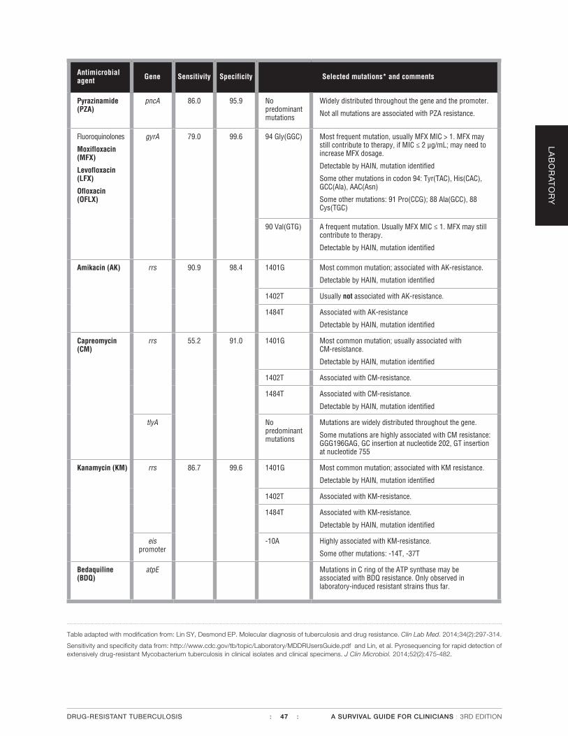
Table adapted with modification from: Lin SY, Desmond EP. Molecular diagnosis of tuberculosis and drug resistance. Clin Lab Med. 2014;34(2):297-314.
Sensitivity and specificity data from: http://www.cdc.gov/tb/topic/Laboratory/MDDRUsersGuide.pdf and Lin, et al. Pyrosequencing for rapid detection of extensively drug-resistant Mycobacterium tuberculosis in clinical isolates and clinical specimens. J Clin Microbiol. 2014;52(2):475-482.
Antimicrobial agent Gene Sensitivity Specificity Selected mutations* and comments
Pyrazinamide (PZA)
pncA 86.0 95.9 No predominant mutations
Widely distributed throughout the gene and the promoter.
Not all mutations are associated with PZA resistance.
Fluoroquinolones
Moxifloxacin (MFX)
Levofloxacin (LFX)
Ofloxacin (OFLX)
gyrA 79.0 99.6 94 Gly(GGC) Most frequent mutation, usually MFX MIC > 1. MFX may still contribute to therapy, if MIC ≤ 2 µg/mL; may need to increase MFX dosage.
Detectable by HAIN, mutation identified
Some other mutations in codon 94: Tyr(TAC), His(CAC), GCC(Ala), AAC(Asn)
Some other mutations: 91 Pro(CCG); 88 Ala(GCC), 88 Cys(TGC)
90 Val(GTG) A frequent mutation. Usually MFX MIC ≤ 1. MFX may still contribute to therapy.
Detectable by HAIN, mutation identified
Amikacin (AK) rrs 90.9 98.4 1401G Most common mutation; associated with AK-resistance.
Detectable by HAIN, mutation identified
1402T Usually not associated with AK-resistance.
1484T Associated with AK-resistance
Detectable by HAIN, mutation identified
Capreomycin (CM)
rrs 55.2 91.0 1401G Most common mutation; usually associated with CM-resistance.
Detectable by HAIN, mutation identified
1402T Associated with CM-resistance.
1484T Associated with CM-resistance.
Detectable by HAIN, mutation identified
tlyA No predominant mutations
Mutations are widely distributed throughout the gene.
Some mutations are highly associated with CM resistance: GGG196GAG, GC insertion at nucleotide 202, GT insertion at nucleotide 755
Kanamycin (KM) rrs 86.7 99.6 1401G Most common mutation; associated with KM resistance.
Detectable by HAIN, mutation identified
1402T Associated with KM-resistance.
1484T Associated with KM-resistance.
Detectable by HAIN, mutation identified
eis promoter
-10A Highly associated with KM-resistance.
Some other mutations: -14T, -37T
Bedaquiline (BDQ)
atpE Mutations in C ring of the ATP synthase may be associated with BDQ resistance. Only observed in laboratory-induced resistant strains thus far.
LA
BO
RA
TO
RY
DRUG-RESISTANT TUBERCULOSIS : 47 : A SURVIVAL GUIDE FOR CLINICIANS : 3RD EDITION
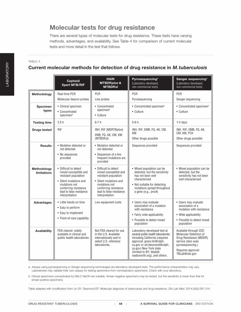
Molecular tests for drug resistanceThere are several types of molecular tests for drug resistance. These tests have varying
methods, advantages, and availability. See Table 4 for comparison of current molecular
tests and more detail in the text that follows.
TABLE 4.
Current molecular methods for detection of drug resistance in M. tuberculosis
CepheidXpert MTB/RIF
HAIN MTBDRplus &
MTBDRsl
Pyrosequencinga
(Laboratory-developed, non-commercial tests)
Sanger sequencinga
(Laboratory-developed, non-commercial tests)
Methodology Real-time PCR
Molecular beacon probes
PCR
Line probes
PCR
Pyrosequencing
PCR
Sanger sequencing
Specimen types
• Clinical specimen
• Concentrated specimenb
• Concentrated specimenb
• Culture
• Concentrated specimenb
• Culture
• Concentrated specimenb
• Culture
Testing time 2.5 h 6-7 h 5-6 h 1-2 days
Drugs tested RIF INH, RIF (MDRTBplus)
EMB, FQ, AK, CM, KM (MTBDRsl)
INH, RIF, EMB, FQ, AK, CM, KM
Other drugs possible
INH, RIF, EMB, FQ, AK, CM, KM, PZA
Other drugs possible
Results • Mutation detected or not detected
• No sequences provided
• Mutation detected or not detected
• Sequences of a few frequent mutations are provided
Sequences provided Sequences provided
Methodology limitations
• Difficult to detect mixed susceptible and resistant population
• Silent mutations and mutations not conferring resistance lead to false resistance interpretation
• Difficult to detect mixed susceptible and resistant population
• Silent mutations and mutations not conferring resistance lead to false resistance interpretation
• Mixed population can be detected, but the sensitivity has not been well characterized
• Not suitable for detecting mutations spread throughout a gene (e.g., pncA)
• Mixed population can be detected, but the sensitivity has not been well characterized
Advantages • Little hands-on time
• Easy to perform
• Easy to implement
• Point-of-care capability
Low equipment costs • Users may evaluate association of a mutation with resistance
• Fairly wide applicability
• Possible to detect mixed population
• Users may evaluate association of a mutation with resistance
• Wide applicability
• Possible to detect mixed population
Availability FDA cleared; widely available in clinical and public health laboratories
Not FDA cleared for use in the U.S. Available internationally and in select U.S. reference laboratories.
Laboratory-developed test at several public health laboratories including California (requires approval: [email protected] or [email protected]) New York state (limited to NY; [email protected]), and others.
Available through CDC Molecular Detection of Drug Resistance (MDDR) service (also uses pyrosequencing.)
Requires approval: [email protected]
a. Assays using pyrosequencing or Sanger sequencing technologies are laboratory-developed tests. The performance characteristics may vary. Laboratories may validate their own assays for testing specimens from nonrespiratory specimens. Check with your laboratory.
b. Clinical specimens concentrated by NALC-NaOH are suitable. Smear-negative specimens may be tested, but the sensitivity is lower than that for smear-positive specimens.
Table adapted with modification from Lin SY, Desmond EP. Molecular diagnosis of tuberculosis and drug resistance. Clin Lab Med. 2014;34(2):297-314.
LA
BO
RA
TO
RY
DRUG-RESISTANT TUBERCULOSIS : 48 : A SURVIVAL GUIDE FOR CLINICIANS : 3RD EDITION

Probe-based testsMolecular beacon assay
• Xpert MTB/RIF (Cepheid, Sunnyvale, CA) detects M. tuberculosis complex and resistance to RIF by real-time PCR with five molecular beacon probes (A-E) that cover the RIF-resistance determining region of rpoB (see Figure 3).
• FDA-approved for testing smear-positive or negative sputum specimens. The sys-tem is easy to operate and results are available within approximately 2.5 hours.
• Sensitivity/specificity of detecting RIF resistance are 95% and 98% respectively (from a 2014 meta-analysis by Steingart, et al., the majority of data from low- or middle-income countries).
• The Xpert MTB/RIF assay detects the presence or absence of mutations within the 81 base pair core region of rpoB. When mutations are detected, the assay issues reports stating “RIF resistance detected.” Certain mutations in the rpoB gene do not confer in vitro RIF resistance (silent or neutral mutations). CDC and APHL recommend confirmation of rpoB mutations with a sequence-based method.
• A frequently encountered silent mutation, 514Phe(TTT), is detectable by
probe B. Although the prevalence of this silent mutation has not been fully
investigated, data from the California Department of Public Health show a
frequency of 16.9% (26 of 154). [From a total of 1,538 specimens sequenced,
of the 154 containing rpoB mutations, 26 had this silent mutation (unpub-
lished data)]. These data suggest a lower positive predictive value of rpoB
mutations detected by Xpert and other nonsequence-based assays for RIF
resistance in an area with low prevalence of RIF resistance.
• Especially for patients in whom TB or drug-resistant TB is not suspected, clini-cians may wish to discuss Xpert MTB/RIF results with the performing laboratory to get more information. A resistant result involving Probe B might indicate a silent mutation (some resistance conferring mutations are also detectable by Probe B). High cycle threshold (Ct) values, corresponding to smaller quantities of mycobacterial DNA or mutations detected by multiple probes (which are rare), should be interpreted with caution.
For further information, see section: Difficulties interpreting results from molecular tests.
LA
BO
RA
TO
RY
DRUG-RESISTANT TUBERCULOSIS : 49 : A SURVIVAL GUIDE FOR CLINICIANS : 3RD EDITION
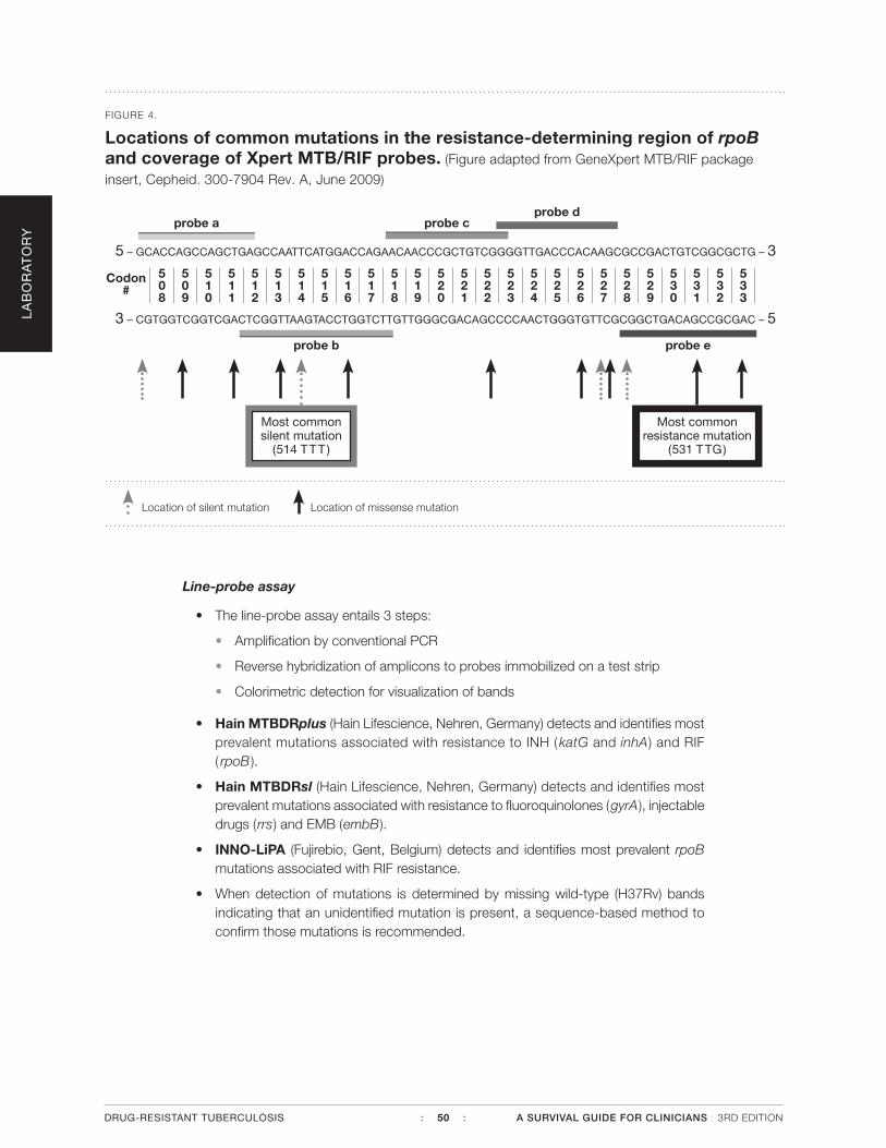
Line-probe assay
• The line-probe assay entails 3 steps:
• Amplification by conventional PCR
• Reverse hybridization of amplicons to probes immobilized on a test strip
• Colorimetric detection for visualization of bands
• Hain MTBDRplus (Hain Lifescience, Nehren, Germany) detects and identifies most prevalent mutations associated with resistance to INH (katG and inhA) and RIF ( rpoB ).
• Hain MTBDRsl (Hain Lifescience, Nehren, Germany) detects and identifies most prevalent mutations associated with resistance to fluoroquinolones (gyrA), injectable drugs (rrs) and EMB (embB).
• INNO-LiPA (Fujirebio, Gent, Belgium) detects and identifies most prevalent rpoB mutations associated with RIF resistance.
• When detection of mutations is determined by missing wild-type (H37Rv) bands indicating that an unidentified mutation is present, a sequence-based method to confirm those mutations is recommended.
FIGURE 4.
Locations of common mutations in the resistance-determining region of rpoB and coverage of Xpert MTB/RIF probes. (Figure adapted from GeneXpert MTB/RIF package
insert, Cepheid. 300-7904 Rev. A, June 2009)
probe a probe cprobe d
Most common silent mutation
(514 TTT )
Most common resistance mutation
(531 TTG)
probe eprobe b
5 – GCACCAGCCAGCTGAGCCAATTCATGGACCAGAACAACCCGCTGTCGGGGTTGACCCACAAGCGCCGACTGTCGGCGCTG – 3
Codon #
508
510
509
511
512
513
514
515
516
517
518
519
520
522
524
526
528
521
523
525
527
529
530
532
531
533
3 – CGTGGTCGGTCGACTCGGTTAAGTACCTGGTCTTGTTGGGCGACAGCCCCAACTGGGTGTTCGCGGCTGACAGCCGCGAC – 5
Location of silent mutation Location of missense mutation
LA
BO
RA
TO
RY
DRUG-RESISTANT TUBERCULOSIS : 50 : A SURVIVAL GUIDE FOR CLINICIANS : 3RD EDITION

TABLE 5.
Interpretation of line-probe assay
Line-probe band pattern Interpretation/comments
All wild-type bands are present with absence of all mutant bands.
• No mutations are present within the targeted DNA segment; this suggests susceptibility to the drug.
Missing at least one wild-type band and presence of one of the mutant bands.
• A specific mutation is present and its sequence is identified. Drug resistance is predicted.
Missing at least one wild-type band but none of the mutant bands are present.
• A mutation is present but not one of the frequent mutations; the identity of the mutation is not given.
• It is likely to be associated with drug resistance, but one cannot rule out silent mutations or other mutations not conferring resistance.
All wild-type bands are present and one of the mutant bands is also present.
• Possibly a mixed population or a mixed infection with two different strains, a wild-type strain and a drug-resistant strain.
• The variable intensity of the band may add difficulties in interpretation for this scenario. It is advisable to repeat the test or to confirm by a sequence-based method, or to defer the interpretation to culture-based drug susceptibility testing results.
(Table adapted with modification from Lin SY, Desmond EP. Molecular diagnosis of tuberculosis and drug resistance. Clin Lab Med. 2014;34(2):297-314.)
Sequence-based testsA sequence-based test not only detects presence or absence of mutations, but also pro-
vides the identity of a mutation. This allows a user to identify if a mutation confers in vitro
resistance. Furthermore, specific mutations may be used to predict a range of MICs.
• Pyrosequencing (PSQ) is a real-time sequencing method that sequences a short stretch of nucleotides and is capable of detecting any mutation within the targeted length with the mutation identity provided. It is not suitable for detecting mutations which are widely spread throughout the gene, such as PZA-resistance associated pncA mutations. A well-designed PSQ assay is sensitive enough to detect muta-tions from concentrated specimens.
• Sanger sequencing is the gold standard of sequencing, using the dye-terminator technology. It is capable of sequencing hundreds of nucleotides. CDC’s MDDR ser-vice provides sequencing that detects mutations associated with resistance to EMB, PZA, AK, CM, KM, and fluoroquinolones by Sanger sequencing and to INH, RIF by PSQ. The service has a short turnaround time (1-2 days).
LA
BO
RA
TO
RY
DRUG-RESISTANT TUBERCULOSIS : 51 : A SURVIVAL GUIDE FOR CLINICIANS : 3RD EDITION

• Next generation sequencing can be used to perform partial genome or whole genome sequencing and can provide the same information as PSQ and Sanger sequencing in addition to information on many other genes; however, it is not yet widely available in clinical laboratories. It requires sophisticated software to handle enormous amounts of data and has a longer turnaround time. At present, it requires higher concentrations of DNA extracted from cultures, so it is not yet sensitive enough for testing direct specimens.
Choice of molecular tests• If a sequence-based method is available locally, it is the method of choice.
• If Xpert MTB/RIF is readily available, it can be used for detection of M. tuberculosis complex and RIF-resistance. When a mutation is detected, confirmation by a sequence-based method is recommended.
• If INH-resistance is suspected, use a method which can at least detect the most common INH-associated mutations, katG and inhA.
• If RIF-resistance is detected, MDR-TB is likely and the specimen should be tested for mutations associated with resistance to other drugs.
Difficulties interpreting results from molecular testsMolecular testing is enabling much more rapid diagnosis of likely drug resistance, yet with
new technologies come new questions. Difficulties interpreting results may arise from the
way tests are reported, clinicians’ lack of familiarity with molecular terminology, and—
most importantly—from evolving knowledge regarding the clinical implications of specific
mutations. Among the most challenging situations for the clinician is when molecular and
growth-based test results are discordant.
• Discordance between molecular and growth-based test results may occur and can be confusing. Examples of this are isolates with certain mutations in the rpoB gene that may test susceptible for rifampin by growth-based methods. These mutations have been referred to as “disputed mutations.” Most laboratories per-forming sequence-based assays should be able to identify these mutations in test reports. However, reporting parameters and language may vary by laboratory.
Several clinical case series have been published reporting poor treatment out-comes for patients with these disputed mutations when treated with standard first-line therapy. In a 2013 study evaluating samples from two countries with a high burden of drug-resistant TB, disputed rpoB mutations were responsible for over 10% of rifampin resistance among first-line failure and relapse cases.
The best clinical approach to managing patients with strains possessing disputed mutations is not known and may depend on the presence of other factors such as additional drug resistance, the extent of disease, comorbidities (diabetes, HIV status and treatment, etc.), serum drug concentration, patient adherence, and nutritional status. Expert clinical and laboratory consultation for patients with a disputed rpoB mutation may be helpful.
Silent and neutral mutations (defined in Types of mutations) are additional causes for discordance between molecular and growth-based test results and can be identified through sequencing as sources of false-positive molecular resistance results.
LA
BO
RA
TO
RY
DRUG-RESISTANT TUBERCULOSIS : 52 : A SURVIVAL GUIDE FOR CLINICIANS : 3RD EDITION

Types of mutations
Silent mutations: alteration in DNA sequence but no resulting amino acid change, and thus, not associated with drug resistance. Also called synon-ymous mutations.
• 514 TTT(Phe) mutation in rpoB is the most common silent mutation. Information regarding this silent mutation contributing to false-positive rifampicin resistance results when using Xpert MTB/RIF can be found in the section: Probe-based assays.
Missense mutations: alteration in DNA sequence results in change in amino acid sequence. Also called nonsynonymous mutations.
• May confer different levels of resistance or no resistance.
• A missense mutation that has no effect on growth-based test results is also called a neutral mutation. Neutral mutations can be present in both drug susceptible and drug resistant strains. This term is used on the CDC’s MDDR report.
• Understanding sequence-based molecular test reports can be challenging. Results can be reported using various formats, abbreviations and numbering sys-tems. Figure 5 shows variations of reporting formats based on the example of an rpoB mutation using the format of CDC MDDR results. All reports should indicate the location (codon number or nucleotide number) and the mutant sequence or amino acid detected. This information can be used to make additional conclusions about the likelihood and extent of resistance (see Table 3: Genes and mutations associated with drug resistance).
FIGURE 5.
Guide to understanding sequence-based molecular test reports based on the example of an rpoB mutation using the format of CDC’s MDDR Service results.
Alternate formats
TCG>TTG;Ser 531Leu
531Leu
531Leu(TTG)
S531L
Wild-type sequence
Wild-type amino acid
3 letter amino acid abbreviation
1 letter amino acid abbreviation
Mutant sequence
Codon number
Mutant amino acid
LA
BO
RA
TO
RY
DRUG-RESISTANT TUBERCULOSIS : 53 : A SURVIVAL GUIDE FOR CLINICIANS : 3RD EDITION

• Both growth-based susceptibility testing and molecular testing are import-ant in constructing treatment regimens. Growth-based testing still plays an integral role in providing crucial additional information and testing drugs for which molecular tests are not yet available.
Clinical scenario:
Long-term elderly resident of the United States who was born in Mexico
presents with 3 months of cough and cavitary lesion on chest radiograph.
He has not been treated for TB before and has no known contact with an
MDR-TB case. Xpert MTB/RIF assay performed on AFB smear-positive
sputum is reported as “MTB detected, RIF resistance detected.”
Confirmatory sequence-based testing is requested prior to starting an
MDR-TB regimen because likelihood of MDR-TB is low given the patient’s
history. Sequencing assay reveals mutation at 514TTT(Phe), a silent
mutation. Growth-based susceptibility testing confirms RIF
susceptibility. The patient does well on standard first-line treatment.
Molecular tests on extrapulmonary specimensMolecular tests for drug resistance can also be performed on non-respiratory specimens.
However, no molecular assay is FDA cleared for use on non-respiratory specimens, and
assays therefore must be validated by individual laboratories. Many laboratories do not
have the capability to validate or run molecular tests on extrapulmonary specimens. Xpert
MTB/RIF performance for testing extrapulmonary specimens has been published (See
Table 7).
LA
BO
RA
TO
RY
DRUG-RESISTANT TUBERCULOSIS : 54 : A SURVIVAL GUIDE FOR CLINICIANS : 3RD EDITION
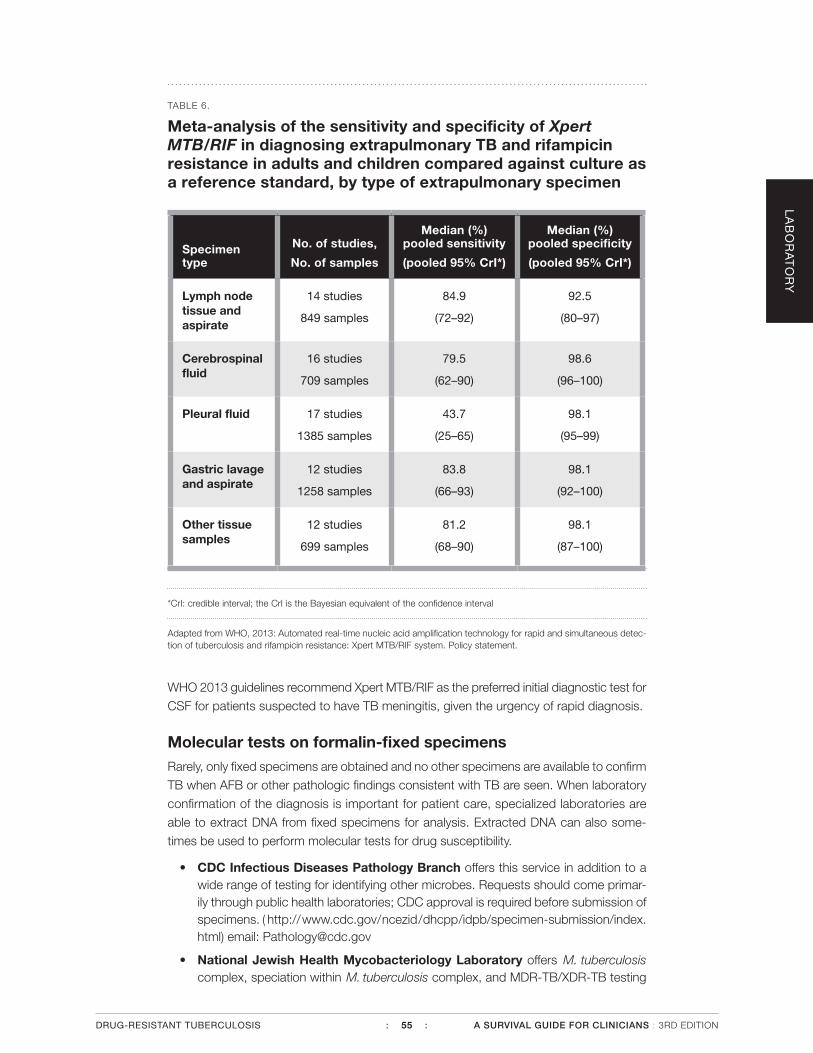
TABLE 6.
Meta-analysis of the sensitivity and specificity of Xpert MTB/RIF in diagnosing extrapulmonary TB and rifampicin resistance in adults and children compared against culture as a reference standard, by type of extrapulmonary specimen
Specimen type
No. of studies,
No. of samples
Median (%) pooled sensitivity
(pooled 95% CrI*)
Median (%) pooled specificity
(pooled 95% CrI*)
Lymph node tissue and aspirate
14 studies
849 samples
84.9
(72–92)
92.5
(80–97)
Cerebrospinal fluid
16 studies
709 samples
79.5
(62–90)
98.6
(96–100)
Pleural fluid 17 studies
1385 samples
43.7
(25–65)
98.1
(95–99)
Gastric lavage and aspirate
12 studies
1258 samples
83.8
(66–93)
98.1
(92–100)
Other tissue samples
12 studies
699 samples
81.2
(68–90)
98.1
(87–100)
*CrI: credible interval; the CrI is the Bayesian equivalent of the confidence interval
Adapted from WHO, 2013: Automated real-time nucleic acid amplification technology for rapid and simultaneous detec-tion of tuberculosis and rifampicin resistance: Xpert MTB/RIF system. Policy statement.
WHO 2013 guidelines recommend Xpert MTB/RIF as the preferred initial diagnostic test for
CSF for patients suspected to have TB meningitis, given the urgency of rapid diagnosis.
Molecular tests on formalin-fixed specimensRarely, only fixed specimens are obtained and no other specimens are available to confirm
TB when AFB or other pathologic findings consistent with TB are seen. When laboratory
confirmation of the diagnosis is important for patient care, specialized laboratories are
able to extract DNA from fixed specimens for analysis. Extracted DNA can also some-
times be used to perform molecular tests for drug susceptibility.
• CDC Infectious Diseases Pathology Branch offers this service in addition to a wide range of testing for identifying other microbes. Requests should come primar-ily through public health laboratories; CDC approval is required before submission of specimens. ( http://www.cdc.gov/ncezid/dhcpp/idpb/specimen-submission/index.html) email: [email protected]
• National Jewish Health Mycobacteriology Laboratory offers M. tuberculosis complex, speciation within M. tuberculosis complex, and MDR-TB/XDR-TB testing
LA
BO
RA
TO
RY
DRUG-RESISTANT TUBERCULOSIS : 55 : A SURVIVAL GUIDE FOR CLINICIANS : 3RD EDITION

on formalin-fixed specimens, available 7 days a week. (http://www.nationaljewish.org/getattachment/professionals/clinical-services/diagnostics/adx/ordering-tests/requisitions/myco_rec_web.pdf.aspx) email: [email protected]
• University of Washington Medical Center Molecular Diagnosis Section offers identification of M. tuberculosis complex and NTM from tissue specimens including fixed specimens. (http://depts.washington.edu/molmicdx/mdx/tests/afbpcr.shtml) email: [email protected]
Therapeutic drug monitoring (TDM) When to order TDM Therapeutic drug monitoring is routinely used for several circumstances:
• Aminoglycoside/CM serum concentrations especially in patients with renal impairment
• CS concentrations in order to minimize risk of CNS toxicity and to safely use opti-mal dose
• Known or suspected malabsorption (e.g., diabetes, gastrointestinal disorders)
• Lack of expected clinical response or relapse while on appropriate drugs and doses, administered by directly observed therapy (DOT)
• Patients with few effective drugs in their regimen, in order to optimize the effect of available drugs
• Patients with potentially significant drug-drug interactions such as rifamycins and antiretrovirals
• EMB concentrations in patients with significant renal impairment
Many drug-resistant TB experts routinely monitor certain TB drug concentrations in antic-
ipation of toxicity and to escalate a drug dose when possible.
Where to send a specimen for TDMMost hospital and commercial laboratories perform AK serum concentrations. Only a few
laboratories perform drug concentrations for other TB drugs.
Drugs tested for first- and second-line therapeutic drug monitoring tests:
Capreomycin Ethionamide p-Aminosalicylic Acid
Ciprofloxacin Isoniazid Pyrazinamide
Clarithromycin Levofloxacin Rifabutin
Clofazimine Linezolid Rifampin
Cycloserine Moxifloxacin Rifapentine
Ethambutol Ofloxacin Streptomycin
LA
BO
RA
TO
RY
DRUG-RESISTANT TUBERCULOSIS : 56 : A SURVIVAL GUIDE FOR CLINICIANS : 3RD EDITION

Laboratories and contact information:
University of Florida National Jewish Health
idpl.pharmacy.ufl.edu/ njlabs.org
[email protected] [email protected]
352- 273-6710 303-398-1422
How to send a specimen for TDMCollecting and processing samples for TDM
• One milliliter of serum (about 2 mL of blood) is required per test. It is advisable to provide some excess serum in case there are technical problems.
• Specimens should be collected after at least 4-5 half-lives have elapsed since the initiation of the drug. In practice, approximately 1 week works well in most cases. A shorter time can be used for adjustments of dose or schedule.
• Random samples generally are not informative.
• The patient should come to clinic with his/her medications and should plan to be at the clinic for at least 2 hours.
• See Table 7 for timing of specimen collection. On the day of blood draws only, rifab-utin (RFB) can be given 1 hour before the other TB drugs so that only 2 venipunc-tures are required.
• Observe the taking or injection of the medications and record the exact time and date.
• Collect the blood by direct venipuncture (timing as described by Table 7) and record the exact time of the blood collection.
• For SM, note if the patient is also receiving ampicillin.
• Label the tubes with the patient’s name, date and time of collection, and the drug(s) to be assayed.
• The specimen should be stored frozen until ready for shipping; –70 degrees C is preferable, but at a minimum –20 degrees C.
• For detailed instructions for processing and submitting specimens for TDM, see:
University of Florida: idpl.pharmacy.ufl.edu
National Jewish Health: njlabs.org
How to interpret results of TDMFor information about how to interpret results of TDM, see Chapter 4, Treatment.
LA
BO
RA
TO
RY
DRUG-RESISTANT TUBERCULOSIS : 57 : A SURVIVAL GUIDE FOR CLINICIANS : 3RD EDITION

TABLE 7.
Suggested time for blood collection after an oral dose.
Drug name Hours after oral dose to “peak”
Time after dose for additional concentration if desired*
Clarithromycin 2-3 hours
Clofazimine 2-3 hours
Cycloserine 2 hours 6 hours
Ethambutol 2-3 hours 6 hours
Ethionamide 2 hours 6 hours
Isoniazid 1-2 hours 4-6 hours
Levofloxacin 2 hours 6 hours
Linezolid 2 hours 6 hours
Moxifloxacin 2 hours 6 hours
PAS 6 hours
Pyrazinamide 2 hours 6 hours
Rifabutin 3-4 hours 7 hours
Rifampin 2 hours 6 hours
Drug name Hours after completion of infusion/injection
to “peak”
Time after dose for additional concentration if desired*
Amikacin
Capreomycin
Kanamycin
Streptomycin
1.5-2 hours (IV)
2 hours (IM)
6 hours (IV or IM)
* An additional concentration may be obtained to evaluate for delayed absorption or to calculate a half-life in order to more accurately prescribe a drug dose and interval.
LA
BO
RA
TO
RY
DRUG-RESISTANT TUBERCULOSIS : 58 : A SURVIVAL GUIDE FOR CLINICIANS : 3RD EDITION

National TB genotyping serviceThe Michigan Department of Community Health is under contract with CDC to provide
genotyping services to TB programs in the United States. TB programs, through their state or county public health laboratories, should submit the initial isolate from each culture-positive TB patient to the genotyping laboratory.
The genotyping laboratory uses the following genotyping methods:
• Spoligotyping (performed at CDC)
• Mycobacterial interspersed repetitive units (MIRU) analysis (performed at Michigan)
• IS6110-based restriction fragment length polymorphism (RFLP) analysis (special request)
• Whole genome sequencing (WGS)
Spoligotyping and MIRU analysis are PCR-based genotyping methods. The genotyping
laboratories will analyze all the submitted isolates by both PCR-based genotyping tests.
Under certain circumstances and upon the request of the TB program, isolates that have
matching genotypes by both spoligotyping and MIRU analysis can be further typed by
RFLP. Whole genome sequencing, having greater discriminatory power, is used to further
type strains having the same genotypes by spoligotyping and MIRU but no epidemiology
links by the conventional contact investigation.
The CDC-supported genotyping services are offered at no cost to TB programs.
The objectives of universal TB genotyping are:
1. To determine the extent and dynamics of ongoing transmission in order to focus program interventions in specific areas and populations
2. To assess TB transmission in outbreaks and to refine contact investigations
3. To identify nosocomial transmission not identified by conventional methods
4. To investigate possible false-positive culture results so that clinicians can be notified of diagnostic errors quickly, allowing for termination of unnecessary TB treatment
These objectives are of particular importance in the care and investigation of drug-resis-
tance cases, and all programs are encouraged to support these efforts toward universal
genotyping.
LA
BO
RA
TO
RY
DRUG-RESISTANT TUBERCULOSIS : 59 : A SURVIVAL GUIDE FOR CLINICIANS : 3RD EDITION
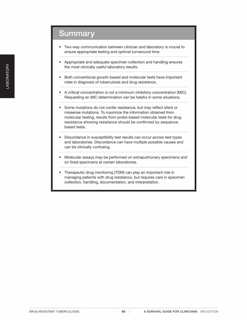
Summary
• Two-way communication between clinician and laboratory is crucial to ensure appropriate testing and optimal turnaround time.
• Appropriate and adequate specimen collection and handling ensures the most clinically useful laboratory results.
• Both conventional growth-based and molecular tests have important roles in diagnosis of tuberculosis and drug resistance.
• A critical concentration is not a minimum inhibitory concentration (MIC). Requesting an MIC determination can be helpful in some situations.
• Some mutations do not confer resistance, but may reflect silent or missense mutations. To maximize the information obtained from molecular testing, results from probe-based molecular tests for drug resistance showing resistance should be confirmed by sequence-based tests.
• Discordance in susceptibility test results can occur across test types and laboratories. Discordance can have multiple possible causes and can be clinically confusing.
• Molecular assays may be performed on extrapulmonary specimens and on fixed specimens at certain laboratories.
• Therapeutic drug monitoring (TDM) can play an important role in managing patients with drug resistance, but requires care in specimen collection, handling, documentation, and interpretation.
LA
BO
RA
TO
RY
DRUG-RESISTANT TUBERCULOSIS : 60 : A SURVIVAL GUIDE FOR CLINICIANS : 3RD EDITION
References• Ando H, Miyoshi-Akiyama T, Watanabe S, Kirikae T. A silent mutation in mabA confers isoniazid resis-
tance on Mycobacterium tuberculosis. Mol Microbiol. 2014; 91:538-547.
• Berrada ZL, Lin SY, Rodwell TC, et al. Rifabutin and rifampin resistance levels and associated rpoB mutations in clinical isolates of Mycobacterium tuberculosis complex. Diagn Microbiol Infect Dis. Forth-coming 2016.
• Centers for Disease Control and Prevention. Availability of an assay for detecting Mycobacterium tuber-culosis, including rifampin resistant strains, and considerations for its use—United States, 2013. MMWR. 2013;62(41);821-824.
• Centers for Disease Control and Prevention. Updated guidelines for the use of nucleic acid amplification tests in the diagnosis of tuberculosis. MMWR. 2009;58(01):7-10.
• Clinical and Laboratory Standards Institute (CLSI). Susceptibility Testing of Mycobacteria, Nocardiae, and Other Aerobic Actinomycetes; Approved Standard—Second Edition. CLSI document M24-A2 (ISBN 1-56238-746-4). Clinical and Laboratory Standards Institute, 940 West Valley Road, Suite 1400, Wayne, PA 19087
• Division of Microbiology Devices, Office of In Vitro Diagnostics and Radiological Health, Center for De-vices and Radiological Health, Food and Drug Administration; Centers for Disease Control and Preven-tion (CDC). Revised device labeling for the Cepheid Xpert MTB/RIF assay for detecting Mycobacterium tuberculosis. MMWR. 2015;64(7):193.
• George PM, Mehta M, Dhariwal J, et al. Post-bronchoscopy sputum: improving the diagnostic yield in smear negative pulmonary TB. Respir Med. 2011;105(11):1726-1731.
• Heartland National Tuberculosis Center. A Clinician’s Guide to the TB Laboratory. (Pending publication.) http://www.heartlandntbc.org/products/
• Lin SY, Desmond EP. Molecular diagnosis of tuberculosis and drug resistance. Clin Lab Med. 2014; 34(2):297-314.
• Lin SY, Desmond E, Bonato D, Gross W, Siddiqi S. Multicenter evaluation of Bactec MGIT 960 system for second-line drug susceptibility testing of Mycobacterium tuberculosis complex. J Clin Microbiol. 2009;47:3630-3634.
• Lin SY, Rodwell TC, Victor TC, et al. Pyrosequencing for rapid detection of extensively drug-resistant Myco-bacterium tuberculosis in clinical isolates and clinical specimens. J Clin Microbiol. 2014;52(2): 475-482.
• Ling D, Zwerling A, Pai M. GenoType MTBDR assays for the diagnosis of multidrug-resistant tubercu-losis: a meta-analysis. Eur Respir J. 2008;32(5):1165-1174.
• Malekmohammad M, Marjani M, Tabarsi P, et al. Diagnostic yield of post-bronchoscopy sputum smear in pulmonary tuberculosis. Scand J Infect Dis. 2012;44(5):369-373.
• Mase SR, Ramsay A, Ng V, et al. Yield of serial sputum specimen examinations in the diagnosis of pulmonary tuberculosis: a systematic review. Int J Tuberc Lung Dis. 2007;11(5):485-495.
• Rodrigues C, Jani J, Shenai S, Thakkar P, Siddiqi S, Mehta A. Drug susceptibility testing of Mycobac-terium tuberculosis against second-line drugs using the Bactec MGIT 960 system. Int J Tuberc Lung Dis. 2008;12(12):1449-1455.
• Rusch-Gerdes S, Pfyffer GE, Casal M, Chadwick M, Siddiqi S. Multicenter laboratory validation of the BACTEC MGIT 960 technique for testing susceptibilities of Mycobacterium tuberculosis to classical second-line drugs and newer antimicrobials. J Clin Microbiol. 2006;44:688-692.
• Steingart KR, Schiller I, Horne DJ, Pai M, Boehme CC, Dendukuri N. Xpert® MTB/RIF assay for pulmo-nary tuberculosis and rifampicin resistance in adults. Cochrane Database of Syst Rev. 2014, Issue 1. Art. No.: CD009593.
• TB CARE I. International Standards for Tuberculosis Care, Edition 3. TB CARE I, The Hague, 2014.
• TREK Diagnostic Systems. Sensititre. Available at: http://www.trekds.com/products/sensititre/c_myco-bacterium.asp Accessed October 12, 2015.
• Van Deun A, Aung KJ, Bola V, et al. Rifampin drug resistance tests for tuberculosis: challenging the gold standard. J Clin Microbiol. 2013;51(8):2633-2640.
• World Health Organization. 2013. Automated real-time nucleic acid amplification technology for rapid and simultaneous detection of tuberculosis and rifampicin resistance: Xpert MTB/RIF assay for the di-agnosis of pulmonary and extrapulmonary TB in adults and children. Policy update.
• World Health Organization. Companion handbook to the WHO guidelines for the programmatic man-agement of drug-resistant tuberculosis. 2014.
LA
BO
RA
TO
RY
DRUG-RESISTANT TUBERCULOSIS : 61 : A SURVIVAL GUIDE FOR CLINICIANS : 3RD EDITION

DRUG-RESISTANT TUBERCULOSIS : 62 : A SURVIVAL GUIDE FOR CLINICIANS : 3RD EDITION