Embed Size (px)
Citation preview
Drug-resistant leprosy: Monitoring and current
status
DIANA L. WILLIAMS & THOMAS P. GILLIS
HRSA, BPHC, National Hansen’s Disease Programs, Laboratory
Research Branch
Accepted for publication 30 August 2012
Introduction
Leprosy control depends solely on case detection and treatment with multi-drug therapy
(MDT).1 – 3 This strategy is based on the principle that identifying and treating chronic
infectious diseases with combinations of effective antibiotics limits the emergence and
spread of new or existing antibiotic resistant pathogens.2 According to the World Health
Organization (WHO), MDT formulated for leprosy has been effective at reducing both the
prevalence and incidence of leprosy globally.3 – 5 According to official reports from 130
countries and territories, the global registered prevalence of leprosy at the beginning of 2011
was 192,246 cases, while the number of new cases detected during 2010 was 228,474.5
The most important indicator for the effectiveness of a chemotherapeutic regimen is the
rate of relapse following successful completion of the scheduled course of treatment.
Information from a number of leprosy control programmes suggests that the relapse rate is very
low for both paucibacillary (PB) leprosy (0·1% per year) and multibacillary (MB) leprosy
(0·06% per year).5 Lessons learned from tuberculosis strongly suggest that relapse cases are at
risk for drug resistance and can undermine existing control measures.6,7 Therefore establishing
the success of a strategy like MDT for leprosy control requires thorough evaluation of
treatment failures, including drug susceptibility testing. Several studies have documented
relapses after MDT8 – 14 and drug-resistant strains of Mycobacterium leprae have been
identified.15 – 26 In contrast to what we know for tuberculosis, the current prevalence of primary
and secondary resistance to rifampicin; dapsone, and clofazimine is virtually unknown for
leprosy. Therefore, surveillance of drug resistance globally is a key factor in monitoring MDT
effectiveness and preventing the spread of drug resistance.
Over the past two decades, rapid DNA-based molecular assays for detection of drug-
resistant M. leprae directly from clinical specimens have been developed [Reviewed in22,23].
Even though these assays are based on sophisticated, modern, molecular biology techniques,
many reference laboratories in leprosy endemic countries have the capability of utilizing
Correspondence to: Diana L. Williams, HRSA, BPHC, National Hansen’s Disease Programs, LaboratoryResearch Branch, Molecular Biology Research Dept., LSU-SVM, Rm 3517W, Skip Bertman, Dr., Baton Rouge, LA70803 USA (Tel: þ1 225 578 9839; Fax: þ 1 225 578 9856; e-mail: [email protected])
Lepr Rev (2012) 83, 269–281
0305-7518/12/064053+13 $1.00 q Lepra 269
these tools for detection of drug resistance. Information gained from their implementation can
now be used as an integral component of an overall public health strategy for better patient
care as well as monitoring the spread of drug-resistant M. leprae. In this review we describe
the antibiotics used to treat leprosy and, where known, the mechanism of resistance for each
in M. leprae. We also describe current DNA-based assays for drug susceptibility testing and
surveillance studies aimed at quantifying the global burden of drug-resistant leprosy.
ANTI-LEPROSY DRUGS AND RESISTANCE MECHANISMS
The WHO Study Group on Chemotherapy of Leprosy for Control Programmes recommended
the introduction of Multi-Drug Therapy (MDT) in 19822 in response to the serious threat to
leprosy control posed by the widespread emergence of dapsone resistance.15,16 Concern has
also been expressed about the development of drug resistance to rifampicin, as it is the most
important component of the MDT regimen.17 – 23 As with tuberculosis, the emergence of
multi-drug resistant strains of M. leprae would pose a serious threat to leprosy control efforts.
FIRST LINE DRUGS
The drugs used in WHO-MDT are a combination of rifampicin; clofazimine and dapsone for
MB leprosy patients and rifampicin and dapsone for PB leprosy patients. Among these drugs,
rifampicin is the most important anti-leprosy drug and, therefore, is included in the treatment
of both types of leprosy. Experience strongly suggest that treatment of leprosy with either
dapsone15,16 or rifampicin alone27 will result in the development of resistance to the
respective drug and therefore should be discouraged.
In the 1950 s, dapsone was introduced as standard chemotherapy for leprosy28 and was
used worldwide for treating both MB and PB forms of the disease. The use of dapsone
required long-term, often life-long, treatment to control infections because of its slow
bacteriostatic effect on M. leprae. Long-term monotherapy with dapsone resulted in poor
compliance in many areas ultimately leading to treatment failures and the emergence of
dapsone-resistant strains of M. leprae in the 1970 s.15,16 This presented serious problems for
leprosy control programmes as resistance levels were reported as high as 40% in some areas
of the world.29,30 By the mid-1970 s it was clear that life-long dapsone monotherapy was
failing. Between the 1960 s and 1970 s, additional antimicrobial agents such as rifampicin31,32
and clofazimine33 were introduced for treating leprosy. Rifampicin proved to be a powerful
anti-leprosy drug however; using rifampicin alone resulted in relapses.27 In addition,
clofazimine proved to be only weakly bactericidal against M. leprae and, therefore, was not a
suitable single drug therapy for leprosy.33
To overcome the threat posed by the worldwide spread of dapsone resistance and to
improve treatment efficacy the WHO recommended MDT for leprosy in 1982.2 The current
WHO recommendations for adults are: daily dapsone (100 mg) and clofazimine (50 mg), with
once monthly rifampin (600 mg) and clofazimine (300 mg) for a duration of 1 year in the
treatment of MB leprosy (skin smears with a bacterial index of $2þ ); and daily dapsone
(100 mg) and once monthly rifampicin (600 mg) used for a duration of 6 months to treat
patients with PB leprosy (skin smears with a bacterial index of ,2þ ). A simple scheme to
define disease type by number of lesions is applied in peripheral clinics, where microscope BI
testing is not available. MB leprosy patients are those with more than five skin lesions and PB
leprosy patients are those with up to five skin lesions.34 These drug formulations are
D.L. Williams and T.P. Gillis270
incorporated into blister packs that can be stored at room temperature. This has made it
possible to distribute drugs to patients in rural or hard to reach locations sufficient for several
months of treatment, thereby improving treatment completion rates.5
DAPSONE
The first effective treatment for leprosy was promin (diamino-azobenzene40-sulfonamide)
introduced in 1941 and given intravenously. Six years later a more effective oral sulphone,
dapsone (diamino-diphenylsulphone), replaced promin and is still a fundamental part of
MDT for leprosy.28 Sulphone drugs target the dihydropteroate synthase (DHPS), a key
enzyme in the folate biosynthesis pathway in bacteria including M. leprae, by acting as a
competitive inhibitor of p-aminobenzoic acid (PABA).35 – 39 Missense mutations within
codons 53 and 55 of the drug resistance determining region (DRDR) of the folP1gene,
encoding the DHPS of M. leprae, have been observed in dapsone-resistant strains (Table 1,
Figure 1).
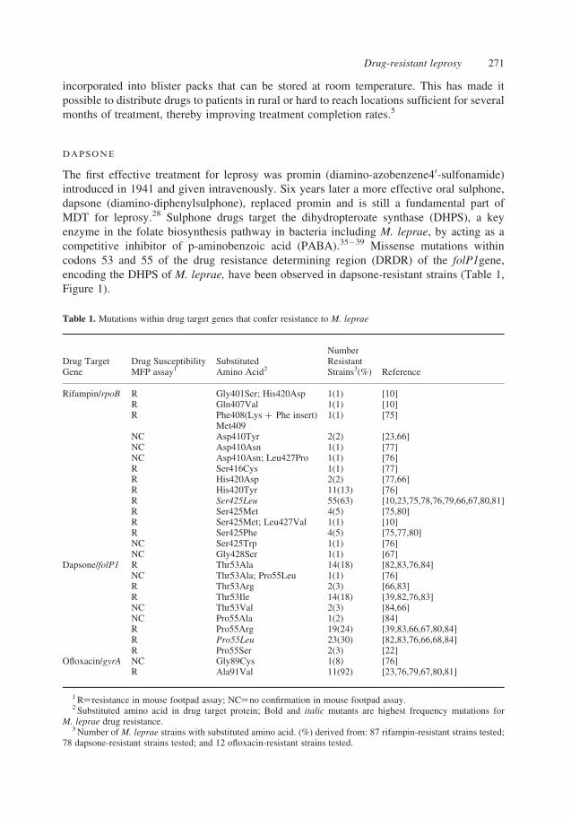
Table 1. Mutations within drug target genes that confer resistance to M. leprae
Drug TargetGene
Drug SusceptibilityMFP assay1
SubstitutedAmino Acid2
NumberResistantStrains3(%) Reference
Rifampin/rpoB R Gly401Ser; His420Asp 1(1) [10]R Gln407Val 1(1) [10]R Phe408(Lys þ Phe insert)
Met4091(1) [75]
NC Asp410Tyr 2(2) [23,66]NC Asp410Asn 1(1) [77]NC Asp410Asn; Leu427Pro 1(1) [76]R Ser416Cys 1(1) [77]R His420Asp 2(2) [77,66]R His420Tyr 11(13) [76]R Ser425Leu 55(63) [10,23,75,78,76,79,66,67,80,81]R Ser425Met 4(5) [75,80]R Ser425Met; Leu427Val 1(1) [10]R Ser425Phe 4(5) [75,77,80]NC Ser425Trp 1(1) [76]NC Gly428Ser 1(1) [67]
Dapsone/folP1 R Thr53Ala 14(18) [82,83,76,84]NC Thr53Ala; Pro55Leu 1(1) [76]R Thr53Arg 2(3) [66,83]R Thr53Ile 14(18) [39,82,76,83]NC Thr53Val 2(3) [84,66]NC Pro55Ala 1(2) [84]R Pro55Arg 19(24) [39,83,66,67,80,84]R Pro55Leu 23(30) [82,83,76,66,68,84]R Pro55Ser 2(3) [22]
Ofloxacin/gyrA NC Gly89Cys 1(8) [76]R Ala91Val 11(92) [23,76,79,67,80,81]
1 R¼ resistance in mouse footpad assay; NC¼no confirmation in mouse footpad assay.2 Substituted amino acid in drug target protein; Bold and italic mutants are highest frequency mutations for
M. leprae drug resistance.3 Number of M. leprae strains with substituted amino acid. (%) derived from: 87 rifampin-resistant strains tested;
78 dapsone-resistant strains tested; and 12 ofloxacin-resistant strains tested.
Drug-resistant leprosy 271
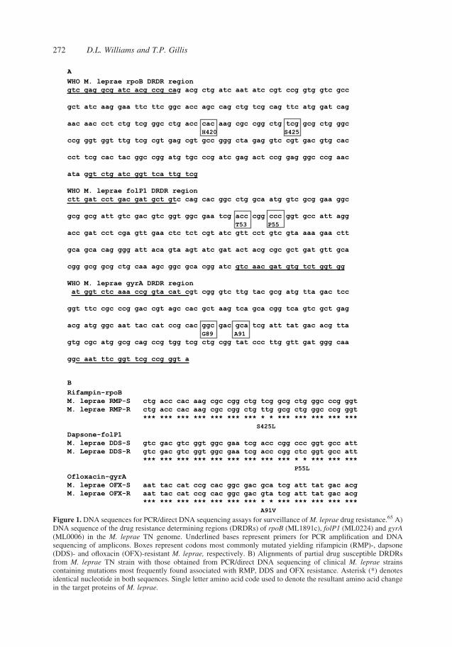
Figure 1. DNA sequences for PCR/direct DNA sequencing assays for surveillance of M. leprae drug resistance.65 A)DNA sequence of the drug resistance determining regions (DRDRs) of rpoB (ML1891c), folP1 (ML0224) and gyrA(ML0006) in the M. leprae TN genome. Underlined bases represent primers for PCR amplification and DNAsequencing of amplicons. Boxes represent codons most commonly mutated yielding rifampicin (RMP)-, dapsone(DDS)- and ofloxacin (OFX)-resistant M. leprae, respectively. B) Alignments of partial drug susceptible DRDRsfrom M. leprae TN strain with those obtained from PCR/direct DNA sequencing of clinical M. leprae strainscontaining mutations most frequently found associated with RMP, DDS and OFX resistance. Asterisk (*) denotesidentical nucleotide in both sequences. Single letter amino acid code used to denote the resultant amino acid changein the target proteins of M. leprae.
D.L. Williams and T.P. Gillis272
In addition, the majority of these patient biopsies were confirmed to harbour M. leprae
with moderate to high-levels of dapsone resistance as demonstrated by the mouse footpad
(MFP) drug susceptibility assay.
RIFAMPICIN
Rifampicin (3-{[(4-methyl-1-piperazinyl)-imino]-methyl}rifamycin) is the key bactericidal
component of all recommended MDT regimens. A single dose of 1,200 mg can reduce the
number of viable bacilli in a patient’s skin to undetectable levels within a few days.32 This
study also showed that a single dose of 600 mg had the same effect as 1200 mg in
approximately 7 days. The target for rifampicin in bacteria is the b-subunit of the DNA-
dependent RNA polymerase encoded by rpoB.40 M. tuberculosis resistance to rifampicin
correlates with changes in the structure of the b-subunit of the RNA polymerase, primarily
due to missense mutations that occur within a highly conserved region of the rpoB gene
referred to the rifampicin resistance determining region (RRDR).6,41 Rifampicin resistance in
M. leprae also correlates with missense mutations within the rpoB RRDR (Table 1, Figure 1).
Substitutions within codon Ser456 have been shown to be the most frequent mutations
associated with the development of the rifampicin-resistant phenotype in M. leprae (Table 1,
Figure 1B).
CLOFAZIMINE
Clofazimine [3-( p-chloroanilino)-10-( p-chlorophenyl)-2,10]-dihydro-2-(isopropylimino)-
phenazine] is a lipophilic riminophenazine antibiotic that possesses antimycobacterial
activities1,42,43 for which the mechanism has not been fully elucidated. Clofazimine attains
high intracellular levels in mononuclear phagocytic cells, its metabolic elimination is slow, it
has an anti-inflammatory effect, and the occurrence of resistance in M. leprae is extremely
low.1,22,23 Clofazimine is highly lipophilic and appears to bind preferentially to
mycobacterial DNA.1 Binding of the drug to DNA appears to occur principally at base
sequences containing guanine, which may explain clofazimine’s preference for the G þ C-
rich genomes of mycobacteria over human DNA. The accumulation of lysophospholipids
(detergent-like agents with membrane-disruptive properties in bacterial cells) appears to
mediate the activity of clofazimine in some gram-positive bacteria.44 However, it is unclear
whether this mechanism of action is operational in M. leprae. Since clofazimine may act
through several different mechanisms, it is not difficult to understand why only a few cases of
clofazimine-resistant leprosy have been reported over the years.
OTHER ANTI -LEPROSY DRUGS
OFLOXACIN
Ofloxacin (4-fluoroquinolone) is a fluorinated carboxyquinolone that has moderate
bactericidal activity for M. leprae.45 – 50 The mechanism of action of ofloxacin on M. leprae
is unknown, but in other bacteria it appears to inhibit DNA replication by inactivating the DNA
gyrase, a tetramer containing two b-subunits (GyrA) and two b-subunits (GyrB).51 Mutations
within a highly conserved region of gyrA, the quinolone resistance-determining region
(QRDR), are associated with the development of ofloxacin resistance in most resistant strains
Drug-resistant leprosy 273
of M. tuberculosis.52 The first ofloxacin-resistant M. leprae was described in 199453 and
subsequently other cases have been found. The DRDR ofM. leprae gyrA is highly homologous
to that ofM. tuberculosis, and missense mutations within codon Ala91 of this region have been
found in the majority of ofloxacin-resistant strains of M. leprae (Table 1, Figure 1).
CLARITHROMYCIN
Clarithromycin is a semisynthetic macrolide that differs from erythromycin in its methyl
substitution at the number six position of the macrolide ring. This drug displays significant
bactericidal activity against M. leprae in humans.54 – 56 In patients with lepromatous leprosy,
daily administration of 500 mg of clarithromycin kills 99% of viable M. leprae within 28 days
and 99·9% by 56 days. Although the mechanism of action against M. leprae is unknown, it is
thought to be similar to that of erythromycin, which acts by inhibiting protein synthesis by
binding to the ribosome.57 Clarithromycin resistance in bacteria and mycobacteria appears to
be due to a decrease in binding of the drug to ribosomes and is associated with missense
mutations within the 23SrRNA gene.57,58,59 This has not been fully investigated in M. leprae
due to the lack of well characterised resistant strains.60
MINOCYCLINE
Minocycline (7-dimethylamino-6-demethyl-6-deoxytetracycline) is the only member of the
tetracycline group of antibiotics to demonstrate significant activity against M. leprae,
presumably due to its lipophilic nature which may enhance cell wall penetration.1,61
Minocycline is bactericidal for M. leprae and its activity is additive when it is combined with
dapsone and rifampicin. The mechanism of action of minocycline against M. leprae is
unknown but is thought to be similar to that of all tetracyclines which act by inhibiting protein
synthesis. Tetracyclines bind reversibly to the 30S ribosomal subunit blocking the binding
of aminoacyl-tRNA to the mRNA ribosome complex.62 Resistance to tetracyclines may be
mediated by three different mechanisms: an energy-dependent efflux of tetracycline brought
about by an integral membrane protein; ribosomal protection by a soluble protein;62 or
enzymatic inactivation of tetracyclines. The molecular mechanism of minocycline resistance
has not been studied in M. leprae due to the lack of resistant mutants, presumably because
minocycline has been primarily used to treat single-lesion PB leprosy in combination with
rifampin and ofloxacin.
RIFAMPICIN, OFLOXACIN AND MINOCYCLINE (ROM) COMBINATION THERAPY
MDT for leprosy has been very practical and successful for both MB and PB leprosy and the
overall prevalence rates of leprosy in the world have fallen dramatically.5 However,
noncompliance is a primary reason that drug-resistant strains develop and relapses with
resistant strains may occur posing a potential problem for MDT in the control of leprosy.
As new drugs have been shown to be active against M. leprae, new combinations have been
tried in attempts to shorten the duration of therapy and improve therapeutic efficacy. For
example, in 1998 a single dose of a combination of rifampicin (600 mg), ofloxacin (400 mg)
and minocycline (100 mg) (ROM) was evaluated for treating single lesion, PB leprosy.63
A recent review of ROM therapy in leprosy concluded that, while ROM therapy has inherent
advantages (potential for improved compliance, absence of skin pigmentation and severe
D.L. Williams and T.P. Gillis274
reactions) it was less protective than WHO MDT in single lesion, PB patients.64 The authors
also concluded that current published data are insufficient to make meaningful comparisons
of monthly ROM therapy vis a vis standard MDT for treating MB leprosy. Future studies with
these drugs and others should be encouraged in an attempt to improve compliance and cure
rates while maintaining a focus on holding drug resistance to a minimum and reducing the
incidence of severe reactions.
DEVELOPMENT OF DRUG RESISTANCE IN M. LEPRAE
Lacking direct evidence for the mechanisms of M. leprae’s resistance to most of the anti-
leprosy drugs, our current understanding is based on studies carried out in M. tuberculosis,6
other bacteria, and a few studies with M. leprae genes in surrogate hosts. From these studies
one can predict that drug resistance in M. leprae is attributable to: 1) chromosomal mutations
in genes encoding drug targets; 2) these mutations occur spontaneously as a result of errors in
DNA replication; and 3) these mutants are enriched in a population of susceptible M. leprae
by inappropriate drug therapy. Drug-resistant M. leprae mutants can be acquired during the
initial infection from an infection source containing drug-resistant leprosy (primary drug
resistance) or from inadequate treatment (secondary drug resistance).
Because M. leprae cannot be cultivated in vitro, the frequency of drug-resistant mutants
in a population of bacteria is also inferred from studies with M. tuberculosis or other
cultivable mycobacteria. For example, the frequency of dapsone-resistant mutants in a
population of M. leprae is estimated to be 106 and the frequency for rifampicin and ofloxacin
resistance is estimated to be 107 and 108,6 respectively. Rates of clofazimine resistance in M.
leprae are unknown but appear to be extremely low. Since untreated MB patients can harbour
large bacterial loads (1011M. leprae), it is feasible that a patient could contain up to 105
dapsone-resistant organisms and thousands of rifampicin- or ofloxacin-resistant mutants in
their tissues. MDT was designed to reduce the development of drug resistance and therefore
these frequencies become less relevant when effective drug combinations are given. However
noncompliance or inadequate therapy of MB patients with high bacterial loads has the
potential to enrich the subpopulations of drug-resistant M. leprae, leading to the spread of one
or more resistant phenotypes.
SURVEILLANCE OF DRUG RESISTANCE IN LEPROSY
The success of leprosy control programmes relies heavily upon MDT. Therefore, it is
important that trends in drug resistance be monitored periodically. If resistance rates are
found to be increasing new strategies should be formulated that arrest its spread.
Acknowledging the growing concern of drug resistance in leprosy, the WHO issued
guidelines for the global surveillance of drug resistance in leprosy using PCR-direct
sequencing of M. leprae DRDRs from patients with characterised relapse from MDT.65
During 2010, a total of 109 relapsed cases were diagnosed at sentinel sites in China,
Colombia, India, Myanmar, Pakistan, The Philippines, Viet Nam and Yemen. Of the 109
cases identified 88 (81%) were tested for drug resistance.4 Nine (10%) were resistant to
dapsone and one (1·1%) case tested positive for resistance to rifampicin. No resistance to
ofloxacin was reported and no MDR cases were detected in this cohort.
Other studies have found similar results for dapsone and rifampicin resistance in patients
who had relapsed with active disease after completion of, or premature termination of,
Drug-resistant leprosy 275
MDT.66 – 69 In addition, very low levels of ofloxacin-resistant and MDR cases were observed.
Three of these studies also evaluated anti-leprosy drug resistance in newly diagnosed
patients.67 – 69 In 565 new cases tested 1·7% of cases were resistant to dapsone and 1% of
cases were resistant to rifampicin. Ofloxacin-resistant and MDR cases were not detected in
newly diagnosed patients in these studies. As more sites in more countries participate in
future surveillance studies, it should be possible to formulate an accurate view of drug-
resistant leprosy and thereby assess the success of current control strategies.
DETECTION OF DRUG-RESISTANT LEPROSY
Leprosy presents a very special problem for detecting drug resistance because M. leprae
cannot be cultured axenically. Accordingly, drug susceptibility testing was non-existent until
1962 when Shepard developed the MFP assay for determining M. leprae’s susceptibility to
anti-leprosy drugs.31 Since its development, the MFP assay has been the ‘gold standard’ for
leprosy drug susceptibility testing. This method requires the recovery of a sufficient number
of viable organisms from a patient to inoculate the footpads of 20 to 40 mice (depending on
the number of drugs to be tested) with each footpad receiving 5£103 organisms. Infected
mice are treated with the appropriate drug(s) orally. Mice are sacrificed after a defined period
of time (usually 6 months or longer) and the numbers of bacilli in the footpads of treated
mice and untreated mice are compared.
The MFP method is the only bacteriological assay for drug susceptibility testing for
M. leprae and presently is the standard for characterising the association of mutations in
target genes with drug resistance in M. leprae. While the MFP gives definitive information
pertaining to the susceptibility of an M. leprae isolate to anti-leprosy drugs, it is laborious,
expensive and often fails due to the need for significant numbers of viable M. leprae in a
patient’s biopsy. Because of the need for special resources to conduct this assay, only a few
facilities in the world have retained high quality mouse footpad laboratories. Their support is
critical as new drug-resistant mutants may evolve requiring corroboration in this model.
The first rapid drug-screening assays for M. leprae were developed based on
radiorespirometry techniques70,71 and have been used successfully to identify new anti-
leprosy drugs.71,46 Both assays are based on detection of 14-CO2 production from M. leprae’s
metabolism of 14-C-labelled palmitic acid in 7H12 medium in the presence and absence of
anti-leprosy drugs. However, the use of these techniques for drug susceptibility testing in
leprosy biopsies is limited by a stringent requirement for very large numbers (2£107) of
viable bacteria from a patient and the use of radioactivity, often restricted in many countries.
The availability of genomic sequence of M. leprae72,73 and an improved understanding of
the genetic basis of drug resistance in mycobacteria led to the development of molecular
methods for detection of mutations associated with dapsone, rifampicin and fluoroquinolone
resistance [reviewed in22,23]. These molecular methods have proven valuable in the rapid and
efficient detection of drug-resistant M. leprae derived directly from clinical specimens. All of
the current molecular methods for drug susceptibility testing of M. leprae are based on PCR
amplification of M. leprae DNA regions containing the DRDRs of gene targets ( folP1, rpoB
and gyrA) for subsequent mutation detection (Figure 1A). Assays can be performed on
purified DNA or crude biological specimens (e.g., skin biopsy specimens or skin slit smears).
Most laboratories use direct DNA sequencing of PCR amplicons containing DRDRs
(described below) to detect mutations causing resistance. Other non DNA sequencing-based
assays have been also developed24 – 26 and are described below.
D.L. Williams and T.P. Gillis276
MUTATION DETECTION BY PCR/DIRECT DNA SEQUENCING
Several laboratories have shown the association of mutations in the M. leprae DRDRs of anti-
leprosy drug targets folP1, rpoB, and gyrA using PCR/direct DNA sequencing (Table 1).
In addition, the majority of these sequenced mutants have been confirmed with the MFP assay.
In 2008 WHO recommended guidelines for global surveillance of drug-resistant
M. leprae using PCR-direct sequencing.65 These guidelines included: 1) DNA isolation from
skin biopsy of MB relapse patients using DNeasy Kit (Qiagen, Germantown, MD); 2) PCR
amplification of the appropriate target DNA fragments containing DRDRs of M. leprae using
specific primers (Figure 1A); 3) automated DNA sequencing of these fragments with both
forward and reverse primers; and 4) alignment of generated sequences to that of reference
DRDR sequences in the M. leprae TN strain (NC002677GenBank) to determine the presence
of drug-resistant mutations.
Figure 1B demonstrates representative partial alignments of sequences of the genomic
M. leprae TN strain and mutant strains showing the mutations most frequently found
associated with rifampicin-, dapsone- and ofloxacin-resistant M. leprae (Table 1). For
example, the most frequently detected mutation associated with rifampin resistance in
M. leprae is TCG ! TTG in codon 425 of rpoB, resulting in the substitution of a leucine amino
acid residue for a serine residue (Ser425Leu) in theb-subunit of the RNA polymerase (Table 1,
Figure 1B). The most frequently detected mutation associated with dapsone resistance in
M. leprae is CCC ! CTC in codon 55 of folP1 resulting in the substitution of leucine for a
proline residue (Pro55Leu) in the DHPS. The most frequently detected mutation associated
with ofloxacin resistance in M. leprae is GCA ! GTA in codon 91 of gyrA resulting in
the substitution of valine for alanine (Ala91Val) in the a-subunit of the DNA gyrase.
As DNA sequencing has become routine in more laboratories around the world it has
become the new ‘gold standard’ for drug susceptibility testing for leprosy. However, other
assays have been developed recently for laboratories unable to perform DNA sequencing.
MUTATION DETECTION BY LEPROSY DRUG SUSCEPTIBILITY-DNA ARRAY
(LDS-DA)
DNA array technology has been exploited to develop a reverse hybridisation method
LDS- DA to detect mutations in the genome of M. leprae that confer resistance to key drugs
for leprosy.24 Briefly, this assay is performed using the following steps: 1) multiplex PCR to
simultaneously amplify the DRDRs of target genes from DNA of clinical specimens and label
the amplicons with biotin; 2) PCR amplicons are heat denatured and quickly chilled; 3) the
chilled mixture is hybridized to the LDS-DA slide containing a series of bound
oligonucleotide probes corresponding to each mutation in the folP1, rpoB and gyrA genes
for dapsone, rifampicin and ofloxacin resistance, respectively; and 4) biotin-labelled
DNA fragments hybridize15 to the capture probes on the LDS-DA and are detected by
avidin-biotin horseradish peroxidase.
Feasibility studies were conducted to evaluate the performance of the LDS-DA in
Myanmar and the Philippines.24 The results of 305 isolates studied showed a high correlation
with that of PCR/direct DNA sequencing. Therefore, the LDS-DA is a rapid method for the
simultaneous susceptibility testing of two of the three front-line drugs for leprosy and
ofloxacin, sometimes used to treat leprosy as well as other common infections.
Drug-resistant leprosy 277
MUTATION DETECTION BY REAL TIME-PCR- HIGH RESOLUTION MELT (RT-PCR HRM)
To enable wider implementation of molecular drug resistance analyses in leprosy a novel
RT- PCR-HRM assay without the need for allele-specific primers, probes or post-PCR
sample handling has been developed.26 This method is based on real-time PCR using
primer sets for amplification of the DRDRs in rpoB, folP1 and gyrA with subsequent
mutation detection using HRM analysis. Briefly, PCR primers generate labelled products
,200 bp for each DRDR RT-PCR. After PCR amplification there is a hetero-duplex
formation step and a melt curve for each product generated. Post-PCR HRM analysis of
the melt curves is performed identifying wild-type and mutant M. leprae. In addition to
identifying homologous susceptible or resistant M. leprae populations, RT-PCR-HRM
analyses aided in recognising samples with mixed or minor alleles. When tested in 121
sequence-characterised clinical strains, HRM identified all the folP1 mutants representing
two mutation types, including one not within the reference panel but associated with
dapsone resistance.26 False positives (,5%) were attributed to low DNA concentrations or
PCR inhibition. The authors concluded that the RT-PCR-HRM is a sensitive, simple,
rapid, and high-throughput tool for routine screening of new and relapsed cases and may
aid in the detection of minor mutant alleles associated with drug resistance in a population
of M. leprae that are fully susceptible.
MUTATION DETECTION BY GENOTYPE LEPRAE-DR
The new commercially available DNAzSTRIPw test (GenoType Leprae-DR from Hain
Lifescience, Nehren, Germany) permits the simultaneous detection of M. leprae and its
resistance to rifampin, dapsone and ofloxacin.25,74 This assay is performed as follows: 1)
DNA is isolated; 2) DRDRs of M. leprae target genes are amplified by PCR; 3) amplicons are
chemically denatured; 4) single-stranded amplicons are bound to the complementary
analogue probes during hybridisation with a DNAzSTRIPw coated with specific mutant and
wild type probes; 5) unbound amplicons are removed by washing; 6) a conjugate reaction is
performed during which bound amplicons are marked with the enzyme alkaline phosphatase;
and 7) wild-type or mutant DRDRs are then made visible in a colorimetric detection reaction.
A feasibility study was conducted to determine the effectiveness of this assay to detect
antibiotic-resistant leprosy.25 Among 120 M. leprae strains previously analysed for resistance
by mouse footpad drug susceptibility assay, 16 were resistant to rifampin, 22 resistant to
dapsone and four resistant to ofloxacin. The GenoType Leprae DR assay was 100%
concordant with DNA sequencing and the MFP assay for DRDRs encoding most of the major
mutations in rpoB, folP1 and gyrA. Two of the susceptible strains, as determined by DNA
sequencing and MFP assays for rifampin resistance, had discordant GenoType Leprae DR
results. This was due to the presence of mutations within a codon in these strains that does not
induce rifampin resistance in M. leprae. The authors concluded that the test is easy to perform
and highly specific for detection of drug resistance in leprosy.
Conclusion
Although drug resistance among new cases appears to be rare, reports of single and multi-
drug-resistant M. leprae among relapse patients continue to appear in the literature. Since the
D.L. Williams and T.P. Gillis278
magnitude of resistance at the global level remains unclear, monitoring of drug resistance in
leprosy is especially important. The understanding of drug resistance in M. leprae has led to
the development of many different assays for its detection. The PCR/direct DNA sequencing
assay is currently the choice of laboratories around the world for detecting drug-resistant
strains of M. leprae. Other molecular assays, not requiring DNA sequencing, have been
developed and show promise for labs unable to perform DNA sequencing. It is anticipated
that these new assays may evolve into much needed low cost, point-of-care diagnostic tools
for monitoring drug resistance in leprosy.
References
1 Ji B. Treatment of leprosy. In: Gangadharam PR, Jenkins PA (eds).Mycobacteria:Vol IIChemotherapy. Chapman& Hall Medical Microbiology Series, International Thomson Publishing Co., New York, 1998; pp. 398–424.
2 World Health Organization Study Group. Chemotherapy of leprosy for control programmes. WHO TechnicalReport Series, 1982; 675.
3 World Health Organization. Global strategy for further reducing the leprosy burden and sustaining leprosy controlactivities: plan period 2006_2010. 2006 [Internet]: [Cited 2012 August 23]. Available at: http://www.searo.who.int/LinkFiles/Guidelines_1-Global_Strategy_Plan_period_06-10.pdf
4 World Health Organization. Surveillance of drug resistance in leprosy: 2010. Wkly Epidemiol Rec, 2011; 86:237–240.
5 WHO MDT Effectiveness FAQ [Internet]: [cited 2012 August 23]. Available from: http://www.who.int/lep/mdt/effectiveness/en/index.html
6 Musser JM. Antimicrobial agent resistance in mycobacteria: molecular genetic insights. Clin Microbiol Rev,1995; 8: 496–514.
7 Menzies D, Benedetti A, Paydar A et al. Effect of duration and intermittency of rifampin on tuberculosis treatmentoutcomes: a systematic review and meta-analysis. PLoS Med, 2009; 6: e1000146.
8 Jamet P, Ji B et al. Relapse after long-term follow up of multibacillary patients treated by WHO multidrugregimen. Int J Lepr Other Mycobact Dis, 1995; 63: 195–201.
9 Chen XS, Li WZ, Jiang C, Ye GY. Studies on risk of leprosy relapses in China: relapses after treatment withmultidrug therapy. Int J Lepr Other Mycobact Dis, 1999; 67: 379–387.
10 Cambau E, Bonnafous P, Perani E et al. Molecular detection of rifampin and ofloxacin resistance for patients whoexperience relapse of multibacillary leprosy. Clin Infect Dis, 2002; 34: 39–45.
11 Cellona RV, Balagon MF, dela Cruz EC et al. Long-term efficacy of 2 year WHO multiple drug therapy (MDT) inmultibacillary (MB) leprosy patients. Int J Lepr Other Mycobact Dis, 2003; 71: 308–319.
12 Norman G, Joseph G, Richard J. Relapses in multibacillary patients treated with multi-drug therapy until smearnegativity: findings after twenty years. Int J Lepr Other Mycobact Dis, 2004; 72: 1–7.
13 Gelber RH, Balagon VF, Cellona RV. The relapse rate in MB leprosy patients treated with 2-years of WHO-MDTis not low. Int J Lepr Other Mycobact Dis, 2004; 72: 493–500.
14 Poojabylaiah M, Marne RB, Varikkodan R et al. Relapses in multibacillary leprosy patients after multidrugtherapy. Lepr Rev, 2008; 79: 320–324.
15 Pettit JHS, Rees RJW. Sulphone resistance in leprosy: an experimental and clinical study.Lancet, 1964; 284: 673–674.16 Pearson TMH, Rees RTW, Waters MFR. Sulphone resistance in leprosy: a review of one hundred proven clinical
cases. Lancet, 1975; 306: 69–72.17 Ji BH. Drug resistance in leprosy—a review. Lep Rev, 1985; 56: 265–278.18 Shepard CC, Rees RJ, Levy L et al. Susceptibility of strains of Mycobacterium leprae isolated prior to 1977 from
patients with previously untreated lepromatous leprosy. Int J Lepr Other Mycobact Dis, 1986; 54: 11–15.19 Butlin CR, Neupane KD, Failbus SS. Drug resistance in Nepali leprosy patients. Int J Lepr Other Mycobact Dis,
1996; 64: 136–141.20 Namisato M, Matsuoka M, Kashiwabara Y et al. Drug resistance in the treatment of leprosy: study in the relapsed
cases found in Japanese leprosaria. Jpn J Natl Med Serv, 2002; 56: 331–337.21 Ebenezer GJ, Norman G, Joseph GA et al. Drug resistant Mycobacterium leprae—results of mouse footpad
studies from a laboratory in south India. Indian J Lepr, 2002; 4: 301–312.22 Scollard D, Adams LB, Gillis TP et al. Continuing Challenges of Leprosy. Clin Microbiol Rev, 2006; 19: 338–382.23 Matsuoka M. Drug resistance in leprosy. Jpn J Infect Dis, 2010; 63: 1–7.24 Matsuoka LM, Khin M, Kyaw SD et al. A novel method for simple detection of mutations conferring drug
resistance in Mycobacterium leprae, based on a DNA microarray, and its applicability in developing countries.J Med Microbiol, 2008; 57: 1213–1219.
Drug-resistant leprosy 279
25 Cambau E, Chaufflour-Nejevans A, Tejmar-Kolar L et al. Detection of Antibiotic Resistance in LeprosyUsing GenoType LepraeDR, a Novel Ready-To-Use Molecular Test. PLoS Negl Trop Dis, 2012; 6: e1739.Epub 2012 Jul 31.
26 Li W, Matsuoka M, Kai M et al. Real-time PCR and high-resolution melt analysis for rapid detection ofMycobacterium leprae drug resistance mutations and strain types. J Clin Microbiol, 2012; 50: 742–753.
27 Grosset JH, Guelpa-Lauras CC, Bobin P et al. Study of 39 documented relapses of multibacillary leprosy aftertreatment with rifampin. Int J Lepr Other Mycobact Dis, 1989; 57: 607–614.
28 Lowe J. Treatment of leprosy with diamino-diphenylsulfone by mouth. Lancet, 1950; 1: 145–150.29 Barrett DF. Lepromatous leprosy: dapsone resistance. Proc R Soc Med, 1976; Jun; 69: 391–392.30 Pearson JM, Cap JA, Haile GS, Rees RJ. Dapsone-resistant leprosy and its implications for leprosy control
programmes. Lepr Rev, 1977; 48: 83–94.31 Shepard CC, Chang YT. Effect of several anti-leprosy drugs on multiplication of human leprosy bacilli in footpads
of mice. Proc Soc Exp Biol Med, 1962; 109: 636–638.32 Levy L, Shepard CC, Fasal P. The bactericidal effect of rifampicin on M. leprae in man: a) single doses of 600,
900 and 1200 mg; and b) daily doses of 300 mg. Int J Lepr Other Mycobact Dis, 1976; 44: 183–187.33 Browne SG, Hogerzeil LM. B663 in the treatment of leprosy. Preliminary report of a pilot trial. Lepr Rev, 1962;
33: 6–10.34 WHO expert committee on leprosy, seventh report Technical report series 874, WHO 1998. [Internet]. [Cited
2012 August 23]. Available at: http://www.searo.who.int/LinkFiles/Reports_2-Leprosy_7th_Geneva1998.pdf35 Dallas WS, Gowen JE, Ray PH et al. Cloning, sequencing, and enhanced expression of the dihydropteroate
synthase gene of Escherichia coli MC4100. J Bacteriol, 1992; 74: 5961–5970.36 Richey DP, Brown GM. The biosynthesis of folic acid. IX. Purification and properties of the enzymes required for
the formation of dihydropteroic acid. J Biol Chem, 1969; 244: 1582–1592.37 Seydel JK, Richter M, Wempe E. Mechanism of action of the folate blocker diaminodiphenylsulfone (dapsone,
DDS) studied in E. coli cell-free enzyme extracts in comparison to sulfonamides (SA). Int J Lepr Other MycobactDis, 1980; 48: 18–29.
38 Kulkarni VM, Seydel JD. Inhibitory activity and mode of action of diaminodiphenylsulfone in cell-free folate-synthesizing systems prepared from Mycobacterium lufu and Mycobacterium leprae. A comparison.Chemotherapy, 1983; 29: 58–67.
39 Williams DL, Spring L, Harris E et al. Dihydropteroate synthase of Mycobacterium leprae and dapsone resistance.Antimicrob Agents Chemother, 2000; 44: 1530–1537.
40 Jin DJ, Gross CA. Mapping and sequencing of mutations in the Escherichia coli rpoB gene that lead to rifampicinresistance. J Mol Biol, 1988; 202: 45–58.
41 Telenti A, Imboden P, Marchesi F et al. Detection of rifampicin-resistance mutations in Mycobacteriumtuberculosis. Lancet, 1993; 341: 647–650.
42 Jamet P, Traore I, Husser JA, Ji B. Short-term trial of clofazimine in previously untreated lepromatous leprosy. IntJ Lepr Other Mycobact Dis, 1992; 60: 542–548.
43 Ji B, Perani EG, Petinom C et al. Clinical trial of ofloxacin alone and in combination with dapsone plusclofazimine for treatment of lepromatous leprosy. Antimicrob Agents Chemother, 1994; 38: 662–667.
44 De Bruyn EE, Steel HC, Van Rensburg EF, Anderson R. The riminophenazines, clofazimine and B669, inhibitpotassium transport in gram-positive bacteria by a lysophospholipid-dependent mechanism. J AntimicrobChemother, 1996; 38: 349–362.
45 Grosset JH, Ji BH, Guelpa-Lauras CC et al. Clinical trial of pefloxacin and ofloxacin in the treatment oflepromatous leprosy. Int J Lepr Other Mycobact Dis, 1990; 58: 281–295.
46 Franzblau SG, White KE. Comparative in vitro activities of 20 fluoroquinolones against Mycobacterium leprae.Antimicrob Agents Chemother, 1990; 34: 229–231.
47 Ji B, Grosset J. Ofloxacin for the treatment of leprosy. Acta Lepra, 1991; 7: 321–326.48 Gelber RH, Iranmanesh A, Murray L et al. Activities of various quinolone antibiotics against Mycobacterium
leprae in infected mice. Antimicrob Agents Chemother, 1992; 36: 2544–2547.49 Ji B, Sow S, Perani E et al. Bactericidal activity of a single-dose combination of ofloxacin plus minocycline, with
or without rifampin, against Mycobacterium leprae in mice and in lepromatous patients. Antimicrob AgentsChemother, 1968; 42: 1115–1120.
50 Fajardo TT, Villahermosa LG, Cruz EC et al. A clinical trial of pefloxacin and ofloxacin in lepromatous leprosy.Lepr Rev, 2004; 75: 389–397.
51 Drlica K, Xu C, Wang JY et al. Fluoroquinolone action in mycobacteria: similarity with effects in Escherichia coliand detection by cell lysate viscosity. Antimicrob Agents Chemother, 1996; 40: 1594–1599.
52 Takiff HE, Salazar L, Guerrero C et al. Cloning and nucleotide sequence of Mycobacterium tuberculosis gyrA andgyrB genes and detection of quinolone resistance mutations. Antimicrob Agents Chemother, 1994; 38: 773–780.
53 Cambau E, Sougakoff W, Jarlier V. Amplification and nucleotide sequence of the quinolone resistance-determining region in the gyrA gene of mycobacteria. FEMS Microbiol Lett, 1994; 116: 49–54.
54 Ji B, Jamet P, Perani EG et al. Powerful bactericidal activities of clarithromycin and minocycline againstMycobacterium leprae in lepromatous leprosy. J Infect Dis., 1993; 168: 188–190.
D.L. Williams and T.P. Gillis280
55 Chan GP, Garcia-Ignacio BY, Chavez VE et al. Clinical trial of clarithromycin for lepromatous leprosy.Antimicrob Agents Chemother, 1994; 38: 515–517.
56 Gelber RH. Successful treatment of a lepromatous patient with clarithromycin. Int J Lepr Other Mycobact Dis,1995; 63: 113–115.
57 Vester B, Douthwaite S. Macrolide resistance conferred by base substitutions in 23S rRNA. Antimicrob AgentsChemother, 2001; 45: 1–12.
58 Wallace RJ, Meier A, Brown BA et al. Genetic basis for clarithromycin resistance among isolates ofMycobacterium chelonae and Mycobacterium abscessus. Antimicrob Agents Chemother, 1996; 40: 1676–1681.
59 Meier A, Heifets L, Wallace RJ et al. Molecular mechanisms of clarithromycin resistance in Mycobacteriumavium: observation of multiple 23S rDNA mutations in a clonal population. J Infect Dis, 1996; 174: 354–360.
60 You EY, Kang TJ, Kim SK et al. Mutations in genes related to drug resistance in Mycobacterium leprae isolatesfrom leprosy patients in Korea. J Infect, 2005; 50: 6–11.
61 Gelber RH. Activity of minocycline in Mycobacterium leprae-infected mice. J Infect Dis, 1987; 156: 236–239.62 Taylor DE, Chau A. Tetracycline resistance mediated by ribosomal protection. Antimicrob Agents Chemother,
1996; 40: 1–5.63 Ramu G. Single-dose rifampicin, ofloxacin and minocycline (ROM) therapy for single leprosy lesions. Lepr Rev,
1998; 69: 78–82.64 Maninder SS, Shinde SS, Jerajani HR, Boivin JF. Is there a role for ROM therapy in the treatment of leprosy?
Systematic review and meta-analysis. J Trop Med and Int Health, 2011; 16: 1541–1551.65 World Health Organization (2009). Guidelines for Global Surveillance of Drug Resistance in Leprosy. EW-GLP
2009. [Internet]: [cited 2012 August 23]. Available from: http://www.searo.who.int/catalogue/2005-2011/pdf/leprosy/sea-glp-2009.2.pdf
66 Matsuoka M, Budiawan T, Aye KS et al. The frequency of drug resistance mutations in Mycobacterium lepraeisolates in untreated and relapsed leprosy patients from Myanmar, Indonesia and the Philippines. Lepr Rev, 2007;78: 343–352.
67 Matsuoka M, Suzuki Y, Estrada Garcia I et al. Possible mode of emergence for drug resistant leprosy revealed byan analysis of samples from Mexico. Jpn J Infect Dis, 2010; 64: 412–416.
68 Kai M, Nguyen NH, Nguyen HA et al. Analysis of drug-resistant strains of Mycobacterium leprae in an endemicarea of Vietnam. CID, 2011; 52: e127.
69 Singh P, Busso P, Paniz-Mondolfi A et al. Molecular drug susceptibility testing and genotyping of Mycobacteriumleprae strains from South America. Antimicrob Agents Chemother, 2011; 55: 2971–2973.
70 Franzblau SG. Drug susceptibility testing of Mycobacterium leprae in the BACTEC 460 system. AntimicrobAgents Chemother, 1989; 33: 2115–2117.
71 Franzblau SG, Biswas AN, Jenner P, Colston MJ. Double-blind evaluation of BACTEC and Buddemeyer-typeradiorespirometric assays for in vitro screening of antileprosy agents. Lepr Rev, 1992; 63: 125–133.
72 Cole ST, Eiglmeier K, Parkhill J et al. Massive gene decay in the leprosy bacillus. Nature, 2001; 409: 1007–1011.73 Eiglmeier K, Parkhill P, Honore N et al. The decaying genome of Mycobacterium leprae. Lepr Rev, 2001; 72:
387–398.74 GenoType LepraeDR, Hain Lifesciences. [Internet]: [citied 2012 August 23] Available from: http://www.
hain-lifescience.de/uploadfiles/file/produkte/mikrobiologie/mykobakterien/LepraeDR.pdf75 Honore N, Cole ST. Molecular basis of rifampin resistance in Mycobacterium leprae. Antimicrob Agents
Chemother, 1993; 37: 414–418.76 Maeda S, Matsuoka M, Nakata N et al. Multi-drug resistant Mycobacterium leprae from patients with leprosy.
Antimicrob Agents Chemother, 2001; 45: 3635–3639.77 Honore N, Roche P, Grosset JH, Cole ST. A method for rapid detection of rifampin resistant isolates of
Mycobacterium leprae. Lepr Rev, 2001; 72: 441–448.78 Williams DL, Waguespack C, Eisenach K et al. Characterization of rifampin resistance in pathogenic
mycobacteria. Antimicrob Agents Chemother, 1994; 38: 2380–2386.79 Cambau E, Perani E, Guillemin I et al. Multidrug resistance to dapsone, rifampin, andofloxacin in Mycobacterium
leprae. Lancet, 1997; 349: 103–104.80 da Silva Rocha A, Cunha MD, Diniz LM et al. Drug and multiple-drug resistance among Mycobacterium leprae
isolates from Brazilian relapsed leprosy patients. J Clin Microbiol, 2012; 50: 1912–1917.81 Cambau E, Perani E, Guillemin I et al. Multi-drug resistant to dapsone, rifampicin, and ofloxacin in
Mycobacterium leprae. Lancet, 2005; 340: 103–104.82 Kai M, Matsuoka M, Nakata N et al. Diaminodiphenylsulfone resistance of Mycobacterium leprae due to
mutations in the dihydropteroate synthase gene. FEMS Microbiol Lett, 1999; 177: 231–235.83 Williams DL, Pittman TL, Gillis TP et al. Simultaneous detection of Mycobacterium leprae and its susceptibility
to dapsone using DNA heteroduplex analysis. J Clin Microbiol, 2001; 39: 2083–2088.84 Cambau E, Carthagena L, Chauffour AJ et al. Dihydroptheroate synthase mutations in the folP1 gene predict
dapsone in relapsed cases of leprosy. CID, 2006; 42: 238–241.
Drug-resistant leprosy 281


![fluxconsole.com · Web viewManage and ensure favorable relationships with federal partners (HRSA [BPHC and HAB] and CDC), local and state health departments, as well as other accreditation](https://img.pdfslide.us/doc/110x75/60086c133880396fe75baa23/web-view-manage-and-ensure-favorable-relationships-with-federal-partners-hrsa-bphc.jpg)