Embed Size (px)
Citation preview

HAL Id: pasteur-02510858https://hal-pasteur.archives-ouvertes.fr/pasteur-02510858
Submitted on 7 Apr 2020
HAL is a multi-disciplinary open accessarchive for the deposit and dissemination of sci-entific research documents, whether they are pub-lished or not. The documents may come fromteaching and research institutions in France orabroad, or from public or private research centers.
L’archive ouverte pluridisciplinaire HAL, estdestinée au dépôt et à la diffusion de documentsscientifiques de niveau recherche, publiés ou non,émanant des établissements d’enseignement et derecherche français ou étrangers, des laboratoirespublics ou privés.
Drug Discovery Targeting Amino Acid RacemasesPaola Conti, Lucia Tamborini, Andrea Pinto, Arnaud Blondel, Paola
Minoprio, Andrea Mozzarelli, Carlo de Micheli
To cite this version:Paola Conti, Lucia Tamborini, Andrea Pinto, Arnaud Blondel, Paola Minoprio, et al.. Drug DiscoveryTargeting Amino Acid Racemases. Chemical Reviews, American Chemical Society, 2011, 111 (11),pp.6919-6946. �10.1021/cr2000702�. �pasteur-02510858�
1
Drug Discovery Targeting Amino Acid Racemases
Paola Conti,a Lucia Tamborini, a Andrea Pinto,a Arnaud Blondel,b Paola Minoprio,c
Andrea Mozzarellid and Carlo De Micheli a*
aDipartimento di Scienze Farmaceutiche ‘P. Pratesi’, via Mangiagalli 25, 20133 Milano, Italy;
bUnité de Bioinformatique Structurale, CNRS-URA 2185, Département de Biologie Structurale et
Chimie, 25 rue du Dr. Roux, 75724 Paris, France.
cInstitut Pasteur, Laboratoire des Processus Infectieux à Trypanosoma; Départment d’Infection
and Epidémiologie; 25 rue du Dr. Roux, 75724 Paris, France
dDipartimento di Biochimica e Biologia Molecolare, via G. P. Usberti 23/A, 43100 Parma, Italy
and
Istituto di Biostrutture e Biosistemi, viale Medaglie d’oro, Rome, Italy.
RECEIVED DATE
Corresponding Author: Prof. Carlo De Micheli, Dipartimento di Scienze Farmaceutiche ‘P. Pratesi’,
via Mangiagalli 25, 20133 Milano (Italy); tel. +39 02 50319330; FAX +39 02 50319326; e-mail:
2
TABLE OF CONTENTS
1. Introduction
2. Classification and Catalytic Mechanisms of Racemases
2.1. PLP-dependent Amino Acid Racemases
2.2. PLP-independent Amino Acid Racemases
3. Amino Acid Racemases as Drug Targets.
3.1 PLP-dependent Racemases
3.1.1 Alanine Racemase
3.1.1.1 Localization, structure and function
3.1.1.2 Inhibitors and Drug perspectives
3.1.2 Serine Racemase
3.1.2.1 Localization, structure and function
3.1.2.2 Inhibitors and Drug perspectives
3.1.3 Arginine Racemase
3.1.3.1 Localization, structure and function
3.1.4 Aspartate Racemase
3.1.4.1 Localization, structure and function
3.2 PLP-independent Racemases
3.2.1 Proline Racemase
3.2.1.1 Localization, structure and function
3
3.2.1.2 Inhibitors and drug perspectives
3.2.2 Glutamate Racemase
3.2.2.1 Localization, structure and function
3.2.2.2 Inhibitors and drug perspectives
3.2.3 Aspartate Racemase
3.2.3.1 Localization, structure and function.
3.2.4 Diaminopimelate epimerase
3.2.4.1 Localization, structure and function.
3.2.4.2 Inhibitors and drug perpectives
4. Conclusions and Future Perspectives
Acronyms
D-aspartate: D -Asp; L-aspartate: L-Asp; D-alanine: D-Ala; L-alanine: L-Ala; D-glutamate: D-Glu; L-glutamate: L-Glu; L-proline: L-Pro; D-proline: D-Pro; D-serine: D-Ser; L –serine: L-Ser; D-phenylalanine: D-Phe; L-phenylalanine: L-Phe; D-arginine: D-Arg; D-methionine: D-Met; D-tyrosine: D -Tyr
aspartate racemase: AspR; alanine racemase: AlaR; glutamate racemase: GluR; proline racemase: ProR; serine racemase: SerR; arginine racemase: ArgR; diaminopimelate epimerase: DAPE; aspartate transcarbamylase: ATC; D-amino acid oxidase: DAAO; Superoxide Dismutase 1: SOD 1; protein kinase C: PKC; phospholipase C: PLC; protein interacting with C kinase 1: PICK1; glutamate receptor interacting protein: GRIP; growth hormone: GH; luteinizing hormone: LH; luteinizing hormone-releasing hormone: LHRH; a-melanocyte-stimulating hormone: a-MSH
(L)-1-aminoethylphosphonic acid: L-Ala-P; (1-aminomethy1)boronic acid: L-Ala-B; (L,L)-2,6-diaminopimelic acid: DAP; pyrrole-2-carboxylic acid: PYC; N-methyl-D-aspartate: NMDA; 2-amino-3-(5-methyl-3-oxo-1,2-oxazol-4-yl)propanoic acid: AMPA; pyridoxal-5’-phosphate: PLP; phosphatidylinositol-(4,5)-bisphosphate: PIP2; adenosine-5'-triphosphate: ATP; adenosine diphosphate: ADP; guanosine diphosphate: GDP; isothermal titration calorimetry: ITC
Trypanosoma cruzi: Tc; Clostridium sticklandii (Cs); Amyotrophic Lateral Sclerosis: ALS
1. Introduction
4
Amino acids are among the most important molecules in nature since they play central roles both as
building blocks of proteins and as intermediates in metabolism. The amino acid sequence dictates
protein folding, the native three dimensional structure, and protein stability. Furthermore, the peculiar
chemical properties of the amino acids forming the active site and their interplay determine protein
function and regulation.
All amino acids found in proteins, except glycine, possess a stereogenic center at the a-carbon atom.
Millions of years of evolution have resulted in the virtually complete homochirality of such a
stereogenic center, i.e. the L-enantiomer, in mammals.1 This selection of the L-amino acids by nature is
generally considered to be a result of chance.2
Since the cornerstone of the protein-ligand recognition is the multi-point attachment theory, it turns
out that the configuration of the a-carbon atom of amino acids strongly affects the protein-ligand
interaction.
Nevertheless, during the last half of the twentieth century, various studies evidenced the presence of D-
amino acids in some plants and bacteria.3,4 These compounds were either found in a free state or in
peptides and proteins. Most bacteria produce significant amounts of D-alanine (D-Ala) and D-glutamate
(D-Glu), which are incorporated into peptidoglycan.5 Peptidoglycan is a strong and elastic polymer of
the bacterial wall, which is capable to counteract the osmotic pressure of the cell, maintaining cell shape
and anchoring components of the cell envelope.6 The number of D-amino acids present in the structure
of peptidoglycan seems to constitute a measure of protection against peptidase and protease attacks. So
far, no peptidase capable of hydrolyzing a peptide bond characterized by the sequence D-D or D-L
amino acids has been isolated in mammals. In addition, several antibiotics produced by prokaryotes (e.g.
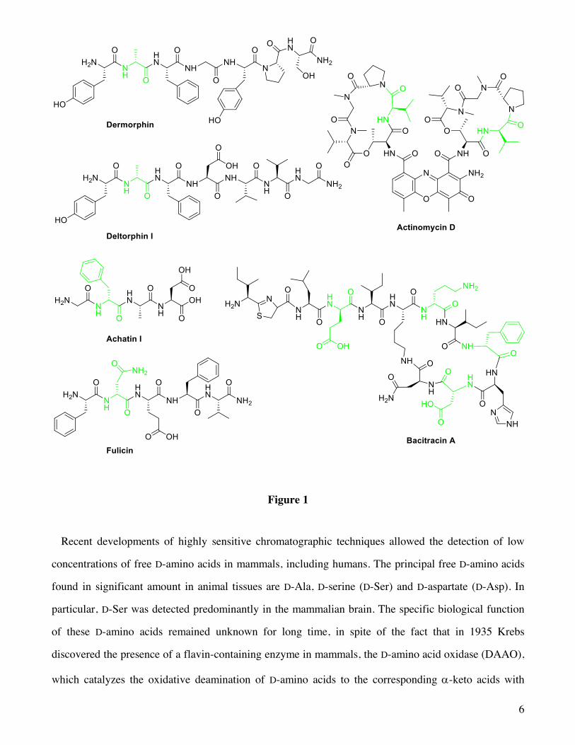
bacitracin, actinomycin D) contain D-amino acids (Figure 1). It has been recently demonstrated that
bacteria synthesize a pool of different D-amino acids, including D-methionine (D-Met) and D-leucine (D-
Leu) in Vibrio cholerae and D-tyrosine (D-Tyr) and D-phenylalanine (D-Phe) in Bacillus subtilis. By
selectively incorporating them in the peptidoglycan cell wall, bacteria cope to different environmental
stresses.7
5
The first reports on D-amino acids in animal tissues were restricted to amphibians and invertebrates.
As a matter of fact, dermorphin (Figure 1) was the first D-amino acid-containing peptide isolated from
the skin of a frog; it is provided with an analgesic activity a thousand times greater than that of
morphine.8 Worth noting, the replacement of D-Ala by its mirror image causes a sharp drop in activity
indicating that D-Ala, located at position 2 of the amino acid sequence, is essential for its activity.
Subsequently, D-amino acids have been detected in a variety of peptides synthesized by animal cells,9
including opioids (deltorphins) (Figure 1), neuropeptides (achatin and fulicin) (Figure 1) and a family
of antimicrobial peptides named bombinins H. For example, a tetrapeptide, termed achatin I (Figure 1),
containing a D-phenylalanine (D-Phe) at position 2, was isolated from the ganglia and atrium of the
African giant snail Achatina fulica.10 Also in this case the excitatory activity on the heart or other
muscles is lost by replacing D-Phe with L-phenylalanine (L-Phe).
6
Figure 1
Recent developments of highly sensitive chromatographic techniques allowed the detection of low
concentrations of free D-amino acids in mammals, including humans. The principal free D-amino acids
found in significant amount in animal tissues are D-Ala, D-serine (D-Ser) and D-aspartate (D-Asp). In
particular, D-Ser was detected predominantly in the mammalian brain. The specific biological function
of these D-amino acids remained unknown for long time, in spite of the fact that in 1935 Krebs
discovered the presence of a flavin-containing enzyme in mammals, the D-amino acid oxidase (DAAO),
which catalyzes the oxidative deamination of D-amino acids to the corresponding a-keto acids with
7
high enantiospecificity.11 DAAO is an ubiquitous enzyme in eukaryotes, with a selective distribution
that, in humans, includes kidney and cerebellum. The biological role of DAAO remained uncertain for a
number of years. Since the presence of D-amino acids in mammalian tissues was initially attributed to
the action of endogenous microbial flora, to ingestion with the diet or to spontaneous racemization of L-
amino acids incorporated in polypeptides during aging,12 it was suggested that such an enzyme acts as a
detoxifying agent to remove D-amino acids. The subsequent detection of substantial amounts of D-
amino acids in various human tissues and in the brain suggested the involvement of DAAO in
modulating the level of D-amino acids. Moreover, it has been shown that the human brain contains
endogenous D-Ser and D-Asp, which are likely to act as modulators of neurotransmission.13,14
Consequently, it has been hypothesized that DAAO may play a role in controlling the level of these
neurotransmitters.
At present, the physiological significance of D-amino acids, i.e. D-Ser and D-Asp, is well-established.
As a consequence, it is conceivable to admit the presence in the human body of a pathway, which
involves specific enzymes, for the synthesis of such D-amino acids. These enzymes have been
discovered, structurally characterized and termed racemases. The different racemases, which have been
discovered in mammals and in bacteria, may differ considerably with respect to substrate specificity.
For example, arginine racemase (ArgR) is mostly active with lysine, arginine and ornithine, and merely
weakly active with alanine, whereas alanine racemase (AlaR) acts on alanine only.
In the following chapters we will discuss the classification of racemases, their diffusion, function,
localization, structural features and the state of the art in the discovery of novel drugs targeting such
enzymes for the treatment of different pathologies.
2. Classification and Catalytic Mechanisms of Racemases
The array of racemases can be divided in two classes on the basis of their mode of action: 1) pyridoxal
5’-phosphate-dependent amino acid racemases and 2) pyridoxal 5’-phosphate-independent racemases.
The first group of racemases depends on pyridoxal 5’-phosphate (PLP), the coenzyme representing the
biologically active form of vitamin B6. Its members are exemplified by eukaryotic enzymes such as
serine racemase (SerR), AlaR, and aspartate racemase (DR-), and by bacterial enzymes such as AlaR,
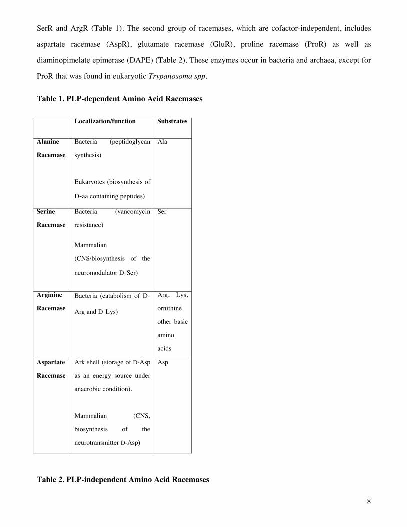
8
SerR and ArgR (Table 1). The second group of racemases, which are cofactor-independent, includes
aspartate racemase (AspR), glutamate racemase (GluR), proline racemase (ProR) as well as
diaminopimelate epimerase (DAPE) (Table 2). These enzymes occur in bacteria and archaea, except for
ProR that was found in eukaryotic Trypanosoma spp.
Table 1. PLP-dependent Amino Acid Racemases
Localization/function Substrates
Alanine
Racemase
Bacteria (peptidoglycan
synthesis)
Eukaryotes (biosynthesis of
D-aa containing peptides)
Ala
Serine
Racemase
Bacteria (vancomycin
resistance)
Mammalian
(CNS/biosynthesis of the
neuromodulator D-Ser)
Ser
Arginine
Racemase
Bacteria (catabolism of D-
Arg and D-Lys)
Arg, Lys,
ornithine,
other basic
amino
acids
Aspartate
Racemase
Ark shell (storage of D-Asp
as an energy source under
anaerobic condition).
Mammalian (CNS,
biosynthesis of the
neurotransmitter D-Asp)
Asp
Table 2. PLP-independent Amino Acid Racemases
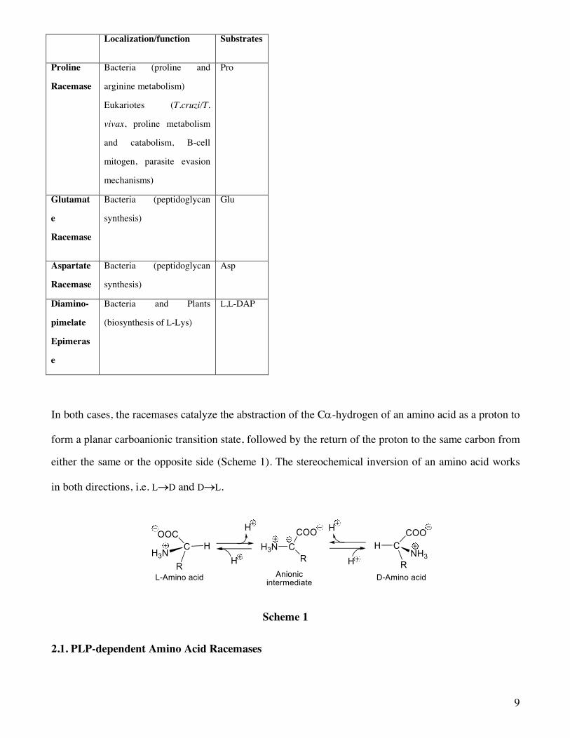
9
Localization/function Substrates
Proline
Racemase
Bacteria (proline and
arginine metabolism)
Eukariotes (T.cruzi/T.
vivax, proline metabolism
and catabolism, B-cell
mitogen, parasite evasion
mechanisms)
Pro
Glutamat
e
Racemase
Bacteria (peptidoglycan
synthesis)
Glu
Aspartate
Racemase
Bacteria (peptidoglycan
synthesis)
Asp
Diamino-
pimelate
Epimeras
e
Bacteria and Plants
(biosynthesis of L-Lys)
L,L-DAP
In both cases, the racemases catalyze the abstraction of the Ca-hydrogen of an amino acid as a proton to
form a planar carboanionic transition state, followed by the return of the proton to the same carbon from
either the same or the opposite side (Scheme 1). The stereochemical inversion of an amino acid works
in both directions, i.e. L®D and D®L.
Scheme 1
2.1. PLP-dependent Amino Acid Racemases
10
Since the pKa value of the amino acidic Ca-hydrogen usually spans the range of 21-34,15 several
enzymes evolved to employ the PLP coenzyme in order to increase the acidity of such a hydrogen.16-19All
known PLP-dependent enzymes exist in their native state as an internal aldimine (Schiff base) (Scheme
2) with the e-amino group of a lysine residue present in the catalytic site. The amino moiety of the
amino acidic substrate displaces the lysine from the internal aldimine to form a new aldimine, termed
external aldimine, which is characterized by a sizable acidity of the Ca-hydrogen of the substrate
(Scheme 2). Typically, the formation of such an external aldimine brings about a sharp drop in the pKa
value of the substrate from 21-34 to 6-17.20,21 This huge reduction in the pKa value is due to the
delocalization of the negative charge of the anionic intermediate by resonance through the conjugated π
system extending on the PLP ring. The scissile bond of the external aldimine must be oriented
perpendicularly to the coenzyme planar π-bonding system to maximize the s-p orbital overlap in the
transition state,22 as recognized by crystallographic studies carried out on PLP-dependent enzymes.23-25
The process of racemization takes place because the a-hydrogen of the external aldimine, due to its
substantial acidity, can be easily abstracted as a proton to form an anionic planar intermediate which,
subsequently, undergoes a re-protonation on either the re- or si-faces of the substrate-cofactor complex.
The mechanism of racemization of the PLP-dependent enzymes as well as the three most significant
resonance forms of the anionic intermediate are depicted in Scheme 2. The quinonoid resonance
structure is commonly considered the major species responsible for the catalytic power of PLP, in
which the electrons from Ca are neutralized by the positively charged pyridine nitrogen.

11
Scheme 2
The PLP-dependent enzymes have been classified, on the basis of their tertiary structures, into five
groups, namely fold type I-V26,27 and they probably evolved from at least three different ancestral
proteins.
The fold type I is the most common and best known structure, and is found in a variety of
aminotransferases and decarboxylases, as well as in enzymes that catalyse an a-, b- or g-elimination
reaction. The prototype of the fold type I enzyme is aspartate aminotransferase. This family includes
enzymes that are catalytically active as homodimers, but some of them assemble into larger complexes.
Each subunit folds into two domains: a large domain, in which the central feature is a seven stranded
b sheet, and a small domain, which folds into a three- or four-stranded b sheet covered with helices on
one side. The active site is located in a cleft between the two domains, at the interface between the two
subunits of the dimer. All enzymes belonging to this fold type show an aspartate interacting with the
pyridine nitrogen. In turn, the enzymes belonging to the fold type I can be divided into eight subtypes.28
12
The fold type II is found mainly in enzymes that catalyse b-replacement and b-elimination reactions.
The prototype of the fold type II enzyme is tryptophan synthase. SerR29-31 belongs to this fold type. Each
subunit consists of two domains: an N-terminal domain containing a four-stranded sheet surrounded by
helices, and a C-terminal domain built up by a six-stranded sheet with flanking helices. The active site
is located in a cleft between the C-terminal ends of the two b sheets. At variance with fold type I
enzymes, a serine residue interacts with the pyridine nitrogen, rather than an aspartate.
The Fold type III family includes AlaR, ArgR and a subset of amino acid decarboxylases. The subunit is
characterized by an eight-stranded a/b barrel and a domain mainly comprising b strands. The side chain
of an arginine residue forms a hydrogen bond with the pyridine nitrogen, indicating that protonation of
the pyridine nitrogen is not crucial for the catalytic activity.
The Fold-type IV subset comprises D-Ala aminotransferase and a few other enzymes. The fold consists
of a two-domain structure with the active site located at the domain interface. Two identical subunits
form a catalytical dimer. However, branched chain aminotransferase further assembles into a hexamer.
The smaller N-terminal domain contains a six-stranded antiparallel b sheets with two a helices on one
side. In the larger C-terminal domain, two four-stranded b sheets form a pseudo-b-barrel that is
surrounded by a few helices.
Finally, the fold-type V group includes glycogen and starch phosphorylases. The C-terminal domain has
a dinucleotide-binding fold and binds the cofactor PLP. In these enzymes, the catalytic role of PLP is
via its phosphate moiety, thus diverging from the coenzyme action in the enzymes belonging to the
other four fold types.
In all the five types, a covalent Schiff-base linkage with lysine, present in the active site, is used to
anchor the cofactor to the enzyme in the resting state and the phosphate group is bound to the N
terminus of an a helix. In four of the five folds, the catalytic lysine residue is located in the connection
between a b strand and the following a helix. In contrast to the classical binding mode of, for example,
NAD(P)H or thiamine diphosphate to open a/b structures, cross-over connections to adjacent parallel
strands in the sheet are, with the exception of fold type II, not utilized for PLP binding.
13
2.2. PLP-independent Amino Acid Racemases
The members of this second class of enzymes, which includes proline, aspartate, and glutamate
racemases as well as diaminopimelate epimerase operates through a “two-base” mechanism in a
cofactor-independent manner, as first proposed by pioneering work on ProR from Abeles’ group32,33
(Scheme 3).
Scheme 3
All these enzymes are members of the class of proteins that contain both a and b secondary structural
elements. In particular, AspR and GluR belong to the aspartate transcarbamylase (ATC)-like fold, while
ProR and DAPE belong to the diaminopimelate epimerase-like fold. The former fold consists of two
similar domains related by a pseudodyad axis, whereas the latter fold consists of mixed beta-sheet folds
into a barrel around the central helix.
ProR has been considered the prototype of PLP-independent racemases, as it interchanges L and D
enantiomers without the aid of any cofactor. A partially purified preparation of ProR from the Gram-
positive bacterium Clostridium sticklandii was first described by Stadtman and Elliot in 1957.34 The
Authors also established the sulfhydryl nature of the enzyme and they speculated that PLP was not
involved in the racemization of Pro. It was only ten years later that Cardinale and Abeles published, for
the first time, the isolation of ProR in a highly purified form and described a conceivable mechanism of
catalysis.32 The reported studies on isotope incorporation and enzyme kinetics in D2O demonstrated a
‘two-base’ mechanism in which one base of the enzyme removes the substrate a-hydrogen as a proton
while the conjugate acid of the second base delivers a proton to the opposite side of the a-carbon. The
Authors also observed that deuterium can not be exchanged while the active site is occupied, indicating
that the two bases of the catalytic site are monoprotic. In 1975, the same group hypothesized that the
active site of ProR is located at the interface of two identical, or nearly identical, subunits.33 Each of
these two subunits supplies one base, which were then identified as the thiol group of two cysteines.

14
Moreover, at saturating substrate levels, the initial rate of the enzyme-catalyzed release of 3H from L-[2-
3H]-proline into the solvent is independent of the initial L-proline (L-Pro) concentration, whereas the
initial rate of the enzyme-catalyzed release of 3H from (±)-[2-3H]-proline decreases by increasing the
(±)-Pro concentration. On the basis of these observations, the Authors speculated that there are two
forms of the enzyme, one which is able to bind L-Pro while the other recognizes solely the D
enantiomer.
Fundamental studies by Knowles and coworkers on ProR35 further clarified the mode of action of this
enzyme deepening the energetics and mechanism of ProR. Subsequently, the same authors extended
their work to GluR,36 confirming the same “two-base” catalytic mechanism of racemization, in which
one cysteine residue behave as a base abstracting the C-a proton to generate a carbanion intermediate,
which is then protonated by the second Cys residue, behaving as a conjugated acid. Such a catalytic
mechanism is also shared by AspR37 and DAPE.38 Although they all operate through a similar acid/base
mechanism, involving two Cys residues, it is possible to separate the PLP-independent racemases into
two subgroups on the basis of sequence and structural similarity: GluR and AspR constitute one
homologous family of enzymes, whereas ProR and DAPE form the other one.
3. Amino Acid Racemases as Drug Targets.
3.1 PLP-dependent Racemases
3.1.1 Alanine Racemase
3.1.1.1 Localization, structure and function
AlaR (EC 5.1.1.1), one of the PLP-dependent racemases, is ubiquitous in bacteria and only a few
similar enzymes have been found in eukaryotes, such as aquatic animals,39 fungi,40,41fission yeast42 and
plants.43 The enzyme produced by pathogenic fungi such as Tolypocladium niveum and Cochliobolus
carbonum are responsible for the synthesis of D-Ala in cyclosporin A, an immunosuppressant drug
widely used in post-allogeneic organ transplant, as well as in HC-toxin, a cyclic peptide isolated from
varieties of maize (Zea mays L.) infected by C. carbonum.
15
Bacterial AlaR is one of the best-studied amino acid racemases. In fact, D-Ala, produced by the action
of AlaR on L-Alanine (L-Ala), is important to both Gram-positive and Gram-negative bacteria, since it
is required for the synthesis of the peptidoglycan in the cell wall. Because peptidoglycan is ubiquitously
distributed in bacteria, but not in mammals, AlaR can be considered a target for new antimicrobial
drugs. Several bacteria, such as Salmonella typhimurium, Escherichia coli and Bacillus subtilis contain
two independent AlaR genes, DadB and Alr,44 belonging to the fold-type III. The expression of DadB is
induced when cells are grown in high concentration of L- or D-Ala and is probably responsible for the
catabolism of D-Ala.45 By contrast, Alr is constitutively expressed and is probably involved in the
synthesis of D-Ala. Moreover, recent studies have indicated that AlaR, which is accessible in the
exosporium, plays a key role in the inhibition of germination in Bacillus spores.46 As a matter of fact,
while L-Ala is an effective germination-promoting compound, its mirror image (D-Ala) is a potent
inhibitor of L-Ala-induced germination.47 Therefore, a compound capable to inhibit the AlaR activity
could trigger a premature germination leading to the death of Bacillum spores in suboptimal
environments.48, 49 Bacillus anthracis, a spore-forming Gram-positive bacterium, is the causative agent of
the zoonotic disease anthrax. Although the disease is most common in wild and domestic mammals, it
can also occur in humans when exposed to infected animals or living spores.50 Inhalation of B. anthracis
spores can lead to the most severe form of the disease, historically associated with a very high mortality
rate.51,52 Triggering the premature germination of B. anthracis spores by spraying a solution of an AlaR
inhibitor on affected areas may therefore be a strategy to speed decontamination efforts and reduce the
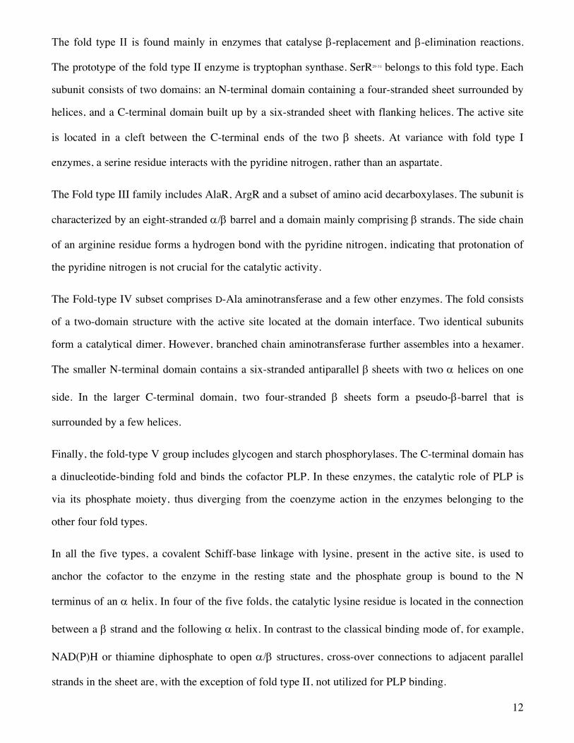
risk of infection in humans.The crystal structure of AlaR from Bacillus stearothermophilus and the
complexes between AlaR and two inhibitors, namely propionate and 1-aminoethylphosphonic acid,
have been reported (Figure 2).53-57 The crystal structure revealed that the enzyme is a homodimer of 388
residues.56 Each monomer is composed of two folded domains; i) a N-terminal domain formed by the
portion 1–240 and ii) a C-terminal domain which involves the remaining portion of the monomer (241–
388). The N-terminal domain is made up of an eight-stranded a/b-barrel while the C-terminal domain
contains mainly b-strands. A water molecule was identified in both active sites of the AlaR
homodimer.57 This water molecule plays an important role and should be taken into account for the
design of specific AlaR inhibitors either by utilizing it as a bridging group or by displacing it by a
suitable moiety appended to the inhibitor.
16
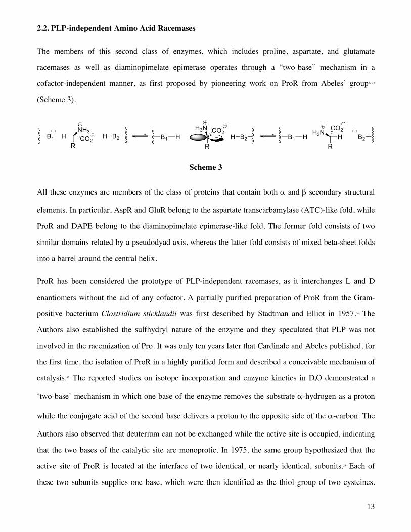
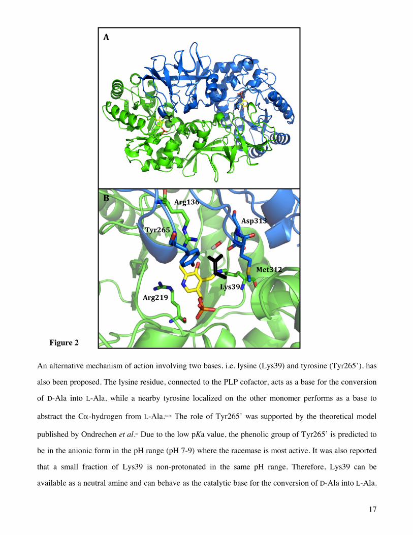
An interesting feature of AlaR is that the pyridine ring of PLP is not protonated due to the contiguity
with Arg219,58 which possesses a pKa value in water of about 13. The X-ray crystal structure evidenced
this structural feature where Arg219 interacts with the PLP pyridine nitrogen through a hydrogen bond,
preventing the formation of the cationic pyridinium ion (Figure 2B).56 Therefore, the main catalytic
effect of PLP in AlaR may be ascribed to solvation.59-61 It has been assumed that in such an enzyme the
non-protonated form generates an intermediate with a limited lifetime which aids the enzyme in
avoiding side reactions.62 Quantum mechanical and molecular dynamics simulations were recently
carried out on the wild type and Arg219Glu mutant.59,63 Results suggest that in the wild type the
stabilization of the quinonoid intermediate is due to solvent effects whereas in the mutant also Glu219
contributes to catalysis.
17
Figure 2
An alternative mechanism of action involving two bases, i.e. lysine (Lys39) and tyrosine (Tyr265’), has
also been proposed. The lysine residue, connected to the PLP cofactor, acts as a base for the conversion
of D-Ala into L-Ala, while a nearby tyrosine localized on the other monomer performs as a base to
abstract the Ca-hydrogen from L-Ala.64-66 The role of Tyr265’ was supported by the theoretical model
published by Ondrechen et al.67 Due to the low pKa value, the phenolic group of Tyr265’ is predicted to
be in the anionic form in the pH range (pH 7-9) where the racemase is most active. It was also reported
that a small fraction of Lys39 is non-protonated in the same pH range. Therefore, Lys39 can be
available as a neutral amine and can behave as the catalytic base for the conversion of D-Ala into L-Ala.
Arg219
Tyr265
Arg136
Met312
Asp313
Lys39
A
B
18
Moreover, AlaR has been shown to be capable to catalyze a transamination reaction.26,68 The enzyme
catalyzes one or the other transformation at different pH values. While the racemization process has its
optimum pH in the range 7-10, the rate of the transamination reaches its peak value around pH 6.5.68
3.1.1.2 Inhibitors and Drug perspectives
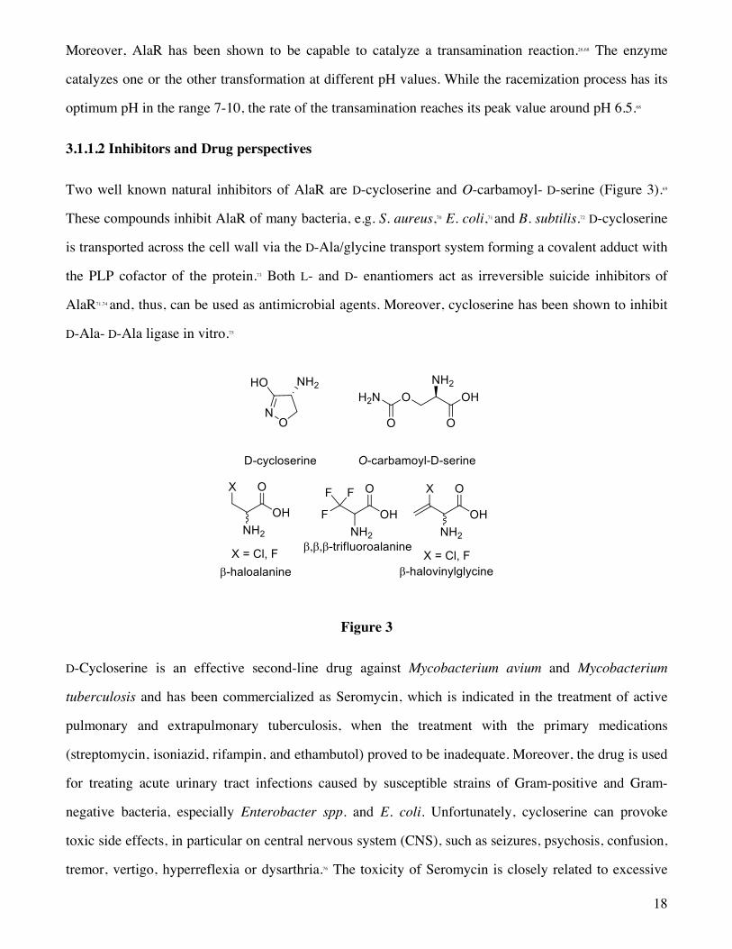
Two well known natural inhibitors of AlaR are D-cycloserine and O-carbamoyl- D-serine (Figure 3).69
These compounds inhibit AlaR of many bacteria, e.g. S. aureus,70 E. coli,71 and B. subtilis.72 D-cycloserine
is transported across the cell wall via the D-Ala/glycine transport system forming a covalent adduct with
the PLP cofactor of the protein.73 Both L- and D- enantiomers act as irreversible suicide inhibitors of
AlaR71,74 and, thus, can be used as antimicrobial agents. Moreover, cycloserine has been shown to inhibit
D-Ala- D-Ala ligase in vitro.75
Figure 3
D-Cycloserine is an effective second-line drug against Mycobacterium avium and Mycobacterium
tuberculosis and has been commercialized as Seromycin, which is indicated in the treatment of active
pulmonary and extrapulmonary tuberculosis, when the treatment with the primary medications
(streptomycin, isoniazid, rifampin, and ethambutol) proved to be inadequate. Moreover, the drug is used
for treating acute urinary tract infections caused by susceptible strains of Gram-positive and Gram-
negative bacteria, especially Enterobacter spp. and E. coli. Unfortunately, cycloserine can provoke
toxic side effects, in particular on central nervous system (CNS), such as seizures, psychosis, confusion,
tremor, vertigo, hyperreflexia or dysarthria.76 The toxicity of Seromycin is closely related to excessive

19
blood levels (above 30 µg/mL) due to a high dosage or to an inadequate renal clearance. The cause of
these neuronal disorders is probably connected to the activity of D-cycloserine as partial agonist of the
glycine site of N-methyl-D-aspartate (NMDA) receptors77 and as an inhibitor of PLP-dependent enzymes
involved in the biosynthesis and metabolism of the neurotransmitter g-aminobutyric acid (GABA).78
Moreover, resistance has been observed in many clinical situations.79 Since cycloserine is not a specific
inhibitor of AlaR but can inhibit several PLP-dependent enzymes,80 the existence of more than one
mechanism leading to cycloserine resistance is likely. Cycloserine may enter the bacterial cell by either
diffusion or uptake via a specific transporter.73 Mutations in the transporter gene which cause a reduced
cycloserine binding or eliminating the transporter from the cell surface affect the drug uptake and
increase the resistance. Analogously, a mutation in a gene coding for an efflux pump may lead to a
higher affinity for cycloserine increasing the efficiency of the drug expulsion. Alternatively, it is
possible that a mutational change could increase the affinity or the levels of a drug-detoxifying enzyme
which would derivatize or hydrolyze cycloserine. The overproduction of a protein target could lead to
increased resistance due to the sequestration or removal of the drug by the excess target protein. To this
end, it was shown that a natural mechanism of resistance to the drug is the overproduction of the AlaR
due to a promoter-up mutation.81
NH
OOH2C
HNLys
N O
HONH2H
NH
OOH2C
N O
HOHN
H
NH
OOH2C
N O
HOHN
H
NH
OOH2C
N O
HOH2N
internal aldimine external aldimine
hydroxy-isoxazole/PLP derivative exocyclic-ketimine
P P
PP
NH
H
NH
H

20
Scheme 4
NMR and IR spectra indicate that cycloserine exists in solution primarily in the lactim form. Recent
studies suggest a mechanism of inactivation that results in aromatization of cycloserine (Scheme 4).82
This mechanism requires an initial transamination step to form an external aldimine which subsequently
undergoes a proton shift to yield an exocyclic ketimine. Such an exocyclic ketimine, which is no more
conjugated with the pyridine ring, proceeds through a second prototropic shift to form the stable
covalent adduct hydroxy-isoxazole/PLP.73
Cycloserine can be considered a model compound for the development of similar derivatives capable to
inactivate PLP-dependent enzymes. The goal is focused on the discovery of drug candidates
characterized by a high selectivity for AlaR.
The most successful approach to design inhibitors of PLP-dependent enzymes consists in incorporating
olefinic, acetylenic, or halogen functional groups in the substrates of the target enzymes. In fact, many
D-Ala analogues have shown to exhibit a remarkable inhibitory activity if they bears a halogen in a
position suitable for a nucleophilic attack, i.e. b-chloroalanine, b-fluoroalanine,83,84 b,b,b–
trifluoroalanine,85 b-halovinylglycine (Figure 3).86 The inactivation of AlaR by b-haloalanines and
trifluoroalanine proceeds via two different mechanisms.85 In both cases, the first step is invariably the
formation of the cross-conjugated enamino-PLP species through the nucleophilic attack of the nascent
aminoacrylate on the PLP-lysine aldimine (Scheme 5).87 Such an intermediate can undergo a bifurcated
route: i) if the b-carbon atom bears a single halogen (b-haloalanines), the electrophilicity at the olefinic
terminus is insufficient to trap the nearby nucleophile, i.e. the e-NH2 of the active-site Lys38, and the
nucleophilic attack occurs at the aldimine carbon atom or ii) if the b-carbon atom bears more halogens,
i.e. trifluoroalanine, the amino group of Lys38 acts as a nucleophile and attacks the highly electron
deficient difluoroenamino moiety. In both cases the enzyme is irreversibly inhibited but, whereas in the
21
first route the inactivation is caused by the alkylation of the PLP-cofactor, in the second route the
inhibition of the enzyme is due to the formation of a covalent bond between the e-NH2 of Lys38 and the
b position of the fluoro-aminoacrylate. An important indicator of potency of irreversible inhibitors is
the number of turnovers per inactivation event, termed partition ratio. This parameter measures the
frequency (for turnover to inactivation) with which an alkylation of the active site occurs causing the
inactivation of the enzyme before the electrophilic species uncovered in turnover can be deactivated
chemically or physically (e.g., product release) at the active site. With a lower partition ratio, the
amount of drug required to fully inhibit a given amount of enzyme is reduced. Consequently, the release
of activated metabolites, which could interact with other cellular constituents, is diminished with an
amelioration of the pharmacological profile. In the case of b-haloalanines, a side reaction that leads to
the formation of pyruvate and ammonia without inactivation of the enzyme can take place (Scheme 5).88

22
Scheme 5
Racemic b-fluoroalanine was the first drug candidate prepared at Merck by Kollonitsch and colleagues;
the D isomer was successfully validated as a potent, wide-spectrum and orally active antibiotic
(partition ratio ~ 800).83,89,90 Unfortunately, safety issues unrelated to its mechanism of action have
hindered its development for clinical applications.
Halovinylglycines represent a class of potent AlaR inhibitors. At variance with haloalanines, in which
the loss of HX leads to the formation of a nucleophile (aminoacrylate), the removal of the halide from
halovinylglycines generates 2-amino-2,3-dienoic acid 1, an allene characterized by a highly
23
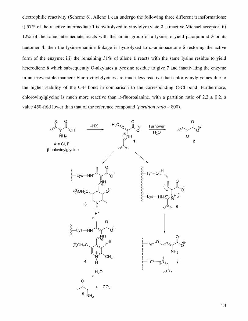
electrophilic reactivity (Scheme 6). Allene 1 can undergo the following three different transformations:
i) 57% of the reactive intermediate 1 is hydrolyzed to vinylglyoxylate 2, a reactive Michael acceptor; ii)
12% of the same intermediate reacts with the amino group of a lysine to yield paraquinoid 3 or its
tautomer 4, then the lysine-enamine linkage is hydrolyzed to a-aminoacetone 5 restoring the active
form of the enzyme; iii) the remaining 31% of allene 1 reacts with the same lysine residue to yield
heterodiene 6 which subsequently O-alkylates a tyrosine residue to give 7 and inactivating the enzyme
in an irreversible manner.91 Fluorovinylglycines are much less reactive than chlorovinylglycines due to
the higher stability of the C-F bond in comparison to the corresponding C-Cl bond. Furthermore,
chlorovinylglycine is much more reactive than D-fluoroalanine, with a partition ratio of 2.2 ± 0.2, a
value 450-fold lower than that of the reference compound (partition ratio ~ 800).

24
Scheme 6
Unfortunately, their potency as antibiotics in vivo is not commensurate to their efficiency as inactivators
of AlaR in vitro, probably because these inhibitors are not well recognized by the amino acid transport
systems of the bacteria.
An interesting compound that selectively inhibits the biosynthesis of the bacterial cell wall is
Alaphosphin (L-alanyl-L-1-aminoethylphosphonic acid, Figure 4). Alaphosphin is representative of a
series of antibacterial phosphonopeptides which were designed to mimic the terminal dipeptide moiety
(D-Ala- D-Ala) of the bacterial cell wall peptidoglycan. Alaphosphin efficiently crosses the bacterial cell
wall and is hydrolyzed to L-Ala and (L)-1-aminoethylphosphonic acid (L-Ala-P) by intracellular
aminopeptidases. The (L,L) stereochemistry of Alaphosphin is necessary for its recognition by the
transporters and to cross the cell wall. Its bactericidal activity arises from the inhibition activity of AlaR
brought about by L-Ala-P. It is well documented that L-Ala-P is a competitive inhibitor of AlaR present
in crude extracts of Gram-negative bacteria (E. coli and S. typhimurium).71,92-94 It is also a time-dependent
and irreversible inhibitor of AlaR isolated from Gram-positive bacterial sources (Streptomyces aureus
and Streptococcus faecalis).92 L-Ala-P is characterized by a slow binding and a slow release process,95,96
with an extremely slow off rate (t1/2 ~ 25 days). The dianionic form of the phosphonate moiety may
mimic a potential transition-state intermediate of the reaction and the two negative charges are crucial
for the observed inhibitory activity of L-Ala-P. In 1998, Stamper et al. published the crystal structure of
the complex of L-Ala-P with AlaR; this was the first report on the X-ray structure of an external
aldimine involving this enzyme.54
Figure 4
The replacement of the carboxylic group of L-Ala with the boronic moiety leads to (1-
aminomethy1)boronic acid (L-Ala-B, Figure 4), which is a powerful slow binding and time-dependent
25
inhibitor of both AlaR from B. stearothermophilus and D-Ala- D-Ala ligase from S. typhimurium.97 L-
Ala-B is less potent than L-Ala-P in terms of both Ki and lifetime of the enzyme-inhibitor complex,
probably, the two bioisosteres differently interacting with the enzyme active site.
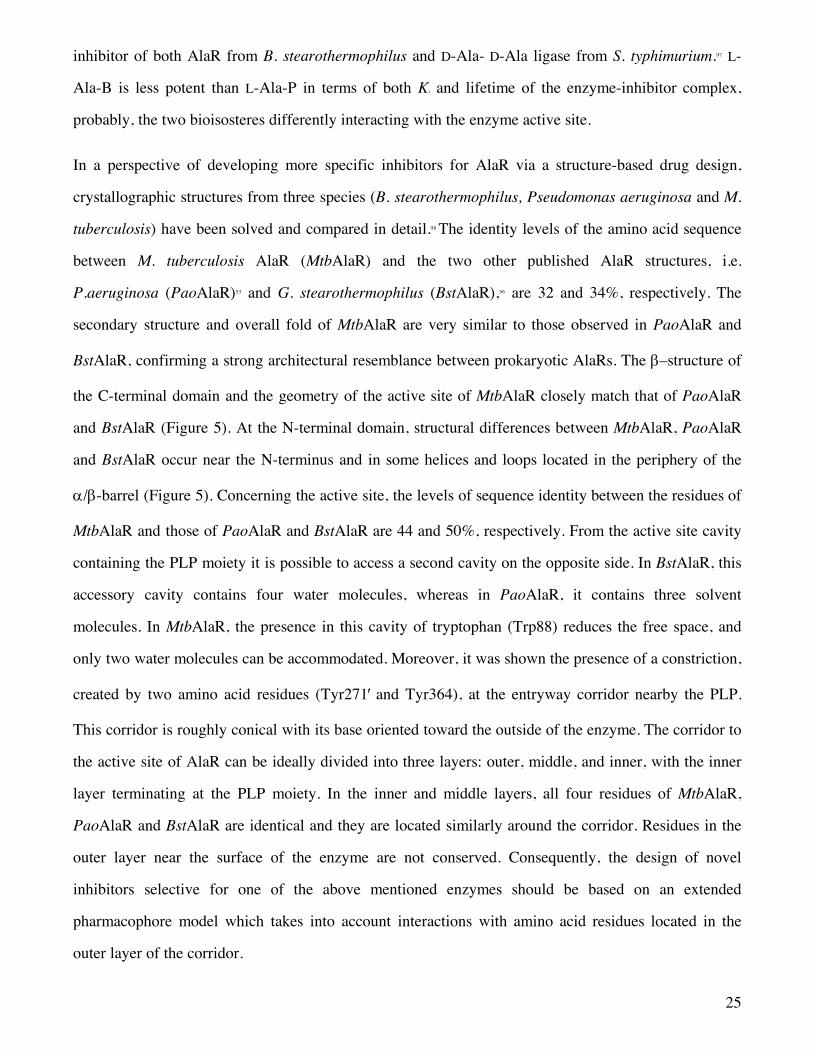
In a perspective of developing more specific inhibitors for AlaR via a structure-based drug design,
crystallographic structures from three species (B. stearothermophilus, Pseudomonas aeruginosa and M.
tuberculosis) have been solved and compared in detail.98 The identity levels of the amino acid sequence
between M. tuberculosis AlaR (MtbAlaR) and the two other published AlaR structures, i.e.
P.aeruginosa (PaoAlaR)53 and G. stearothermophilus (BstAlaR),56 are 32 and 34%, respectively. The
secondary structure and overall fold of MtbAlaR are very similar to those observed in PaoAlaR and
BstAlaR, confirming a strong architectural resemblance between prokaryotic AlaRs. The b–structure of
the C-terminal domain and the geometry of the active site of MtbAlaR closely match that of PaoAlaR
and BstAlaR (Figure 5). At the N-terminal domain, structural differences between MtbAlaR, PaoAlaR
and BstAlaR occur near the N-terminus and in some helices and loops located in the periphery of the
a/b-barrel (Figure 5). Concerning the active site, the levels of sequence identity between the residues of
MtbAlaR and those of PaoAlaR and BstAlaR are 44 and 50%, respectively. From the active site cavity
containing the PLP moiety it is possible to access a second cavity on the opposite side. In BstAlaR, this
accessory cavity contains four water molecules, whereas in PaoAlaR, it contains three solvent
molecules. In MtbAlaR, the presence in this cavity of tryptophan (Trp88) reduces the free space, and
only two water molecules can be accommodated. Moreover, it was shown the presence of a constriction,
created by two amino acid residues (Tyr271¢ and Tyr364), at the entryway corridor nearby the PLP.
This corridor is roughly conical with its base oriented toward the outside of the enzyme. The corridor to
the active site of AlaR can be ideally divided into three layers: outer, middle, and inner, with the inner
layer terminating at the PLP moiety. In the inner and middle layers, all four residues of MtbAlaR,
PaoAlaR and BstAlaR are identical and they are located similarly around the corridor. Residues in the
outer layer near the surface of the enzyme are not conserved. Consequently, the design of novel
inhibitors selective for one of the above mentioned enzymes should be based on an extended
pharmacophore model which takes into account interactions with amino acid residues located in the
outer layer of the corridor.
26
Given the key role of AlaR in the formation of the bacterial wall, it is not surprising that activities
aimed at developing efficient enzyme inhibitors are covering more than two decades. In spite of many
unsuccessful strategies, new avenues are opened by the recent determinations of the crystal structures of
AlaR from Bacillus anthracis in the presence and absence of L-Ala-P,99 AlaR from Streptococcus
pneumonia.100 AlaR from Enterococcus faecalis,101 AlaR from Escherichia coli in the absence and in the
presence of cycloserine and four mutants of the active site residues Pro219 and Glu221,102 the
characterization and preliminary crystallographic studies on AlaR from Bacillus pseudofirmus OF4.103, 104
These recent structural studies indicate that the entryway of the active site is very narrow, conserved
residues are present in the active site as well as a string of water molecules, and dimer formation is a
critical step for AlaR activity Thus, it has been proposed100 that strategies for AlaR inhbitors should be
aimed at a high throughput screening (HTS) and structure-based methods for the identification of
compounds that i) occupy and block the entryway of the active site, ii) interfere with active site
assembly by preventing dimer formation,105 iii) enter in the active site with a specific conformation and
by interacting with active site residue change conformations, eventually displacing water molecules
27
Figure 5
Recently, modified forms of AlaR have been used as catalysts for chemical transformation. In 2003
Hilvert and co-workers showed that AlaR from Geobacillus stearothermophilus can be converted into a
retro-aldolase enzyme by a single point mutation.106 As a matter of fact, replacement of Tyr265, one of
the catalytic bases, with alanine (alrY265A) decreased the original epimerase activity by more than
three orders of magnitude.107 In a following paper from the same group,108 the alrY265A enzyme was used
for the semi-preparative production of b-hydroxy-a-amino acids. In fact, the mutant racemase promotes
the PLP-dependent aldol condensation of glycine with a range of aromatic aldehydes, working similarly
to natural D-threonine aldolases when substrate specificity and stereoselectivity are taken into account.
3.1.2 Serine Racemase
3.1.2.1 Localization, structure and function
SerR (EC 5.1.1.18), another PLP-dependent racemase, was first discovered in 1998 in pupae of the
silkworm Bombyx mori,109 while the first purification of brain mammalian SerR was performed by
Wolosker and co-workers.110,111 SerR is the enzyme involved both in the reversible conversion of L- to D-
Ser and serine catabolism by a,b-elimination of water, thereby regulating D-Ser levels.
In their pioneering work, Wolosker et al.110 showed that SerR is abundant in the glial cells of young rats.
Recent immunohistochemical and in situ hybridization studies revealed the localization of the enzyme112
and its mRNA113 in neurons and glia. Expression of SerR has also been shown in the peripheral tissues
including retinal ganglion cells,114 Schwann cells,115 and chondrocytes.116
The existence of free D-Ser in the mammalian brain of both rats and humans was first reported by two
Japanese laboratories.117-119 Sizable amounts of D-Ser are released by astrocytes, a type of glia cells, where
SerR was later detected.110 D-Ser behaves as a ‘neuromodulator’ acting as an agonist at the glycine site of
NMDA receptors in the mammalian nervous system.120 L-glutamate (L-Glu) is the main excitatory
neurotransmitter in the CNS, where it is involved in the modulation of many physiological processes
such as learning, memory, and synaptic plasticity.121 However, a massive influx of L-Glu into the
synapses can lead to acute and chronic neurodegenerative diseases (for example, cerebral ischemia,
traumatic brain injury, stroke, spinal injury, epilepsy, and Parkinson’s, Alzheimer’s and Huntington’s

28
diseases).122 Memantine, a non competitive antagonist of the NMDA receptors, is the only glutamatergic
drug currently approved for the treatment of moderate to severe Alzheimer’s disease. Due to its low
affinity for the NMDA receptors and capability to block the channel in the open state, memantine does
not substantially accumulate in the channel to interfere with the normal synaptic transmission.123 An
alternative approach to reduce glutamatergic hyperactivity is the use of blockers of the glycine site of
the NMDA receptors. Unfortunately, the development of drugs with such a mode of action was
discontinued since clinical trials evidenced the appearance of heavy adverse effects, such as
hallucinations.124 These side effects were attributed to an excessive blockage of the NMDA receptors and
alteration of the normal neurotransmission. In this context, inhibition of SerR may provide an
alternative therapeutic approach,125 since inhibitors of SerR could offer a more gentle and indirect way to
decrease NMDA receptor function, with less unwanted side effects.
A few years ago, Sasabe et al.126 demostrated that elevated levels of D-Ser in the glia may enhance Glu
toxicity in Amyotrophic Lateral Sclerosis (ALS). Although the discovery of mutations in the gene
encoding superoxide dismutase 1 (SOD 1) resulted in a considerable number of studies, the reason of
the selective motoneuronal death is still unclear. An inefficient GluA2 RNA editing has been proposed as a
cause of the death of motoneurons in sporadic ALS patients.127 In addition, based on the experimental
evidences that elevated level of Glu remained unchanged during the progression of the disease at
variance with D-Ser whose level increases progressively along with the course of the pathology, Sasabe
et al. speculated that the amount of D-Ser can be used to test the progress of ALS.126 Consequently, the
reduction of the concentration of D-Ser could be set forth to slow down or to halt the progression of
ALS.
The increased glial production of D-Ser seems to be caused by pro-inflammatory stimuli that induce
release of D-Ser from microglia by elevating the transcription of SerR.128 At the same time, the excessive
amounts of L-Glu in the spinal cord of ALS patients can improve the enzymatic activity of SerR in
astrocytes, determining an increased level of D-Ser. A further cause of increased levels of D-Ser in ALS
patients could be a failure in the D-Ser metabolic pathway, caused by the inactivation of DAAO, the
degradation enzyme, and/or a decrease in the glial uptake. Thus, decreasing the D-Ser levels by
29
inhibiting the enzymatic activity of SerR or by stimulating the activity of DAAO may represent a new
therapeutic strategy to treat patients affected by ALS.126
An altered level of D-Ser seems to be involved also in schizophrenia.129 In this severely debilitating
psychiatric disorder reduced levels of D-Ser have been observed in the cerebrospinal fluid and serum,
with a corresponding increase in its precursor, L-Ser.130,131 This finding points out the need to deepen the
investigation on the involvement of D-Ser modulation in schizophrenia. Some recent studies indicated
the misfunctioning of enzymes such as SerR, DAAO and G72, a putative activator of DAAO, as risk
factors for the appearance of schizophrenia.132,133 SerR knockout mice have been used for determining
the biological role of SerR and D-Ser. A recent review has summarized the phenotypes of the presently
available three SerR-KO mice and discusses the role of SerR and D-Ser in vivo.134
The initial isolation of SerR from mammalian brains110 also allowed the establishment of its molecular
weight (37 kDa), optimum operational pH and Km value. Subsequent studies from the same group13 led to
the identification of Lys56 as the residue involved in the formation of the internal Schiff base with the
PLP moiety. The crystal structure of a homolog of mammalian SerR from Schizosaccharomyces pombe
was determined in the absence and presence of serine and in the absence and presence of an adenosine-
5'-triphosphate (ATP) analogue.135 More recently, the first crystal structure of mammalian SerR, both in
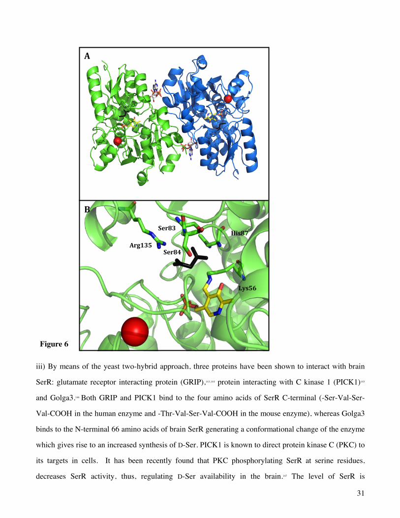
the presence and absence of the inhibitor malonate, was determined (Figure 6).136 A conformational
change affecting the small domain and the active site was detected upon ligand binding with the volume
of the active site significantly decreasing in the transition from the open to the closed state. Moreover,
Ser84 was identified as the residue that plays a critical role in the catalytic racemization of serine,
providing the C-a proton (Figure 6B). The X-ray data show that human and rat SerRs are 90% identical
in sequence and are structurally almost indistinguishable confirming the dimeric structure of the
enzyme.
SerR, like AlaR, is a bifunctional enzyme since, in addition to the racemization of serine, it is able to
catalyze the dehydration of serine to produce pyruvate and ammonia. The physiological relevance of
this dual activity has been investigated.137 By generating a SerR mutant (Q155D) with impaired
elimination activity, it was demonstrated that the levels of D-Ser were several fold higher both in vitro
and in vivo. This finding suggests that the irreversible elimination reaction, coupled to the reversible

30
racemization controls the physiological level of D-Ser especially in the brain tissues lacking DAAO.
Random mutagenesis studies on human SerR provided further evidence on the role of Ser84 in the
enzyme activity and Cys217 and Lys221 in Mg2+ binding and protein stability.138
A peculiar feature of SerR is the multiple catalytic regulatory mechanisms, involving the interaction
with i) divalent cations, ii) nucleotides, iii) specific proteins, and iv) NO.
i) Divalent cations, i.e. calcium, magnesium and manganese were shown to bind to the enzyme at a
cation-binding site (Fig. 6) through a hexavalent coordination composed by two carboxylate anions of
amino acid residues, a carbonyl oxygen of the main-chain and three well-ordered water molecules.139 In
human SerR, the residues directly involved in the coordination of calcium are: Glu210, Asp216 and
Ala214. Divalent cations at physiological concentrations activate 5 to 10 fold the racemization and
elimination reactions,140 with EDTA favouring racemization over elimination. Since the cation binding
site is relatively far from the active site, the cation effect is likely associated to a conformational change
more than a direct involvement in the catalytic reaction. This behaviour resembles the scenario
previously observed in tryptophan synthase where monovalent cations bind to a site distinct from the
active site affecting catalytic rates and intermediate equilibrium distribution.141,142
ii) Nucleotides, such as ATP, adenosine diphosphate (ADP) and guanosine diphosphate (GDP), bind at
a site localized at the subunit interface, although with different affinities (Fig. 6A), increasing 5 to 10
fold the racemase and elimination activity. Since ATP is not hydrolized during the catalytic cycle, ATP
acts as an allosteric effector.
31
Figure 6
iii) By means of the yeast two-hybrid approach, three proteins have been shown to interact with brain
SerR: glutamate receptor interacting protein (GRIP),143,144 protein interacting with C kinase 1 (PICK1)145
and Golga3.146 Both GRIP and PICK1 bind to the four amino acids of SerR C-terminal (-Ser-Val-Ser-
Val-COOH in the human enzyme and -Thr-Val-Ser-Val-COOH in the mouse enzyme), whereas Golga3
binds to the N-terminal 66 amino acids of brain SerR generating a conformational change of the enzyme
which gives rise to an increased synthesis of D-Ser. PICK1 is known to direct protein kinase C (PKC) to
its targets in cells. It has been recently found that PKC phosphorylating SerR at serine residues,
decreases SerR activity, thus, regulating D-Ser availability in the brain.147 The level of SerR is
Arg135
Ser84
Ser83
His87
Lys56
B
A
32
dynamically regulated by glutamate. In fact, activation of metabotropic glutamate receptors leads to the
cleavage of phosphatidylinositol (4,5)-bisphosphate (PIP2) by phospholipase C (PLC); the reduced
concentration of PIP2 diminishes its inhibition effect on SerR.148 On the other hand, GRIP, which is
normally bound to 2-amino-3-(5-methyl-3-oxo-1,2-oxazol-4-yl)propanoic acid (AMPA) receptors,
dissociates from them upon neuronal depolarization to induce SerR activation as a consequence of its
association with the enzyme.149 The activation of NMDA receptors by L-Glu (the agonist) and D-Ser (the
co-agonist) leads to the formation of nitric oxide which, by binding to SerR, inactivates the enzyme;
this mechanism is a feedback homeostatic regulation of SerR.149
iv) SerR is selectively nitrosylated at Cys113,149 that is localized near the ATP binding site. The Cys-133
nitrosylation inhibits the enzyme activity likely by decreasing the stimulatory effect brought about by
ATP. It was found that such a nitrosylation interferes with ATP binding causing a 40-fold increase in Km
for ATP without affecting Vmax. The S-nitrosylation does not affect Km for L-Ser but causes a 2-fold
decrease in Vmax. Because NMDA receptors stimulate NO synthase, the SerR nitrosylation seems to act
as a negative feedback in regulating the level of D-Ser.
In bacteria, SerR has been implicated in vancomycin resistance.150 Vancomycin is a glycopeptide
antibiotic that inhibits peptidoglycan synthesis by formation of a complex with the D-Ala- D-Ala residue
of the peptidoglycan precursors. Resistance to vancomycin is acquired by modification of the D-Ala- D-
Ala residue. Depending on the phenotype, the second D-Ala residue can be mutated in D-lactate or in D-
Ser, in turn produced by the action of SerR (VanT).150
3.1.2.2 Inhibitors and drug perspectives
Very few potent and specific inhibitors of SerR have been identified to date; the two most potent
competitive inhibitors, malonic acid and L-erythro-3-hydroxyaspartic acid,151 (Figure 7) possess
inhibitory constants in the micromolar range (Ki = 77 µM and 49 µM, respectively). Recently, a series
of hydroxamic and dihydroxamic acids (Figure 7) with potent inhibitory activity was identified by
Hoffman et al.152 In this series, succinodihydroxamic acid 9 appears to be the most potent competitive
SerR inhibitor identified to date (Ki = 3.6 ± 0.6 µM). Unfortunately, although some dihydroxamic acids
are effective SerR inhibitors (e.g compounds 8 and 9), their lack of specificity renders them unattractive

33
candidates for further drug development. Their potent inhibition activity was initially attributed to their
capacity to chelate Mg2+, a potent SerR activator present in the screening assay. Subsequently, this
hypothesis was discarded by the experimental evidence that the hydroxamic acid derivatives maintained
their activity even in the presence of a large molar excess of Mg2+, indicating that the capacity of the
substrate to chelate Mg2+ plays a marginal role in SerR inhibition. Probably, the inhibition is due to a
modification of the cofactor, which forms a catalytically inactive aldoxime species. Among the
hydroxamic acid inhibitors, compound 10 (Figure 7) exhibits a moderate selectivity for mouse and
human SerR. Compound 10 (Ki = 98 µM) is an L-aspartic acid (L-Asp) analogue and is 20-fold more
potent as an inhibitor of mouse SerR than L-Asp (Ki = 1900 µM). For these reasons, compound 10 could
serve as a lead compound for the development of the next generation of SerR inhibitors.
Figure 7
The very recent determination of the structure of SerR in the absence and presence of ligands has
triggered a few studies aimed at the development of SerR inhibitors. In this quest an added difficulty is
represented by the need for such compounds to cross the blood-brain barrier.
The comparison of the structures of SerR in the absence and presence of malonate135, 136 indicates that the
enzyme undergoes an open to closed transition upon inhibitor binding. A similar conformational change
is likely to occur upon L-Ser binding. Ligand-induced open-closed transitions are common in PLP-
dependent enzymes and usually involve the reorientation of the small and large domains that form a
34
subunit. As a result the volume of the active site is significantly reduced and active site residues are
relocated. This molecular event might hamper or make more difficult the identification of high affinity
ligands. In fact, if a virtual screening is carried using the open conformation, less specific compounds
might be identified, whereas if the screening is carried out using the closed conformation, the reduced
space may lead to the identification of ligands with reduced size and affinity. In an effort to overcome
these limitations and to include SerR conformational flexibility in virtual screening investigations,
targeted molecular dynamics of SerR was combined with conformational sampling and docking
studies.153 Results suggest that a virtual screenings of SerR carried out sampling a defined number of
protein conformations along the open-to-closed state pathway might lead to the identification of
selective and high affinity enzyme inhibibitors.
An ongoing investigation is carrying out an in silico screening exploiting the the open structure of
human SerR and the searching engine FLAP.154 Selected hits were docked in the active site using
GOLD155 and the free energy of binding was evaluated by HINT.156-158 Compounds that exhibit dissociation
constants in the low micromolar range were identified.159 A similar approach was applied to the
identification of PLP-dependent enzyme inhibitors of O-acetylserine sulfhydrylase.160
Finally, SerR activation by ATP and inhibition by Cys133 nitrosylation suggest an alternative route for
the modulation of the D-Ser level in the brain. Compounds that target the ATP allosteric site displacing
ATP or capable to cause selective nitrosylation of Cys 133 are predicted to inhibit SerR activity.
3.1.3 Arginine Racemase
3.1.3.1 Localization, structure and function
ArgR (EC 5.1.1.9) is an example of a PLP-dependent racemase with broad substrate specificity because
it catalyzes the racemization of arginine, lysine, ornithine and various other amino acids, including
ethionine and citrulline. However, ArgR is unable to recognize and transform hydrophobic, acidic or
aromatic amino acids.. The enzyme was first isolated from Pseudomonas graveolens as early as 1971.161
It was shown that four moles of the cofactor are bound to one mole of ArgR. In 2009, Matsui et al.162
established that in both Pseudomonas taetrolens and Escherichia coli, ArgR resided almost exclusively
in the periplasm, a feature so far unknown for any other amino acid racemase. The experiments
35
performed with P. taetrolens on the utilization of D- and L- amino acids as a carbon source
demonstrated that ArgR is a catabolic enzyme necessary for the efficient utilization of the two basic
amino acids D-Lys, and D-arginine (D-Arg). ArgR from P. taetrolens was found to contain a disulfide
bridge. However, enzyme activity and stability were unaffected by either removal of the disulfide bond
through a reduction process or by mutating the involved cysteines.163
The existence of an ArgR in Pseudomonas aeruginosa, an opportunistic human pathogen with an
enormous catabolic capability, was suggested by Jann et al in 1988.164 Generalized inflammation and
sepsis are the symptoms of infections by P. aeruginosa. If colonization occurs in organs such as lungs,
urinary tract, and kidneys, the result can be fatal. P. aeruginosa is capable of growing on D-Arg as the
sole source of carbon and nitrogen.165
3.1.3.2 Inhibitors and drug perspectives
The observed broad substrate specificity of ArgR suggests the presence in the active site of only a few
well defined anchor points. This feature and the lack of three-dimensional structure of ArgR has so far
prevented studies aimed at the identification of enzyme inhibitors. Towards this goal, protein
expression, careful biochemical investigations and crystallization trials are the pre-requisites. A
possible short-cut might be a homology modelling study using as a parent structure the PLP-dependent
enzyme that more closely matches ArgR sequence.
3.1.4 Aspartate Racemase
3.1.4.1 Localization, structure and function
D-Asp has been detected in the brain and neuroendocrine tissues.163,165-168 This finding triggered a genomic
sequence analysis that led to the identification and cloning of a PLP-dependent enzyme.14 The
recombinant enzyme exhibits a Km value for L-Asp of 3.1 mM and a Vmax of 0.46 mmol/mg/min at the
optimum pH and temperature of 7.5 and 37°C, respectively. Based on in vivo experiments in which the
enzyme was depleted, it was proposed that D-Asp may function as a modulator of adult neurogenesis.14
Furthermore, biochemical investigations, coupled to tissue immunostaining, suggest that D-Asp is a
novel endogenous neurotransmitter,169 thus indicating that AspR is a potential target for
neuropathological disorders.
36
D-Asp is an agonist of the NMDA receptors, equipotent with L-Glu and NMDA itself.120,170,171 It has been
demonstrated that in human brain very high levels of D-Asp occur transiently during the last stage of
embryonic life or in the early postnatal life.117 In human fetal cortex, the concentration of D-Asp
exceeded that of L-Asp, but diminished rapidly to trace levels after birth. D-Asp is crucial for
neurotransmission and neurosecretion in the CNS, as well as for the biosynthesis and/or secretion of
hormones in endocrine glands.172-174 D-Asp is present in some neuronal populations of the brain and of
neuroendocrine tissues, such as the catecholaminergic cells of the adrenal medulla, the anterior and
posterior lobes of pituitary gland, the pineal gland, and the testes.120,167 D-Asp is highly concentrated in the
supraoptic and paraventricular hypothalamic nuclei, whose axons terminate in the posterior pituitary.175
D-Asp occurs also in a high concentration in the pineal gland,176 where it modulates melatonin synthesis
in rat pinealocytes.177 Its localization in different areas of the hypothalamic–pituitary axis suggested that
D-Asp might have a role in neuroendocrine modulation. In fact, D-Asp has been shown to increase
serum growth hormone (GH), luteinizing hormone (LH) and prolactin levels, throughmodulation of the
release of some of the neuropeptides and neurotransmitters involved in their regulation, such as
luteinizing hormone-releasing hormone (LHRH), a-melanocyte-stimulating hormone (a-MSH), GABA
and dopamine.170,178 The high concentrations of D-Asp in the cortical plate, subventricular zone, and
discrete portions of the hippocampal formation, during early neonatal stages, imply an important role
during the developmental phase.179 In adult hippocampus, D-Asp persists in dentate gyrus where new
neurons are generated180,181and integrated into existing neural circuitries which are involved in process of
learning and memory formation. If D-Asp is physiologically formed by AspR, the enzyme should have
similar localizations.14,182,183It was demonstrated that AspR plays an important role in neuronal
development, consistent with the high levels of D-Asp in early neonatal stages.14
Although many studies on the physiological relevance of D-Asp have been published, at present, there
are no reports dealing with the connection of altered levels of D-Asp with any pathological state.
Yamada and his co-workers also reported the occurrence of a PLP-dependent AspR in an ark shell,
Scapharca broughtonii.184 A peculiar characteristic of AspR from S. broughtonii, is that it is markedly
affected by AMP, which maximally enhances its activity up to seven-fold. By contrast, ATP lowers the
activity to less than 7%.185
37
3.1.4.2 Inhibitors and drug perspectives
The interest in AspR as a potential drug target for neurological disorders is very recent.14 So far, no high
yield expression system for AspR has been reported, thus preventing detailed enzyme characterization,
biochemical assays and extensive crystallization campaign for the determination of enzyme structure.
Given the AspR role in modulating NMDA-associated signals, intensive activities should be first
focussed on a deep understanding of enzyme structure-function relationships.
3.2 PLP-independent Racemases
3.2.1 Proline Racemase
3.2.1.1 Localization, structure and function
A partially purified preparation of ProR from the Gram-positive bacterium Clostridium sticklandii (Cs)
was first described by Stadtman and Elliot in 1957.34 The first eukaryotic ProR was identified in 2000 by
Minoprio et al. in the human parasite Trypanosoma cruzi (Tc),186 the etiological agent of Chagas
disease.187 Investigating the in vitro-induced differentiated metacyclic trypomastigotes of Tc, Reina San
Martin et al.188 were able to isolate a 45 kDa protein (Tc45) which is involved in the non-specific
polyclonal activation of B lymphocytes. Subsequently, it was hypothesized the existence of two
homologous of the Tc45 genes (Tc45-A and Tc45-B),186 where the Tc45-A gene copy shares a high
sequence homology with CsProR, the only ProR then described in the literature. Worthnoting, since the
active site of CsProR33,186 is fully conserved in the Tc45 protein, both proteins catalyze the racemization
of L- and D-proline (D-Pro) without the aid of any cofactor while leaving unaffected the stereochemistry
of the other amino acids. Furthermore, specific or non-specific inhibitors, such as pyrrole-2-carboxylate
(PYC), iodoacetamide, and iodoacetate, abolish or severely compromise both the mitogenic activity of
TcProR45 and its enzymatic activity.186 Through a site-directed mutagenesis investigation, the Cys330
residue was identified as the key amino acid, since its replacement completely abolished the ProR
activity;189 the catalytic mechanism was further deepened in 2006.190 The crystal structure of the TcProR-
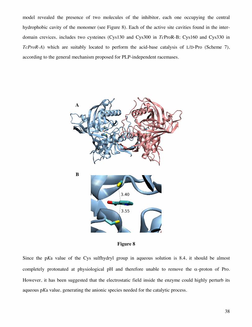
A was obtained with its competitive inhibitor PYC and showed that the enzyme is in fact a homo-dimer,
with each monomer folded in two equally sized a/b domains separated by a deep crevice. The refined
38
model revealed the presence of two molecules of the inhibitor, each one occupying the central
hydrophobic cavity of the monomer (see Figure 8). Each of the active site cavities found in the inter-
domain crevices, includes two cysteines (Cys130 and Cys300 in TcProR-B; Cys160 and Cys330 in
TcProR-A) which are suitably located to perform the acid-base catalysis of L/D-Pro (Scheme 7),
according to the general mechanism proposed for PLP-independent racemases.
Figure 8
Since the pKa value of the Cys sulfhydryl group in aqueous solution is 8.4, it should be almost
completely protonated at physiological pH and therefore unable to remove the a-proton of Pro.
However, it has been suggested that the electrostatic field inside the enzyme could highly perturb its
aqueous pKa value, generating the anionic species needed for the catalytic process.
A
B

39
Scheme 7
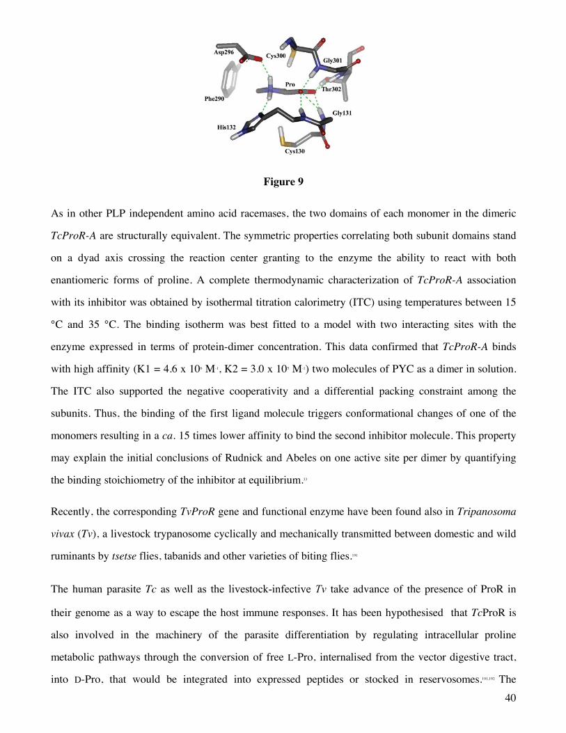
In the crystal structure of the complex of ProR with PYC, an inhibitor representing a transition state
analogue, the C2 atom of PYC is in close contact with the two sulfhydryl groups of residues Cys130
and Cys300 from equivalent a-helices in each structural domain (Figure 9).190 This experimental
evidence strongly supports the ‘two-base’ mechanism. PYC is firmly fixed in place through H-bonding
interactions of its carboxylate group with five amino acidic residues of the enzyme; additionally, the Pro
quaternary amine group is involved in hydrogen bonding interactions with His132 and Asp296. The
pKa of proline inside the ProR active side may be estimated to be about 23.4, reflecting a reduction of
more than 7 pKa units when compared to the calculated value of 30.9 in aqueous solution. This
relatively high pKa of Pro in the active site raises the question of why Ca-deprotonation occurs and not
deprotonation of the ammonium group of the zwitterionic Pro (pKa ≈ 9.6). Probably the answer is the
tight interactions with His132 and Asp296, which prevent Pro from reorienting in a position enabling
ammonium deprotonation.61,190
40
Figure 9
As in other PLP independent amino acid racemases, the two domains of each monomer in the dimeric
TcProR-A are structurally equivalent. The symmetric properties correlating both subunit domains stand
on a dyad axis crossing the reaction center granting to the enzyme the ability to react with both
enantiomeric forms of proline. A complete thermodynamic characterization of TcProR-A association
with its inhibitor was obtained by isothermal titration calorimetry (ITC) using temperatures between 15
°C and 35 °C. The binding isotherm was best fitted to a model with two interacting sites with the
enzyme expressed in terms of protein-dimer concentration. This data confirmed that TcProR-A binds
with high affinity (K1 = 4.6 x 10⁶ M-1, K2 = 3.0 x 10⁵ M-1) two molecules of PYC as a dimer in solution.
The ITC also supported the negative cooperativity and a differential packing constraint among the
subunits. Thus, the binding of the first ligand molecule triggers conformational changes of one of the
monomers resulting in a ca. 15 times lower affinity to bind the second inhibitor molecule. This property
may explain the initial conclusions of Rudnick and Abeles on one active site per dimer by quantifying
the binding stoichiometry of the inhibitor at equilibrium.33
Recently, the corresponding TvProR gene and functional enzyme have been found also in Tripanosoma
vivax (Tv), a livestock trypanosome cyclically and mechanically transmitted between domestic and wild
ruminants by tsetse flies, tabanids and other varieties of biting flies.191
The human parasite Tc as well as the livestock-infective Tv take advance of the presence of ProR in
their genome as a way to escape the host immune responses. It has been hypothesised that TcProR is
also involved in the machinery of the parasite differentiation by regulating intracellular proline
metabolic pathways through the conversion of free L-Pro, internalised from the vector digestive tract,
into D-Pro, that would be integrated into expressed peptides or stocked in reservosomes.191,192 The
41
replacement of the L-Pro moiety with its mirror image, i.e. D-Pro, into a peptide sequence increases its
half-life, stability and resistance to host proteases. Furthermore, the presence of D-Pro would make the
peptide less immunogenic than the corresponding one made up exclusively by L-amino acids.193,194
In addition to the above mentioned enzymatic properties, ProR of both Tc and Tv possesses a T-cell-
independent B-cell mitogenic activity.
Attempts to better explore the direct or indirect association between TcProR mitogenic and enzymatic
activities have demonstrated that they are dissociated186,189 It is believed that the structure of TcProR
active site may expose protein conformational motifs that bind to B-cell expressed molecules, thus
triggering lymphocyte activation. Indeed, modification of key catalytic site residues of the enzyme
aiming at abolishing catalysis without altering TcProR conformational structure, produced TcProR
mutants in which the mitogenic activity was preserved. On the other hand, B cell proliferation assays
showed that TcProR inactivated by PYC was unable to trigger B-cell proliferative activity. These data
confirmed that conformational changes of TcProR takes place upon inhibitor binding preventing its
direct interaction with B-cell expressed ligand(s) and that polyclonal B-cell activation observed with
TcProR is unambigously dependent on conformational epitopes displayed by the active protein.
3.2.1.2 Inhibitors and drug perspectives
In the previous section we have discussed how the first eukaryotic ProR was identified in Tc. This
intracellular protozoan causes Chagas disease, one of the most neglected diseases. Endemic in several
regions of Latin America, this disease persists as the major infectious heart disease in the world.195,196 It is
estimated that 13 million people are currently infected in Central and South America and that the global
incidence of the disease is 300,000 new cases per year.197 Natural transmission of the disease occurs
through faeces of the vector (a haematophagous bug belonging to the subfamily Triatominae, family
Reduviidae) deposited near a skin lesion or mucosa (80–90%) or via organ transplantation/blood
transfusion (5–20%) or congenital transmission (0.5–8%). The majority of infections occur during early
childhood, and around 30% infected people develop chronic cardiac involvement, usually after decades
of asymptomatic infection.
42
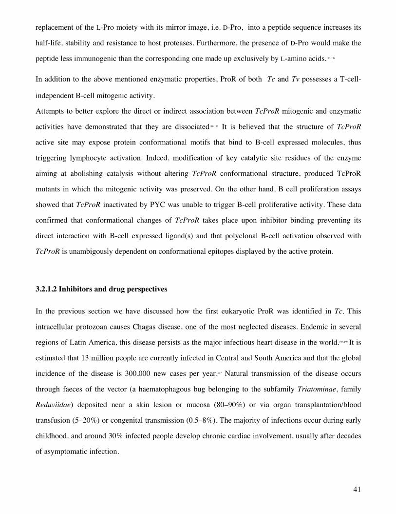
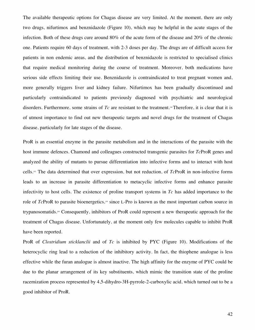
The available therapeutic options for Chagas disease are very limited. At the moment, there are only
two drugs, nifurtimox and benznidazole (Figure 10), which may be helpful in the acute stages of the
infection. Both of these drugs cure around 80% of the acute form of the disease and 20% of the chronic
one. Patients require 60 days of treatment, with 2-3 doses per day. The drugs are of difficult access for
patients in non endemic areas, and the distribution of benznidazole is restricted to specialised clinics
that require medical monitoring during the course of treatment. Moreover, both medications have
serious side effects limiting their use. Benznidazole is contraindicated to treat pregnant women and,
more generally triggers liver and kidney failure. Nifurtimox has been gradually discontinued and
particularly contraindicated to patients previously diagnosed with psychiatric and neurological
disorders. Furthermore, some strains of Tc are resistant to the treatment.198 Therefore, it is clear that it is
of utmost importance to find out new therapeutic targets and novel drugs for the treatment of Chagas
disease, particularly for late stages of the disease.
ProR is an essential enzyme in the parasite metabolism and in the interactions of the parasite with the
host immune defences. Chamond and colleagues constructed transgenic parasites for TcProR genes and
analyzed the ability of mutants to pursue differentiation into infective forms and to interact with host
cells.192 The data determined that over expression, but not reduction, of TcProR in non-infective forms
leads to an increase in parasite differentiation to metacyclic infective forms and enhance parasite
infectivity to host cells. The existence of proline transport systems in Tc has added importance to the
role of TcProR to parasite bioenergetics,199 since L-Pro is known as the most important carbon source in
trypanosomatids.200 Consequently, inhibitors of ProR could represent a new therapeutic approach for the
treatment of Chagas disease. Unfortunately, at the moment only few molecules capable to inhibit ProR
have been reported.
ProR of Clostridium sticklanclii and of Tc is inhibited by PYC (Figure 10). Modifications of the
heterocyclic ring lead to a reduction of the inhibitory activity. In fact, the thiophene analogue is less
effective while the furan analogue is almost inactive. The high affinity for the enzyme of PYC could be
due to the planar arrangement of its key substituents, which mimic the transition state of the proline
racemization process represented by 4,5-dihydro-3H-pyrrole-2-carboxylic acid, which turned out to be a
good inhibitor of ProR.
43
Figure 10
Crystallographic data have shown that incubation of TcProR in the presence of PYC promotes
considerable alteration of the enzyme structure, thus interfering with its binding to B-cell expressed
ligands.190 Furthermore, studies of TcProR localization showed that the enzyme localizes in the
cytoplasm of intracellular dividing parasites (tissue amastigotes) and nearby the flagellar pocket, the
principal site of excretion/secretion of trypanosomes. Interestingly, the addition of PYC to Tc-infected
cultures results in a dose-dependent decrease of the conversion of amastigotes to circulating
trypomastigotes, suggesting that the enzyme, which is up-regulated in actively multiplying parasites,
may be related to the parasite differentiation triggering steps.201 The addition of PYC during the initial
steps of the parasite-host cell interaction led to a significant initial reduction of Tc invasion in vitro. The
inhibition of the enzyme has a direct or an indirect influence on the parasite metabolism, differentiation
and virulence. Unfortunately, PYC has a very poor solubility in water and this problem limits its further
development as a drug. Minoprio’s and Blondel’s groups and collaborators had dedicated lots of efforts
to produce by medicinal chemistry more soluble PYC variants and to design pharmocophoric models
based on the catalytic site of TcProR to be further used in virtual screening of compound libraries.
However, at present, those attempts have not yet yielded better inhibitors with a higher affinity for the
enzyme than PYC.202 The active site of TcProR in its complexed form defines a small pocket of 100 Å3
presenting a very specific set of oriented functional groups. Hence, the design of active inhibitors for
this enzyme by the classical strategies of drug design represents a particular challenge. Therefore, a
more aggressive and less traditional approach has been conceived. It takes advantage of the
conformational changes observed in the TcProR upon ligand binding. In effect, ongoing innovative
approaches and modeling technologies making use of Molecular dynamics and transition path
44
calculations203 can be used to build new docking models for enzymes, either in the catalytic site or by
identifying previously unknown allosteric sites. This approach, based on a new powerful transition Path
Optimization and Exploration method (POE), has proved to allow a drastic expansion of virtual
screening strategies and, thus, of the chemical space accessible through computational aided drug
design, as successfully validated in other systems.204
A different approach was first reported by Minoprio’s group, which tried to use TcProR45 as a
‘vaccine’.205 The aim was to induce specific responses against TcProR45 and neutralise its mitogenic
activity. They developed two protocols. In the first one, intramuscular DNA ‘vaccination’ containing
the TcProR45 gene was used. It turned out that ‘immunoprotection’ is possible, since an 85% decrease
in the levels of parasitaemia, after a challenge with infective forms of the parasite, was obtained.
Moreover, even higher levels of parasitaemia control have been observed when sub-mitogenic doses of
the protein were injected. In the second protocol, an i.p. injection of low doses of recombinant protein
(rTcProR45) was used. The outcome was a decrease up to 95% of the mice parasitaemia, after challenge
with a lethal inoculum of Tc. Therefore, both protocols of immunization were able to trigger specific B
cells and consequently high levels of antibodies anti-rTcProR45 were detected in sera.
In 2010, Bryan et al showed that TcProR may contribute to delay host immune effector mechanisms by
non-specifically activating B cells, especially marginal zone B cells, causing hypergammaglobulinemia
and inducing production of interleukin-10, a cytokine known to increase host susceptibility to Tc.206
While TcProR45 is apparently non-antigenic during experimental T. cruzi infection, it can be delivered
as a potent antigen via genetic immunization. Genetic immunization with TcProR did not lead to
detectable systemic B-cell expansion and did not interfere with the immune response to a different test
of immunogenity. Moreover, genetic vaccination with TcProR induces the triggering of memory B cells,
underlying the usefulness of TcProR as a target to the development of new therapies against
trypanosomiasis.
Finally, it is also possible to suppose that the parasite uses the racemization process to synthesise and/or
express new peptides containing D-Pro to avoid or reduce the host-induced proteolysis. The identities of
the parasite proteins bearing D-Pro should be discovered and investigated since they may become
45
attractive targets involved in parasite evasion.193 These last studies offer the basis for further
development of novel strategies to treat Chagas and other infectious diseases.
3.2.2 Glutamate Racemase
3.2.2.1 Localization, structure and function
GluR (EC 5.1.1.3) is another PLP-independent racemase that occurs in prokaryotes. The first evidence
of GluR from Lactobacillus arabinosus dates back to 1952, but its biochemical function was not
clarified.207,208 In early nineties, it was demonstrated that, like ProR, GluR uses a ‘two-base’ mechanism in
which the sulfhydryl groups of two cysteines serve as the general base/acid catalysts.36 The involvement
of the two Cys residues was demonstrated with the enzyme from Lactobacillus fermenti, since their
replacement, one at a time, with an alanine completely abolished the racemase activity.209 Similar results
were obtained by replacing the Cys residues with Thr/Ala in the enzyme from E. coli.210,211 It was also
suggested that Cys73 is responsible for the deprotonation of D-Glu, while Cys184 is responsible for the
deprotonation of L-Glu. Moreover, it was shown that neighboring residues, i.e. Asp10 for the
conversion of D into L and His186 for the reverse conversion, assist the catalytic thiol groups in acting
as bases.212
The structure determination of GluR from Aquifex pyrophilus in the absence and presence of D-
glutamine, a weak inhibitor, provided the first structural elucidation of the architecture of the active
site.213 However, no strong interactions between D-glutamine and the amino acid residues present in the
active site were noticed. In 2005 it was reported the crystal structure of the GluR from L. fermenti,214
which shares approximately 32% sequence identity with A. pyrophilus GluR. It represents the first three
dimensional structure of a cofactor-independent racemase from a Gram-positive bacterium.
NMR studies carried out on B. anthracis GluR215 and on monomeric H. pylori GluR,216 allowed the
establishment that the enzyme is quite flexible, adopting multiple conformations. The enzyme active site
is rather small, therefore substrate and product can access it exploiting the enzyme flexibility.
GluR is essential to the bacterial cell wall biosynthesis pathway, since the incorporation of D-Glu as the
second residue of the pentapeptide is strictly conserved across the bacterial kingdom.6 GluR has been
implicated in the production and maintenance of sufficient D-Glu pool levels required for growth.
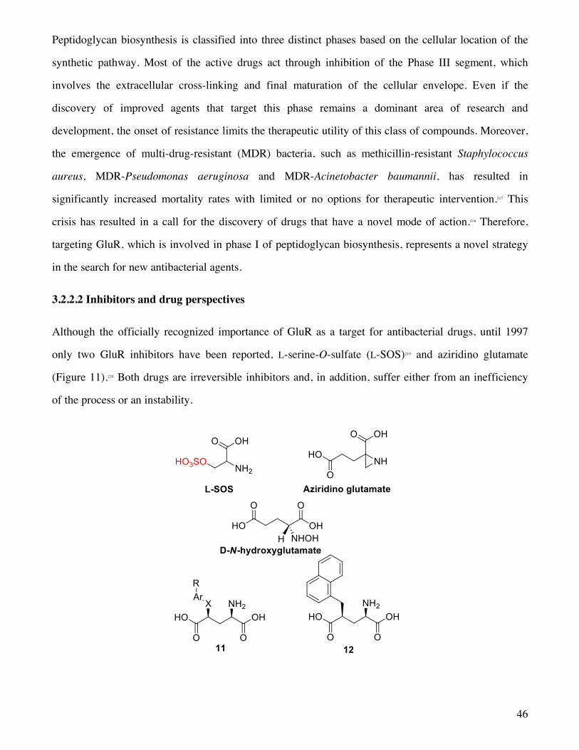
46
Peptidoglycan biosynthesis is classified into three distinct phases based on the cellular location of the
synthetic pathway. Most of the active drugs act through inhibition of the Phase III segment, which
involves the extracellular cross-linking and final maturation of the cellular envelope. Even if the
discovery of improved agents that target this phase remains a dominant area of research and
development, the onset of resistance limits the therapeutic utility of this class of compounds. Moreover,
the emergence of multi-drug-resistant (MDR) bacteria, such as methicillin-resistant Staphylococcus
aureus, MDR-Pseudomonas aeruginosa and MDR-Acinetobacter baumannii, has resulted in
significantly increased mortality rates with limited or no options for therapeutic intervention.217 This
crisis has resulted in a call for the discovery of drugs that have a novel mode of action.218 Therefore,
targeting GluR, which is involved in phase I of peptidoglycan biosynthesis, represents a novel strategy
in the search for new antibacterial agents.
3.2.2.2 Inhibitors and drug perspectives
Although the officially recognized importance of GluR as a target for antibacterial drugs, until 1997
only two GluR inhibitors have been reported, L-serine-O-sulfate (L-SOS)219 and aziridino glutamate
(Figure 11).220 Both drugs are irreversible inhibitors and, in addition, suffer either from an inefficiency
of the process or an instability.
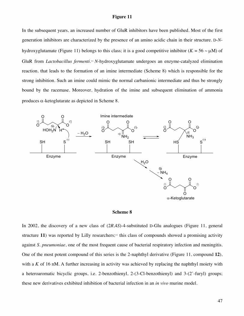
47
Figure 11
In the subsequent years, an increased number of GluR inhibitors have been published. Most of the first
generation inhibitors are characterized by the presence of an amino acidic chain in their structure. D-N-
hydroxyglutamate (Figure 11) belongs to this class; it is a good competitive inhibitor (Ki = 56 ~ µM) of
GluR from Lactobacillus fermenti.221 N-hydroxyglutamate undergoes an enzyme-catalyzed elimination
reaction, that leads to the formation of an imine intermediate (Scheme 8) which is responsible for the
strong inhibition. Such an imine could mimic the normal carbanionic intermediate and thus be strongly
bound by the racemase. Moreover, hydration of the imine and subsequent elimination of ammonia
produces a-ketoglutarate as depicted in Scheme 8.
Scheme 8
In 2002, the discovery of a new class of (2R,4S)-4-substituted D-Glu analogues (Figure 11, general
structure 11) was reported by Lilly researchers;222 this class of compounds showed a promising activity
against S. pneumoniae, one of the most frequent cause of bacterial respiratory infection and meningitis.
One of the most potent compound of this series is the 2-naphthyl derivative (Figure 11, compound 12),
with a Ki of 16 nM. A further increasing in activity was achieved by replacing the naphthyl moiety with
a heteroaromatic bicyclic groups, i.e. 2-benzothienyl, 2-(3-Cl-benzothienyl) and 3-(2’-furyl) groups;
these new derivatives exhibited inhibition of bacterial infection in an in vivo murine model.

48
In the last few years, the research was focused mostly on non amino acidic scaffolds. In this contest,
high-throughput screening of the AstraZeneca collection compounds identified a number of hits
including 9-benzyl purines that showed good activity against Enterococcus faecalis and Enterococcus
faecium GluR. Despite a number of efforts to improve the pharmacological profile of the lead
compound through strategies based on an iterative structure-based drug design, only modest
improvements in potency was observed for the Enterococcus spp and no activity was detected against
the S. aureus enzyme. Furthermore, many members of the series exhibited relatively poor solubility and
only moderate antibacterial activity. In 2008, researchers from the same company reported a successful
‘scaffold-hopping’ endeavor from the 9-benzyl purines to the 8-benzyl pteridinediones and a hit-to-lead
work on this series (Figure 12). Among the tested compounds, 2-benzylthio 8-benzyl pteridinedione
showed activity against the S. aureus enzyme as well as E. faecalis and E. faecium.223 Moreover, by
decorating the scaffold with different substituents at the 2-, 4- and 8-positions emerged some derivatives
provided with a relevant inhibition potency and improved physical properties. The broad spectrum of
action exhibited by some derivatives of this series represents a breakthrough in the development of
small molecule inhibitors. Unfortunately, this series of compounds generally exhibits a poor solubility.
Attempts to improve their physical properties by incorporating polar substituents gave a moderate
success.
49
Figure 12
Recently, a very interesting research plan focused on the design and synthesis of selective H. Pylori
GluR inhibitors has been published.224 H. pylori erodes the stomach mucosa and causes gastritis and
peptic ulcers.225 Moreover, a relationship between infection by such a bacterium and an increased
incidence of a gastric cancer has been proposed.226,227 Current therapies, based on the combined
administration of two antibiotics with a proton pump inhibitor (Triple Therapy), gave rise to numerous
drawbacks, such as stomach upset, diarrhea, headache, a metallic taste and sensitivity to the sun, that
consequently involve a poor patient compliance.228 Additionally, the appearance of H. pylori strains
resistant to the drugs diminished the effectiveness of the multiple therapies. In this contest, researchers
at AstraZeneca focused their attention on the design of new H. pylori GluR inhibitors. An initial high-
throughput screening of the AstraZeneca compound collection and a subsequent lead optimization
process allowed the identification of pyrazolopyrimidinedione 13 (Figure 13). Such a compound
possesses a low nanomolar inhibitory activity at GluR (IC50 = 26 nM) but, due to its low aqueous
solubility and bioavailability, it is not suitable for oral administration. With the aim to improve its
bioavailability, a series of analogues bearing polar functionalities onto the fused pyrimidinedione
scaffold were prepared and tested.

50
Figure 13
In Figure 13 are reported the structures of some derivatives characterized by a high inhibitory activity
against H. pylori GluR. These inhibitors are time-independent, fully reversible and insensitive to
changes in enzyme or detergent concentration. Remarkably, studies of kinetic inhibition on compound
14 indicated that it does not compete with glutamate and, quite interesting, glutamate binding was
required for inhibition. H. pylori GluR was co-crystallized with both a substrate (D-Glu) (Figure 14A,
B) and an inhibitor (compound 14) (Figure 14C) and the structure of the complexes were solved at
high-resolution.229 The major feature of the crystalline structure is the presence of a cryptic pocket,
formed by a dislocation of the C-terminal helix, capable to accommodate the inhibitor and located ~9 Å

51
away from the catalytic centre. The inhibitor binding pocket is formed by displacement and rotation of
the Trp252 side-chain with the pyrazolopyrimidinedione core of derivative 14, as well as the other
members of the cluster, forming a p-stacking interaction which involves its naphthyl moiety and the
indole ring of Trp252 (Figure 14C). Worth of note, the structure of the active site did not change on
binding of the inhibitor.
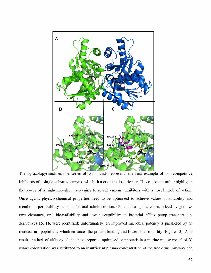
52
Figure 14
The pyrazolopyrimidinedione series of compounds represents the first example of non-competitive
inhibitors of a single-substrate enzyme which fit a cryptic allosteric site. This outcome further highlights
the power of a high-throughput screening to search enzyme inhibitors with a novel mode of action.
Once again, physico-chemical properties need to be optimized to achieve values of solubility and
membrane permeability suitable for oral administration.224 Potent analogues, characterized by good in
vivo clearance, oral bioavailability and low susceptibility to bacterial efflux pump transport, i.e.
derivatives 15, 16, were identified; unfortunately, an improved microbial potency is paralleled by an
increase in lipophilicity which enhances the protein binding and lowers the solubility (Figure 13). As a
result, the lack of efficacy of the above reported optimized compounds in a murine mouse model of H.
pylori colonization was attributed to an insufficient plasma concentration of the free drug. Anyway, the
A
B
C
Trp252
Leu186 Ser152
Glu150
Phe13
Lys17
Val10 Cys70
Cys181
Ser152
Cys181
Glu150
Cys70Phe13
Lys17
Trp252Leu186
Val10
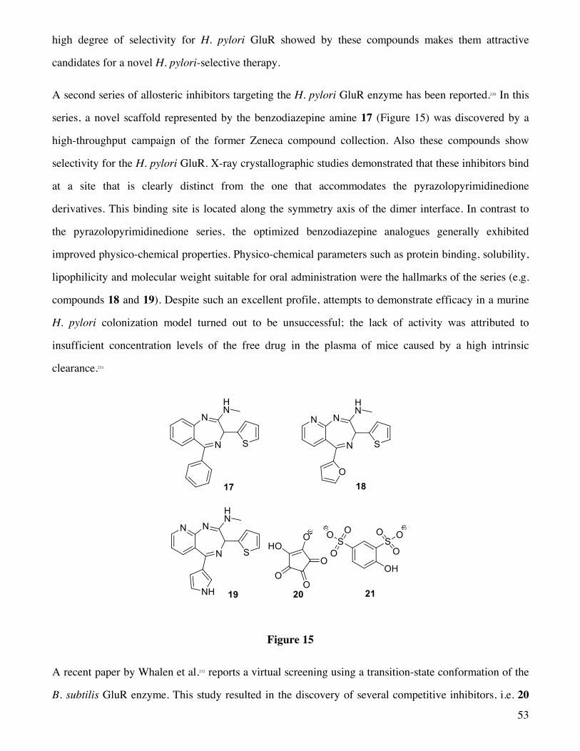
53
high degree of selectivity for H. pylori GluR showed by these compounds makes them attractive
candidates for a novel H. pylori-selective therapy.
A second series of allosteric inhibitors targeting the H. pylori GluR enzyme has been reported.230 In this
series, a novel scaffold represented by the benzodiazepine amine 17 (Figure 15) was discovered by a
high-throughput campaign of the former Zeneca compound collection. Also these compounds show
selectivity for the H. pylori GluR. X-ray crystallographic studies demonstrated that these inhibitors bind
at a site that is clearly distinct from the one that accommodates the pyrazolopyrimidinedione
derivatives. This binding site is located along the symmetry axis of the dimer interface. In contrast to
the pyrazolopyrimidinedione series, the optimized benzodiazepine analogues generally exhibited
improved physico-chemical properties. Physico-chemical parameters such as protein binding, solubility,
lipophilicity and molecular weight suitable for oral administration were the hallmarks of the series (e.g.
compounds 18 and 19). Despite such an excellent profile, attempts to demonstrate efficacy in a murine
H. pylori colonization model turned out to be unsuccessful; the lack of activity was attributed to
insufficient concentration levels of the free drug in the plasma of mice caused by a high intrinsic
clearance.231
Figure 15
A recent paper by Whalen et al.232 reports a virtual screening using a transition-state conformation of the
B. subtilis GluR enzyme. This study resulted in the discovery of several competitive inhibitors, i.e. 20

54
and 21 (Figure 15), provided with IC50 values in the micromolar range (Ki = 42 µM for 20 and 59 µM for
21). Since the structures of these novel inhibitors are dissimilar from those of current amino acid like
inhibitors, they could represent novel scaffolds to be used in the drug discovery process.
Besides these classes of small molecules that inhibit GluR, two reports on large-molecule inhibitors
(molecular weight > 650) have been published. The first report identified haemin as a moderately potent
inhibitor of the P. pentosaceus GluR enzyme.233 Haemin was found to be a non-competitive inhibitor (Ki
= 3.7 µM), hypothesized to bind either to the orthosteric site, or to a distal site inducing a
conformational change. The second report, published in 2000, showed a series of peptides234 provided
with a weak inhibitory potency (IC50 = 160 µM) for the E. coli GluR enzyme.
3.2.3 Aspartate Racemase
3.2.3.1 Localization, structure and function.
The PLP-independent AspR (EC 5.1.1.13) shares with the other members of the AspR group the same
‘two-base’ mode of action. AspR was discovered in Streptococcus faecalis and partially purified in
1972.235 In 1991, Yohda et al. found that AspR occurs exclusively in lactic acid bacteria, such as
Streptococcus species and Lactobacillus species, and succeeded to purify the enzyme to homogeneity
from Streptococcus thermophilus.236 Lactic acid bacteria are characterized by the presence of a D-Asp
residue in the peptidoglycan crossbridge, which is produced from L-Asp by the action of AspR. The
same Authors were able to clone the AspR gene of S. thermophilus and to determine the nucleotide
sequence of the gene.237
The crystal structure of the Pyrococcus horikoshii OT3 AspR (PhAspR), disclosed in 2002, exhibits a
dimeric assembly (Figure 16A).238 As expected for a PLP-independent racemase, it showed two strictly
conserved Cys residues (Cys82 and Cys194) in its catalytic site (Figure 16B). These two cysteine
residues as well as other surrounding amino acid residues, i.e. Asn83, Thr84, Gly193, and Thr195, are
highly conserved among all AspR isoforms and some other PLP-independent racemases (Figure 16B).

55
Figure 16
Furthermore, the active site of all known AspRs contains two basic amino acids, i.e. Arg48 and Lys164
(Figure 16B), which are essential to the functionality of the enzyme since they seem to play an
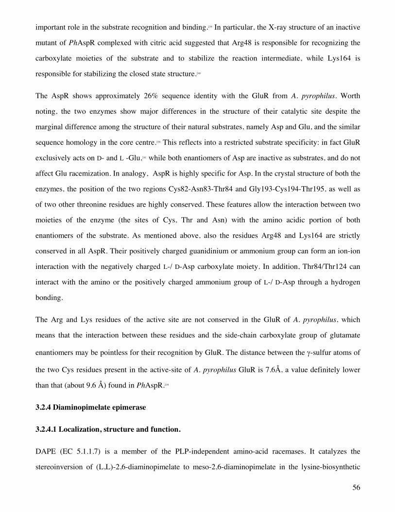
56
important role in the substrate recognition and binding.239 In particular, the X-ray structure of an inactive
mutant of PhAspR complexed with citric acid suggested that Arg48 is responsible for recognizing the
carboxylate moieties of the substrate and to stabilize the reaction intermediate, while Lys164 is
responsible for stabilizing the closed state structure.240
The AspR shows approximately 26% sequence identity with the GluR from A. pyrophilus. Worth
noting, the two enzymes show major differences in the structure of their catalytic site despite the
marginal difference among the structure of their natural substrates, namely Asp and Glu, and the similar
sequence homology in the core centre.238 This reflects into a restricted substrate specificity: in fact GluR
exclusively acts on D- and L -Glu,241 while both enantiomers of Asp are inactive as substrates, and do not
affect Glu racemization. In analogy, AspR is highly specific for Asp. In the crystal structure of both the
enzymes, the position of the two regions Cys82-Asn83-Thr84 and Gly193-Cys194-Thr195, as well as
of two other threonine residues are highly conserved. These features allow the interaction between two
moieties of the enzyme (the sites of Cys, Thr and Asn) with the amino acidic portion of both
enantiomers of the substrate. As mentioned above, also the residues Arg48 and Lys164 are strictly
conserved in all AspR. Their positively charged guanidinium or ammonium group can form an ion-ion
interaction with the negatively charged L-/ D-Asp carboxylate moiety. In addition, Thr84/Thr124 can
interact with the amino or the positively charged ammonium group of L-/ D-Asp through a hydrogen
bonding.
The Arg and Lys residues of the active site are not conserved in the GluR of A. pyrophilus, which
means that the interaction between these residues and the side-chain carboxylate group of glutamate
enantiomers may be pointless for their recognition by GluR. The distance between the g-sulfur atoms of
the two Cys residues present in the active-site of A. pyrophilus GluR is 7.6Å, a value definitely lower
than that (about 9.6 Å) found in PhAspR.239
3.2.4 Diaminopimelate epimerase
3.2.4.1 Localization, structure and function.
DAPE (EC 5.1.1.7) is a member of the PLP-independent amino-acid racemases. It catalyzes the
stereoinversion of (L,L)-2,6-diaminopimelate to meso-2,6-diaminopimelate in the lysine-biosynthetic

57
pathway in plants and bacteria (Scheme 9). Since lysine biosynthesis does not occur in animals, the
members of such a pathway, such as DAPEs, are attractive targets for the rational design of
antibiotics.242,243
Scheme 9
DAPE is a unique member of this family of racemases, because its substrates [(L,L)-DAP and (D,L)-
DAP)] contain two stereocenters but the enzyme recognizes the sole two stereoisomers with the L
configuration at the distal site. The DAP epimerase was detected over 50 years ago in E. coli,244 but its
purification and characterization was made roughly 30 years ago.245 Similarly to the other PLP-
independent racemases, the catalytic mechanism of DAPE involves two cysteine residues, in a ‘two-
base’ mechanism.38 This mechanism was later elucidated by Stenta et al. through QM/MM studies.246 The
structure of DAPE isoforms from many bacteria247-249 and from a plant have been determined recently.250
Particular attention has to be paid to the crystal structure of DAPE from Mycobacterium tuberculosis,
since it represents a new potential target to overcome the problem of drug resistance in the treatment of
tuberculosis.248 Differently from Asp and Glu racemases, in which the cysteine residues are assisted in
the racemization by a hystidine (His132) and an aspartate (Asp296), it was proposed that the active site
of DAPE exists as a thiol–thiolate pair at neutral pH and does not use any neighboring active-site base
to assist the deprotonation process.250
DAPE catalyses the interconversion of (L,L)-2,6-diaminopimelic acid (DAP) into meso-DAP. The
meso-DAP, a precursor of L-Lys, is a key component of the pentapeptide linker in bacterial
peptidoglycan.251 Interestingly, mycobacteria incorporate two distinct interpeptide linkages, the common
D-Ala/meso-DAP linkage during exponential growth and a meso-DAP/meso-DAP linkage, a penicillin-
resistant mode of ligation, upon entering the stationary phase.252 This unique structural feature could be
the cause of the b-lactam resistance of M. tuberculosis.253 Tuberculosis is a common and often deadly
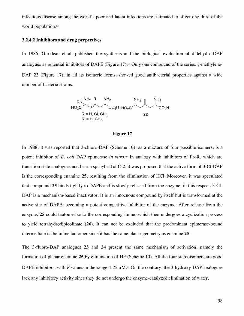
58
infectious disease among the world’s poor and latent infections are estimated to affect one third of the
world population.254
3.2.4.2 Inhibitors and drug perpectives
In 1986, Girodeau et al. published the synthesis and the biological evaluation of didehydro-DAP
analogues as potential inhibitors of DAPE (Figure 17).255 Only one compound of the series, g-methylene-
DAP 22 (Figure 17), in all its isomeric forms, showed good antibacterial properties against a wide
number of bacteria strains.
Figure 17
In 1988, it was reported that 3-chloro-DAP (Scheme 10), as a mixture of four possible isomers, is a
potent inhibitor of E. coli DAP epimerase in vitro.256 In analogy with inhibitors of ProR, which are
transition state analogues and bear a sp2 hybrid at C-2, it was proposed that the active form of 3-Cl-DAP
is the corresponding enamine 25, resulting from the elimination of HCl. Moreover, it was speculated
that compound 25 binds tightly to DAPE and is slowly released from the enzyme; in this respect, 3-Cl-
DAP is a mechanism-based inactivator. It is an innocuous compound by itself but is transformed at the
active site of DAPE, becoming a potent competitive inhibitor of the enzyme. After release from the
enzyme, 25 could tautomerize to the corresponding imine, which then undergoes a cyclization process
to yield tetrahydrodipicolinate (26). It can not be excluded that the predominant epimerase-bound
intermediate is the imine tautomer since it has the same planar geometry as enamine 25.
The 3-fluoro-DAP analogues 23 and 24 present the same mechanism of activation, namely the
formation of planar enamine 25 by elimination of HF (Scheme 10). All the four stereoisomers are good
DAPE inhibitors, with Ki values in the range 4-25 µM.257 On the contrary, the 3-hydroxy-DAP analogues
lack any inhibitory activity since they do not undergo the enzyme-catalyzed elimination of water.
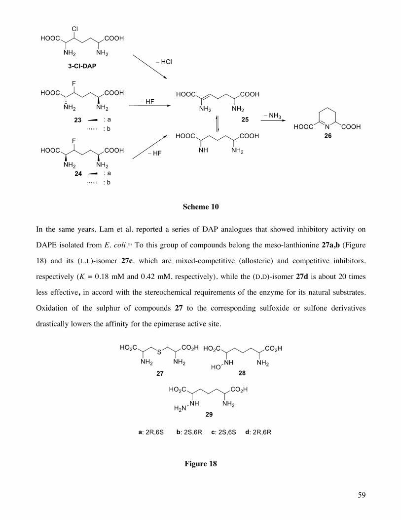
59
Scheme 10
In the same years, Lam et al. reported a series of DAP analogues that showed inhibitory activity on
DAPE isolated from E. coli.258 To this group of compounds belong the meso-lanthionine 27a,b (Figure
18) and its (L,L)-isomer 27c, which are mixed-competitive (allosteric) and competitive inhibitors,
respectively (Ki = 0.18 mM and 0.42 mM, respectively), while the (D,D)-isomer 27d is about 20 times
less effective, in accord with the stereochemical requirements of the enzyme for its natural substrates.
Oxidation of the sulphur of compounds 27 to the corresponding sulfoxide or sulfone derivatives
drastically lowers the affinity for the epimerase active site.
Figure 18
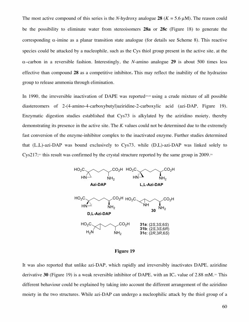
60
The most active compound of this series is the N-hydroxy analogue 28 (Ki = 5.6 µM). The reason could
be the possibility to eliminate water from stereoisomers 28a or 28c (Figure 18) to generate the
corresponding a-imine as a planar transition state analogue (for details see Scheme 8). This reactive
species could be attacked by a nucleophile, such as the Cys thiol group present in the active site, at the
a–carbon in a reversible fashion. Interestingly, the N-amino analogue 29 is about 500 times less
effective than compound 28 as a competitive inhibitor. This may reflect the inability of the hydrazino
group to release ammonia through elimination.
In 1990, the irreversible inactivation of DAPE was reported259,260 using a crude mixture of all possible
diastereomers of 2-(4-amino-4-carboxybutyl)aziridine-2-carboxylic acid (azi-DAP, Figure 19).
Enzymatic digestion studies established that Cys73 is alkylated by the aziridino moiety, thereby
demonstrating its presence in the active site. The Ki values could not be determined due to the extremely
fast conversion of the enzyme-inhibitor complex to the inactivated enzyme. Further studies determined
that (L,L)-azi-DAP was bound exclusively to Cys73, while (D,L)-azi-DAP was linked solely to
Cys217;261 this result was confirmed by the crystal structure reported by the same group in 2009.250
Figure 19
It was also reported that unlike azi-DAP, which rapidly and irreversibly inactivates DAPE, aziridine
derivative 30 (Figure 19) is a weak reversible inhibitor of DAPE, with an IC50 value of 2.88 mM. 261 This
different behaviour could be explained by taking into account the different arrangement of the aziridino
moiety in the two structures. While azi-DAP can undergo a nucleophilic attack by the thiol group of a

61
cysteine at its unhindered methylene position of the aziridine ring, the same reaction occurs on
derivative 30 with the involvement of a tertiary carbon atom which, by definition, is more crowded.
Another class of compounds that have been designed as inhibitors of DAPE is represented by 2,3-
methano-2,6-diaminopimelic acids (31a-c, Figure 19). These compounds did not exhibit any
antibacterial properties; probably they mimic DAP and act as substrates for the enzyme; as a matter of
fact, they are incorporated into the cell wall.262
4. Conclusions and Future Perspectives
L-amino acids are the common building blocks of proteins and are also deeply involved in metabolism
to produce energy. They are currently termed “natural amino acids” in contrast to their mirror images,
i.e. D-amino acids, which could be tagged as “unnatural amino acids” and, according to a long-standing
belief, have been considered inactive substances. Along the years there have been compelling evidences
of the presence of D-amino acids in bacterial cell wall, in antibiotics as well as in peptides produced by
molluscs and amphibians. These peptides are provided with an array of biological properties spanning
from antimicrobial to analgesic activity. Worth noting, the biological activity of the peptides is strictly
dependent, not only on the presence of D-amino acids, but also upon the position of such building
blocks along the amino acid sequence. Furthermore, free D-amino acids have been detected in
mammals, including humans, where they play physiological roles of utmost importance. For example,
substantial amounts of D-Asp and D-Ser have been found in the brain of newborn rats and humans,
where they perform as neurotransmitter. Their concentration decreases gradually with the development.
The present review deals with the enzymes, termed amino acid racemases, involved in the production
of D-amino acids through a racemization process of the L counterparts. We took into account solely the
amino acid racemases which are specific for a single amino acid or recognize as a substrate a selected
number of amino acids. These enzymes have been classified into two groups according to the
mechanism used to interconvert the stereocenter of the amino acids: i) PLP-dependent racemases and
ii) PLP-independent racemases.
Among the PLP-dependent racemases, bacterial AlaR is one of the best-studied amino acid racemases.
The crystal structure of AlaR from Bacillus stearothermophilus as well as its complexes with two
62
inhibitors have been reported and allowed the definition of the active site features. This information is
of utmost importance to design new antimicrobial drugs since AlaR, ubiquitously distributed in bacteria
and not in mammals, produces D-Ala, which is an essential component of peptidoglycan. D-Cycloserine,
an irreversible suicide inhibitor of AlaR, is clinically used as a second-line drug against Mycobacterium
avium and Mycobacterium tuberculosis. The major drawbacks associated to the administration of this
drug is related to its capacity to cross the blood brain barrier causing side effects probably due to an
interaction with the NMDA receptors as well as to its low specificity for AlaR; it is able to inhibit
several PLP-dependent enzymes. Novel structural information has opened new avenues for the design
of specific and potent AlaR inhibitors. In the future we expect antimicrobial drugs characterized by a
higher selectivity for AlaR and with reduced side effects. In this set of enzymes, SerR and AspR will be
the targets of novel drugs capable to treat neuropathological disorders. Sizable amount of D-Ser and D-
Asp have been detected in the mammalian brain of both rats and humans where they perform as
neuromodulators of the NMDA receptors.
Within the set of PLP-independent racemases in depth investigations have been carried out both in
academia and in industry with the goal to find out new antiparasitic, antibiotic, and antibacterial agents.
Most of the antibacterial drugs present in the market act through inhibition of the Phase III segment of
the peptidoglycan biosynthetic route. Even if this phase remains the dominant area of research and drug
development, the onset of resistance limits the therapeutic utility of this class of compounds. Therefore,
targeting GluR, which is involved in phase I, represents a novel strategy in the search for new
antibacterial agents. The major problem associated to the development of drugs targeting the cofactor-
independent family is the need to find out potent inhibitors selective for a single species of racemases.
In the field of GluR, selective inhibitors of the H. Pylori GluR have been discovered through a high-
throughput screening of a large collection of compounds. Quite interesting, the drug candidates are non-
competitive inhibitors, since they fit a cryptic allosteric site. This outcome further highlights the power
of a high-throughput screening to search enzyme inhibitors with a novel mode of action. In addition,
investigations carried out with the aim to uncover allosteric inhibitors of racemases could be a fruitful
way to obtain compounds provided with a selective profile.
63
5. Acknowledgments
Financial supports from Ministry of Education (MIUR – Rome) and Università degli Studi di Milano
(PUR) are gratefully acknowledged. We thank Dr. Francesca Spyrakis for the preparation of figures and
the graphic abstract.
6. References
(1) Lamzin, V. S.; Dauter, Z.; Wisoln, K. S. Curr. Opin. Struct. Biol. 1995, 5, 830.
(2) Prelog, V. Science 1976, 193, 17.
(3) Corrigan, J. J. Science 1969, 164, 142.
(4) Meister, A. Biochemistry of the amino acids; Meister, A., 2nd ed/Academic Press: New York,
London, 1965; vol 1, pp 113-139.
(5) Holtje, J. V. Microbiol. Mol. Biol. Rev. 1998, 62, 181.
(6) van Heijenoort, J. Nat. Prod. Rep. 2001, 18, 503.
(7) Lam, H.; Oh, D.C.; Cava, F.; Takacs, C. N.; Clardy, J.; de Pedro, M. A.; Waldor, M. K.
Science 2009, 325, 1552.
(8) Broccardo, M.; Erspamer, V.; Falconieri Erspamer, G.; Improta, G.; Linari, G.; Melchiorri, P.;
Montecucchi, P. C. Br. J. Pharmacol. 1981, 73, 625.
(9) G. Kreil Annu. Rev. Biochem. 1997. 66, 337.
(10) Kamatani, Y.; Minakata, H.; Kenny, P. T.; Iwashita, T.; Watanabe, K.; Funase, K.; Sun,
X. P.; Yongsiri, A.; Kim, K. H.; Novales-Li, P.; Novales, E. T.; Kanapi, C. G.; Takeuchi, H.;
Nomoto, K. Biochem. Biophys. Res. Commun. 1989, 160, 1015.
(11) Krebs, H. A. Biochem. J. 1935, 29, 1620.
(12) Helfman, P. M.; Bada, J. L.; Shou, M. Y. Gerontology 1977, 23, 419.
64
(13) Wolosker, H.; Blackshaw, S.; Snyder, S. H. Proc. Natl. Acad. Sci. U.S.A. 1999, 96,
13409.
(14) Kim, P. M.; Duan, X.; Huang, A. S.; Liu, C. Y.; Ming, G.-l; Song, H.; Snyder, S. H.,
Proc. Natl. Acad. Sci. U.S.A. 2010, 107, 3175.
(15) Tanner, M. E. Acc. Chem. Res. 2002, 35, 237.
(16) John, R. A. Biochim. Biophys. Acta 1995, 1248, 81.
(17) Eliot, A. C.; Kirsch, J. F. Annu. Rev. Biochem. 2004, 73, 383.
(18) Toney, M. D. Arch. Biochem. Biophys. 2005, 433, 279.
(19) Amadasi, A.; Bertoldi, M.; Contestabile, R.; Bettati, S.; Cellini, B.; di Salvo, M. L.;
Borri-Voltattorni, C.; Bossa, F.; Mozzarelli, A. Curr. Med. Chem. 2007, 14, 1291.
(20) Dixon, J. E.; Bruice, T. C. Biochemistry 1973, 12, 4762.
(21) Toth, K.; Richard, J. P. J. Am. Chem. Soc. 2007, 129, 3013.
(22) Dunathan, H. C. Adv. Enzymol. Relat. Areas Mol. Biol. 1971, 35, 79.
(23) Kirsch, J. F.; Eichele, G.; Ford, G. C.; Vincent, M. G.; Jansonius, J. N.; Gehring, H.;
Christen, P. J. Mol. Biol. 1984, 174, 497.
(24) Momany, C.; Ernst, S.; Ghosh, R.; Chang, N. L.; Hackert, M. L. Protein Sci. 1995, 4, 84.
(25) Toney, M. D.; Hohenester, S. W.; Cowan, S. W.; Jansonius, J. N. Science 1993, 261,
756.
(26) Grishin, N. V.; Phillips, M.A.; Goldsmith, E. J. Protein Sci. 1995, 4, 1291.
(27) Soda, K.; Yoshimura, T.; Esaki, N. Chem. Rec. 2001, 1, 373.
(28) Schneider, G.; Käck, H.; Lindqvist Y. Structure 2000, 8, R1.
(29) Hyde, C.C.; Ahmed, S. A.; Padlan, E. A.; Miles, E. W.; Davies, D. R. J. Biol. Chem.
1988, 263, 17857.
65
(30) Gallagher, D. T.; Gilliland, G. L.; Xiao, G.; Zondlo, J.; Fisher, K. E.; Chinchilla, D.;
Eisenstein, E. Structure 1998, 6, 465.
(31) Burkhard, P.; Rao, G. S. J.; Hohenester, E.; Schnackerz, K. D.; Cook, P. F.; Jansonius, J.
N. J. Mol. Biol. 1998, 283, 121.
(32) Cardinale, G. J.; Abeles, R. H. Biochemistry 1968, 7, 3970.
(33) Rudnick, G.; Abeles, R. H. Biochemistry 1975, 14, 4515.
(34) Stadtman, T. C.; Elliot, P. J. Biol. Chem. 1957, 228, 983.
(35) Albery, W. J.; Knowles, J. R. Biochemistry 1986, 25, 2572.
(36) Gallo, K. A.; Tanner, M. E.; Knowles, J. R. Biochemistry 1993, 32, 3991.
(37) Yamauchi, T.; Choi, S. Y.; Okada, H.; Yohda, M.; Kumagai, H.; Esaki, N.; Soda, K. J.
Biol. Chem. 1992, 267, 18361.
(38) Pillai, B.; Cherney, M. M.; Diaper, C. M.; Sutherland, A.; Blanchard, J. S.; Vederas, J.
C.; James, M. N. G. Proc. Natl Acad. Sci. U.S.A. 2006, 103, 8668.
(39) Abe, H.; Yoshikawa, N.; Sarower, M. G.; Okada, S. Biol. Pharm. Bull. 2005, 28, 1571.
(40) Hoffmann, K.; Schneider-Scherzer, E.; Kleinkauf, H.; Zocher, R. J. Biol. Chem. 1994,
269, 12710.
(41) Cheng, Y. Q.; Walton, J. D. J. Biol. Chem. 2000, 275, 4906.
(42) Uo, T.; Yoshimura, T.; Tanaka, N.; Takegawa, K.; Esaki, N. J. Bacteriol. 2001, 183,
2226.
(43) Ono, K.; Yanagida, K.; Oikawa, T.; Ogawa, T.; Soda, K. Phytochemistry 2006, 67, 856.
(44) Walsh, C. T. J. Biol. Chem. 1989, 64, 2393.
(45) McFall, E.; Newman, E. B. Escherichia coli and Salmonella: Cellular and Molecular
biology; Neidhart, F. C./Am. Soc. Microbiol.: Washington, D.C., 1996; pp. 358-379.
66
(46) Yan, X.; Gai, Y.; Liang, L.; Liu, G.; Tan, H. Arch. Microbiol. 2007, 187, 371.
(47) Preston, R. A.; Douthit, H. A. J. Gen. Microbiol. 1988, 134, 3001.
(48) Huang, C. M.; Elmets, C. A.; Tang, D. C.; Li, F.; Yusuf, N. Gen. Prot. Bioinfo. 2004, 2,
143.
(49) Chesnokova, O. N.; McPherson, S. A.; Steichen, C. T.; Turnbough, C. L. Jr. J Bacteriol.
2009, 191, 1303.
(50) Dixon, T. C.; Meselson, M.; Guillemin, J.; Hanna, P. C. N. Engl. J. Med. 1999, 341,
815.
(51) Dahlgren, C. M.; Buchanan, L. M.; Decker, H. M.; Freed, S. W.; Phillips, C. R.;
Brachman, P. S. Am. J. Hyg. 1960, 72, 24.
(52) Meselson, M.; Guillemin, J.; Hugh-Jones, M.; Langmuir, A.; Popova, I.; Shelokov, A.;
Yampolskaya, O. Science 1994, 266, 1202.
(53) Morollo, A. A.; Petsko, G. A.; Ringe, D. Biochemistry 1999, 38, 3293.
(54) Stamper, C. G. F.; Morollo, A. A.; Ringe, D. Biochemistry 1998, 37, 10438.
(55) Watanabe, A.; Yoshimura, T.; Mikami, B.; Hayashi, H.; Kagamiyama, H.; Esaki, N. J.
Biol. Chem. 2002, 277, 19166.
(56) Shaw, J. P.; Petsko, G. A.; Ringe, D. Biochemistry 1997, 36, 1329.
(57) Iurcu Mustata, G.; Soares, T. A.; Briggs, J. M. Biopolymers 2003, 70, 186.
(58) Spies, M. A.; Woodward, J. J.; Watnik, M. R.; Toney, M. D. J. Am. Chem. Soc. 2004,
126, 7464.
(59) Rubinstein, A.; Major, D. T. Biochemistry 2010, 49, 3957.
(60) Major, D. T.; Gao, J. J. Am. Chem. Soc. 2006, 128, 16345.
(61) Rubinstein, A.; Major, D. T. J. Am. Chem. Soc. 2009, 131, 8513.
67
(62) Toney, M. D. Arch. Biochem. Biophys. 2005, 433, 279.
(63) Lin, Y. L.; Gao, J.; Rubinstein, A.; Major, D. T. Biochim Biophys Acta. 2011, In press
(64) Strych, U.; Davlieva, M.; Longtin, J. P.; Murphy, E. L.; Im, H.; Benedik, M. J.; Krause,
K. L. BMC Microbiol. 2007, 7, 40.
(65) Watanabe, A.; Yoshimura, T. Mikami, B.; Esaki, N. J. Biochem. 1999, 125, 987.
(66) Yoshimura, T.; Goto, M. FEBS Journal 2008, 275, 3527.
(67) Ondrechen, M. J.; Briggs, J. M.; McCammon, J. A. J. Am. Chem. Soc. 2001, 123, 2830.
(68) Kurokawa, Y.; Watanabe, A.; Yoshimura, T.; Esaki, N.; Soda, K. J. Biochem. 1998, 124,
1163.
(69) Neuhaus, F. C. Antimicrob. Agents Chemother. 1967, 7, 304.
(70) Roze, U.; Strominger, J. L. Mol. Pharmacol. 1966, 2, 92.
(71) Lambert, M. P.; Neuhaus, F. C. J. Bacteriol. 1972, 110, 978.
(72) Johnston, R. B.; Scholz, J. J.; Diven, W. F.; Shepard, S.: Pyridoxal Catalysis: Enzymes
and Model Systems; Snell, E. E.; Braunstein, A. E.; Severin, E. S.; Torchinsky, Y. M./ Wiley-
Interscience: New York, 1968, p 537.
(73) Wargel, R. J.; Shadur, C. A.; Neuhaus, F. C. J. Bacteriol. 1970, 103, 778.
(74) Fenn, T. D.; Stamper, G. F.; Morollo, A. A.; Ringe, D. Biochemistry 2003, 42, 5775.
(75) David, H. L.; Takayama, K.; Goldman, D. S. Am. Rev. Respir. Dis. 1969, 100, 579.
(76) Lee, H. K. Chest 1960, 37, 378.
(77) Pitkanen, M.; Sirvio, J.; MacDonald, E.; Ekonsalo, T.; Riekkinen, P., Sr. J. Neural
Transm. 1995, 9, 133.
(78) Wood, J. D.; Peesker, S. J.; Gorecki, D. K. J.; Tsui, D. Can. J. Physiol. Pharmacol.
1978, 52, 62.

68
(79) Reitz, R. H.; Slade, H. D.; Neuhaus, F. C. Biochemistry 1967, 6, 2561.
(80) Neuhaus, F. C.: Antibiotics, vol. 1. Mechanisms of action; Gottlieb, D.; Shaw P.
L./Springer-Verlag: Heidelberg, 1967; p 40.
(81) Cáceres, N. E.; Harris, N. B.; Wellehan, J. F.; Feng, Z.; Kapur, V.; Barletta, R. G. J.
Bacteriol. 1997, 179, 5046.
(82) Peisach, D.; Chipman, D. M.; Van Ophem, P. W.; Manning, J. M.; Ringe, D. J. Am.
Chem. Soc. 1998, 120, 2268.
(83) Kollonitsch, J.; Barash, L.; Kahan, F. H.; Kropp, H. Nature 1973, 243, 346.
(84) Badet, B.; Roise, D.; Walsh, C. T. Biochemistry 1984, 23, 5188.
(85) Faraci, W. S.; Walsh, C. T. Biochemistry 1989, 28, 431.
(86) Thornberry, N. A.; Bull, H. G.; Taub, D.; Greenlee, W. J.; Patchett, A. A.; Cordes E. H.
J. Am. Chem. Soc. 1987, 109,1543.
(87) Wang, E.; Walsh, C. T. Biochemistry 1981, 20, 7539.
(88) Kaczorowski, G.; Shaw, L.; Laura, R.; Walsh, C. T. J. Biol. Chem. 1975, 250, 8921.
(89) Wang, E.; Walsh, C. Biochemistry 1978, 17, 1313.
(90) Kollonitsch, J.; Barash, L. J. Am. Chem. Soc. 1976, 98, 5591.
(91) Thornberry, N. A.; Bull, H. G.; Taub, D.; Wilson, K. E.; Giménęz-Gallego, G.; Rosegay,
A.; Soderman, D. D.; Patchett, A. A. J. Biol. Chem. 1991, 266, 21657.
(92) Atherton, F. R.; Hall, M. J.; Hassall, C. H.; Lambert, R. W.; Lloyd, W. J.; Ringrose, P. S.
Antimicrob. Agents Chemother. 1979, 15, 696.
(93) Adams, E.; Mukherjee, K. L.; Dunathan, H. C. Arch. Biochem. Biophys. 1974, 165, 126.
(94) Atherton, F. R.; Hall, M. J.; Hassall, C. H.; Lambert, R. W.; Ringrose, P. S. Antimicrob.
Agents Chemother. 1979, 15, 677.
69
(95) Badet, B.; Inagaki, K.; Soda, D.; Walsh, C. T. Biochemistry 1986, 25, 3275.
(96) Copie, V.; Faraci, W. S.; Walsh, C. T.; Griffin, R. G. Biochemistry 1988, 27, 4966.
(97) Duncan, K.; Faraci, S.; Matteson, D. S.; Walsh, C. T. Biochemistry 1989, 28, 3541.
(98) LeMagueres, P.; Im, H.; Ebalunode, J.; Strych, U.; Benedik, M. J.; Briggs, J. M.; Kohn,
H.; Krause, K. L.; Biochemistry 2005, 44, 1471.
(99) Au, K.; Ren, J.; Walter, T. S.; Harlos, K.; Nettleship, J. E.; Owens, R. J.; Stuart, D. I.;
Esnouf, R. M. Acta Crystallogr Sect F Struct Biol Cryst Commun. 2008, 64, 327.
(100) Im, H.; Sharpe, M. L.; Strych, U.; Davlieva, M.; Krause, K. L. BMC Microbiol. 2011,
11, 116 (online journal).
(101) Priyadarshi, A.; Lee, E. H.; Sung, M. W.; Nam, K. H.; Lee, W. H.; Kim, E. E.; Hwang,
K. Y. Biochim Biophys Acta. 2009, 1794, 1030.
(102) Wu, D.; Hu, T.; Zhang, L.; Chen, J.; Du, J.; Ding, J.; Jiang, H.; Shen, X. Protein Sci.
2008, 17, 1066.
(103) Ju, J.; Qi, J.; Xu, S.; Ohnishi, K.; Benedik, M. J.; Xue, Y.; Ma, Y. Acta Crystallogr Sect
F Struct Biol Cryst Commun. 2009, 65, 166.
(104) Ju, J.; Xu, S.; Wen, J.; Li, G.; Ohnishi, K.; Xue, Y.; Ma, Y. J Biosci Bioeng. 2009, 107,
225.
(105) Ju, J.; Xu, S. ; Furukawa, Y.; Zhang, Y.; Misono, H.; Minamino, T.; Namba, K.; Zhao,
B.; Ohnishi, K. J Biochem. 2011, 149, 83.
(106) Seebeck, F. P.; Hilvert, D. J. Am. Chem. Soc. 2003, 125, 10158.
(107) Watanabe, A.; Yoshimura, T.; Mikami, B.; Esaki, N. J. Biochem. 1999, 126, 781.
(108) Fesko, K.; Giger, L.; Hilvert, D. Bioorg. Med. Chem. Lett. 2008, 18, 5987.

70
(109) Uo, T.; Yoshimura, T.; Shimizu, S.; Esaki, N. Biochem. Biophys. Res. Commun. 1998,
246, 31.
(110) Wolosker, H.; Sheth, K. N.; Takahashi, M.; Mothet, J. P.; Brady, R. O. Jr; Ferris, C. D.;
Snyder, S. H. Proc. Natl. Acad. Sci. U.S.A. 1999, 96, 721.
(111) Wolosker, H. Biochim. Biophys. Acta 2011, In Press.
(112) Kartvelishvily, E.; Shleper, M.; Balan, L.; Dumin, E.; Wolosker, H. J. Biol. Chem. 2006,
281, 14151.
(113) Yoshikawa, M.; Takayasu, N.; Hashimoto, A.; Sato, Y.; Tamaki, R.; Tsukamoto, H.;
Kobayashi, H.; Noda, S. Arch. Histol. Cytol. 2007, 70, 127.
(114) Stevens, E. R.; Esguerra, M.; Kim, P. M.; Newman, E. A.; Snyder, S. H.; Zahs, K. R.;
Miller, R. F. Proc. Natl. Acad. Sci. U.S.A. 2003, 100, 6789.
(115) Wu, S.; Barger, S. W.; Sims, T. J. Brain Res. 2004, 1020, 161.
(116) Takarada, T.; Takahata, Y.; Iemata, M.; Hinoi, E.; Uno, K.; Hirai, T.; Yamamoto, T.;
Yoneda, Y. J. Cell. Physiol. 2009, 220, 756.
(117) Hashimoto, A.; Kumashiro, S.; Nishikawa, T.; Oka, T.; Takahashi, K.; Mito, T.;
Takashima, S.; Doi, N.; Mizutani, Y.; Yamazaki, T.; Kaneko, T.; Ootomo, E. J. Neurochem.
1993, 1, 348.
(118) Hashimoto, A.; Oka, T.; Nishikawa, T. Eur. J. Neurosci. 1995, 7, 1657.
(119) Nagata, Y.; Horiike, K.; Maeda, T. Brain Res. 1994, 634, 291.
(120) Dunlop, D. S.; Neidle, A.; McHale, D.; Dunlop, D. M., Lajtha, A. Biochem. Biophys.
Res. Commun. 1986, 141, 27.
(121) Excitatory Amino Acids and Synaptic Transmission Wheal, H. V.; Thomson A.
M./Academic Press: London, 1995.
71
(122) Bräuner-Osborne, H.; Egebjerg, J.; Nielsen, E. Ø.; Madsen, U.; Krogsgaard-Larsen, P.; J.
Med. Chem. 2000, 43, 2609.
(123) Lipton S. A. J. Alzheimer’s Dis. 2004, 6, S61.
(124) Kemp, J. A.; McKernan, R. M. Nat. Neurosci. 2002, 5, 1039.
(125) Hashimoto, K.; Fukushima, T.; Shimizu, E.; Okada, S.; Komatsu, N.; Okamura, N.;
Koike, K.; Koizumi, H.; Kumakiri, C.; Imai, K.; Iyo, M. Biol. Psychiatry 2004, 28, 385.
(126) Sasabe, J.; Chiba, T.; Yamada, M.; Okamoto, K.; Nishimoto, I.; Matsuoka, M.; Aiso, S.
EMBO J. 2007, 26, 4149.
(127) Kwak, S.; Hideyama, T.; Yamashita, T.; Aizawa, H. Neuropathology, 2010, 30, 182.
(128) Wu, S.; Barger, S. W. Ann. NY Acad. Sci. 2004, 1035, 133.
(129) Labrie, V.; Fukumura, R.; Rastogi, A.; Fick, L. J.; Wang, W.; Boutros, P. C.; Kennedy, J.
L.; Semeralul, M. O.; Lee, F. H.; Baker, G. B.; Belsham, D. D.; Barger, S. W.; Gondo, Y.;
Wong, A. H. C.; Roder J. C. Hum. Mol. Gen. 2009, 18, 3227.
(130) Bendikov, I.; Nadri, C.; Amar, S.; Panizzutti, R.; De Miranda, J.; Wolosker, H.; Agam,
G. Schizophr. Res. 2007, 90, 41.
(131) Hashimoto, K.; Fukushima, T.; Shimizu, E.; Komatsu, N.; Watanabe, H.; Shinoda, N.;
Nakazato, M.; Kumakiri, C.; Okada, S.; Hasegawa, H.; Kazuhiro, I.; Masaomi, I. Arch. Gen.
Psychiatry 2003, 60, 572.
(132) Morita, Y.; Ujike, H.; Tanaka, Y.; Otani, K.; Kishimoto, M.; Morio, A.; Kotaka, T.;
Okahisa, Y.; Matsushita, M.; Morikawa, A.; Hamase, K.; Zaitsu, K.; Kuroda, S. Biol.
Psychiatry 2007, 61, 1200.
(133) Chumakov, I.; Blumenfeld, M.; Guerassimenko, O.; Cavarec, L.; Palicio, M.;
Abderrahim, H. Bougueleret, L.; Barry, C.; Tanaka, H.; La Rosa, P.; Puech, A.; Tahri, N.;
Cohen-Akenine, A.; Delabrosse, S.; Lissarrague, S.; Picard, F.-P.; Maurice, K.; Essioux, L.;
Millasseau, P.; Grel, P.; Debailleul, V.; Simon, A.-M.; Caterina, D.; Dufaure, I.; Malekzadeh,

72
K.; Belova, M.; Luan, J.-J.; Bouillot, M.; Sambucy, J.-L.; Primas, G.; Saumier, M.; Boubkiri,
N.; Martin-Saumier, S.; Nasroune, M.; Peixoto, H.; Delaye, A.; Pinchot, V.; Bastucci, M.;
Guillou, S.; Chevillon, M.; Sainz-Fuertes, R.; Meguenni, S.; Aurich-Costa, J.; Cherif, D.;
Gimalac, A.; Van Duijn, C.; Gauvreau, D.; Ouelette, G.; Fortier, I.; Realson, J.; Sherbatich, T.;
Riazanskaia, N.; Rogaev, E.; Raeymaekers, P.; Aerssens, J.; Konings, F.; Luyten, W.;
Macciardi, F.; Sham, P. C.; Straub, R. E.; Weinberger, D. R.; Cohen, N.; Cohen, D. Proc. Natl.
Acad. Sci. U.S.A. 2002, 99, 13675.
(134) Mori, H.; Inoue, R. Chem. Biodiv. 2010, 7, 1573.
(135) Goto, M.; Yamauchi, T.; Kamiya, N.; Miyahara, I.; Yoshimura, T.; Mihara, H.; Kurihara,
T.; Hirotsu, K.; Esaki, N. J Biol Chem. 2009, 284, 25944.
(136) Smith, M. A.; Mack, V.; Ebneth, A.; Moraes, I.; Felicetti, B.; Wood, M.; Schonfeld, D.;
Mather, O.; Cesura, A.; Barker, J. J. Biol. Chem. 2010, 285, 12873.
(137) Foltyn, V. N.; Bendikov, I.; De Miranda, J.; Panizzutti, R.; Dumin, E.; Shleper, M.; Li,
P.; Toney, M. D.; Kartvelishvily, E.; Wolosker, H. J Biol Chem. 2005, 280,1754.
(138) Hoffman, H. E.; Jiraskova, J.; Zvelebil, M.; Konvalinka, J. Collect. Czech. Chem. Comm.
2010, 75, 59.
(139) Baumgart, F.; Mancheno, J. M.; Rodriguez-Crespo, I. FEBS J. 2007, 274, 4561.
(140) De Miranda, J.; Panizzutti, R.; Foltyn, V. N.; Wolosker, H. Proc. Natl. Acad. Sci. U.S.A.
2002, 99, 14542.
(141) Peracchi, A.; Mozzarelli, A; Rossi, G. L. Biochemistry 1995, 34, 9459.
(142) Raboni, S.; Bettati, S.; Mozzarelli, A. Cellular and Molecular Life Science, 2009, 66,
2391.
(143) Boehning, D.; Snyder, S. H. Annu. Rev. Neurosci. 2003, 26, 105.
73
(144) Kim, P. M.; Aizawa, H.; Kim, P. S.; Huang, A. S.; Wickramasinghe, S. R.; Kashani, A.
H.; Barrow, R. K.; Huganir, R. L.; Ghosh, A.; Snyder, S. H. Proc. Natl. Acad. Sci. U.S.A.
2005, 102, 2105.
(145) Fujii, K.; Maeda, K.; Hikida, T.; Mustafa, A. K.; Balkissoon, R.; Xia, J.; Yamada, T.;
Ozeki, Y.; Kawahara, R.; Okawa, M.; Huganir, R. L.; Ujike, H.; Snyder, S. H.; Sawa, A. Mol.
Psychiatry 2006, 11, 150.
(146) Dumin, E.; Bendikov, I.; Foltyn, V. N.; Misumi, Y.; Ikehara, Y.; Kartvelishvily, E.;
Wolosker, H. J. Biol. Chem. 2006, 281, 20291.
(147) Vargas-Lopes, C.; Madeira, C.; Kahn, S. A.; Albino do Couto, I.; Bado, P.; Houzel, J. C.;
De Miranda, J.; de Freitas, M. S.; Ferreira, S. T.; Panizzutti, R. J. Neurochem. 2011 116, 281.
(148) Mustafa, A. K.; van Rossum D. B.; Patterson, R. L.; Maag, D.; Ehmsen, J. T.; Gazi, S.
K.; Chakraborty, A.; Barrow, R. K.; Amzel, L. M.; Snyder, S. H. Proc. Natl. Acad. Sci. U.S.A.
2009, 106, 2921.
(149) Mustafa A. K.; Kumar, M.; Selvakumar, B.; Ho, G. P.; Ehmsen, J. T.; Barrow, R. K.;
Amzel, L. M.; Snyder, S. H. Proc. Natl. Acad. Sci. U.S.A. 2007, 104, 2950.
(150) Arias, C. A.; Martin-Martinez, M.; Blundell, T. L.; Arthur, M.; Courvalin, P.; Reynolds,
P. E. Mol. Microbiol. 1999, 31, 1653.
(151) Strisovsky, K.; Jiraskova, J.; Mikulova, A.; Rulisek, L.; Konvalinka, J. Biochem. 2005,
44, 13091.
(152) Hoffman, H. E.; Jirásková, J.; Cígler, P.; Šanda, M.; Schraml, J.; Konvalinka, J. J. Med.
Chem. 2009, 52, 6032.
(153) Bruno, A.; Amori, L.; Costantino, G. Molecular Informatics 2011, 30, 317.
(154) Baroni, M.; Cruciani, G.; Sciabola, S.; Perruccio, F.; Mason, J. S. J. Chem. Inf. Model.
2007, 47, 279.
(155) Jones,G.;Willett,P.;Glen,R.C.J.Mol.Biol.1995,245,43.

74
(156) Kellogg,E.G.;Abraham,D.J.Eur.J.Med.Chem.2000,35,651.
(157) Cozzini,P.;Fornabaio,M.;Marabotti,A.;Abraham,D.J.;Kellogg,G.E.;Mozzarelli,A.J.
Med.Chem.2002,45,2469.
(158) Spyrakis,F.;Amadasi,A.;Fornabaio,M.;Abraham,D.J.;Mozzarelli,A.;Kellogg,G.E.;
Cozzini,P.Eur.J.Med.Chem.2007,42,921.
(159) Spyrakis, F.; Dellafiora, L.; Cozzini. P.; Singh, R.; Salsi, E.; Benedetti, P.; Cruciani, G.;
Carosati, E.; Campanini, B.; Cellini, B.; Mozzarelli, A. 2nd Workshop in New Trends in
Computational Chemsitry for Industry Applications; Barcelona, 2011
(160) Salsi, E.; Bayden, A.; Spyrakis, F.; Amadasi, A.; Campanini, B.; Bettati, S.; Cozzini, P.;
Kellogg, G. E.; Cook, P. F.; Dodatko, T.; Roderick, S. L.; Mozzarelli, A. J. Med. Chem. 2010,
53, 345.
(161) Yorifuji, T.; Misono, H.; Soda, K. J. Biol. Chem. 1971, 246, 5093.
(162) Matsui, D.; Oikawa, T.; Arakawa, N.; Osumi, S.; Lausberg, F.; Stäbler, N.; Freudl, R.;
Eggeling, L. Appl. Microbiol. Biotechnol. 2009, 83, 1045.
(163) Matsui, D.; Oikawa, T. Chem Biodivers. 2010, 7, 1591.
(164) Jann, A.; Matsumoto, H.; Haas, D. J. Gen. Microbiol. 1988, 134, 1043.
(165) Haas, D.; Matsumoto, H.; Moretti, P.; Stalon, V.; Mercenier, A. Mol. Gen. Genet. 1984,
193, 437.
(166) Dunlop, D. S.; Neidle, A.; McHale, D.; Dunlop, D. M.; Lajtha, A. 1986. Biochem
Biophys Res Commun. 1996, 141, 27.
(167) Hashimoto, A. Nishikawa, T.; Konno, R.; Niwa, A.; Yasumura, Y.; Oka, T.; Takahashi,
K. Neurosci. Lett. 1993, 152, 33.
75
(168) D'Aniello, A.; Di Cosmo, A.; Di Cristo, C.; Annunziato, L.; Petrucelli, L.; Fisher, G.;
D’Aniello, A. Life Sci. 1996, 59, 97.
(169) D'Aniello, S.; Somorjai, I.; Garcia-Fernàndez, J; Topo, E.; D'Aniello, A. FASEB J. 2011,
25, 1014.
(170) Erreger, K.; Geballe, M. T.; Kristensen, A.; Chen, P. E.; Hansen, K. B.; Lee, C. J.; Yuan,
H.; Le, P.; Lyuboslavsky, P. N.; Micale, N.; Jorgensen, L.; Clausen, R. P.; Wyllie, D. J. A.;
Snyder, J. P.; Traynelis, S. F. Mol. Pharmacol. 2007, 72, 907.
(171) Skerritt, J. H.; Johnston, G. A. J. Neurochem. 1981, 36, 881.
(172) D’Aniello, A.; Di Fiore, M. M.; Fisher, G. H.; Milone, A.; Seleni, A.; D’Aniello, S.;
Perna, A. F.; Ingrosso, D. FASEB J. 2000, 14, 699.
(173) Wang, H.; Wolosker, H.; Pevsner, J.; Snyder, S. H.; Selkoe, D. J. J. Endocrinol. 2000,
167, 247.
(174) Topo, E.; Soricelli, A.; D'Aniello, A.; Ronsini, S.; D'Aniello, G. Reprod. Biol. End.
2009, 7, 120.
(175) Schell, M. J.; Cooper, O. B.; Snyder, S. H. Proc. Natl. Acad. Sci. U.S.A. 1997, 94, 2013.
(176) Imai, K.; Fukushima, T.; Hagiwara, K.; Santa, T. Biomed. Chromatogr. 1995, 9, 106.
(177) Ishio, S.; Yamada, H.; Hayashi. M.; Yatsushiro, S.; Noumi, T.; Yamaguchi, A.;
Moriyama, Y. Neurosci. Lett. 1998, 249, 143.
(178) Pampillo, M.; Scimonelli, T.; Bottin, M. C.; Duvilanski, B. H.; Rettori, V.; Seilicovich,
A.; Lasaga, M. Neuroreport 2002, 13, 2341.
(179) Wolosker, H.; D’Aniello, A.; Snyder, S. H. Neurosci. 2000, 100, 183.
(180) Ming G. L.; Song, H. Annu. Rev. Neurosci. 2005, 28, 223.
(181) Zhao, C.; Deng, W; Gage, F. H. Cell 2008, 132, 645.
(182) Lee, J. A.; Homma H.; Tashiro, K.; Iwatsubo, T.; Imai, K. Brain Res. 1999, 838, 193.
76
(183) Sakai, K; Homma, H.; Lee, J. A.; Fukushima, T. Santa, T.; Tashiro, K.; Iwatsubo, T.;
Imai, K. Arch. Biochem. Biophys. 1998, 351, 96.
(184) Shibata, K.; Watanabe, T.; Yoshikawa, H.; Abe, K.; Takahashi, S.; Kera, Y.; Yamada,
R.-h. Comp. Biochem. Physiol. 2003, Part B 134, 307.
(185) Shibata, K.; Watanabe, T.; Yoshikawa, H.; Abea, K.; Takahashia, S.; Kera, Y.; Yamada
R.-h. Comp. Biochem. Physiol. 2003, Part B 134, 713.
(186) Reina-San-Martín, B.; Degrave, W.; Rougeot, C.; Cosson, A.; Chamond, N.; Cordeiro-
Da-Silva, A.; Arala-Chaves, M.; Coutinho, A.; Minoprio, P. Nature Medicine 2000, 6, 890.
(187) Minoprio, P.; Itohara, S.; Heusser, C.; Tonegawa, S.; Coutinho, A. Immunol. Rev. 1989,
112, 183.
(188) Reina-San-Martin, B.; Cosson, A.; Minoprio, P. Parasitol. Today 2000, 16, 62.
(189) Chamond, N.; Grégoire, C.; Coatnoan, N.; Rougeot, C.; Holanda Freitas-Junior, L.;
Franco da Silveira, J.; Degrave, W. M.; Minoprio, P. J. Biol. Chem. 2003, 278, 15484.
(190) Buschiazzo, A.; Goytia, M.; Schaeffer, F.; Degrave, W.; Shepard, W.; Grégoire, C.;
Chamond, N.; Cosson, A.; Berneman, A.; Coatnoan, N.; Alzari, P. M.; Minoprio, P. Proc. Nat.
Acad. Sci. U.S.A. 2006, 103, 1705.
(191) Chamond, N.; Cosson, A.; Coatnoan, N.; Minoprio, P. Mol. Biochem. Parasitology 2009,
165, 170.
(192) Chamond, N.; Goytia, M.; Coatnoan, N.; Barale, J.-C.; Cosson, A.; Degrave, W. M.;
Minoprio, P. Mol. Microbiol. 2005, 58, 46.
(193) Coatnoan, N.; Berneman, A.; Chamond, N.; Minoprio P. Mem. Inst. Oswaldo Cruz, Rio
de Janeiro, 2009, 104, 295.
(194) Sela, M.; Zisman, E. FASEB J. 1997, 11, 449.

77
(195) Working to overcome the global impact of neglected tropical diseases. First WHO report
on neglected tropical diseases, World Health Organization, David W.T. Crompton, Ed. , 2010,
p. 75.
(196) Kirchhoff, L. V.; Weiss, L. M.; Wittner, M.; Tanowitz, H. B. Front. Biosci. 2004, 9, 706.
(197) Gutierrez, F. R. S.; Guedes, P. M. M.; Gazzinelli, R. T.; Silva, J. S. Parasite Immunology
2009, 31, 673.
(198) Coura, J. R.; Pinto Dias, J. C. Mem. Inst. Oswaldo Cruz, Rio de Janeiro, 2009, 104, 31.
(199) Silber, A.M.; Tonelli, R.R.; Martinelli, M; Colli, W.; Alves, M.J. J. Eukaryot. Microbiol.
2002, 49, 441.
(200) Sylvester, D.; Krassner, S. M. Comp. Biochem. Physiol. 1976, 55, 443.
(201) Coutinho, L.; Alves Ferreira, M.; Cosson, A.; Meuser Batista, M.; da Gama Jaén Batista,
D.; Minoprio, P.; Degrave, W. M.; Berneman, A.; de Nazaré Correia Soeiro M. Mem. Inst.
Oswaldo Cruz, Rio de Janeiro, 2009, 104, 1055.
(202) Berneman, A.; Alves-Ferreira, M.; Coatnoan, N.; Chamond, N.; Minoprio, P. J.
Microbial. Biochem. Technol. 2010, 2, 139.
(203) Blondel, A.; Renaud, J. P.; Fischer, S.; Moras, D.; Karplus, M. J. Mol. Biol. 1999, 291,
101.
(204) Laine, E.; Goncalves, C.; Karst, J. C.; Lesnard, A.; Rault, S.; Tang, W. J.; Malliavin, T.
E.; Ladant, D.; Blondel, A. Proc. Natl. Acad. Sci. U.S.A. 2010, 107, 11277.
(205) Chamond, N.; Coatnoan, N.; Minoprio, P. Curr. Drug Targets Immune Endocr. Metabol.
Disord. 2002, 2, 247.
(206) Bryan, M. A.; Norris, K. A. Infect. Imm. 2010, 78, 810.
(207) Ayengar, P.; Roberts, E. J. Biol. Chem. 1952, 197, 453.
(208) Narrod, S. A.; Wood, W. A. Arch. Biochem. Biophys. 1952, 35, 462.

78
(209) Tanner, M. E.; Gallo, K. A.; Knowles, J. R. Biochemistry 1993, 32, 3998.
(210) Doublet, P.; van Heijenoort, J.; Mengin-Lecreulx, D. Microb. Drug Resist. 1996, 2, 43.
(211) Ho, H.-T.; Falk, P. J.; Ervin, K. M.; Krishnan, B. S.; Discotto, L. F.; Dougherty, T. J.;
Pucci, M. J. Biochem. 1995, 34, 2464.
(212) Glavas, S.; Tanner, M. E. Biochemistry 2001, 40, 6199.
(213) Hwang, K. Y.; Cho, C. S.; Kim, S. S.; Sung, H. C.; Yu, Y. G.; Cho, Y. Nature Struct.
Biol. 1999, 6, 422.
(214) Lee, K.-S.; Park, S.-M.; Yeon Hwang, K.; Chi, Y.-M. Acta Cryst. 2005, F61, 199.
(215) Mehboob, S.; Guo, L.; Fu, W.; Mittal, A.; Yau, T.; Truong, K.; Johlfs, M.; Long, F.;
Fung, L. W.-M.; Johnson, M. E. Biochemistry 2009, 48, 7045.
(216) Fisher, S. L.; Kern, G.; Newton, T.; Lundqvist, T.; Folmer, R.; Xue, Y.: 45th Annual
ICAAC, American Society for Microbiology: Washington, DC, 2005.
(217) Klevens, R. M.; Morrison, M. A.; Nadle, J.; Petit, S.; Gershman, K.; Ray, S.; Harrison,
L. H; Lynfield, R.; Dumyati, G.; Townes, J. M.; Craig, A. S.; Zell, E. R.; Fosheim, G. E.;
McDougal, L. K.; Carey, R. B.; Fridkin, S. K. J. Am. Med. Assoc. 2007, 98, 1763.
(218) Spellberg, B.; Powers, J. H.; Brass, E. P.; Miller, L. G.; Edwards, J. E. Jr Clin. Infect.
Dis. 2004, 38, 1279.
(219) Ashiuchi, M.; Yoshimura, T.; Esaki, N.; Ueno, H.; Soda, K. Biosci. Biotech. Biochem.
1993, 57, 1978.
(220) Tanner, M. E.; Miao, S. Tetrahedron Lett. 1994, 35, 4073.
(221) Glavas, S.; Tanner, M. E. Bioorg. Med. Chem. Lett. 1997, 7, 2265.
(222) de Dios, A.; Prieto, L.; Martín, J. A.; Rubio, A.; Ezquerra, J.; Tebbe, M.; López de
Uralde, B.; Martín, J.; Sánchez, A.; LeTourneau, D. L.; McGee, J. E.; Boylan, C.; Parr, T. R.
Jr.; Smith, M. C. J. Med. Chem. 2002, 45, 4559.

79
(223) Breault, G. A.; Comita-Prevoir, J.; Eyermann, C. J.; Geng, B.; Petrichko, R.; Doig, P.;
Gorseth, E.; Noonan, B. Bioorg. Med. Chem. Lett. 2008, 18, 6100.
(224) Basarab, G. S.; Hill, P. J.; Rastagar, A.; Webborn, P. J. H. Bioorg. Med. Chem. Lett.
2008, 18, 4716.
(225) Megraud, F. Cell 2005, 123, 975.
(226) Peek, R. M. Jr. Springer Semin. Immunopathol. 2005, 2, 197.
(227) Nomura, A. M. Y.; Lee, J.; Stemmermann, G. N.; Nomura, Y.; Perez-Perez, G. I.; Blaser,
M. J. J. Infect. Dis. 2002, 186, 1138.
(228) Suerbaum, S.; Michetti, P. New Engl. J. Med. 2002, 347, 1175.
(229) Lundqvist, T.; Fisher, S. L.; Kern, G.; Folmer, R. H. A.; Xue, Y.; Newton, D. T.;
Keating, T. A.; Alm, R.A.; de Jonge, B. L. M. Nature 2007, 447, 817.
(230) Geng, B.; Basarab, G.; Comita-Prevoir, J.; Gowravaram, M.; Hill, P.; Kiely, A.; Loch, J.;
MacPherson, L.; Morningstar, M.; Mullen, G.; Osimboni, E.; Satz, A.; Eyermann, C.;
Lundqvist, T. Med. Chem. Lett. 2009, 19, 930.
(231) Newman, J. V.; Rooney, M. T.; Cederberg, C. 46th ICAAC Meeting, San Francisco, CA,
USA, 2006, Abs: A-1106.
(232) Whalen, K. L.; Pankow, K. L.; Blanke, S. R.; Spies, M. A. ACS Med. Chem. Lett. 2010,
1, 9.
(233) Choi, S.-Y.; Esaki, N.; Ashiuchi, M.; Yoshimura, T.; Soda, K. Proc. Natl. Acad. Sci.
U.S.A. 1994, 91, 10144.
(234) Kim, W.-C.; Rhee, H.-I.; Park, B.-K.; Suk, K.-H.; Cha, S.-H. J. Biomol. Screening 2000,
5, 435.
(235) Lamont, H. C.; Staudenbauer, W. L.; Strominger, J. L. J. Biol. Chem. 1972, 247, 5103.
80
(236) Okada, H.; Yohda, M.; Giga-Haama, Y.; Ueno, Y.; Ohdo, S.; Kumagai, H. Biochim.
Biophys. Acta 1991, 1078, 377.
(237) Yohda, M.; Okada, H.; Kumagal, H. Biochem. Biophys. Acta 1991, 1089, 234.
(238) Liu, L.; Iwata, K.; Kita, A.; Kawarabayasi, Y.; Yohda, M.; Miki, K. J. Mol. Biol. 2002,
319, 479.
(239) Yoshida, T.; Seko, T.; Okada, O.; Iwata, K.; Liu, L.; Miki, K.; Yohda, M. Proteins
Struct., Funct. Bioinfo. 2006, 64, 502.
(240) Ohtaki, A.; Nakano, Y.; Iizuka, R.; Arakawa, T.; Yamada, K.; Odaka, M.; Yohda, M.
Proteins Struct., Funct. Bioinfo 2008, 70, 1167.
(241) Ashiuchi, M.; Tani, K.; Soda, K.; Risono, H. J. Biochem. 1998, 123, 1156.
(242) Hutton, C. A.; Perugini, M. A.; Gerrard, J. A. Mol. Biosyst. 2007, 3, 458.
(243) Hutton, C. A.; Southwood, T. J.; Turner, J. J. Mini Rev. Med. Chem. 2003, 3, 115.
(244) Antia, M.; Hoare, D. S.; Work, E. J. Biochem. 1957, 65, 448.
(245) Wiseman, J. S.; Nichols, J. S. J. Biol. Chem. 1984, 259, 8907.
(246) Stenta, M.; Calvaresi, M.; Altoè, P.; Spinelli, D.; Garavelli, M.; Galeazzi, R.; Bottoni, A.
J. Chem. Theory Comput. 2009, 5, 1915.
(247) Cirilli, M.; Zheng, R.; Scapin, G.; Blanchard, J. S. Biochemistry 1998, 37, 16452.
(248) Usha, V.; Dover, L. G.; Roper, D. I.; Fütterer, K.; Besra, G. S. Acta Cryst. 2009, D65,
383.
(249) Hor, L.; Dobson, R. C. J.; Dogovski, C.; Huttona, C. A.; Perugini, M. A. Acta Cryst.
2010, F66, 37.
81
(250) Pillai, B.; Moorthie, V. A.; van Belkum, M. J.; Marcus, S. L.; Cherney, M. M.; Diaper,
C. M.; Vederas, J. C.; James, M. N. G. J. Mol. Biol. 2009, 385, 580.
(251) Schleifer, K. H.; Kandler, O. Bacteriol. Rev. 1972, 36, 407.
(252) Wietzerbin, J.; Das, B. C.; Petit, J. F.; Lederer, E.; Leyh-Bouille, M.; Ghuysen, J. M.
Biochemistry 1974, 13, 3471.
(253) Goffin, C.; Ghuysen, J. M. Microbiol. Mol. Biol. Rev. 2002, 66, 702.
(254) Dye, C.; Scheele, S.; Dolin, P.; Pathania, V.; Raviglione, M. C. J. Am. Med. Assoc.
1999, 282, 677.
(255) Girodeau, J.-M.; Agouridas, C.; Masson, M.; Pineau, R.; Le Goffict, F. J. Med. Chem.
1986, 29, 1023.
(256) Baumann, R. J.; Bohme, E. H.; Wiseman, J. S.; Vaal, M.; Nichols, J. S. Antimicrob.
Agents Chemot. 1988, 32, 1119.
(257) Gelb, M. H.; Lin, Y.; Pickard, M. A.; Song, Y.; Vederas J. C. J. Am. Chem. Soc. 1990,
112, 4932.
(258) Lam, L. K. P.; Arnold, L. D.; Kalantar, T. H.; Kelland, J. G.; Lane-Bell, P. M.; Palcic,
M. M.; Pickard, M. A.; Vederas, J. C. J. Biol. Chem. 1988, 263, 11814.
(259) Gerhart, F.; Higgins, W.; Tardif, C.; Ducep, J. B. J. Med. Chem. 1990, 33, 2157.
(260) Higgins, W.; Tardif, C.; Richaud, C.; Krivanek; M.; Cardin, A. Eur. J. Biochem. 1989,
186, 137.
(261) Diaper, C. M.; Sutherland, A.; Pillai, B.; James, M. N. G.; Semchuk, P.; Blanchard, J. S.;
Vederas, J. C. Org. Biomol. Chem. 2005, 3, 4402.
(262) Williams, R. M.; Fegley, G. J.; Gallegos, R.; Schaefert, F.; Pruess, D. L. Tetrahedron
1996, 52, 1149.
82
LIST OF CAPTIONS
Scheme 1. Racemization of an amino acid.
Scheme 2. Role of PLP in the racemization of amino acids.
Scheme 3. Two-base mechanism for PLP-independent racemases.
Scheme 7. Mechanism of action of ProR.
Figure 2. A) Three-dimensional structure of the dimeric AlaR from B. stearothermophilus complexed
with propionate (PDB code 2sfp57). B) Close up of the binding pocket. The PLP is coloured in yellow
and the propionate in black. The residues involved in PLP and ligand binding, and participating to the
catalytic processes are represented in capped sticks. The water molecule located in the binding pocket
and likely involved in the stabilization of the complex is also highlighted.
Figure 5. Least-squares superposition, reported by LeMagueres et al.98 of MtbAlaR (blue), PaoAlaR
(green), and BstAlaR (red) of (a) the N-terminal domain and (b) the C-terminal domain. Reprinted with
permission from ref 98, Copyright 2005 American Chemical Society.
Figure 6. A) Three-dimensional structure of human SerR complexed with malonate (PDB code 3l6b136).
B) Close up view of the binding pocket. The PLP is coloured in yellow, the malonate in black, and the
manganese ion is represented as a red sphere. The nucleotide analogue phosphomethylphosphonic acid
adenylate ester (AMP-PCP) molecules located at the dimer interface were extracted from the
crystallographic structure of mammalian SerR from Schizosaccharomyces pombe (PDB code 1wtc135)
and superposed to the human structure.136 The residues involved in the ligand binding and in the catalytic
processes are highlighted in capped sticks.
Figure 8. Ribbon diagram of the ProR (PDB: 1W61) in complex with two PYC molecules, displayed as
sticks. (a) a global view of the enzyme, (b) a close view of the catalytic site. The catalytic Cys 300 and
Cys 130, are displayed below and above, respectively. A broken line shows the distances (in Å)
between the two catalytic sulfur atoms and the Pro a carbon.
Figure 9. Active-site interactions of ProR with proline in the transition state (see Scheme 7).
Figure 14. A) Three-dimensional structure of GluR from H. pylori complexed with D-Glu (PDB code
2jfy229). B) Close up view of the enzyme active site in the presence of the substrate and C) in the

83
presence of the substrate and an inhibitor (PDB code 2jfz). The catalytic cysteines are coloured in
yellow, D-Glu in black and the pyrazolopyrimidinedione-based inhibitor in magenta. The residues lining
the binding pocket are represented in capped sticks. The pocket vacated by the indole ring movement of
Trp252 is filled with the naphthyl moiety of the inhibitor and further stabilized by interactions with
Val10, Gly11 (not shown), His183, Leu186, and Trp244 (not shown). Additional interactions are
formed between the isobutyl moiety and the Phe13, Ser14 (not shown), Lys17, Leu253 (not shown)
residues, while the pyridyl ring substituent makes contacts with the main-chain atoms of residues
Glu150 and Ser152.
Figure 16. A) Three-dimensional structure of AspR from P. horikoshii OT3 (PDB code 1jfl238). B) Close
up view of the binding pocket. The catalytic cysteines are coloured in yellow, the residues lining the
binding pocket are represented in capped sticks.