Embed Size (px)
Citation preview
DR.RAHIMI
Maldigestion and Malabsorption

The only clinical situations in which absorption is increased are hemochromatosis and Wilson's disease, in which absorption of iron and copper, respectively, are increased.
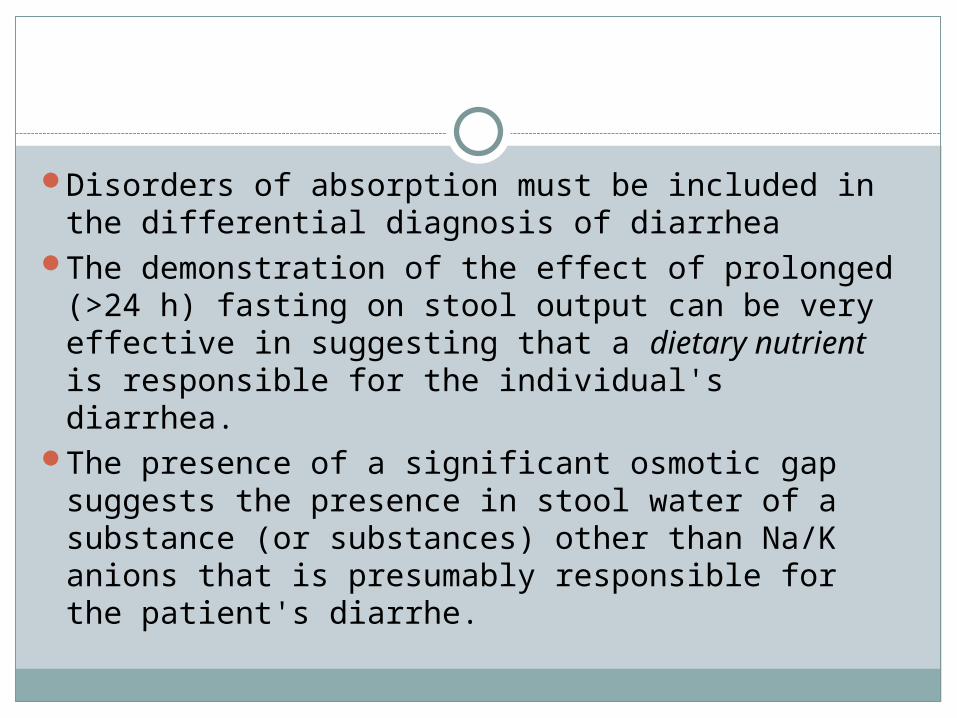
Disorders of absorption must be included in the differential diagnosis of diarrhea
The demonstration of the effect of prolonged (>24 h) fasting on stool output can be very effective in suggesting that a dietary nutrient is responsible for the individual's diarrhea.
The presence of a significant osmotic gap suggests the presence in stool water of a substance (or substances) other than Na/K anions that is presumably responsible for the patient's diarrhe.
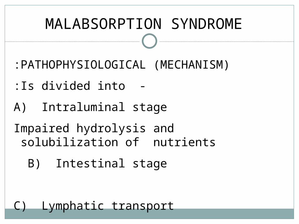
MALABSORPTION SYNDROME
PATHOPHYSIOLOGICAL (MECHANISM):
-Is divided into:
A) Intraluminal stage
Impaired hydrolysis and solubilization of nutrients
B) Intestinal stage
C) Lymphatic transport
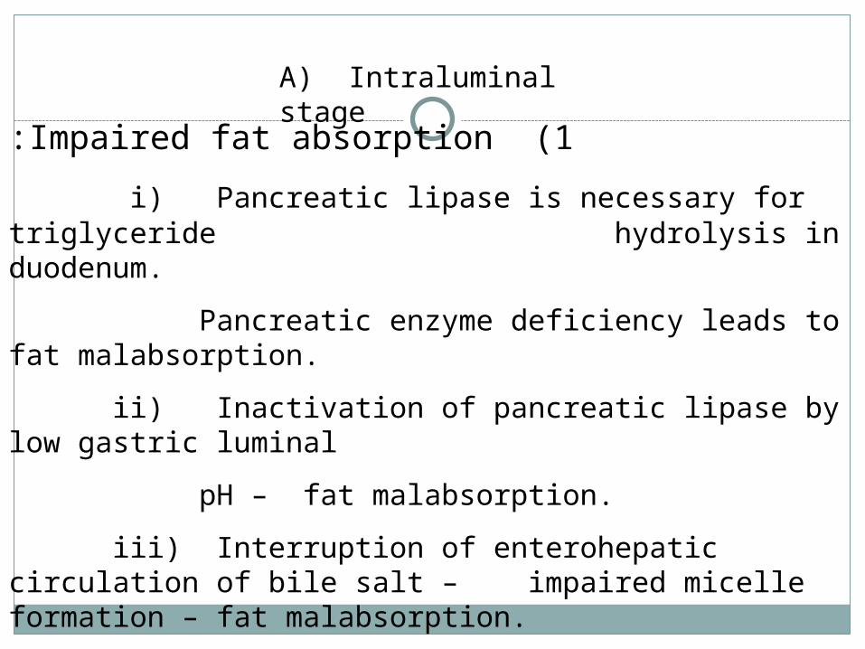
1 (Impaired fat absorption:
i) Pancreatic lipase is necessary for triglyceride hydrolysis in duodenum.
Pancreatic enzyme deficiency leads to fat malabsorption.
ii) Inactivation of pancreatic lipase by low gastric luminal
pH – fat malabsorption.
iii) Interruption of enterohepatic circulation of bile salt – impaired micelle formation – fat malabsorption.
Absorption of fat soluble vitamins may be impaired as well.
A) Intraluminal stage
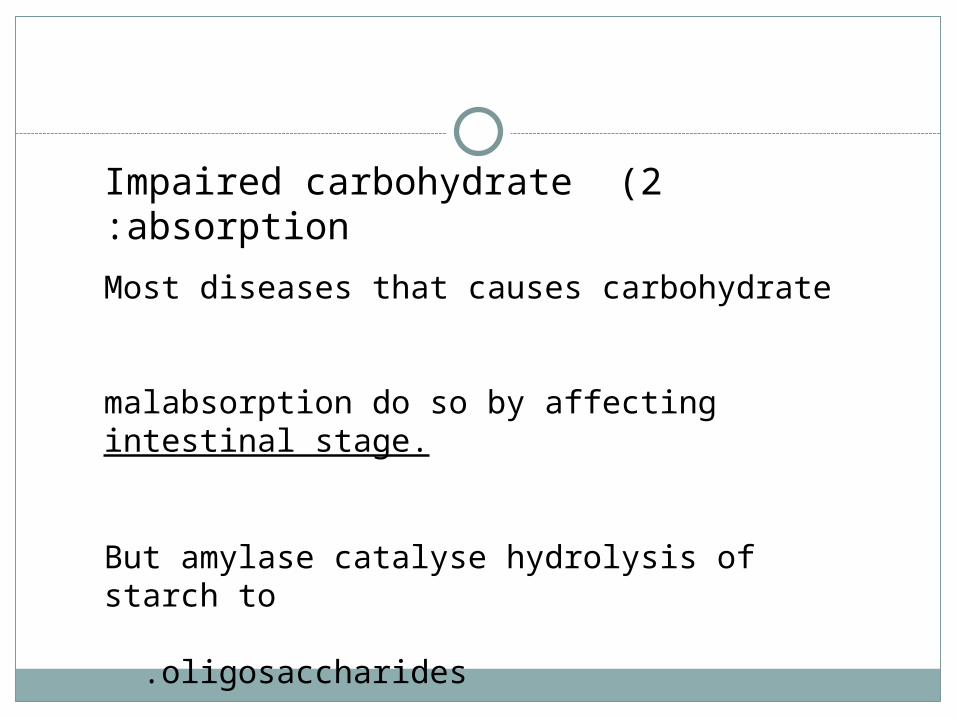
2 (Impaired carbohydrate absorption:
Most diseases that causes carbohydrate malabsorption do so by affecting intestinal stage.
But amylase catalyse hydrolysis of starch to
oligosaccharides .

3 (Impaired protein absorption:
Hydrolysis of polypeptides occurs mainly in small intestine by action of pancreatic enzyme trypsin, chymotrypsin.
Deficiency of pancreatic proteases – impaired protein absorption.
Diseases like :
Chronic pancreatitis
Cystic fibrosis
Ca. pancreatic resection
- Protein malnutrition

B) Intestinal stageB) Intestinal stage
1 (Abnormalities of small intestinal mucosa.
Lactase deficiency
e.g. Congenital or acquired
Result – malabsorption of lactose.
Acquired:- i) Celiac disease
ii) Crohn’s disease
iii) Infective enteritis

2 (Impaired epithelial cell transport:
Many diseases cause loss of intestinal surface area
- malabsorption of many nutrients.
e.g. i) Celiac disease
ii) Tropical spure
iii) Extensive surgical resection
iv) Drugs

C) Lymphatic transportC) Lymphatic transport::
Lymphatic obstruction – fat malabsorption
e.g. i) Intestinal lymphangiectasia
iii) Tuberculous enteritis
iv) Intestinal lymphoma

D)D) Decreased availability of ingested nutrients and
cofactors for absorption.
i) Vitamin B12 malabsorption if intrinsic factor is deficient. e.g. gastrectomy, antiparietal cell Ab.
ii) Bacterial overgrowth –can bind B12.
iii) Patient infected with fist tapeworm – B12 deficiency.
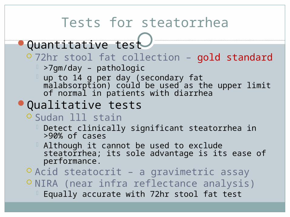
Tests for steatorrheaQuantitative test
72hr stool fat collection – gold standard >7gm/day – pathologic up to 14 g per day (secondary fat malabsorption) could
be used as the upper limit of normal in patients with diarrhea
Qualitative tests Sudan lll stain
Detect clinically significant steatorrhea in>90% of cases
Although it cannot be used to exclude steatorrhea; its sole advantage is its ease of performance.
Acid steatocrit – a gravimetric assay NIRA (near infra reflectance analysis)
Equally accurate with 72hr stool fat test
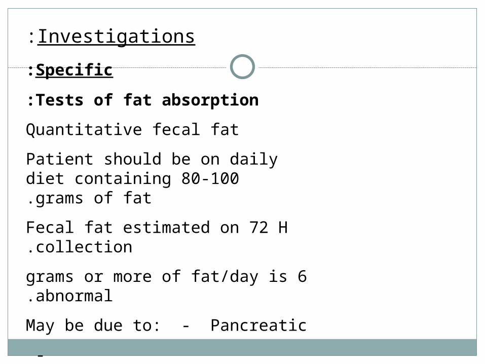
Investigations:
Specific:
Tests of fat absorption:
Quantitative fecal fat
Patient should be on daily diet containing 80-100 grams of fat.
Fecal fat estimated on 72 H collection.
6 grams or more of fat/day is abnormal.
May be due to: - Pancreatic
- Small intestinal
- Hepatobiliary disease
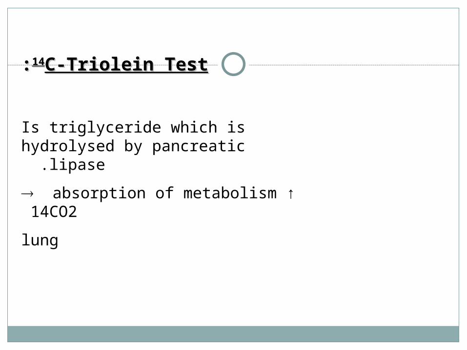
1414C-Triolein TestC-Triolein Test::
Is triglyceride which is hydrolysed by pancreatic lipase .
absorption of metabolism ↑ 14CO2
lung

Tests for pancreatic function:
1 (Bentiromide test:
Chymotrypsin
PABA + pepside
PABA absorbed and conjugated in liver
urine excretion
2 (Schilling test
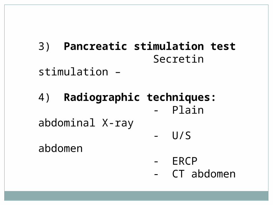
3) Pancreatic stimulation test Secretin stimulation –
4) Radiographic techniques: - Plain abdominal X-ray - U/S abdomen - ERCP - CT abdomen

Schilling test
The Schilling test can be used clinically to distinguish between gastric and ileal causes of vitamin B12 deficiency.
Alternative approaches to diagnosing pernicious anemia are to document atrophic gastritis by endoscopy and biopsy, to confirm achlorhydria by acid secretion analysis and increased serum gastrin levels, and to look for antibodies in the serum directed against parietal cells or intrinsic factor.
Schilling test results are normal in patients with dietary vitamin B12 deficiency, in protein-bound (food-bound) vitamin B12 malabsorption,[24] and sometimes in congenital transcobalamin II deficiency.[124]
False-positive results on the Schilling test can result from renal dysfunction or inadequate urine collection
Schilling test
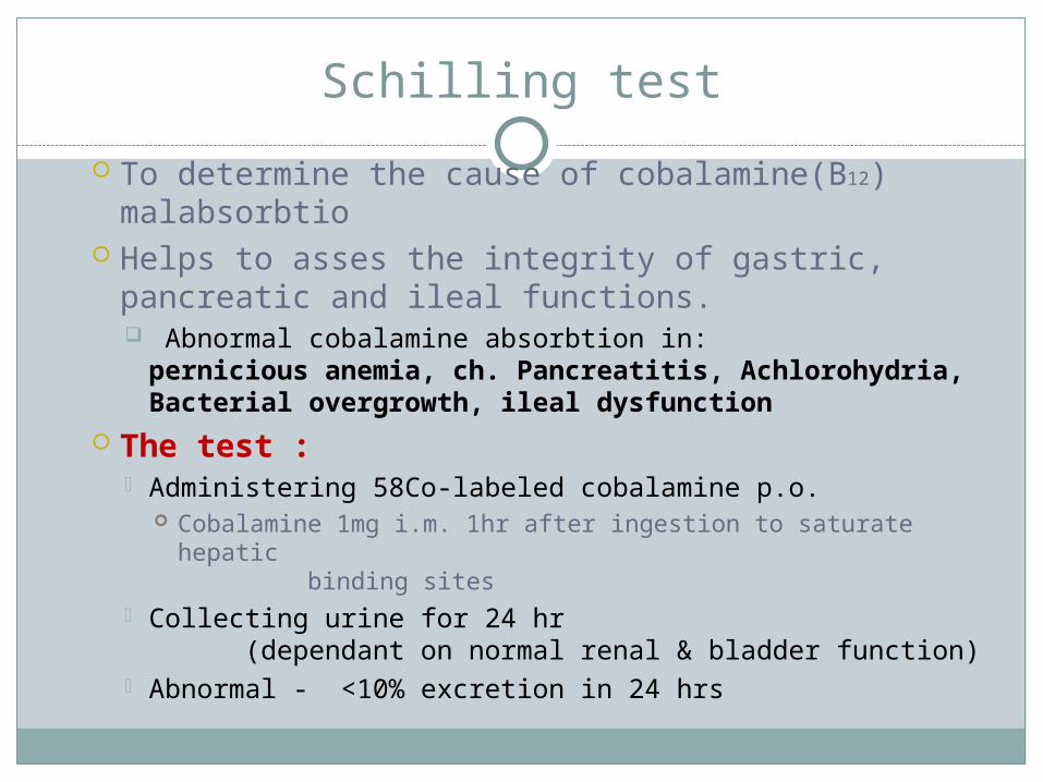
To determine the cause of cobalamine(B12) malabsorbtio
Helps to asses the integrity of gastric, pancreatic and ileal functions. Abnormal cobalamine absorbtion in:
pernicious anemia, ch. Pancreatitis, Achlorohydria, Bacterial overgrowth, ileal dysfunction
The test : Administering 58Co-labeled cobalamine p.o.
Cobalamine 1mg i.m. 1hr after ingestion to saturate hepatic binding sites
Collecting urine for 24 hr (dependant on normal renal & bladder function)
Abnormal - <10% excretion in 24 hrs
Schilling Test
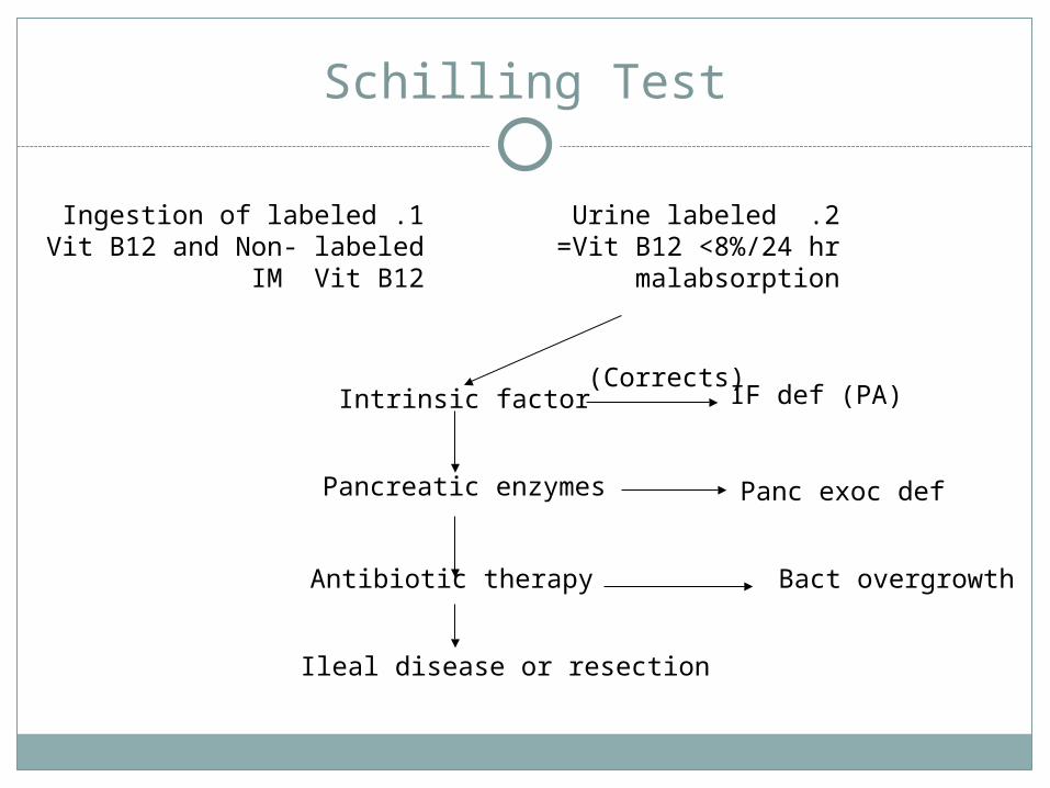
1 .Ingestion of labeledVit B12 and Non- labeled
IM Vit B12
2 .Urine labeledVit B12 <8%/24 hr=
malabsorption
Intrinsic factor
Pancreatic enzymes
Antibiotic therapy
Ileal disease or resection
IF def (PA)
Panc exoc def
Bact overgrowth
)Corrects(
Carbohydrate absorption test
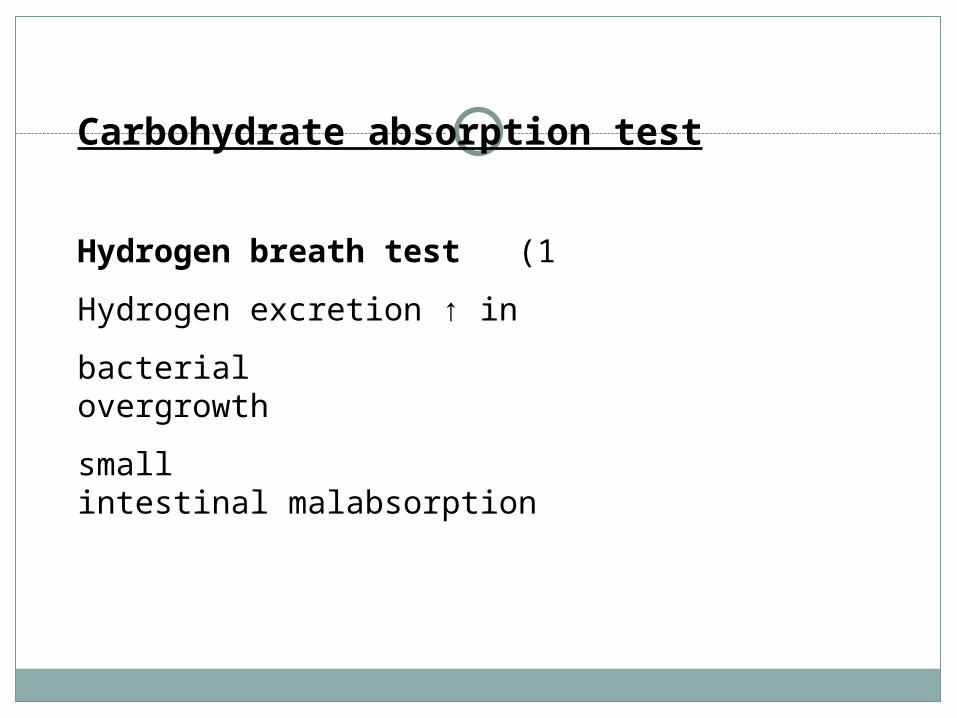
1 (Hydrogen breath test
Hydrogen excretion ↑ in
bacterial overgrowth
small intestinal malabsorption

Carbohydrate absorption test
2 (D-xylose test
5-carbon sugar excreted unchanged in urine
25 grams given
Urine collected for 5 hours
Normally 25% is excreted
In patients with fat malabsorption, this test
differentiates pancreatic from small intestinal malabsorpton.
D-xylose is normal in pancreatic disease
Serum level of D-xylose at 1-2 hours after ingestion can be measured .

Test for bacterial overgrowth:1) Intestinal aspiration and culture2) Breath test3) C-D xylose breath test

Glucose Hydrogen
most widely used breath test in clinical practice With bacterial overgrowth, : the glucose is cleaved by bacteria into
carbon dioxide and hydrogen. rise of 20 parts per million (ppm) above the baseline is regarded as diagnostic of SIBO.
Fasting breath hydrogen levels of more than 20 ppm also are considered positive. High baseline hydrogen levels also are common in untreated celiac disease and normalize after gluten withdrawal for as-yet-unknown reasons.[
Patients must avoid smoking and ingestion of nonfermentable carbohydrates, such as pasta and bread, the night before the test, because these factors can raise baseline breath hydrogen values. Exercise can induce hyperventilation, thereby reducing baseline breath hydrogen values, and should be avoided for two hours before the test.
sensitivity of 62% and specificity of 83%. Very rapid intestinal transit can lead to a false-positive test result, because glucose can reach the colon before it can be absorbed.
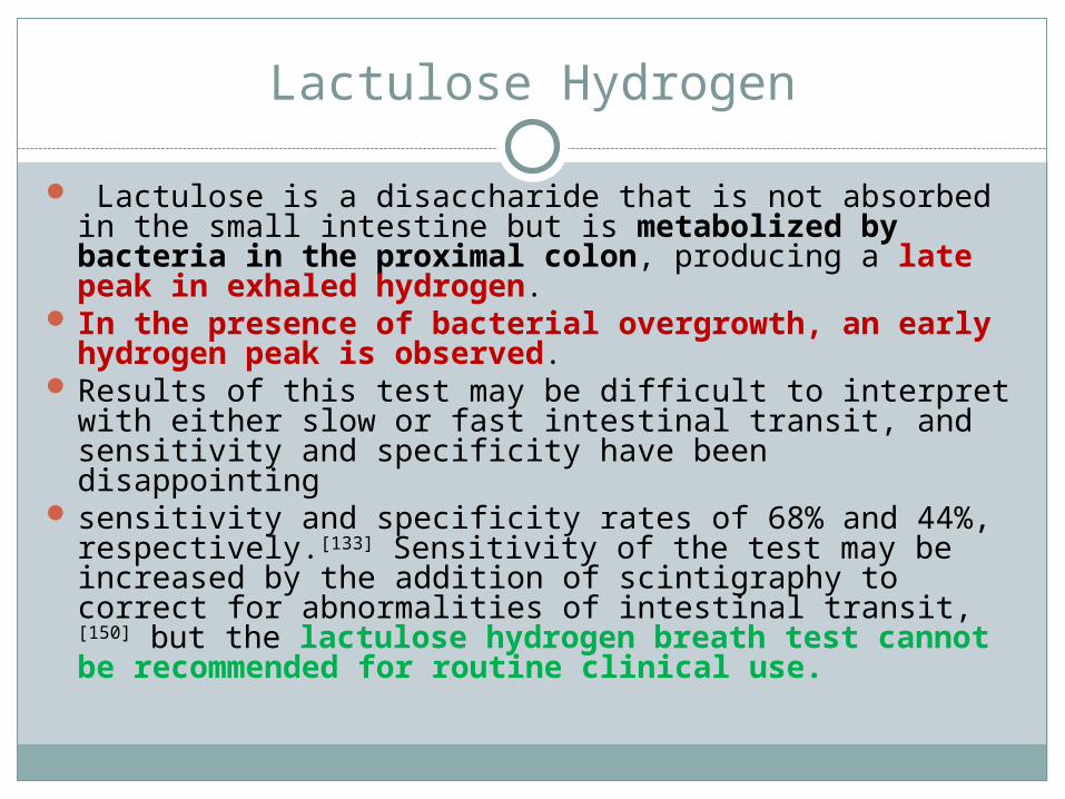
Lactulose Hydrogen
Lactulose is a disaccharide that is not absorbed in the small intestine but is metabolized by bacteria in the proximal colon, producing a late peak in exhaled hydrogen.
In the presence of bacterial overgrowth, an early hydrogen peak is observed.
Results of this test may be difficult to interpret with either slow or fast intestinal transit, and sensitivity and specificity have been disappointing
sensitivity and specificity rates of 68% and 44%, respectively.[133] Sensitivity of the test may be increased by the addition of scintigraphy to correct for abnormalities of intestinal transit,[150] but the lactulose hydrogen breath test cannot be recommended for routine clinical use.
Xylose
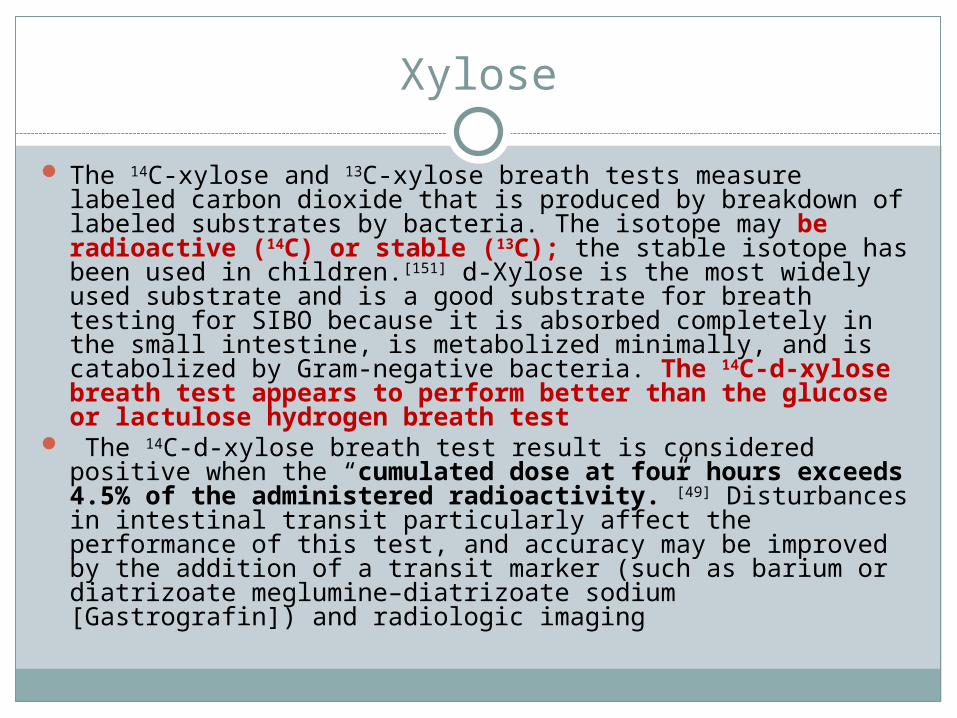
The 14C-xylose and 13C-xylose breath tests measure labeled carbon dioxide that is produced by breakdown of labeled substrates by bacteria. The isotope may be radioactive (14C) or stable (13C); the stable isotope has been used in children.[151] d-Xylose is the most widely used substrate and is a good substrate for breath testing for SIBO because it is absorbed completely in the small intestine, is metabolized minimally, and is catabolized by Gram-negative bacteria. The 14C-d-xylose breath test appears to perform better than the glucose or lactulose hydrogen breath test
The 14C-d-xylose breath test result is considered positive when the “cumulated dose at four hours exceeds 4.5% of the administered radioactivity.”[49] Disturbances in intestinal transit particularly affect the performance of this test, and accuracy may be improved by the addition of a transit marker (such as barium or diatrizoate meglumine–diatrizoate sodium [Gastrografin]) and radiologic imaging

1) Radiography of small intestine:barium contrast (small-bowel series or study)– to see- Blind loop- Strictures and fistulas (as in Crohn's disease)- J. diverticular
A normal barium contrast study does not exclude the possibility of small-intestinal disease.
2) CT enteroclysis and magnetic resonance (MR) enteroclysis

2) Intestinal mucosal biopsy: indications ;
A small-intestinal mucosal biopsy is essential in the evaluation of a patient with documented steatorrhea or chronic diarrhea (lasting >3 weeks)
Diffuse or focal abnormalities of the small intestine defined on a small-intestinal series
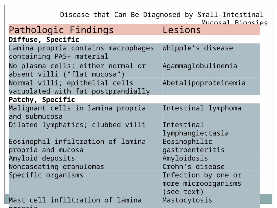
Disease that Can Be Diagnosed by Small-Intestinal Mucosal Biopsies
LesionsPathologic FindingsDiffuse, Specific
Whipple's diseaseLamina propria contains macrophages containing PAS+ material
AgammaglobulinemiaNo plasma cells; either normal or absent villi ("flat mucosa")
AbetalipoproteinemiaNormal villi; epithelial cells vacuolated with fat postprandiallyPatchy, Specific
Intestinal lymphomaMalignant cells in lamina propria and submucosa
Intestinal lymphangiectasiaDilated lymphatics; clubbed villiEosinophilic gastroenteritisEosinophil infiltration of lamina propria
and mucosaAmyloidosisAmyloid depositsCrohn's diseaseNoncaseating granulomasInfection by one or more microorganisms (see text)
Specific organisms
MastocytosisMast cell infiltration of lamina propria
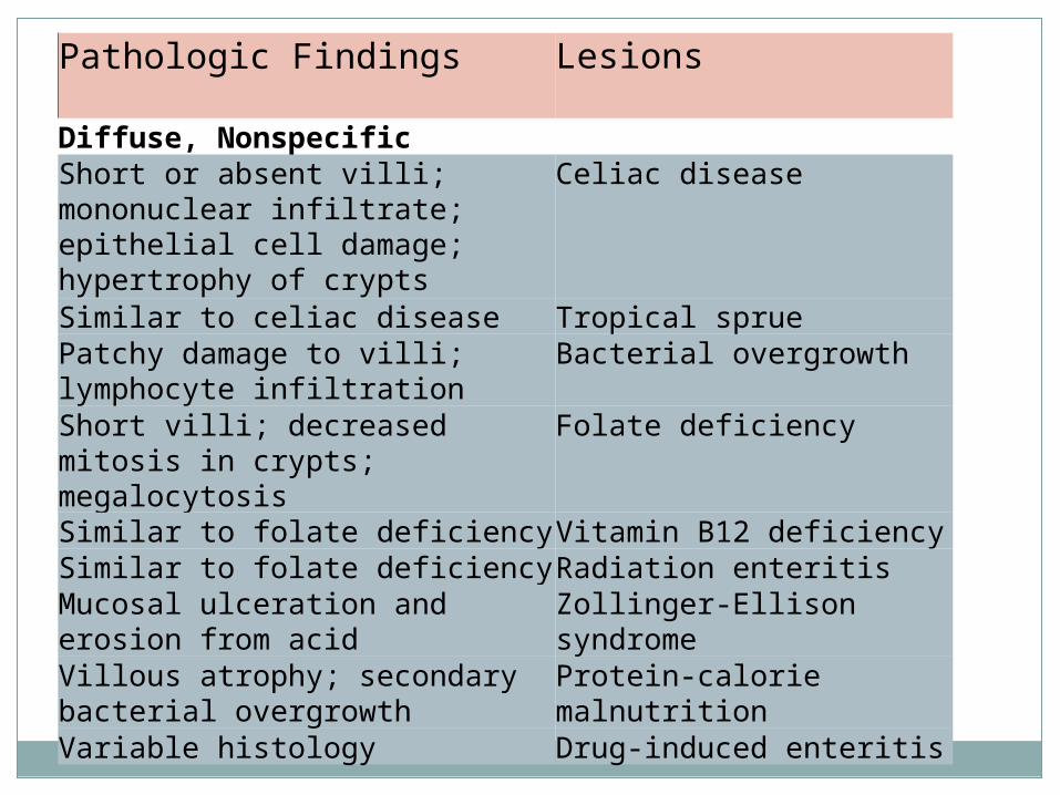
LesionsPathologic Findings
Diffuse, Nonspecific Celiac diseaseShort or absent villi;
mononuclear infiltrate; epithelial cell damage; hypertrophy of crypts
Tropical sprueSimilar to celiac diseaseBacterial overgrowthPatchy damage to villi;
lymphocyte infiltrationFolate deficiencyShort villi; decreased mitosis in
crypts; megalocytosisVitamin B12 deficiencySimilar to folate deficiencyRadiation enteritisSimilar to folate deficiencyZollinger-Ellison syndrome
Mucosal ulceration and erosion from acid
Protein-calorie malnutrition
Villous atrophy; secondary bacterial overgrowth
Drug-induced enteritisVariable histology

D-Xylose Test Schilling TestDuodenal Mucosal Biopsy
Chronic pancreatitis
Normal50% abnormal; if abnormal, normal with pancreatic enzymes
Normal
Bacterial overgrowth syndrome
Normal or only modestly abnormal
Often abnormal; if abnormal, normal after antibiotics
Usually normal
Ileal diseaseNormalAbnormalNormalCeliac diseaseDecreasedNormalAbnormal:
probably "flat"Intestinal lymphangiectasia
NormalNormalAbnormal: "dilated lymphatics"
Results of Diagnostic Studies in Different Causes of Steatorrhea
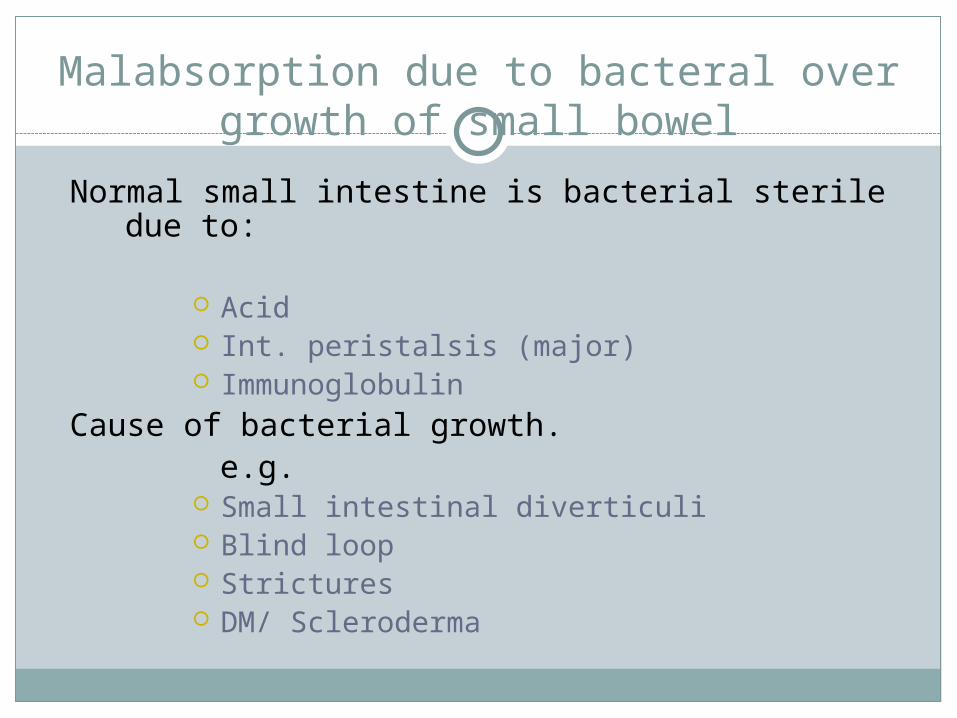
Malabsorption due to bacteral over growth of small bowel
Normal small intestine is bacterial sterile due to:
Acid Int. peristalsis (major) Immunoglobulin
Cause of bacterial growth. e.g.
Small intestinal diverticuli Blind loop Strictures DM/ Scleroderma
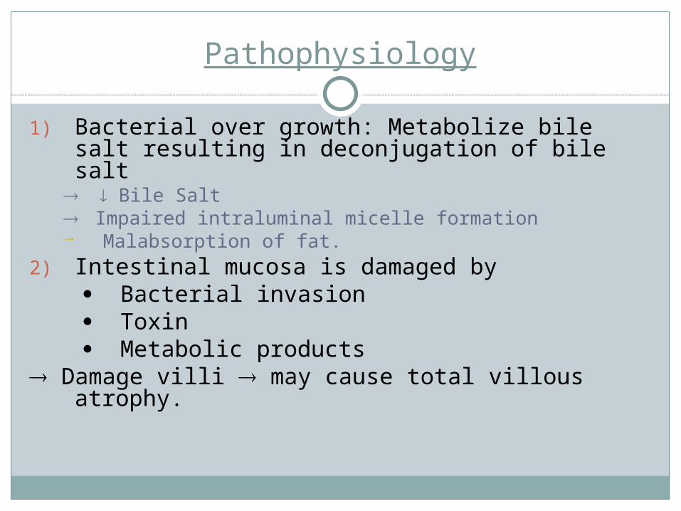
Pathophysiology
1) Bacterial over growth: Metabolize bile salt resulting in deconjugation of bile salt
Bile Salt Impaired intraluminal micelle formation Malabsorption of fat.
2) Intestinal mucosa is damaged by Bacterial invasion Toxin Metabolic products Damage villi may cause total villous atrophy.

Clinically: Steatorrhea Anaemia B12 def.Reversed of symptom after antibiotic
treatment. Diagnosis: Breath test Cxylose test Culture of aspiration (definitive)Treatment: Antibiotic Tetracyclin Ciproflexacin Metronidazole Amoxil
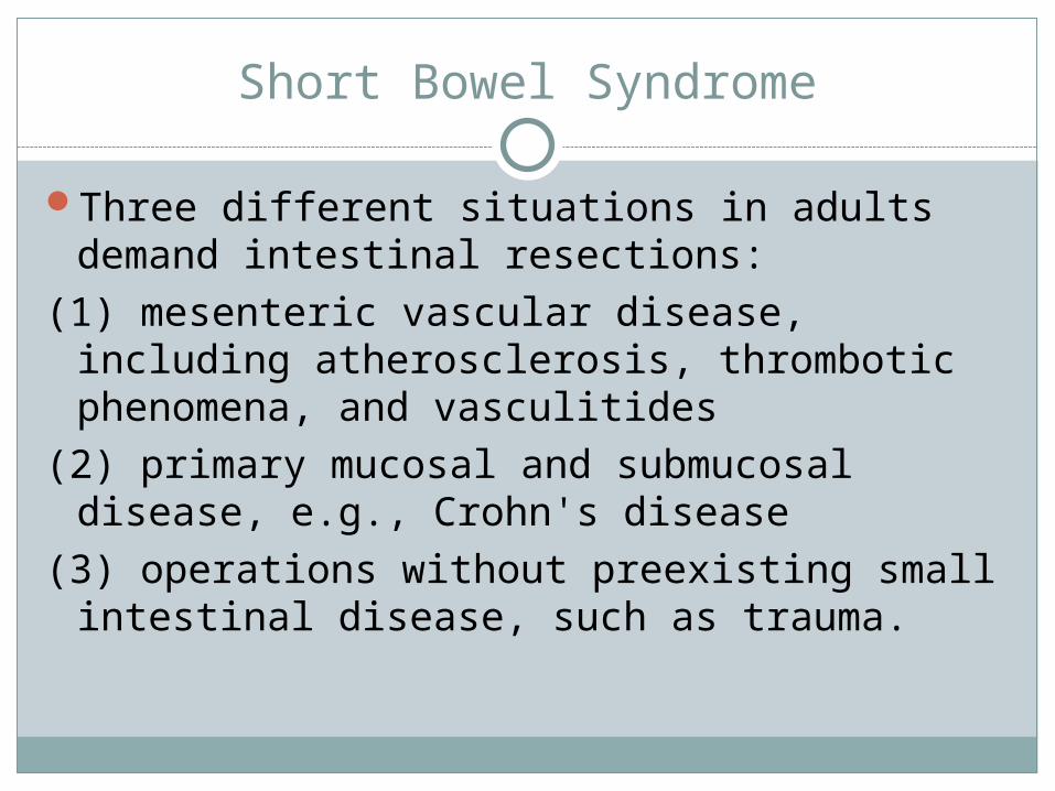
Short Bowel Syndrome
Three different situations in adults demand intestinal resections:
(1) mesenteric vascular disease, including atherosclerosis, thrombotic phenomena, and vasculitides
(2) primary mucosal and submucosal disease, e.g., Crohn's disease
(3) operations without preexisting small intestinal disease, such as trauma.
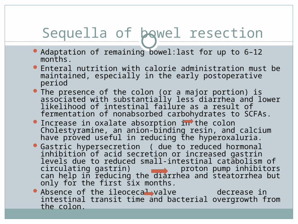
Sequella of bowel resection Adaptation of remaining bowel:last for up to 6–12
months. Enteral nutrition with calorie administration must be
maintained, especially in the early postoperative period The presence of the colon (or a major portion) is
associated with substantially less diarrhea and lower likelihood of intestinal failure as a result of fermentation of nonabsorbed carbohydrates to SCFAs.
Increase in oxalate absorption in the colon Cholestyramine, an anion-binding resin, and calcium have proved useful in reducing the hyperoxaluria.
Gastric hypersecretion ( due to reduced hormonal inhibition of acid secretion or increased gastrin levels due to reduced small-intestinal catabolism of circulating gastrin) proton pump inhibitors can help in reducing the diarrhea and steatorrhea but only for the first six months.
Absence of the ileocecal valve decrease in intestinal transit time and bacterial overgrowth from the colon.

Whipple's Disease
Etiology:gram-positive bacillus, T. whipplei Clinical Presentation:diarrhea, steatorrhea, abdominal
pain, weight loss, migratory large-joint arthropathy, and fever as well as ophthalmologic and CNS symptoms (dementia,..).
The steatorrhea in these patients is generally believed secondary to both small-intestinal mucosal injury and lymphatic obstruction secondary to the increased number of PAS-positive macrophages in the lamina propria of the small intestine.
Diagnosis:tissue biopsies from the small intestine and/or other organs . PAS-positive macrophages containing the characteristic small (0.25—1–2 mm) bacilli is suggestive of this diagnosis.
Treatment:trimethoprim/sulfamethoxazole for approximately 1 year.

Protein-Losing Enteropathy
Normally, about 10% of total protein catabolism occurs via the gastrointestinal tract.
Increased protein loss into the gastrointestinal in more than 65 different diseases with mucosal ulceration or nonulcerated mucosa or lymphatic dysfunction
Peripheral edema and low serum albumin and globulin levels in the absence of renal and hepatic disease.
Treatment: treat the underlying disease process .