Embed Size (px)
Citation preview
Current Biology, Vol. 14, 1694–1702, October 5, 2004, 2004 Elsevier Ltd. All rights reserved. DOI 10.1016/j .cub.2004.09.048
Drosophila T Box Proteins Break the Symmetryof Hedgehog-Dependent Activation of wingless
mation in the ventral epidermis. In odd-numbered ab-dominal segments, Mid/H15 activity plays an importantrole in restricting the expression of Wg to a single domain.
Marita Buescher,1,4,* Pia C. Svendsen,2,4 Murni Tio,1
Cindy Miskolczi-McCallum,2 Guy Tear,1
William J. Brook,2 and William Chia1,3
1Medical Research Council Centrefor Developmental Neurobiology Background
King’s College London4th Floor New Hunts House The larval cuticle of Drosophila melanogaster is a model
system for generating patterns from fields of cells. TheGuy’s Hospital CampusLondon SE1 1UL ventral cuticle exhibits a segmentally reiterated array of
six rows of unique denticles separated by areas of nakedUnited Kingdom2 Genes and Development Research Group and cuticle. These external structures reflect the cellular di-
versity within the underlying epidermis, and defectiveDepartment of Biochemistry and Molecular BiologyUniversity of Calgary cuticle patterns are indicative of incorrectly specified
cell fates [1]. The secreted products of two segment3330 Hospital Drive NWCalgary, Alberta T2N 4N1 polarity genes, wg and hh, are the key players that initi-
ate progressive patterning events that ultimately resultCanada3 Temasek Lifesciences Laboratory in epidermal differentiation at the single-cell level [2–9].
Thus, patterning requires the tight regulation of the spa-1 Research LinkNational University of Singapore Campus tial limits of Wg and Hh expression. In early embryogene-
sis, pair-rule gene activity initiates the expression of WgSingaporeSingapore 117604 and Hh in adjacent stripes, with Wg just anterior to the
Hh-expressing cells (the spatial arrangement of theseexpression domains is shown in Figure 7G). After stage9, reciprocal signaling between Wg- and Hh-expressingSummarycells stabilizes their expression domains. Acting aniso-tropically, Hh signaling activates Wg anterior, but notBackground: Segmentation of the Drosophila embryo
is a classic paradigm for pattern formation during devel- posterior, to the Hh stripe. Finally, Wg expression be-comes independent of Hh and is maintained through anopment. The Wnt-1 homolog Wingless (Wg) is a key
player in the establishment of a segmentally reiterated autoregulatory feedback loop. Previous studies haveled to the conclusion that Hh signaling is bidirectionalpattern of cell type specification. The intrasegmental
polarity of this pattern depends on the precise position- because it maintains patched (ptc)-gene expression innarrow stripes anterior and posterior to the En/Hh stripeing of the Wg signaling source anterior to the Engrailed
(En)/Hedgehog (Hh) domain. Proper polarity of epider- [10, 11]. The ptc gene product is a repressor of Wgexpression, and maintenance of Wg expression at stagemal segments requires an asymmetric response to the
bidirectional Hh signal: wg is activated in cells anterior 9 requires the Hh-mediated derepression. However, de-spite the symmetry of Hh signaling, the outcome withto the Hh signaling source and is restricted from cells
posterior to this signaling source. respect to Wg expression is asymmetric and resultsin a single Wg stripe anterior to the En/Hh stripe. ToResults: Here we report that Midline (Mid) and H15, two
highly related T box proteins representing the orthologs rationalize the differential response of Wg to the Hhsignal, Ingham et al. have put forward a model thatof zebrafish hrT and mouse Tbx20, are novel negative
regulators of wg transcription and act to break the sym- subdivides each parasegment into two domains: theposterior half of the parasegment represents the wg-metry of Hh signaling. Loss of mid and H15 results in
the symmetric outcome of Hh signaling: the establish- competent domain, and the anterior half is the en-com-petent domain [12]. The wg-competent domain encom-ment of wg domains anterior and posterior to the signal-passes those cells that express Wg in a ptc mutanting source predominantly, but not exclusively, in odd-background. Later studies showed that wg competencenumbered segments. Accordingly, loss of mid and H15requires the activity of the pair-rule/segment polarityproduces defects that mimic a wg gain-of-function phe-genes sloppy-paired 1, 2 (slp1, 2), which are expressednotype. Misexpression of mid represses wg and pro-in broad stripes anterior to the En/Hh stripe (see modelduces a weak/moderate wg loss-of-function pheno-Figure 7G for the location of the slp expression domain)copy. Furthermore, we show that loss of mid and H15[13]. It has been suggested that Slp permits the activa-results in an anterior expansion of the expression oftion of Wg anterior to the En/Hh stripe by antagonizingserrate (ser) in every segment, representing a secondone (or several) putative repressor(s) of Wg. Here weinstance of target gene repression downstream of Hhreport that Mid and H15 act to repress the Hh-dependentsignaling in the establishment of segment polarity.activation of Wg in the en-competent domain predomi-Conclusions: The data we present here indicate thatnantly in odd-numbered segments. Furthermore, our re-mid and H15 are important components in pattern for-sults suggest that the Slp-mediated repression of Mid/H15 anterior to the En/Hh stripe is an important compo-*Correspondence: [email protected]
4These authors contributed equally to the work. nent of wg competence.
T Box Proteins Repress wingless Expression1695
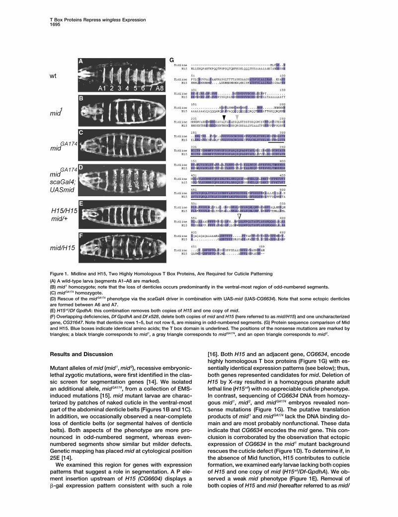
Figure 1. Midline and H15, Two Highly Homologous T Box Proteins, Are Required for Cuticle Patterning
(A) A wild-type larva (segments A1–A8 are marked).(B) mid1 homozygote; note that the loss of denticles occurs predominantly in the ventral-most region of odd-numbered segments.(C) midGA174 homozygote.(D) Rescue of the midGA174 phenotype via the scaGal4 driver in combination with UAS-mid (UAS-CG6634). Note that some ectopic denticlesare formed between A6 and A7.(E) H15x4/Df GpdhA: this combination removes both copies of H15 and one copy of mid.(F) Overlapping deficiencies, Df GpdhA and Df x528, delete both copies of mid and H15 (here referred to as mid/H15) and one uncharacterizedgene, CG31647. Note that denticle rows 1–5, but not row 6, are missing in odd-numbered segments. (G) Protein sequence comparison of Midand H15. Blue boxes indicate identical amino acids; the T box domain is underlined. The positions of the nonsense mutations are marked bytriangles; a black triangle corresponds to mid1, a gray triangle corresponds to midGA174, and an open triangle corresponds to mid2.
Results and Discussion [16]. Both H15 and an adjacent gene, CG6634, encodehighly homologous T box proteins (Figure 1G) with es-sentially identical expression patterns (see below); thus,Mutant alleles of mid (mid1, mid2), recessive embryonic-
lethal zygotic mutations, were first identified in the clas- both genes represented candidates for mid. Deletion ofH15 by X-ray resulted in a homozygous pharate adultsic screen for segmentation genes [14]. We isolated
an additional allele, midGA174, from a collection of EMS- lethal line (H15x4) with no appreciable cuticle phenotype.In contrast, sequencing of CG6634 DNA from homozy-induced mutations [15]. mid mutant larvae are charac-
terized by patches of naked cuticle in the ventral-most gous mid1, mid2, and midGA174 embryos revealed non-sense mutations (Figure 1G). The putative translationpart of the abdominal denticle belts (Figures 1B and 1C).
In addition, we occasionally observed a near-complete products of mid1 and midGA174 lack the DNA binding do-main and are most probably nonfunctional. These dataloss of denticle belts (or segmental halves of denticle
belts). Both aspects of the phenotype are more pro- indicate that CG6634 encodes the mid gene. This con-clusion is corroborated by the observation that ectopicnounced in odd-numbered segment, whereas even-
numbered segments show similar but milder defects. expression of CG6634 in the mid1 mutant backgroundrescues the cuticle defect (Figure 1D). To determine if, inGenetic mapping has placed mid at cytological position
25E [14]. the absence of Mid function, H15 contributes to cuticleformation, we examined early larvae lacking both copiesWe examined this region for genes with expression
patterns that suggest a role in segmentation. A P ele- of H15 and one copy of mid (H15x4/Df-GpdhA). We ob-served a weak mid phenotype (Figure 1E). Removal ofment insertion upstream of H15 (CG6604) displays a
�-gal expression pattern consistent with such a role both copies of H15 and mid (hereafter referred to as mid/
Current Biology1696
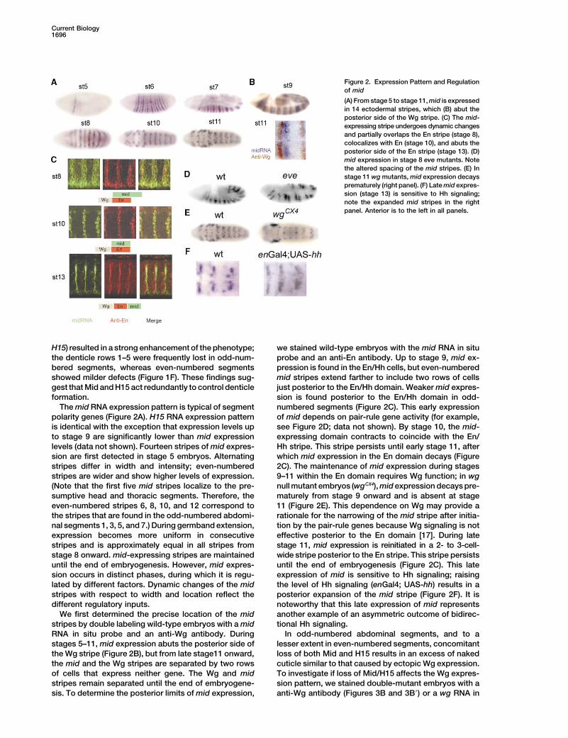
Figure 2. Expression Pattern and Regulationof mid
(A) From stage 5 to stage 11, mid is expressedin 14 ectodermal stripes, which (B) abut theposterior side of the Wg stripe. (C) The mid-expressing stripe undergoes dynamic changesand partially overlaps the En stripe (stage 8),colocalizes with En (stage 10), and abuts theposterior side of the En stripe (stage 13). (D)mid expression in stage 8 eve mutants. Notethe altered spacing of the mid stripes. (E) Instage 11 wg mutants, mid expression decaysprematurely (right panel). (F) Late mid expres-sion (stage 13) is sensitive to Hh signaling;note the expanded mid stripes in the rightpanel. Anterior is to the left in all panels.
H15) resulted in a strong enhancement of the phenotype; we stained wild-type embryos with the mid RNA in situprobe and an anti-En antibody. Up to stage 9, mid ex-the denticle rows 1–5 were frequently lost in odd-num-
bered segments, whereas even-numbered segments pression is found in the En/Hh cells, but even-numberedmid stripes extend farther to include two rows of cellsshowed milder defects (Figure 1F). These findings sug-
gest that Mid and H15 act redundantly to control denticle just posterior to the En/Hh domain. Weaker mid expres-sion is found posterior to the En/Hh domain in odd-formation.
The mid RNA expression pattern is typical of segment numbered segments (Figure 2C). This early expressionof mid depends on pair-rule gene activity (for example,polarity genes (Figure 2A). H15 RNA expression pattern
is identical with the exception that expression levels up see Figure 2D; data not shown). By stage 10, the mid-expressing domain contracts to coincide with the En/to stage 9 are significantly lower than mid expression
levels (data not shown). Fourteen stripes of mid expres- Hh stripe. This stripe persists until early stage 11, afterwhich mid expression in the En domain decays (Figuresion are first detected in stage 5 embryos. Alternating
stripes differ in width and intensity; even-numbered 2C). The maintenance of mid expression during stages9–11 within the En domain requires Wg function; in wgstripes are wider and show higher levels of expression.
(Note that the first five mid stripes localize to the pre- null mutant embryos (wgCX4), mid expression decays pre-maturely from stage 9 onward and is absent at stagesumptive head and thoracic segments. Therefore, the
even-numbered stripes 6, 8, 10, and 12 correspond to 11 (Figure 2E). This dependence on Wg may provide arationale for the narrowing of the mid stripe after initia-the stripes that are found in the odd-numbered abdomi-
nal segments 1, 3, 5, and 7.) During germband extension, tion by the pair-rule genes because Wg signaling is noteffective posterior to the En domain [17]. During lateexpression becomes more uniform in consecutive
stripes and is approximately equal in all stripes from stage 11, mid expression is reinitiated in a 2- to 3-cell-wide stripe posterior to the En stripe. This stripe persistsstage 8 onward. mid-expressing stripes are maintained
until the end of embryogenesis. However, mid expres- until the end of embryogenesis (Figure 2C). This lateexpression of mid is sensitive to Hh signaling; raisingsion occurs in distinct phases, during which it is regu-
lated by different factors. Dynamic changes of the mid the level of Hh signaling (enGal4; UAS-hh) results in aposterior expansion of the mid stripe (Figure 2F). It isstripes with respect to width and location reflect the
different regulatory inputs. noteworthy that this late expression of mid representsanother example of an asymmetric outcome of bidirec-We first determined the precise location of the mid
stripes by double labeling wild-type embryos with a mid tional Hh signaling.In odd-numbered abdominal segments, and to aRNA in situ probe and an anti-Wg antibody. During
stages 5–11, mid expression abuts the posterior side of lesser extent in even-numbered segments, concomitantloss of both Mid and H15 results in an excess of nakedthe Wg stripe (Figure 2B), but from late stage11 onward,
the mid and the Wg stripes are separated by two rows cuticle similar to that caused by ectopic Wg expression.To investigate if loss of Mid/H15 affects the Wg expres-of cells that express neither gene. The Wg and mid
stripes remain separated until the end of embryogene- sion pattern, we stained double-mutant embryos with aanti-Wg antibody (Figures 3B and 3B�) or a wg RNA insis. To determine the posterior limits of mid expression,
T Box Proteins Repress wingless Expression1697
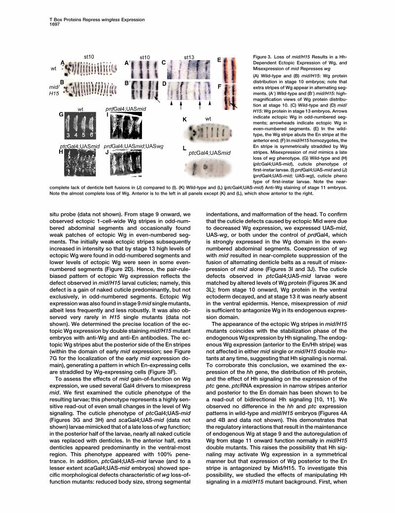
Figure 3. Loss of mid/H15 Results in a Hh-Dependent Ectopic Expression of Wg, andMisexpression of mid Represses wg
(A) Wild-type and (B) mid/H15: Wg proteindistribution in stage 10 embryos; note thatextra stripes of Wg appear in alternating seg-ments. (A�) Wild-type and (B�) mid/H15: high-magnification views of Wg protein distribu-tion at stage 10. (C) Wild-type and (D) mid/H15: Wg protein in stage 13 embryos. Arrowsindicate ectopic Wg in odd-numbered seg-ments; arrowheads indicate ectopic Wg ineven-numbered segments. (E) In the wild-type, the Wg stripe abuts the En stripe at theanterior end. (F) In mid/H15 homozygotes, theEn stripe is symmetrically straddled by Wgstripes. Misexpression of mid mimics a lateloss of wg phenotype. (G) Wild-type and (H)(ptcGal4;UAS-mid), cuticle phenotype offirst-instar larvae. (I) prdGal4;UAS-mid and (J)(prdGal4;UAS-mid; UAS-wg), cuticle phenotype of first-instar larvae. Note the near-
complete lack of denticle belt fusions in (J) compared to (I). (K) Wild-type and (L) (ptcGal4;UAS-mid) Anti-Wg staining of stage 11 embryos.Note the almost complete loss of Wg. Anterior is to the left in all panels except (K) and (L), which show anterior to the right.
situ probe (data not shown). From stage 9 onward, we indentations, and malformation of the head. To confirmthat the cuticle defects caused by ectopic Mid were dueobserved ectopic 1-cell-wide Wg stripes in odd-num-
bered abdominal segments and occasionally found to decreased Wg expression, we expressed UAS-mid,UAS-wg, or both under the control of prdGal4, whichweak patches of ectopic Wg in even-numbered seg-
ments. The initially weak ectopic stripes subsequently is strongly expressed in the Wg domain in the even-numbered abdominal segments. Coexpression of wgincreased in intensity so that by stage 13 high levels of
ectopic Wg were found in odd-numbered segments and with mid resulted in near-complete suppression of thefusion of alternating denticle belts as a result of misex-lower levels of ectopic Wg were seen in some even-
numbered segments (Figure 2D). Hence, the pair-rule- pression of mid alone (Figures 3I and 3J). The cuticledefects observed in ptcGal4;UAS-mid larvae werebiased pattern of ectopic Wg expression reflects the
defect observed in mid/H15 larval cuticles; namely, this matched by altered levels of Wg protein (Figures 3K and3L); from stage 10 onward, Wg protein in the ventraldefect is a gain of naked cuticle predominantly, but not
exclusively, in odd-numbered segments. Ectopic Wg ectoderm decayed, and at stage 13 it was nearly absentin the ventral epidermis. Hence, misexpression of midexpression was also found in stage 9 mid single mutants,
albeit less frequently and less robustly. It was also ob- is sufficient to antagonize Wg in its endogenous expres-sion domain.served very rarely in H15 single mutants (data not
shown). We determined the precise location of the ec- The appearance of the ectopic Wg stripes in mid/H15mutants coincides with the stabilization phase of thetopic Wg expression by double staining mid/H15 mutant
embryos with anti-Wg and anti-En antibodies. The ec- endogenous Wg expression by Hh signaling. The endog-enous Wg expression (anterior to the En/Hh stripe) wastopic Wg stripes abut the posterior side of the En stripes
(within the domain of early mid expression; see Figure not affected in either mid single or mid/H15 double mu-tants at any time, suggesting that Hh signaling is normal.7G for the localization of the early mid expression do-
main), generating a pattern in which En-expressing cells To corroborate this conclusion, we examined the ex-pression of the hh gene, the distribution of Hh protein,are straddled by Wg-expressing cells (Figure 3F).
To assess the effects of mid gain-of-function on Wg and the effect of Hh signaling on the expression of theptc gene. ptcRNA expression in narrow stripes anteriorexpression, we used several Gal4 drivers to misexpress
mid. We first examined the cuticle phenotype of the and posterior to the En domain has been shown to bea read-out of bidirectional Hh signaling [10, 11]. Weresulting larvae; this phenotype represents a highly sen-
sitive read-out of even small changes in the level of Wg observed no difference in the hh and ptc expressionpatterns in wild-type and mid/H15 embryos (Figures 4Asignaling. The cuticle phenotype of ptcGal4;UAS-mid
(Figures 3G and 3H) and scaGal4;UAS-mid (data not and 4B and data not shown). This demonstrates thatthe regulatory interactions that result in the maintenanceshown) larvae mimicked that of a late loss of wg function;
in the posterior half of the larvae, nearly all naked cuticle of endogenous Wg at stage 9 and the autoregulation ofWg from stage 11 onward function normally in mid/H15was replaced with denticles. In the anterior half, extra
denticles appeared predominantly in the ventral-most double mutants. This raises the possibility that Hh sig-naling may activate Wg expression in a symmetricalregion. This phenotype appeared with 100% pene-
trance. In addition, ptcGal4;UAS-mid larvae (and to a manner but that expression of Wg posterior to the Enstripe is antagonized by Mid/H15. To investigate thislesser extent scaGal4;UAS-mid embryos) showed spe-
cific morphological defects characteristic of wg loss-of- possibility, we studied the effects of manipulating Hhsignaling in a mid/H15 mutant background. First, whenfunction mutants: reduced body size, strong segmental
Current Biology1698
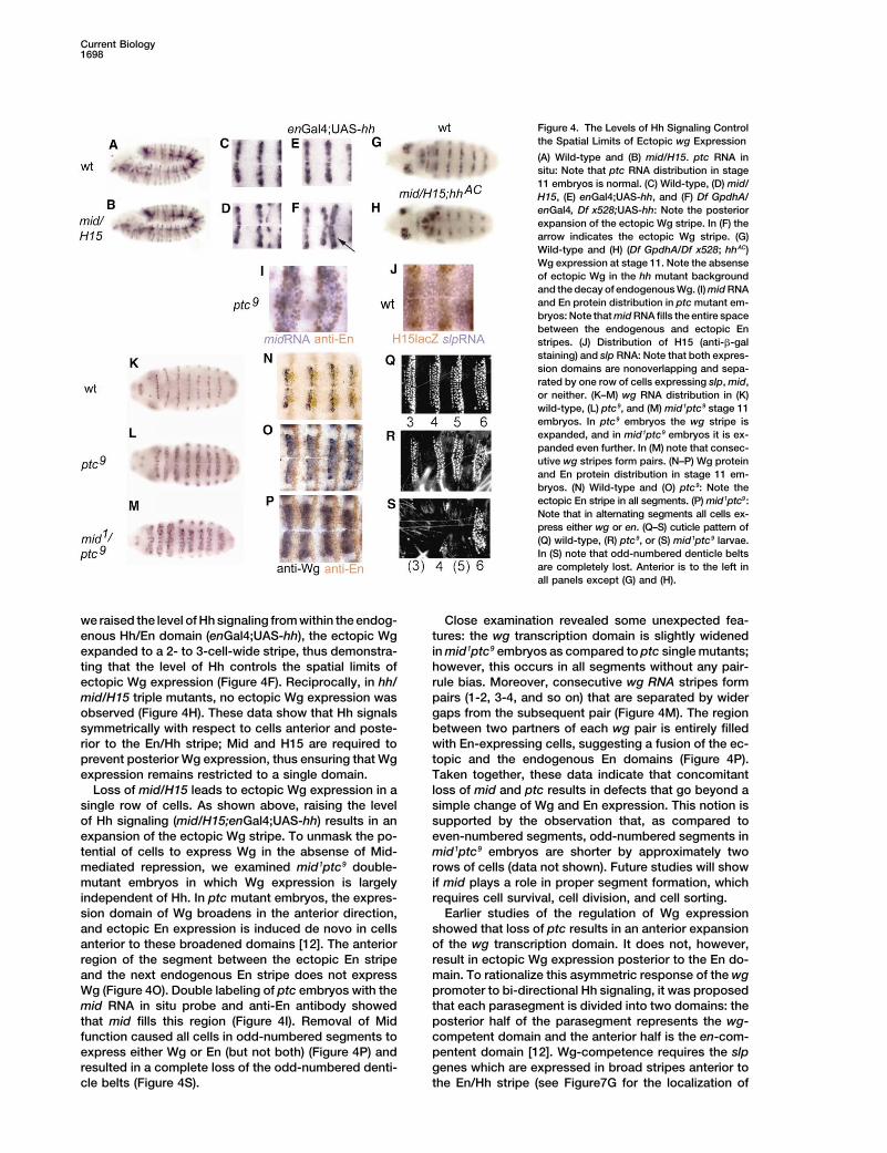
Figure 4. The Levels of Hh Signaling Controlthe Spatial Limits of Ectopic wg Expression
(A) Wild-type and (B) mid/H15. ptc RNA insitu: Note that ptc RNA distribution in stage11 embryos is normal. (C) Wild-type, (D) mid/H15, (E) enGal4;UAS-hh, and (F) Df GpdhA/enGal4, Df x528;UAS-hh: Note the posteriorexpansion of the ectopic Wg stripe. In (F) thearrow indicates the ectopic Wg stripe. (G)Wild-type and (H) (Df GpdhA/Df x528; hhAC)Wg expression at stage 11. Note the absenseof ectopic Wg in the hh mutant backgroundand the decay of endogenous Wg. (I) mid RNAand En protein distribution in ptc mutant em-bryos: Note that mid RNA fills the entire spacebetween the endogenous and ectopic Enstripes. (J) Distribution of H15 (anti-�-galstaining) and slp RNA: Note that both expres-sion domains are nonoverlapping and sepa-rated by one row of cells expressing slp, mid,or neither. (K–M) wg RNA distribution in (K)wild-type, (L) ptc9, and (M) mid1ptc9 stage 11embryos. In ptc9 embryos the wg stripe isexpanded, and in mid1ptc9 embryos it is ex-panded even further. In (M) note that consec-utive wg stripes form pairs. (N–P) Wg proteinand En protein distribution in stage 11 em-bryos. (N) Wild-type and (O) ptc9: Note theectopic En stripe in all segments. (P) mid1ptc9:Note that in alternating segments all cells ex-press either wg or en. (Q–S) cuticle pattern of(Q) wild-type, (R) ptc9, or (S) mid1ptc9 larvae.In (S) note that odd-numbered denticle beltsare completely lost. Anterior is to the left inall panels except (G) and (H).
we raised the level of Hh signaling from within the endog- Close examination revealed some unexpected fea-tures: the wg transcription domain is slightly widenedenous Hh/En domain (enGal4;UAS-hh), the ectopic Wg
expanded to a 2- to 3-cell-wide stripe, thus demonstra- in mid1ptc9 embryos as compared to ptc single mutants;however, this occurs in all segments without any pair-ting that the level of Hh controls the spatial limits of
ectopic Wg expression (Figure 4F). Reciprocally, in hh/ rule bias. Moreover, consecutive wg RNA stripes formpairs (1-2, 3-4, and so on) that are separated by widermid/H15 triple mutants, no ectopic Wg expression was
observed (Figure 4H). These data show that Hh signals gaps from the subsequent pair (Figure 4M). The regionbetween two partners of each wg pair is entirely filledsymmetrically with respect to cells anterior and poste-
rior to the En/Hh stripe; Mid and H15 are required to with En-expressing cells, suggesting a fusion of the ec-topic and the endogenous En domains (Figure 4P).prevent posterior Wg expression, thus ensuring that Wg
expression remains restricted to a single domain. Taken together, these data indicate that concomitantloss of mid and ptc results in defects that go beyond aLoss of mid/H15 leads to ectopic Wg expression in a
single row of cells. As shown above, raising the level simple change of Wg and En expression. This notion issupported by the observation that, as compared toof Hh signaling (mid/H15;enGal4;UAS-hh) results in an
expansion of the ectopic Wg stripe. To unmask the po- even-numbered segments, odd-numbered segments inmid1ptc9 embryos are shorter by approximately twotential of cells to express Wg in the absense of Mid-
mediated repression, we examined mid1ptc9 double- rows of cells (data not shown). Future studies will showif mid plays a role in proper segment formation, whichmutant embryos in which Wg expression is largely
independent of Hh. In ptc mutant embryos, the expres- requires cell survival, cell division, and cell sorting.Earlier studies of the regulation of Wg expressionsion domain of Wg broadens in the anterior direction,
and ectopic En expression is induced de novo in cells showed that loss of ptc results in an anterior expansionof the wg transcription domain. It does not, however,anterior to these broadened domains [12]. The anterior
region of the segment between the ectopic En stripe result in ectopic Wg expression posterior to the En do-main. To rationalize this asymmetric response of the wgand the next endogenous En stripe does not express
Wg (Figure 4O). Double labeling of ptc embryos with the promoter to bi-directional Hh signaling, it was proposedthat each parasegment is divided into two domains: themid RNA in situ probe and anti-En antibody showed
that mid fills this region (Figure 4I). Removal of Mid posterior half of the parasegment represents the wg-competent domain and the anterior half is the en-com-function caused all cells in odd-numbered segments to
express either Wg or En (but not both) (Figure 4P) and pentent domain [12]. Wg-competence requires the slpgenes which are expressed in broad stripes anterior toresulted in a complete loss of the odd-numbered denti-
cle belts (Figure 4S). the En/Hh stripe (see Figure7G for the localization of
T Box Proteins Repress wingless Expression1699
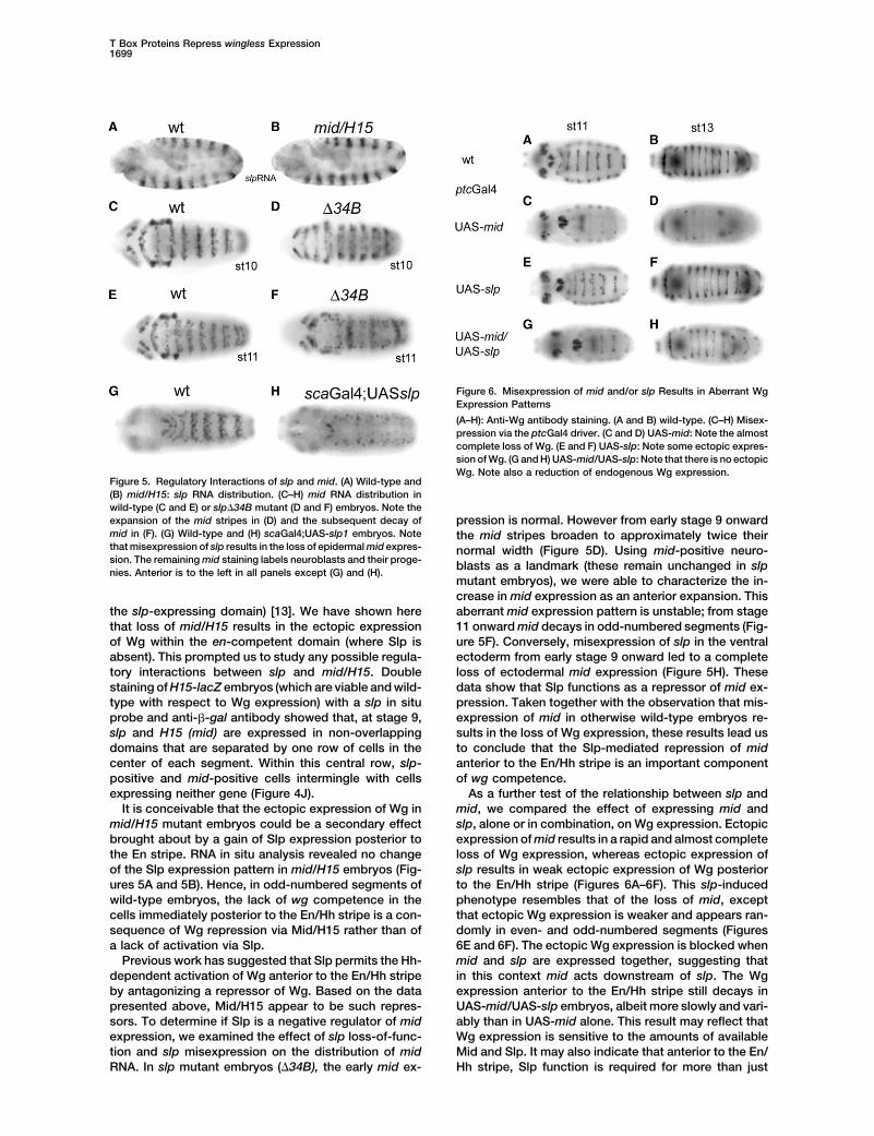
Figure 6. Misexpression of mid and/or slp Results in Aberrant WgExpression Patterns
(A–H): Anti-Wg antibody staining. (A and B) wild-type. (C–H) Misex-pression via the ptcGal4 driver. (C and D) UAS-mid: Note the almostcomplete loss of Wg. (E and F) UAS-slp: Note some ectopic expres-sion of Wg. (G and H) UAS-mid/UAS-slp: Note that there is no ectopicWg. Note also a reduction of endogenous Wg expression.
Figure 5. Regulatory Interactions of slp and mid. (A) Wild-type and(B) mid/H15: slp RNA distribution. (C–H) mid RNA distribution inwild-type (C and E) or slp�34B mutant (D and F) embryos. Note theexpansion of the mid stripes in (D) and the subsequent decay of pression is normal. However from early stage 9 onwardmid in (F). (G) Wild-type and (H) scaGal4;UAS-slp1 embryos. Note the mid stripes broaden to approximately twice theirthat misexpression of slp results in the loss of epidermal mid expres- normal width (Figure 5D). Using mid-positive neuro-sion. The remaining mid staining labels neuroblasts and their proge- blasts as a landmark (these remain unchanged in slpnies. Anterior is to the left in all panels except (G) and (H).
mutant embryos), we were able to characterize the in-crease in mid expression as an anterior expansion. Thisaberrant mid expression pattern is unstable; from stagethe slp-expressing domain) [13]. We have shown here
that loss of mid/H15 results in the ectopic expression 11 onward mid decays in odd-numbered segments (Fig-ure 5F). Conversely, misexpression of slp in the ventralof Wg within the en-competent domain (where Slp is
absent). This prompted us to study any possible regula- ectoderm from early stage 9 onward led to a completeloss of ectodermal mid expression (Figure 5H). Thesetory interactions between slp and mid/H15. Double
staining of H15-lacZ embryos (which are viable and wild- data show that Slp functions as a repressor of mid ex-pression. Taken together with the observation that mis-type with respect to Wg expression) with a slp in situ
probe and anti-�-gal antibody showed that, at stage 9, expression of mid in otherwise wild-type embryos re-sults in the loss of Wg expression, these results lead usslp and H15 (mid) are expressed in non-overlapping
domains that are separated by one row of cells in the to conclude that the Slp-mediated repression of midanterior to the En/Hh stripe is an important componentcenter of each segment. Within this central row, slp-
positive and mid-positive cells intermingle with cells of wg competence.As a further test of the relationship between slp andexpressing neither gene (Figure 4J).
It is conceivable that the ectopic expression of Wg in mid, we compared the effect of expressing mid andslp, alone or in combination, on Wg expression. Ectopicmid/H15 mutant embryos could be a secondary effect
brought about by a gain of Slp expression posterior to expression of mid results in a rapid and almost completeloss of Wg expression, whereas ectopic expression ofthe En stripe. RNA in situ analysis revealed no change
of the Slp expression pattern in mid/H15 embryos (Fig- slp results in weak ectopic expression of Wg posteriorto the En/Hh stripe (Figures 6A–6F). This slp-inducedures 5A and 5B). Hence, in odd-numbered segments of
wild-type embryos, the lack of wg competence in the phenotype resembles that of the loss of mid, exceptthat ectopic Wg expression is weaker and appears ran-cells immediately posterior to the En/Hh stripe is a con-
sequence of Wg repression via Mid/H15 rather than of domly in even- and odd-numbered segments (Figures6E and 6F). The ectopic Wg expression is blocked whena lack of activation via Slp.
Previous work has suggested that Slp permits the Hh- mid and slp are expressed together, suggesting thatin this context mid acts downstream of slp. The Wgdependent activation of Wg anterior to the En/Hh stripe
by antagonizing a repressor of Wg. Based on the data expression anterior to the En/Hh stripe still decays inUAS-mid/UAS-slp embryos, albeit more slowly and vari-presented above, Mid/H15 appear to be such repres-
sors. To determine if Slp is a negative regulator of mid ably than in UAS-mid alone. This result may reflect thatWg expression is sensitive to the amounts of availableexpression, we examined the effect of slp loss-of-func-
tion and slp misexpression on the distribution of mid Mid and Slp. It may also indicate that anterior to the En/Hh stripe, Slp function is required for more than justRNA. In slp mutant embryos (�34B), the early mid ex-
Current Biology1700
repression of mid and may possibly have independentactivating functions. An analysis of slp1,slp2;mid/H15quadruple mutants would be highly helpful in clarifyingthe relationship between slp genes and mid/H15. Unfor-tunately, the generation of such a quadruple mutant bygenetic recombination is impossible because the slpdeletion that removes these genes (�34B) is on a bal-ancer chromosome that precludes recombination.
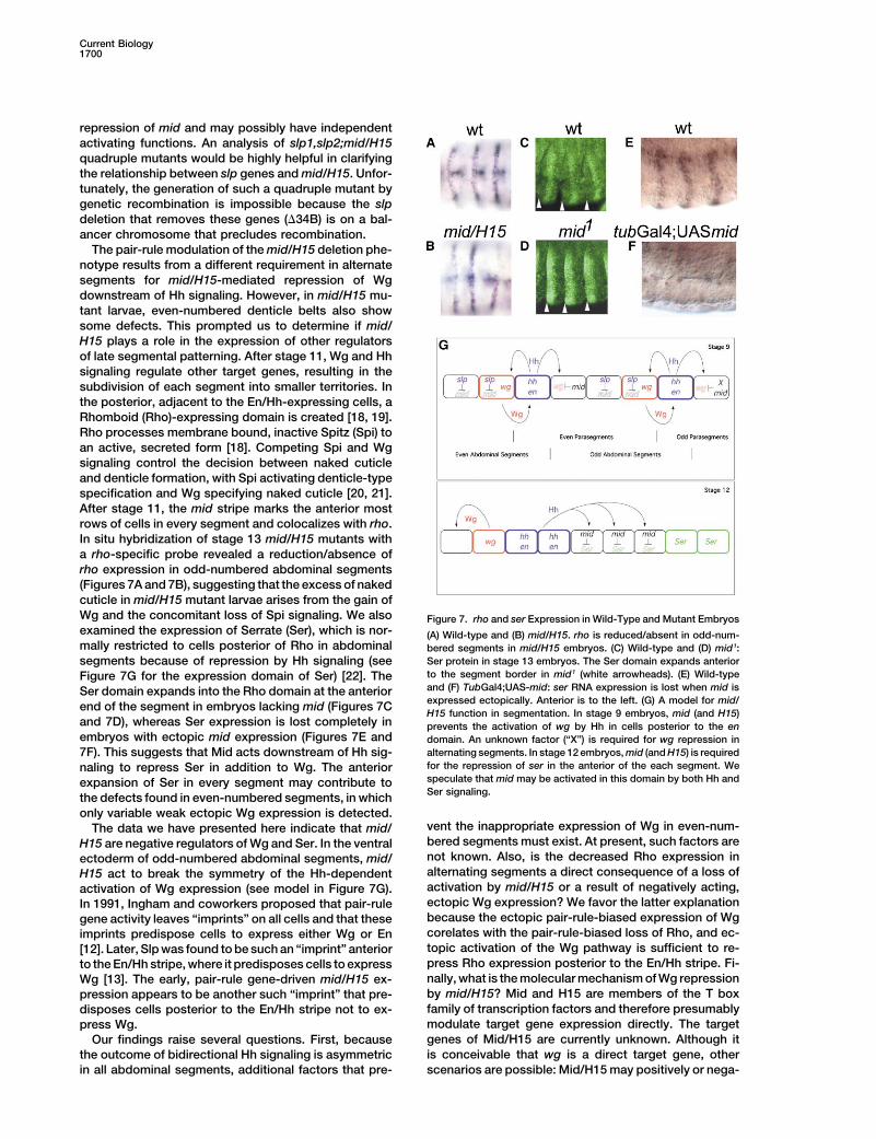
The pair-rule modulation of the mid/H15 deletion phe-notype results from a different requirement in alternatesegments for mid/H15-mediated repression of Wgdownstream of Hh signaling. However, in mid/H15 mu-tant larvae, even-numbered denticle belts also showsome defects. This prompted us to determine if mid/H15 plays a role in the expression of other regulatorsof late segmental patterning. After stage 11, Wg and Hhsignaling regulate other target genes, resulting in thesubdivision of each segment into smaller territories. Inthe posterior, adjacent to the En/Hh-expressing cells, aRhomboid (Rho)-expressing domain is created [18, 19].Rho processes membrane bound, inactive Spitz (Spi) toan active, secreted form [18]. Competing Spi and Wgsignaling control the decision between naked cuticleand denticle formation, with Spi activating denticle-typespecification and Wg specifying naked cuticle [20, 21].After stage 11, the mid stripe marks the anterior mostrows of cells in every segment and colocalizes with rho.In situ hybridization of stage 13 mid/H15 mutants witha rho-specific probe revealed a reduction/absence ofrho expression in odd-numbered abdominal segments(Figures 7A and 7B), suggesting that the excess of nakedcuticle in mid/H15 mutant larvae arises from the gain ofWg and the concomitant loss of Spi signaling. We also Figure 7. rho and ser Expression in Wild-Type and Mutant Embryosexamined the expression of Serrate (Ser), which is nor- (A) Wild-type and (B) mid/H15. rho is reduced/absent in odd-num-mally restricted to cells posterior of Rho in abdominal bered segments in mid/H15 embryos. (C) Wild-type and (D) mid1:
Ser protein in stage 13 embryos. The Ser domain expands anteriorsegments because of repression by Hh signaling (seeto the segment border in mid1 (white arrowheads). (E) Wild-typeFigure 7G for the expression domain of Ser) [22]. Theand (F) TubGal4;UAS-mid: ser RNA expression is lost when mid isSer domain expands into the Rho domain at the anteriorexpressed ectopically. Anterior is to the left. (G) A model for mid/end of the segment in embryos lacking mid (Figures 7CH15 function in segmentation. In stage 9 embryos, mid (and H15)
and 7D), whereas Ser expression is lost completely in prevents the activation of wg by Hh in cells posterior to the enembryos with ectopic mid expression (Figures 7E and domain. An unknown factor (“X”) is required for wg repression in
alternating segments. In stage 12 embryos, mid (and H15) is required7F). This suggests that Mid acts downstream of Hh sig-for the repression of ser in the anterior of the each segment. Wenaling to repress Ser in addition to Wg. The anteriorspeculate that mid may be activated in this domain by both Hh andexpansion of Ser in every segment may contribute toSer signaling.the defects found in even-numbered segments, in which
only variable weak ectopic Wg expression is detected.vent the inappropriate expression of Wg in even-num-The data we have presented here indicate that mid/bered segments must exist. At present, such factors areH15 are negative regulators of Wg and Ser. In the ventralnot known. Also, is the decreased Rho expression inectoderm of odd-numbered abdominal segments, mid/alternating segments a direct consequence of a loss ofH15 act to break the symmetry of the Hh-dependentactivation by mid/H15 or a result of negatively acting,activation of Wg expression (see model in Figure 7G).ectopic Wg expression? We favor the latter explanationIn 1991, Ingham and coworkers proposed that pair-rulebecause the ectopic pair-rule-biased expression of Wggene activity leaves “imprints” on all cells and that thesecorelates with the pair-rule-biased loss of Rho, and ec-imprints predispose cells to express either Wg or Entopic activation of the Wg pathway is sufficient to re-[12]. Later, Slp was found to be such an “imprint” anteriorpress Rho expression posterior to the En/Hh stripe. Fi-to the En/Hh stripe, where it predisposes cells to expressnally, what is the molecular mechanism of Wg repressionWg [13]. The early, pair-rule gene-driven mid/H15 ex-by mid/H15? Mid and H15 are members of the T boxpression appears to be another such “imprint” that pre-family of transcription factors and therefore presumablydisposes cells posterior to the En/Hh stripe not to ex-modulate target gene expression directly. The targetpress Wg.genes of Mid/H15 are currently unknown. Although itOur findings raise several questions. First, becauseis conceivable that wg is a direct target gene, otherthe outcome of bidirectional Hh signaling is asymmetric
in all abdominal segments, additional factors that pre- scenarios are possible: Mid/H15 may positively or nega-
T Box Proteins Repress wingless Expression1701
Cuticles of first-instar larvae were prepared essentially as de-tively regulate the expression of unidentified genes andscribed [7] except that the vitelline membranes were removed bythereby modulate Wg or Hh pathway activities, and hy-vigorous agitation in methanol/heptane (1:1). The larvae were thenperactivity of either pathway could produce an ectopicplaced in a microtube and incubated overnight at 55�C in a mixture
stripe of Wg expression. It is noteworthy that a different of Hoyer’s medium/lactic acid (1:1). Subsequently, the larvae weregroup of T box genes, the dorsocross genes, has been mounted on slides and viewed with dark-field microscopy.identified as a negative regulator of Wg expression in the
Acknowledgmentsdorsolateral epidermis [23]. However, the Dorsocrosstarget genes are also unknown. Further studies are re-
We thank A. Gould, M. Frasch, A. Simmonds, K. Irvine, X.H. Yang,quired to elucidate the mechanisms by which T boxand the Bloomington Stock Center for providing materials; P.W.
proteins negatively regulate Wg expression. Ingham, S. DiNardo, and P. Overton for helpful comments on themanuscript; and P. Overton for help in preparing Figure 1. W.J.B.
Experimental Procedures is supported by the Canadian Institutes of Health Research and theAlberta Heritage Foundation for Medical Research.
Fly StrainsThe alleles mid1 and mid2 were obtained from the Bloomington stock Received: March 12, 2004center [14]. midGA174 was isolated from a collection of EMS-induced Revised: August 6, 2004mutations [15]. The deficiencies H15x4 and Df(2L)x528 were gener- Accepted: August 6, 2004ated by X-irradiation, and the breakpoints were determined by PCR Published: October 5, 2004analysis. Df-GpdhA (breakpoints 25D7; 26A2-5) was obtained fromthe Bloomington Stock Center, and the breakpoints were deter- Referencesmined by PCR analysis. ptc9, wgCX4, hhAC and slp�34B are null alleles(see Flybase: http://www.flybase.org). All mutant alleles were bal- 1. St Johnston, D., and Nusslein-Volhard, C. (1992). The origin ofanced over CyOftzlacZ or CyOwglacZ to facilitate the identification of pattern and polarity in the Drosophila embryo. Cell 68, 201–219.homozygotes. enGal4 (a gift from A. Gould; originally generated by 2. Lee, J.J., von Kessler, D.P., Parks, S., and Beachy, P.A. (1992).A. Brand), scaGal4 (a gift from X. Yang; scaGal4 drives expression Secretion and localized transcription suggest a role in positionalin the ventral ectoderm from early stage 9 onward), ptcGal4, Tub- signaling for products of the segmentation gene hedgehog. CellGal4, and prdGal4 (Bloomington Stock Center) were used to drive 71, 33–50.the expression of transgenes. UAS-hh.1 and UAS-slp1 (on the third 3. Mohler, J., and Vani, K. (1992). Molecular organization and em-chromosome) were a gift from M. Frasch; UAS-slp1 (on the second bryonic expression of the hedgehog gene involved in cell-cellchromosome) was a gift from M. Leptin. UAS-wg was a gift from A. communication in segmental patterning of Drosophila. Develop-Simmonds. A transgenic stock carrying UAS-CG6634 (UAS-mid.1.4; ment 115, 957–971.insertion on the third chromosome) was generated from the full- 4. Bejsovec, A., and Martinez Arias, A. (1991). Roles of winglesslength CG6634 cDNA (derived from RE27439) subcloned into the in patterning the larval epidermis of Drosophila. DevelopmentpUAST vector. 113, 471–485.
All strains were raised at 22�C on standard corn meal agar 5. Martizenz Arias, A., Baker, N.E., and Ingham, P.W. (1988). Rolemedium. of segment polarity genes in the definition and maintenance of
cell states in the Drosophila embryo. Development 103,Cloning of mid 157–170.mid1, mid2, and midGA174 were balanced over CyOActGFP to facilitate 6. Baker, N.E. (1988). Localization of transcripts from the winglessthe identification of homozygous mutant embryos. Genomic DNA gene in whole Drosophila embryos. Development 103, 289–298.was isolated according to standard protocols and analyzed by auto- 7. DiNardo, S., Heemskerk, J., Dougan, S., and O’Farrell, P.H.mated sequencing. mid1 was found to have a C-to-T transition at (1994). The making of a maggot: patterning the Drosophila em-position 659, mid2 to have a C-to-T transition at position 1758, and bryonic epidermis. Curr. Opin. Genet. Dev. 4, 529–534.midGA174 to have a C-to-T transition at position 680; numbering is 8. Hatini, V., and DiNardo, S. (2001). Divide and conquer: patternaccording to the cDNA clone RE27439. All three base pair substitu- formation in Drosophila embryonic epidermis. Trends Genet.tions result in nonsense codons. 17, 574–579.
9. Sanson, B. (2001). Generating patterns from fields of cells. Ex-amples from Drosophila segmentation. EMBO Rep. 2, 1083–Phenotypic Analysis
Immunohistochemistry. Embryos were collected, fixed, and immu- 1088.10. Hidalgo, A., and Ingham, P. (1990). Cell patterning in the Dro-nostained as previously described [24]. Primary antibodies were
polyclonal rat anti-Serrate (a gift from K. Irvine) and monoclonal sophila segment: spatial regulation of the segment polarity genepatched. Development 110, 291–301.mouse anti-Engrailed/Invected (4D9) and mouse anti-Wingless
(4D4). The respective hybridomas, developed by C. Goodman (4D9) 11. Forbes, A.J., Nakano, Y., Taylor, A.M., and Ingham, P.W. (1993).Genetic analysis of hedgehog signalling in the Drosophila em-and S.M. Cohen (4D4), were obtained from the Developmental Stud-
ies Hybridoma Bank developed under the auspices of the National bryo. Dev. Suppl., 115–124.12. Ingham, P.W., Taylor, A.M., and Nakano, Y. (1991). Role of theInstitute of Child Health and Human Development (NICHD) and main-
tained by The University of Iowa, Department of Biological Sciences, Drosophila patched gene in positional signalling. Nature 353,184–187.Iowa City, IA 52242. Histochemical detection was performed with
Jackson Immunoresearch HRP-conjugated secondary antibodies 13. Cadigan, K.M., Grossniklaus, U., and Gehring, W.J. (1994). Lo-calized expression of sloppy paired protein maintains the polar-and visualized by the glucose-oxidase-DAB-nickel method as pre-
viously described [24]. ity of Drosophila parasegments. Genes Dev. 8, 899–913.14. Nusslein-Volhard, C., Wieschaus, E., and Kluding, H. (1984).RNA in situ hybridization was carried out as described previously
[25]. Fluorescent RNA in situ hybridization was performed with the Mutations affecting the pattern of the larval cuticle in Drosophilamelanogaster. I. Zygotic loci on the second chromosome. RouxTSA Fluorescein System from PerkinElmer according to the manu-
facturer’s instruction. For generation of a mid-specific RNA in situ Arch. Dev. Biol. 193, 267–282.15. Seeger, M., Tear, G., Ferres-Marco, D., and Goodman, C.S.probe, 1.1 kb of 5� cDNA sequence (derived from RE27439) was
subcloned into Bluescript vector and subsequently transcribed (1993). Mutations affecting growth cone guidance in Drosophila:genes necessary for guidance toward or away from the midline.in vitro with T7 RNA polymerase. A construct for the generation of
a rho-specific RNA probe was a gift from A. Gould. A slp-specific Neuron 10, 409–426.16. Griffin, K.J., Stoller, J., Gibson, M., Chen, S., Yelon, D., Stainier,RNA in situ probe was generated by in vitro transcription of full-
length slp cDNA with SP6 polymerase. D.Y., and Kimelman, D. (2000). A conserved role for H15-related
Current Biology1702
T-box transcription factors in zebrafish and Drosophila heartformation. Dev. Biol. 218, 235–247.
17. Sanson, B., Alexandre, C., Fascetti, N., and Vincent, J.P. (1999).Engrailed and Hedgehog make the range of Wingless asymmet-ric in Drosophila embryos. Cell 98, 207–216.
18. Schweitzer, R., Shaharabany, M., Seger, R., and Shilo, B.Z.(1995). Secreted Spitz triggers the DER signaling pathway andis a limiting component in embryonic ventral ectoderm determi-nation. Genes Dev. 9, 1518–1529.
19. Alexandre, C., Lecourtois, M., and Vincent, J. (1999). Winglessand Hedgehog pattern Drosophila denticle belts by regulatingthe production of short-range signals. Development 126, 5689–5698.
20. Szuts, D., Freeman, M., and Bienz, M. (1997). Antagonism be-tween EGFR and Wingless signalling in the larval cuticle ofDrosophila. Development 124, 3209–3219.
21. Payre, F., Vincent, A., and Carreno, S. (1999). ovo/svb integratesWingless and DER pathways to control epidermis differentia-tion. Nature 400, 271–275.
22. Wiellette, E.L., and McGinnis, W. (1999). Hox genes differentiallyregulate Serrate to generate segment-specific structures. De-velopment 126, 1985–1995.
23. Reim, I., Lee, H.H., and Frasch, M. (2003). The T-box-encodingDorsocross genes function in amnioserosa development andthe patterning of the dorsolateral germ band downstream ofDpp. Development 130, 3187–3204.
24. Yang, X., Bahri, S., Klein, T., and Chia, W. (1997). Klumpfuss,a putative Drosophila zinc finger transcription factor, acts todifferentiate between the identities of two secondary precursorcells within one neuroblast lineage. Genes Dev. 11, 1396–1408.
25. Tautz, D., and Pfeifle, C. (1989). A non-radioactive in situ hybrid-ization method for the localization of specific RNAs in Drosoph-ila embryos reveals translational control of the segmentationgene hunchback. Chromosoma 98, 81–85.
![Cell Type-Specific Responses to Wingless, Hedgehog and ... · specifically recognizes Futsch/22C10(Fig.1E),aneuronalMAP12B-like protein[33],and did notexpress GFPdrivenbybreathless-Gal4[34,35].This](https://img.pdfslide.us/doc/110x75/5ec80f497dedbb0fcc771de0/cell-type-specific-responses-to-wingless-hedgehog-and-specifically-recognizes.jpg)