Embed Size (px)
Citation preview

Drosophila P-element transposase is anovel site-specific endonucleaseEileen L. Beall and Donald C. Rio1
Department of Molecular and Cell Biology, University of California, Berkeley, California 94720-3204 USA
We developed in vitro assays to study the first step of the P-element transposition reaction: donor DNAcleavage. We found that P-element transposase required both 5* and 3* P-element termini for efficient DNAcleavage to occur, suggesting that a synaptic complex forms prior to cleavage. Transposase made a staggeredcleavage at the P-element termini that is novel for all known site-specific endonucleases: the 3* cleavage siteis at the end of the P-element, whereas the 5* cleavage site is 17 bp within the P-element 31-bp invertedrepeats. The P-element termini were protected from exonucleolytic degradation following the cleavagereaction, suggesting that a stable protein complex remains bound to the element termini after cleavage. Thesedata are consistent with a cut-and-paste mechanism for P-element transposition and may explain why Pelements predominantly excise imprecisely in vivo.
[Key Words: P transposon; transposition; cleavage site; recombinase]
Received April 23, 1997; revised version accepted June 27, 1997.
Transposons are mobile genetic elements that are pres-ent in many organisms. In general, the transposable ele-ment encodes a recombinase that mediates the DNAcleavage and strand transfer reactions during transposi-tion. The recombinase can be a single polypeptide, likethe transposase proteins encoded by the Tc elements,phage Mu, Tn10, and P elements, or a combination ofpolypeptides, like with Tn7. The type of DNA cleavagemade by the transposase protein, however, is common toall transposition reactions. All transposases cleave at theend of the transposon DNA to expose a 38-hydroxylgroup that is covalently joined to a new target site. Themechanism of transposition is determined by whethercleavage occurs at the 58 ends. If the 58 end of the ele-ment is cleaved to generate an excised transposon inter-mediate, the element transposes by a cut-and-pastemechanism. Tn7 (Bainton et al. 1991), Tn10 (Benjaminand Kleckner 1992), the Tc elements (van Luenen et al.1994; Vos et al. 1996), and the P element (Kaufman andRio 1992) all transpose by such a mechanism. The natureof the nontransferred strand cleavage, however, is vari-able and occurs either 3 bp outside [for Tn7 (Bainton etal. 1991; Gary et al. 1996)], 2 bp inside [for the Tc ele-ments (van Luenen et al. 1994; Vos et al. 1996)], or at thetransposon termini [for Tn10 (Benjamin and Kleckner1992)]. If the 58 end of the element is not cleaved, theelement transposes by a replicative transposition mecha-nism in which the transposon remains attached to boththe donor site and the target site. The intermediate isreplicated by host replication proteins to produce two
copies of the element in a structure called a cointegrate.The bacteriophage Mu can transpose by this type ofmechanism (Craigie and Mizuuchi 1985; Mizuuchi1992a). Analogous to Mu, retroviral integration, like HIVintegration, does not require processing at the 58 end ofthe reverse-transcribed genome by the integrase proteinprior to insertion into the host genome (Engelman et al.1991). A staggered target-site cleavage is made by mosttransposases and integrases, and DNA repair of the gapsthat flank the newly inserted element generates thecharacteristic target-site duplications present after inser-tion.
The P transposable element of Drosophila melanogas-ter is one of the best characterized eukaryotic trans-posons. Full-length P elements are 2.9 kb in length andencode an 87-kD transposase protein. Transposase syn-thesis is restricted to the germ line by a regulated, tissue-specific pre-mRNA splicing mechanism (Laski et al.1986). P elements transpose by a cut-and-paste mecha-nism and create an 8-bp target site duplication upon in-sertion (O’Hare and Rubin 1983; Engels et al. 1990; Kauf-man and Rio 1992). The P-element transposase is uniquein that GTP is required as a cofactor, in addition to mag-nesium, for activity (Kaufman and Rio 1992; Mul andRio 1997). P-element transposition requires ∼150 bp ofsequence at each end of the P element. These sequencesinclude 31-bp terminal inverted repeats, internal trans-posase-binding sites, and internal 11-bp inverted repeats(O’Hare and Rubin 1983; Kaufman et al. 1989; Mullins etal. 1989). The terminal 31-bp inverted repeats are notbound by the transposase protein (Kaufman et al. 1989)but instead are bound site-specifically by a Drosophila-encoded protein, the inverted repeat binding protein
1Corresponding author.E-MAIL don [email protected]; FAX (510) 642-6062.
GENES & DEVELOPMENT 11:2137–2151 © 1997 by Cold Spring Harbor Laboratory Press ISSN 0890-9369/97 $5.00 2137
Cold Spring Harbor Laboratory Press on April 7, 2018 - Published by genesdev.cshlp.orgDownloaded from
(IRBP) (Rio and Rubin 1988). This finding was surprisingbecause most transposase proteins bind within the in-verted repeat regions to sequences at or near the terminalnucleotides of the transposable element. For instance,the Tn10 transposase binds to sequences within the IS10terminal 23-bp inverted repeats, with the most criticalcontacts made at 6–13 bp (Kleckner et al. 1996). Simi-larly, the Tc1 and Tc3 transposases recognize sequenceswithin 20–25 bp of either the Tc1 or Tc3 transposonterminal inverted repeats (van Luenen et al. 1993; Vosand Plasterk 1994). The sites to which transposase bindsat the P-element termini are of unequal distance fromthe sites of cleavage (either 40 or 52 bp away) and mayaccount for the inability of a P element with two 58 endsto transpose in vivo (Mullins et al. 1989). During themechanistically similar V(D)J recombination reaction(van Gent et al. 1996b), the Rag-1 and Rag-2 proteins alsobind to sites of unequal distance from the site of cleavage(either 19 or 30 bp away) (Difilippantonio et al. 1996;Spanopoulou et al. 1996; Hiom and Gellert 1997).
For many transposable elements, such as Tn10 (Hani-ford et al. 1991; Sakai et al. 1995), Tn7 (Bainton et al.1993; Sarnovsky et al. 1996), and Mu (Craigie and Mi-zuuchi 1987; Surette et al. 1987), and during V(D)J rear-rangement (Eastman et al. 1996; van Gent et al. 1996a;Steen et al. 1997), the first step of the rearrangementreaction, DNA cleavage, requires synapsis of the twoends to form a stable nucleoprotein complex prior tocleavage. The subsequent breakage and joining reactionsoccur in the context of this synaptic complex or trans-pososome. For instance, during Mu transposition, a dis-tinct series of stable nucleoprotein complexes have beenobserved for each step of the transposition reaction (Mi-zuuchi 1992b; Lavoie and Chaconas 1996). The earliestdetectable complex, or LER complex, contains a tran-sient species with the Mu left and right ends, and theinternal enhancer element (Watson and Chaconas 1996).The type 0, or stable synaptic complex (SSC), containsthe two Mu ends bound by a tetramer of transposase.The type 1, or cleaved donor complex (CDC), containsboth transposase and the cleaved 38 ends of the MuDNA. The last, or strand transfer complex (STC), con-tains the Mu genome inserted into a new target site.Completion of the reaction involves disassembly of thetransposase tetramer from the DNA by a host encodedprotein, ClpX, and replication across the Mu DNA togenerate a second copy of the Mu genome (Levchenko etal. 1995; Nakai and Kruklitis 1995; Kruklitis et al. 1996).Similarly, during V(D)J recombination, a stable nucleo-protein complex remains bound to the cleaved DNA in-termediates following Rag-1/Rag-2 mediated cleavage(Zhu et al. 1996; Agrawal and Schatz 1997). Formation ofa stable synaptic complex prior to, and after cleavage, isa way to ensure reaction fidelity through a properly po-sitioned recombinase.
To characterize the P-element transposition reactionin more detail, we developed in vitro cleavage assayswith transposase purified from Drosophila cell culture.Here, we show that P-element transposase requires both58 and 38 P-element termini for efficient DNA cleavage
to occur, suggesting that a synaptic complex forms onthe P-element termini prior to cleavage. We mapped thecleavage sites made by transposase and show that trans-posase makes a staggered cleavage at the P-element ter-mini that is novel for all known site-specific endonucle-ases. Similar to all transposable elements, the 38 cleavagesite occurs at the end of the P element. However, unlikeany other known transposable element, the 58 cleavagesite occurs 17 bp within the P-element 31-bp invertedrepeats, directly adjacent to the IRBP-binding site. The17-nucleotide, 38 extensions left behind at the donor siteprovide an explanation for the imprecise P-element ex-cisions observed in vivo. Finally, we show that the P-element termini are protected from exonucleolytic deg-radation following the cleavage reaction, which suggeststhat a stable protein complex remains bound to the P-element termini following cleavage.
Results
Transposase only cleaves substrates containing bothP-element termini
To better understand the mechanism of P-element trans-position, in vitro assays were developed to study P-ele-ment transposase-mediated donor DNA cleavage activ-ity. P-element transposase was partially purified from astable cell line expressing transposase under the controlof the metallothionein promoter (Mul and Rio 1997).This cell line differs from the previously reported trans-posase-producing cell line (Kaufman et al. 1989) in that25% of the transposase amino terminus was chemicallyresynthesized to alter codon usage to the most fre-quently used codons in Drosophila (Lee et al. 1996). Thepartially purified transposase fraction (referred to asH0.1FT) was highly active for P-element transpositionby use of a genetic-based in vitro assay (Kaufman and Rio1992).
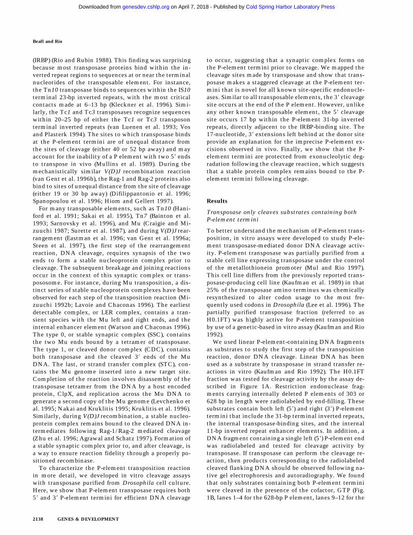
We used linear P-element-containing DNA fragmentsas substrates to study the first step of the transpositionreaction, donor DNA cleavage. Linear DNA has beenused as a substrate by transposase in strand transfer re-actions in vitro (Kaufman and Rio 1992). The H0.1FTfraction was tested for cleavage activity by the assay de-scribed in Figure 1A. Restriction endonuclease frag-ments carrying internally deleted P elements of 303 or628 bp in length were radiolabeled by end-filling. Thesesubstrates contain both left (58) and right (38) P-elementtermini that include the 31-bp terminal inverted repeats,the internal transposase-binding sites, and the internal11-bp inverted repeat enhancer elements. In addition, aDNA fragment containing a single left (58) P-element endwas radiolabeled and tested for cleavage activity bytransposase. If transposase can perform the cleavage re-action, then products corresponding to the radiolabeledcleaved flanking DNA should be observed following na-tive gel electrophoresis and autoradiography. We foundthat only substrates containing both P-element terminiwere cleaved in the presence of the cofactor, GTP (Fig.1B, lanes 1–4 for the 628-bp P element, lanes 9–12 for the
Beall and Rio
2138 GENES & DEVELOPMENT
Cold Spring Harbor Laboratory Press on April 7, 2018 - Published by genesdev.cshlp.orgDownloaded from

Figure 1. Transposase requires both 58 and 38 P-element termini during cleavage. (A) Schematic diagram of the assay used to studythe first step in the P-element transposition reaction: donor DNA cleavage. Restriction endonuclease fragments carrying internallydeleted P elements of 303 or 628 bp in length (as indicated by arrows), along with varying lengths of flanking DNA (hatched and shadedboxes), were radiolabeled at the 38 end as indicated by an asterisk. These substrates contain both P-element termini that include the31-bp terminal inverted repeats, the internal transposase binding sites, and the internal 11-bp inverted repeat enhancer elements. ADNA fragment containing a single 58 P-element end was also radiolabeled and tested as a cleavage substrate for transposase. Thepredicted sizes for each cleavage product are as indicated. (B) Partially purified transposase (H0.1FT) was tested for double-strandedDNA cleavage activity as diagrammed in A. Reactions were performed at 27°C for 2 hr, and products analyzed by native PAGE andautoradiography. Shown is an autoradiograph from an experiment in which three different substrates were tested for cleavage activity:the 628-bp P element-containing fragment (lanes 1–4), the 175-bp single left (58) P-element end-containing fragment (lanes 5–8), andthe 303-bp P-element-containing fragment (lanes 9–12). For each substrate, two different amounts of the H0.1FT were tested foractivity (either ∼2 µg, or 4 µg total protein, as indicated by + or ++). GTP-dependent cleavage products are indicated by arrows. (M)Radiolabeled pBR322 MspI molecular weight markers. (C) Schematic diagram of the assay used to test whether P-element transposaserequires both 58 and 38 P-element termini for DNA cleavage to occur. Plasmid substrates containing the 303-bp P element (P 5838, left),the left end only (P58, middle), or the right end only (P38, right) are as diagrammed. The predicted sizes for each product followingtransposase-mediated cleavage and restricion endonuclease digestion with the indicated enzymes are outlined at the bottom. (D)Partially purified transposase (H0.1FT) was tested for cleavage activity as diagrammed in Fig. 1C. Reactions were performed at 27°Cfor 2 hr, and products analyzed by DNA blot hybridization with a radiolabeled P-element probe following native PAGE. Shown is anautoradiograph from an experiment in which substrates containing both P-element termini (P 5838, lanes 1,2; only the 251-bp AccIfragment is shown), each terminus alone (P 58, lanes 3,4; P 38, lanes 5,6), or a mixture containing equimolar amounts of both P 58 andP 38 (lanes 7 and 8) were tested for cleavage by transposase. For each substrate, ∼2 µg of the H0.1FT was tested for activity. GTP-dependent cleavage products are indicated by arrows. (M) radiolabeled pBR322 MspI molecular weight markers.
GENES & DEVELOPMENT 2139
Cold Spring Harbor Laboratory Press on April 7, 2018 - Published by genesdev.cshlp.orgDownloaded from

303-bp P element). The single left (58) P element end-containing fragment was not used as a substrate (Fig. 1B,lanes 5–8). These data suggest that during P-elementtransposition, assembly of a cleavage complex requiresboth P-element termini and are consistent with the find-ing that substrates containing two left (58) P-elementends are not active for transposition in vivo (Mullins etal. 1989).
To further examine the requirements for DNA cleav-age, plasmid substrates containing both P-element ter-mini on one plasmid, each terminus alone, or mixturesof each terminus on separate plasmids were tested in thecleavage assay as diagrammed in Figure 1C. If trans-posase requires synapsis of both a left (58) and a right (38)end for cleavage, then products should only be detectedwhen substrates containing both ends are present in thereaction. DNA blot hybridization analysis of restrictionendonuclease-cleaved reaction products is shown in Fig-ure 1D. We found that efficient DNA cleavage only oc-curred in the presence of GTP and when both a left (58)and right (38) end were present in the reaction (Fig. 1D, cf.lanes 4 and 6 with lanes 2 and 8). Cleavage at the right(38) end-containing substrate (lane 6) is approximatelyfourfold reduced than when both ends are present. Thefinding that a mixture containing a single left (58) end-containing plasmid and a single right (38) end-containingplasmid was active for cleavage by transposase (Fig. 1D,lane 8), but each single end-containing plasmid substratealone was inefficiently cleaved by transposase (Fig. 1D,lanes 4 and 6), demonstrates that transposase can cleaveP-element end-containing substrates both in cis (Fig. 1D,lane 2) and in trans (Fig. 1D, lane 8). These data areconsistent with synapsis of both a left (58) and right (38)P-element end during the cleavage step of the P-elementtransposition reaction. The presence of both ends on thesame DNA molecule, however, is not a prerequisite forcleavage. The two P-element ends differ in the spacingbetween the transposase-binding site and the 31-bp in-verted repeat. A requirement for synapsis of both a left(58) and right (38) end for efficient cleavage to occur sug-gests that the nature of these spacing differences may becritical for synapsis (see Discussion). A similar observa-tion has been made for cleavage substrates during V(D)Jrecombination (Eastman et al. 1996; van Gent et al.1996a; Steen et al. 1997; van Gent et al. 1997).
Transposase makes a 17-bp staggered cleavage at eachP-element end
To determine the exact position of the double-strandDNA breaks made by transposase, large scale in vitrocleavage reactions were performed with both the 628-and 303-bp P-element-containing plasmid substrates andthe H0.1FT transposase fraction. The reaction productscorresponding to the excised element and cleaved plas-mid vector were isolated from gel slices following aga-rose gel electrophoresis. A PCR-based primer extensionanalysis of the purified cleavage products is shown inFigure 2. Primer extension experiments were used to de-
termine the sites of cleavage for both the Tn7 trans-posase (Bainton et al. 1991) and the Tc1 transposase (Voset al. 1996). We found that the 38 cleavage site for eitherthe left (58) end (Fig. 2A) or the right (38) end (Fig. 2B) wasmade precisely at the end of the transposon. This findingis consistent with the 38 cleavage site of all known trans-posases and previous in vitro data by use of dideoxy-substituted substrate DNAs during P-element transposi-tion (Kaufman and Rio 1992). Primer extension productsare observed near the P-element termini in the laneslacking transposase (Fig. 2, A, lane 2, and B, lane 4). Webelieve that these products are the result of secondarystructures in the DNA that cause Taq polymerase toprematurely terminate extension, because the sequenc-ing ladder around the terminal P-element nucleotidehas the same background bands (Fig. 2A,B). In addition,the same strong stop near the terminal P-elementnucleotide can be seen in the reactions that examine the58 cleavage sites (Fig. 2, C, lanes 2 and 4, and D, lanes 6and 8).
Surprisingly, we found that the 58 cleavage site for ei-ther the left (58) end (Fig. 2C), or the right (38) end (Fig.2D) occurs 17 bp within the P-element 31-bp invertedrepeat sequence, directly adjacent to the IRBP-bindingsite (Rio and Rubin 1988). The finding that transposasemakes a staggered cleavage that generates a 17-nucleo-tide, 38 extension is novel. In addition, we found that atthe left (58) end only for both the 303-bp P element (E.L.Beall and D.C. Rio, unpubl.) and the 628-bp P element(Fig. 2C, lane 3), there is a clustering of primer extensionproducts surrounding the proposed 17-bp cleavage site. Itis possible that P-element transposase has reduced fidel-ity at the left (58) end. The Tc1 transposase displays simi-lar endonuclease activity near the site of cleavage (Vosand Plasterk 1994).
Because of the unusual nature of the 17-bp staggeredcleavage made by P-element transposase, we wanted toconfirm the primer extension results by an independentmethod. Cleavage reactions were performed with theH0.1FT transposase fraction and both the 303-bp and628-bp P-element-containing plasmid substrates. Reac-tion products were analyzed by ligation-mediated PCR(LMPCR) as outlined in Figure 3A. An analogous assayhas been used to identify the sites of cleavage made byRag-1 and Rag-2 during V(D)J rearrangement (Roth et al.1993; Schlissel et al. 1993). Reaction products were madeflush with T4 DNA polymerase treatment prior to liga-tion of the annealed linkers and PCR by use of the primerpairs indicated in Figure 3A. LMPCR products were iso-lated from gel slices following agarose gel electrophore-sis, inserted into a plasmid vector, and sequenced. Wefound that the sequence from 27 of 30 independent iso-lates derived from either the 58 cleavage site at the left(58) end or right (38) end of the element, for both the303-bp and 628-bp elements, began at nucleotide 18 ofthe P-element 31-bp inverted repeat (E.L. Beall and D.C.Rio, unpubl.). Two of the remaining DNAs began atnucleotide 19 and the third began at nucleotide 3 of theP-element 31-bp inverted repeat. These DNAs may havebeen derived from either exonucleolytic degradation of
Beall and Rio
2140 GENES & DEVELOPMENT
Cold Spring Harbor Laboratory Press on April 7, 2018 - Published by genesdev.cshlp.orgDownloaded from

the terminus after cleavage, or from cleavages made byP-element transposase at another site within the 31-bpinverted repeat, similar to the alternative cleavage sitesobserved by the primer extension analysis (Fig. 2C, lane3). The finding that 27/30 independent LMPCR productsbegan at nucleotide 18 of the 31-bp inverted repeat con-firms that the transposase makes a 17-bp staggered cleav-age at the P-element termini during transposition.
The P-element termini are protected after cleavage
Efficient cleavage by Rag1 and Rag2 at the recombina-tion signal sequences (RSS) requires a 12/23 RSS pair andoccurs in a coordinated fashion during V(D)J rearrange-ment (Eastman et al. 1996; van Gent et al. 1996a, 1997;Steen et al. 1997). These data suggest that a synapticcomplex forms at the RSS pair that allows for proper
Figure 2. Transposase makes 17-bp stag-gered cleavages at the P-element termini.Large-scale cleavage reactions were per-formed with partially purified transposaseand plasmid substrates containing eitherthe 628-bp or 303-bp P elements. Reactionproducts corresponding to both the cleavedplasmid vector and the excised elementwere isolated from agarose gel slices. APCR-based primer extension analysis wasperformed on each product in order to de-termine the transposase cleavage sites.Shown are autoradiographs of sequencinggels that display primer extension productsfrom reactions in which nontemplated ad-dition of a single nucleotide by Taq poly-merase occurs (as indicated by +1 in eachpanel). The authentic cleavage sites are in-dicated by C. Sequencing reactions (ACGT)are shown as markers. The relevant se-quence is indicated, with P-element-de-rived sequences boxed and numbered fromthe terminal P-element nucleotide, and thecleavage sites indicated by arrows. Sche-matic diagrams of the direction for primerextension and the cleavage positions foreach strand are indicated below each panel.(A,B) Extension products to determine the38 cleavage site at the left (58) P-elementend (A) or the 38 cleavage site at the right(38) P-element end (B). Products were ana-lyzed for the 303-bp P-element-derivedcleavage product only. (Odd-numberedlanes) + transposase; (even-numbered lanes)− transposase. Extension yields a productthat terminates exactly at the 38 end of theP-element inverted repeat. (C,D) Extensionproducts to determine the 58 cleavage sitesat the left (58) P-element end (C) or right (38)P-element end (D). (Lanes 1,2,5,6) Productsderived from the 303-bp P element. (Lanes3,4,7,8) Products derived from the 628-bp Pelement. (Odd-numbered lanes) + trans-posase, (even-numbered lanes) − trans-posase. Extension yields a product that ter-minates at nucleotide 18 of the P-elementinverted repeat.
DNA cleavages by P-element transposase
GENES & DEVELOPMENT 2141
Cold Spring Harbor Laboratory Press on April 7, 2018 - Published by genesdev.cshlp.orgDownloaded from

double-strand DNA break formation during the cleavagestep of the reaction. Following cleavage by Rag-1 andRag-2, the signal and coding ends are held in a stableprotein complex that protects them from exonucleasedegradation (Zhu et al. 1996; Agrawal and Schatz 1997).
To determine whether a stable protein complex pres-ent in the H0.1FT transposase fraction is protecting thecleaved ends, LMPCR was performed on purified cleav-age products. As shown in Figure 3B, LMPCR productsderived from all four possible cleaved ends are only pro-duced in the presence of GTP (Fig. 3B, cf. lanes 1–4 with5–8). T4 DNA polymerase treatment was required to de-tect P-element end-derived LMPCR products (Fig. 3C, cf.lanes 2 and 3 with 7 and 8) but not for the detection offlanking DNA end-derived products (Fig. 3C, lanes1,2,5,6). Identical results were obtained for the 628-bpP-element-containing plasmid substrate (E.L. Beall andD.C. Rio, unpubl.). These data suggest that only the P-element termini are protected by a stable protein com-plex following transposase-mediated cleavage. Sequenceanalysis of 27 flanking DNA-derived LMPCR productsshowed that 33% had small deletions extending into theflanking DNA (E.L. Beall and D.C. Rio, unpubl.), whichis consistent with the idea that the flanking DNA endsare not part of a stable protein complex containing thecleaved P element following excision. Similar smallflanking donor DNA deletions were observed in vivo fol-lowing P-element excision in embryos (Beall and Rio1996).
Purified transposase can perform cleavage
To determine whether highly purified P-element trans-
posase can catalyze the cleavage reaction, the H0.1FTtransposase-containing fraction was further purified on anonspecific DNA affinity column (TdT). The bound pro-tein was eluted with increasing KCl steps. Analysis ofthe purified transposase fractions either by silver stain-ing following SDS–polyacrylamide gel electrophoresis(Fig. 4A) or immunoblot analysis (Fig. 4B) is shown. Mostof the purified transposase protein eluted in the first 0.6M KCl fraction (Fig. 4, A, lane 2, and B, lane 4), andappears to be the predominant species present in thisfraction. Immunoblot analysis of the TdT column frac-tions with anti-IRBP polyclonal antibodies revealed thatmost of the IRBP protein was present in the protein frac-tion not bound by the TdT column and the 0.3 M KClfraction (Fig. 4C, lanes 2,3), with little or no IRBP presentin the 0.6 M KCl fraction (Fig. 4C, lane 4). DNase I foot-printing analysis of the TdT fractions revealed that theonly site protected from DNase I digestion was the trans-posase-binding site (E.L. Beall and D.C. Rio, unpubl.).
The cleavage activity of the H0.1FT and DNA affinity-purified (TdT0.6) transposase-containing fractions werecompared with the 628-bp P-element-containing sub-strate. The excised element was detected by DNA blothybridization with a radiolabeled P-element DNA frag-ment probe. Cleavage products were not detected in theabsence of GTP (Fig. 4D, lanes 1,4). The purified trans-posase-containing fraction was able to perform the cleav-age reaction (Fig. 4D, lane 5). Several products are pres-ent in the reaction containing the purified TdT0.6 frac-tion, however, with the predominant size correspondingto that of the excised element. The heterogeneous prod-ucts are no longer detected with the DNA affinity-puri-fied transposase fraction when the reactions were per-
Figure 3. P-element termini are protected after transposase-mediatedcleavage. (A) Schematic diagram of the LMPCR assay. P-element-containingplasmid substrates were incubated with partially purified transposase(H0.1FT) in the presence or absence of GTP. Products were treated with T4DNA polymerase to remove the 17-bp, 38 extension, or ligated directly toannealed linkers as shown. LMPCR was performed with the indicated prim-ers to detect all four possible cleavage products of the sizes indicated.(Shaded boxes) P-element-derived sequences; (black boxes) 8-bp target siteduplication. (B) Photograph of a native polyacrylamide gel following ethid-ium bromide staining. Cleavage reactions were performed in the presence (+)or absence (−) of GTP as indicated. All samples were treated with T4 DNApolymerase prior to LMPCR. (M) pBR322 MspI molecular weight markers.(58F) Amplification to detect the cleavage product at the 58 flanking DNA
end (85 bp); (38F) amplification to detect the cleavage product at the 38 flanking DNA end (78 bp); (58E) amplification to detect thecleavage product at the 58 P-element DNA end (81 bp); (38E) amplification to detect the cleavage product at the 38 P-element DNA end(86 bp). (C) Photograph of a native polyacrylamide gel following ethidium bromide staining. Cleavage reactions were performed in thepresence of GTP (+), and either in the presence (+) or absence (−) of T4 polymerase treatment prior to LMPCR analysis. Abbreviationsare as outlined in B.
Beall and Rio
2142 GENES & DEVELOPMENT
Cold Spring Harbor Laboratory Press on April 7, 2018 - Published by genesdev.cshlp.orgDownloaded from

formed with the addition of Drosophila nuclear extractslacking the transposase protein (Fig. 4D, lane 6). Prelimi-nary analysis suggests that these heterogeneous productsmay represent autointegration events (E.L. Beall andD.C. Rio, unpubl.) as has been observed with theMoloney murine leukemia virus integrase (Lee andCraigie 1994) , Tn10 transposase (Chalmers and Kleck-ner 1996), and Tn7 transposase (M. Biery, M. Loptata,and N. L. Craig, pers. comm.). These data suggest thatthe purified transposase protein can perform the cleavagereaction on its own. In the absence of Drosophila DNA-binding proteins or excess nonspecific target DNA (likethat present in crude nuclear extracts), it is possible thatautointegration can occur.
Discussion
Here, we report the development of in vitro assays tostudy the cleavage step of the P-element transpositionreaction. Transposase requires that both left (58) andright (38) P-element termini are present on DNA sub-strates for efficient donor DNA cleavage to occur. In ad-dition, transposase cleaves the P-element termini to gen-
erate 17-nucleotide, 38 extensions. By using LMPCR, wefound that the P-element termini are protected from exo-nucleolytic degradation following donor DNA cleavage,suggesting that the termini are bound by a stable proteincomplex following the cleavage reaction. Finally, wefound that highly purified transposase-containing frac-tions can perform the cleavage reaction, suggesting thatthe P-element transposase protein can perform the cleav-age reaction in the absence of other Drosophila-encodedproteins.
The P-element termini are functionally distinct
Synapsis of the termini is a general prerequisite for cleav-age during V(D)J recombination (Eastman et al. 1996;Steen et al. 1997; van Gent et al. 1996a, 1997) and duringtransposition, as has been observed for the transposonsMu (Mizuuchi et al. 1992; Surette et al. 1987) and Tn10(Sakai et al. 1995). The finding that efficient transposase-mediated cleavage requires both left (58) and right (38)P-element termini on substrates both in vitro (Fig. 1) andin vivo (Mullins et al. 1989) suggests that synapsis of thetwo termini is required for efficient donor DNA cleavage
Figure 4. Purified P-element transposase can performcleavage in vitro. (A) SDS-PAGE analysis of the purifiedtransposase fractions. Samples were subjected to elec-trophoresis on a 7.5% acrylamide gel and stained withsilver. Transposase was purified from Drosophila cellculture nuclear extracts with heparin-agarose chroma-tography (see Materials and Methods for details). Theflowthrough (H0.1FT) fraction containing highamounts of transposase activity was chromatographedon a nonspecific DNA-affinity column (TdT), and theprotein eluted with increasing KCl. (M) Molecularweight markers, with relative molecular mass in kD(left). (Lane 1) 0.3 M KCl fraction; (lane 2) 0.6 M KClelution 1; (lane 3) 0.6 M KCl elution 2; (lane 4) 1.0 M KClfraction. BSA was added to each fraction at 50 µg/ml.Approximately 1⁄50 of the total fraction was loaded ineach lane. (B) Immunoblot analysis of the purifiedtransposase fractions. Samples from the TdT columnwere subjected to electrophoresis on a 7.5% acrylamidegel, transferred to nitrocellulose and probed with anti-transposase affinity-purified polyclonal antibodies. Mo-lecular weight markers are described in A. (Lane 1) TdTcolumn input; (lane 2) TdT column flowthrough; (lane3) 0.3 M KCl fraction; (lane 4) 0.6 M KCl elution 1; (lane5) 0.6 M KCl elution 2; (lane 6) 1.0 M KCl fraction. Ap-proximately 1⁄1000 of the input and flowthrough, and1⁄100 of each fraction was loaded in each lane. (C) Im-munoblot analysis of the purified transposase fractions.Samples from the TdT column were subjected to elec-trophoresis on a 7.5% acrylamide gel, transferred to
nitrocellulose and probed with anti-IRBP affinity-purified polyclonal antibodies. Molecular weight markers are described in A. (Lane1) TdT column input; (lane 2) TdT column flowthrough; (lane 3) 0.3 M KCl fraction; (lane 4) 0.6 M KCl elution 1; (lane 5) 0.6 M KClelution 2. Approximately 1⁄1000 of the input and flow-through, and 1⁄50 of each fraction was loaded in each lane. (D) Both partiallypurified (H0.1FT) and DNA affinity-purified (TdT0.6 E1) transposase-containing fractions were tested for cleavage activity with the628-bp P-element-containing plasmid substrate. The excised element was detected by DNA blot hybridization by use of a radiolabeledP-element fragment as a probe. (M) Molecular weight markers in bp, as indicated. (−GTP) Control reactions lacking the cofactor, GTP.Reactions were performed either in the presence (+) or absence (−) of nuclear extract (NE) derived from a Drosophila somatic cell linelacking transposase (Kc cells). (NP) Reaction lacking transposase.
DNA cleavages by P-element transposase
GENES & DEVELOPMENT 2143
Cold Spring Harbor Laboratory Press on April 7, 2018 - Published by genesdev.cshlp.orgDownloaded from

to occur during P-element transposition. The experi-ments reported here, however, indicate that the cleavagereaction can rarely bypass the requirement for synapsisof both P-element termini. Low-level cleavage activity(about fourfold reduced) can be detected with a singleright (38) end-containing substrate (Fig. 1D). Precedentsfor uncoupled cleavage have been observed in other sys-tems, such as during V(D)J recombination both in vitrowhen Mn2+ is used as the divalent metal ion (van Gent etal. 1995, 1996a; Eastman et al. 1996) or recently in vivoon substrates that contain both single or double RSS(Steen et al. 1997). It is noteworthy that the two P-ele-ment termini differ in the spacing between the trans-posase-binding site and the 31-bp inverted repeat (Fig. 6,below). Analogous to our observed low-level cleavage atthe 38 end-containing substrates (Fig. 1D, lane 6) inwhich the spacing between the transposase-binding siteand the 31-bp inverted repeat is 9 bp, cleavage at an iso-lated 12-bp RSS has been observed (van Gent et al. 1995;Steen et al. 1997). It is possible that the spacing differ-ences between the binding and cleavage sites at the twotermini is critical for coupled cleavage, and that at a lowfrequency, the two 38 ends or 12-bp RSS elements can besynapsed and cleaved because the longer spacer-contain-ing substrate requires additional factor(s) for cleavage tooccur. In support of this hypothesis, increased activity ata 23-bp RSS in the presence of the DNA-bending proteinsHMG-1 and HMG-2 has been observed during in vitrocleavage assays (van Gent et al. 1997).
Our data also show that cleavage can occur in trans onsubstrates that contain a left (58) or a right (38) end lo-cated on separate molecules. Interestingly, a model hasbeen proposed to explain P element-induced male re-combination in Drosophila that involves synapsis of apair of P-element ends from two different elements lo-cated on homologous chromosomes (Gray et al. 1996;Preston and Engels 1996; Preston et al. 1996). In thismodel, P-element transposase synapses and cleaves the58 terminus from an element on one homolog, and the 38terminus of a second element on the homologous chro-mosome. These two ends can be inserted into a newtarget site to produce the observed recombinant chromo-somes. Together, these data suggest that P-elementtransposase requires synapsis of both left (58) and right(38) P-element termini for DNA cleavage to occur, butdoes not necessarily require that the two termini arepresent on the same DNA molecule. The Tn10 trans-posase has also recently been shown to synapse, cleave,and strand transfer two transposon ends derived fromdifferent DNA molecules in vitro (Chalmers and Kleck-ner 1996).
A model for P-element transposition
The donor DNA cleavage site has been precisely deter-mined for several transposable elements. The 38 cleavagesite is always at the end of the transposon, whereas the 58cleavage site can occur at different positions surroundingthe transposon end. For instance, the 58 cleavage siteoccurs at the end of the Tn10 element to generate a flush
end excised transposon intermediate (Benjamin andKleckner 1992), 3 bp outside of the Tn7 element end togenerate 3-nucleotide, 58 extensions (Bainton et al. 1991;Gray et al. 1996), and 2 bp within the element for bothTc1 and Tc3 to generate 2-nucleotide, 38 extensions (vanLuenen et al. 1994; Vos et al. 1996). Cleavage at the P-element ends follows the same general rule, the 38 site isat the transposon end, whereas the 58 site is 17 bp withinthe P-element ends. However, the 17-nucleotide stag-gered cleavages are without precedent for all knowntransposase and restriction endonuclease cleavage sitesdetermined to date. Based on the structure of the excisedP-element intermediate, we propose that P-elementtransposition occurs by the mechanism outlined in Fig-ure 5. In this model, P-element transposase binds to se-quences within both P-element termini and initiates adouble-strand DNA break at each end, as indicated. Theexcised element can then be inserted into a new targetsite. Gap repair will generate the characteristic 8-bp tar-get site duplications as well as regenerate the P-elementsequences at each end. We have recently developed invitro strand transfer assays by use of oligonucleotide sub-strates that mimic the cleaved P-element termini (E.L.Beall and D.C. Rio, in prep.), which demonstrates thatthe excised element is an intermediate in the transposi-tion reaction.
In the presence of homologous P-element sequences,repair of the double-strand DNA breaks at the donor sitefollowing transposition is proposed to occur by a synthe-sis-dependent strand annealing (SDSA) pathway inwhich each end initiates DNA synthesis from the tem-plate independently (Formosa and Alberts 1986; Nassifet al. 1994). In the absence of homologous sequences,Drosophila is thought to repair double-strand DNAbreaks by an end-joining mechanism similar to that usedby mammals. In this process, double-strand DNA breaksare repaired after direct abutting of the broken ends or byannealing short complementary sequences (Roth andWilson 1986; Nicolas et al. 1995). We propose that P-element-derived sequences found at the donor site fol-lowing excision arise from end-joining repair of the 17-nucleotide staggered breaks. In fact, a similar model wasproposed to explain chromosomal excision events at thevestigial locus (Staveley et al. 1995), except that thecleavage site was proposed to be ∼16-nucleotides withinthe 31-bp P-element inverted repeats. Our data supportand extend the previously proposed model to include theprecise cleavage site made by P-element transposase: 17-nucleotides into the 31-bp P-element inverted repeats.
The donor DNA ends are not protected followingcleavage (Fig. 3C) and may be substrates for exonucleo-lytic degradation. Consistent with this finding are sev-eral reports of varying lengths of P-element-derived se-quences left behind at the donor site following excisionin vivo. These studies include excision from chromo-somal locations (Takasu-Ishikawa et al. 1992; Staveley etal. 1995) as well as excisions from extrachromosomalplasmid substrates (Rio et al. 1986; O’Brochta et al. 1991;Beall and Rio 1996). In the majority of cases from chro-mosomal P-element excision events, 15, 16, or 17
Beall and Rio
2144 GENES & DEVELOPMENT
Cold Spring Harbor Laboratory Press on April 7, 2018 - Published by genesdev.cshlp.orgDownloaded from

nucleotides of P-element sequences from each end of theP element (for a total of 30, 32, or 34 bp) were found atthe donor site (Takasu-Ishikawa et al. 1992; Staveley etal. 1995). The most frequently observed product, 16nucleotides derived from each end (Staveley et al. 1995),may arise through annealing the terminal two nucleo-tides of the 17-nucleotide extensions (TA) followed byextension and ligation, to give a product that appears tobe recessed by one nucleotide but is, in fact, derived fromtwo 17-nucleotide ends. The remaining truncated prod-ucts can be explained by varying amounts of exonucleo-lytic degradation and/or annealing of small regions ofcomplementary sequence followed by gap repair. Thelarge stretches of single-stranded DNA at the donor sitefollowing excision may also explain the extremely lowfrequency of precise excision events in the absence ofhomologous sequences (Engels et al. 1990; O’Brochta etal. 1991). To generate a precise P-element excision, both17-nucleotide extensions must be removed from eachend, as well as one of the 8-bp target site duplications,prior to repair.
Some of the P-element-derived sequences found at thedonor site are larger than 17 nucleotides derived fromeach end. At a low frequency, both 18 and 19 nucleotidesfrom each end have been observed (Staveley et al. 1995).In addition, it was found that a polymorphism located 33nucleotides within the donor site P element was retainedfollowing excision and template-dependent repair fromectopically located P elements, suggesting that cleavageoccurred at least 33 nucleotides within the P-elementends (Keeler and Gloor 1997). We propose that theseproducts can either arise from alternative transposase-induced cleavage products similar to those observed inour primer extension analysis (Fig. 2), or from extensionof the accurately cleaved end by the repair polymeraseprior to dissociation of the cleavage complex followingexcision. These extended ends then can be repaired di-rectly or further extended from a second, ectopically lo-cated P element, to generate the observed products.
The prematurely terminated primer extension prod-ucts that we observed near the terminal nucleotides ofthe P-element inverted repeat (Fig. 2C) suggest that the
Figure 5. Model for P-element transposition. A model for nonreplicative P-element transposition is diagrammed. P-element trans-posase binds to sequences within both P-element termini and initiates a double-stranded DNA break at each end, as indicated byarrows. The excised element, which contains 17-nt, 38 extensions at each end, can be inserted into a new target site (left). Gap repairwill generate the characteristic 8-bp target site duplications as well as regenerate the P-element sequences at each end. The 17-nt, 38
extensions left behind at the donor site can be used for repair either from homologous P-element sequences located elsewhere in thegenome by SDSA repair, or by end-joining in the absence of homologous P-sequences, (right). Imprecise repair of the double-strandedDNA break left at the donor site can produce products that contain varying lengths of P-element-derived sequences, e.g., a singlerepaired donor site is shown in which the cleaved termini were adjoined, extended and ligated to leave behind 34 nts of P-elementsequence at the donor site. (Darkly shaded boxes) 8-bp target site duplications. (Lightly shaded boxes) P-element-derived sequences.
DNA cleavages by P-element transposase
GENES & DEVELOPMENT 2145
Cold Spring Harbor Laboratory Press on April 7, 2018 - Published by genesdev.cshlp.orgDownloaded from

DNA structure at the ends of the element may be dis-torted. Whether the P-element termini are unwound ordistorted prior to DNA cleavage has yet to be deter-mined. The unusual structure of the cleaved termini,however, suggests that the active site of P-element trans-posase can perform transesterification reactions on un-paired, or single-stranded regions of DNA, at least duringthe strand transfer step of the reaction. The ability ofsingle-stranded DNA to stimulate cleavage is not with-out precedent. Unpaired DNA sequences in model oli-gonucleotide substrates facilitates DNA cleavage by Mutransposase (Savilahti et al. 1995), HIV integrase (Scotto-line et al. 1997), and the Rag proteins (Cuomo et al. 1996;Ramsden et al. 1996) in vitro. It is possible that localdistortions in the DNA structure at both the terminalelement nucleotides and the target site sequence prior tothe cleavage and strand transfer reactions occurs for Mutransposase, P-element transposase, HIV integrase, andthe Rag proteins during catalysis.
Roles for Drosophila-encoded proteins in transposition
It is interesting that transposase cleaves the P-elementtermini directly adjacent to the IRBP-binding site (Rioand Rubin 1988). IRBP is homologous to the p70 subunitof the mammalian Ku antigen (Beall et al. 1994; Jacobyand Wensink 1994). Ku, a heterodimer composed of 70-and 80-kD proteins, is the DNA-binding subunit of theDNA-dependent protein kinase (DNA–PK) (Gottlieb andJackson 1993). DNA–PK phosphorylates a number ofnuclear DNA-binding proteins (Lees-Miller et al. 1990;Anderson and Lees-Miller 1992; Boubnov and Weaver1995), and radiosensitive mutant mammalian cell linesdefective for either the 80-kD Ku subunit (xrs-6/sxi-3) orthe 465-kD DNA-PKcs (V-3/scid) both display hypersen-sitivity to ionizing radiation and exhibit defects in im-munoglobulin V(D)J recombination (Taccioli et al. 1994;Blunt et al. 1995; Boubnov et al. 1995; Kirchgessner et al.1995; Errami et al. 1996). Similar to the mammalian Kuprotein, Drosophila IRBP (Ku p70) mutants, calledmus309, are hypersensitive to DNA-damaging agentsand show defects in the repair of DNA breaks after P-element excision in vivo (Beall and Rio 1996).
There are several possible roles for IRBP and for DNA–PK in P-element transposition. IRBP may serve as anend-aligning factor that helps to properly position theP-element ends prior to cleavage by transposase. Host-encoded proteins have been shown to perform similarfunctions during Mu transposition (Mizuuchi and Mi-zuuchi 1989; Surette and Chaconas 1992; Lavoie andChaconas 1993). It is possible that the Drosophila Kuprotein possesses DNA helicase activity like that re-ported for the mammalian Ku protein (Tuteja et al.1994). The properly positioned protein could facilitateunwinding of the 17-bp staggered ends following cleav-age. Several studies have shown that the mammalian Kuprotein efficiently binds to staggered double-strand DNAbreaks (Mimori and Hardin 1986; Falzon et al. 1993;Morozov et al. 1994). The nature of the cleaved P-ele-ment termini suggests that the Drosophila Ku protein
will remain bound to the cleaved P-element termini. Inaddition, a new Ku heterodimer can also bind to thecleaved donor site following excision. Repair of the DNAbreaks at both the donor site, and at the new target sitemay be facilitated by Drosophila Ku bound at both ofthese sites.
The consensus phosphorylation recognition sequencefor DNA–PK was determined to be a serine or threonineresidue adjacent to glutamine for the substrate, p53(Lees-Miller et al. 1992). There are eight potential DNA–PK phosphorylation sites located within the first 144amino acids of the transposase protein (Rio et al. 1986),the portion of the protein determined to have site-spe-cific DNA-binding activity (Lee et al. 1996). It is possiblethat phosphorylation by DNA–PK may regulate DNAbinding by transposase, such that phosphorylation pro-motes disassembly of the complex following transposi-tion. Disassembly of the cleavage complex is required forcompleting the Mu transposition reaction (Levchenko etal. 1995). However, the role of phosphorylation, if any,during P-element transposition has yet to be determined.
Similarities to V(D)J recombination
V(D)J rearrangement occurs in two steps: site-specificDNA cleavage that is followed by joining or repair of thecleaved double-strand DNA breaks. Efficient cleavage invitro requires the synapsis of a pair of RSS elements byRag-1 and Rag-2, and requires the divalent metal ion,Mg2+ (Eastman et al. 1996; van Gent et al. 1996a) (Fig. 6).The RSS consists of conserved heptamer and nonamerelements that are separated by 12 or 23 nucleotide spacersequences. Rag-1 has been shown to recognize the non-amer (Difilippantonio et al. 1996; Spanopoulou et al.1996; Hiom and Gellert 1997). In the presence of Rag-2and Mg2+, cleavage occurs precisely at the heptamer togenerate the two reaction intermediates: blunt-endedsignal ends and hairpin-coding ends. Recombination pri-marily occurs between pairs of RSS that contain differentspacer lengths, the so-called 12/23 rule (Eastman et al.1996; van Gent et al. 1996a).
The arrangement of the Rag-1/Rag-2 genes in themammalian genome (Schatz et al. 1989; Oettinger et al.1990), the chemical mechanism of the DNA rearrange-ment reaction (van Gent et al. 1996b), and the similaritybetween the RSS nonamer and the bacterial HIN inver-tase-binding site, HixL (Difilippantonio et al. 1996;Spanopoulou et al. 1996), have led to the proposal thatthe V(D)J recombination system may have evolved froma transposon-like system (Oettinger et al. 1990; Craig1996; Lewis and Wu 1997). In light of this idea, it isinteresting to note that the overall architecture betweenthe P-element termini and the RSS is similar (Fig. 6). The58 end of the P element contains a 21-nucleotide spacerthat separates the internal 10-bp consensus transposase-binding site from the 31-bp terminal inverted repeat andthe 38 end of the P element contains a 9-bp spacer thatseparates these two sequence elements. While sequencesimilarities between the V(D)J RSS nonamer and the Tc1transposon termini have been noted (Difilippantonio et
Beall and Rio
2146 GENES & DEVELOPMENT
Cold Spring Harbor Laboratory Press on April 7, 2018 - Published by genesdev.cshlp.orgDownloaded from

al. 1996; Spanopoulou et al. 1996), the spacial relation-ship between the recombinase-binding site and thecleavage site is not similar, as can be observed for theV(D)J RSS and the P-element termini. Different spacerlengths between the recombinase-binding site and therecombination site may facilitate alignment or assemblyof the termini prior to cleavage by the synaptic complex.A high degree of specificity can be achieved by requiringtwo termini that differ in the length of DNA that sepa-rates the binding site from the cleavage site. The 12/23rule may simply be an important mechanism to ensurereaction fidelity in a more complex genome.
Materials and methods
Recombinant DNA
The plasmids used in the P-element cleavage assays are as fol-lows: pISP-2/Km (Beall and Rio 1996) contains the 628-bp Pelement. pN/P175XTpB (Rio and Rubin 1988) contains the 58
P-element end only. pUC18–C4 38D20 (Kaufman et al. 1989)contains the 38 P-element end only. pHSX–P5838 was derivedfrom several plasmids: the 58 P-element end-containing frag-ment (nucleotides 1–141) was isolated from pN/P150XTpB fol-lowing XhoI and BamHI cleavage. The 38 P-element end-con-taining fragment (P-element nucleotides 2754–2907) was gener-ated from D20 (Mullins et al. 1989) following XbaI and BamHIcleavage. The 58 and 38 end-containing fragments were insertedinto XhoI and XbaI cleaved pHSX to generate the 303-bp-con-taining plasmid, pHSX–P5838.
The substrates used in the cleavage assay described in Figure1A were generated as follows: 25 µg of pISP-2/Km was cleavedwith EcoRI and EagI to generate a 749-bp P-element-containingfragment. 25 µg of pHSX–P5838 was cleaved with XbaI and XhoIto generate a 436-bp P element-containing fragment. 25 µg ofpN/P175XTpB was cleaved with XhoI and KpnI to generate a225-bp 58 P-element end-containing fragment. All fragmentswere isolated from agarose gel slices according to the manufac-
turer (Qiagen Gel Extraction Kit). Fragments were eluted indH2O, and stored at −20°C.
Protein purification and immunoblot analysis
P-element transposase was purified from the DrosophilaSchneider L2 stable cell line, pUChygMT-Tnp, followingCuSO4 induction as described (Mul and Rio 1997). This cell linediffers from the previously reported transposase-producing cellline (Kaufman et al. 1989) in that 25% of the amino terminus oftransposase was chemically resynthesized to alter codon usageto the most frequently used codons in Drosophila (Lee et al.1996). Briefly, nuclear extracts were precipitated with ammo-nium sulfate and fractions containing transposase pooled, dia-lyzed, and chromatographed on heparin-agarose (Kaufman et al.1989). All buffers contained 50 mM NaF to inhibit phosphataseactivity. The flow through (H0.1FT) contained highly activetransposase, as determined by a genetic-based plasmid assay(Kaufman and Rio 1992). The H0.1FT transposase-containingfraction was chromatographed on a nonspecific DNA affinityresin (TdT) essentially as described (Kaufman et al. 1989), withthe following modifications: Two oligonucleotides carryingthree TdT sites were synthesized, one containing a biotin moi-ety at the 58 end. These two DNAs were annealed and bound tostreptavidin–agarose (Pierce) as described (Chen et al. 1994).Transposase was eluted, batchwise, with increasing amounts ofKCl. The 0.6 M KCl fraction (TdT0.6) contained peak amountsof transposase, at ∼10 ng/µl as judged by silver-stained SDS–polyacrylamide gels containing known amounts of bovine se-rum albumin.
For the immunoblot analysis in Figure 4B, 1⁄1000 of each TdTfraction and 1⁄1000 of the input and flow-through fractions wereanalyzed on a 7.5% SDS-polyacrylamide gel and transferred tonitrocellulose. Immunoblot analysis was performed with rabbitanti-KP affinity-purified polyclonal antibodies (kindly providedby S. Roche, University of California, Berkeley) prepared fromthe amino-terminal transposase KP fragment (amino acids1–209) expressed in Escherichia coli as described (Harlow andLane 1989; Lee et al. 1996). For the immunoblot analysis in
Figure 6. Similarities between the V(D)J RSS andP-element transposon termini. Schematic diagramfor synapsis of the V(D)J RSS is shown on the left.Pairing of a 12-bp spacer RSS and 23-bp spacer RSSis as indicated. Rag-1 (lightly shaded oval) is showninteracting with the nonamer element (blackboxes). In the presence of Rag-2 (darkly shadedoval) and Mg2+, cleavage occurs at the heptamer(medium gray stippled boxes) to produce blunt sig-nal ends and hairpin coding ends. (Right) A sche-matic diagram of the synapsed P-element termini.Pairing of the left (58) end that contains a 21-bpspacer between the transposase-binding site (blackbox) and the 31-bp inverted repeat (black arrow)with the right (38) end containing a 9-bp spacer be-tween the two sequence elements is shown. In thepresence of Mg2+ and GTP, transposase (shadedoval) makes a staggered 17-bp cleavage within the31-bp inverted repeats, adjacent to the IRBP-bind-ing site [(IRBP) hatched oval, p70 subunit. (Blackoval) p80 subunit].
DNA cleavages by P-element transposase
GENES & DEVELOPMENT 2147
Cold Spring Harbor Laboratory Press on April 7, 2018 - Published by genesdev.cshlp.orgDownloaded from

Figure 4C, 1⁄50 of each TdT fraction and 1⁄1000 of the input andflow-through fractions were analyzed on a 7.5% SDS-polyacryl-amide gel and transferred to nitrocellulose. Immunoblot analy-sis was performed with rabbit anti-IRBP affinity-purified poly-clonal antibodies as described (Beall et al. 1994).
In vitro cleavage assays
The cleavage substrates described in Figure 1A were prepared asfollows: 200 ng of the restriction endonuclease fragmentspHSX–P5838 XbaI–XhoI fragment, or pISP-2/Km EcoRI–EagIfragment, and 125 ng of pN/P175XTpB XhoI–KpnI fragment,were radiolabeled by end-filling with the Klenow fragment ofDNA polymerase I and [a-32P]dCTP. Radiolabeled fragmentswere extracted with 25:24:1 phenol-chloroform-isoamyl alcoholand ethanol precipitated. The pellets were resuspended to 10fmoles/µl in dH2O. Reaction conditions for the cleavage assaywere as follows: 10 fmoles of radiolabeled restriction fragmentwas incubated with ∼2 µg of total protein H0.1FT transposase-containing fraction in a volume of 6 µl in chromatographybuffer (HGKED: 20 mM HEPES-KOH at pH 7.6, 20% glycerol,100 mM KCl, 0.5 mM EGTA, 0.5 mM EDTA, 1 mM DTT, 0.2 mM
PMSF, with the addition of 100 µg/ml of bovine serum albu-min). Binding was carried out on ice for 15 min. The reactionwas initiated by addition of 0.35 × HGKED (0M KCl), 10 mM
MgCl2, and 2 mM GTP to make a total volume of 20 µl, and[KCl] ø 35 mM. Reactions were performed at 27°C for 2 hr andterminated by the addition of 125 µl of 50 mM Tris-HCl at pH7.5, 10 mM EDTA, 0.3 M NaCl, 1% SDS, 250 µg/ml of yeastRNA, 0.1 mg/ml of proteinase K, and incubated at 37°C for 30min. The reactions were extracted with 25:24:1 phenol/chloro-form/isoamyl alcohol and ethanol precipitated. The pelletswere resuspended in 10 µl of TE containing 100 µg/ml of RNaseA and analyzed by native acrylamide gel electrophoresis andautoradiography.
For the DNA blot hybridization in Figure 1D, the standardcleavage assay was performed with 100 ng of circular plasmid(pHSX–P5838, pN/P175XTpB, or pUC18–C4 38D20) as the sub-strate, and ∼2 µg H0.1FT transposase-containing fractions. Halfof the reaction products were cleaved with either AccI (pHSX–P5838) or BamHI (pN/P175XTpB or pUC18–C4 38D20) and ana-lyzed by native gel electrophoresis and standard DNA blot hy-bridization following electrophoretic transfer to Hybond N+
membrane (Amersham). Products were detected with a 32P-ran-dom hexamer-labeled EcoRI/EagI pISP-2/Km restriction frag-ment.
For the DNA blot hybridization in Figure 4, the standardcleavage assay was performed with 35 fmole of circular plasmidpISP-2/Km as the substrate, and ∼2 µg of H0.1FT or ∼10 ng ofTdT0.6 transposase-containing fractions. Half of the reactionproducts were analyzed by agarose gel electrophoresis and stan-dard DNA blot hybridization following capillary transfer to Hy-bond N+ membrane (Amersham). Products were detected with a32P-random hexamer-labeled EcoRI/EagI pISP-2/Km restrictionfragment and quantitated using a PhosphorImager (Fuji).
Oligonucleotide primers and primer extension analysis
The primers used for both sequencing and primer extensionanalysis are as follows: to detect the 58 cleavage site at the left(58) end of the element for both pISP-2/Km and pHSX–P5838, theprimer 58IN was used, which is complementary to nts 56–79 ofthe 2.9-kb P-element sequence (O’Hare and Rubin 1983). Todetect the 38 cleavage site at the right (38) end of the element forboth pISP-2/Km and pHSX–P5838, the primer 38IN was used,
which corresponds to nts 2937–2857 of the 2.9-kb P-elementsequence (O’Hare and Rubin 1983). To detect the 38 cleavagesite at the left (58) end of the element for pHSX–P5838, the primer58EX was used, with the sequence 58-GGTCGATAGATAGG-TAGTATTG. To detect the 38 cleavage site at the right (38) endof the element for pHSX–P5838, the primer 38EX was used, withthe sequence 58-GAAGTCTTAGAGCCAGATATGCG.
Oligonucleotides were gel purified and radiolabeled at their 58
ends with T4 polynucleotide kinase and [g-32P]ATP (6000 Ci/mmole, ICN radionucleotides) according to the manufacturer’sinstructions (GIBCO BRL). Large scale cleavage reactions (five-fold increase) were performed with either pISP-2/Km or pHSX–P5838 plasmid substrates and the H0.1FT transposase-contain-ing fraction, and both the excised P element or cleaved plasmiddonor were extracted from gel slices according to the manufac-turer’s instructions (Qiagen Gel Extraction Kit). Extension re-actions contained 0.5 pmole of radiolabeled oligonucleotide,one-sixth of the total purified fragment, 2.5 units of Taq poly-merase (Perkin Elmer Cetus), 200 mM dNTPs, and PCR buffer(10 mM Tris-HCl at pH8.3, 50 mM KCl, 2.0 mM MgCl2, 0.01%gelatin). Under these conditions, we found that Taq polymeraseadds one nontemplated additional nucleotide to the 38 ends ofthe amplified products, as previously observed (Bainton et al.1991; Vos et al. 1996). Linear PCR amplification was performedin a total volume of 20 µl for 20 cycles: 30 sec at 95°C, 30 sec at50°C, 1 min at 72°C followed by a final extension of 5 min at72°C. DNA sequencing reactions were performed with circularplasmid templates and radiolabeled primer as described by themanufacturer (GIBCO BRL). Both extension products and se-quencing reactions were analyzed on 6% polyacrylamide-ureasequencing gels.
LMPCR analysis
Large scale cleavage reactions (fivefold increase) were performedwith either pHSX–P5838 or pISP-2/Km circular plasmid sub-strates and the dried DNA pellets resuspended in 30 µl TE. 5 µl(1/6 of the total cleaved substrate) was treated with T4 DNApolymerase to create flush termini. The T4-treated sampleswere extracted with 25:24:1 phenol-chloroform-isoamyl alcoholand ethanol precipitated with 10 µg of glycogen. The pelletswere resuspended in 5 µl of dH2O and ligated to 50 pmoles ofannealed linkers (FM25-2: 58-GCGGTGACTCGGGAGATCT-GAGATG, and FM11-2: 58-CATCTCAGATC) overnight at14°C with 1 unit of T4 DNA ligase. Ligation reactions wereextracted with 25:24:1 phenol-chloroform-isoamyl alcohol andethanol precipitated with 10 µg of glycogen. The pellets wereresuspended in 30 µl of dH2O. PCR was performed on 5 µl of thetreated DNA (∼15 ng, or 5 fmoles) with Taq polymerase and 25cycles: 30 sec at 95°C, 30 sec at 50°C, 1 min at 72°C followed bya final extension of 5 min at 72°C with the following primerpairs: FM25-2 and either 58 IN or 38 IN to detect the cleavageproducts corresponding to the left or right P-element ends, re-spectively for the pHSX–P5838 plasmid substrate; FM25-2 andeither 58 EX or 38 EX to detect the cleavage products correspond-ing to the left or right flanking DNA ends, respectively for thepHSX–P5838 plasmid substrate. PCR products were analyzed bynative acrylamide gel electrophoresis and ethidium bromidestaining.
PCR products were purified from agarose gel slices and clonedinto EcoRV cleaved, T-tailed pBSKS(+) as described (Marchuk etal. 1991). Standard sequencing reactions were performed withthe standard T3 sequencing primer and Sequenase 2.0 as de-scribed by the manufacturer (U.S. Biochemical) on plasmidscontaining LMPCR product inserts.
Beall and Rio
2148 GENES & DEVELOPMENT
Cold Spring Harbor Laboratory Press on April 7, 2018 - Published by genesdev.cshlp.orgDownloaded from

Acknowledgments
We thank Paul Kaufman for initiating the work that led to thedevelopment of the assays used in this paper. We also thankCharles Lee for help with the transposase protein purification,and Matt Mahoney for help with sequencing the LMPCR prod-ucts. We thank G. Barnes, M. Botchan, K. Collins, A. Copeland,P. Kaufman, J. Sekelsky, and members of the Rio lab for manyhelpful discussions and suggestions on the manuscript. Thiswork was supported by a grant from the National Institutes ofHealth (R01GM48862).
The publication costs of this article were defrayed in part bypayment of page charges. This article must therefore be herebymarked ‘‘advertisement’’ in accordance with 18 USC section1734 solely to indicate this fact.
References
Agrawal, A. and D.G. Schatz. 1997. RAG1 and RAG2 form astable postcleavage synaptic complex with DNA containingsignal ends in V(D)J recombination. Cell 89: 43–53.
Anderson, C.W. and S.P. Lees-Miller. 1992. The nuclear serine/threonine protein kinase DNA-PK. Crit. Rev. Eukaryot.Gene Expression 2: 283–314.
Bainton, R., P. Gamas, and N.L. Craig. 1991. Tn7 transpositionin vitro proceeds through an excised transposon intermedi-ate generated by staggered breaks in DNA. Cell 65: 805–816.
Bainton, R.J., K.M. Kubo, J.N. Feng, and N.L. Craig. 1993. Tn7transposition: Target DNA recognition is mediated by mul-tiple Tn7-encoded proteins in a purified in vitro system. Cell72: 931–943.
Beall, E.L. and D.C. Rio. 1996. Drosophila IRBP/Ku p70 corre-sponds to the mutagen-sensitive mus309 gene and is in-volved in P-element excision in vivo. Genes & Dev. 10: 921–933.
Beall, E.L., A. Admon, and D.C. Rio. 1994. A Drosophila proteinhomologous to the human p70 Ku autoimmune antigen in-teracts with the P transposable element inverted repeats.Proc. Natl. Acad. Sci. 91: 12681–12685.
Benjamin, H.W. and N. Kleckner. 1992. Excision of Tn10 fromthe donor site during transposition occurs by flush double-strand cleavages at the transposon termini. Proc. Natl. Acad.Sci. 89: 4648–4652.
Blunt, T., N.J. Finnie, G.E. Taccioli, G.C. Smith, J. Demengeot,T.M. Gottlieb, R. Mizuta, A.J. Varghese, F.W. Alt, P.A. Jeggoet al. 1995. Defective DNA-dependent protein kinase activ-ity is linked to V(D)J recombination and DNA repair defectsassociated with the murine scid mutation. Cell 80: 813–823.
Boubnov, N.V. and D.T. Weaver. 1995. scid cells are deficient inKu and replication protein A phosphorylation by the DN8A-dependent protein kinase. Mol. Cell. Biol. 15: 5700–5706.
Boubnov, N.V., K.T. Hall, Z. Wills, S.E. Lee, D.M. He, D.M.Benjamin, C.R. Pulaski, H. Band, W. Reeves, E.A. Hendrick-son et al. 1995. Complementation of the ionizing radiationsensitivity, DNA end binding, and V(D)J recombination de-fects of double-strand break repair mutants by the p86 Kuautoantigen. Proc. Natl. Acad. Sci. 92: 890–894.
Chalmers, R.M. and N. Kleckner. 1996. IS10/Tn10 transposi-tion efficiently accommodates diverse transposon end con-figurations. EMBO J. 15: 5112–5122.
Chen, J.L., L.D. Attardi, C.P. Verrijzer, K. Yokomori, and R.Tjian. 1994. Assembly of recombinant TFIID reveals differ-ential coactivator requirements for distinct transcriptionalactivators. Cell 79: 93–105.
Craig, N.L. 1996. V(D)J recombination and transposition: Closerthan expected. Science 271: 1512.
Craigie, R. and K. Mizuuchi. 1985. Mechanism of transpositionof bacteriophage Mu: Structure of a transposition interme-diate. Cell 41: 867–876.
———. 1987. Transposition of Mu DNA: Joining of Mu to targetDNA can be uncoupled from cleavage at the ends of Mu. Cell51: 493–501.
Cuomo, C.A., C.L. Mundy, and M.A. Oettinger. 1996. DNAsequence and structure requirements for cleavage of V(D)Jrecombination signal sequences. Mol. Cell. Biol. 16: 5683–5690.
Difilippantonio, M.J., C.J. McMahan, Q.M. Eastman, E.Spanopoulou, and D.G. Schatz. 1996. RAG1 mediates signalsequence recognition and recruitment of RAG2 in V(D)J re-combination. Cell 87: 253–262.
Eastman, Q.M., T.M. Leu, and D.G. Schatz. 1996. Initiation ofV(D)J recombination in vitro obeying the 12/23 rule. Nature380: 85–88.
Engelman, A., K. Mizuuchi, and R. Craigie. 1991. HIV-1 DNAintegration: Mechanism of viral DNA cleavage and DNAstrand transfer. Cell 67: 1211–1221.
Engels, W.R., D.M. Johnson-Schlitz, W.B. Eggleston, and J. Sved.1990. High-frequency P element loss in Drosophila is homo-log dependent. Cell 62: 515–525.
Errami, A., V. Smider, W.K. Rathmell, D.M. He, E.A. Hendrick-son, M.Z. Zdzienicka and G. Chu. 1996. Ku86 defines thegenetic defect and restores X-ray resistance and V(D)J recom-bination to complementation group 5 hamster cell mutants.Mol. Cell. Biol. 16: 1519–1526.
Falzon, M., J.W. Fewell, and E.L. Kuff. 1993. EBP-80, a transcrip-tion factor closely resembling the human autoantigen Ku,recognizes single- to double-strand transitions in DNA. J.Biol. Chem. 268: 10546–10552.
Formosa, T. and B.M. Alberts. 1986. DNA synthesis dependenton genetic recombination: Characterization of a reactioncatalyzed by purified bacteriophage T4 proteins. Cell47: 793–806.
Gary, P.A., M.C. Biery, R.J. Bainton, and N.L. Craig. 1996. Mul-tiple DNA processing reactions underlie Tn7 transposition.J. Mol. Biol. 257: 301–316.
Gottlieb, T.M. and S.P. Jackson. 1993. The DNA-dependent pro-tein kinase: Requirement for DNA ends and associationwith Ku antigen. Cell 72: 131–142.
Gray, Y.H.M., M.M. Tanaka, and J.A. Sved. 1996. P element-induced recombination in Drosophila melanogaster: Hybridelement insertion. Genetics 144: 1601–1610.
Haniford, D.B., H.W. Benjamin, and N. Kleckner. 1991. Kineticand structural analysis of a cleaved donor intermediate and astrand transfer intermediate in Tn10 transposition. Cell64: 171–179.
Harlow, E. and D. Lane. 1989. Antibodies: A laboratorymanual. Cold Spring Harbor Laboratory Press, Cold SpringHarbor, NY.
Hiom, K. and M. Gellert. 1997. A stable RAG1-RAG2-DNAcomplex that is active in V(D)J cleavage. Cell 88: 65–72.
Jacoby, D.B. and P.C. Wensink. 1994. Yolk protein factor 1 is aDrosophila homolog of Ku, the DNA-binding subunit of aDNA-dependent protein kinase from humans. J. Biol. Chem.269: 11484–11491.
Kaufman, P.D. and D.C. Rio. 1992. P element transposition invitro proceeds by a cut-and-paste mechanism and uses GTPas a cofactor. Cell 69: 27–39.
Kaufman, P.D., R.F. Doll, and D.C. Rio. 1989. Drosophila Pelement transposase recognizes internal P element DNA se-quences. Cell 59: 359–371.
Keeler, K.J. and G.B. Gloor. 1997. Efficient gap repair in Dro-sophila melanogaster requires a maximum of 31 nucleotides
DNA cleavages by P-element transposase
GENES & DEVELOPMENT 2149
Cold Spring Harbor Laboratory Press on April 7, 2018 - Published by genesdev.cshlp.orgDownloaded from

of homologous sequence at the searching ends. Mol. Cell.Biol. 17: 627–634.
Kirchgessner, C.U., C.K. Patil, J.W. Evans, C.A. Cuomo, L.M.Fried, T. Carter, M.A. Oettinger, and J.M. Brown. 1995.DNA-dependent kinase (p350) as a candidate gene for themurine SCID defect. Science 267: 1178–1183.
Kleckner, N., R.M. Chalmers, D. Kwon, J. Sakai, and S. Bolland.1996. Tn10 and IS10 transposition and chromosome rear-rangements: Mechanism and regulation in vivo and in vitro.Curr. Top. Microbiol. Immunol. 204: 49–82.
Kruklitis, R., D.J. Welty, and H. Nakai. 1996. ClpX protein ofEscherichia coli activates bacteriophage Mu transposase inthe strand transfer complex for initiation of Mu DNA syn-thesis. EMBO J. 15: 935–944.
Laski, F.A., D.C. Rio, and G.M. Rubin. 1986. Tissue specificityof Drosophila P element transposition is regulated at thelevel of mRNA splicing. Cell 44: 7–19.
Lavoie, B.D. and G. Chaconas. 1993. Site-specific HU binding inthe Mu transpososome: Conversion of a sequence-indepen-dent DNA-binding protein into a chemical nuclease. Genes& Dev. 7: 2510–2519.
———. 1996. Transposition of phage Mu DNA. Curr. Top. Mi-crobiol. Immunol. 204: 83–102.
Lee, C.C., Y.M. Mul, and D.C. Rio. 1996. The Drosophila P-element KP repressor protein dimerizes and interacts withmultiple sites on P-element DNA. Mol. Cell. Biol. 16: 5616–5622.
Lee, M.S. and R. Craigie. 1994. Protection of retroviral DNAfrom autointegration: Involvement of a cellular factor. Proc.Natl. Acad. Sci. 91: 9823–9827.
Lees-Miller, S.P., Y.R. Chen, and C.W. Anderson. 1990. Humancells contain a DNA-activated protein kinase that phos-phorylates simian virus 40 T antigen, mouse p53, and thehuman Ku autoantigen. Mol. Cell. Biol. 10: 6472–6481.
Lees-Miller, S.P., K. Sakaguchi, S.J. Ullrich, E. Appella, andC.W. Anderson. 1992. Human DNA-activated protein ki-nase phosphorylates serines 15 and 37 in the amino-terminaltransactivation domain of human p53. Mol. Cell. Biol.12: 5041–5049.
Levchenko, I., L. Luo, and T.A. Baker. 1995. Disassembly of theMu transposase tetramer by the ClpX chaperone. Genes &Dev. 9: 2399–2408.
Lewis, S.M. and G.E. Wu. 1997. The origins of V(D)J recombi-nation. Cell 88: 159–162.
Marchuk, D., M. Drumm, A. Saulino, and F.S. Collins. 1991.Construction of T-vectors, a rapid and general system fordirect cloning of unmodified PCR products. Nucleic AcidsRes. 19: 1154.
Mimori, T. and J.A. Hardin. 1986. Mechanism of interactionbetween Ku protein and DNA. J. Biol. Chem. 261: 10375–10379.
Mizuuchi, K. 1992a. Polynucleotidyl transfer reactions in trans-positional DNA recombination. J. Biol. Chem. 267: 21273–21276.
———. 1992b. Transpositional recombination: Mechanistic in-sights from studies of mu and other elements. Annu. Rev.Biochem. 61: 1011–1051.
Mizuuchi, M. and K. Mizuuchi. 1989. Efficient Mu transposi-tion requires interaction of transposase with a DNA se-quence at the Mu operator: Implications for regulation. Cell58: 399–408.
Mizuuchi, M., T.A. Baker, and K. Mizuuchi. 1992. Assembly ofthe active form of the transposase-Mu DNA complex: Acritical control point in Mu transposition. Cell 70: 303–311.
Morozov, V.E., M. Falzon, C.W. Anderson, and E.L. Kuff. 1994.DNA-dependent protein kinase is activated by nicks and
larger single-stranded gaps. J. Biol. Chem. 269: 16684–16688.Mul, Y.M. and D.C. Rio. 1997. Reprogramming the purine
nucleotide cofactor requirement of Drosophila P elementtransposase in vivo. EMBO J. (in press).
Mullins, M.C., D.C. Rio, and G.M. Rubin. 1989. cis-acting DNAsequence requirements for P-element transposition. Genes& Dev. 3: 729–738.
Nakai, H. and R. Kruklitis. 1995. Disassembly of the bacterio-phage Mu transposase for the initiation of Mu DNA replica-tion. J. Biol. Chem. 270: 19591–19598.
Nassif, N., J. Penney, S. Pal, W.R. Engels, and G.B. Gloor. 1994.Efficient copying of nonhomologous sequences from ectopicsites via P-element-induced gap repair. Mol. Cell. Biol.14: 1613–1625.
Nicolas, A.L., P.L. Munz, and C.S.H. Young. 1995. A modifiedsingle-strand annealing model best explains the joining ofDNA double-strand breaks in mammalian cells and cell ex-tracts. Nucleic Acids Res. 23: 1036–1043.
O’Brochta, D.A., S.P. Gomez, and A.M. Handler. 1991. P ele-ment excision in Drosophila melanogaster and related dro-sophilids. Mol. Gen. Genet. 225: 387–394.
O’Hare, K. and G.M. Rubin. 1983. Structures of P transposableelements and their sites of insertion and excision in the Dro-sophila melanogaster genome. Cell 34: 25–35.
Oettinger, M.A., D.G. Schatz, C. Gorka, and D. Baltimore. 1990.RAG-1 and RAG-2, adjacent genes that synergistically acti-vate V(D)J recombination. Science 248: 1517–1523.
Preston, C.R. and W.R. Engels. 1996. P-element-induced malerecombination and gene conversion in Drosophila. Genetics144: 1611–1622.
Preston, C.R., J.A. Sved, and W.R. Engels. 1996. Flanking dupli-cations and deletions associated with P-induced male recom-bination in Drosophila. Genetics 144: 1623–1638.
Ramsden, D.A., J.F. McBlane, D.C. van Gent, and M. Gellert.1996. Distinct DNA sequence and structure requirementsfor the two steps of V(D)J recombination signal cleavage.EMBO J. 15: 3197–3206.
Rio, D.C. and G.M. Rubin. 1988. Identification and purificationof a Drosophila protein that binds to the terminal 31-base-pair inverted repeats of the P transposable element. Proc.Natl. Acad. Sci. 85: 8929–8933.
Rio, D.C., F.A. Laski, and G.M. Rubin. 1986. Identification andimmunochemical analysis of biologically active DrosophilaP element transposase. Cell 44: 21–32.
Roth, D.B. and J.H. Wilson. 1986. Nonhomologous recombina-tion in mammalian cells: Role for short sequence homolo-gies in the joining reaction. Mol. Cell. Biol. 6: 4295–4304.
Roth, D.B., C. Zhu, and M. Gellert. 1993. Characterization ofbroken DNA molecules associated with V(D)J recombina-tion. Proc. Natl. Acad. Sci. 90: 10788–10792.
Sakai, J., R.H. Chalmers, and N. Kleckner. 1995. Identificationand characterization of a pre-cleavage synaptic complex thatis an early intermediate in Tn10 transposition. EMBO J.14: 4374–4383.
Sarnovsky, R.J., E.W. May, and N.L. Craig. 1996. The Tn7 trans-posase is a heteromeric complex in which DNA breakageand joining activities are distributed between different geneproducts. EMBO J. 15: 6348–6361.
Savilahti, H., P.A. Rice, and K. Mizuuchi. 1995. The phage Mutranspososome core: DNA requirements for assembly andfunction. EMBO J. 14: 4893–4903.
Schatz, D.G., M.A. Oettinger, and D. Baltimore. 1989. The V(D)Jrecombination activating gene, RAG-1. Cell 59: 1035–1048.
Schlissel, M., A. Constantinescu, T. Morrow, M. Baxter, and A.Peng. 1993. Double-strand signal sequence breaks in V(D)Jrecombination are blunt, 58-phosphorylated, RAG-depen-
Beall and Rio
2150 GENES & DEVELOPMENT
Cold Spring Harbor Laboratory Press on April 7, 2018 - Published by genesdev.cshlp.orgDownloaded from

dent, and cell cycle regulated. Genes & Dev. 7: 2520–2532.Scottoline, B.P., S. Chow, V. Ellison, and P.O. Brown. 1997.
Disruption of the terminal base pairs of retroviral DNA dur-ing integration. Genes & Dev. 11: 371–382.
Spanopoulou, E., F. Zaitseva, F.H. Wang, S. Santagata, D. Balti-more, and G. Panayotou. 1996. The homeodomain region ofRag-1 reveals the parallel mechanisms of bacterial and V(D)Jrecombination. Cell 87: 263–276.
Staveley, B.E., T.R. Heslip, R.B. Hodgetts, and J.B. Bell. 1995.Protected P-element termini suggest a role for inverted-re-peat-binding protein in transposase-induced gap repair inDrosophila melanogaster. Genetics 139: 1321–1329.
Steen, S.B., L. Gomelsky, S.L. Speidel, and D.B. Roth. 1997.Initiation of V(D)J recombination in vivo: Role of recombi-nation signal sequences in formation of single and paireddouble-strand breaks. EMBO J. 16: 2656–2664.
Surette, M.G. and G. Chaconas. 1992. The Mu transpositionalenhancer can function in trans: Requirement of the enhancerfor synapsis but not strand cleavage. Cell 68: 1101–1108.
Surette, M.G., S.J. Buch, and G. Chaconas. 1987. Transposo-somes: Stable protein-DNA complexes involved in the in vitrotransposition of bacteriophage Mu DNA. Cell 49: 253–262.
Taccioli, G.E., T.M. Gottlieb, T. Blunt, A. Priestley, J. Demen-geot, R. Mizuta, A.R. Lehmann, F.W. Alt, S.P. Jackson, andP.A. Jeggo. 1994. Ku80: Product of the XRCC5 gene and itsrole in DNA repair and V(D)J recombination. Science265: 1442–1445.
Takasu-Ishikawa, E., M. Yoshihara, and Y. Hotta. 1992. Extrasequences found at P element excision sites in Drosophilamelanogaster. Mol. Gen. Genet. 232: 17–23.
Tuteja, N., R. Tuteja, A. Ochem, P. Taneja, N.W. Huang, A.Simoncsits, S. Susic, K. Rahman, L. Marusic, J. Chen et al.1994. Human DNA helicase II: A novel DNA unwindingenzyme identified as the Ku autoantigen. EMBO J. 13: 4991–5001.
van Gent, D.C., J.F. McBlane, D.A. Ramsden, M.J. Sadofsky, J.E.Hesse, and M. Gellert. 1995. Initiation of V(D)J recombina-tion in a cell-free system. Cell 81: 925–934.
van Gent, D.C., D.A. Ramsden and M. Gellert. 1996a. TheRAG1 and RAG2 proteins establish the 12/23 rule in V(D)Jrecombination. Cell 85: 107–113.
van Gent, D.C., K. Mizuuchi, and M. Gellert. 1996b. Similari-ties between initiation of V(D)J recombination and retroviralintegration. Science 271: 1592–1594.
van Gent, D.C., K. Hiom, T.T. Paull, and M. Gellert. 1997.Stimulation of V(D)J cleavage by high mobility group pro-teins. EMBO J. 16: 2665–26670.
van Luenen, H.G., S.D. Colloms and R.H. Plasterk. 1993. Mo-bilization of quiet, endogenous Tc3 transposons of Cae-norhabditis elegans by forced expression of Tc3 transposase.EMBO J. 12: 2513–2520.
———. 1994. The mechanism of transposition of Tc3 in C. el-egans. Cell 79: 293–301.
Vos, J.C. and R.H. Plasterk. 1994. Tc1 transposase of Cae-norhabditis elegans is an endonuclease with a bipartiteDNA binding domain. EMBO J. 13: 6125–6132.
Vos, J.C., I. De Baere, and R.H. Plasterk. 1996. Transposase isthe only nematode protein required for in vitro transpositionof Tc1. Genes & Dev. 10: 755–761.
Watson, M.A. and G. Chaconas. 1996. Three-site synapsis dur-ing Mu DNA transposition: A critical intermediate preced-ing engagement of the active site. Cell 85: 435–445.
Zhu, C., M.A. Bogue, D.S. Lim, P. Hasty, and D.B. Roth. 1996.Ku86-deficient mice exhibit severe combined immunodefi-ciency and defective processing of V(D)J recombination in-termediates. Cell 86: 379–389.
DNA cleavages by P-element transposase
GENES & DEVELOPMENT 2151
Cold Spring Harbor Laboratory Press on April 7, 2018 - Published by genesdev.cshlp.orgDownloaded from

10.1101/gad.11.16.2137Access the most recent version at doi: 11:1997, Genes Dev.
Eileen L. Beall and Donald C. Rio endonuclease
P-element transposase is a novel site-specificDrosophila
References
http://genesdev.cshlp.org/content/11/16/2137.full.html#ref-list-1
This article cites 89 articles, 39 of which can be accessed free at:
License
ServiceEmail Alerting
click here.right corner of the article or
Receive free email alerts when new articles cite this article - sign up in the box at the top
Cold Spring Harbor Laboratory Press
Cold Spring Harbor Laboratory Press on April 7, 2018 - Published by genesdev.cshlp.orgDownloaded from